Article

Experimental Inactivation of Microalgae in Marine Ballast Water by Microbubbles Generated through Hydrodynamic Cavitation

1
Marine Engineering College, Dalian Maritime University, Dalian 116026, China
2
Dalian Baiyi Technology Co., Ltd., Dalian 116021, China
*
Author to whom correspondence should be addressed.
J. Mar. Sci. Eng. 2023, 11(2), 241; https://doi.org/10.3390/jmse11020241
Submission received: 11 December 2022 / Revised: 5 January 2023 / Accepted: 11 January 2023 / Published: 17 January 2023
(This article belongs to the Special Issue Advanced Technologies for Green Maritime Transportation)

Abstract

:
This paper presents a novel approach to microbubble technology for the treatment of aquatic invasive organisms in ship ballast water. The microbubbles are produced by hydrodynamic cavitation with a sudden and dramatic water pressure drop. The air and ozone microbubbles, respectively, verified the bioavailability of ship ballast water treatment using marine microalgae as an indicator. Besides the effects of an ozone injection dose, the morphological changes of cells and the effluent toxicity were investigated. Compared with the ozone microbubble treatment, the inactivation of marine microalgae by air microbubbles required a long treatment time. In the storage experiment, it was found that air microbubbles did not inhibit the growth of microalgae cells, and that the injection of active matter such as ozone was still necessary to ensure the validity of biological invasion. However, even with very low doses of ozone, the inactivation effect of ozone microbubbles was still very evident. Overall, it helps to minimize the use of active matter to reduce the toxicity of treated water, and this has the capability to develop into an environmentally acceptable and practical ballast water treatment technology.

1. Introduction

At present, electrolytic chlorination disinfection and ultraviolet radiation are mainstream strategies for commercial ballast water treatment systems, and their amount accounts for over 70% of approved treatment systems in the market [1,2]. The International Convention for the Control and Management of Ships’ Ballast Water and Sediments sets out five requirements for ship ballast water treatment systems that are “safe, economical, effective, practical and environmentally acceptable” to regulate the application of methods for the inactivation of aquatic invasive organisms. However, the commonly used UV radiation method when ballast water is rich in plankton and the water quality is turbid may be rendered useless by the reduced penetration of UV radiation [3]. In addition, microorganisms in ship ballast water have been found to recover from damage caused by UV radiation through their resilience [4]. Therefore, normally, UV radiation has to be combined with mechanical filtration methods such as hydrocyclone separation to improve its effectiveness [5,6]. The use of active substances such as electrolytic chlorination, hypochlorous acid, and ozone is still considered to be limited in ballast water treatment of ships due to high costs of preventing harmful algal blooms [7], difficulties in controlling cysts [8], and high-risk of by-products [9], which means they are not environmentally acceptable enough.
In recent years, the emerging fine bubble technology has shown unique advantages in the application of water treatments. Many groundbreaking studies have documented that micro-nanobubbles have different characteristics from ordinary bubbles and are widely used in environmental engineering. For example, in urban water pollution management, the application of pressurized dissolved air flotation can effectively eliminate the water bloom phenomenon caused by algal blooms and purify water bodies [10]. Ozone has been investigated as an inactivation gas for ship ballast water treatment, but the results of ozone treatment have not been satisfactory, mainly because the bubbles produced by conventional aeration are not fine enough, and it is difficult for the gas to diffuse into all the water inside the ship’s cabins. Conventional aeration also causes large turbulent perturbations which can stir up impurities and make it harder for ozone to dissolve in water, so ozonation is limited in practice. However, this microbubble technology can generate a large number of fine bubbles, which may help to address this issue for the gas processing method. It is fast and efficient in raising the level of dissolved air or ozone and thus inactivates microalgae while reducing the turbulence scale without disturbing the substrate, which contributes to gaseous treatment in the water. Experiments and analyses show that the collapse of these cavitation bubbles near rigid boundaries can also lead to high-speed re-entry of liquid jets, which can penetrate highly deformed bubbles and hit nearby boundaries, creating a water hammer-like impact pressure. Cavitation of air microbubbles was found to inhibit the growth of microalgae and to be inactivated. In addition, air microbubbles are lethal to bacteria and viruses when inflated [11]. However, this technique has little application in ballast water treatment in marine engineering.
In this paper, an innovative method of treating invasive microorganisms in ballast water of ships by air cavitation is proposed to combat invasive marine organisms. The overall objective of this study was to determine the capability of microbubbles to eliminate a range of natural marine organisms and to explore the potential and specific application of methods for ballast water treatment projects. To investigate the potential application of microbubbles to inactivate marine phytoplankton, the inactivation of Platymonas subcordiformis, Nitzschia closterium, and Phaeodactylum tricornutum by the air and ozone microbubbles processes was studied, respectively. Furthermore, the effects of storage and an ozone injection dose were examined in this system, and the morphological changes of algae cells and effluent toxicity were closely monitored.

2. Materials and Methods

2.1. Apparatus and Species

Figure 1 shows the schematic of the entire microbubble generation apparatus and the testing process for this experiment. The whole generation system consists of a cylindrical water tank with a capacity of 10 L, a circulating pump (Guling, China), a Venturi tube, a cavitation microbubble nozzle (Baiyi, China), a water bath cooler, a flow meter, a pressure gauge, and a thermometer. The outlet of the pump is connected to the bubble generator, and the inlet is connected to the bottom of the experimental tank. An additional pipe is designed as a closed-loop circuit to allow ballast water to be recycled. The experimental reaction tank was placed in a cooling tank for water bath cooling to keep the experimental temperature in a certain range. Gas enters the pipe through a Venturi ejector, and a check valve is installed at the air inlet to prevent liquid from backflow.
A large number of microbubbles are produced by hydrodynamic cavitation under a pump pressure of 2–3 bar. An amount of 6 L of simulated seawater is added to cylindrical containers along with biological samples and circulated through pumps. The impeller circulation drives the flow of water, which creates negative pressure at the gas inlet of the Venturi tube, thus drawing the gas into the pipe [12,13]. As the gas–liquid mixture passes through the Venturi tube, bubbles form as a result of a sudden drop in local pressure from the nozzle. Cavitation is the formation of a cavity or bubble of gas or vapor within a liquid or at the liquid–solid interface when the local pressure inside the liquid decreases.
Phytoplankton are often used as experimental species to test the effectiveness of ballast water treatment in a wide range of marine microbes, as they are considered to be more resistant to harsh environments and more resilient than zooplankton due to their outer cell walls and, in some cases, photosynthetic repair mechanisms. In this experiment, three types of microalgae, Platymonas subcordiformis, Nitzschia closterium, and Phaeodactylum tricornutum, were used to simulate microbubbles in the treatment of marine phytoplankton. Platymonas subcordiformis belongs to the Chlorophyta, with 20–24 μm in length, and can be observed to be extremely active under the microscope; Nitzschia closterium belongs to the Bacillariophyta, with 12–23 μm in length; Phaeodactylum tricornutum belongs to the Bacillariophyta, with 10–18 μm in length. These microalgae can be the indicator species for the removal of invasive species from ballast water, consistent with the D-2 standard of 10–50 μm size. All algal species and the simulated seawater for the experiment were purchased from the Institute of Marine Aquatic Sciences in Liaoning Province.

2.2. Test Method

The use of fluorescein diacetate (FDA) dual staining is regulated by the Environmental Technology Verification Program (ETV) issued by the US Environmental Protection Agency (US EPA) for the detection of aquatic invasive organism activity. In practical applications, single staining is sufficient to fully observe microbial survival, so single staining was used for the activity assay of algal cells. Fluorescein diacetate (FDA) is a cell-membrane-permeable esterase substrate that is activated by esterase to produce green fluorescence and remains fluorescent only in intact membranes and is, therefore, commonly used for cell viability analysis.
FDA staining solution configuration: fluorescein diacetate (FDA) solid powder weighing 0.05 g, dissolved in 1 mL acetone, mixed with 9 mL sterile water, prepare 5 mg/mL of FDA parent grain, stored in a brown bottle, and refrigerate at 4 °C. FDA was added to the cytosol at a final concentration of 100 μg/mL, mixed evenly, placed at room temperature for 5 min, then observed under fluorescence microscopy and tested within 10 min to ensure the sample was free of fluorescein toxicity. The fluorescence microscope used for the assay was a Nikon (Japan) with a blue light filter, where protoplasts that fluoresced green were observed to be viable under the microscope, and individuals that did not fluoresce were considered non-viable. The samples were observed via a fluorescence microscope, and the number and concentration of active cells were determined via the direct counting method. Microbial counting methods and procedures were tested in accordance with the Marine Monitoring Code Part 7: Ecological Survey and Biological Monitoring of Offshore Pollution (GB17378.7-2007, China).

3. Results and Discussion

3.1. Efficiency Analysis of Microbubbles Treatment of Marine Microalgae

The inactivation efficiency of Platymonas subcordiformis, Nitzschia closterium, and Phaeodactylum tricornutum, measured during algae inactivation by the air microbubble process was compared under identical conditions (Figure 2). In this experiment, ozone was injected into the circulation pipeline through the Venturi tube during the generation of fine bubbles, followed by the formation of ozone microbubbles. Ozone was delivered at an inlet flow rate of 10 mL/min. The initial biological concentration of the samples in each group was approximately 4 × 10³–8 × 10³ cells/mL. Furthermore, to exclude the possibility that the pump operation would also inactivate microalgae organisms, resulting in errors, an additional blank control group was set for the experiment, and biological concentrations in control samples and treated samples were compared to determine the inactivation efficiency of each variety. Parameters measured during the tests: the pressure of 2.3 bar, the flow rate of 1300 L/h, and the temperature maintained at 26–28 °C.
According to the control group in Figure 2, it can be seen that only the centrifugal pump circulates the seawater and samples without the generation of microbubbles via the hydrodynamic cavitation method, which has little influence on the inactivation effect, and centrifugal pump interference can be ruled out.
Figure 2 shows that the treatment was effective for all three species of microalgae, and the decreasing trend in biological density was similar for all of them. The effect on Phaeodactylum tricornutum was particularly significant, with an order of magnitude decrease in its bioconcentration before the 20th min, and it was also the fastest species to reach 99% inactivation. We also found that the inactivation times for the three microalgae at the same initial bioconcentration were similar for this treatment. Overall, while it is efficient to inactivate microalgae solely using cavitation-generated micro-nano bubbles, which can achieve an order-of-magnitude reduction in biological density, this is a relatively long treatment time, especially when the biological concentration is 103–104 cells/mL, and it is only possible to remove microalgae after continuous treatment for approximately 40 min. In the experiment of inactivation of three kinds of microalgae by air microbubbles, the biological concentration of microalgae can be reduced by an order of magnitude at the 20th minute, and the log rate of algae elimination can reach −3.1 (inactivation rate of 99.9%) when the continuous treatment time is increased to 40 min.
Table 1 shows the results of an experiment in which three algae were treated with air microbubbles when the initial bioconcentration was the variable. The experiment can be found to be valid for both higher and lower biological concentrations, and while the treatment time required can be found to increase with increasing biological concentration, the treatment time using air alone is still relatively long. Furthermore, the times of the treatments are almost identical between the different biological samples, perhaps due to the small differences in the individual sizes of the species chosen, and the effectiveness of microbubbles may also be related to the size of the species treated.
The inactivation effect of ozone microbubbles can be found to be very fast and efficient (Figure 3). The inactivation time for the three microalgae at the same initial bioconcentration was substantially reduced by this treatment compared to that using air, and the decreasing trend was still similar. An order of magnitude reduction in the biological concentration of each of the three algae species is achieved within one minute. This represents a four orders of magnitude reduction in biological density for all experimental algal species, and the rate of inactivation can be achieved within 5 min to over 99.9%. In comparison to the results from aerial bubbles, the experiment that explores the influence of the initial biological density can fully demonstrate that ozone injection can significantly increase the rate of inactivation of microalgae.
Compared to the treatment effect of the air, when using ozone microbubbles on samples of various biological concentrations, the treatment time is significantly reduced and is very effective in inactivating a wide range of organisms (Table 2). The treatment time required when the inactivation rate reaches 99% also increases with increasing biological concentration.

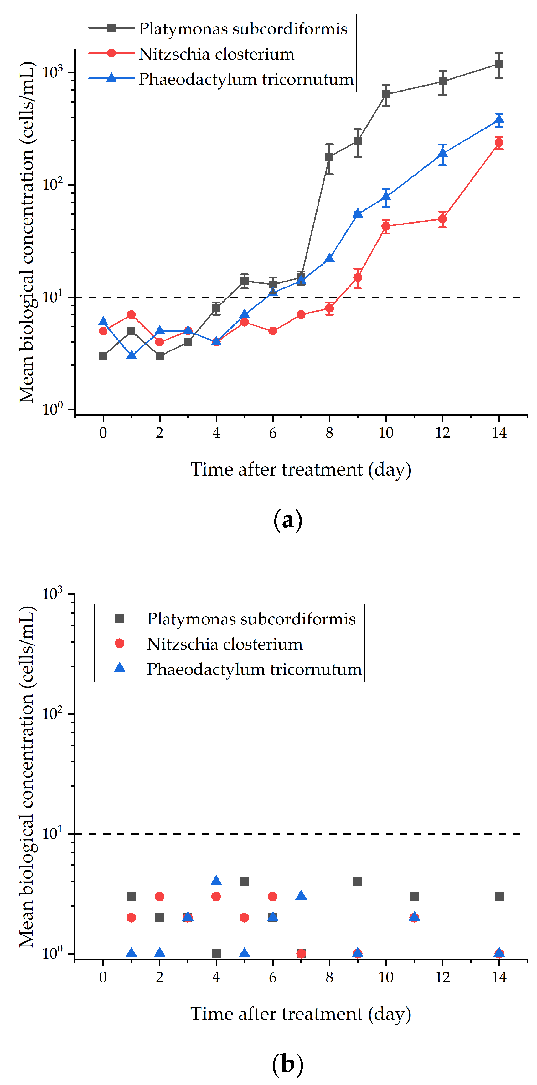
3.2. Storage Experimental Results

The viability of treated algal samples was monitored continuously to further simulate ballast water sample storage. As the completely dark environment in the ballast tank will inhibit algal photosynthesis as well and render them inactive, which may affect the actual assessment results of microbubble processing, the experiment was set up to store the processed samples in an area with natural light and a stable temperature. Three kinds of microalgae samples with a greater than 99.9% inactivation rate after treatment were stored in this experiment, and sample activity was detected for 2 consecutive weeks to test for follow-up effects of this method of treatment.
The biological concentration of the samples treated with ozone microbubbles did not increase by orders of magnitude over the course of 14 days after 2 weeks of natural light preservation and cultivation and could always be kept very low for a long time (Figure 4), which was also consistent with the results of using the active matter method. Active matter (usually oxidizing substances) may dissolve to a certain extent in seawater, creating an aquatic environment that is not suitable for the long-term growth of organisms.
However, even though the biological concentration of samples processed by airborne microbubbles had been significantly reduced, there was an order of magnitude rebound in the biological concentration of the three kinds of microalgae over time, of which the number of revived microalgae samples within 14 days was the highest. The mean biological concentration of all samples reached the single digit per milliliter level after 40 min of microbubble treatment and remained at the same level over the first week of treatment. From the 8th day, the biological concentration of all the samples started to revive one after the other, and it started to increase dramatically. The sample of Platymonas subcordiformis had the highest activity and ultimately increased by three orders of magnitude, whereas both diatoms also increased by two orders of magnitude in activity. This indicates that while airborne microbubbles generated by cavitation have continuous stability to some extent and may promote free radical generation in water, their inactivation effect for ballast water treatment is still inadequate.

3.3. Effect of Ozone Injection Dose

The effect of the injection dose of ozone on the inactivation was further investigated after verifying the bioavailability of ozone microbubbles. The ozone generator used in this experiment had a tunable concentration, but because of the limited samples and long cycle time of the biological experiments, ozone concentration was only fitted as a variable under the condition that intake volume was unchanged, and the three experimental groups with concentrations of 100%, 75%, 50%, and 25%, respectively, were studied. The testing species of the sample is Platymonas subcordiformis.
The inactivation rate of ozone microbubbles is accelerated with increasing ozone concentration, and even the lowest concentration of 25% ozone can still efficiently and rapidly remove active microalgae (Figure 5). Furthermore, the results indicate that ozone injection can greatly increase the inactivation efficiency of microbubbles on the microalgae.
When using active matter as a treatment method, it should be ensured that the treated water will not harm the personnel or equipment, for which the total residual oxidant (TRO) is an important parameter for detection after using the ozone-based treatment. The experimental group received their results from the samples treated with micro-nano bubbles from the established experimental system, with the gas flow rate maintained at 10 mL/min. The TRO concentration was determined using a standard DPD (N, N-diethyl-p-phenylenediamine) colorimetric analysis.
The Venturi tube inlet rate of ozone is not high due to the throttling effect of the cavitation nozzle, so the ozone concentration of the microbubble treatment sample is relatively low (Table 3). Nevertheless, these results indicate that the amount of active substance can be reduced in order to reduce toxicity when using microbubble inactivation processing.

3.4. Morphological Changes of Microalgae Cells after Microbubble Treatment

After a period of standing and producing algal scum deposits, the upper transparent layer was extracted. The samples from right to left are the five groups of samples treated from 0 to 60 min in that order (Figure 6). It was observed that with increasing treatment time at each sampling site, the treated seawater showed a marked fade in greenness and a decrease in chlorophyll a, which represents the viability of the algal cells, allowing direct detection of the effects of treatment on the phytoplankton. It is worth noting that there was no obvious degradation of chlorophyll observed in the algal samples, suggesting that free radicals with oxidation play a minimal role in the inactivation process.
In order to directly verify the effect of air microbubble treatment on microalgae, the morphological changes of algae were observed by optical biological microscopy. Figure 7a–c shows untreated algal cells with an intact cytoplasm and cell wall, which can be observed as inactive. Figure 7b,d show varying degrees of damage to algal cells after microbubble treatment. In the experimental group treated with it at the initial biological concentration of 103–104 cells/mL, after 20 min of treatment, some cells had collapsed. At this stage, after 40 min of treatment, cell disruption became more severe and had disaggregated, due to which intracellular material had been lost. Almost all of the cell surfaces were visibly ruptured. Moreover, all the algal cells were found to be immobile and inactive. This was seen in all treated microalgal species but barely occurred in the control samples.
Figure 8 shows that in the treated Chlorophyta samples, chlorophyll metamorphism can be observed directly. Figure 9 shows that the algal cells treated with ozone microbubbles still had no complete cell structure and suffered physical damage from bubble collapse and cavitation. Therefore, microalgae treated with ozone microbubbles are subjected to both mechanical destruction and oxidation.

3.5. The Mechanism of Microbubbles on the Inactivation Efficiency

Many groundbreaking studies have documented the sources of the destructive power of microbubbles. When bubbles interact with shock waves, they collapse because the surface tension does not provide sufficient resilience. Experimental studies of micron-sized bubbles show that the effect of a pulse shock on the proximal end of the bubble contracts and accelerates in the direction of shock propagation, leading to the formation of jets in surrounding liquids [14]. Local bubble ejections can affect the surface of the material, and bubble ruptures can produce high-speed shock waves and high-pulsed pressures. When the local shock pressure is high enough, a high effect force higher than the material yield stress is generated, creating craters on the surface of the material [15]. In addition, the collapsing cavitation bubbles emit significant amounts of heat into surrounding liquids while creating local hot zones at temperatures up to gigabytes, where shock waves and microjets with pressures up to gigabytes have a strong impact and generate highly reactive free radicals [16,17]. This high energy density often has adverse effects on fluid dynamics, such as additional pressure loss, vibration, noise, and erosion damage. On the other hand, cavitation has been found to be an effective tool for many environmental, chemical, and biological industry-scale applications, such as accelerated chemical reactions, mixing and separation, sterilization, cell inactivation, water treatment, and heating. Importantly, microbubbles’ collapse has the capability to produce radiation force and hydroxyl radical (OH) at the sample edge (2H2O→OH + H3O+), which provides a cost-effective non-reagent method to generate multiple reactive oxygen species (ROS) during fine bubbles’ collapse [18,19].
In general, in order to effectively prevent and control the invasion of invasive alien species, ballast water treatment systems use oxidizing chemically active matter to enhance the inactivation effect. For example, easily soluble oxidizing substances, such as chlorine gas and hypochlorite, can be easily added to the water to be treated. However, normal-sized bubbles can cause the gas to float up and out of the water quickly, resulting in low exposure to aquatic microorganisms, especially for gases that are insoluble or slightly soluble in water, making it difficult to take full advantage of the gas oxidation and inactivation effects. In contrast, the long residence time, large contact area, and high mass transfer efficiency [20,21,22] of fine bubbles in water effectively extend the oxidizing range and active gases so that they can also be used in ballast water treatment. Ozone is used as a disinfectant and, of all commercially available oxidants, has the highest oxidation potential; therefore, ballast treatment of ozone for commercial ships is currently being studied [23,24]. Ozone in the form of fine bubbles is widely used in drinking water treatment, helping to completely remove finer microorganisms such as planktonic viruses, phages, and bacteria from ballast water. It is a naturally occurring oxygen isomer and can therefore be obtained from the atmosphere, making it as widely available as electrolytic seawater with chlorine production.

4. Conclusions

Microbubble technology is an innovative method for ballast water treatment, which has demonstrated that the use of microbubbles can effectively inactivate some planktonic microalgae in seawater. Experiments have shown that the inactivation of marine microalgae by air microbubbles generated by hydrodynamic cavitation requires a long treatment time, while ozone injection can significantly improve inactivation efficiency. Moreover, even at very low doses of ozone combined with microbubbles, the inactivation effect is still very obvious and rapid. The inactivation mechanism is mainly mechanically damaged when only air microbubbles are used, while the treated microalgae are not only mechanically damaged but also oxidized by the ozone. A two-week storage experiment showed that air microbubbles did not inhibit the growth of microalgae cells and that the injection of active matter, such as ozone, was still required to ensure valid control of biological invasion. However, it helps to reduce the use of active matter to weaken the toxicity of the treated water. Microbubble technology is able to perfectly meet the requirements of the IMO Ballast Water Convention. These works suggest that it offers a great opportunity for the prevention of shipping invasive organisms and application to ship ballast water treatment.

Author Contributions

Conceptualization, B.W. and H.L.; methodology, B.W., H.L. and M.C.; formal analysis, H.Z.; investigation, H.L.; resources, W.L., J.H. and M.C.; writing—original draft preparation, B.W.; writing—review and editing, H.L. and H.Z.; visualization, H.L.; supervision, B.W., H.Z., W.L. and J.H.; funding acquisition, B.W. and H.Z. All authors have read and agreed to the published version of the manuscript.

Funding

This research was supported by the National Natural Science Foundation of China, grant number 51679022, and the National Natural Science Foundation of China, grant number 52271303.

Institutional Review Board Statement

Not applicable.

Informed Consent Statement

Not applicable.

Data Availability Statement

Data are available upon request.

Conflicts of Interest

The authors declare no conflict of interest. The funders had no role in the design of the study; in the collection, analyses, or interpretation of data; in the writing of the manuscript; or in the decision to publish the results.

References

  1. Hess-Erga, O.K.; Moreno-Andrés, J.; Enger, Ø.; Vadstein, O. Microorganisms in ballast water: Disinfection, community dynamics, and implications for management. Sci. Total Environ. 2019, 657, 704–716. [Google Scholar] [CrossRef] [PubMed]
  2. Batista, W.R.; Fernandes, F.C.; Lopes, C.C.; Lopes, R.S.; Miller, W.; Ruiz, G. Which ballast water management system will you put aboard? Remnant anxieties: A mini-review. Environments 2017, 4, 54. [Google Scholar] [CrossRef] [Green Version]
  3. Andreadakis, A.; Mamais, D.; Christoulas, D.; Kabylafka, S. Ultraviolet disinfection of secondary and tertiary effluent in the Mediterranean region. Water Sci. Technol. 1999, 40, 253–260. [Google Scholar] [CrossRef]
  4. Rastogi, R.P.; Madamwar, D.; Nakamoto, H.; Incharoensakdi, A. Resilience and self-regulation processes of microalgae under UV radiation stress. J. Photochem. Photobiol. C: Photochem. Rev. 2020, 43, 100322. [Google Scholar] [CrossRef]
  5. Taylor, A.; Rigby, G.; Gollasch, S.; Voigt, M.; Hallegraeff, G.; McCollin, T.; Jelmert, A. Preventive treatment and control techniques for ballast water. In Invasive Aquatic Species of Europe. Distribution, Impacts and Management; Springer: Dordrecht, The Netherlands, 2002; pp. 484–507. [Google Scholar]
  6. Pećarević, M.; Mikuš, J.; Prusina, I.; Juretić, H.; Cetinić, A.B.; Brailo, M. New role of hydrocyclone in ballast water treatment. J. Clean. Prod. 2018, 188, 339–346. [Google Scholar] [CrossRef] [Green Version]
  7. Takahashi, C.K.; Lourenco, N.G.G.S.; Lopes, T.F.; Rall, V.L.M.; Lopes, C.A.M. Ballast water: A review of the impact on the world public health. J. Venom. Anim. Toxins Incl. Trop. Dis. 2008, 14, 393–408. [Google Scholar] [CrossRef]
  8. Lin, L.; Wang, Q.; Wu, H. Study on the dinoflagellate cysts in ballast tank sediments of international vessels in Chinese shipyards. Mar. Environ. Res. 2021, 169, 105348. [Google Scholar] [CrossRef]
  9. Zhu, Y.; Ling, Y.; Peng, Z.; Zhang, N. Formation of emerging iodinated disinfection by-products during ballast water treatment based on ozonation processes. Sci. Total. Environ. 2020, 743, 140805. [Google Scholar] [CrossRef]
  10. Hanotu, J.; Bandulasena, H.H.; Zimmerman, W.B. Microflotation performance for algal separation. Biotechnol. Bioeng. 2012, 109, 1663–1673. [Google Scholar] [CrossRef] [Green Version]
  11. Mimura, H.; Abe, A.; Katakura, R.; Kawasaki, H.; Yoshida, K.; Ishida, H. Lethality of shock pressures to a marine Vibrio sp. isolated from a ship’s ballast water. Biocontrol. Sci. 2006, 11, 159–166. [Google Scholar] [CrossRef]
  12. Khan, P.; Zhu, W.; Huang, F.; Gao, W.; Khan, N.A. Micro–nanobubble technology and water-related application. Water Supply 2020, 20, 2021–2035. [Google Scholar] [CrossRef]
  13. Peng, K.; Qin, F.G.; Jiang, R.; Kang, S. Interpreting the influence of liquid temperature on cavitation collapse intensity through bubble dynamic analysis. Ultrason. Sonochem. 2020, 69, 105253. [Google Scholar] [CrossRef]
  14. Vedadi, M.; Choubey, A.; Nomura, K.I.; Kalia, R.K.; Nakano, A.; Vashishta, P.; Van Duin, A.C.T. Structure and dynamics of shock-induced nanobubble collapse in water. Phys. Rev. Lett. 2010, 105, 014503. [Google Scholar] [CrossRef]
  15. Hsiao, C.T.; Jayaprakash, A.; Kapahi, A.; Choi, J.K.; Chahine, G.L. Modelling of material pitting from cavitation bubble collapse. J. Fluid Mech. 2014, 755, 142–175. [Google Scholar] [CrossRef] [Green Version]
  16. Sun, X.; Kang, C.H.; Park, J.J.; Kim, H.S.; Om, A.S.; Yoon, J.Y. An experimental study on the thermal performance of a novel hydrodynamic cavitation reactor. Exp. Therm. Fluid Sci. 2018, 99, 200–210. [Google Scholar] [CrossRef]
  17. Li, P.; Song, Y.; Yu, S. Removal of Microcystis aeruginosa using hydrodynamic cavitation: Performance and mechanisms. Water Res. 2014, 62, 241–248. [Google Scholar] [CrossRef]
  18. Lyu, T.; Wu, S.; Mortimer, R.J.; Pan, G. Nanobubble technology in environmental engineering: Revolutionization potential and challenges. Environ. Sci. Technol. 2019, 53, 7175–7176. [Google Scholar] [CrossRef] [Green Version]
  19. Jin, J.; Wang, R.; Tang, J.; Yang, L.; Feng, Z.; Xu, C.; Yang, F.; Gu, N. Dynamic tracking of bulk nanobubbles from microbubbles shrinkage to collapse. Colloids Surf. A Physicochem. Eng. Asp. 2020, 589, 124430. [Google Scholar] [CrossRef]
  20. Li, C.; Zhang, H. A review of bulk nanobubbles and their roles in flotation of fine particles. Powder Technol. 2022, 395, 618–633. [Google Scholar] [CrossRef]
  21. Zhang, F.; Sun, L.; Yang, H.; Gui, X.; Schönherr, H.; Kappl, M.; Cao, Y.; Xing, Y. Recent advances for understanding the role of nanobubbles in particles flotation. Adv. Colloid. Interface Sci. 2021, 291, 102403. [Google Scholar] [CrossRef]
  22. Xiao, W.; Xu, G. Mass transfer of nanobubble aeration and its effect on biofilm growth: Microbial activity and structural properties. Sci. Total. Environ. 2020, 703, 134976. [Google Scholar] [CrossRef]
  23. Perrins, J.C.; Cordell, J.R.; Ferm, N.C.; Grocock, J.L.; Herwig, R.P. Mesocosm experiments for evaluating the biological efficacy of ozone treatment of marine ballast water. Mar. Pollut. Bull. 2006, 52, 1756–1767. [Google Scholar] [CrossRef]
  24. Wright, D.A.; Gensemer, R.W.; Mitchelmore, C.L.; Stubblefield, W.A.; van Genderen, E.; Dawson, R.; Orano-Dawson, C.E.; Bearr, J.S.; Mueller, R.A.; Cooper, W.J. Shipboard trials of an ozone-based ballast water treatment system. Mar. Pollut. Bull. 2010, 60, 1571–1583. [Google Scholar] [CrossRef]
Figure 1. Treating apparatus of the microbubble laboratory test.
Jmse 11 00241 g001
Figure 2. Treatment results of air microbubbles produced by hydrodynamic cavitation at initial biological concentration of 103–104 cells/mL.
Jmse 11 00241 g002
Figure 3. Treatment results of O3 microbubbles produced by hydrodynamic cavitation at initial biological concentration of 103–104 cells/mL.
Jmse 11 00241 g003
Figure 4. Comparison results of organism viability during sample storge after (a) air and (b) ozone microbubbles treatment.
Jmse 11 00241 g004
Figure 5. The effect of ozone injection dose on inactivation rate of ozone microbubbles treatment.
Jmse 11 00241 g005
Figure 6. Treatment effect of air microbubbles on Platymonas subcordiformis.
Jmse 11 00241 g006
Figure 7. Comparison of morphological results of microalgal cells before and after air microbubbles treatment. (a) Optical micrograph of Platymonas subcordiformis before air microbubbles treatment; (b) optical micrograph of Platymonas subcordiformis after 40 min air microbubbles treatment; (c) optical micrograph of Phaeodactylum tricornutum before air microbubbles treatment; (d) optical micrograph of Phaeodactylum tricornutum after 40 min of air microbubble treatment.
Jmse 11 00241 g007
Figure 8. The observation of chlorophyll metamorphism after ozone microbubble treatment.
Jmse 11 00241 g008
Figure 9. Comparison of morphological results of microalgal cells before and after ozone microbubbles treatment (100% ozone dose). (a) Optical micrograph of Platymonas subcordiformis before ozone microbubbles treatment; (b) optical micrograph of Platymonas subcordiformis after 5 min ozone microbubble treatment; (c) optical micrograph of Phaeodactylum tricornutum before ozone microbubble treatment; (d) optical micrograph of Phaeodactylum tricornutum after 5 min ozone microbubble treatment.
Jmse 11 00241 g009
Table 1. Effect of initial biological concentration on the treatment of air microbubbles.
SpeciesThe Processing Time Required When the Inactivation Rate Reaches 99%
Initial Biological Concentration:
104–105 Cells/mL
Initial Biological Concentration:
103–104 Cells/mL
Initial Biological Concentration:
102–103 Cells/mL
Platymonas subcordiformis65 min40 min20 min
Nitzschia closterium65 min40 min15 min
Phaeodactylum tricornutum65 min40 min20 min
Table 2. Effect of initial biological concentration on the treatment of ozone microbubbles.
SpeciesThe Processing Time Required When the Inactivation Rate Reaches 99%
Initial Biological Concentration:
104–105 Cells/mL
Initial Biological Concentration:
103–104 Cells/mL
Initial Biological Concentration:
102–103 Cells/mL
Platymonas subcordiformis300 s180 s60 s
Nitzschia closterium300 s180 s60 s
Phaeodactylum tricornutum300 s180 s60 s
Table 3. TRO concentration of effluent after O3 microbubble treatment with 10 mL/min injection inflow.
Injection DoseTotal Residual Oxidant (mg/L)
InitialAfter 1 DayAfter 2 DaysAfter 5 DaysAfter 7 DaysAfter 14 Days
100%1.360.880.560.090.050.02
75%0.960.740.400.050.020.01
50%0.520.110.070.040.010.00
25%0.070.030.010.010.000.00
Disclaimer/Publisher’s Note: The statements, opinions and data contained in all publications are solely those of the individual author(s) and contributor(s) and not of MDPI and/or the editor(s). MDPI and/or the editor(s) disclaim responsibility for any injury to people or property resulting from any ideas, methods, instructions or products referred to in the content.

