Next Article in Journal
Hydraulic Performance of Seawater Intake System Using CFD Modeling
Next Article in Special Issue
Correction: Cima, F. Searching for the Origin and the Differentiation of Haemocytes before and after Larval Settlement of the Colonial Ascidian Botryllus schlosseri: An Ultrastructural Viewpoint. J. Mar. Sci. Eng. 2022, 10, 987
Previous Article in Journal
A K-Nearest Neighbors Algorithm in Python for Visualizing the 3D Stratigraphic Architecture of the Llobregat River Delta in NE Spain
Previous Article in Special Issue
Biocide vs. Eco-Friendly Antifoulants: Role of the Antioxidative Defence and Settlement in Mytilus galloprovincialis
 
 
Correction published on 29 August 2022, see J. Mar. Sci. Eng. 2022, 10(9), 1207.
Article

Searching for the Origin and the Differentiation of Haemocytes before and after Larval Settlement of the Colonial Ascidian Botryllus schlosseri: An Ultrastructural Viewpoint

Laboratory of Ascidian Biology, Department of Biology (DiBio), University of Padova, Via U. Bassi 58/B, 35131 Padova, Italy
J. Mar. Sci. Eng. 2022, 10(7), 987; https://doi.org/10.3390/jmse10070987
Submission received: 30 May 2022 / Revised: 14 July 2022 / Accepted: 17 July 2022 / Published: 19 July 2022 / Corrected: 29 August 2022

Abstract

:
The colonial ascidian Botryllus schlosseri possesses an innate immunity, which plays fundamental roles in its survival, adaptability, worldwide spread and ecological success. Three lines of differentiation pathways of circulating haemocytes are known to be present in the haemolymph, starting from undifferentiated haemoblasts: (i) the phagocytic line (hyaline amoebocytes and macrophage-like cells), (ii) the cytotoxic line (granular amoebocytes and morula cells) and (iii) the storage cell line (pigment cells and nephrocytes). Many questions remain about their origin, and thus, observations during various stages of development were undertaken in this study. Haemocytes were detected beginning from the early tailbud embryo stage. Haemoblasts were always present and morula cells were the first differentiated haemocytes detected. In both the next stage, just before hatching, and the swimming tadpole larva stage, hyaline amoebocytes and pigment cells were also recognisable. Some morula cells containing active phenoloxidase migrated from the haemolymph into the tunic after having crossed the epidermis, and this behaviour could be related to the preparation of a defensive function for spatial competition. During larval metamorphosis, macrophage-like cells appeared with their phagosomes positive to acid phosphatase activity and containing apoptotic cells from tail tissue degeneration. After metamorphosis, in the filter-feeding oozoid stage, nephrocytes involved in nitrogen catabolism finally appeared. In both the subendostylar sinus and the peripheral blind-sac vessels (ampullae), clusters of haemoblasts were recognisable, some of which showed incipient specialisations, considering the hypothesis of the presence of putative niches of haemolymph stem cells.

1. Introduction

The haemocytes of many ascidian species that are protagonists of soft fouling—such as the cosmopolitan colonial species Botryllus schlosseri [1,2]—have long been studied and characterised, from a functional point of view, as being responsible for the defence responses of innate immunity, from which the survival of individuals and the ability to compete for the substratum after settlement derive [3,4]. In ascidians, which are the closest relatives to vertebrates [5,6], haemocytes are responsible for all vital activities because they are involved in nutrition [7], excretion [8], asexual reproduction [9,10,11,12,13] and part of tunic formation [14]. In defence responses [15], haemocytes are able to phagocytise foreign materials [16,17,18,19,20]; release opsonins [21], cytokines [22,23,24], agglutinins [25,26], complement factors [27,28] and antimicrobial peptides [29]; actively participate with cytotoxic substances in inflammatory reactions [30]; and are involved in allorecognition between contacting colonies that are genetically incompatible [31,32,33,34,35].
The haemocytes of ascidians, due to their high polymorphism, have led to a very complex and uncertain classification by various authors. However, in the case of B. schlosseri, the numerous cell types have finally been grouped into differentiation pathways on the basis of their ultrastructural features [36,37] and cytochemical and morpho-functional properties [38]. On this basis, three haemocyte lines have been defined, the origin of which has been hypothesised from undifferentiated cells (Figure 1). The first two are involved in defence responses (‘immunocytes’), and the latter is involved in catabolite storage and excretion: (i) phagocytic cell lines, represented by hyaline amoebocytes (previously known as ‘microgranular amoebocytes’ [36]) and macrophage-like cells; (ii) cytotoxic cell lines, represented by granular amoebocytes (previously known as ‘macrogranular amoebocytes’ [36]) and morula cells; and (iii) storage cell lines, represented by pigment cells and nephrocytes.
The hypotheses concerning the origin of the various differentiation pathways in ascidians, B. schlosseri included, are currently still controversial [8,39,40,41,42], especially regarding both the moment of appearance of the various cell types during embryo and larva development and the search for stem cells. The most likely candidate of the haemocyte stem cell seems to be represented by the haemoblast (Figure 1), a circulating undifferentiated cell recognised in B. schlosseri by the anti-CD34 antibody, which has been employed as a marker because CD34 is a transmembrane phosphoglycoprotein of the haematopoietic stem and progenitor cells of vertebrates [38]. During ascidian embryogenesis, haemocytes originate from trunk mesenchymal cells derived from the A7.6 blastomere [43], and the differentiation of haemocytes begins from the developmental stage of the early tailbud embryo. For example, in Clavelina picta [44] and Ecteinascidia turbinate [45] haemocytes have been observed migrating and distributing around the coelom in contact with the endoderm. Starting from the late tailbud embryo stage, before the hatching of the tadpole larva, the real differentiation of haemocytes begins, which is completed during the period of free life and metamorphosis. The activation of many innate immunity genes during metamorphosis, and trans-epidermal haemocyte migrations into the tunic during both the swimming larval period and the settlement, have been previously reported in Boltenia villosa [46,47]. These events were probably related to (i) the programmed maturation of the adult immune system, (ii) the high phagocytosis activity during tissue remodelling at metamorphosis, and (iii) the potential inflammatory response to environmental settlement cues. The latter are mainly due to the influence of the surface characteristics, e.g., the microbial film or biofilm, as observed in ascidians and other fouling invertebrates during settlement on the substratum [48,49,50,51].
After metamorphosis, a true haematopoietic tissue is less clear in ascidian adults. Regions with clusters of haemogenic cells (‘lymphatic nodules’) have been recognised both in the crossbars of the branchial and connective tissue surrounding the intestine of Ciona intestinalis and Styela clava and in the blood vessels in Polyclinum planum and Pyura austor [52,53,54]. A lymphatic or haematopoietic nodule is formed of undifferentiated cells in the centre, represented by haemoblasts, which are externally surrounded by differentiating blood cells [52].
The question of the origin of differentiation pathways during embryo-larval development was the main purpose of the present paper. It was dealt with using the cosmopolitan colonial ascidian B. schlosseri, and was concerned with the embryo, larva and oozoid, with the latter considered the result of metamorphosis and the founding individual of the colony. The choice of this species was made because it is ovoviviparous and easily reared in the laboratory. Entire embryonic development can be studied within parental individuals after obtaining colonies with embryos at well-determined stages of development [55,56,57]. Moreover, the metamorphosis stages of the hatched tadpole larva can be easily recognised [58]. In particular, the following stages of embryo–larval development have been considered: (i) early tailbud embryo, with the tail forming an initial turn around the cephalenteron; (ii) late tailbud embryo, with the tail at its maximum extension (1.5 turns around the cephalenteron); (iii) newly hatched tadpole larva (early metamorphosis); (iv) late metamorphosis larva; and (v) oozoid. The occurrence of various types of haemocytes was studied by means of ultrastructural observations, including characterisation of the enzymatic content, taking into account the functional peculiarities of the differentiation pathways of the immunocytes. The activity of lysosomal acid phosphatase has been assayed as a marker of the phagocytic line, and that of phenoloxidase has been assayed as a marker of the cytotoxic line. Finally, since the adult of B. schlosseri, despite belonging to the family Styelidae, does not possess haematopoietic or lymphatic nodules, the presence and localisation of the accumulation and differentiation zones of haemoblasts have been investigated inside the oozoid.

2. Materials and Methods

2.1. Animals

The animals used in this study were colonies and larvae of B. schlosseri from our laboratory cultures and wild colonies from the Lagoon of Venice. Colonies were kept in aerated aquaria attached to 5 × 5 cm glass slides filled with filtered sea water (salinity of 35 ± 1 psu, temperature of 19 ± 0.5 °C, pH of 8.1), which was renewed every other day. They were fed with microalgae mixtures. Stages of embryonic development from fragmented colonies and larvae were easily identified under a dissecting binocular stereomicroscope (Wild Heerbrugg Ltd., Heerbrugg, Switzerland) with 50× maximum magnification.

2.2. Transmission Electron Microscopy

Selected colonies and larvae were fixed for 2 h at 4 °C in 1% glutaraldehyde, buffered with 0.2 M sodium cacodylate buffer (CAB) containing 1.7% NaCl at a pH of 7.4 plus 1% caffeine to prevent the leakage of phenols contained inside morula cell vacuoles [59]. Since single zooids are 1–2 mm long in the colony, after washing in buffer with 1.7% NaCl, small fragments (approximately 5–6 mm) of the colonies were cut and inserted into glass vials, whereas entire larvae were inserted inside tubes with a plankton filter at the bottom. After postfixation in 1% OsO4 in CAB, all specimens were dehydrated and embedded in Epon 812 (Fluka Chemie, Buchs, Switzerland). Thick sections (1 µm), obtained using an LKB ultrotome, were counterstained with 1% toluidine blue and observed under an Olympus CX31 light microscope equipped with a DV Lumenera Infinity 2 and Infinity Capture Application software version 5.0.0 (Lumenera Co. 2002–2009, Ottawa, ON, Canada) for image capturing. Thin sections (80 nm) were briefly stained with uranyl acetate and lead citrate. Micrographs were taken with an FEI Tecnai G2 transmission electron microscope operating at 75 kV.

2.3. Histoenzymatic Assays

Both intact larvae and lacerated colonies were preincubated in 0.1% Triton X-100 (Sigma-Aldrich, St. Louis, MO, USA) in CAB for 10 min. For each enzymatic assay, control specimens were prepared by omitting the specific enzyme substrate. The following assays are reported in brief according to the methods described in detail by Cima [60].

2.3.1. Assay for Acid Phosphatase

Among the hydrolytic enzymes, acid phosphatase was chosen for its presence in the lysosomes and phagosomes of the phagocytic line. Fixed specimens were incubated for 15 h at 30 °C in the following reaction mixture [61]: 5 mg of naphtol AS-BI phosphate (Sigma-Aldrich) previously dissolved in 200 µL of dimethylformamide (DMF); 200 µL of solution A (0.4 g of hexazonium p-rosanilin, Fluka dissolved in 2 mL of 36% HCl and 8 mL of distilled water); 200 µL of solution B (4% NaNO2 in distilled water); and 10 mL of 0.1 M sodium acetate buffer plus 1.7% NaCl and 1% saccharose at a pH of 5.2. Positive sites were stained red under light microscopy and revealed an electron-dense granular precipitate under electron microscopy.

2.3.2. Assay for Phenoloxidase
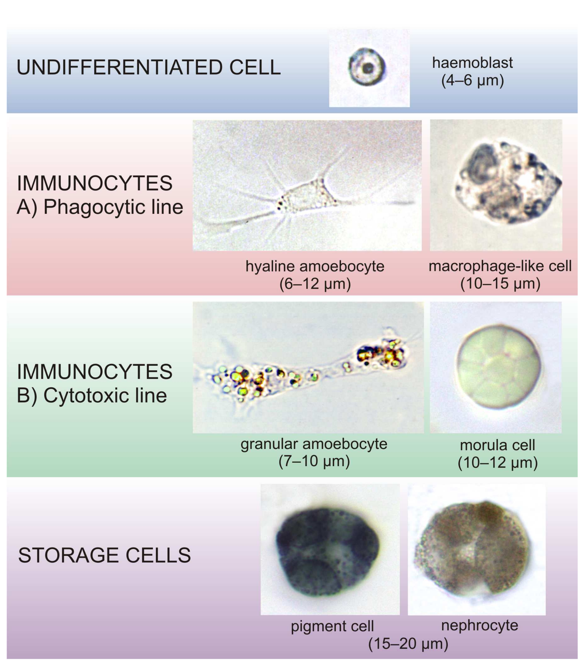
Among the oxidative enzyme activities, phenoloxidase was chosen as the enzyme typical of the cytotoxic line. The enzyme substrate was β-(3,4-dihydroxyphenyl)-L-alanine (L-DOPA), purchased from Sigma-Aldrich. Fixed specimens were incubated for 15 h at 4 °C with L-DOPA-saturated 0.01 M CAB plus 5 mM CaCl2 (CAB-Ca) at a pH of 7.0 [62], and then for 1 h at 37 °C in the same renewed medium before dehydration and embedding. In this case, postfixation in OsO4 was omitted because of the high reducing activity inside morula cell vacuoles, which causes osmium precipitation and impedes the detection of electron-dense positive sites under electron microscopy.

3. Results

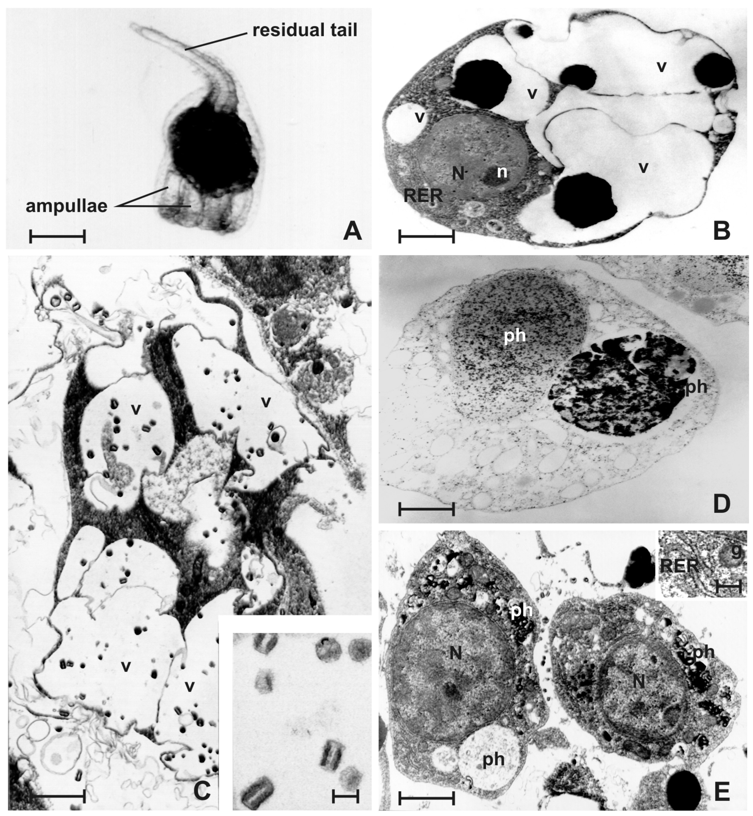
3.1. Early Tailbud Embryo

This stage immediately follows that of the neurula with the closure of the neuropore. The embryo appears elongated, and the cephalenteron region and tail can be distinguished, the latter appearing short and bending at 90° to the right side (Figure 2A). The chordamesodermal cells differentiate into cells of the notochord, whereas those of the mesoderm differentiate into myocytes in the tail and into a group of mesenchymal cells that will later give rise to the pericardium, heart and mantle mesenchyme. The archenteron cavity is also recognisable [56,63].
This is the first stage of embryonic development in which differentiating haemocytes are detected and distinguished with respect to the germ layers. They are mainly observed in the haemocoel, the space between the epidermis and the organ primordia (Figure 2B,C), and they are represented by haemoblasts and putative precursors of morula cells.
Haemoblasts appear as small cells (2–5 μm in diameter) resembling undifferentiated cells. The large nucleus occupies almost the entire volume of the cell and is placed in a central position, with chromatin mainly scattered or organised in small clusters close to the nuclear envelope. Within the nucleus, a nucleolus is recognisable with a granular structure. The plasmalemma shows an irregular contour, and the cytoplasm, which is represented by a thin peripheral layer, has no details of differentiation. Only some granules of the yolk, mitochondria and ribosomes, the latter both organised into polysomes and adherent to small cisternae of the rough endoplasmic reticulum (RER), can be found (Figure 2D).
Morula cells appear as vacuolated cells and have shared features with haemoblasts, such as a large spherical nucleus with a nucleolus (Figure 2E) and yolk granules in the cytoplasm. Vacuoles are small and scant and contain the typical material of this cell type, and form strongly electron-dense aggregates due to their high osmiophilicity (Figure 2F). In addition, long RER profiles and mitochondria with parallel cristae are present.

3.2. Late Tailbud Embryo

The tail is completely turned around the cephalenteron of the embryo. The first muscular movements of the tail begin [56,63]. Both the larval organs (such as the larval nervous system and photolith) and primordia of future organs of the adult (such as the great expansion of the peribranchial chambers that converge medially and an evident intestine) can be recognised (Figure 3A,B). Haemocytes are very numerous in the defined spaces of the haemocoel (Figure 3C). At this stage, haemoblasts, morula cells (Figure 3D) and, for the first time, hyaline amoebocytes and storage cells can be found.
Morula cells occur with their typical features, i.e., large vacuoles containing strongly electron-dense granules (Figure 3E). The cytoplasm and nucleus are displaced into the spaces among the vacuoles. However, as a feature of young cells, the nucleus is still large and has a nucleolus; moreover, in the cytoplasm, mitochondria and cisternae of the RER, as well as some granules of yolk can still be observed. Beginning from this developmental stage, some morula cells are observed to cross the epidermis and migrate into the definitive tunic. The latter is secreted by the epidermis and is placed below the embryonic tunic, which is secreted by the egg envelopes. Moreover, the definitive tunic displays a cuticle, which is densely covered by small electron-dense spherical papillae (Figure 3F).
Hyaline amoebocytes are cells characterised by a large nucleus, occasionally reniform with clustered chromatin (Figure 4A). The cytoplasm contains scant granules of yolk and small granules of approximately 0.3–0.7 μm in diameter bound by an undulated membrane, as well as containing homogeneous and moderately electron-dense material (inset in Figure 4A). Small, clear vesicles of approximately 0.2 μm in diameter and many parallel cisternae of the RER, mitochondria and some multivesicular bodies are also present.
Storage cells (Figure 4B) are only represented by pigment cells. They have large vacuoles of 3.5 μm in diameter containing small pigment crystals (approximately 180 nm in length) formed of overlapping electron-dense layers (inset in Figure 4B). In the cytoplasm, numerous mitochondria, free ribosomes and scant yolk granules are present.
Various putative precursors of hyaline amoebocytes (Figure 4C) and morula cells (Figure 4D) are also frequent. Both have characteristics similar to haemoblasts, that is, 5 μm in size, a high ratio of nucleus/cytoplasm, and the presence of a nucleolus in the nucleus. Moreover, the putative precursors of hyaline amoebocytes show, in the cytoplasm, small electron-dense membrane-bound granules and scattered clear vesicles, the latter budding from the Golgi apparatus (inset in Figure 4C). The putative precursors of morula cells have vacuoles with strongly electron-dense material inside (Figure 4D).

3.3. Newly Hatched Tadpole Larva (Early Metamorphosis)

The swimming larva represents the dispersal phase of the species (Figure 5A). It is anteriorly equipped with three transient sensory adhesive organs, namely ‘papillae’, for substratum detection and larval settlement, before the beginning of metamorphosis to the filter-feeding adult, i.e., the ‘oozoid’. In the anterior part of cephalenteron, the larva has eight compacted ampullae, i.e., the primordium of the colonial vascular network. Ampullae are blind-sac structures that are, afterwards, involved both in the process of settlement and in the formation of the circulatory system of the colony [64,65,66]. They arise, before larval hatching, from a haemocoelic lacuna ventrally placed with respect to the branchial basket primordium. Subsequently, the ampullae spread into the newly formed definitive tunic and, during metamorphosis, protrude away from the area of origin, remaining connected to it through thin vascular peduncles. At the end of metamorphosis, the peduncles merge into a single vessel that is arranged to form an arch around the oozoid, namely, the marginal vessel, which communicates with the eight peripheral ampullae that are spreading onto the substratum [64]. As a clear sign of the beginning of metamorphosis, the epidermis thickens, and the cells of the notochord and musculature of the proximal area of the tail, are disrupted and begin to accumulate into the cephalenteron [67].
At this stage, haemoblasts, morula cells, hyaline amoebocytes and pigment cells continue to be present (Figure 5B). In the cytoplasm of these cells, the yolk granules are small and very scarce. Morula cells still have extensive RER and many mitochondria in the cytoplasm. Beginning from this stage, intense enzymatic activity of phenoloxidase in the vacuoles of some morula cells is revealed (Figure 5C). The specific histochemical labelling highlights the strong electron-density of the material inside the vacuoles. In the tunic, in addition to morula cells (Figure 5D), some granulocytes are detected (Figure 5E). The latter are large cells with a spherical nucleus in the peripheral position. In the cytoplasm, numerous large membrane-bound granules of 1.5 μm in diameter are recognised, the content of which shows various degrees of homogeneity and electron-density, occasionally with a more dense core. The salient feature of this cell type is the presence of a Golgi apparatus, which is enormously developed and displaced in the central area of the cytoplasm. Numerous cisternae of the RER and scant small mitochondria are also visible. This cell strongly resembles the cuboidal cells of the apical epithelium of the ampullae (Figure 5F).

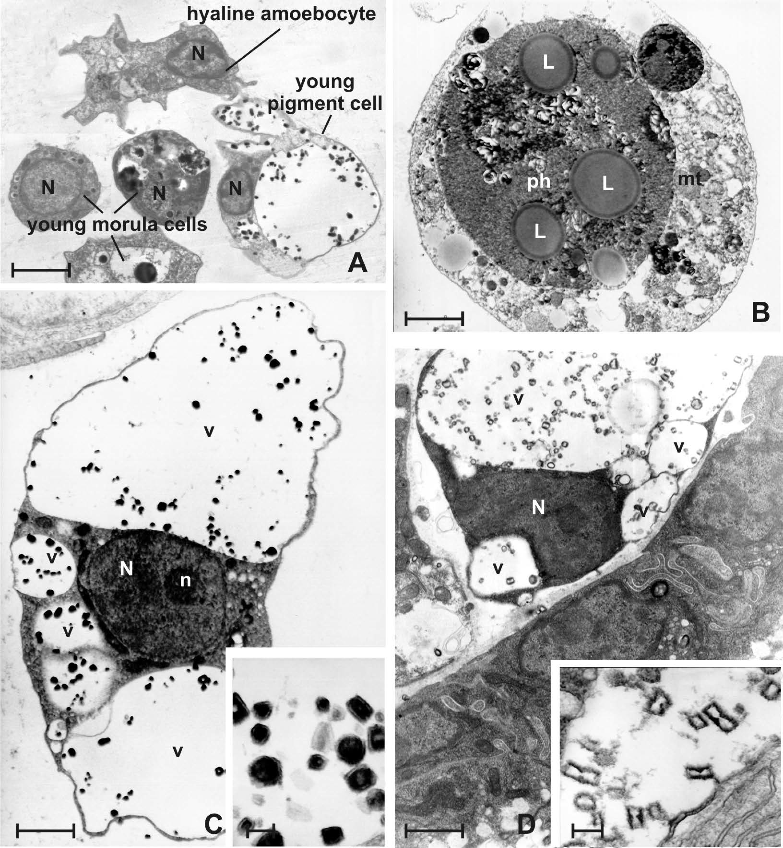
3.4. Late Larval Metamorphosis

Tail resorption occurs for two thirds of its length, involving dismantlement of the notochord, posterior neural tube and musculature. Anteriorly, ampullae begin to expand on the substratum, contributing to the formation of a bell-shaped cephalenteron (Figure 6A). At this stage, heart contractions and haemolymph circulation begin. Almost all cell types of the haemolymph characteristic of the species are present and have a more advanced degree of differentiation in comparison with the previous stages. For example, morula cells (Figure 6B) show numerous large vacuoles with electron-dense content; however, a nucleolus is still present, and storage cells (Figure 6C)—still represented only by pigment cells—become remarkable in size (10 μm in diameter), although they are still smaller than the pigment cells observed in adults (maximum 20 μm in diameter). The nucleus is pushed to the periphery because the cell volume is almost entirely occupied by vacuoles variable in size, which often protrude towards the plasmalemma, deform the surface of the cell and contain small, multilayered granules.
In addition, the first occurrence of macrophage-like cells can be detected. They are large scavenger cells (about 13 µm in diameter) mainly involved in tail resorption by engulfing the dead cells detaching from the tissues. In this cell type, the large phagosomes displace the nucleus at the cell periphery and appear to mainly contain both necrotic masses and membrane systems, arranged to form myelin figures. In these heterophagic vacuoles, an intense activity of acid phosphatase is found inside their content, which appears to be labelled by an electron-dense product of the reaction (Figure 6D). Moreover, numerous cells with intermediated characteristics between those of hyaline amoebocytes and macrophage-like cells are observed (Figure 6E). They are represented by a medium-sized cell type (about 6 μm in diameter) and are variable in shape for their amoeboid activity and phagocytic function. The features shared with hyaline amoebocytes are a central nucleus, scattered clear vesicles, cisternae of the RER and moderately electron-dense membrane-bound granules in the cytoplasm (inset in Figure 6E). The features shared with macrophage-like cells are the presence of many digestive vacuoles with heterogeneous content (phagosomes) in the cytoplasm, which are positive to acid phosphatase activity (data not shown) and smaller than those of the macrophage-like cells.

3.5. Oozoid

The tail is not yet recognisable due to its complete resorption. The eight ampullae continue to protrude onto the substratum with the formation of the marginal vessel (Figure 7A). A blastogenetic bud is already visible on the right side that will later form a new zooid. On the left side, only a primordium of a bud appears, which will be reabsorbed. Siphons open, and the activity of filtration begins. The oozoid is the founding individual of the new colony and possesses all cell types of the adult tissues, as well as those of the haemolymph.
In particular, haemoblasts tend to accumulate in some areas at the level of the subendostylar sinus (Figure 7B,C) and along the lateral epithelium (i.e., the epidermis) of the peripheral ampullae (Figure 7D). Inside the ampullae, haemoblasts appear to closely adhere to each other (Figure 7E,F) and to the basal lamina of the epithelium (Figure 7F), forming small clusters. Some haemoblasts facing the lumen show specialisations, such as minute electron-dense granules in the cytoplasm and short pseudopodia (inset in Figure 7G). In the lumen of the ampullae, various free cells can be recognised. They are young cell types (Figure 8A) that include hyaline amoebocytes, small morula cells with vacuolar material in the process of aggregation, and small pigment cells. In the latter, the nucleus still shows organised chromatin and is not pycnotic as in the large forms of pigment cells at the end differentiation.
In the haemolymph, circulating macrophage-like cells (Figure 8B) become larger (15 μm in diameter). Phagosomes appear of various sizes, some of which are so large that the nucleus is not always visible because it is pushed to the periphery by their presence. They contain entire cells, cell fragments, heterogenous and electron-dense material and lipid droplets. In the scant cytoplasm, RER, free ribosomes, some mitochondria and scattered clear vesicles are still present.
Storage cells are represented by pigment cells and a new cell type, the nephrocyte, the latter appearing from this stage for the first time. Pigment cells (Figure 8C) become large (average size of 15 μm in diameter), with a few vacuoles containing a large number of crystalline granules, which reveal a prismatic structure (approximately 500 nm in length) with electron-dense layers (inset of Figure 8C). The nucleus is peripheral and often has a nucleolus. The cytoplasm is scarce and confined around the nucleus and vacuoles. The morphology of the nephrocyte is very similar to that of the pigment cell (Figure 8D). The most important difference lies in the structure of the crystalline granule (inset of Figure 8D), which appears with a typical hourglass shape and is transparent to electrons because it is composed of uric acid crystals.

4. Discussion

The problem of the origin of haemocytes in B. schlosseri still presents important questions, as specific haematopoietic tissues are unknown in adults of this species. During embryonic development, haemocytes become identifiable when the tail begins to form in the embryo, that is, at an advanced stage of organogenesis. They appear among the embryonic germ layers, and contain large yolk granules in the cytoplasm. The origin of the yolk is mainly exogenous and occurs during the vitellogenesis stage of the oocyte. Before ovulation, the outer follicle cells of the egg envelopes are engaged in the intense synthesis of proteins, which may be transferred and taken, via endocytosis, into the oocyte for yolk formation. The oocyte can also receive a protein contribution from the haemolymph via an intercellular pathway, overcoming the egg envelopes [68]. For the whole period of the embryo-larval development, the yolk granules inside the cytoplasm of all cell tissues, haemocytes included, progressively decrease in number and volume until they disappear for total consumption during metamorphosis.
As summarised in Table 1, the various haemocyte morphotypes appear progressively in the various stages of development, and at each stage, are always recognisable putative precursors, i.e., cell types with intermediate characteristics between the haemoblast and other cell types belonging to the three haemocyte lines.
The haemoblast can be considered a stem cell, a multipotent, self-renewing entity that generates one or more differentiated daughter cell types and is able to contribute to the germline and various somatic tissues of the zooid, the haemolymph included [69,70,71]. It possesses the typical characteristics of an undifferentiated cell with a large nucleus occupying the entire cytoplasm, the latter always showing scant organelles represented by a few cisternae of the RER and small mitochondria. In deuterostomes, this cell type differentiates into other haemocytes, as has been demonstrated in echinoderms [72,73] and has been hypothesised to occur in ascidians by various authors [8,36,38,39,74,75,76,77]. Differentiation towards other cell types is characterised by the development of long cisternae of the RER, elongated mitochondria, large Golgi apparatus, and the occurrence of granules, vacuoles and small vesicles, the latter appearing optically empty or filled with variously electron-dense material.
In the oozoid, which represents the first adult individual of the colony, we observed and documented, for the first time, the occurrence of clusters of undifferentiated cells of the haemoblast type, leaning against the flattened cells of the lateral epithelium of the ampullae, the basal lamina of which acquires close relationships through the intercalation of extracellular material. Mitotic figures that could demonstrate haematopoietic activities were not observed in the present study, although they could not be excluded. The presence of some haemoblasts showing incipient specialisations would indicate a centrifugal differentiation from the clusters of haemoblasts towards the ampullar lumen, where numerous young cell types represented by putative precursors of the main haemocyte cell lines (phagocytic, cytotoxic and storage lines) are recognisable. Beside the heart contractions, which give rise to haemolymph oscillating (advisceral and abvisceral) circulation, the ampullae play a role in the circulatory flow. They are associated with the colonial vascular network and exhibit coordinated swelling and contraction in regular cycles. Bundles of microfilaments in the ampullar cells are involved in contraction of the epithelium. During swelling, a decrease in blood pressure occurs in the lumen of the ampullae, causing haemolymph stagnation inside it, whereas during contraction, the blood pressure increases and haemolymph flows from the lumen of the ampullae within the connecting vessels [78]. The swelling phase could be useful for the storage of haemocytes inside the ampullae, and the contraction phase could cause the inflow of haemocytes in the bloodstream, modulating the release of newly formed haemocytes. From these preliminary observations, it is evident that further experimental and molecular data are required to support the hypothesis that ampullae could host a microenvironment of stem cells of the haemolymph. The origin of haemoblasts could be elsewhere, for example, in the endostylar niche. A ‘niche’ is a local tissue microenvironment that maintains and regulates stem cells in their ability to self-renew and to generate differentiated cell populations. In B. schlosseri, a highly coordinated asexual budding process, which occurs every week, generates new zooids. In this cyclic process, adult stem cells are involved in the generation of all somatic organs and the germline. In particular, the anterior subendostylar sinus, as reported for this species by Voskoboynik et al. [79], would represent a niche of populations of somatic stem cells, in which cells morphologically similar to haemoblasts would be able to proliferate and migrate to regenerate organs in the growing buds. Recently, molecular studies demonstrated the expression of genes of haematopoietic stem cells in the endostylar niche [70]. Moreover, it cannot be excluded that haemoblasts could also proliferate and differentiate towards functional immunocytes in the bloodstream after an immune stimulus, as has been shown for other invertebrates lacking haematopoietic organs, such as the bivalve mollusc Ruditapes philippinarum [80,81].
Morula cells, which belong to the cytotoxic line and are the most frequent circulating cells, occur very early during embryonic development, i.e., soon after the neurula stage. However, these precursors are different from the typical ones in the haemolymph of the adult called ‘granular amoebocytes’. The latter have numerous cytoplasmic vacuoles with strongly electron-dense content and a central ovoidal nucleus without a nucleolus. As an important difference from the adult, the embryonic differentiation of this cell type probably starts very early, directly from the haemoblast. In the stages that precede the completion of larval formation, numerous transitional forms of haemoblasts were observed showing small vacuoles containing strongly electron-dense granules. Subsequently, in the swimming larva, morula cells lost yolk granules, and some revealed an intense activity of the enzyme phenoloxidase within their vacuoles. This enzyme is involved in the inflammatory responses of many invertebrates [82] and, with regard to ascidians, has been shown in the haemocytes of many species, such as Goniocarpa rustica and Halocynthia aurantium [83], C. intestinalis [84] and B. schlosseri [85]. In B. schlosseri, morula cells are the first cells to sense nonself molecules. As a consequence of recognition, they produce and release, via degranulation, the following: (i) cytokines able to recruit and activate phagocytes and (ii) the enzyme phenoloxidase and its polyphenol substrates, which are responsible for the formation of quinones and reactive oxygen species. The latter cause cytotoxicity against bacteria and nonfusion (rejection) reactions with melanin formation between two contacting colonies that are genetically incompatible [15,86,87,88,89].
At the late tailbud embryo stage, both embryonic and definitive tunics wrap the embryo. The definitive tunic is progressively populated by peculiar cells, some of which migrate from the haemocoel [90,91]. The classification of this cell population is even more difficult than that of circulating haemocytes because, once settled in the matrix, the cells significantly change their morphology and often appear at a more-or-less advanced stage of degeneration. Therefore, some tunic cells are very different from any haemocyte, and they most likely differentiate within the tunic [92]. They may also originate from epidermal cells [93,94] or from the mantle epithelium [95]. In B. schlosseri, according to the observations of histological sections stained via several methods, tunic cells have been classified into three types: (i) the ‘vacuolated cell’ [91] or ‘vacuo-granular tunic cell’ [92], which has many vacuoles each containing a round granule and long filopodia radiating from the cell body; (ii) the ‘fibrocyte’ [91] or ‘amoeboid tunic cell’ [92], which has small pseudopodia and a large nucleus with a nucleolus; and the ‘fusiform cell’ [91], which is found along the vessel wall of the colonial haemolymph circulation. In the present paper, morula cells were observed to cross the epidermis. This behaviour has been observed throughout the colonial life cycle. In adults, morula cells continuously undergo transmigration from the haemolymph to the tunic, where they play a role in immune defence as sentinel cells [96,97,98] responding to some reactions, such as lesions, bacterial invasion or allorecognition, after contacting genetically incompatible colonies of the same species [99,100,101,102]. The consequent rejection reaction is preceded by partial fusion of the contacting tunics, in front of the opposite, facing marginal ampullae, after the local disappearance of the respective cuticles. This enables the diffusion of soluble factors from the circulation of one colony to the other, which triggers the following events: (i) the selective crowding of morula cells inside the facing ampullae; (ii) the morula cell-crossing of the ampullar epithelium; and (iii) the degranulation of morula cells and release of their vacuolar contents, which are directly responsible for the formation of the cytotoxic foci (points of rejection) and consequent tissue necrosis in front of opposite contacting ampullae [31,32,103,104]. The early transmigrating activity observed in the present study could be related to the preparation of a defensive function for spatial competition in the larva during contact, exploration and settlement on the substratum. A mobilisation of phenoloxidase-containing morula cells via spatial competition by heterospecifics was demonstrated as a defensive mechanism, involving the recruitment of these sentinel cells in the tunic near competitor contact zones in colonies of the ascidian Didemnum perlucidum [96].
On the other hand, the other cell type observed in the tunic, i.e., the granulocyte, seems to derive from the cuboidal epithelial cells of the apical cap of the ampullae. This is supported by the fact that during vascular formation in this species, some epidermal cells detach from the apex of the tunic vessels and migrate into the tunic, possibly guiding vessel growth [105]. In this process, each migrating epithelial cell protrudes towards the tunic, but the basal lamina maintains its continuity. Tunic granulocytes, epithelial cells of both the ampullae and growing vessels, share many ultrastructural features. These cells show a basal nucleus, a developed rough endoplasmic reticulum (RER) and Golgi apparatus, and many membrane-bound secretory granules with similar electron-dense content. They could play an important role in the production of the constituents of the tunic since these ultrastructural aspects support the hypothesis of protein synthesis and secretion. In Botryllus primigenus, the granules in the ampullar epidermis were described as ‘adhesive vesicles’ and were assumed to be involved in the attachment to the substratum [65]. Therefore, the content of the granules will require further morpho-functional and molecular analyses to better characterise this cell type and understand not only its origin, but also its function; the latter concerns the immunity, formation and maintenance of the tunic, and adhesion to the substratum.
Hyaline amoebocytes, belonging to the phagocytic line, have been recognised starting from the late tailbud embryo. This cell type can be distinguished by the presence of a few membrane-bound granules and numerous small, clear vesicles, both characteristics observed even in adult individuals. The appearance of macrophage-like cells, derived from activated hyaline amoebocytes [38], was observed during the metamorphosis of the larva, confirming the observations in other ascidian species of the massive presence of this type of cell as soon as resorption of the tail tissues begins [106,107]. These giant cells also appear in the haemolymph when the regression of old zooids occurs in the colony. The latter are periodically replaced altogether from the next generation of zooids. During colonial regression, namely take-over, as well as during larval metamorphosis, the progressive phenomenon of apoptosis occurs, in which scavenger phagocytes infiltrate between degenerating organs for tissue resorption [108,109]. During the colonial take-over, the increase in macrophage-like cells is counterbalanced by a corresponding decrease in hyaline amoebocytes, confirming the role of the latter as precursors within the phagocytic line [110,111]. Hyaline amoebocytes can phagocytise various target particles in vitro. As they engulf foreign material, they increase in size, withdraw pseudopodia and acquire a spherical shape with large phagosomes containing hydrolytic enzymes [112]. To confirm what has been previously observed in this cell type in the colonies [38], even the phagosomes of the hyaline amoebocytes and macrophage-like cells of the larva in metamorphosis are rich in hydrolases such as acid phosphatase, supporting early functional activity.
Pigment cells are the first cells of the storage line to have been observed during embryonic development, just before larval hatching. They can be identified by the presence of giant vacuoles containing multi-layered, small granules of pigment, the phenotype of which—red or blue—is genetically regulated by two alleles [113]. They are specialised haemocytes, and the pigment contributes to the pigmentation of the colony and protects it against radiation light [114]. The transitional forms from the haemoblast to pigment cells are not easily identifiable. These young cells always show characteristics peculiar to adults [37] but, as a difference, show a small volume of the entire cell and vacuoles, few stratified pigment granules, and a nucleus that is still well-represented morphologically and functionally. Nephrocytes appear only after metamorphosis in the oozoid, and their occurrence is related to the metabolism change with the beginning of filter-feeding activity. The granules have a typical hourglass shape with an electron-dense edge. Nephrocytes are cells specialised in the storage of nitrogenous catabolites such as uric acid and purines [115], and are localised in the connective tissue of the mantle lacunae and in the ampullae of both the oozoid and the blastozoids [36]. Even in this case, transitional forms from the haemoblast were not detected; there was only the occasional occurrence of circulating small, vacuolated cells with optically empty vacuoles, in which the germs of the future granules could be formed.

5. Conclusions

This study on the origin and differentiation of haemocytes by means of ultrastructural observations in both the embryo and larva of B. schlosseri made it possible to establish a timescale of the appearance of the various cell types. This approach represents a starting point for future studies on haematopoiesis, which still remains to be clarified with regard to many aspects, distinguishing the possible roles of the haemoblast. Many questions still remain open about this stem cell, such as (i) the mode and time of separation of the somatic line from the germline in the embryo and in blastogenetic generations; (ii) the reduction of pluripotency in somatic stem cells towards the haemolymph stem cell line; (iii) the niches in which the formation of various cell types of the haemolymph occur in both the embryos and adults, with a particular focus on their locations inside the individuals during the blastogenetic stages of the colonial life-cycle; (iv) the presence of differentiation sites different from those of formation; and (v) the capacity of the haemoblasts to proliferate and differentiate in the bloodstream after an immune stimulus. Therefore, further studies are necessary to understand and characterise these aspects from a functional and molecular point of view.
The early transcellular migration of certain cell types of both the haemolymph and epidermis into the tunic for immune defence and tunic repair is another aspect that requires in-depth analysis to understand the mechanism behind this behaviour, which appears to be very similar to that of the motile cells of the connective tissues of vertebrates. Confocal microscopy with fluorescent molecular probes specific to the various haemocyte morphotypes, and 3D-reconstructions, could be useful to show the location of these cells in the individuals during embryonic development, metamorphosis and blastogenesis.
Finally, the presence of immunocytes with active enzymes in the larva demonstrates the great potential of innate immunity, beginning from the settlement on the substratum. This is probably the basis of the ecological success of this species, which is competitive and widespread along the coasts in all the world’s seas and is able to cope with the challenges of climate change, pollutants and the settlement of other fouling competitors.

Funding

This research was supported by grants from the Italian MIUR (DOR 2021) to F.C.

Acknowledgments

The author wishes to thank Andrea Sambo, the technician in charge of the staff at the ‘Umberto D’Ancona’ Hydrobiological Station of Chioggia (Venice, Italy), for their assistance with ascidian collection and boat driving.

Conflicts of Interest

The author declares no conflict of interest. The funder had no role in the design of the study; in the collection, analyses, or interpretation of data; in the writing of the manuscript; or in the decision to publish the results.

References

  1. Cima, F.; Ballarin, L.; Caicci, F.; Franchi, N.; Gasparini, F.; Rigon, F.; Schiavon, F.; Manni, L. Life history and ecological genetics of the colonial ascidian Botryllus schlosseri. Zool. Anz. 2015, 257, 54–70. [Google Scholar] [CrossRef]
  2. Reem, E.; Douek, J.; Rinkevich, B. A critical deliberation of the ‘species complex’ status of the globally spread colonial ascidian Botryllus schlosseri. J. Mar. Biol. Assoc. UK 2021, 101, 1047–1060. [Google Scholar] [CrossRef]
  3. Rosengarten, R.D.; Nicotra, M.L. Model systems of invertebrate allorecognition. Curr. Biol. 2011, 21, R82–R92. [Google Scholar] [CrossRef] [PubMed]
  4. Ballarin, L.; Cima, F.; Franchi, N. Origin and functions of tunicate hemocytes. In The Evolution of the Immune System: Conservation and Diversification, 1st ed.; Malagoli, D., Ed.; Elsevier Academic Press: Amsterdam, The Netherlands, 2016; Chapter 2; pp. 29–49. [Google Scholar]
  5. Delsuc, F.; Tsagkogeorga, G.; Lartillot, N.; Philippe, H. Additional molecular support for the new chordate phylogeny. Genesis 2008, 46, 592–604. [Google Scholar] [CrossRef]
  6. Putnam, N.; Butts, T.; Ferrier, D.; Furlong, R.F.; Hellsten, U.; Kawashima, T.; Robinson-Rechavi, M.; Shoguchi, E.; Terry, A.; Dubchak, I.; et al. The amphioxus genome and the evolution of the chordate karyotype. Nature 2008, 453, 1064–1071. [Google Scholar] [CrossRef]
  7. Fujimoto, H.; Watanabe, H. The characterization of granular amebocytes and their possible roles in the asexual reproduction of the plystyelid ascidian, Polyzoa vesiculiphora. J. Morphol. 1976, 150, 623–638. [Google Scholar] [CrossRef]
  8. Goodbody, I. The physiology of ascidians. Adv. Mar. Biol. 1974, 12, 1–149. [Google Scholar]
  9. Oka, H.; Watanabe, H. Vascular budding, a new type of budding in Botryllus. Biol. Bull. 1957, 112, 225–240. [Google Scholar] [CrossRef]
  10. Freeman, G. The role of blood cells in the process of asexual reproduction in the tunicate Perophora viridis. J. Exp. Zool. 1964, 156, 157–183. [Google Scholar] [CrossRef]
  11. Sabbadin, A.; Zaniolo, G. Sexual differentiation and germ cell transfer in the colonial ascidian Botryllus schlosseri. J. Exp. Zool. 1979, 207, 289–304. [Google Scholar] [CrossRef]
  12. Kassmer, S.H.; Langenbacher, A.D.; De Tomaso, A.W. Integrin-alpha-6+ candidate stem cells are responsible for whole body regeneration in the invertebrate chordate Botrylloides diegensis. Nat. Commun. 2020, 11, 4435. [Google Scholar] [CrossRef] [PubMed]
  13. Manni, L.; Anselmi, C.; Cima, F.; Gasparini, F.; Voskoboynik, A.; Martini, M.; Peronato, A.; Burighel, P.; Zaniolo, G.; Ballarin, L. Sixty years of experimental studies on the blastogenesis of the colonial tunicate Botryllus schlosseri. Dev. Biol. 2019, 448, 293–308. [Google Scholar] [CrossRef] [PubMed]
  14. Hirose, E.; Taneda, Y.; Ishii, T. Two modes of tunic cuticle formation in a colonial ascidian Aplidium yamazii, responding to wounding. Dev. Comp. Immunol. 1997, 21, 25–34. [Google Scholar] [CrossRef]
  15. Franchi, N.; Ballarin, L. Immunity in protochordates: The tunicate perspective. Front. Immunol. 2017, 8, 674. [Google Scholar] [CrossRef] [PubMed]
  16. Anderson, R.S. Cellular responses to foreign bodies in the tunicate Molgula manhattensis (DeKay). Biol. Bull. 1971, 141, 91–98. [Google Scholar] [CrossRef]
  17. Wright, R.K.; Cooper, E.L. Immunological maturation in the tunicate Ciona intestinalis. Am. Zool. 1975, 15, 21–27. [Google Scholar] [CrossRef]
  18. Wright, R.K.; Cooper, E.L. Inflammatory reactions of protochordata. Am. Zool. 1983, 23, 205–211. [Google Scholar] [CrossRef]
  19. Ballarin, L.; Cima, F.; Sabbadin, A. Phagocytosis in the colonial ascidian Botryllus schlosseri. Dev. Comp. Immunol. 1994, 18, 467–481. [Google Scholar] [CrossRef]
  20. Cima, F.; Ballarin, L.; Sabbadin, A. New data on phagocytes and phagocytosis in the compound ascidian Botryllus schlosseri (Tunicata: Ascidiacea). Ital. J. Zool. 1996, 63, 357–364. [Google Scholar] [CrossRef]
  21. Kelly, K.L.; Cooper, E.L.; Raftos, D.A. Purification and characterization of a humoral opsonin from the solitary urochordate Styela clava. Comp. Biochem. Physiol. B Biochem. Mol. Biol. 1992, 103, 749–753. [Google Scholar] [CrossRef]
  22. Raftos, D.A.; Cooper, E.L.; Habicht, G.S.; Beck, G. Invertebrate cytokines: Tunicate cell proliferation stimulated by an interleukin-1-like molecule. Proc. Natl. Acad. Sci. USA 1991, 88, 9518–9522. [Google Scholar] [CrossRef] [PubMed]
  23. Menin, A.; Ballarin, L. Immunomodulatory molecules in the compound ascidian Botryllus schlosseri: Evidence from conditioned media. J. Invertebr. Pathol. 2008, 99, 275–280. [Google Scholar] [CrossRef]
  24. Melillo, D.; Marino, R.; Della Camera, G.; Italiani, P.; Boraschi, D. Assessing immunological memory in the solitary ascidian Ciona robusta. Front. Immunol. 2019, 10, 1977. [Google Scholar] [CrossRef] [PubMed]
  25. Gasparini, F.; Franchi, N.; Spolaore, B.; Ballarin, L. Novel rhamnose-binding lectins from the colonial ascidian Botryllus schlosseri. Dev. Comp. Immunol. 2008, 32, 1177–1191. [Google Scholar] [CrossRef] [PubMed]
  26. Franchi, N.; Schiavon, F.; Carletto, M.; Gasparini, F.; Bertoloni, G.; Tosatto, S.C.E.; Ballarin, L. Immune roles of a rhamnose-binding lectin in the colonial ascidian Botryllus schlosseri. Immunobiology 2011, 216, 725–736. [Google Scholar] [CrossRef] [PubMed]
  27. Franchi, N.; Ballarin, L. Preliminary characterization of complement in a colonial tunicate: C3, Bf and inhibition of C3 opsonic activity by compstatin. Dev. Comp. Immunol. 2014, 46, 430–438. [Google Scholar] [CrossRef]
  28. Peronato, A.; Drago, L.; Rothbächer, U.; Macor, P.; Ballarin, L.; Franchi, N. Complement system and phagocytosis in a colonial protochordate. Dev. Comp. Immunol. 2020, 103, 103530. [Google Scholar] [CrossRef] [PubMed]
  29. Lehrer, R.I.; Lee, I.H.; Menzel, L.; Waring, A.; Zhao, C. Clavanins and styelins, α-helical antimicrobial peptides from the hemocytes of Styela clava. In Phylogenetic Perspectives on the Vertebrate Immune System. Advances in Experimental Medicine and Biology; Beck, G., Sugumaran, M., Cooper, E.L., Eds.; Springer: Boston, MA, USA, 2001; Volume 484, pp. 71–76. [Google Scholar]
  30. Parrinello, N.; Cammarata, M.; Parrinello, D. The inflammatory response of Urochordata: The basic process of the ascidians’ innate immunity. In Advances in Comparative Immunology; Cooper, E.L., Ed.; Springer: Cham, Switzerland, 2018; pp. 521–590. [Google Scholar]
  31. Sabbadin, A.; Zaniolo, G.; Ballarin, L. Genetic and cytological aspects of histocompatibility in ascidians. Ital. J. Zool. 1992, 59, 167–173. [Google Scholar] [CrossRef]
  32. Cima, F.; Sabbadin, A.; Ballarin, L. Cellular aspects of allorecognition in the compound ascidian Botryllus schlosseri. Dev. Comp. Immunol. 2004, 28, 881–889. [Google Scholar] [CrossRef]
  33. De Tomaso, A.W.; Nyholm, S.V.; Palmeri, K.J.; Ishizuka, K.J.; Ludington, W.B.; Mitchel, K.; Weissman, I.L. Isolation and characterization of a protochordate histocompatibility locus. Nature 2005, 438, 454–459. [Google Scholar] [CrossRef]
  34. Nyholm, S.V.; Passegue, E.; Ludington, W.B.; Voskoboynik, A.; Mitchel, K.; Weissman, I.L.; De Tomaso, A.W. fester, a candidate allorecognition receptor from a primitive chordate. Immunity 2006, 25, 163–173. [Google Scholar] [CrossRef] [PubMed]
  35. McKitrick, T.R.; Muscat, C.C.; Pierce, J.D.; Bhattacharya, D.; De Tomaso, A.W. Allorecognition in a basal chordate consists of independent activating and inhibitory pathways. Immunity 2011, 34, 616–626. [Google Scholar] [CrossRef] [PubMed]
  36. Milanesi, C.; Burighel, P. Blood cell ultrastructure of the ascidian Botryllus schlosseri. I. Hemoblast, granulocytes, macrophage, morula cell and nephrocyte. Acta Zool. 1978, 59, 135–147. [Google Scholar] [CrossRef]
  37. Burighel, P.; Milanesi, C.; Sabbadin, A. Blood cell ultrastructure of the ascidian Botryllus schlosseri. II. Pigment cells. Acta Zool. 1983, 64, 15–23. [Google Scholar] [CrossRef]
  38. Ballarin, L.; Cima, F. Cytochemical properties of Botryllus schlosseri haemocytes: Indications for morpho-functional characterisation. Eur. J. Histochem. 2005, 49, 255–264. [Google Scholar] [PubMed]
  39. Wright, R.K. Urochordates. In Invertebrate Blood Cells; Ratcliffe, N.A., Rowley, A.F., Eds.; Academic Press: New York, NY, USA, 1981; Volume 2, pp. 565–626. [Google Scholar]
  40. De Leo, G. Ascidian hemocytes and their involvement in defense reactions. Boll. Zool. 1992, 59, 195–213. [Google Scholar] [CrossRef]
  41. Burighel, P.; Cloney, R.A. Urochordata: Ascidiacea. In Microscopic Anatomy of Invertebrates: Hemichordata, Chaetognatha and the Invertebrate Chordates; Harrison, F.W., Ruppert, E.E., Eds.; Wiley-Liss, Inc.: New York, NY, USA, 1997; Volume 15, pp. 221–347. [Google Scholar]
  42. Jiménez-Merino, J.; de Abreu, I.S.; Hiebert, L.S.; Allodi, S.; Tiozzo, S.; De Barros, C.M.; Brown, F.D. Putative stem cells in the hemolymph and in the intestinal submucosa of the solitary ascidian Styela plicata. Evo Devo 2019, 10, 31. [Google Scholar] [CrossRef]
  43. Hirano, T.; Nishida, H. Developmental fates of larval tissues after metamorphosis in ascidian Halocynthia roretzi. I. Origin of mesodermal tissues of the juvenile. Dev. Biol. 1997, 192, 199–210. [Google Scholar] [CrossRef]
  44. Cowden, R.R. The embryonic origin of blood cells in the tunicate Clavelina. Trans. Am. Microsc. Sci. 1968, 87, 521–524. [Google Scholar] [CrossRef]
  45. Andrew, W. Phase microscope studies of living blood cells of the tunicates under normal and experimental conditions with a description of a new type of motile cell appendage. Q. J. Microsc. Sci. 1961, 102, 89–105. [Google Scholar] [CrossRef]
  46. Davidson, B.; Swalla, B.J. A molecular analysis of ascidian metamorphosis reveals activation of an innate immune response. Development 2002, 129, 4739–4751. [Google Scholar] [CrossRef] [PubMed]
  47. Roberts, B.; Davidson, B.; MacMaster, G.; Lockhart, V.; Ma, E.; Wallace, S.S.; Swalla, B.J. A complement response may activate metamorphosis in the ascidian Boltenia villosa. Dev. Genes Evol. 2007, 217, 449–458. [Google Scholar] [CrossRef] [PubMed]
  48. Wieczorek, S.; Todd, C. Inhibition and facilitation of bryozoan and ascidian settlement by natural multi-species biofilms: Effects of film age and the roles of active and passive larval attachment. Mar. Biol. 1997, 128, 463–473. [Google Scholar] [CrossRef]
  49. Chase, A.L.; Dijkstra, J.A.; Harris, L.G. The influence of substrate material on ascidian larval settlement. Mar. Pollut. Bull. 2016, 106, 35–42. [Google Scholar] [CrossRef]
  50. Freckelton, M.; Nedved, B.; Hadfield, M. Induction of invertebrate larval settlement; different bacteria, different mechanisms? Sci. Rep. 2017, 7, 42557. [Google Scholar] [CrossRef]
  51. Dobretsov, S.; Rittschof, D. Love at first taste: Induction of larval settlement by marine microbes. Int. J. Mol. Sci. 2020, 21, 731. [Google Scholar] [CrossRef]
  52. Ermak, T.H. An autoradiographic demonstration of blood cell renewal in Styela clava (Urochordata: Ascidiacea). Experientia 1975, 31, 837–838. [Google Scholar] [CrossRef]
  53. Ermak, T.H. The hematogenic tissues of tunicates. In Phylogeny of Thymus and Bone Marrow-Bursa Cells; Wright, R.K., Cooper, E.L., Eds.; North-Holland Publishing Company: Amsterdam, The Netherlands, 1976; pp. 45–56. [Google Scholar]
  54. Wright, R.K.; Ermak, T.H. Cellular defense systems of the protochordata. In Phylogeny and Ontogeny; Cohen, N., Sigel, M.M., Eds.; Springer: Boston, MA, USA, 1982; pp. 283–320. [Google Scholar]
  55. Zaniolo, G.; Burighel, P.; Martinucci, G.B. Ovulation and placentation in Botryllus schlosseri (Ascidiacea): An ultrastructural study. Can. J. Zool. 1987, 65, 1181–1190. [Google Scholar] [CrossRef]
  56. Manni, L.; Lane, N.J.; Sorrentino, M.; Zaniolo, G.; Burighel, P. Mechanism of neurogenesis during the embryonic development of a tunicate. J. Comp. Neurol. 1999, 412, 527–541. [Google Scholar] [CrossRef]
  57. Manni, L.; Burighel, P. Common and divergent pathways in alternative developmental processes of ascidians. Bioessays 2006, 28, 902–912. [Google Scholar] [CrossRef]
  58. Cima, F.; Varello, R. Effects of exposure to trade antifouling paints and biocides on larval settlement and metamorphosis of the compound ascidian Botryllus schlosseri. J. Mar. Sci. Eng. 2022, 10, 123. [Google Scholar] [CrossRef]
  59. Müller, W.C.; Greenwood, A.D. The ultrastructure of phenolic-storing cells fixed with caffeine. J. Exp. Bot. 1978, 29, 757–764. [Google Scholar] [CrossRef]
  60. Cima, F. Enzyme histochemistry for functional histology in invertebrates. In Single Molecule Histochemistry: Methods and Protocols; Pellicciari, C., Biggiogera, M., Eds.; Methods in Molecular Biology; Springer Science: New York, NY, USA, 2017; Volume 1560, pp. 69–90. [Google Scholar]
  61. Barka, T.; Anderson, P.J. Histochemical methods for acid phosphatase using hexazonium pararosanilin as coupler. J. Histochem. Cytochem. 1962, 10, 741–753. [Google Scholar] [CrossRef]
  62. Hose, J.E.; Martin, G.G.; Nguyen, V.A.; Lucas, J.; Rosenstein, A.T. Cytochemical features of shrimp hemocytes. Biol. Bull. 1987, 173, 178–187. [Google Scholar] [CrossRef]
  63. Kowarsky, M.; Anselmi, C.; Hotta, K.; Burighel, P.; Zaniolo, G.; Caicci, F.; Rosental, B.; Neff, N.F.; Ishizuka, K.J.; Palmeri, K.J.; et al. Sexual and asexual development: Two distinct programs producing the same tunicate. Cell Rep. 2021, 34, 108681. [Google Scholar] [CrossRef]
  64. Burighel, P.; Brunetti, R. The circulatory system in the blastozooid of the colonial ascidian Botryllus schlosseri (Pallas). Boll. Zool. 1971, 38, 273–289. [Google Scholar] [CrossRef]
  65. Katow, H.; Watanabe, H. Fine structure and possible role of ampullae on tunic supply and attachment in a compound ascidian, Botryllus primigenus Oka. J. Ultrastruct. Res. 1978, 64, 23–34. [Google Scholar] [CrossRef]
  66. Rodriguez, D.; Nourizadeh, S.; De Tomaso, A.W. The biology of the extracorporeal vasculature of Botryllus schlosseri. Dev. Biol. 2019, 448, 309–319. [Google Scholar] [CrossRef]
  67. Schiaffino, S.; Burighel, P.; Nunzi, M.G. Involution of the caudal musculature during metamorphosis in the ascidian, Botryllus schlosseri. Cell Tissue Res. 1974, 153, 293–305. [Google Scholar] [CrossRef]
  68. Manni, L.; Zaniolo, G.; Burighel, P. 1993 Egg envelope cytodifferentiation in the colonial ascidian Botryllus schlosseri (Tunicata). Acta Zool. 1993, 74, 103–113. [Google Scholar] [CrossRef]
  69. Rinkevich, Y.; Voskoboynik, A.; Rosner, A.; Rabinowitz, C.; Paz, G.; Oren, M.; Douek, J.; Alfassi, G.; Moiseeva, E.; Ishizuka, K.J.; et al. Repeated, long-term cycling of putative stem cells between niches in a basal chordate. Dev. Cell. 2013, 24, 76–88. [Google Scholar] [CrossRef]
  70. Rosental, B.; Kowarsky, M.; Seita, J.; Corey, D.M.; Ishizuka, J.K.; Palmeri, K.J.; Chen, S.Y.; Sinha, R.; Okamoto, J.; Mantalas, G.; et al. Complex mammalian-like haematopoietic system found in a colonial chordate. Nature 2018, 564, 425–429. [Google Scholar] [CrossRef] [PubMed]
  71. Ballarin, L.; Karahan, A.; Salvetti, A.; Rossi, L.; Manni, L.; Rinkevich, B.; Rosner, A.; Voskoboynik, A.; Rosental, B.; Canesi, L.; et al. Stem cells and innate immunity in aquatic invertebrates: Bridging two seemingly disparate disciplines for new discoveries in biology. Front. Immunol. 2021, 12, 688106. [Google Scholar] [CrossRef] [PubMed]
  72. Fontane, A.R.; Lambert, P. The fine structure of the leucocytes of the holothurian Cucumaria miniata. Can. J. Zool. 1977, 55, 1530–1544. [Google Scholar] [CrossRef] [PubMed]
  73. Smith, U.Z. The echinoderms. In Invertebrate Blood Cells; Ratcliffe, N.A., Rowley, A.F., Eds.; Academic Press: New York, NY, USA, 1981; Volume 2, pp. 513–562. [Google Scholar]
  74. Azéma, M. Le sang des Botrylles. Comptes Rendus Séances Soc. Biol. Paris 1929, 192, 823–825. [Google Scholar]
  75. Azéma, M. Recherches sur le sang et l’excrétion chez les ascidies. Ann. Inst. Océan. Monaco 1937, 17, 1–150. [Google Scholar]
  76. Endean, R. The blood cells of the ascidian Phallusia mammillata. Q. J. Microsc. Sci. 1960, 101, 177–197. [Google Scholar]
  77. George, W.C. A comparative study of the blood of the tunicates. Q. J. Microsc. Sci. 1939, 81, 391–428. [Google Scholar]
  78. Mackie, G.O.; Singla, C.L. Coordination of compound ascidians by epithelial conduction in the colonial blood vessels. Biol. Bull. 1983, 165, 209–220. [Google Scholar] [CrossRef]
  79. Voskoboynik, A.; Soen, Y.; Rinkevich, Y.; Rosner, A.; Ueno, H.; Reshef, R.; Ishizuka, K.J.; Palmeri, K.J.; Moiseeva, E.; Rinkevich, B.; et al. Identification of the endostyle as a stem cell niche in a colonial chordate. Cell Stem Cell 2008, 3, 456–464. [Google Scholar] [CrossRef]
  80. Matozzo, V.; Marin, M.G.; Cima, F.; Ballarin, L. First evidence of cell division in circulating haemocytes from the Manila clam Tapes philippinarum. Cell Biol. Int. 2008, 32, 865–868. [Google Scholar] [CrossRef] [PubMed]
  81. Cima, F.; Matozzo, V. Proliferation and differentiation of circulating haemocytes of Ruditapes philippinarum as a response to bacterial challenge. Fish Shellfish Immunol. 2018, 81, 73–82. [Google Scholar] [CrossRef] [PubMed]
  82. Cerenius, L.; Söderhäll, K. Immune properties of invertebrate phenoloxidases. Dev. Comp. Immunol. 2021, 122, 104098. [Google Scholar] [CrossRef] [PubMed]
  83. Chaga, O.Y. Ortho-diphenoloxidase system of ascidians. Tsitologia 1980, 22, 619–625. [Google Scholar]
  84. Smith, V.J.; Söderhäll, K. A comparison of phenoloxidase activity in the blood of marine invertebrates. Dev. Comp. Immunol. 1991, 15, 251–262. [Google Scholar] [CrossRef]
  85. Ballarin, L.; Cima, F.; Sabbadin, A. Phenoloxidase and cytotoxicity in the compound ascidian Botryllus schlosseri. Dev. Comp. Immunol. 1998, 22, 479–492. [Google Scholar] [CrossRef]
  86. Ballarin, L.; Franchini, A.; Ottaviani, E.; Sabbadin, A. Morula cells as the major immunomodulatory hemocytes in ascidians: Evidences from the colonial species Botryllus schlosseri. Biol. Bull. 2001, 201, 59–64. [Google Scholar] [CrossRef]
  87. Ballarin, L.; Cima, F.; Sabbadin, A. Morula cells and histocompatibility in the colonial ascidian Botryllus schlosseri. Zool. Sci. 2005, 12, 757–764. [Google Scholar] [CrossRef]
  88. Ballarin, L.; Franchi, N.; Schiavon, F.; Tosatto, S.C.E.; Mičetić, I.; Kawamura, K. Looking for putative phenoloxidases of compound ascidians: Haemocyanin-like proteins in Polyandrocarpa misakiensis and Botryllus schlosseri. Dev. Comp. Immunol. 2012, 38, 232–242. [Google Scholar] [CrossRef]
  89. Taketa, D.A.; De Tomaso, A.W. Botryllus schlosseri allorecognition: Tackling the enigma. Dev. Comp. Immunol. 2014, 48, 254–265. [Google Scholar] [CrossRef]
  90. Izzard, C.S. Contractile filopodia and in vivo cell movement in the tunic of the ascidian, Botryllus schlosseri. J. Cell Sci. 1974, 15, 513–535. [Google Scholar] [CrossRef]
  91. Zaniolo, G. Histology of the ascidian Botryllus schlosseri: In particular, the test cells. Boll. Zool. 1981, 48, 169–178. [Google Scholar] [CrossRef]
  92. Hirose, E. Ascidian tunic cells: Morphology and functional diversity of free cells outside the epidermis. Invertebr. Biol. 2009, 128, 83–96. [Google Scholar] [CrossRef]
  93. Hirose, E.; Mukai, H. An ultrastructural study on the origin of glomerulocytes, a type of blood cells in a styelid ascidian, Polyandrocarpa misakiensis. J. Morphol. 1992, 211, 269–273. [Google Scholar] [CrossRef] [PubMed]
  94. Kimura, S.; Itoh, T. Evidence for the role of glomerulocyte in cellulose synthesis in the tunicate, Metandrocarpa uedai. Protoplasma 1995, 186, 24–33. [Google Scholar] [CrossRef]
  95. Di Bella, M.A.; Carbone, M.C.; De Leo, G. Aspects of cell proliferation in mantle tissue of Ciona intestinalis L. (Tunicata, Ascidiacea). Micron 2005, 36, 477–481. [Google Scholar] [CrossRef] [PubMed]
  96. Dias, G.M.; Yokoyama, L.Q. Spatial competition induces the mobilization of morula cells in the colonial ascidian Didemnum perlucidum (Tunicata: Didemnidae). Invertebr. Biol. 2011, 130, 186–192. [Google Scholar] [CrossRef]
  97. Ballarin, L. Ascidian cytotoxic cells: State of the art and research perspectives. Invertebr. Surv. J. 2012, 9, 1–6. [Google Scholar]
  98. Melillo, D.; Marino, R.; Italiani, P.; Boraschi, D. Innate immune memory in invertebrate metazoans: A critical appraisal. Front. Immunol. 2018, 9, 1915. [Google Scholar] [CrossRef]
  99. Franchi, N.; Ballarin, L.; Cima, F. Insights on cytotoxic cells of the colonial ascidian Botryllus schlosseri. Invertebr. Surv. J. 2015, 12, 109–117. [Google Scholar]
  100. Franchi, N.; Ballarin, L.; Peronato, A.; Cima, F.; Grimaldi, A.; Girardello, R.; de Eguileor, M. Functional amyloidogenesis in immunocytes from the colonial ascidian Botryllus schlosseri: Evolutionary perspective. Dev. Comp. Immunol. 2019, 90, 108–120. [Google Scholar] [CrossRef] [PubMed]
  101. Taneda, Y.; Watanabe, H. Studies on colony specificity in the compound ascidian, Botryllus primigenus Oka. 1. Blood cells infiltration. Dev. Comp. Immunol. 1982, 6, 43–52. [Google Scholar] [CrossRef]
  102. Hirose, E.; Saito, Y.; Watanabe, H. Subcuticular rejection: An advanced mode of the allogeneic rejection in the compound ascidians Botrylloides simodensis and B. fuscus. Biol. Bull. 1997, 192, 53–61. [Google Scholar] [CrossRef] [PubMed]
  103. Ballarin, L.; Cima, F.; Floreani, M.; Sabbadin, A. Oxidative stress induces cytotoxicity during rejection reaction in the compound ascidian Botryllus schlosseri. Comp. Biochem. Physiol. C Toxicol. Pharmacol. 2002, 133, 411–418. [Google Scholar] [CrossRef]
  104. Cima, F.; Sabbadin, A.; Zaniolo, G.; Ballarin, L. Colony specificity and chemotaxis in the compound ascidian Botryllus schlosseri. Comp. Biochem. Physiol. A Mol. Integr. Physiol. 2006, 145, 376–382. [Google Scholar] [CrossRef]
  105. Gasparini, F.; Longo, F.; Manni, L.; Burighel, P.; Zaniolo, G. Tubular sprouting as a mode of vascular formation in a colonial ascidian (Tunicata). Dev. Dyn. 2007, 236, 719–731. [Google Scholar] [CrossRef]
  106. Grimm, L.M.; Cloney, R.A. Ultrastructural study of ascidian metamorphosis: Blood cell migration across the epidermis. Am. Zool. 1965, 5, 644. [Google Scholar]
  107. Cloney, R.A. Ascidian larvae and the events of metamorphosis. Am. Zool. 1982, 22, 817–826. [Google Scholar] [CrossRef]
  108. Burighel, P.; Schiavinato, A. Degenerative regression of the digestive tract in the colonial ascidian Botryllus schlosseri (Pallas). Cell Tissue Res. 1984, 235, 309–318. [Google Scholar] [CrossRef]
  109. Cima, F.; Ballarin, L. Apoptosis and pattern of Bcl-2 and Bax expression in the alimentary tract during the colonial blastogenetic cycle of Botryllus schlosseri (Urochordata, Ascidiacea). Ital. J. Zool. 2009, 76, 28–42. [Google Scholar] [CrossRef]
  110. Cima, F.; Basso, G.; Ballarin, L. Apoptosis and phosphatidylserine-mediated recognition during the take-over of the colonial life-cycle in the ascidian Botryllus schlosseri. Cell Tissue Res. 2003, 312, 369–376. [Google Scholar] [CrossRef] [PubMed]
  111. Cima, F.; Manni, L.; Basso, G.; Fortunato, E.; Accordi, B.; Schiavon, F.; Ballarin, L. Hovering between death and life: Natural apoptosis and phagocytes in the blastogenetic cycle of the colonial ascidian Botryllus schlosseri. Dev. Comp. Immunol. 2010, 34, 272–285. [Google Scholar] [CrossRef] [PubMed]
  112. Ballarin, L.; Cima, F.; Sabbadin, A. Histoenzymatic staining and characterization of the colonial ascidian Botryllus schlosseri hemocytes. Boll. Zool. 1993, 60, 19–24. [Google Scholar] [CrossRef]
  113. Sabbadin, A. Formal genetics of ascidians. Am. Zool. 1982, 22, 765–773. [Google Scholar] [CrossRef]
  114. Mukai, H. Photo-induced accumulation of pigment cells in a compound ascidian, Botryllus primigenus. Ann. Zool. Jpn. 1974, 47, 43–47. [Google Scholar]
  115. Sabbadin, A.; Tontodonati, A. Nitrogenous excretion in the compound ascidians Botryllus schlosseri (Pallas) and Botrylloides leachi (Savigny). Monit. Zool. Ital. 1967, 1, 185–190. [Google Scholar]
Figure 1. Classification of circulating morphotypes in the haemolymph of adult individuals (i.e., zooids) of the colonial ascidian Botryllus schlosseri. Representative living haemocytes shown with their size range.
Jmse 10 00987 g001
Figure 2. Early tailbud embryos: (A) embryos collected from parental zooids of the colony and viewed in toto; (B) longitudinal thick section of an embryo stained with toluidine blue; (C) detail of (B) showing numerous haemocytes in the haemocoel; (D) haemoblast showing scant cisternae of RER (arrowheads), mitochondria and yolk granules in the cytoplasm; and (E,F) putative precursors of morula cells with small vacuoles containing electron-dense aggregates. mt—mitochondrion, N—nucleus, n—nucleolus, v—vacuole, and y—yolk granule. Bar length: 150 µm in (A); 30 µm in (B); 10 µm in (C); 1.4 µm in (DF); and 2 µm in (E).
Jmse 10 00987 g002
Figure 3. Late tailbud embryos: (A) embryo collected from a parental zooid of the colony and viewed in toto; (B) longitudinal thick section of an embryo showing both larval and adult organ primordia; (C) detail of (B) showing various types of haemocytes (arrowheads) in the haemocoel; (D) haemoblast (in the centre) and morula cell (on the top); (E) morula cell with a few vacuoles containing electron-dense aggregates occasionally clustered and merged (inset); and (F) morula cell (dotted circle) migrating from the haemocoel across the epidermis towards the definitive tunic, which can be distinguished from the embryonic tunic above for the presence of an electron-dense papillose cuticle. amp—ampulla, br—branchial chamber primordium, c—tunic cuticle, dT—definitive tunic, end—endostyle primordium, ep—epidermis, eT—embryonic tunic, gp—gut primordium, mt—mitochondrion, N—nucleus, n—nucleolus, pbr—peribranchial chamber primordium, SV—sensory vesicle, v—vacuole, VG—visceral ganglion, and y—yolk granule. Bar length: 60 µm in (A) and (B); 12 µm in (C); 1.7 µm in (D,E); 0.3 µm in inset of (E); and 2.7 µm in (F).
Jmse 10 00987 g003
Figure 4. Late tailbud embryos: (A) hyaline amoebocyte with a large, reniform or horseshoe-shaped nucleus. Membrane-bound granules contain homogeneous and electron-dense material (inset). Numerous small, clear vesicles (arrowheads) are present in the cytoplasm; (B) pigment cell with large vacuoles containing few stratified pigment granules of ‘blue phenotype’ [1,37] (inset); (C) putative precursor of hyaline amoebocyte for the occurrence of scant, membrane-bound granules in the cytoplasm of a small cell (see text for details); and (D) putative precursor of morula cell, in which small vacuoles with highly electron-dense aggregates are forming. g—membrane-bound granule, G—Golgi apparatus, mb—multivesicular body, mt—mitochondrion, N—nucleus, RER—cisternae of rough endoplasmic reticulum, v—vacuole, and y—yolk granule. Bar length: 1 µm in (A,C,D); 1.7 µm in (B); 0.3 µm in inset of (A); 0.5 µm in inset of (B); and 0.2 µm in inset of (C).
Jmse 10 00987 g004
Figure 5. Larvae at early metamorphosis: (A) swimming larva viewed in toto. Note the presence of the anterior papillae, i.e., the organs involved in the settlement on the substratum; (B) haemoblasts (H), hyaline amoebocytes (HA) and pigment cells (PC) crowding in the haemocoel. Scant yolk granules are still recognisable in their cytoplasm; (C) two morula cells after the histochemical assay for phenoloxidase activity. The one on the left shows clear, i.e., not labelled, floccular granules and the one on the right reveals the vacuolar content positive to enzyme activity, which appears as highly electron-dense aggregates; (D) morula cell resident in the definitive tunic; and (E,F) granulocyte resident in the tunic (E) and cuboidal cells of the apical epithelium of the ampullae (F). Both share numerous large, membrane-bound granules showing a content with various degrees of homogeneity and electron-density, occasionally with a denser core, a well-developed Golgi apparatus and widened RER cisternae (arrowheads). BL—basal lamina, c—tunic cuticle, dT—definitive tunic, eT—embryonic tunic, g—granule, G—Golgi apparatus, mv—microvilli, N—nucleus, RER—cisternae of rough endoplasmic reticulum, v—vacuole, and y—yolk granule. Bar length: 350 µm in (A); 2.5 µm in (B); 1.5 µm (C) and (D); 1.6 µm in (E); and 1 µm in (F).
Jmse 10 00987 g005
Figure 6. Late larval metamorphosis; (A) metamorphosing bell-shaped larva viewed in toto, showing partial resorption of the tail and protrusion of the ampullae; (B) morula cell; (C) pigment cell with evident pigment crystals of ‘blue phenotype’ inside vacuoles (inset); (D) macrophage-like cell. Phagosomes contain heterogeneous engulfed material and cell debris, and are labelled by the histochemical assay for the hydrolytic enzyme, acid phosphatase; and (E) two cells with intermediate characteristics between hyaline amoebocyte and macrophage-like cell (see text for details). g—membrane-bound granule, N—nucleus, n—nucleolus, ph—phagosome, RER—cisternae of rough endoplasmic reticulum, and v—vacuole. Bar length: 200 µm in (A); 1.2 µm in (B); 1 µm in (C) and (E); 1.7 µm in (D); 0.1 µm in inset of (C); and 0.2 µm in inset of (E).
Jmse 10 00987 g006
Figure 7. Oozoid: (A) individual at the end of metamorphosis viewed in toto, showing the maximum protrusion of the eight ampullae onto the substratum. Ampullae join with the vessels of the colonial circulatory system that are growing and arranging in the tunic; (B) transversal thick section at the level of the endostyle stained with toluidine blue. Note clusters of haemocytes in the subendostylar sinus; (C) ultrastructural detail of the selected area in (B) showing the presence of crowding haemoblasts; (D) longitudinal thick section at the level of ampullae; (E) detail of the selected area in (D) showing clusters of haemoblasts (arrowheads); and (F,G) ultrastructural images of these areas confirm the presence of groups of haemoblasts along the lateral epithelium of the ampullae (F), in which these cells adhere tightly to each other and with the basal lamina of the epithelium (arrow in inset). Specialisations such as granules and pseudopodia (arrowheads) are recognisable in haemoblasts facing the ampullar lumen (G). AE—ampullar epithelium, g—membrane-bound granule, mt—mitochondrion, N—nucleus, and n—nucleolus. Bar length: 65 µm in (A); 130 µm in (B); 5 µm in (C); 120 µm in (D); 8 µm in (E); 1.7 µm in (F); 0.7 µm in (G); and 0.3 µm in inset of (F).
Jmse 10 00987 g007
Figure 8. Oozoid: (A) young haemocyte types free in the lumen of the ampullae; (B) macrophage-like cell with a large phagosome containing digested cell debris and lipid droplets; (C) pigment cell with prismatic granules of ‘blue phenotype’ showing electron-dense layers (inset); and (D) nephrocyte showing the typical hourglass-shaped granules of uric acid in the large vacuoles. L—lipid droplet, N—nucleus, n—nucleolus, ph—phagosome, and v—vacuole. Bar length: 4 µm in (A); 3 µm in (B); 1.5 µm in (C); 1.6 µm in (D); 0.2 µm in inset of (C); and 0.3 µm in inset of (D).
Jmse 10 00987 g008
Table 1. Occurrence of the various haemocyte types and enzymatic activities of phenoloxidase (PO) and acid phosphatase (AP) during embryonic development and metamorphosis.
Early
Tailbud Embryo
Late
Tailbud Embryo
Early MetamorphosisLate MetamorphosisOozoid
Haemoblasts
Morula cells POPOPO
Hyaline amoebocytes APAP
Pigment cells
Macrophage-like cells APAP
Nephrocytes
Publisher’s Note: MDPI stays neutral with regard to jurisdictional claims in published maps and institutional affiliations.

