Review

Functional and Structural Biological Methods for Palytoxin Detection

Department of Life Sciences, University of Trieste, 34127 Trieste, Italy
*
Author to whom correspondence should be addressed.
J. Mar. Sci. Eng. 2022, 10(7), 916; https://doi.org/10.3390/jmse10070916
Submission received: 13 May 2022 / Revised: 27 June 2022 / Accepted: 30 June 2022 / Published: 1 July 2022
(This article belongs to the Special Issue Chemistry, Toxicology and Etiology of Marine Biotoxins)

Abstract

:
Palytoxin (PLTX) and its analogues are marine polyethers identified in Palythoa and Zoanthus corals, Ostreopsis dinoflagellates, and Trichodesmium cyanobacteria. Humans can be exposed to these toxins by different routes with a series of adverse effects but the most severe risk is associated with poisonings by the consumption of edible marine organisms accumulating these toxins, as occurs in (sub)-tropical areas. In temperate areas, adverse effects ascribed to PLTXs have been recorded after inhalation of marine aerosols and/or cutaneous contact with seawater during Ostreopsis blooms, as well as during cleaning procedures of Palythoa-containing home aquaria. Besides instrumental analytical methods, in the last years a series of alternative or complementary methods based on biological/biochemical tools have been developed for the rapid and specific PLTX detection required for risk assessment. These methods are usually sensitive, cost- and time-effective, and do not require highly specialized operators. Among them, structural immunoassays and functional cell-based assays are reviewed. The availability of specific anti-PLTX antibodies allowed the development of different sensitive structural assays, suitable for its detection also in complex matrices, such as mussels. In addition, knowing the mechanism of PLTX action, a series of functional identification methods has been developed. Despite some of them being limited by matrix effects and specificity issues, biological methods for PLTX detection represent a feasible tool, suitable for rapid screening.

1. Introduction

Palytoxin (PLTX) is a complex marine poly-ol and polyether with a molecular weight of 2680.13 Da. Originally identified in the 1970s in soft corals of the species Palythoa toxica, it is considered one of the most potent non-polymeric natural toxins currently known. The high toxicity of PLTX has been demonstrated by toxicity studies in rodents. After a single oral administration in mice, its LD50 (median lethal dose) ranged between 510 and 767 μg/kg [1,2,3], with adverse effects observable at a dose of 36 μg/kg [3]. Moreover, after daily oral administration for 7 days, lethality in mice was observed at 30 μg/kg/day [4], whereas 28-day administration resulted in an LD50 of 0.44 µg/kg and a NOAEL (no-observed-adverse-effect level) of 0.03 µg/kg, with tissues alterations recorded even at 0.1 µg/kg [5].
PLTX and a series of its analogues were also identified in various marine organisms, such as Zoantharia belonging to the genera Palythoa and Zoanthus [6], benthic dinoflagellates of the genus Ostreopsis [7,8,9,10], and cyanobacteria of the genus Trichodesmium [11]. Currently, several PLTX structural analogues have been identified (PLTXs; Figure 1). They differ for the number and position of hydroxyl and/or methyl groups, or for chirality, which could also significantly affect the toxic potency within two stereoisomers [12]. Only a few of them have been studied from a toxicological point of view, including: (i) 42-hydroxy-PLTX (42S-OH-50S-PLTX), isolated from Palythoa toxica [13,14], and its stereoisomer (42S-OH-50R-PLTX), isolated from P. tuberculosa [15]; (ii) ostreocin-D (OST-D), produced by Ostreopsis siamensis [16,17]; and (iii) ovatoxin-a (OVTX-a), the most abundant toxin produced by Ostreopsis cf. ovata [18,19,20]. Moreover, a series of OVTX-a analogues, such as OVTX-b to -k and isobaric palytoxin, were also identified in Ostreopsis cf. ovata in the Mediterranean Sea, but in much lower concentrations than OVTX-a [7,8].
The main risk associated with these toxins is related to their accumulation in edible marine organisms with the possible entrance into the food web up to humans. Indeed, a series of even fatal poisonings, characterized by gastrointestinal, respiratory, and cardiac symptoms, have been ascribed to ingestion of PLTX-contaminated fish or crustaceans in subtropical and tropical areas [14,21,22,23]. In temperate areas, such as the Mediterranean Sea, PLTXs have been frequently detected in edible mollusks and echinoderms during Ostreopsis blooms, but no foodborne poisonings ascribed to these toxins have been documented, so far. On the contrary, adverse effects have been recorded after inhalation of marine aerosol and/or cutaneous or ocular contact with seawater during Ostreopsis blooms [14,24], as well as after cleaning procedures of Palythoa-containing home aquaria [6,25].
Despite the high toxic potential of PLTXs for humans, these toxins are still not regulated in seafood. Only the European Food Safety Authority (EFSA) Panel on Contaminants in the Food Chain assessed the risks to human health related to the presence of PLTXs in shellfish and suggested a maximum allowable level of 30 µg PLTXs/Kg meat, also recommending the development of suitable, specific, and rapid methods for the toxins’ detection in seafood [26]. Several methods for PLTX detection have been set up, including the in vivo mouse bioassay, in vitro biological/biochemical assays, and analytical chemical methods, each of them having advantages and disadvantages [27]. However, none of them have been formally validated by inter-laboratory validation studies, probably because of the poor availability of certified reference standards or their insufficient sensitivity for PLTX detection at the EFSA-recommended safe levels [27,28]. PLTXs can be efficiently detected by analytical chemical methods, such as liquid chromatography–mass spectrometry [27,29,30]. The latter has completely substituted the mouse bioassay for PLTX detection [31] because of ethical reasons, as well as its poor specificity and the low sensitivity. Moreover, analytical chemical methods are able to discriminate different PLTX analogues, but one of their disadvantages is the need for individual standards of each toxin analogue [27,28,29,30]. In addition, they have a series of limitations, such as: (i) need of expensive equipment; (ii) necessity of highly qualified operators; (iii) inability to carry out simultaneous analysis of different samples; and (iv) proneness to matrix effects. In general, these limitations can be overcome by cell-based and immuno-based methods, which are usually cost- and time-effective without the need of highly specialized operators and can simultaneously analyze multiple samples. However, the big limitation of these detection methods is their inability to discriminate between the different structural analogues.
In vitro biological/biochemical detection methods can be divided into functional and structural assays. The formers are based on the mechanism of PLTX toxicity: the interaction with the Na+/K+ ATPase and its conversion to an unselective cation channel, leading to cell death caused by an altered ionic homeostasis and oxidative mitochondrial dysfunction [32]. Thus, functional assays allow to quantify the functional activity of PLTXs in a sample under analysis, at the basis of its toxic potential. On the contrary, biological or biochemical structural assays are based on toxin detection by molecules able to recognize specific part of their structure, usually by anti-PLTX antibodies, and do not provide information on PLTX-induced functional changes. This review is focused on functional and structural assays for PLTX detection and monitoring, which may be suitable for screening purposes before confirmatory chemical analysis.

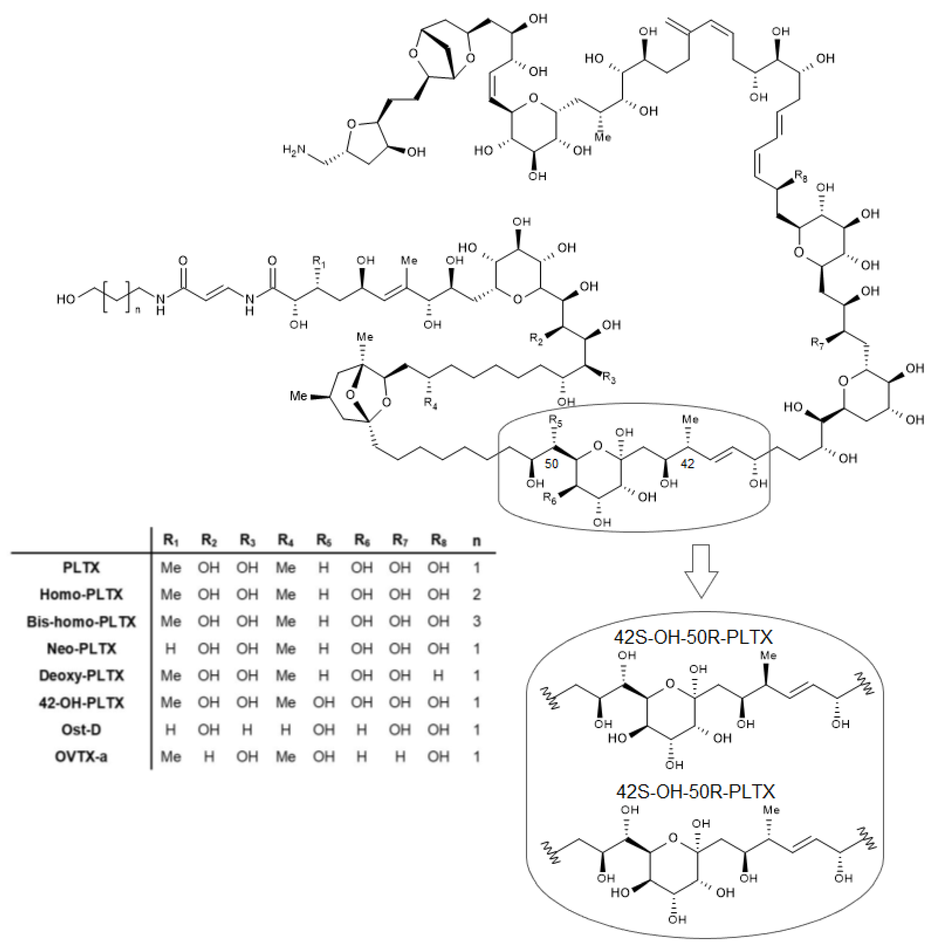
2. Palytoxins Mechanism of Action

The mechanism of action of PLTX and its analogues is quite well described in literature and represents one of the most active research fields on PLTXs [33]. It has been used as a starting point to set up functional assays for PLTX detection.
The main molecular target of PLTX is the Na+/K+ ATPase, a transmembrane transporter belonging to the P-type ATPase family of animal cells that actively maintain cellular ions homeostasis. It transports three Na+ ions out of the cell in trade for two K+ ions in the cytoplasm against their concentration gradient, due to chemical energy derived from ATP hydrolysis [33]. Na+/K+ ATPase consists in a hetero-oligomer made by three subunits: α, the catalytic one, β, and γ. PLTX binds to the extracellular side of this pump with a high affinity [33,34]. It has been demonstrated that PLTX downstream effects (i.e., sustained cytotoxicity) appear to be correlated with the expression of specific subunit isoforms in the pump. In particular, the expression of the β2—and to a lesser extent the α3—isoform positively correlates with increased cell sensitivity to PLTX cytotoxicity [35]. PLTX interaction with the α-β ATPase heterodimer causes a permanent conformational change of the transmembrane pump into a non-selective monovalent cation channel [36,37]. This event results in a strong cellular ion imbalance: the increasing intracellular concentration of Na+ causes cell membrane depolarization, enhanced by the massive efflux of K+ and influx of Ca2+. The intracellular overload of Ca2+ seems to be mediated by an inverted activity of the Na+/Ca2+ exchanger, caused by the increased intracellular Na+ concentration. In turn, Ca2+ ions overload seems to induce the opening of K+ and Cl channels, whereas the increased intracellular Na+ ions can lead to reverse functioning of the Na+/H+ exchanger with subsequent cytoplasm acidification [32]. This ion imbalance leads to functional and structural cell alterations, depending on the cell type (i.e., excitable or non-excitable cells), such as actin cytoskeleton disorganization, mitochondrial damage, and leakage of membrane integrity, up to necrotic cell death [38,39,40,41,42,43,44,45].

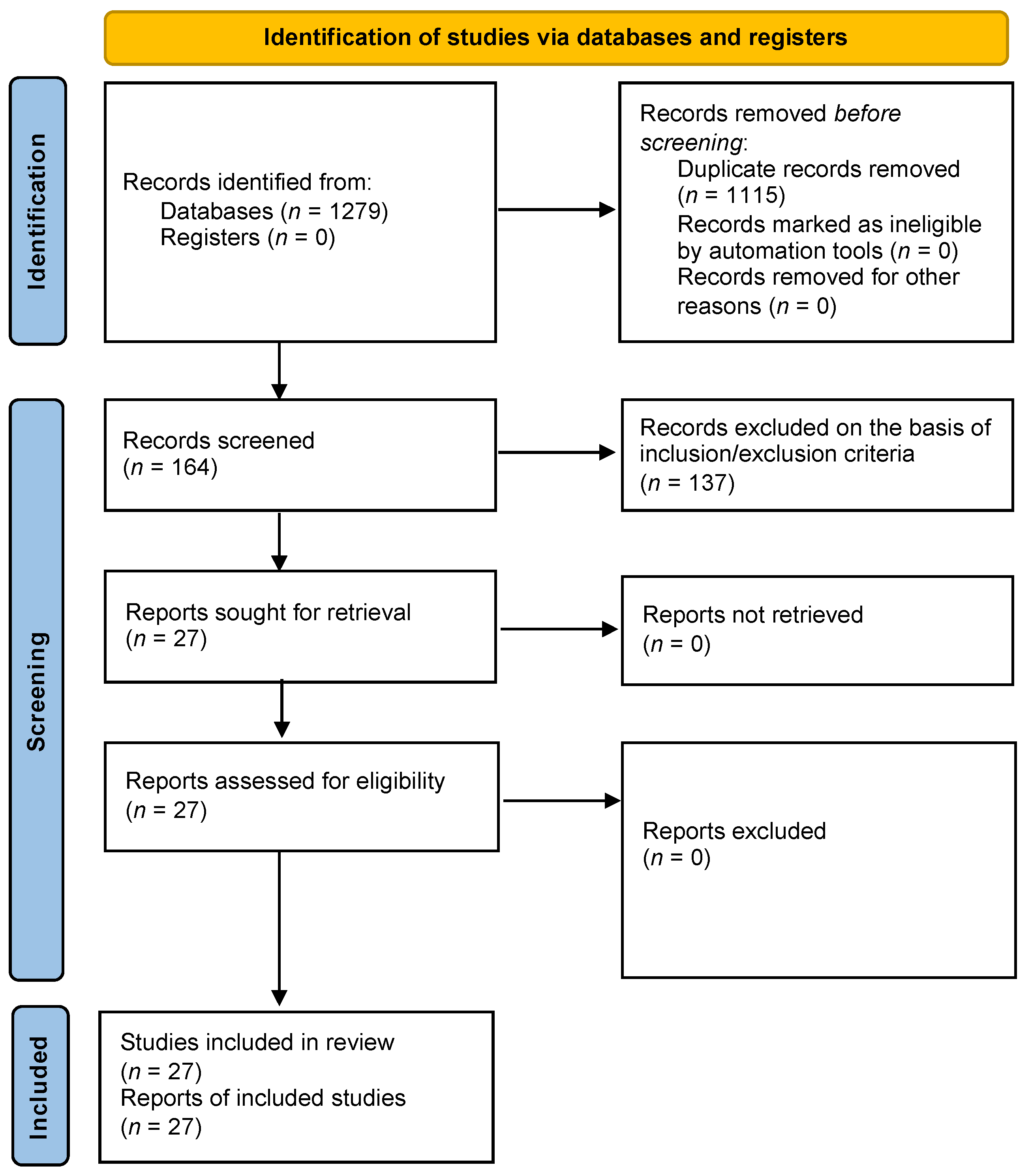
3. Method of Literature Review

A systematic review of the literature on functional and structural biological detection methods for PLTX was performed with no time restriction, according to the preferred reporting items for systematic reviews and meta-analyses (PRISMA) guidelines [46]. The electronic databases (PubMed and Scopus) were used as data sources, using the terms “palytoxin”, “Ostreopsis”, “detection method”, and “biological method”.
Inclusion criteria were: (1) biological detection methods; (2) methods originally set up for PLTX; (3) functional and structural detection methods; (4) full-text articles; and (5) English language. Exclusion criteria included: (1) editorials; (2) not-related abstracts; and (3) chemical-based detection methods and/or non-biological methods.

4. Biological Detection Methods for Palytoxins

Results of the database search are reported in the PRISMA flow diagram (Figure 2). A total of 1279 articles were identified from the electronic databases and 1252 of them were removed because of duplication among the two databases or on the basis of the above reported exclusion criteria. Thus, 27 articles were included to review the biological methods to detect palytoxins.

4.1. Functional Assays

In general, functional assays for PLTX detection include cell-based assays, mainly haemolysis and cytotoxicity assays (Table 1). These methods have been developed considering the mechanism of action of these toxins, i.e., interaction with the Na+/K+ ATPase and the subsequent conformational changes leading to cytolysis and/or other cytotoxic effects [32]. Moreover, the binding of PLTX to Na+/K+ ATPase has been exploited, including its inhibition by the cardioactive glycoside ouabain as a negative allosteric modulator or as a noncompetitive antagonist against high or low PLTX concentrations, respectively [34]. A cell-based biosensor exploiting the haemolytic activity of PLTX has been also set up.

4.1.1. Haemolysis Assays

Haemolysis assays detect PLTXs by exploiting the characteristic delayed erythrocytes lysis consequent to the toxin binding to Na+/K+ ATPase and its conversion into a nonselective monovalent cation channel, which leads to osmotic lysis and haemoglobin release, measurable spectrophotometrically. The first haemolysis assay was set up using murine erythrocytes: the haemolytic action of PLTX was exploited to develop a simple, sensitive assay with a good selectivity that was due to the use of an anti-PLTX monoclonal antibody (73D3 mAb) able to neutralize the PLTX effect. PLTX induced a time and temperature dependent haemolysis, with a detection limit (LOD) of 1 pg/mL (0.37 pM) after 24 h exposure [47]. This assay represented the basis for the development of other haemolytic assays, with slight modifications in the origin of erythrocytes and some experimental parameters. Thus, the method was slightly modified using human erythrocytes, in suspension [48] or blocked in an agar support [49]. In both cases, no specific information on the assay sensitivity was provided, but the latter was used to detect PLTXs in several species of bacteria collected from soft corals, without a careful matrix effect evaluation. Riobó and co-workers optimized the hemolysis assay using sheep erythrocytes and ouabain to antagonize PLTX effect on the Na+/K+ ATPase. Based on the working range (10–1200 pg/mL), this haemolysis assay resulted very sensitive at the optimum temperature of 25 °C, but no information on LOD or limit of quantitation (LOQ) are available [50]. However, the haemolytic assay was prone to suffer a significant matrix effect when used to quantify PLTX in shellfish methanol extracts. Indeed, quite recently, the haemolysis assay has been re-optimized using human erythrocytes, varying the buffer osmolarity and assay temperature to increase its sensitivity [51]. This assay was able to detect PLTX and its analogues in accordance with their toxic potency: it had a good sensitivity for PLTX and 42S-OH-50S-PLTX (from P. toxica), but the stereoisomer 42S-OH-50R-PLTX (from P. tuberculosa), OVTX-a, and an OVTXs mixture (composed by OVTX-a, -d, and –e) demonstrated lower haemolytic activity than PLTX. Despite an excellent sensitivity to detect PLTX as standard solution (LOD and LOQ equal to 1.4 × 10⁻¹⁰ M and 3.4 × 10⁻¹⁰ M, corresponding to 0.38 μg/mL and 0.91 μg/mL, respectively), it failed to accurately and precisely quantify the toxin in a complex matrix such as mussel meat at levels below the safe limit proposed by EFSA (30 μg/kg) because of the high matrix interference (LOQ = 640 μg/kg in Mytilus galloprovincialis), which imposes at least 1:50 dilution of the mussels extract [51]. In general, the use of haemolytic assays for PLTX quantitation in seafood requires a careful evaluation of the specific matrix effect exerted by each natural sample.

4.1.2. Cell-Based Assays

To develop functional cell-based assays for PLTX detection, cell death or earlier cytotoxic effects, such as cell membrane depolarization, have been considered as measurable end points. In this view, the Neuro-2a assay, which uses mouse neuroblastoma cells and was previously developed for other algal toxins detection, has been studied for its suitability to also detect PLTXs by its ability to reduce cell viability [52,53,54,55,56,57]. Cañete and Diogène carried out a comparative study of Neuro-2a and the mouse–rat hybrid neuroblastoma-glioma NG108-15 cell line, in an effort to extend a neuronal cell assay able to quantify PLTX by means of MTT reduction in viable cells [52]. Both NG108-15 and Neuro-2a cells treated with ouabain (Na+/K+ ATPase inhibitor) and veratridine (Na+ channels opener) were more sensitive to PLTX than those exposed only to the latter. A high sensitivity was achieved in NG108-15 or Neuro-2a cells exposed to PLTX for 24 h in the presence of ouabain and veratridine (EC50 = 0.05 and 0.04 nM, corresponding to 0.13 and 0.11 ng/mL, respectively). However, no information on LOD or LOQ and no data on possible cross-reactivity with other toxins are available for this assay. Using the MTT assay, Ledreux and co-workers showed a potent PLTX cytotoxicity toward Neuro-2a cells exposed to the toxin for 19 h in the absence of ouabain (EC50 = 42.9 ± 3.8 pM; LOD = 20 pM, corresponding to 0.05 ng/mL). The cytotoxic potency of PLTX increased by simultaneous cell exposure to 500 μM ouabain (EC50 = 6.3 ± 4.7 pM), whereas pre-incubation with ouabain for 2 h increased cell viability as compared to cell exposure to PLTX alone (EC50 = 290.7 ± 50.2 pM) [53]. The assay was able to detect PLTX and its analogues in spiked algal and mussels extracts with a LOD of 5 ng/mL extract (corresponding to 50 μg PLTX/kg shellfish meat) without matrix effects. The reduced PLTX cytotoxicity in Neuro-2a cells pre-exposed to ouabain for 2 h was exploited to selectively detect this toxin, differentiating its effect from that of other neurotoxic phycotoxins, such as saxitoxins, brevetoxins, and ciguatoxins: exposure of Neuro-2a cells to a toxin with or without appropriate timed ouabain allow the specific detection of PLTXs, whereas appropriate cell treatment with or without ouabain/veratridine allow to detect the other algal toxins acting on voltage-gated sodium channels [53]. In a similar manner, Espiña and colleagues developed a microplate-based dynamic assay using BE(2)-M17 human neuroblastoma cells without or with ouabain pretreatment able to prevent cell viability reduction induced by PLTXs. The assay uses the Alamar blue fluorescent probe, which allowed continuous measurement of cell viability. The assay, carried out exposing the cells to PLTX for 4 h, was able to detect the toxin at 0.4 ng/mL, while 72 h exposure allowed toxin detection within the range of 0.2–40 ng/mL. Moreover, it was used to differentially detect PLTXs and okadaic acid in naturally contaminated mussels, based on selective ouabain inhibition of cell viability reduction induced by PLTXs. The method was applied also to detect PLTXs in Ostreopsis cf. siamensis cells cultured from field samples, which was due to the lack of matrix effect [54].
More recently, Nicolas and co-workers explored the suitability of new functional endpoints in the Neuro-2a assay to detect PLTX and other neurotoxins (saxitoxin and tetrodotoxin): changes of cellular membrane potential and of genes expression. However, none of these endpoints were more useful than cytotoxicity as a final readout for the detection of these toxins [55]. Exploiting the neurotoxic effects of PLTX, the same research group built a functional bioassay based on the multielectrode array (MEA) technique, able to measure local field potentials in neuronal networks of electrically active rat cortical neurons [56]. The method turned out to be more sensitive than the Neuro-2a assay for PLTX detection (EC50 = 12 pM; LOD = 1 pM, corresponding to 2.7 pg/mL), but its ability to also detect other neurotoxins, such as saxitoxin, ciguatoxin, and tetrodotoxin, as well as its sensitivity for PLTX equal to that displayed for ciguatoxin (LOD = 1 pM), argue against a suitable selectivity. Notwithstanding, the same technique was applied to analyze cultured Ostreopsis cf. ovata cells submitted to three different treatments: (i) filtered and resuspended algal cells; (ii) filtered, resuspended, and sonicated algal cells, or (iii) conditioned growth medium devoid of algal cells. Exposure of neuronal networks to algal cells releasing PLTXs up to 20 min affected the spontaneous electrical activity inhibiting firing rate, burst rate, and percentage of spikes in burst, while increasing the burst duration and the inter-spike intervals within a burst. However, a large variability in the effects was observed, probably because of the not homogeneous distribution of entire cells in the MEA chip, although an effect caused by cell debris could not be excluded [57].
Cell-based functional assays for PLTX were also developed on cell models other than neurons. For instance, the MCF-7 human breast adenocarcinoma cell line was used by Bellocci’s group to develop a cytotoxicity assay for PLTX detection based on the cytolytic release of lactate dehydrogenase (LDH) induced by the toxin, with or without ouabain pretreatment. The method was sensitive (LOD of approximately 0.5 ng/mL and LOQ of 0.08 µg/kg) and was able to quantify PLTX in field samples of O. ovata, mussels (M. galloprovincialis), and sea urchins (Paracentrotus lividus). Moreover, it did not show any cross-reactivity with maitotoxin, tetrodotoxin, okadaic acid, or yessotoxin [58,59]. However, PLTX levels in naturally contaminated samples measured by the MCF-7 cytolytic assay were significantly higher than those quantified by LC–MS. These differences are probably due to the presence of PLTX analogues not detected by the analytical chemical method, even though a revision based on a high quality PLTX standard indicated that PLTX quantitation made by cytolytic or LC–MS methods were similar [59].

4.1.3. Cell-Based Biosensors

On the basis of the haemolytic properties of PLTX, Volpe and co-workers set up an electrochemical biosensor for PLTX detection through an amperometric measurement of LDH release from sheep erythrocytes exposed to the toxin. Exposure of diluted sheep blood to PLTX for 24 h or 4 h allowed the toxin detection within the concentration ranges of 0.007–0.02 ng/mL or 0.16–1.3 ng/mL, respectively. The assay specificity toward PLTXs was also confirmed: saxitoxin, brevetoxin, tetrodotoxin, okadaic acid, and yessotoxin were analyzed and did not induce erythrocytes lysis detectable by the biosensor. However, a significant matrix effect was recorded for 50% aqueous methanol extracts of mussel (Mytilus galloprovincialis) digestive glands (1:50 dilution), which was removed if 10% aqueous methanol extracts from the whole mussels’ tissue were analyzed, but with a low (35%) toxin recovery [60].
Table 1. Functional assays for PLTX quantitation.
AssayLOD in Standard SolutionLOQ in Standard SolutionMatricesLOQ in MatricesCross-ReactivityReference
Functional AssaysHaemolysis assay1 pg/mL [47]
[48]
(wr: 10–1200 pg/mL) [50]
Bacteria [49]
0.38 ng/mL0.91 ng/mLMussels640 µg/Kg42S-OH-50R-PLTX
ovatoxins
[51]
Hemolysis-based biosensor(wr: 0.16–1.3 ng/mL, 4h;
0.007–0.02 ng/mL, 24h)
Mussels No cross-reactivity with saxitoxin, brevetoxin, tetrodotoxin, okadaic acid and yessotoxin[60]
Cytotoxicity assay (NG108-15)(EC50 = 0.13 ng/mL) [52]
Cytotoxicity assay (Neuro-2a)(EC50 = 0.11 ng/mL)
0.05 ng/mL Mussels(LOD = 5 ng/mL)No cross-reactivity with phycotoxins, such as saxitoxins, brevetoxins and ciguatoxins[53]
[55]
Cytotoxicity assay (BE(2)-M17)0.2 ng/mL Mussels
O. siamensis
No cross-reactivity with okadaic acid[54]
Cytotoxicity assay (MCF-7)0.5 ng/mL Mussels
Sea archins
O. ovata
0.08 µg/KgNo cross-reactivity with maitotoxin, tetrodotoxin, okadaic acid or yessotoxin[58]
Multielectrode array2.7 pg/mL CTX
No cross-reactivity with tetrodotoxin and saxitoxin
[56]
O. ovata [57]
Abbreviations and notes: LOD = limit of detection; LOQ = limit of quantitation; wr = working range; other parameters are reported in brackets; empty cells indicate not determined data.

4.2. Structural Assays

The majority of structural assays for PLTX detection relies on biochemistry and exploits the ability of anti-PLTX antibodies to bind to a specific toxin structure (i.e., immunoassays and immuno-based biosensors), even though few methods are based on other kinds of structural detection (Table 2). The large size of PLTX is an advantage for anti-PLTX antibodies production via animal immunization, thereby simplifying the development of immunoassays. However, the molecule conjugation with proteins is still a requirement to trigger an immunogenic response necessary to stimulate generation of anti-PLTX antibodies. Indeed, all the anti-PLTX antibodies obtained by animal immunization used immunogens formed via conjugation of the C-115 PLTX terminal amino functional group [61,62,63,64,65], that can be easily derivatized to form well-defined N-acyl haptens.

4.2.1. Immunoassays

Levine and co-workers were the first researchers who obtained anti-PLTX polyclonal antibodies from immunized rabbits. These antibodies were used to develop a competitive radioimmunoassay (RIA) based on the use of a 125I-labeled PLTX derivative [66]. These anti-PLTX antibodies did not distinguish the homologous PLTX of P. caribaeorum from the heterologous palytoxin of P. tuberculosa, whose structure differed only in hemiketal ring at position C55, but were able to detect PLTX without cross-reactivity with other toxins, such as maitotoxin, teleocidin, okadaic acid, and debromoaplysiatoxin. Despite its extraordinary sensitivity for PLTX detection (PLTX concentration inhibiting 125I-PLTX binding to anti-PLTX antibodies by 50%, IC50 = 0.27 pM, corresponding to 0.72 pg/mL), the requirement for a radioiodine-labeled PLTX antigen makes this assay impractical in terms of managing, costs, antigen stability, and radioactivity concerns.
To overcome the limitations posed by radioactive reagents, a series of enzyme-linked immunosorbent assays (ELISAs) for PLTX detection have been developed in the last three decades. Both competitive and sandwich enzyme immunoassays have been set up [63,64,65,67]. Bignami and co-workers developed competitive and sandwich immunoassays based upon a murine anti-PLTX monoclonal antibody (73D3 mAb; isotype IgG1, κ). In particular, two direct competitive immunoassays were developed, using the monoclonal anti-PLTX antibody conjugated with alkaline phosphatase (ALP; mAb-ALP) or PLTX conjugated with ALP (PLTX-ALP). Both strategies showed good sensitivities toward PLTX (IC50, toxin concentration producing 50% signal inhibition = 3.5 and 10.1 ng/mL for mAb-ALP and PLTX-ALP, respectively), and were applied to quantify the toxin in naturally contaminated P. tuberculosa extracts. In addition, an indirect competitive ELISA was also developed, allowing PLTX quantitation with an IC50 of 6.2 ng/mL. The assay was applied for the quantitation of the toxin in naturally contaminated P. tuberculosa extracts, without showing any cross-reactivity with selected marine toxins (tetrodotoxin, okadaic acid, or lyngbyatoxin A) [63]. While Bignami’s group used the classical PLTX-BSA conjugate as a coating agent, Frolova and colleagues took advantage of PLTX’s large size to directly immobilize the toxin in the microwells and set up an indirect competitive ELISA able to detect PLTX with an IC50 of 20 ng/mL and to quantify it in bacteria (Aeromonas sp. and Vibrio sp.) isolated from Palythoa corals [64]. A decade later, a novel indirect competitive ELISA was set up by Garet and co-workers using recombinant human anti-PLTX single-chain (scFv) antibodies obtained via a phage display technique and PLTX directly absorbed to the solid phase. Garet’s method was reported to be extraordinarily sensitive, with a LOD of 0.5 pg/mL, but the variable toxin recovery from mussels (64−113%) and clams (84−181%) argues against its reproducibility and would likely impair its suitability for monitoring purposes [67].
Concerning the noncompetitive sandwich-type ELISAs, Bignami’s group also developed a direct and an indirect immunoassay using the murine 73D3 mAb as capture antibody, and ALP-labelled polyclonal anti-PLTX antibody (pAb-ALP) as detection antibody. The two ELISAs were able to detect PLTX with an IC50 of 4.8 ng/mL and 0.6 ng/mL, respectively, but cross-reactivity with other toxins has not been verified. Moreover, these ELISAs were applied to quantify PLTXs only in naturally contaminated P. tuberculosa extracts and to monitor toxin isolation [63]. Two decades later, a new indirect sandwich ELISA was developed using the same 73D3 mAb as a capture antibody and novel rabbit anti-PLTX pAb targeted with horseradish (HRP)-conjugated anti-rabbit antibody as detection agents. This ELISA was thoroughly characterized for PLTX detection, providing an IC50 of 7.6 ng/mL, an LOD of 1.1 ng/mL, and an LOQ of 2.2 ng/mL [65]. The assay was also able to quantify 42-OH-PLTX from P. toxica [15], ovatoxin-a and ostreocin D, the latter in a lesser extent with respect to PLTX [19]. Beside a good sensitivity, this assay demonstrated good accuracy (bias of 2.1%), repeatability (relative standard deviation of repeatability, RSDr = 6% and 9% for intra- and inter-assay variability, respectively), and specificity as it did not cross-react with okadaic acid, domoic acid, saxitoxin, brevetoxin-3, or yessotoxin. Furthermore, it was applied to quantify PLTX in spiked mussels, microalgae, and seawater extracts, defining LOQs of 11.0, 9.6, and 2.4 ng/mL, respectively. The LOQ in mussels was 11 μg PLTX equivalents/kg edible parts. Recently, the murine 73D3 mAb was used as a detecting antibody to set up a novel indirect cell-based ELISA: PLTX binding to Na+/K+ ATPase of HaCaT keratinocytes was detected by the primary 73D3 mAb, targeted by a secondary HRP-conjugated anti-mouse antibody as detection agent. The method turned out to be highly sensitive (LOD and LOQ = 1.2 × 10⁻¹¹ M and 2.8 × 10⁻¹¹ M, respectively, corresponding to 32.2 and 75 pg/mL, respectively), accurate (bias = 2.5%), and repeatable (15% and 9% interday and intraday RSDr, respectively). In addition, minimal interference of mussels’ extract allows PLTX quantitation with a LOQ of 9.1 µg/kg mussel meat [68], meeting the limit suggested by the EFSA.

4.2.2. Immuno-Based Biosensors

As a further development of immunoassays for PLTX detection, a series of antibody-based biosensors have been developed. The sandwich ELISA set up by Boscolo and colleagues was resized by Zamolo and co-workers into a highly sensitive biosensor based on a sandwich immunoassay architecture and an electrochemiluminescence (ECL) detection [69]. Its sensitivity was significantly increased by covalently linking the 73D3 mAb to an optically transparent transduction electrode through functionalized multiwalled carbon nanotubes (CNTs). The rabbit pAb conjugated to a ruthenium complex served as the ECL signal generating detector. The biosensor was characterized by instrumental LOD and LOQ of 0.07 and 0.24 ng/mL, respectively. Although the biosensor cross-reactivity with other PLTX analogues was not tested, the 73D3 mAb selectivity toward PLTX had been demonstrated by Boscolo and colleagues [65]. No matrix effect was observed for mussel (Mytilus galloprovincialis) or microalgal extracts spiked with PLTX after 1:10 dilution, giving LOQs of 0.22 ng/mL for mussel extract (2.2 μg/kg mussel meat) and 0.23 ng/mL for algal extract. Later on, the 73D3 mAb was employed to develop an immuno-based method for PLTX detection using microspheres coupled to flow-cytometry detection (Luminex 200™). This method displays an IC50 of 1.83 ± 0.21 nM with a LOD of 0.47 ± 0.15 nM, corresponding to 1.27 ± 0.39 ng/mL. The anti-PLTX-mAb, used in this assay, did not interact with okadaic acid, dinophysistoxin-1, domoic acid, saxitoxin, tetrodotoxin, maitotoxin, pectenotoxin, yessotoxin, or brevetoxin-3, whereas the detection of some PLTX analogues (42-OH-PLTX and ovatoxins) appeared to be not very efficient. Nevertheless, this method was able to detect PLTX in contaminated mussels (M. galloprovincialis; working range: 374–4430 μg/kg), as well as in Ostreopsis cf. siamensis and Palythoa tuberculosa extracts, for which no information on the assay sensitivity were reported [70]. The 73D3 mAb had been previously exploited to also build a surface plasmon resonance (SPR) immunosensor. The SPR-based immunosensor was characterized by an instrumental LOD of 0.52 ng PLTX/mL, which was higher for PLTX detection in 10% spiked grouper or 10% spiked clam extracts (2.8 and 1.4 ng/mL, respectively). Moreover, it did not cross-react with saxitoxin, tetrodotoxin, maitotoxin, pectenotoxin, okadaic acid, or dinophysistoxin-1 [71]. Quite recently, the SPR-based immunosensor was further optimized to set up a multiplex structural assay for a simultaneous detection of PLTX and other algal toxins (saxitoxins, okadaic acid, and domoic acid) in mussels (Mytilus edulis), cockles (Cerastoderma edule), oysters (Crassostrea gigas), and scallops (Pecten maximus). The antibodies’ specificity and the absence of measurable cross-reactivity with other marine biotoxins by the multiplex SPR biosensor allowed the detection of PLTX and the other three toxins with different sensitivities. The IC50 for PLTX detection in buffer was equal to 12 ng/mL, whereas an IC50 of 215 µg/kg was calculated for PLTX-spiked mussel extracts [72].

4.2.3. Receptor-Based Biosensors

The surface plasmon resonance (SPR) technology has been exploited to also detect PLTX by the affinity to its molecular target (Na+/K+ ATPase). An SPR biosensor based on PLTX interaction over immobilized Na+/K+ ATPase has been set up. The LOD and LOQ of this biosensor are reported as 3.73 and 11.2 pg, respectively (the measure unit of the volume is not specified). This Na+/K+ ATPase SPR-biosensor represents one the first attempts to detect PLTX in marine organisms, such as Ostreopsis siamensis microalgae, exploiting its mechanism of action [73]. The same research group developed another biosensor based on PLTX interaction with its molecular target. Using Na+/K+ ATPase labeled with a reactive succinimidyl carboxyfluorescein esther and fluorescence polarization (FP) as a spectroscopic technique to measure the molecular interactions, a biosensor has been set up. The biosensor, with LOD and LOQ of 2 nM and 10 nM PLTX, respectively (corresponding to 5.3 and 26.8 ng/mL, respectively), was used to quantify PLTXs in naturally contaminated Ostreopsis siamensis and mussel samples after different clean-up steps on extracts to avoid matrices interferences. Nevertheless, no information on the assay sensitivity in these matrices and the possible cross-reactivity with other toxins are available, whereas 86.6% toxin recovery was determined in PLTX-spiked mussels [74].

4.2.4. Aptamer-Based Biosensors

An enzyme-linked aptamer-based biosensor for PLTX detection has been set up, coupling biolayer interferometry (BLI) with a competitive binding assay through an enzyme-linked aptamer. The biosensor, based on horseradish peroxidase-labeled aptamers as biorecognition receptors for competitive binding to PLTX, displayed a linear detection range of 200–700 pg PLTX/mL, with an exceptional LOD of 0.04 pg/mL. Okadaic acid, microcystin-LR, saxitoxin, and brevetoxin-A/B did not interfere with the detection of PLTX. This biosensor was also able to detect PLTX in spiked mussel, scallop, and clam extracts with a recovery of about 100% [75].
Table 2. Structural assays for PLTX quantitation.
AssayLOD in Standard SolutionLOQ in Standard SolutionMatricesLOQ in MatricesCross-ReactivityReference
Structural AssaysRIA(IC50 = 0.72 pg/mL) No cross-reactivity with maitotoxin, teleocidin, okadaic acid, debromoaplysiatoxin and 12-O-tetradecanoylphorbol-l3-acetate[66]
MAb-ALP competitive direct ELISA(IC50 = 3.5 ng/mL) P. tuberculosa No cross-reactivity with tetrodotoxin, okadaic acid or lyngbyatoxin A[63]
PLTX-ALP competitive direct ELISA(IC50 = 10.1 ng/mL) P. tuberculosa
Indirect competitive ELISA(IC50 = 6.2 ng/mL) P. tuberculosa
(IC50 = 20 ng/mL) Vibro sp.
Aeromonas sp.
[64]
0.5 pg/mL Mussels
Clams
[67]
Direct sandwich ELISA(IC50 = 4.8 ng/mL) P. tuberculosa [63]
Indirect sandwich ELISA(IC50 = 0.6 ng/mL) P. tuberculosa
1.1 ng/mL2.2 ng/mLMussels11.0 ng/mLNo cross-reactivity with okadaic acid, domoic acid, saxitoxin, brevetoxin-3 and yessotoxin[65]
Microalgae9.6 ng/mL
Seawater2.4 ng/mL
Cell-based ELISA32.2 pg/mL75.0 pg/mLMussels9.1 µg/kgNo cross-reactivity with yessotoxin, okadaic acid, domoic acid, brevetoxin-3, saxitoxin, azaspiracid-1, and maitotoxin[68]
ECL-based immunoassay0.07 ng/mL0.24 ng/mLMussels0.22 ng/mLNo cross-reacivity with okadaic acid, domoic acid, saxitoxin, brevetoxin-3, and yessotoxin[69]
Microalagae0.23 ng/mL
Cytometry-based
immunoassay
1.27 ± 0.39 ng/mL Mussels(wr: 374–4430 mg/kg)42-OH-PLTX
Ovatoxins;
No cross-reactivity with okadaic acid, dinophysistoxin-1, domoic acid, saxitoxin, tetrodotoxin, maitotoxin, pectenotoxin, yessotoxin and brevetoxin-3
[70]
O. siamensis
P. tuberculosa
SPR-based immunoassay0.52 ng/mL Grouper(LOD = 2.8 ng/mL)No cross-reacivity with saxitoxin, tetrodotoxin, maitotoxin, pectenotoxin, okadaic acid, and dinophysistoxin-1[71]
Clam(LOD = 1.4 ng/mL)
Multiplex SPR-based immunoassay(IC50 = 12 ng/mL) Mussels(IC50 = 215 µg/Kg)No cross-reactivity with saxitoxins, okadaic acid and domoic acid[72]
Na+/K+ ATPase SPR-biosensor3.73 pg11.2 pgMicroalgae [73]
FP biosensor5.3 ng/mL26.8 ng/mLO. siamensis [74]
Aptamer-based biosensor0.04 pg/mL Mussels
Scallops
Clams
No cross-reactivity with okadaic acid, microcystin-LR, saxitoxin, and brevetoxin-A/B[75]
Abbreviations and notes: LOD = limit of detection; LOQ = limit of quantitation; wr = working range; other parameters are reported in brackets; empty cells indicate not determined data.

5. Conclusions

In general, the use of biological and biochemical methods for the detection of algal toxins in field samples is becoming a fruitful tool for an initial screening approach before confirmatory analysis by reference chemical methods. However, these detection methods are not available for all the algal toxins because of: (1) their unknown mechanism of action and/or molecular target, needed as starting points to set up functional methods; and (2) the lack of specific anti-toxin antibodies used to detect the toxins in the structural biochemical methods. These requirements are satisfied for PLTXs and a series functional and structural biological/biochemical assays have been set up for their detection, as summarized in Figure 3.
Indeed, the availability of the murine 73D3 monoclonal antibody (in addition to others less characterized) allowed the development of different structural immunoassays. Although some of these assays have not been fully characterized, in general they demonstrated a good sensitivity for PLTX detection, in some cases also in complex matrices such as mussel meat. However, there are limited cross-reactivity data showing the assay’s ability to detect or differentiate PLTX structural analogues. Thus, future studies could be directed on this aspect and to the production of antibodies with high affinity for different PLTX analogues, useful to set up new enzyme immunoassays able to provide results highly correlated to the actual toxic potential of the sample under analysis.
In addition, the good knowledge gained on the mode of PLTX action allowed the development of several functional methods for toxin detection. However, functional assays are characterized by some limitations that are due to matrix effects and low specificity. The former is particularly evident for the haemolysis assay, which is frequently used for PLTX detection but may suffer from significant matrix interferences during toxin quantitation in seafood. Lack of specificity is the main limitation of cytotoxicity assays: some progress has been made to differentiate PLTX cytotoxicity from that of other algal toxins by using ouabain and/or veratridine, but the lack of robust data supporting the assay specificity does not allow its use for monitoring purposes. The available biological/biochemical methods for PLTXs quantitation appear to be a quite promising and fruitful tools than can be exploited for a rapid and sensitive detection of these toxins for screening purposes. Anyway, some of them should be fully characterized or optimized to provide the additional data needed for their validation, such as sensitivity, specificity, matrix effect, accuracy, precision, repeatability, and robustness. These aspects should be addressed also during the development of new biological/biochemical assays for PLTX detection.

Author Contributions

Conceptualization, A.T.; writing—original draft preparation, M.C., M.P.; writing—review and editing, S.S., C.P.; supervision, A.T. All authors have read and agreed to the published version of the manuscript.

Funding

This research received no external funding.

Institutional Review Board Statement

Not applicable.

Informed Consent Statement

Not applicable.

Data Availability Statement

Not applicable.

Conflicts of Interest

The authors declare no conflict of interest.

References

  1. Munday, R. Occurrence and toxicology of palytoxin. In Seafood and Freshwater Toxins. Pharmacology, Physiology and Detection; Botana, L.M., Ed.; CRC Press: Boca Raton, FL, USA, 2008; pp. 693–713. [Google Scholar]
  2. Sosa, S.; Del Favero, G.; De Bortoli, M.; Vita, F.; Soranzo, M.R.; Beltramo, D.; Ardizzone, M.; Tubaro, A. Palytoxin toxicity after acute oral administration in mice. Toxicol. Lett. 2009, 191, 253–259. [Google Scholar] [CrossRef] [PubMed]
  3. Boente-Juncal, A.; Vale, C.; Camiña, M.; Cifuentes, J.M.; Vieytes, M.R.; Botana, L.M. Reevaluation of the acute toxicity of palytoxin in mice: Determination of lethal dose 50 (LD50) and No-observed-adverse-effect level (NOAEL). Toxicon 2020, 177, 16–24. [Google Scholar] [CrossRef] [PubMed]
  4. Del Favero, G.; Beltramo, D.; Sciancalepore, M.; Lorenzon, P.; Coslovich, T.; Poli, M.; Testai, E.; Sosa, S.; Tubaro, A. Toxicity of palytoxin after repeated oral exposure in mice and in vitro effects on cardiomyocytes. Toxicon 2013, 75, 3–15. [Google Scholar] [CrossRef] [PubMed]
  5. Boente-Juncal, A.; Raposo-García, S.; Vale, C.; Louzao, M.C.; Otero, P.; Botana, L.M. In vivo evaluation of the chronic oral toxicity of the marine toxin palytoxin. Toxins 2020, 12, 489. [Google Scholar] [CrossRef]
  6. Pelin, M.; Brovedani, V.; Sosa, S.; Tubaro, A. Palytoxin-containing aquarium soft corals as an emerging sanitary problem. Mar. Drugs 2016, 14, 33. [Google Scholar] [CrossRef] [Green Version]
  7. Tartaglione, L.; Dello Iacovo, E.; Mazzeo, A.; Casabianca, S.; Ciminiello, P.; Penna, A.; Dell’Aversano, C. Variability in toxin profiles of the Mediterranean Ostreopsis cf. ovata and in structural features of the produced ovatoxins. Environ. Sci. Technol. 2017, 51, 13920–13928. [Google Scholar] [CrossRef]
  8. Patocka, J.; Nepovimova, E.; Wu, Q.H.; Kuca, K. Palytoxin congeners. Arch. Toxicol. 2018, 92, 143–156. [Google Scholar] [CrossRef]
  9. Terajima, T.; Uchida, H.; Abe, N.; Yasumoto, T. Simple structural elucidation of ostreocin-B, a new palytoxin congener isolated from the marine dinoflagellate Ostreopsis siamensis, using complementary positive and negative ion liquid chromatography/quadrupole time-of-flight mass spectrometry. Rapid Commun. Mass Spectrom. RCM 2018, 32, 1001–1007. [Google Scholar] [CrossRef]
  10. Terajima, T.; Uchida, H.; Abe, N.; Yasumoto, T. Structure elucidation of ostreocin-A and ostreocin-E1, novel palytoxin analogs produced by the dinoflagellate Ostreopsis siamensis, using LC/Q-TOF MS. Biosci. Biotechnol. Biochem. 2019, 83, 381–390. [Google Scholar] [CrossRef]
  11. Kerbrat, A.S.; Amzil, Z.; Pawlowiez, R.; Golubic, S.; Sibat, M.; Darius, H.T.; Chinain, M.; Laurent, D. First evidence of palytoxin and 42-hydroxy-palytoxin in the marine cyanobacterium Trichodesmium. Mar. Drugs 2011, 9, 543–660. [Google Scholar] [CrossRef] [Green Version]
  12. Tartaglione, L.; Pelin, M.; Morpurgo, M.; Dell’Aversano, C.; Montenegro, J.; Sacco, G.; Sosa, S.; Reimer, J.D.; Ciminiello, P.; Tubaro, A. An aquarium hobbyist poisoning: Identification of new palytoxins in Palythoa cf. toxica and complete detoxification of the aquarium water by activated carbon. Toxicon 2016, 121, 41–50. [Google Scholar] [CrossRef]
  13. Ciminiello, P.; Dell’Aversano, C.; Dello Iacovo, E.; Fattorusso, E.; Forino, M.; Grauso, L.; Tartaglione, L.; Florio, C.; Lorenzon, P.; De Bortoli, M.; et al. Stereostructure and biological activity of 42-Hydroxy-palytoxin: A new palytoxin analogue from Hawaiian Palythoa subspecies. Chem. Res. Toxicol. 2009, 22, 1851–1859. [Google Scholar] [CrossRef]
  14. Tubaro, A.; Del Favero, G.; Beltramo, D.; Ardizzone, M.; Forino, M.; De Bortoli, M.; Pelin, M.; Poli, M.; Bignami, G.; Ciminiello, P.; et al. Acute oral toxicity in mice of a new palytoxin analog: 42-hydroxy-palytoxin. Toxicon 2011, 57, 755–763. [Google Scholar] [CrossRef]
  15. Ciminiello, P.; Dell’Aversano, C.; Dello Iacovo, E.; Forino, M.; Tartaglione, L.; Pelin, M.; Sosa, S.; Tubaro, A.; Chaloin, O.; Poli, M.; et al. Stereoisomers of 42-hydroxy palytoxin from Hawaiian Palythoa toxica and P. tuberculosa: Stereostructure elucidation, detection, and biological activities. J. Nat. Prod. 2014, 77, 351–357. [Google Scholar] [CrossRef]
  16. Ukena, T.; Satake, M.; Usami, M.; Oshima, Y.; Naoki, H.; Fujita, T.; Kan, Y.; Yasumoto, T. Structure elucidation of ostreocin D, a palytoxin analog isolated from the dinoflagellate Ostreopsis siamensis. Biosci. Biotechnol. Biochem. 2001, 65, 2585–2588. [Google Scholar] [CrossRef] [Green Version]
  17. Ito, E.; Yasumoto, T. Toxicological studies on palytoxin and ostreocin-D administered to mice by three different routes. Toxicon 2009, 54, 244–251. [Google Scholar] [CrossRef]
  18. Ciminiello, P.; Dell’Aversano, C.; Dello Iacovo, E.; Fattorusso, E.; Forino, M.; Grauso, L.; Tartaglione, L.; Guerrini, F.; Pezzolesi, L.; Pistocchi, R.; et al. Isolation and structure elucidation of ovatoxin-a, the major toxin produced by Ostreopsis ovata. J. Am. Chem. Soc. 2012, 134, 1869–1875. [Google Scholar] [CrossRef]
  19. Pelin, M.; Forino, M.; Brovedani, V.; Tartaglione, L.; Dell’Aversano, C.; Pistocchi, R.; Poli, M.; Sosa, S.; Florio, C.; Ciminiello, P.; et al. Ovatoxin-a, A palytoxin analogue isolated from Ostreopsis cf. ovata Fukuyo: Cytotoxic activity and ELISA detection. Environ. Sci. Technol. 2016, 50, 1544–1551. [Google Scholar] [CrossRef]
  20. Poli, M.; Ruiz-Olvera, P.; Nalca, A.; Ruiz, S.; Livingston, V.; Frick, O.; Dyer, D.; Schellhase, C.; Raymond, J.; Kulis, D.; et al. Toxicity and pathophysiology of palytoxin congeners after intraperitoneal and aerosol administration in rats. Toxicon 2018, 150, 235–250. [Google Scholar] [CrossRef]
  21. Deeds, J.R.; Schwartz, M.D. Human risk associated with palytoxin exposure. Toxicon 2010, 56, 150–162. [Google Scholar] [CrossRef] [Green Version]
  22. Wu, M.L.; Yang, C.C.; Deng, J.F.; Wang, K.Y. Hyperkalemia, hyperphosphatemia, acute kidney injury, and fatal dysrhythmias after consumption of palytoxin-contaminated goldspot herring. Ann. Emerg. Med. 2014, 64, 633–636. [Google Scholar] [CrossRef]
  23. Patocka, J.; Gupta, R.C.; Wu, Q.H.; Kuca, K. Toxic potential of palytoxin. J. Huazhong Univ. Sci. Technolog. Med. Sci. 2015, 35, 773–780. [Google Scholar] [CrossRef]
  24. Del Favero, G.; Sosa, S.; Pelin, M.; D’Orlando, E.; Florio, C.; Lorenzon, P.; Poli, M.; Tubaro, A. Sanitary problems related to the presence of Ostreopsis spp. in the Mediterranean Sea: A multidisciplinary scientific approach. Ann. Ist. Super. Sanità 2012, 48, 407–414. [Google Scholar] [CrossRef]
  25. Barrett, R.T.; Hastings, J.P.; Ronquillo, Y.C.; Hoopes, P.C.; Moshirfar, M. Coral keratitis: Case report and review of mechanisms of action, clinical management and prognosis of ocular exposure to palytoxin. Clin. Ophthalmol. 2021, 15, 141–156. [Google Scholar] [CrossRef]
  26. EFSA. Scientific Opinion on marine biotoxins in shellfish-Palytoxin group: Marine Biotoxins in Shellfish-Palytoxin group. EFSA J. 2009, 7, 1393. [Google Scholar] [CrossRef]
  27. Klijnstra, M.D.; Gerssen, A. A sensitive LC-MS/MS method for palytoxin using lithium cationization. Toxins 2018, 10, E537. [Google Scholar] [CrossRef] [Green Version]
  28. Riobó, P.; Franco, J.M. Palytoxins: Biological and chemical determination. Toxicon 2011, 57, 368–375. [Google Scholar] [CrossRef]
  29. Ciminiello, P.; Dell’Aversano, C.; Dello Iacovo, E.; Fattorusso, E.; Forino, M.; Tartaglione, L. LC-MS of palytoxin and its analogues: State of the art and future perspectives. Toxicon 2011, 57, 376–389. [Google Scholar] [CrossRef]
  30. Ciminiello, P.; Dell’Aversano, C.; Dello Iacovo, E.; Forino, M.; Tartaglione, L. Liquid chromatography-high-resolution mass spectrometry for palytoxins in mussels. Anal. Bioanal. Chem. 2015, 407, 1463–1473. [Google Scholar] [CrossRef]
  31. Riobó, P.; Paz, B.; Franco, J.M.; Vázquez, J.A.; Murado, M.A.; Cacho, E. Mouse bioassay for palytoxin. Specific symptoms and dose-response against dose-death time relationships. Food Chem. Toxicol. 2008, 46, 2639–2647. [Google Scholar] [CrossRef]
  32. Rossini, G.P.; Bigiani, A. Palytoxin action on the Na+,K+-ATPase and the disruption of ion equilibria in biological systems. Toxicon 2011, 57, 429–439. [Google Scholar] [CrossRef] [PubMed]
  33. Habermann, E. Palytoxin acts through Na+, K+-ATPase. Toxicon 1989, 27, 1171–1187. [Google Scholar] [CrossRef]
  34. Pelin, M.; Boscolo, S.; Poli, M.; Sosa, S.; Tubaro, A.; Florio, C. Characterization of palytoxin binding to HaCaT cells using a monoclonal anti-palytoxin antibody. Mar. Drugs 2013, 11, 584–598. [Google Scholar] [CrossRef] [PubMed] [Green Version]
  35. Pelin, M.; Stocco, G.; Florio, C.; Sosa, S.; Tubaro, A. In vitro cell sensitivity to palytoxin correlates with high gene expression of the Na+/K+-ATPase β2 subunit isoform. Int. J. Mol. Sci. 2020, 21, 5833. [Google Scholar] [CrossRef]
  36. Rodrigues, A.M.; Almeida, A.-C.G.; Infantosi, A.F.C.; Teixeira, H.Z.; Duarte, M.A. Model and simulation of Na+/K+ pump phosphorylation in the presence of palytoxin. Comput. Biol. Chem. 2008, 32, 5–16. [Google Scholar] [CrossRef]
  37. Wu, C.H. Palytoxin: Membrane mechanisms of action. Toxicon 2009, 54, 1183–1189. [Google Scholar] [CrossRef]
  38. Ares, I.R.; Louzao, M.C.; Vieytes, M.R.; Yasumoto, T.; Botana, L.M. Actin cytoskeleton of rabbit intestinal cells is a target for potent marine phycotoxins. J. Exp. Biol. 2005, 208, 4345–4354. [Google Scholar] [CrossRef] [Green Version]
  39. Schilling, W.P.; Snyder, D.; Sinkins, W.G.; Estacion, M. Palytoxin-induced cell death cascade in bovine aortic endothelial cells. Am. J. Physiol. Cell Physiol. 2006, 291, C657–C667. [Google Scholar] [CrossRef]
  40. Sheridan, R.E.; Deshpande, S.S.; Adler, M. Cytotoxic actions of palytoxin on aortic smooth muscle cells in culture. J. Appl. Toxicol. 2005, 25, 365–373. [Google Scholar] [CrossRef]
  41. Bellocci, M.; Sala, G.L.; Prandi, S. The cytolytic and cytotoxic activities of palytoxin. Toxicon 2011, 57, 449–459. [Google Scholar] [CrossRef]
  42. Pelin, M.; Zanette, C.; De Bortoli, M.; Sosa, S.; Loggia, R.D.; Tubaro, A.; Florio, C. Effects of the marine toxin palytoxin on human skin keratinocytes: Role of ionic imbalance. Toxicology 2011, 282, 30–38. [Google Scholar] [CrossRef]
  43. Pelin, M.; Ponti, C.; Sosa, S.; Gibellini, D.; Florio, C.; Tubaro, A. Oxidative stress induced by palytoxin in human keratinocytes is mediated by a H+-dependent mitochondrial pathway. Toxicol. Appl. Pharmacol. 2013, 266, 1–8. [Google Scholar] [CrossRef]
  44. Pelin, M.; Sosa, S.; Pacor, S.; Tubaro, A.; Florio, C. The marine toxin palytoxin induces necrotic death in HaCaT cells through a rapid mitochondrial damage. Toxicol. Lett. 2014, 229, 440–450. [Google Scholar] [CrossRef]
  45. Del Favero, G.; Florio, C.; Codan, B.; Sosa, S.; Poli, M.; Sbaizero, O.; Molgó, J.; Tubaro, A.; Lorenzon, P. The stretch-activated channel blocker Gd3+ reduces palytoxin toxicity in primary cultures of skeletal muscle cells. Chem. Res. Toxicol. 2012, 25, 1912–1920. [Google Scholar] [CrossRef]
  46. Page, M.J.; McKenzie, J.E.; Bossuyt, P.M.; Boutron, I.; Hoffmann, T.C.; Mulrow, C.D.; Shamseer, L.; Tetzlaff, J.M.; Akl, E.A.; Brennan, S.E.; et al. The PRISMA 2020 statement: An updated guideline for reporting systematic reviews. BMJ 2021, 372, n71. [Google Scholar] [CrossRef]
  47. Bignami, G.S. A rapid and sensitive hemolysis neutralization assay for palytoxin. Toxicon 1993, 31, 817–820. [Google Scholar] [CrossRef]
  48. Malagoli, D. A full-length protocol to test hemolytic activity of palytoxin on human erythrocytes. Invertebr. Surviv. J. 2007, 4, e92–e94. [Google Scholar]
  49. Seemann, P.; Gernert, C.; Schmitt, S.; Mebs, D.; Hentschel, U. Detection of hemolytic bacteria from Palythoa caribaeorum (Cnidaria, Zoantharia) using a novel palytoxin-screening assay. Antonie Van Leeuwenhoek 2009, 96, 405–411. [Google Scholar] [CrossRef]
  50. Riobò, P.; Paz, B.; Franco, J.M.; Vazquez, J.A.; Murado, M.A. Proposal for a simple and sensitive haemolytic assay for palytoxin: Toxicological dynamics, kinetics, ouabain inhibition and thermal stability. Harmful Algae 2008, 7, 415–429. [Google Scholar] [CrossRef] [Green Version]
  51. Brovedani, V.; Sosa, S.; Poli, M.; Forino, M.; Varello, K.; Tubaro, A.; Pelin, M. A revisited hemolytic assay for palytoxin detection: Limitations for its quantitation in mussels. Toxicon 2016, 119, 225–233. [Google Scholar] [CrossRef] [Green Version]
  52. Cañete, E.; Diogène, J. Comparative study of the use of neuroblastoma cells (Neuro-2a) and neuroblastomaxglioma hybrid cells (NG108-15) for the toxic effect quantification of marine toxins. Toxicon 2008, 52, 541–550. [Google Scholar] [CrossRef]
  53. Ledreux, A.; Krys, S.; Bernard, C. Suitability of the Neuro-2a cell line for the detection of palytoxin and analogues (neurotoxic phycotoxins). Toxicon 2009, 53, 300–308. [Google Scholar] [CrossRef]
  54. Espiña, B.; Cagide, E.; Louzao, M.C.; Fernandez, M.M.; Vieytes, M.R.; Katikou, P.; Villar, A.; Jaen, D.; Maman, L.; Botana, L.M. Specific and dynamic detection of palytoxins by in vitro microplate assay with human neuroblastoma cells. Biosci. Rep. 2009, 29, 13–23. [Google Scholar] [CrossRef] [Green Version]
  55. Nicolas, J.; Bovee, T.F.; Kamelia, L.; Rietjens, I.M.; Hendriksen, P.J. Exploration of new functional endpoints in neuro-2a cells for the detection of the marine biotoxins saxitoxin, palytoxin and tetrodotoxin. Toxicol. Vitro 2015, 30, 341–347. [Google Scholar] [CrossRef]
  56. Nicolas, J.; Hendriksen, P.J.; van Kleef, R.G.; de Groot, A.; Bovee, T.F.; Rietjens, I.M.; Westerink, R.H. Detection of marine neurotoxins in food safety testing using a multielectrode array. Mol. Nutr. Food Res. 2014, 58, 2369–2378. [Google Scholar] [CrossRef]
  57. Alloisio, S.; Giussani, V.; Nobile, M.; Chiantore, M.; Novellino, A. Microelectrode array (MEA) platform as a sensitive tool to detect and evaluate Ostreopsis cf. ovata toxicity. Harmful Algae 2016, 55, 230–237. [Google Scholar] [CrossRef]
  58. Bellocci, M.; Ronzitti, G.; Milandri, A. Melchiorre, N.; Grillo, C.; Poletti, R.; Yasumoto, T.; Rossini, G. A cytolytic assay for the measurement of palytoxin based on a cultured monolayer cell line. Anal. Biochem. 2008, 374, 48–55. [Google Scholar] [CrossRef]
  59. Bellocci, M.; Ronzitti, G.; Milandri, A.; Ronzitti, G.; Milandri, A.; Melchiorre, N.; Grillo, C.; Poletti, R.; Yasumoto, R.; Rossini, G. Addendum to “A cytolytic assay for the measurement of palytoxin based on a cultured monolayer cell line”. Anal. Biochem. 2008, 381, 178. [Google Scholar] [CrossRef]
  60. Volpe, G.; Cozzi, L.; Migliorelli, D.; Croci, L.; Palleschi, G. Development of a haemolytic-enzymatic assay with mediated amperometric detection for palytoxin analysis: Application to mussels. Anal. Bioanal. Chem. 2014, 406, 2399E–2410E. [Google Scholar] [CrossRef]
  61. Levine, L.; Fujiki, H.; Gjika, H.B.; Van Vunakis, H. Production of antibodies to palytoxin: Neutralization of several biological properties of palytoxin. Toxicon 1987, 25, 1273–1282. [Google Scholar] [CrossRef]
  62. Raybould, T.J. Toxin Production and Immunoassay Development. 1. Palytoxin; Hawaii Biotechnology Group Inc. Aiea: Hawaii, HL, USA, 1991; 163p. [Google Scholar]
  63. Bignami, G.S.; Raybould, T.J.; Sachinvala, N.D.; Grothaus, P.G.; Simpson, S.B.; Lazo, C.B.; Byrnes, J.B.; Moore, R.E.; Vann, D.C. Monoclonal antibody-based enzyme-linked immunoassays for the measurement of palytoxin in biological samples. Toxicon 1992, 30, 687–700. [Google Scholar] [CrossRef]
  64. Frolova, G.M.; Kuznetsova, T.A.; Mikha˘ılov, V.V.; Eliakov, G.B. Immunoenzyme method for detecting microbial producers of palytoxin. Bioorg. Khim. 2000, 26, 315–320. [Google Scholar] [PubMed]
  65. Boscolo, S.; Pelin, M.; De Bortoli, M.; Fontanive, G.; Barreras, A.; Berti, F.; Sosa, S.; Chaloin, O.; Bianco, A.; Yasumoto, T.; et al. Sandwich ELISA assay for the quantitation of palytoxin and its analogs in natural samples. Environ. Sci. Technol. 2013, 47, 2034–2042. [Google Scholar] [CrossRef] [PubMed]
  66. Levine, L.; Fujiki, H.; Gjika, H.B.; Van Vunakis, H. A radioimmunoassay for palytoxin. Toxicon 1988, 26, 1115–1121. [Google Scholar] [CrossRef]
  67. Garet, E.; Cabado, A.G.; Vieites, J.M.; González-Fernández, A. Rapid isolation of single-chain antibodies by phage display technology directed against one of the most potent marine toxins: Palytoxin. Toxicon 2010, 55, 1519–1526. [Google Scholar] [CrossRef] [PubMed]
  68. Pelin, M.; Sosa, S.; Brovedani, V.; Fusco, L.; Poli, M.; Tubaro, A. A novel sensitive cell-based immunoenzymatic assay for palytoxin quantitation in mussels. Toxins 2018, 10, E329. [Google Scholar] [CrossRef] [PubMed] [Green Version]
  69. Zamolo, V.A.; Valenti, G.; Venturelli, E.; Chaloin, O.; Marcaccio, M.; Boscolo, S.; Castagnola, V.; Sosa, S.; Berti, F.; Fontanive, G.; et al. Highly sensitive electrochemiluminescent nanobiosensor for the detection of palytoxin. ACS Nano 2012, 6, 7989–7997. [Google Scholar] [CrossRef]
  70. Fraga, M.; Vilariño, N.; Louzao, M.C.; Fernández, D.A.; Poli, M.; Botana, L.M. Detection of palytoxin-like compounds by a flow cytometry-based immunoassay supported by functional and analytical methods. Anal. Chim. Acta 2016, 903, 1–12. [Google Scholar] [CrossRef]
  71. Yakes, B.J.; DeGrasse, S.L.; Poli, M.; Deeds, J.R. Antibody characterization and immunoassays for palytoxin using an SPR biosensor. Anal. Bioanal. Chem. 2011, 400, 2865–2869. [Google Scholar] [CrossRef]
  72. Campbell, K.; McNamee, S.E.; Huet, A.C.; Delahaut, P.; Vilarino, N.; Botana, L.M.; Poli, M.; Elliott, C.T. Evolving to the optoelectronic mouse for phycotoxin analysis in shellfish. Anal. Bioanal. Chem. 2014, 406, 6867–6881. [Google Scholar] [CrossRef]
  73. Alfonso, A.; Pazos, M.J.; Fernández-Araujo, A.; Tobio, A.; Alfonso, C.; Vieytes, M.R.; Botana, L.M. Surface plasmon resonance biosensor method for palytoxin detection based on Na+,K+-ATPase affinity. Toxins 2014, 6, 96–107. [Google Scholar] [CrossRef] [Green Version]
  74. Alfonso, A.; Fernández-Araujo, A.; Alfonso, C.; Caramés, B.; Tobio, A.; Louzao, M.C.; Vieytes, M.R.; Botana, L.M. Palytoxin detection and quantification using the fluorescence polarization technique. Anal. Biochem. 2012, 424, 64–70. [Google Scholar] [CrossRef]
  75. Gao, S.; Zheng, X.; Hu, B.; Sun, M.; Wu, J.; Jiao, B.; Wang, L. Enzyme-linked, aptamer-based, competitive biolayer interferometry biosensor for palytoxin. Biosens. Bioelectron. 2017, 89, 952–958. [Google Scholar] [CrossRef]
Figure 1. Structure of palytoxin and its analogues.
Jmse 10 00916 g001
Figure 2. PRISMA flow diagram outlining the process used to identify and screen the articles obtained from electronic database and the subsequent selection of those included in the review [46].
Jmse 10 00916 g002
Figure 3. Overview of the functional and structural assays for PLTX detection.
Jmse 10 00916 g003
Publisher’s Note: MDPI stays neutral with regard to jurisdictional claims in published maps and institutional affiliations.

