Review

Evolutionary Rates in the Haptophyta: Exploring Molecular and Phenotypic Diversity

1
Department of Earth Sciences, Uppsala University, Villavägen 16, 75236 Uppsala, Sweden
2
Marine Biological Association of the United Kingdom, The Laboratory, Citadel Hill, Plymouth PL1 2PB, UK
3
School of Ocean and Earth Science, University of Southampton, Southampton SO14 3ZH, UK
4
Section for Aquatic Biology and Toxicology (AQUA), Department of Biosciences, University of Oslo, P.O. Box 1066 Blindern, 0316 Oslo, Norway
5
Institute of Environmental Science and Technology (ICTA), Universitat Autònoma de Barcelona (UAB), 08193 Bellaterra, Spain
*
Author to whom correspondence should be addressed.
J. Mar. Sci. Eng. 2022, 10(6), 798; https://doi.org/10.3390/jmse10060798
Submission received: 20 February 2022 / Revised: 28 May 2022 / Accepted: 30 May 2022 / Published: 9 June 2022
(This article belongs to the Special Issue Marine Phytoplankton and Their Evolution)

Abstract

:
Haptophytes are photosynthetic protists found in both freshwater and marine environments with an origin possibly dating back to the Neoproterozoic era. The most recent molecular phylogeny reveals several haptophyte “mystery clades” that await morphological verification, but it is otherwise highly consistent with morphology-based phylogenies, including that of the coccolithophores (calcifying haptophytes). The fossil coccolith record offers unique insights into extinct lineages, including the adaptive radiations that produced extant descendant species. By combining molecular data of extant coccolithophores and phenotype-based studies of their ancestral lineages, it has become possible to probe the modes and rates of speciation in more detail, although this approach is still limited to only few taxa because of the lack of whole-genome datasets. The evolution of calcification likely involved several steps, but its origin can be traced back to an early association with organic scales typical for all haptophytes. Other key haptophyte traits, including the haplo-diplontic life cycle, are herein mapped upon the coccolithophorid phylogeny to help navigate a discussion of their ecological benefits and trade-offs in a rapidly changing ocean.

1. Introduction

Marine ecosystems are fine-tuned between, and dependent upon, different phytoplankton functional groups that support the marine food webs and drive important global biogeochemical cycles. Changes in the abundance and composition of any of these primary producers caused by (accelerated) environmental change may have substantial and far-reaching impacts within such a complex system. Marine ecosystem resilience, therefore, needs to be assessed in terms of extant phytoplankton abundance and diversity, but also through a better understanding of the processes and rates of climatic adaptation and long-term evolution.
Haptophytes are a prominent group of photosynthetic protists found in both freshwater and marine environments, where they thrive in coastal and open ocean niches. For example, blooms of toxin-producing haptophytes can have catastrophic effects on coastal areas and aquaculture [1], whereas pelagic blooms of calcifying haptophytes, such as Emiliania huxleyi, are visible from space [2]. Of the estimated >300 extant haptophyte morphospecies known to date, about two-thirds reside within the clade of calcifying haptophytes, also known as coccolithophores (Figure 1). Coccolithophores are predominantly marine, and supply at least 20% of total phytoplankton primary production in today’s oceans [3]. Their carbonate production represents another 1–10% of total annual marine carbon fixation [4], and impacts ocean carbonate chemistry by reducing alkalinity and removing inorganic carbon efficiently from the surface to the deep ocean, but releasing CO2 as a byproduct of calcification in the photic zone. Shifts in the abundance of heavily or lightly calcified coccolithophores directly impact both the organic and inorganic “carbon pumps” in the global ocean. A unique haplo-diplontic life cycle (Figure 2), alternating between haploid (N) and diploid (2N) stages that each have the possibility to reproduce asexually and form massive populations whose genetic diversity is maintained by repeated sexual recombinations, is confirmed by ploidy levels for several coccolithophores [5] and is suspected, but not yet confirmed, for other key haptophyte clades [6,7]. Although they belong to the same species, each life-stage displays distinct morphological, physiological and molecular traits that likely convey different ecological advantages and adaptations [8]. It is important to be able to understand this diversity in form and function, not only in terms of haptophyte ecologies today, but also in context of their evolutionary history and shared common ancestry, in order to assess how marine biodiversity and ecosystem function may change in future oceans.
The Haptophyta are a monophyletic group that, according to molecular clock methods, likely originated within the Neoproterozoic era (~1 billion to ~800 million years ago, Ma) [9,10,11]. A major component of the nanoplankton (2–20 µm), haptophyte cells are characterized by two flagella and a haptonema, a unique appendage that is related to functions of attachment and food handling (phagotrophy) [6]. The size and prominence of this organelle is highly variable between taxa, but always associated with the presence of two other flagella, which are partially lost in some coccolithophore lineages. Haptophyte cells are commonly covered with organic plates, exhibiting species-specific morphology [6]. In coccolithophores, the cover of organic plates is supplemented with at least one layer of calcite scales, the so-called coccoliths (Figure 1). The fossil record of coccolithophores, based mostly on individual coccoliths, dates back to the late Triassic (~210 Ma) [12,13] and offers unique opportunities to constrain the evolutionary history of the group. Not only is the fossil record an indispensable tool for calibrating molecular phylogenies, but it is also the only way to study the phenotypes and diversification histories of extinct lineages.
Numerous reviews, many of them published in recent years, provide detailed overviews of haptophyte biology [6,14], life cycle strategies [5] and cell physiology; including the ins- and outs of calcification in coccolithophores [15,16,17,18]. Here, we review the latest haptophyte phylogeny data before discussing the evolutionary history of coccolithophores in terms of diversification rates, functional traits and their unique life cycle. The following four sub-chapters (Section 2, Section 3, Section 4 and Section 5) are meant to provide the reader with an overview of the fundamental approaches and the main ideas.
Figure 1. A selection of SEM images illustrating the ranges in cell size, coccolith size and morphology, number of coccoliths per cell and polymorphism found within this group of calcifying haptophytes. (a) Calcidiscus leptoporus, (b) Helicosphaera carteri, (c) Braarudosphaera bigelowii, (d) Syracosphaera anthos, showing dithecal and polymorphic coccosphere in diploid phase (left) and holococcolith-bearing haploid phase (right), (e) Umbellosphaera irregularis (f) Emiliania huxleyi-Gephyrocapsa species complex; specimens of E. huxleyi showing the contrast between multi-layered (top left) and single-layered coccosphere (right) and G. oceanica with a typical bridge structure across the central area of its placoliths (bottom left), (g) Rhabdopshaera clavigera, illustrating dimorphism with spine-bearing and spine-less coccoliths. Image (e) was sourced from the Nannotax3 web resource [19] (image credit: Jeremy Young). All images are on the same scale; scale bar is 5 µm.
Jmse 10 00798 g001
Figure 2. Schematic representation of the coccolithophore haplo-diplontic life cycle, using Syracosphaera anthos as an example. 2N = diploid, heterococcolith-bearing cell, N = haploid, holococcolith-bearing cell.
Jmse 10 00798 g002

2. Molecular Diversity and Phylogeny of the Haptophyta

Our knowledge of haptophyte diversity, their geographic distribution and abundances at the species level is heavily dependent on the techniques with which we sample and study them in both natural and laboratory settings [20,21]. Nevertheless, molecular studies have provided a rather robust evolutionary framework of major extant haptophyte lineages, based on environmental sequencing of clone libraries, sequencing of morphologically verified culture strains, and metabarcoding studies [7,9,10,11,22,23,24,25,26,27,28,29]. Molecular phylogenies are often based on the slow-evolving 18S rRNA (SSU) and faster evolving 28S rRNA (LSU) gene sequences because they provide reliable higher-taxonomic distinctions, while at the same time enabling differentiation down to species level in many lineages [23,30,31]. Faster evolving plastid gene sequences (TufA) have been used to resolve subspecies or (pseudo)cryptic species within several extant coccolithophores [28].
The rRNA genes of E. huxleyi and Gephyrocapsa oceanica are identical [30], confirming their recent divergence. Intra-specific genetic diversity within this globally prominent species-complex has been established using immunological techniques and coccolith morphology [32,33] as well as a range of DNA “microsatellites”, plastid genes and mitochondrial markers [34,35,36,37,38]. To date, environmental sequencing and metabarcoding techniques have revealed a much larger molecular haptophyte diversity than previously expected [21,23,26,39], although some caution in interpreting these molecular sequences is required until the putative new taxa are morphologically identified and formally described.

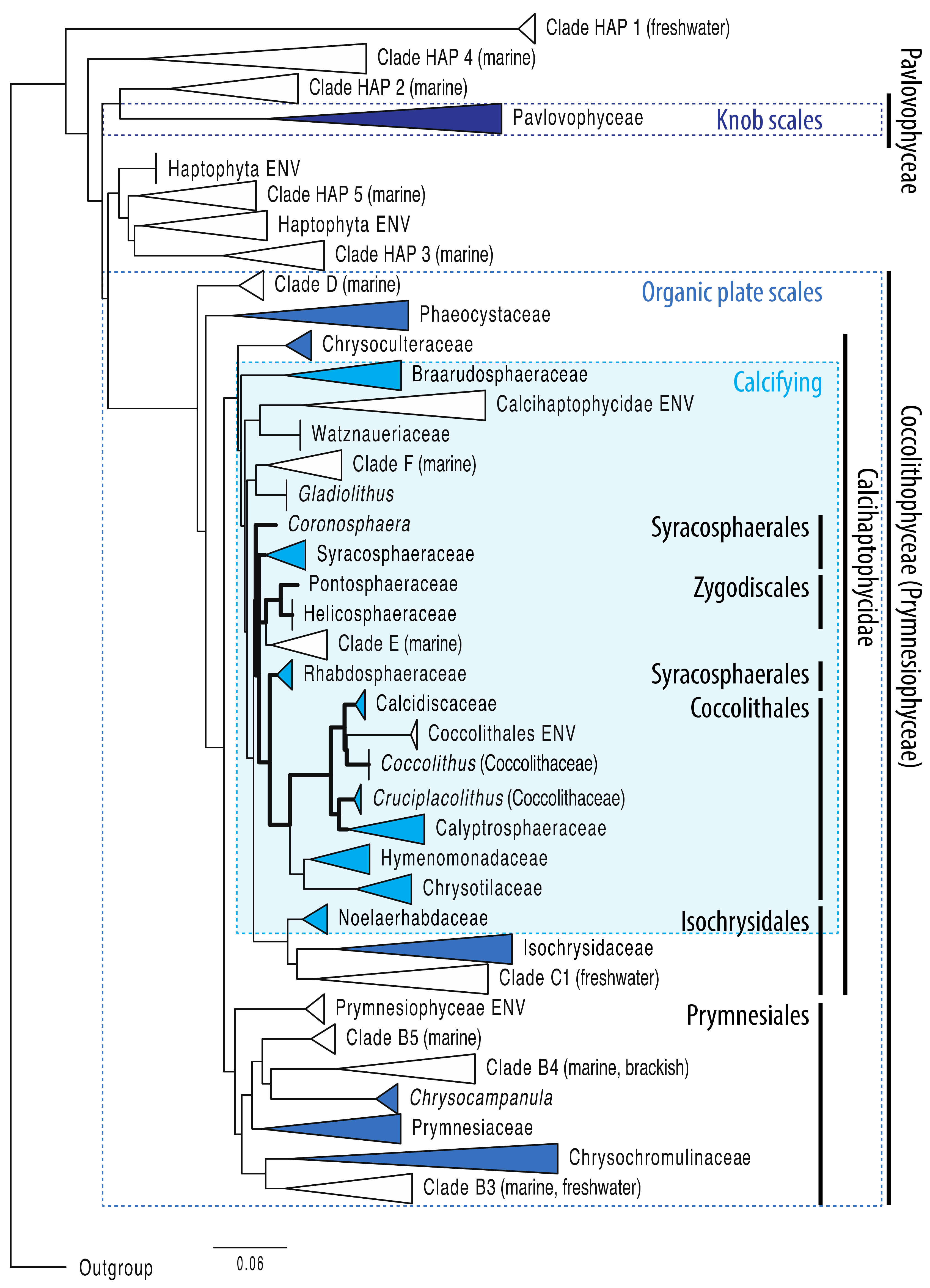
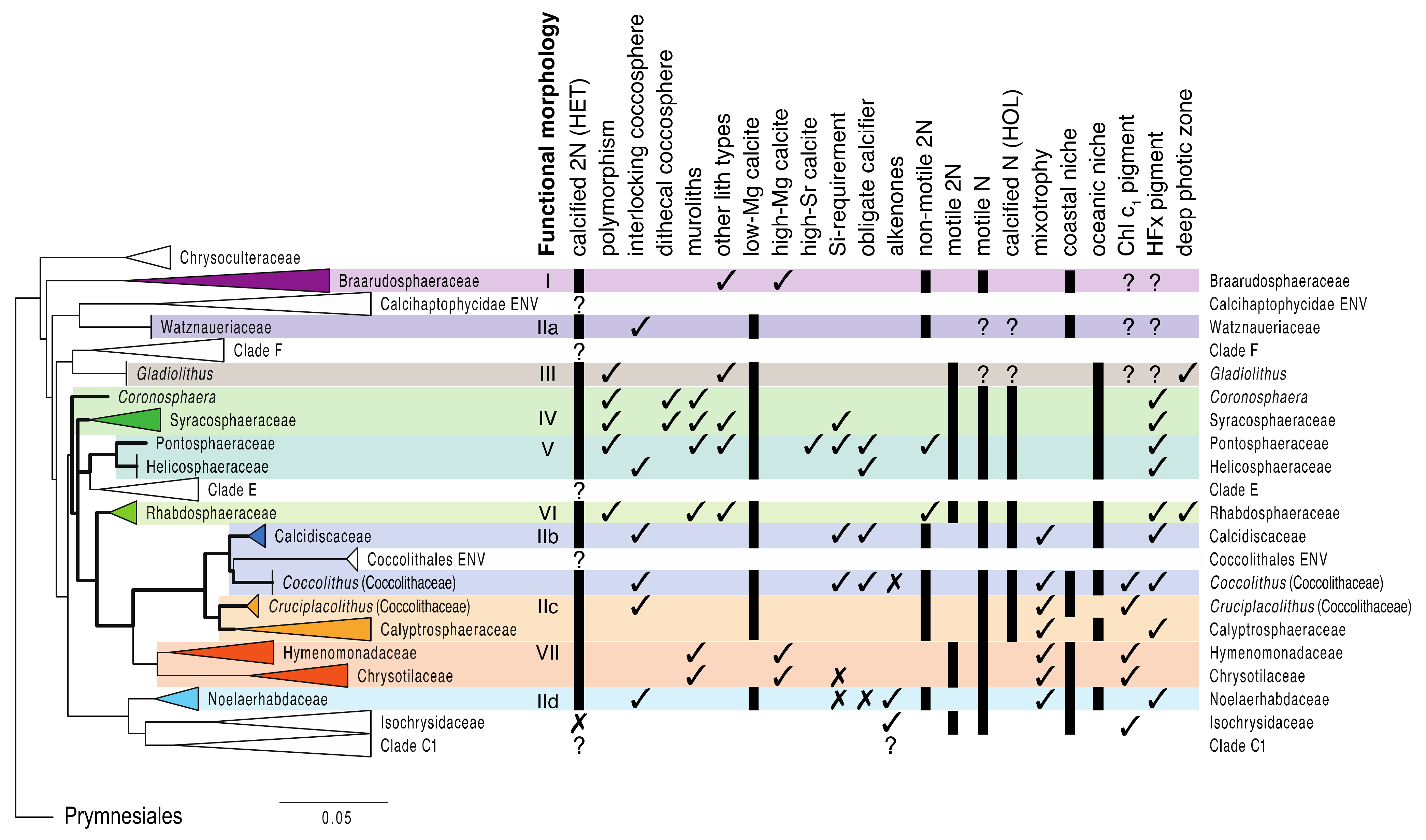
2.1. Haptophytes in Brief: Main Phylogenetic Clades

The division Haptophyta has two well-established classes, the Pavlovophyceae and Prymnesiophyceae [23,40], although Coccolithophyceae has replaced the Prymnesiophyceae by priority [6] (Figure 3). The Pavlovophyceae are distinguished by asymmetrical cells covered in organic knob scales, whereas the Coccolithophyceae (Prymnesiophyceae) have symmetrical cells covered by organic plate scales [6,7]. The prymnesiophyte subclass Calcihaptophycidae was erected by de Vargas et al. [41] to accommodate “potentially calcifying haptophytes”, including all coccolithophores as well as genetically closely related, non-calcifying clades, such as the Isochrysidaceae. The organic plate scales typical for all prymnesiophytes are formed intracellularly and co-evolved into “organic base plates” for the calcite structures (coccoliths) that are formed in specialized intracellular compartments (coccolith vesicle, CV) [16,42,43,44,45]. The siliceous scales of Hyalolithus neolepis [46] form an apparent exception to the rule within the predominantly non-mineralised Order Prymnesiales.
A heteromorphic alternation of generation is known in many prymnesiophyte species, but this is most pronounced in the coccolithophores, where many of the clades produce distinct coccoliths in the haploid or diploid stage called holococcoliths (HOL) and heterococcoliths (HET), respectively (Figure 2). Often, the diploid stage is non-motile and the haploid stage is motile, but various combinations of (non-)motile and (non-)calcified, diploid and haploid states exist across the clades [5,47].
Coccolith morphology is genetically constrained and is, therefore, an excellent first-order tool to establish coccolithophore taxonomy and phylogeny at the family- and genus-levels, especially when based on homologous calcite ultrastructures of heterococcoliths [48,49,50,51,52]. To date, these morphology-based phylogenies have proven to be highly consistent with molecular phylogenies, which is very reassuring for both approaches. Coccolith morphology remains the primary tool for deriving (tentative) evolutionary relationships between clades of uncertain phylogenetic placement (incertae sedis) that lack cultured representatives, as well as the large number of extinct lineages observed in the fossil record (see Section 3). The molecular approach is, of course, restricted to extant taxa, and remains, despite tremendous progress made in recent years, heavily dependent on the availability and sampling density of representative culture strains, with some improvements in linking morphotypes to genotypes using single cell polymerase chain reaction (PCR) approaches [31,53].

2.2. Molecular Phylogeny: Consistent Groupings and Hidden Surprises

The most current molecular phylogeny shown in Figure 3 is based on all 971 18S rRNA (SSU) gene sequences in the Haptophyta database, 451 of which are from cultured strains and 520 from marine and freshwater environmental samples obtained through sequencing of clone libraries [23]. A striking feature is that several distinct clades without any cultured representatives (i.e., not morphologically verified) are embedded within established (sub)classes, or that they appear as more basal clades, possibly representing entirely novel haptophyte classes [23]. For example, Clade HAP1 falls basal to all haptophytes and sequences assigned to this clade have only been detected in freshwater sediments, whereas Clades HAP2, a sister-clade to the well-established Pavlovophyceae, and HAP3 to HAP5 have only been recorded in marine plankton samples [26]. There are several such “mystery” clades within the Coccolithophyceae as well (Figure 3).
It is important to note that several molecular clades are based on only single or two culture strains, compared to the much greater morphological diversity known from plankton observations. This is especially true for the Calcihaptophycidae, because their coccosphere morphologies are more easily sampled and preserved for detailed taxonomic analysis [20,21]. Even if the relative distance (molecular divergence) between clades is delineated quite robustly, the molecular diversity within clades is clearly still very much under-sampled. For example, of the at least 55 extant morphospecies described, the species Syracosphaera pulchra is, with two culture strains, the sole representative of the Syracosphaeraceae family, and together with a single strain of Coronosphaera mediterranea, places the Syracosphaerales order at a basal position of the tree (Figure 3).
Single culture strains also represent distinct clades of the Rhabdosphaeraceae (Algirosphaera robusta) and Pontosphaeraceae (Scyphosphaera apsteinii) that have several extant species and a rich fossil diversity as well. The latter family shares a most recent common ancestor with the Helicosphaeraceae, represented by two isolates of the same species Helicosphaera carteri, within the Zygodiscales order. The phylogenetic positions between these molecular clades may, therefore, shift as more species and environmental sequences are added. For example, the Rhabdosphaeraceae are considered part of the Order Syracosphaerales, based on morphological characteristics, but the molecular divergence model in Figure 3 suggests that this family is more closely related to the Order Coccolithales.
The Order Coccolithales represents a distinct cluster in the tree with five known families; two of them, the Hymenomonadaceae and Chrysotilaceae (previously Pleurochrysidaceae), including the model species Chrysotila (Pleurochrysis) carteri, have lost the ability to calcify in the haploid phase, stand out by producing many small coccoliths composed of high-Mg calcite in the diploid phase [54], and are considered to occupy mainly coastal waters. The Coccolithaceae are a polyphyletic family comprising the genera Coccolithus and Cruciplacolithus, with an early branching between Coccolithus/Calcidiscaceae and Cruciplacolithus/Calyptrosphaeraceae (Figure 3). The Coccolithales ENV is an intriguing “unknown” nested within the Coccolithus/Calcidiscaceae clade, which may harbor previously un-affiliated incertae sedis coccolithophores or completely unknown taxa.
The Isochrysidales order contains two well-studied families, the Noelarhabdaceae and Isochrysidaceae, and one molecularly diverse, but yet to be morphologically verified, Clade C1 (Figure 3). The Noelarhabdaceae include the species E. huxleyi and G. oceanica that have lost the ability to calcify in the haploid phase but form heterococcoliths in the diploid phase. The Isochrysidaceae are confirmed to be non-calcifying in both life phases and thus exemplify the “potentially calcifying” character defined for the Calcihaptophycidae subclass [41] and the fact that calcification mechanisms may have been selected against in either one, or both, life cycle phases over the course of evolution. The gene sequences in Clade C1 were sampled from freshwater environments [23], which further points to the outgroup character of the Isochrysidales within the Calcihaptophycidae that are predominantly marine.
One very robust feature of the current phylogenetic tree is that it connects the ability to calcify in haploid phase (holococcolith formation) to one single node and common ancestry (thick black lines in Figure 3). Hence, it will be important to learn if the as yet unidentified molecular clades (e.g., Clades C1, E, F and ENV clusters; [23]) hold calcifying and/or non-calcifying species. This would confirm, or challenge, the continuity of the holococcolith- and heterococcolith traits within the phylogeny of the Calcihaptophycidae.
To date, no cultures exist for calcifying incertae sedis taxa, such as Umbellosphaera spp. and Florisphaera profunda. However, given their dominance in tropical and oligotrophic plankton assemblages, it is highly likely that such taxa belong to one of the unidentified clades. For example, Young et al. [20] presented a feasible case pairing a distinct molecular clade based on environmental 28S rRNA (LSU) sequences to Umbellosphaera spp. abundances counted under SEM. These authors suggested that the putative Umbellosphaeraceae clade is a sister taxon of the Rhabdosphaeraceae (i.e., one could speculate on the possibility of Clade E in Figure 3). It is clear that more paired morphological and environmental molecular analyses are needed; not only to constrain such tentative correlations (who-is-who), but also to improve our understanding of the inherent sampling and analytical biases of molecular vs. morphological plankton-based studies. One challenge is that molecular (metabarcoding) and quantitative morphological analyses may render very different results in terms of the relative abundance of different taxonomic groups [20,21], which is problematic if the goal is to measure biodiversity routinely in the plankton using only one method, or infer the identity of unknown clades by linking relative abundances of molecular taxonomic units (OTUs or ASVs) and morphology-based abundance data in the same samples.

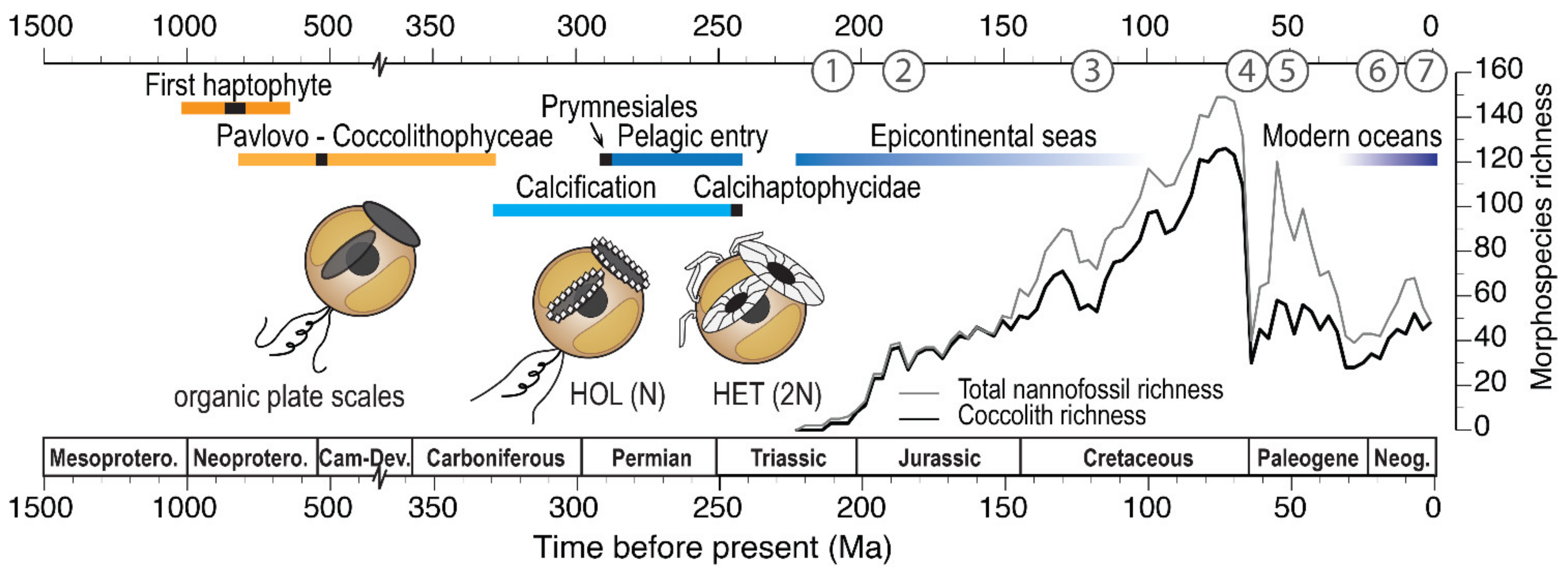
2.3. Molecular Clock Estimates

Molecular clocks are based on the genetic divergence (an estimate of the number of mutations) between species and estimates of average mutation rates, as a way to date major branching events within a molecular phylogeny. Most mutations are neutral and will not express as new phenotypes, and therefore will not be “visible” to natural selection (in fact, most genes are dormant within genomes). Therefore, genetic divergence between taxa will steadily increase over time. As mentioned above, nuclear gene sequences mutate at slower rates than plastid genes, whereas mitochondrial genes evolve the fastest [38]. Mutation rates operate on a generational timescale and are thus closely linked to cell division rates (growth rates). Estimates of whole-genome mutation rates have been obtained in the laboratory only for one culture strain of E. huxleyi [55]. At an average rate of 5.55 × 10⁻¹⁰ per nucleotide per cell division, this value was confirmed to be similar to other eukaryotic plankton groups [55], so it can be speculated to be also comparable between different species of haptophytes. Whole-genome data are currently only available for E. huxleyi and its closest relatives within the Noelaerhabdaceae [56,57,58]. Efforts are underway to include other haptophyte species, but until then we can only speculate about the expected similarities or speculated differences in mutation rates between different clades.
A multigene analysis including both nuclear (SSU and LSU) and plastid (tufA and rbcL) gene sequences derived from the exact same culture strains [11] confirms the robustness of current phylogenetic reconstructions, and supports the idea that nuclear and plastid genomes share the same history within the Haptophyta, despite evolving at different rates. Molecular clock methods use a Bayesian framework for estimating divergence dates with probability curves between a minimum and a maximum bound (time priors) based on fossil calibrations. The oldest assigned fossil of a taxon is taken as the basis for its minimum age and then constructs these other parameters around it. Using such Bayesian models, a timeline of the divergences between the major haptophyte clades and inferred emergences of key traits is reconstructed [9,11] (Figure 4).
In summary, the divergence between the Pavlovophyceae and Coccolithophyceae (Prymnesiophyceae) occurred sometime between the Neoproterozoic and Carboniferous (average estimate ~543 Ma, close to the Neoproterozoic-Cambrian boundary). The development of organic scale plates likely happened within the same time window. The Order Prymnesiales originated by ~291 Ma (Carboniferous), and a hypothesized expansion from predominantly coastal habitats to the pelagic realm has been molecularly dated to between 291–243 Ma [11] (Figure 4). However, the oldest known fossil record of the Prymnesiales, in the form of siliceous scales, is much younger and dates back to the middle Eocene (~42 Ma) [59]. The fossil record of coccolithophores provides a minimum age constraint for the onset of calcification in haptophytes. The most recent common ancestor of calcifying haptophytes has been molecularly dated to ~243 Ma (middle Triassic) and, according to these models, the ability to calcify may have evolved as early as the Carboniferous, ~329 Ma [11] (Figure 4). Yet, the first unambiguous heterococcolith fossils are found in sediments much later in the late Triassic, with an age of ~210 Ma (Figure 4; #1). A much earlier molecular-inferred origin has been argued to reflect primarily preservational biases—i.e., some haptophytes were already calcifying, but their coccoliths were not preserved as fossils until the late Triassic [11,41]; however, other biases, inherent to the molecular clock method, cannot be ruled out.
For the younger part of coccolithophore evolutionary history, plastid molecular clocks [28] have constrained the middle to late Miocene origins of extant species in Helicosphaera (10.2 Ma), Calcidiscus (11.6 Ma) and Umbilicosphaera (5.6 Ma), as well as Chrysotila (formerly Pleurochrysis), the coastal-dwelling members of the Coccolithales (12.9 Ma). A more recent divergence within Coccolithus occurred during the Pleistocene (2.1 Ma) (Figure 5). Even though some of these morphospecies are separated by very subtle morphological differences, they are genetically distinct sibling species [28,60]. In the following, we will first outline fossil evidence that further supports the phylogenetic relationships within the Calcihaptophycidae (Figure 3) and comment on a series of unique historical events which, in hindsight, are called evolution (numbers #1–7 in Figure 4), before revisiting the question of the origin(s) and timing(s) of calcification in haptophytes (or jump to Section 4).

3. The Fossil Record of Coccolithophores

Compared to other major phytoplankton groups, the fossil record is uniquely complete for most, but not all, coccolithophores [12,48,49,61] (Figure 4; Figure 5). This offers robust ways to calibrate molecular phylogenies with first fossil occurrences and divergence-based constraints [10,11,28], and empirically constrain time series of phenotypic change and calcification in ancient lineages. Molecular phylogenies are based on extant taxa only, and by definition reconstruct the history of the evolutionary “winners”. The fossil record offers detailed insights into extinct lineages, many of which established the successful and diverse clades that we know today.
Fossil age constraints are provided by biostratigraphy, the science of dating rocks and sediments based on their fossil content. Nannofossil biostratigraphy is especially reliable and detailed because of its early development and use in hydrocarbon exploration in the 1960s and 1970s, but also as a result of the concerted efforts of more than 50 years of scientific ocean drilling [62] that has provided systematic age calibration of nannofossil ranges with magneto- and isotope stratigraphies, as well as astronomically tuned sediment records from all ocean basins.

3.1. Telling Time: The Dates of Ancient History

Fossil first occurrences (FO) and last occurrences (LO) are well constrained for most Mesozoic (Triassic to Cretaceous) lineages (as reviewed in [48,63,64,65]) and offer a high-resolution biostratigraphic framework for the Cenozoic era (see for example, Paleogene: [66]; Neogene: [67,68]). Within these biostratigraphic frameworks, it is important to remember that age calibrations of standard stratigraphic units are continuously updated and may be revised in future. It is, therefore, important to apply the latest geological timescale for consistent correlations and interpretations [69]. Age constraints may change and the chronostratigraphic units themselves (with standard names) may also be formally amended.
This is the case for the Upper Triassic sediments in which evidence for the first heterococcoliths has been reported. Without considering the latest revisions, the first occurrence of coccoliths within the Norian stage (cf. original reports in [70], based on a previously accepted stratigraphic standard) have converged on an age estimate of ~225 Ma in many subsequent citations in the literature; but this age is actually closer to ~210 Ma, near the Norian-Rhaetian boundary, according to the latest stratigraphic investigations (see full discussion in [13]; Figure 4; #1). It is noteworthy, however, that the ~225 Ma date remains valid for µm-scale calcispheres (most likely affiliated with dinoflagellates rather than haptophytes) that did appear earlier within the Norian stage [12]. The earliest heterococcoliths were very small, ~2 µm, and had a simple murolith morphology [63]. Crucirhabdus primulus was the only survivor of the Triassic–Jurassic boundary extinction event and must have given rise to early Jurassic lineages. The first fossil occurrence of holococcoliths (Anfractus youngii [64]) is recorded in the early Jurassic (lower Toarcian, ~182 Ma; Figure 4; #2).
In addition recovering near-continuous records of calcareous nannofossils, deep-sea drilling has helped to establish the earliest occurrences of several biomarkers, including haptophyte-specific alkenones in Cretaceous sediments (lower Aptian to Cenomanian, ~120–95 Ma; Figure 4; #3) [71,72]. Today, the production of alkenones appears exclusive to the Isochrysidales, so that the most likely explanation is that this trait must have arisen after the divergence of this clade from other coccolithophores (~195 Ma cf. [10]) and may help explain the “outgroup” characteristics of today’s most prolific species E. huxleyi and G. oceanica when compared to other coccolithophores [73,74,75].

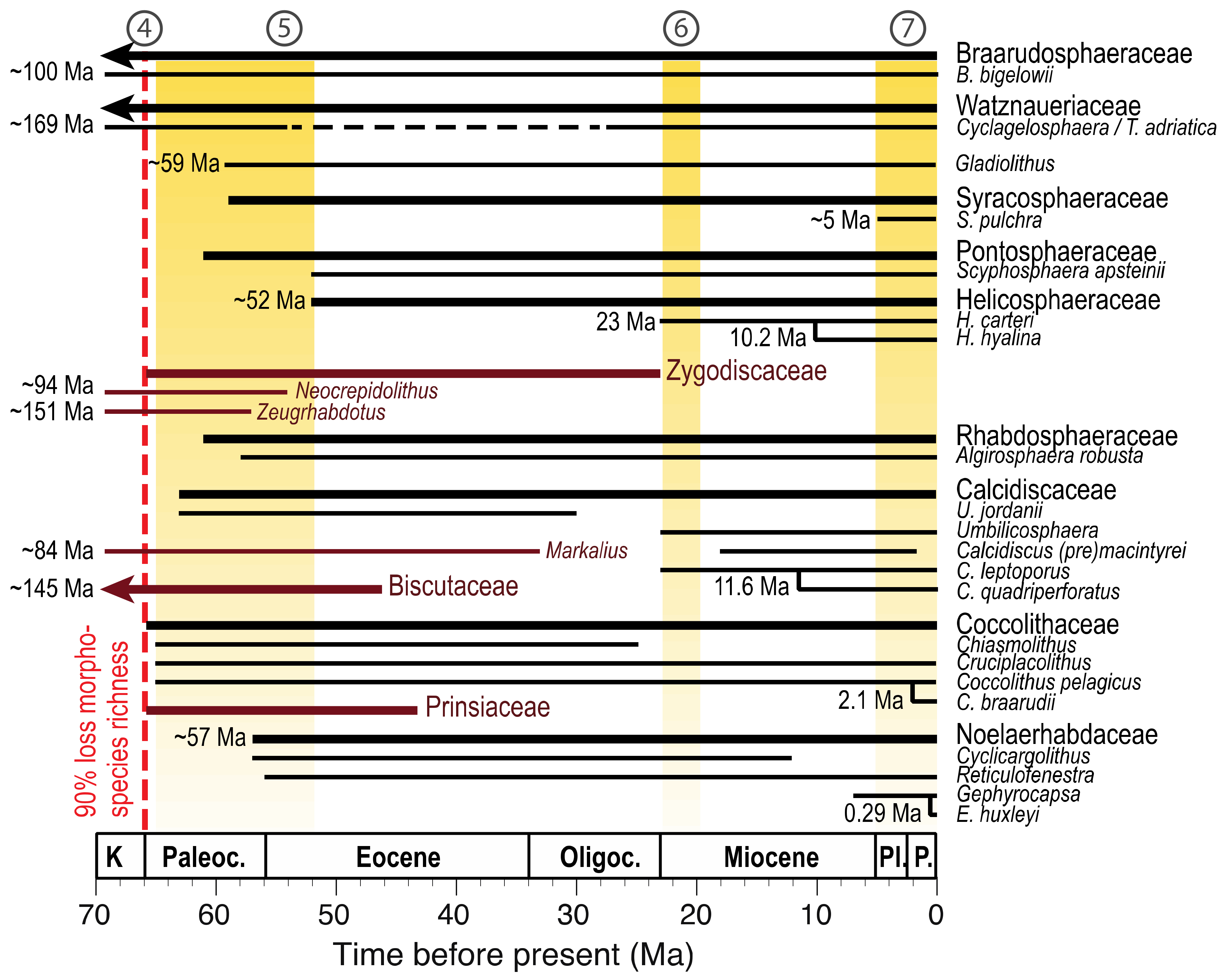
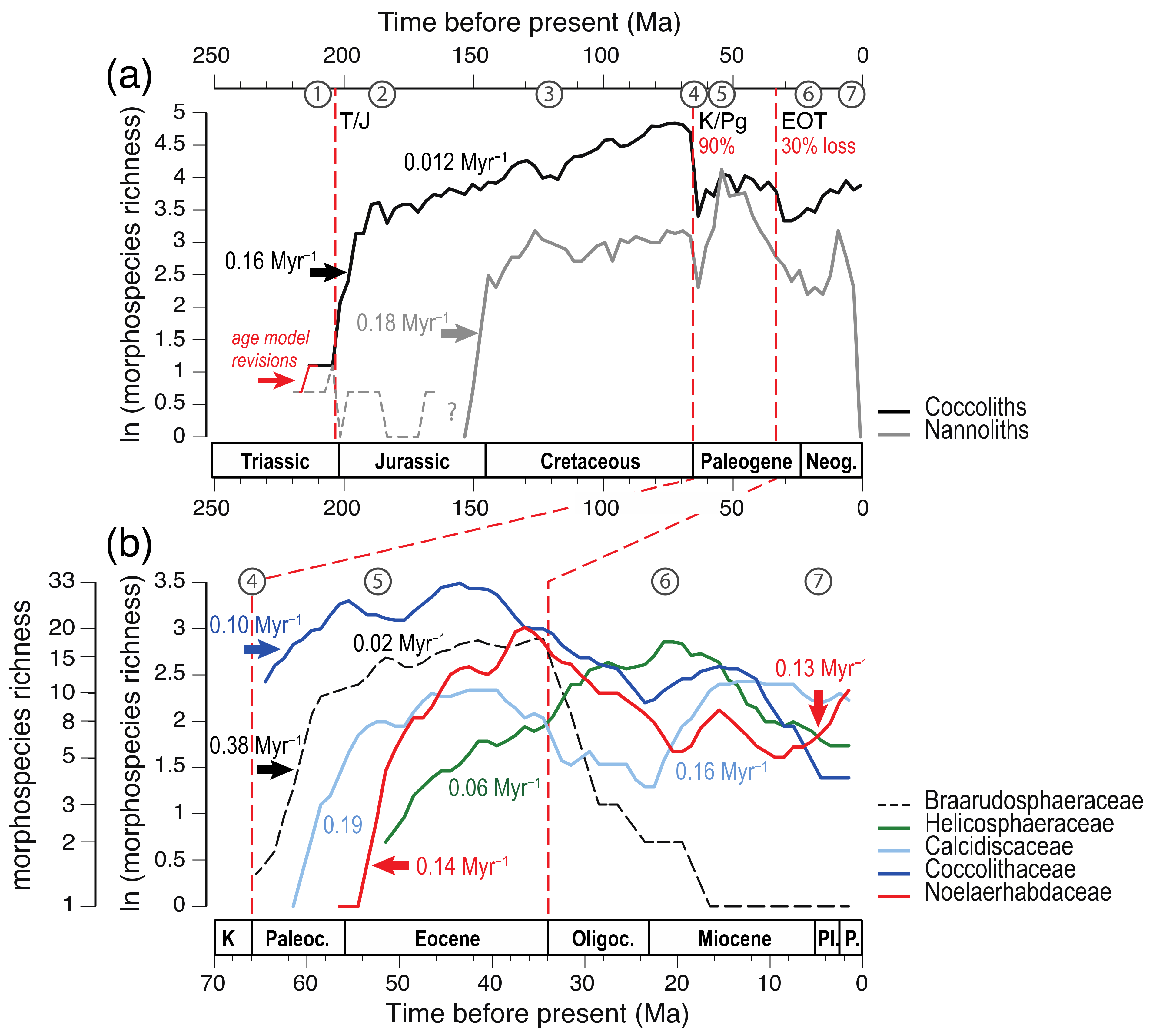
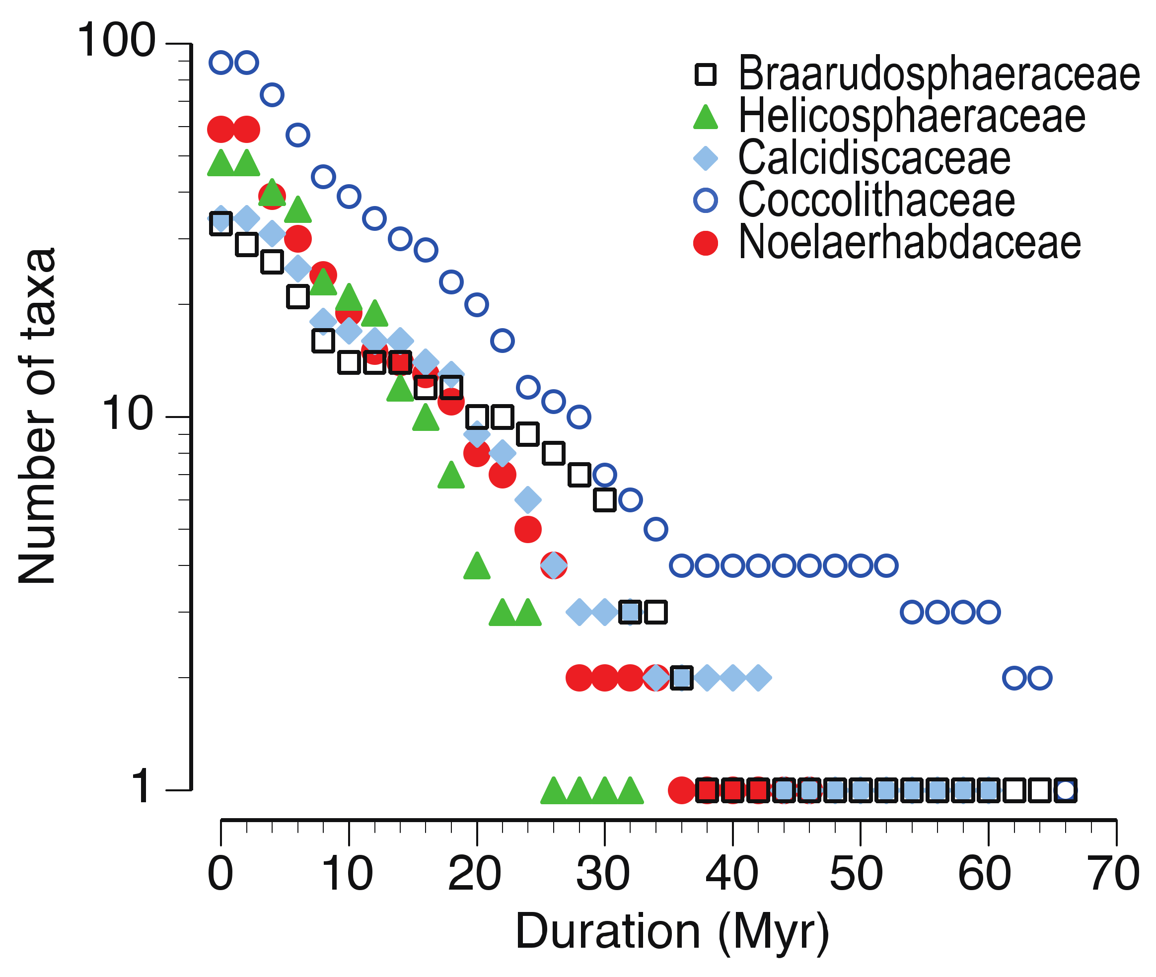
3.2. The Fossil Lineages of Extant Clades

Fossil data supporting the minimum ages of molecular divergence nodes (presented in Section 2.3) are especially robust for the Cenozoic, the geologic era that started abruptly following the Cretaceous–Paleogene (K–Pg) mass extinction event ~66 Ma. This mass extinction caused a ~90% loss in morphospecies richness (Figure 4; #4) and left only few survivors into the early Paleocene [12]. At least two of those survivors are still represented in the modern plankton, namely Braarudosphaera bigelowii and Tergestiella adriatica (Cyclagelosphaera), for which a “living fossil” status is confirmed by molecular analysis [31,53]. These (morpho)species represent the clades of the Braarudosphaeraceae and Watznaueriaceae (Figure 3), which have fossil records dating back to the Cretaceous (Cenomanian stage, ~100 Ma; [65]) and Middle Jurassic (Bajocian stage, ~169 Ma; [64]), respectively (Figure 5). Today, they are rare and found in coastal waters only [31,53], but they are known to have occupied open ocean, pelagic niches in the past. Braarudosphaera was especially abundant immediately following the K-Pg boundary event [76] and also during parts of the Oligocene, when it formed near-monospecific chalks in the South Atlantic [77]—suggesting ecological resilience throughout its evolutionary history and the possibility of ecological dominance through massive blooms.
Another basal clade is formed by Gladiolithus, a lower photic zone (LPZ) dweller with a fossil record dating back to the late Paleocene (Thanetian stage, nannofossil zone NP6, ~59 Ma [78]; Figure 5). Usually, the delicately thin and small coccoliths of LPZ taxa have low fossilization potential, but exceptional preservation in Tanzania drill cores has greatly extended the fossil ranges for a number of otherwise “invisible” taxa, including the rhapdosphaerid Algirosphaera robusta [78]. A diverse sister clade to Gladiolithus is highlighted by Clade F (Figure 3), based on four uncultured eukaryotes and haptophytes from the Cariaco Basin (Caribbean Sea), the Marmara Sea (sampled at 100 m depth) and from sea ice in the Bothnian Sea [23]. It can only be speculated, but this may show a first glimpse of the molecular diversity within LPZ communities that is yet to be uncovered. In any case, a relatively deep divergence between LPZ taxa and other Calcihaptophycidae, much earlier than previously thought, is confirmed by both molecular and fossil data.
The Syracosphaeraceae, Pontosphaeraceae and Rhabdosphaeraceae all have fossil records dating back to the Paleocene, with FOs in the Selandian (NP5) to Thanetian stages (NP7) (~61.5–58.8 Ma; [79]). The sister-clade status of the Pontosphaeraceae and Helicosphaeraceae (within the Zygodiscales order) is confirmed by the divergence of Scyphosphaera apsteinii and the Helicosphaeraceae during the early Eocene (Ypresian stage (NP12), ~52 Ma; Figure 5). The Order Coccolithales contains only two families with reliable fossil records: the Coccolithaceae and the Calcidiscaceae. The former is a polyphyletic group and possibly represents direct descendants of K-Pg survivors, with the fossil lineages of Coccolithus pelagicus and Cruciplacolithus neohelis both reaching back into the earliest Paleocene (Danian stage, ~65 Ma; Figure 5).
Now extinct lineages of the Calcidiscaceae were also established during the Paleocene (~63 Ma), but the extant lineages (e.g., Calcidiscus leptoporus and Umbilicosphaera) arose later during the early Miocene (Aquitanian stage (NN2), ~23 Ma), together with extant lineages of the Helicosphaeraceae (Helicosphaera carteri). Last but not least, the ancestral lineages of modern Gephyrocapsa and E. huxleyi, all found within the Noelaerhabdaceae family (e.g., Cyclicargolithus and Reticulofenestra) first appeared by the late Paleocene (Thanetian stage (NP9), ~57 Ma), most likely diverging from the early Paleocene Toweius lineage (Prinsiaceae extinct family; Figure 5).

3.3. The Adaptive Radiations That Established Modern Clades

All clades in existence today experienced a diversification “burst” (adaptive radiation) at some point—most likely near the clade’s origin—establishing many lineages in order for at least a few to have survived until today [80,81]. Most of this evolutionary history is pruned from the tree through extinction and can only be addressed by studying the fossil record (extinct lineages). Despite the fact that measuring “true” biological species diversity in coccolithophores is an impossible task because of systematic preservational biases, cryptic species, polymorphism and dimorphic life cycles (as reviewed in [61]), valuable information can be gleaned from the collection of extinct morphospecies that were consistently more resistant to dissolution or globally abundant and widespread [82].
When recast on the natural logarithmic scale (Figure 6a), fossil morphospecies richness data [12] (Figure 4) accentuate the times of accelerated clade growth, namely the adaptive radiations during the early Jurassic (coccolith-bearing taxa) and late Jurassic (nannoliths). The pattern of steady diversification until the end of the Cretaceous occurred at an average rate that was at least ten times slower than the initial radiations. The K-Pg mass extinction decimated nearly all nannofossil morphospecies, and the diversification trajectories that followed established most modern coccolithophorid families. Of those, several families stand out as having consistent and abundant fossil records. Here, we want to illustrate a few key points with the help of an inventory based on the stratigraphic ranges (longevities) and taxonomic data available on the Nannotax3 web resource [19] (see also Supplementary Materials).
The tabulated average rates of speciation (S), extinction (E) and diversification (D) (Table 1) serve as a first-order exploratory tool of evolutionary rates within different families. As long as speciation rates exceed extinction rates, high evolutionary turnover rates (S + E)—such as is the case for the Noelaerhabdaceae and Helicosphaeraceae in this example—imply that these taxa may be, on average, faster at adapting to environmental change than the other families under comparison. Unfortunately, members of the Syracosphaeraceae are rare in the fossil record, most likely because of their small size (thus prone to dissolution) and rare occurrences in open ocean, pelagic settings. This precludes calculation of most rates. Nevertheless, based on their Paleocene origin (Section 4.1) and current morphospecies richness, they have the highest exponential diversification rates (Table 1).
Time series of exponential clade growth (or decay; Figure 6b) highlight the adaptive radiations of the Paleocene (#5), early Miocene (#6) and Pliocene–Pleistocene (#7), as well as general trends of diversity decline across the Eocene-Oligocene boundary for most families (but not all; compare, for example, the Helicosphaeraceae). Clearly not all coccolithophore families considered here are in “the midst of unbridled radiation” today [80] (Supplementary Materials), but they have been in the past. Although not originating at the same time, the diversification trajectories of all families show accelerated diversification at their origins, or in the case of the Braarudosphaeraceae, during their recovery following survival of the K-Pg extinction event (Figure 6b). Diversification trajectories following these initial “bursts” vary over time, and may correlate to different eco-physiological adaptive strategies under concurrent environmental forcings, such as the coccolith (and cell) size reductions in relation to long-term, global cooling [83]. This analysis also highlights that the Noelaerhabdaceae appear to be the only family that are in a current trajectory of adaptive radiation, whereas all others are in a state of “stationary clade growth” (Coccolithaceae and Calcidiscaceae) or seemingly at the final stages of a steady decline (Braarudosphaeraceae).
A final observation is that although average background extinction rates do differ between families (Table 1), the probability of going extinct within a family is fairly constant, as predicted by the Law of Constant Extinction (also known as the Red Queen Hypothesis) by Van Valen [84]. This is illustrated by the log-linear relationships in “survivorship curves” shown in Figure 7, which imply that the chance to go extinct is equal for all morphospecies: It does not make a difference if a morphospecies is short-lived or long-lived, because they all share very similar traits within a family and are bound to the same niches [84,85].
It may be intuitively obvious that speciation and extinction rates determine the final outcome of species diversity. However, the question of what determines different evolutionary rates is far from understood. Extinction may seem random and a species can lose this evolutionary game at any age, but extinction rates are affected by traits and environments that vary over time, so that the rate of extinction from time interval to time interval is not constant [86]. The rates of speciation may be impacted by several factors, including growth and mutation rates, sexual recombination rates, the genetic variation within species and their effective population size, the level of dispersal or isolation of populations, as well as their interactions with other biota and the physical environment [58,86,87].

3.4. Modes of Speciation in Coccolithophores

Speciation is the key process in understanding whether we are interested in species diversity, resilience and adaptation in the phytoplankton. If changes in genetic variation within populations—through the microevolutionary processes of mutation, natural selection, gene flow and genetic drift—lead to different phenotypes (the ”variability” that Darwin [88] documented), these will be under natural selection and this may lead to speciation (e.g., [87]). Cryptic diversity (of biological species with highly similar morphologies) is common and likely because of strong stabilizing selection of phenotypes in the plankton [28,89]. It often “hides” genotypes that are adapted to certain environments or have specific niches—adding resilience to change on the morphospecies level. For example, the first whole-genome comparisons between strains of the cosmopolitan species E. huxleyi [56] suggested that what was previously considered a single species is actually composed of multiple species able to thrive under a wide variety of environmental conditions (see also [30,36,90,91] for discussions of the different E. huxleyi morphotypes). Whether all cryptic species eventually fully diverge to form new morphospecies or represent “species-complexes”, rather than the “normal” manifestation of a species, is unknown.
However, microevolution operates on the generational timescale (2–100 generations), microfossil assemblages within a single sediment sample may represent thousands of years—and (at least) ~50-fold that in terms of population generations. Such time-averaging aspects need to be carefully considered when studying phenotypic rates of evolution [92]. Yet, the question of the modes of speciation, whether new species arise through slow and gradual change of entire populations (phyletic change or anagenesis [88]) or whether they evolve more abruptly in genetically isolated populations (punctuated equilibrium or cladogenesis [93]) is arguably best addressed by studying the actual outcomes of past phenotypic evolution, especially in near-continuous records of marine microfossils [94,95].
For example, repeated sets of gradually increasing maximum coccolith size that are (on geological timescales) abruptly truncated by reductions in size variance have been documented within the Noelaerhabdaceae across the Neogene [82,96,97,98,99] and Paleogene [100,101]. Such events have been widely adopted in the field of biostratigraphy, because the size increases and reductions appear to be global events occurring within geologically short time spans. Yet, to date, relatively little has been done to understand the processes behind these phenotypic patterns: are such increases in size an indication of adaptive evolution through speciation, followed by extinction events of large-sized taxa; or do these patterns rather reflect ecological changes in the relative abundance of closely related morphospecies or ecophenotypes?
Recently, both working hypotheses have been proposed to explain so-called “pulsed events” in the evolution of Noelaerhabdaceae during the Pleistocene. Bendif et al. [57] suggest that speciation within the Gephyrocapsa lineage (including E. huxleyi) happened in sets of gradual diversifications that are truncated by abrupt extinctions. These ‘Matsuoka–Okada’ cycles [57,98] occur at ca. 0.5 Myr intervals and are associated with stepwise changes in global sea surface temperature. Whole-genome sequence analyses of extant Gephyrocapsa species further suggest that speciation within this group may occur by reproductive isolation followed by secondary contact of closely related species [57,58]. It was also shown that interspecific hybridization and gene exchange is a very recent phenomenon in these blooming coccolithophores [58], confirming that the group continues to diversify today, on the adaptive radiation trajectory that started in the latest Miocene-Pliocene [102] (see also Figure 6b). A second, alternative hypothesis involves a role of long (405,000 year) orbital cycles forcing the repeated pattern of size expansions and contractions within the Noelaerhabdaceae over the past 2.8 Myr [103]. The eccentricity of Earth’s orbit around the sun influences seasonal contrasts, which in turn determine the phytoplankton seasonal succession and the number of available niches in the photic zone. This hypothesis invokes long-term forcing of climate on phytoplankton ecology and productivity, determining the presence of one or more morphospecies (that evolve over longer timescales), rather than repeated cycles of speciation and extinction.
It is difficult to judge which of the two models is best—if a choice were required—because both have merits and are most likely not mutually exclusive. Nor is it known whether the same models and rates of change observed in the Noelaerhabdaceae are applicable across all coccolithophores (or even haptophytes). It will be important to (re-)investigate time series of phenotypic change in terms of rates of mean trait divergence and variations in concurrent environmental selection pressures. When comparing the different coccolithophore families and long-lived lineages, such as Calcidiscus [104] and Helicosphaera [83] in combination with genome-based and population genetic theory approaches [57,87], we may soon be able to explore these processes across the diversity of coccolithophores and perhaps derive a more unifying model for phenotypic change and speciation in this phytoplankton group. Such models may also provide better insights into how speciation (and extinction) rates may be affected by the current and future rapid rises in ocean temperature. Different lines of evidence already suggest that E. huxleyi is increasingly expanding its range into the polar oceans under the influence of global change during the past decades [105,106].

4. Origins and Evolutionary History of Key Haptophyte Traits

Modern species have inherited molecular and physiological traits from their ancestors (“evolutionary baggage”), which may translate to different ecological niches and shape their responses to environmental change. Some functional traits, such as calcification, may confer lower fitness in today’s oceans than they did in the past. For example, there is a rich literature available on the impacts of ocean acidification on coccolithophores (not reviewed herein, but see for example [107,108]), including the observation that species that depend on calcification to survive (“obligate calcifiers” [75]) are likely to be more sensitive to changes in the carbonate chemistry and lowering of ocean pH in current and future oceans [109]. In contrast, non-obligate calcifiers, such as E. huxleyi, seem more versatile and less bound to their evolutionary inheritance of the calcification machinery. How and when this calcification machinery arose, but also how other key haptophyte functional traits are related to their evolutionary history, remain key questions to navigate the benefits and trade-offs of phytoplankton traits in a changing world—today and in the past.
Aubry [102] was the first to describe and group extant coccolithophores in terms of morphostructural characters (cell size, number of coccoliths per cell, coccolith morphology and size) and their hypothesized functional traits, rather than species diversity patterns per se, and concluded that there is a macroevolutionary trend (or “morphological strategy”) towards small-sized cells and coccoliths over the course of the Neogene (note that a similar decrease in size has been documented in diatoms [110]). Indeed, the selection for smaller morphotypes appears to be a common long-term adaptive response across multiple coccolithophore lineages over the past 15 million years. Detailed biometric time series data of several key taxa support that this selection for smaller cells occurred across various scales: (a) the (morpho)species level, (b) the community level, as well as (c) the evolutionary level through extinction of larger species and speciation of smaller species [83].

4.1. The Origin and Evolution of Calcification in Haptophytes

There is a gap of at least 30 million years between the molecular clock age estimate of the primary divergence in the Calcihaptophycidae, ~243 Ma [11], and the first fossil occurrence of undisputed coccoliths (muroliths), ~210 Ma [12,13] (Figure 4). Indeed, according to molecular clock models, intracellular calcification may have evolved much earlier, before the divergence of the Calcihaptophycidae and Prymnesiales (between 329 and 291 Ma) [11]. This raises several pertinent questions (see also [41]): What does this gap between the molecular inferences and the fossil record mean? Is it simply explained by preservational biases of the fossil record? What would the earliest (precursor) calcification processes have been like? Did calcification arise several times within the haptophyte phylogeny or only once and was the trait subsequently lost in some lineages?
First, it is important to stress that calcification is not one process, nor is it operated by one single “calcification mechanism”. Instead, calcification in coccolithophores is best understood as a modular system of several physiological traits that, together, lead to the formation of coccoliths (coccolithogenesis) [16,41]. These traits include a specialized intracellular compartment (a vesicle), production of specific proteins and polysaccharides that are employed for calcite nucleation and crystal growth, and several (upregulated) ion-pumps (calcium and carbonate ions into, and protons out of, the vesicle and cell) [16,18]. Westbroek and Marin [111] first postulated that calcification in coccolithophores (and other calcifying biota) was made possible by the recruitment of ancestral biochemical processes. In other words, biochemical and physiological traits that were already in existence and, at some point of evolution, became redirected, recombined or upregulated to operate new processes; rather than major evolutionary innovations in cellular traits or biochemical pathways.
It is speculated that the common ancestor of all haptophytes most likely lived in coastal/neritic environments, had a mixotrophic nutrition mode and did not calcify, but it may have had some type of organic coverings already [11]. These organic plate scales, typical for all Coccolithophyceae (Prymnesiophyceae), are formed intracellularly and would be excellent “precursors” for mineralized scales, both in terms of their functionality when placed around a cell (protective cover, cell stability, etc.) as well as a potential substrate for calcite precipitation. Until recently, it was argued that the simple, inorganic-looking rhombic crystallites typical for holococcoliths (Figure 2) were formed extracellularly [112,113,114] and represented a distinctively different calcification process from the formation of heterococcoliths that takes place in specialized intracellular vesicles [45]. New observations have now revealed that holococcolith calcification actually also happens within intracellular vesicles, prompting revisions to a long-standing paradigm [43].
The new insights may actually simplify the hypothesized evolutionary steps tremendously, compared with the work of de Vargas et al. [41]. It can be postulated that mineralization started intracellularly in a compartment that already produced organic scales. The first calcite precipitation within a vesicle likely resulted in simple rhombic crystals (similar to inorganic calcite precipitates) that became associated with the organic plate scale produced in the same vesicle. This holococcolith-like calcification [43] evolved first and required no crystal shaping machinery. Heterococcolith-like calcification evolved later, introducing a novel control of crystal growth after a silicon-dependent crystal shaping mechanism was in place [43].
It is easy to argue that early HOL-type liths had bad preservation potential (because it is still the case for modern holococcoliths [61]) and that is why they are not seen in the fossil record. The first HET-type coccoliths were likely also fragile and not well-preserved. Because of rather simple crystals and their arrangement, the first murolith fossils (Crucirhabdus) look like transition stages between HOL and the sophisticated HET crystals of later morphologies. Crucirhabdus went extinct by the Pliensbachian-Toarcian boundary (183 Ma), within the early Jurassic adaptive radiation (Figure 6a) that saw the first occurrences of placoliths (forming tightly interlocking spheres) and their rapid diversification. We postulate that this is strong evidence that an important next step of crystal-shape control in HET coccolithogenesis (resulting in the placolith type) was recruited near the Triassic–Jurassic boundary (~201 Ma). Minor changes to that machinery rapidly led to increased diversity in shape and size, polymorphism and “all the bells and whistles” observed in the fossil record and until today. The fact that minor changes in the crystal shaping machinery can have significant effects on coccolith morphology is illustrated by malformations in response to environmental changes [115,116,117]. The placolith-bearing taxa (within the Watznaueriaceae) rose to ecological dominance by the late Jurassic [118], and this may relate to the fact that their sturdy interlocking spheres rendered increased fitness by offering mechanical support, armoring against predation or other benefits [119,120]. Yet, both HET and HOL calcification continue to be well-represented traits in modern coccolithophores, so their products (coccoliths) must represent a range of advantages in order for both to have survived natural selection processes over hundreds of millions of years. Although the HOL coccosphere will not display a damage tolerance comparable to that of HET interlocking ones (no measurements are available but SEM observations are telling), there are other features, such as scattering of ultraviolet light, that might be beneficial [121].

4.2. Functional Morphology: Exploring the Traitscape of Coccolithophores

Although the intricate workings of calcification are still mostly unknown, we can interpret the product of this machinery, the coccoliths, in terms of their form, size and hypothesized functions. Importantly, coccoliths and their morphologies are the main tool to connect modern species (with a range of physiological traits that we can study in the field or laboratory) and the fossil record (from which we seek to reconstruct biological and environmental parameters in the past). So, if coccolith morphologies can be linked to known physiological traits and specific ecologies, this would be a major advantage in interpreting ecological and evolutionary patterns. It is already generally accepted that coccolith size is a good proxy for cell diameter and volume (and thus inorganic carbon quota, cellular resource uptake rates, etc.) in many placolith-bearing taxa [122,123,124,125] and that the degree of calcification of a cell can also be reconstructed from individual coccoliths [83,126,127,128,129].
The observed morphological diversity in coccolithophores, both in size and shape, is nothing short of amazing (Figure 1; Figure 2) but has left the pertinent questions “why?” and “for what purpose?” largely unanswered. The reason why coccoliths are so diverse could relate to the fact that they may fulfill many possible functions, in some cases, several at the same time. Numerous hypotheses on the functions of coccoliths have been reviewed in depth [17,119], and these may indeed be as diverse as the coccoliths themselves. Here, we explore the putative links between coccolith morphology and other functional traits in context of their evolutionary history, in the form of a “traitscape” (Figure 8). This traitscape reveals at a glance that there are several distinct groupings of traits confirmed in modern species, summarized here as “functional morphologies”, that are highly consistent with the molecular phylogeny; although it is certainly not all black and white and several exceptions to the “rules” can be identified. Indeed, the traitscape may help to identify what traits to expect (or look for) in the molecular clades without any cultured representatives—or, in reverse, once identified, these “mystery clades” could serve as test cases for the conservatism of the listed functional traits and their phylogenetic relationships.
There is a clear distinction between clades that comprise non-motile, placolith-bearing taxa with tightly interlocking coccospheres and those that are motile and have polymorphic coccospheres in diploid phase (Figure 8). Exceptions to this “rule” are found within the Pontosphaeraceae and Rhabdosphaeraceae (polymorphic and non-motile 2N) as well as the Helicosphaeraceae (interlocking sphere and motile 2N). Murolith-type coccoliths (arguably the most “ancient” heterococcolith morphology) are typical for the polymorphic groups, as well as in coastal-dwelling clades of the Hymenomonadaceae and Chrysotilaceae (Figure 8). The latter, together with the Braarudosphaeraceae, do not calcify in the haploid phase but produce coccoliths with high-Mg calcite [54] during the diploid phase, suggesting different cellular ion transport pathways or rates of ion uptake compared to other coccolithophores. In a similar vein, strontium is an interesting element that has been demonstrated to have relatively low concentrations in the coccoliths of most taxa, except for one striking example, the Pontosphaeraceae [130,131] and an extinct Jurassic morphospecies [132]. A silicon dependency in the heterococcolith formation appears to be correlated with the status of “obligate calcifier” [75], whereas those that lack the same can survive without calcifying (Figure 8).
Coccolithophores all share an ancient haptophyte trait of phagotrophy, the haptonema. Phagocytosis likely constitutes a basal trait, which was inherited by the Calcihaptophycidae from non-calcifying haptophytes [133,134]. Ingesting organic compounds in addition to carrying out photosynthesis is a way of alleviating inorganic nutrient stress in oligotrophic waters and has been described in detail for non-calcifying haptophytes [133,135,136]. Mixotrophy may prevent the sole dependence on inorganic nutrient availability, and be the reason why most, if not all, coccolithophores are deemed specialists of oligotrophic conditions in the open ocean, pelagic realm. Prey ingestion through phagotrophy was observed in culture in both life phases of E. huxleyi, and C. leptoporus, haploid Calyptrosphaera sphaeroidea [134,137], as well as haploid C. braarudii [138]. Uptake of organic molecules through osmotrophy has been observed in diploid Ochrosphaera neopolitana, diploid Cruciplacolithus neohelis, and haploid G. oceanica [139] (Figure 8). In addition, diploid C. braarudii was found to survive over extended periods while exposed to darkness, suggesting an alternate mode of energy acquisition [140]. Particle/prey ingestion in these instances was generally low, and likely does not represent a major proportion of energy consumption except in limited taxa, such as the polar coccolithophore family Papposphaeraceae, which lack chloroplasts, or the deep photic zone (DPZ) dwelling Florisphaera profunda and Gladiolithus spp. [141,142,143]. However, conclusive experimental proof for this assumption is yet to be gained. Although the proportion of energy acquired by mixotrophy may be low, it is not surprising that both life cycle stages seem to utilize it. Last but not least, the accessory pigments chlorophyll c1 (Chl c1) and 19′-hexanoyloxyfucoxanthin (HFx) are strongly correlated to coastal rather than pelagic habitats [144] (Figure 8), whereas deep photic zone taxa remain mostly a conundrum for the lack of cultures.

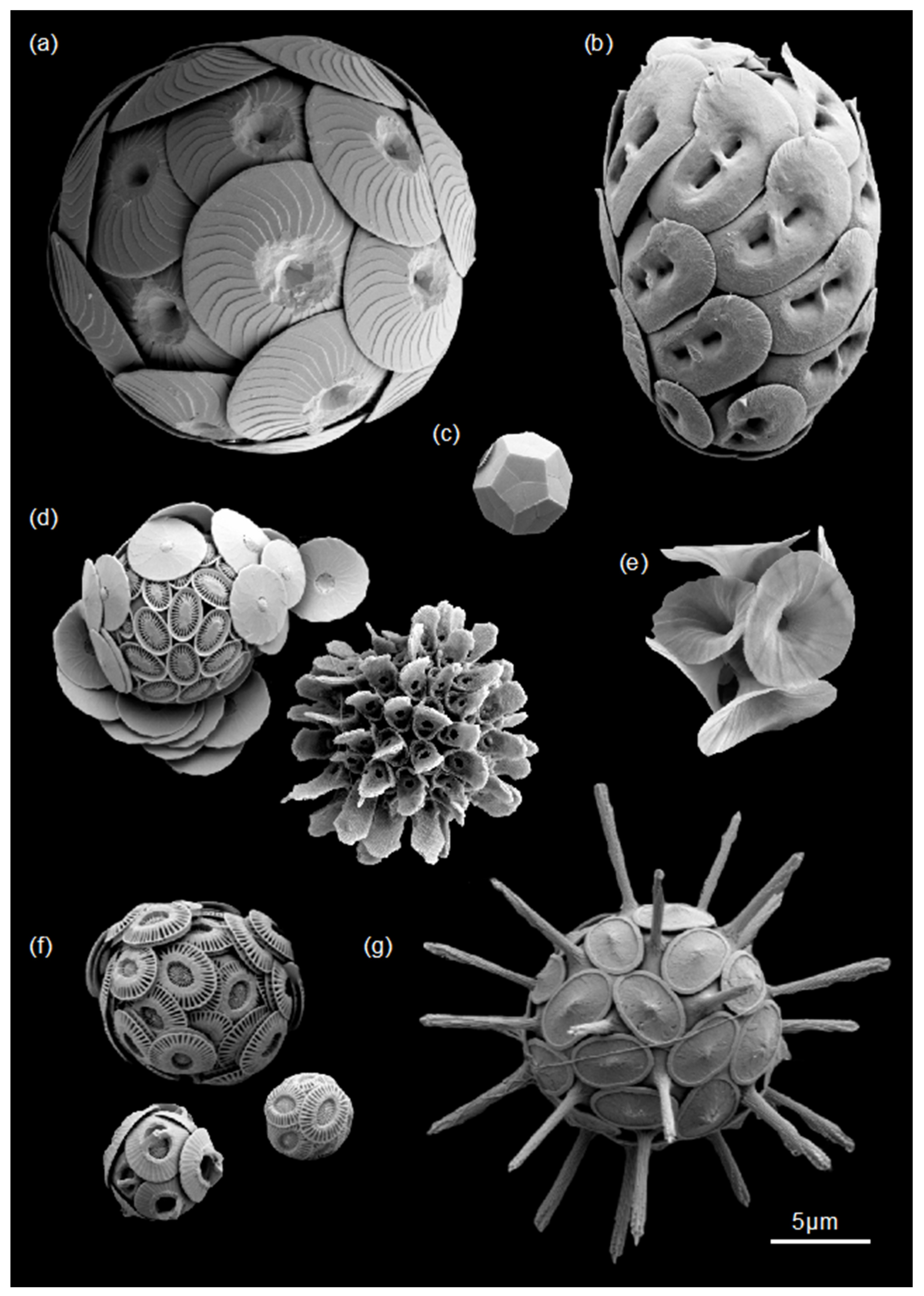
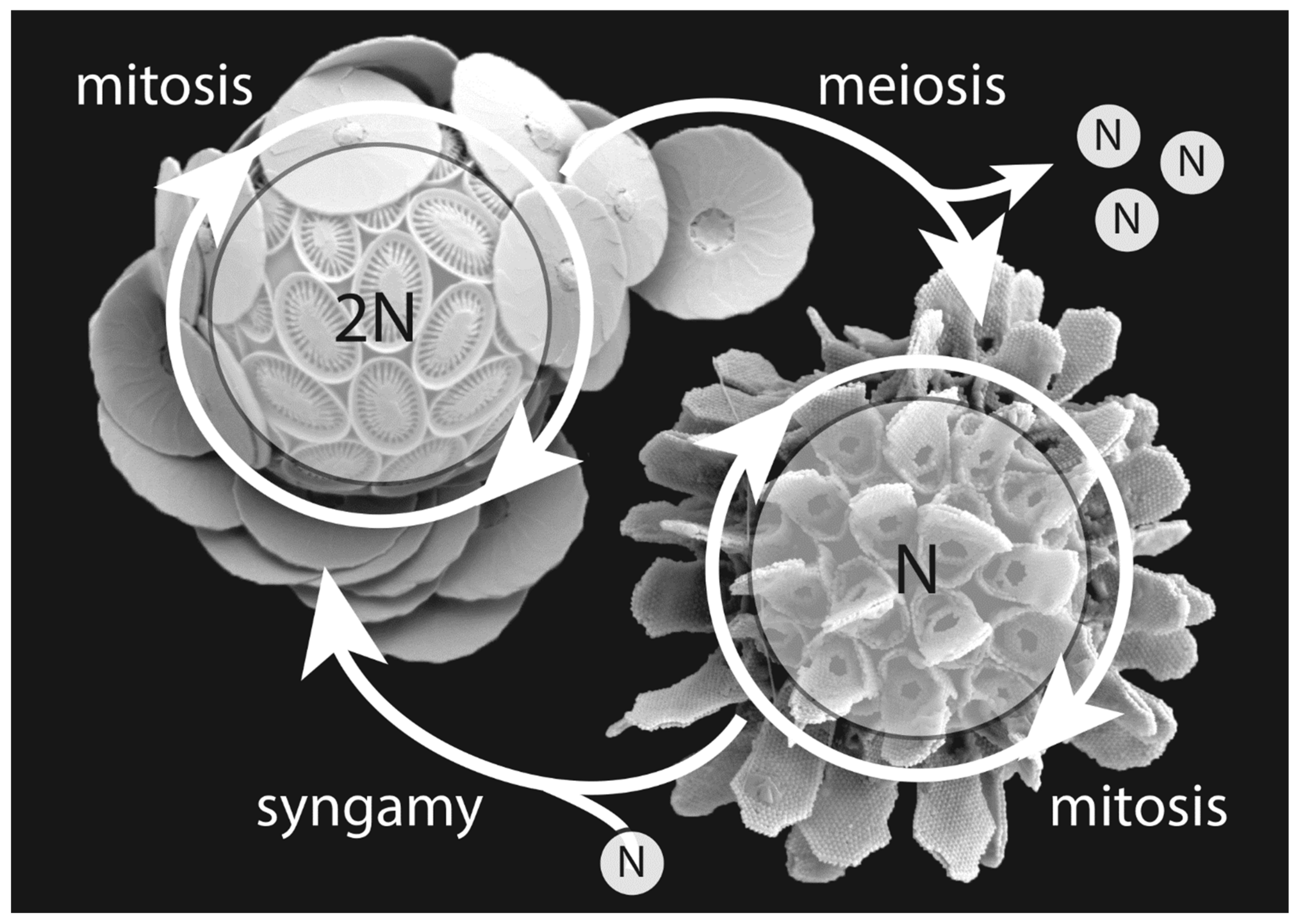
5. Evolutionary Benefits of the Haplo-Diplontic Life Cycle

An important aspect of coccolithophore biology that has—until recently—been given little attention in the literature is their complex life cycle (but see review by Frada et al. [5]). The two life phases have a different number of chromosome sets (ploidy level), a by-and-large non-motile diploid (2N) and a motile haploid (N) phase, in which they can asexually reproduce independently and maintain populations. This makes coccolithophores so called “haplo-diplontic” (Figure 2; Figure 9). The fact that coccolithophores lack a cyst-forming or resting stage makes them stand out from other abundant phytoplankton groups, such as the dinoflagellates and diatoms, which predominantly carry out vegetative reproduction or mitosis only during either the haploid or diploid life cycle phase (making them haplontic or diplontic, respectively) [145].
The coccolithophore life cycle is heteromorphic and most taxa show substantial differences in coccolith and cell size, coccolith morphology, and calcite quota between haploid and diploid cells. The diploid phase has a higher calcite quota and produces larger, intricate crystals in their heterococcoliths (Figure 2). The haploid phase has a lower calcite quota (holococcoliths) or is even non-calcified. This difference in cell calcite content influences the impact of each life phase on the calcite standing stock and may, therefore, have global implications for biogeochemical cycles. Each coccolithophore life phase is capable of vegetative reproduction for years without phase changes, as demonstrated in long-lived monoclonal cultures of both HET and HOL strains, but little is known about the conditions triggering life cycle transitions or the frequency of sexual reproduction in nature [5,146,147].
The first evidence of a motile haploid and a coccolith-bearing diploid phase within the same species was described for Coccolithus pelagicus in 1960 [148] and other species soon followed [149,150]. This recognition has led to substantial revisions of extant coccolithophore taxonomy, because taxa that were previously thought to be distinct morphospecies were actually two phases in the life history of the same biological species [61,151]. The first observation of naked, motile cells and non-motile coccolith-bearing E. huxleyi was made in 1970; however, at the time it was concluded that these distinct morphologies did not represent different life phases, but rather a permanent genetic modification [152]. Yet, by 1996 flow cytometry confirmed that the different E. huxleyi cell types were in fact haploid and diploid relatives [153]. Since then and to date, the life cycle of E. huxleyi is without a doubt the most researched of all coccolithophorid species. In the following we want to draw attention to the putative advantages coccolithophores may gain from undergoing a haplo-diplontic life cycle. We also collate some of the latest information in supplement to that already reviewed in [5].

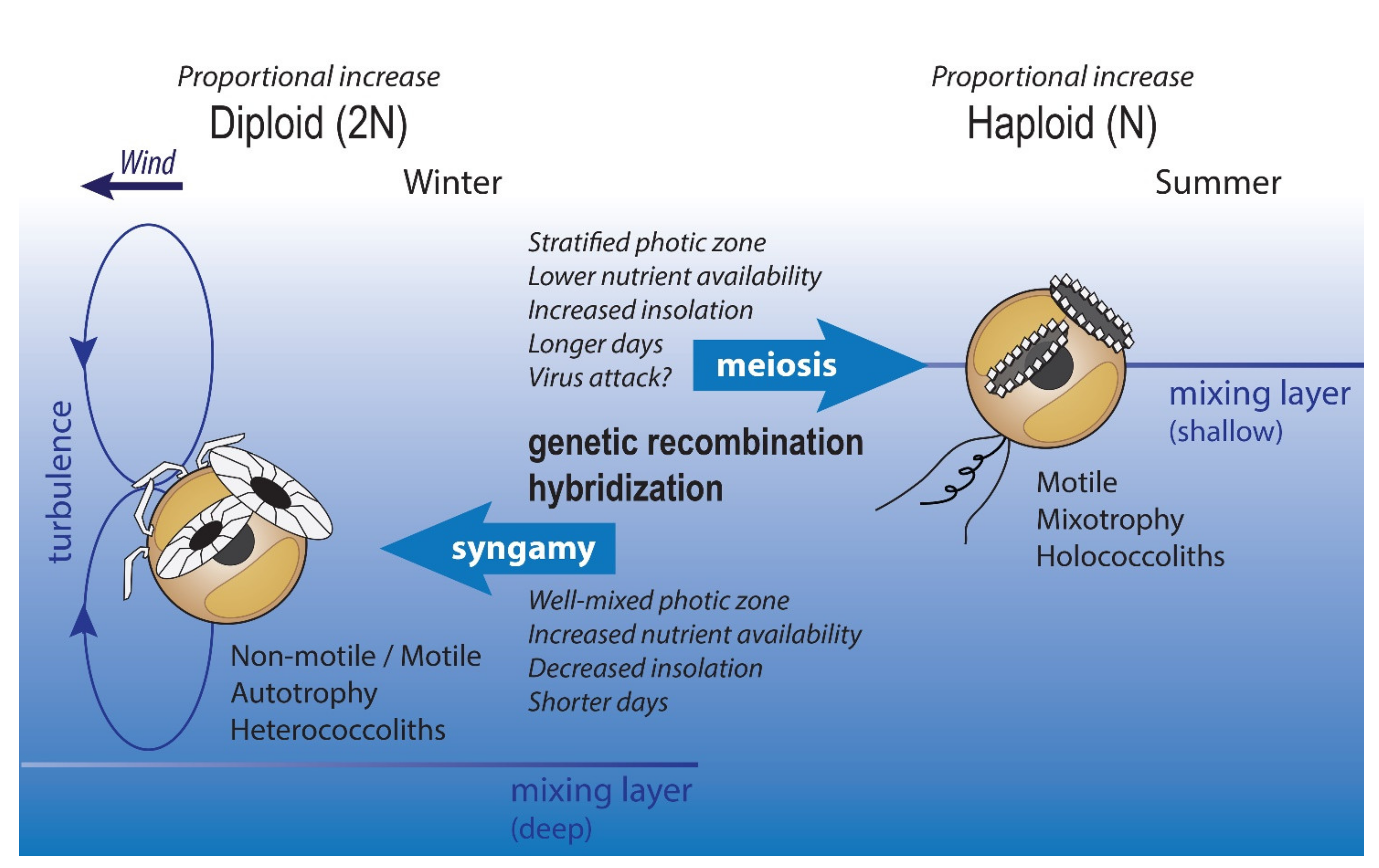
5.1. What Is the Haplo-Diplontic Life Cycle Good for?

Observations of natural populations indicate that the two coccolithophorid life phases may have different environmental preferences and tolerances [5]. At the same time, both life phases generally co-occur in the plankton at any time of sampling. Therefore, the most fundamental way of describing possible drivers behind life phase distribution is by comparing the ratio between haploid and diploid coccolithophores in situ (the so-called HOLP-index). In summary, these observations have established that the haploid cells are typically more commonly found in warmer, oligotrophic, stratified waters in the upper photic zone. Diploid cells, on the other hand, dominate in well-mixed, nutrient-rich environments [154,155,156,157] (Figure 9). Culture experiments have supported this hypothesis, where two heavily calcified coccolithophore species (Calcidiscus leptoporus and Coccolithus braarudii) increased growth under turbulent, nutrient-rich conditions [138]. Additionally, recent evidence suggests that the HOL phase of C. braarudii is more tolerant to high irradiance as well as phosphate limitation compared to its HET phase [140]. The non-motile, calcified diploid phase of E. huxleyi is commonly found in stratified surface waters, where it is known to form extensive blooms following nutrient depletion by other phytoplankton groups [158,159], whereas its non-calcified, biflagellate cells become progressively more abundant during late-stage bloom periods [160].
The different physiological and phenotypic traits are underpinned by substantial genomic differences between the life phases, although these observations remain limited to E. huxleyi and no data are available yet for other coccolithophores. This is in part because of the availability of a fully sequenced genome for E. huxleyi [56], whereas to date only transcriptome information of the diploid life phase is available for other prominent coccolithophore species [161]. Key studies of E. huxleyi have established substantial differences between the haploid and diploid transcriptome, the full range of messenger RNA (mRNA) molecules expressed by the cell. In the diploid phase, the transcriptome is 20% richer in expressed genes than in the haploid phase, and the phases have less than 50% of the transcripts in common. This may further point towards specialization to ecological niches. The more basic transcriptome of the haploid phase, which requires less energy, may increase fitness in post-bloom, oligotrophic environments [137]. Haploid-specific transcripts include genes putatively involved in motility and signal transduction [137,162,163]. Transcripts that are highly specific for the diploid phase include Ca2+, H+, and HCO3 pumps, which are most likely involved in the biomineralization of heterococcoliths. Some of the haploid-specific functions seem to have been lost in 2N genotypes of E. huxleyi, which lacked the expression of flagella-related genes. These traits were predominantly absent in strains inhabiting stable, oligotrophic environments where clonal populations of the calcifying life phase dominate [164]. Interestingly, without the ability to produce flagella, such E. huxleyi “mutants” may still be able to undergo life phase transitions and retain a sexual life cycle [165]. The reason behind the loss of flagellar genes in some 2N E. huxleyi strains, therefore, remains unexplained.
In conclusion, one major advantage of switching between two life phases, each able to reproduce vegetatively and often form blooms, is that it broadens the environmental tolerances of the species [5,166] so that it can quickly respond to, and cope with, changes in a dynamic plankton niche on daily, seasonal or longer timescales (Figure 9). Whether this effect is globally measurable has been recently assessed by quantification of niche overlap and niche expansion of both life phases in the plankton [8] and it was concluded that the haplo-diplontic life cycle might expand coccolithophore niche space by around 17%.

5.2. How Do Coccolithophores Live Their Haplo-Diplontic Lifestyle?

The different proportions of haploid and diploid cells in situ raise the question of how coccolithophores live their haplo-diplontic lifestyle. We cannot yet answer this question definitively, but we identify three potential options: (a) both diploid and haploid cell populations are kept up at all times and environmental conditions dictate which life phase prevails through natural selection, (b) only one or the other life stage is present until environmental cues trigger a switch between the life cycle phases, or (c) both strategies are used (Figure 9).
Most observations of life cycle shifts under laboratory culture conditions remain anecdotal and non-reproducible [147]. Inducing life cycle transitions by introducing environmental stress have so far not been successful. However, an important biotic factor that may trigger a life cycle switch in E. huxleyi is possibly the most well-known concept within the coccolithophorid community and is dubbed the “Cheshire Cat” escape strategy. Therein, the motile haploid E. huxleyi cell is hypothesized to resist viral infection and subsequent lysis, a process typically terminating large-scale blooms of diploid scale-bearing E. huxleyi [160,165,167]. This would allow E. huxleyi populations to evade infection by transitioning from the diploid to the haploid life phase when exposed to E. huxleyi viruses (EhVs). However, Frada et al. [165] described that in response to viral infection, diploid E. huxleyi produced cells with a haploid-resembling phenotype without undergoing meiosis.

5.3. Sexual vs. Asexual Reproduction

As with most phytoplankton groups, coccolithophores produce large populations by undergoing asexual vegetative reproduction (mitosis and binary fission); however, the frequency of sexual reproduction in coccolithophores is poorly understood [168,169]. The advantages and disadvantages of sexual vs. asexual reproduction are reviewed in detail in [145]. Disadvantages of sexual reproduction include the associated high genetic and energetic costs. Asexually produced offspring share 100% of their genes with their parent, rather than only 50% in a sexual generation. Additionally, meiosis is more prone to chromosome loss and mutations, and proceeds at a much slower rate than mitosis (von Dassow et al. [145] and references therein). Despite the high cost of sexual reproduction and the apparent choice whether to reproduce asexually or sexually, sex remains ubiquitous within phytoplankton populations. The most likely explanation is that the generation of new genetic combinations results in adaptive advantages over longer timescales. This matches with the observation that declining fitness of an asexually reproducing population induces sexual reproduction [170].

6. Outlook

Haptophytes, and especially the coccolithophores, are studied on widely ranging timescales, from generations to millions of years, and from molecular to global scales. Navigating the breadth of this exciting research field can be challenging at first. However, it has long been recognized that key questions involving the molecular, morphological and physiological diversity of the group are best explored with combinations of expertise found within the fields of biology and earth sciences [171,172]. The promise of combining molecular techniques with detailed fossil time series data is exemplified in recent studies of E. huxleyi and its closest relatives [57,58,103]. Expanding this approach to include other key coccolithophore clades will be crucial in exploring the hypothesized differences in evolutionary rates or adaptive strategies between clades [83,173]. When it comes to observations in the plankton, the main challenges include seasonal and spatial sampling coverage [8] and the techniques with which haptophyte diversity is measured [20,21]. Establishing a library of clonal cultures has been a pivotal part of haptophyte research [7,23,174], greatly advancing our knowledge of the molecular diversity and physiological traits of the group. New additions and access to these live monoclonal cultures remains an important resource and necessary bridge between biological and paleobiological investigations. Combined, these research efforts will help to answer the questions of how the global abundance, distribution and species composition of calcified and non-calcified haptophytes may continue to be impacted by global change.

Supplementary Materials

The following supporting information can be downloaded at: https://www.mdpi.com/article/10.3390/jmse10060798/s1; Supplement S1—Calculating evolutionary rates.

Author Contributions

J.H. wrote the manuscript and drafted all figures, with feedback and input from all authors; D.S. and G.L. contributed with additional text for Section 4.2 and Section 5; L.Š. created Figure 1 and performed the phylogenetic analysis for Figure 3. All authors contributed to critical discussions. All authors have read and agreed to the published version of the manuscript.

Funding

This research received no external funding.

Institutional Review Board Statement

Not applicable.

Informed Consent Statement

Not applicable.

Data Availability Statement

This review involves no original data.

Acknowledgments

J.H. is grateful for feedback from Luc Beaufort at different stages of this review project.

Conflicts of Interest

The authors declare no conflict of interest.

References

  1. Karlson, B.; Andersen, P.; Arneborg, L.; Cembella, A.; Eikrem, W.; John, U.; West, J.J.; Klemm, K.; Kobos, J.; Lehtinen, S.; et al. Harmful algal blooms and their effects in coastal seas of Northern Europe. Harmful Algae 2021, 102, 101989. [Google Scholar] [CrossRef] [PubMed]
  2. Brown, C.W.; Yoder, J.A. Coccolithophorid blooms in the global ocean. J. Geophys. Res. 1994, 99, 7467–7482. [Google Scholar] [CrossRef]
  3. Rousseaux, S.C.; Gregg, W.W. Interannual variation in phytoplankton primary production at a global scale. Remote Sens. 2014, 6, 1–19. [Google Scholar] [CrossRef] [Green Version]
  4. Poulton, A.J.; Adey, T.R.; Balch, W.M.; Holligan, P.M. Relating coccolithophore calcification rates to phytoplankton community dynamics: Regional differences and implications for carbon export. Deep.-Sea Res. II 2007, 54, 538–557. [Google Scholar] [CrossRef]
  5. Frada, M.J.; Bendif, E.M.; Keuter, S.; Probert, I. The private life of coccolithophores. Perspect. Phycol. 2018, 6, 11–30. [Google Scholar] [CrossRef]
  6. Eikrem, W.; Medlin, L.K.; Henderiks, J.; Rokitta, S.; Rost, B.; Probert, I.; Throndsen, J.; Edvardsen, B. Haptophyta. In Handbook of the Protists; Archibald, J.M., Simpson, A.G.B., Slamovits, C.H., Eds.; Springer: Cham, Switzerland, 2017; pp. 1–61. [Google Scholar]
  7. Bendif, E.M.; Probert, I.; Hervé, A.; Billard, C.; Goux, D.; Lelong, C.; Cadoret, J.-P.; Véron, B. Integrative taxonomy of the Pavlovophyceae (Haptophyta): A reassessment. Protist 2011, 162, 738–761. [Google Scholar] [CrossRef]
  8. De Vries, J.; Monteiro, F.; Wheeler, G.; Poulton, A.; Godrijan, J.; Cerino, F.; Malinverno, E.; Langer, G.; Brownlee, C. Haplo-diplontic life cycle expands coccolithophore niche. Biogeosciences 2021, 18, 1161–1184. [Google Scholar] [CrossRef]
  9. Medlin, L.K.; Kooistra, W.H.C.F.; Potter, D.; Saanders, G.; Wandersen, R.A. Phylogenetic relationships of the ‘golden algae’ (haptophytes, heterokont chromophytes) and their plastids. In The Origin of the Algae and Their Plastids (Plant Systematics and Evolution Supplement); Bhattacharya, D., Ed.; Springer: New York, NY, USA, 1997; pp. 187–219. [Google Scholar]
  10. Medlin, L.K.; Sáez, A.G.; Young, J.R. A molecular clock for coccolithophores and implications for selectivity of phytoplankton extinctions across the K/T boundary. Mar. Micropaleontol. 2008, 67, 69–86. [Google Scholar] [CrossRef]
  11. Liu, H.; Aris-Brosou, S.; Probert, I.; de Vargas, C. A time line of the environmental genetics of the Haptophytes. Mol. Biol. Evol. 2010, 27, 161–176. [Google Scholar] [CrossRef] [Green Version]
  12. Bown, P.R.; Lees, J.A.; Young, J.R. Calcareous nannoplankton evolution and diversity through time. In Coccolithophores: From Molecular Processes to Global Impact; Thierstein, H.R., Young, J.R., Eds.; Springer: Berlin/Heidelberg, Germany, 2004; pp. 481–508. [Google Scholar]
  13. Gardin, S.; Krystyn, L.; Richoz, S.; Bartolini, A.; Galbrun, B. Where and when the earliest coccolithophores? Lethaia 2012, 45, 507–523. [Google Scholar] [CrossRef]
  14. Billard, C.; Inouye, I. What is new in coccolithophore biology? In Coccolithophores: From Molecular Processes to Global Impact; Thierstein, H.R., Young, J.R., Eds.; Springer: Berlin/Heidelberg, Germany, 2004; pp. 1–29. [Google Scholar]
  15. Brownlee, C.; Wheeler, G.L.; Taylor, A.H. Coccolithophore biomineralization: New questions, new answers. Semin. Cell Dev. Biol. 2015, 46, 11–16. [Google Scholar] [CrossRef] [Green Version]
  16. Brownlee, C.; Langer, G.; Wheeler, G.L. Coccolithophore calcification: Changing paradigms in changing oceans. Acta Biomater. 2021, 120, 4–11. [Google Scholar] [CrossRef]
  17. Monteiro, F.M.; Bach, L.T.; Brownlee, C.; Bown, P.; Rickaby, R.E.M.; Poulton, A.J.; Tyrrell, T.; Beaufort, L.; Dutkiewicz, S.; Gibbs, S.; et al. Why marine phytoplankton calcify. Sci. Adv. 2016, 2, e1501822. [Google Scholar] [CrossRef] [Green Version]
  18. Taylor, A.R.; Brownlee, C.; Wheeler, G. Coccolithophore cell biology: Chalking up progress. Annu. Rev. Mar. Sci. 2017, 9, 283–310. [Google Scholar] [CrossRef] [Green Version]
  19. Young, J.R.; Bown, P.R.; Lees, J.A. Nannotax 3 (Online Resource and Guide to the Biodiversity and Taxonomy of Coccolithophores). Available online: https://www.mikrotax.org/Nannotax3 (accessed on 24 December 2021).
  20. Young, J.R.; Liu, H.; Probert, I.; Aris-Brosou, S.; de Vargas, C. Morphospecies versus phylospecies concepts for evaluating phytoplankton diversity: The case of the Coccolithophores. Cryptogam. Algol. 2014, 35, 353–377. [Google Scholar] [CrossRef]
  21. Gran-Stadniczenko, S.; Šupraha, L.; Egge, E.D.; Edvardsen, B. Haptophyte diversity and vertical distribution explored by 18S and 28S ribosomal RNA gene metabarcoding and scanning electron microscopy. J. Eukaryot. Microbiol. 2017, 64, 514–532. [Google Scholar] [CrossRef] [Green Version]
  22. Battacharya, D.; Medlin, L.K. The phylogeny of plastids: A review based on comparison of small-subunit ribosomal RNA coding regions. J. Phycol. 1995, 31, 489–498. [Google Scholar] [CrossRef] [Green Version]
  23. Edvardsen, B.; Egge, E.S.; Vaulot, D. Diversity and distribution of haptophytes revealed by environmental sequencing and metabarcoding—A review. Perspect. Phycol. 2016, 3, 77–91. [Google Scholar] [CrossRef]
  24. Edvardsen, B.; Eikrem, W.; Green, J.C.; Andersen, R.A.; Moon-van der Staay, S.Y.; Medlin, L.K. Phylogenetic reconstructions of the Haptophyta inferred from 18S ribosomal DNA sequences and available morphological data. Phycologia 2000, 39, 19–35. [Google Scholar] [CrossRef]
  25. Edvardsen, B.; Medlin, L.K. Molecular systematics of the Haptophyta. In Unravelling the Algae—The Past, Present and Future of Algal Molecular Systematics; Lewis, J., Brodie, J., Eds.; The Systematics Association; Taylor and Francis: London, UK; New York, NY, USA, 2007; pp. 183–196. [Google Scholar]
  26. Egge, E.S.; Eikrem, W.; Edvardsen, B. Deep-branching novel lineages and high diversity of haptophytes in the Skagerrak (Norway) uncovered by 454 pyrosequencing. J. Eukaryot. Microbiol. 2015, 62, 121–140. [Google Scholar] [CrossRef]
  27. Fujiwara, S.; Tsuzuki, M.; Kawachi, M.; Minaka, N.; Inouye, I. Molecular phylogeny of the Haptophyta based on the rbcL gene and sequence variation in the spacer region of the Rubisco operon. J. Phycol. 2001, 37, 121–129. [Google Scholar] [CrossRef]
  28. Sáez, A.G.; Probert, I.; Geisen, M.; Quinn, P.; Young, J.; Medlin, L.K. Pseudo-cryptic speciation in coccolithophores. Proc. Natl. Acad. Sci. USA 2003, 100, 7163–7168. [Google Scholar] [CrossRef] [PubMed] [Green Version]
  29. Sáez, A.G.; Probert, I.; Young, J.R.; Edvardsen, B.; Eikrem, W.; Medlin, L.K. A review of the phylogeny of the Haptophyta. In Coccolithophores: From Molecular Processes to Global Impact; Thierstein, H.R., Young, J.R., Eds.; Springer: Berlin/Heidelberg, Germany, 2004; pp. 251–269. [Google Scholar]
  30. Medlin, L.K.; Barker, G.L.A.; Campbell, L.; Green, J.C.; Hayes, P.K.; Marie, D.; Wrieden, S.; Vaulot, D. Genetic characterisation of Emiliania huxleyi (Haptophyta). J. Mar. Syst. 1996, 9, 13–31. [Google Scholar] [CrossRef] [Green Version]
  31. Hagino, K.; Takano, Y.; Horiguchi, T. Pseudo-cryptic speciation in Braarudosphaera bigelowii (Gran and Braarud) Deflandre. Mar. Micropaleontol. 2009, 72, 210–221. [Google Scholar] [CrossRef] [Green Version]
  32. Van Bleijswijk, J.; van der Wal, P.; Kempers, R.; Veldhuis, M.; Young, J.; Muyzer, G.; de Vrind-de Jong, E.; Westbroek, P. Distribution of two types of Emiliania huxleyi (Prymnesiophyceae) in the Northeast Atlantic region as determined by immunofluorescence and coccolith morphology. J. Phycol. 1991, 27, 566–570. [Google Scholar] [CrossRef]
  33. Young, J.; Westbroek, P. Genotypic variation in the coccolithophorid species Emiliania huxleyi. Mar. Micropaleontol. 1991, 18, 5–23. [Google Scholar] [CrossRef]
  34. Schroeder, D.C.; Biggi, G.F.; Hall, M.; Davy, J.; Martínez Martínez, J.; Richardson, A.J.; Malin, G.; Wilson, W.H. A genetic marker to separate Emiliania huxleyi (Prymnesiophyceae) morphotypes. J. Phycol. 2005, 41, 874–879. [Google Scholar] [CrossRef]
  35. Iglesias-Rodríguez, M.D.; Schofield, O.; Batley, J.; Medlin, L.K.; Hayes, P.K. Intraspecific genetic diversity in the marine coccolithophore Emiliania huxleyi (Prymnesiophyceae): The use of microsatellite analysis in marine phytoplankton population studies. J. Phycol. 2006, 42, 526–536. [Google Scholar] [CrossRef]
  36. Hagino, K.; Bendif, E.M.; Young, J.; Kogame, K.; Probert, I.; Takano, H.; Horiguchi, T.; de Vargas, C.; Okada, H. New evidence for morphological and genetic variation in the cosmopolitan coccolithophore Emiliania huxleyi (Prymnesiophyceae) from the COX1b-ATP4 genes. Phycologia 2011, 47, 1164–1176. [Google Scholar] [CrossRef]
  37. Cook, S.S.; Whittock, L.; Wright, S.W.; Hallegraeff, G.M. Photosynthetic pigment and genetic differences between two Southern Ocean morphotypes of Emiliania huxleyi (Haptophyta). J. Phycol. 2011, 47, 615–626. [Google Scholar] [CrossRef]
  38. Bendif, E.M.; Probert, I.; Carmichael, M.; Romac, S.; Hagino, K.; de Vargas, C. Genetic delineation between an within the widespread coccolithophore morpho-species Emiliania huxleyi and Gephyrocapsa oceanica (Haptophyta). J. Phycol. 2014, 50, 140–148. [Google Scholar] [CrossRef]
  39. Liu, H.; Probert, I.; Uitz, J.; Claustre, H.; Aris-Brosou, S.; Frada, M.; Not, F.; de Vargas, C. Extreme diversity in noncalcifying haptophytes explains a major pigment paradox in open oceans. Proc. Natl. Acad. Sci. USA 2009, 106, 12803–12808. [Google Scholar] [CrossRef] [Green Version]
  40. Jordan, R.W.; Cros, L.; Young, J.R. A revised classification scheme for living haptophytes. Micropaleontology 2004, 50, 55–79. [Google Scholar] [CrossRef]
  41. De Vargas, C.; Aubry, M.-P.; Probert, I.; Young, J. Origin and evolution of Coccolithophores: From coastal hunters to oceanic farmers. In Evolution of Primary Producers in the Sea; Falkowski, P.G., Knoll, A., Eds.; Academic Press: Cambridge, MA, USA, 2007; pp. 251–285. [Google Scholar]
  42. Klaveness, D.; Paasche, E. Two different Coccolithus huxleyi cell types incapable of coccolith formation. Arch. Mikrobiol. 1971, 75, 382–385. [Google Scholar] [CrossRef]
  43. Langer, G.; Taylor, A.R.; Walker, C.E.; Meyer, E.M.; Ben Joseph, O.; Gal, A.; Harper, G.M.; Probert, I.; Brownlee, C.; Wheeler, G.L. Role of silicon in the development of complex crystal shapes in coccolithophores. New Phytol. 2021, 231, 1845–1857. [Google Scholar] [CrossRef]
  44. Van der Wal, P.; de Jong, E.W.; Westbroek, P. Ultrastructural polysaccharide localization in calcifying and naked cells of the coccolithophorid Emiliania huxleyi. Protoplasma 1983, 118, 157–168. [Google Scholar] [CrossRef]
  45. Wilbur, K.M.; Watabe, N. Experimental studies on calcification in Molluscs and the alga Coccolithus huxleyi. Ann. N. Y. Acad. Sci. 1963, 109, 82–112. [Google Scholar] [CrossRef]
  46. Yoshida, M.; Noël, M.-H.; Nakayama, T.; Naganuma, T.; Inouye, I. A haptophyte bearing siliceous scales: Ultrastructure and phylogenetic position of Hyalolithus neolepis gen. et sp. nov. (Prymnesiophyceae, Haptophyta). Protist 2006, 157, 213–234. [Google Scholar] [CrossRef]
  47. De Vargas, C.; Probert, I. New keys to the Past: Current and future DNA studies in Coccolithophores. Micropaleontology 2004, 50, 45–54. [Google Scholar] [CrossRef]
  48. Perch-Nielsen, K. Mesozoic calcareous nannofossils. In Plankton Stratigraphy; Bolli, H.M., Saunders, J.B., Perch-Nielsen, K., Eds.; Cambridge University Press: Cambridge, UK, 1985; pp. 329–426. [Google Scholar]
  49. Perch-Nielsen, K. Cenozoic calcareous nannofossils. In Plankton Stratigraphy; Bolli, H.M., Saunders, J.B., Perch-Nielsen, K., Eds.; Cambridge University Press: Cambridge, UK, 1985; pp. 427–554. [Google Scholar]
  50. Aubry, M.-P. Phylogenetically based calcareous nannofossil taxonomy: Implications for the interpretation of geological events. In Nannofossils and Their Applications; Crux, J.A., Van Heck, S.E., Eds.; Ellis Horwood Limited: London, UK, 1989; pp. 21–40. [Google Scholar]
  51. Young, J.R.; Didymus, J.M.; Bown, P.R.; Prins, B.; Mann, S. Crystal assembly and phylogenetic evolution in heterococcoliths. Nature 1992, 356, 516–518. [Google Scholar] [CrossRef]
  52. Young, J.; Geisen, M.; Cros, L.; Kleijne, A.; Sprengel, C.; Probert, I.; Ostergaard, J. A guide to extant coccolithophore taxonomy. J. Nannoplankton Res. Spec. Issue 2003, 1, 1–132. [Google Scholar]
  53. Hagino, K.; Young, J.R.; Bown, P.R.; Godrijan, J.; Kulhanek, D.K.; Kogame, K.; Horiguchi, T. Re-discovery of a “living fossil” coccolithophore from the coastal waters of Japan and Croatia. Mar. Micropaleontol. 2015, 116, 28–37. [Google Scholar] [CrossRef] [Green Version]
  54. Stanley, S.M.; Ries, J.B.; Hardie, L.A. Seawater chemistry, coccolithophore population growth, and the origin of Cretaceous chalk. Geology 2005, 33, 593–596. [Google Scholar] [CrossRef] [Green Version]
  55. Krasovec, M.; Rickaby, R.E.M.; Filatov, D.A. Evolution of mutation rate in astronomically large phytoplankton populations. Genome Biol. Evol. 2020, 12, 1051–1059. [Google Scholar] [CrossRef]
  56. Read, B.A.; Kegel, J.; Klute, M.J.; Kuo, A.; Lefebvre, S.C.; Maumus, F.; Mayer, C.; Miller, J.; Monier, A.; Salamov, A.; et al. Pan genome of the phytoplankton Emiliania underpins its global distribution. Nature 2013, 499, 209–213. [Google Scholar] [CrossRef] [Green Version]
  57. Bendif, E.M.; Nevado, B.; Wong, E.L.Y.; Hagino, K.; Probert, I.; Young, J.R.; Rickaby, R.E.M.; Filatov, D.A. Repeated species radiations in the recent evolution of the key marine phytoplankton lineage Gephyrocapsa. Nat. Commun. 2019, 10, 4234. [Google Scholar] [CrossRef] [Green Version]
  58. Filatov, D.A.; Bendif, E.M.; Archontikis, O.A.; Hagino, K.; Rickaby, R.E.M. The mode of speciation during a recent radiation in open-ocean phytoplankton. Curr. Biol. 2021, 31, 5439–5449.e5. [Google Scholar] [CrossRef]
  59. Abe, K.; Tsutsui, H.; Jordan, R.W. Hyalolithus tumescens sp. nov., a siliceous scale-bearing haptophyte from the middle Eocene. J. Micropalaeontol. 2016, 35, 143–149. [Google Scholar] [CrossRef] [Green Version]
  60. Geisen, M.; Young, J.R.; Probert, I.; Sáez, A.G.; Baumann, K.-H.; Sprengel, C.; Bollmann, J.; Cros, L.; de Vargas, C.; Medlin, L.K. Species level variation in coccolithophores. In Coccolithophores: From Molecular Processes to Global Impact; Thierstein, H.R., Young, J., Eds.; Springer: Berlin/Heidelberg, Germany, 2004; pp. 327–366. [Google Scholar]
  61. Young, J.; Geisen, M.; Probert, I. A review of selected aspects of coccolithophore biology with implications for paleobiodiversity estimation. Micropaleontology 2005, 51, 267–288. [Google Scholar] [CrossRef] [Green Version]
  62. Becker, K.; Austin, J.A.J.; Exon, N.; Humphris, S.; Kastner, M.; McKenzie, J.A.; Miller, K.G.; Suyehiro, K.; Taira, A. Fifty years of scientific ocean drilling. Oceanography 2019, 32, 17–21. [Google Scholar] [CrossRef]
  63. Bown, P.R. Triassic. In Calcareous Nannofossil Biostratigraphy; Bown, P.R., Ed.; Chapman & Hall: Cambridge, UK, 1998; pp. 29–33. [Google Scholar]
  64. Bown, P.R.; Cooper, M.K.E. Jurassic. In Calcareous Nannofossil Biostratigraphy; Bown, P.R., Ed.; Chapman & Hall: Cambridge, UK, 1998; pp. 34–85. [Google Scholar]
  65. Burnett, J.A. Upper Cretaceous. In Calcareous Nannofossil Biostratigraphy; Bown, P.R., Ed.; Chapman & Hall: Cambridge, UK, 1998; pp. 132–199. [Google Scholar]
  66. Agnini, C.; Fornaciari, E.; Raffi, I.; Catanzariti, R.; Pälike, H.; Backman, J.; Rio, D. Biozonation and biochronology of Paleogene calcareous nannofossils from low and middle latitudes. Newsl. Stratigr. 2014, 47, 131–181. [Google Scholar] [CrossRef]
  67. Raffi, I.; Backman, J.; Fornaciari, E.; Pälike, H.; Rio, D.; Lourens, L.; Hilgen, F. A review of calcareous nannofossil astrobiochronology encompassing the past 25 million years. Quat. Sci. Rev. 2006, 25, 3113–3137. [Google Scholar] [CrossRef]
  68. Backman, J.; Raffi, I.; Rio, D.; Fornaciari, E.; Pälike, H. Biozonation and biochronology of Miocene through Pleistocene calcareous nannofossils from low and middle latitudes. Newsl. Stratigr. 2012, 47, 131–181. [Google Scholar] [CrossRef]
  69. Gradstein, F.M.; Ogg, J.G.; Schmitz, M.D.; Ogg, G.M. The Geologic Time Scale 2012; Elsevier: Amsterdam, The Netherlands, 2012; p. 1176. [Google Scholar] [CrossRef]
  70. Bown, P.R. Taxonomy, evolution, and biostratigraphy of late Triassic-early Jurassic calcareous nannofossils. Spec. Pap. Palaeontol. 1987, 38, 118. [Google Scholar]
  71. Farrimond, P.; Eglinton, G.; Brassell, S.C. Alkenones in Cretaceous black shales, Blake-Bahama Basin, western North Atlantic. Org. Geochem. 1986, 10, 897–903. [Google Scholar] [CrossRef]
  72. Brassell, S.C.; Dumitrescu, M.; the ODP Leg 198 Shipboard Scientific Party. Recognition of alkenones in a lower Aptian porcellanite from the west-central Pacific. Org. Geochem. 2004, 35, 181–188. [Google Scholar] [CrossRef]
  73. Paasche, E. A review of the coccolithophorid Emiliania huxleyi (Prymnesiophyceae), with particular reference to growth, coccolith formation, and calcification-photosynthesis interactions. Phycologia 2002, 40, 503–529. [Google Scholar] [CrossRef]
  74. Durak, G.M.; Taylor, A.R.; Walker, C.E.; Probert, I.; de Vargas, C.; Audic, S.; Schroeder, D.; Brownlee, C.; Wheeler, G.L. A role for diatom-like silicon transporters in calcifying coccolithophores. Nat. Commun. 2016, 7, 10543. [Google Scholar] [CrossRef] [Green Version]
  75. Walker, C.E.; Taylor, A.R.; Langer, G.; Durak, G.M.; Heath, S.; Probert, I.; Tyrrell, T.; Brownlee, C.; Wheeler, G.L. The requirement for calcification differs between ecologically important coccolithophore species. New Phytol. 2018, 220, 147–162. [Google Scholar] [CrossRef] [Green Version]
  76. Jones, H.L.; Lowery, C.M.; Bralower, T.J. Delayed calcareous nannoplankton boom-bust successions in the earliest Paleocene Chicxulub (Mexico) impact crater. Geology 2019, 47, 753–756. [Google Scholar] [CrossRef]
  77. Kelly, D.C.; Norris, R.D.; Zachos, J. Deciphering the paleoceanographic significance of Early Oligocene Braarudosphaera chalks in the South Atlantic. Mar. Micropaleontol. 2003, 49, 49–63. [Google Scholar] [CrossRef]
  78. Bown, P.R.; Dunkley Jones, T.; Young, J.R.; Randell, R. A Palaeogene record of extant lower photic zone calcareous nannoplankton. Palaeontology 2009, 52, 457–469. [Google Scholar] [CrossRef]
  79. Bown, P.R. Paleocene calcareous nannofossils from Tanzania (TDP sites 19, 27 and 38). J. Nannoplankton Res. 2016, 36, 1–32. [Google Scholar]
  80. Stanley, S.M. Macroevolution, Pattern and Process; Freeman and Company: San Francisco, CA, USA, 1979; p. 332. [Google Scholar]
  81. Budd, G.E.; Mann, R.P. History is written by the victors: The effect of the push of the past on the fossil record. Evolution 2018, 72, 2276–2291. [Google Scholar] [CrossRef] [Green Version]
  82. Hannisdal, B.; Henderiks, J.; Liow, L.H. Long-term evolutionary and ecological responses of calcifying phytoplankton to changes in atmospheric CO2. Glob. Chang. Biol. 2012, 18, 3504–3516. [Google Scholar] [CrossRef]
  83. Šupraha, L.; Henderiks, J. A 15-million-year-long record of phenotypic evolution in the heavily calcified coccolithophore Helicosphaera and its biogeochemical implications. Biogeosciences 2020, 17, 2955–2969. [Google Scholar] [CrossRef]
  84. Van Valen, L. A New Evolutionary Law. Evol. Theory 1973, 1, 1–30. [Google Scholar]
  85. Raup, D.M. Taxonomic survivorship curves and Van Valen’s Law. Paleobiology 1975, 1, 82–96. [Google Scholar] [CrossRef]
  86. Liow, L.H.; Van Valen, L.; Stenseth, N.C. Red Queen: From populations to taxa and communities. Trends Ecol. Evol. 2011, 26, 349–358. [Google Scholar] [CrossRef]
  87. Rengefors, K.; Kremp, A.; Reusch, T.B.H.; Wood, A.M. Genetic diversity and evolution in eukaryotic phytoplankton: Revelations from population genetic studies. J. Plankton Res. 2017, 39, 165–179. [Google Scholar] [CrossRef] [Green Version]
  88. Darwin, C. On the Origin of Species by Means of Natural Selection, or, the Preservation of Favoured Races in the Struggle for Life; John Murray: London, UK, 1859. [Google Scholar]
  89. De Vargas, C.; Sáez, A.G.; Medlin, L.K.; Thierstein, H.R. Super-species in the calcareous plankton. In Coccolithophores: From Molecular Processes to Global Impact; Thierstein, H.R., Young, J.R., Eds.; Springer: Berlin/Heidelberg, Germany, 2004; pp. 271–298. [Google Scholar]
  90. Van Bleijswijk, J.D.L.; Kempers, R.S.; Veldhuis, M.J. Cell and growth characteristics of types A and B of Emiliania huxleyi (Prymnesiophyceae) as determined by flow cytometry and chemical analyses. J. Phycol. 1994, 30, 230–241. [Google Scholar] [CrossRef]
  91. Langer, G.; Nehrke, G.; Probert, I.; Ly, J.; Ziveri, P. Strain-specific responses of Emiliania huxleyi to changing seawater carbonate chemistry. Biogeosciences 2009, 6, 2637–2646. [Google Scholar] [CrossRef] [Green Version]
  92. Gingerich, P.D. Rates of evolution on the time scale of the evolutionary process. Genetica 2001, 112/113, 127–144. [Google Scholar] [CrossRef] [Green Version]
  93. Eldredge, N.; Gould, S.J. Punctuated equilibria: An alternative to phyletic gradualism. In Models in Paleobiology; Schopf, T.J.M., Thomas, J.M., Eds.; Freeman, Cooper, & Co.: San Francisco, CA, USA, 1972; pp. 82–115. [Google Scholar]
  94. Malmgren, B.A.; Berggren, W.A.; Lohmann, G.P. Evidence for punctuated gradualism in the Late Neogene Globorotalia tumida lineage of planktonic foraminifera. Paleobiology 1983, 9, 377–389. [Google Scholar] [CrossRef]
  95. Hunt, G.; Rabosky, D.L. Phenotypic evolution in fossil species: Pattern and Process. Annu. Rev. Earth Planet. Sci. 2014, 42, 421–441. [Google Scholar] [CrossRef]
  96. Beaufort, L. Size variations in Late Miocene Reticulofenestra and implication for paleoclimatic interpretation. Mem. Di Sci. Geol. 1992, 43, 339–350. [Google Scholar]
  97. Imai, R.; Farida, M.; Sato, T.; Iryu, Y. Evidence for eutrophication in the northwestern Pacific and eastern Indian oceans during the Miocene to Pleistocene based on the nannofossil accumulation rate, Discoaster abundance, and coccolith size distribution of Reticulofenestra. Mar. Micropaleontol. 2015, 116, 15–27. [Google Scholar] [CrossRef]
  98. Matsuoka, H.; Okada, H. Time progressive morphometric changes of the genus Gephyrocapsa in the Quaternary sequence of the tropical Indian Ocean, Site 709. In Proceedings of the Ocean Drilling Program, Scientific Results; Duncan, R.A., Backman, J., Peterson, L.C., Eds.; Ocean Drilling Program: College Station, TX, USA, 1990; Volume 115, pp. 225–270. [Google Scholar]
  99. Young, J. Size variation of Neogene Reticulofenestra coccoliths from Indian Ocean DSDP Cores. J. Micropaleontol. 1990, 9, 71–86. [Google Scholar] [CrossRef] [Green Version]
  100. Backman, J.; Hermelin, J.O.R. Morphometry of the Eocene nannofossil Reticulofenestra umbilicus lineage and its biochronological consequences. Palaeogeogr. Palaeoclimatol. Palaeoecol. 1986, 57, 103–116. [Google Scholar] [CrossRef]
  101. Henderiks, J.; Pagani, M. Coccolithophore cell size and the Paleogene decline in atmospheric CO2. Earth Planet. Sci. Lett. 2008, 269, 575–583. [Google Scholar] [CrossRef]
  102. Aubry, M.-P. A major Pliocene coccolithophore turnover: Change in morphological strategy in the photic zone. In Large Ecosystem Perturbations: Causes and Consequences; Monechi, S., Coccioni, R., Rampino, M.R., Eds.; The Geological Society of America Special Paper: Boulder, CO, USA, 2007; Volume 424, pp. 25–51. [Google Scholar]
  103. Beaufort, L.; Bolton, C.T.; Sarr, A.C.; Suchéras-Marx, B.; Rosenthal, Y.; Donnadieu, Y.; Barbarin, N.; Bova, S.; Cornuault, P.; Gally, Y.; et al. Cyclic evolution of phytoplankton forced by changes in tropical seasonality. Nature 2022, 601, 79–84. [Google Scholar] [CrossRef] [PubMed]
  104. Knappertsbusch, M. Morphologic evolution of the coccolithophorid Calcidiscus leptoporus from the Early Miocene to Recent. J. Paleontol. 2000, 74, 712–730. [Google Scholar] [CrossRef]
  105. Rivero-Calle, S.; Gnanadesikan, A.; Del Castillo, C.E.; Balch, W.M.; Guikema, S.D. Multidecadal increase in North Atlantic coccolithophores and the potential role of rising CO2. Science 2015, 350, 1533–1537. [Google Scholar] [CrossRef] [PubMed] [Green Version]
  106. Winter, A.; Henderiks, J.; Beaufort, L.; Rickaby, R.E.M.; Brown, C.W. Poleward expansion of the coccolithophore Emiliania huxleyi. J. Plankton Res. 2014, 36, 316–325. [Google Scholar] [CrossRef]
  107. Bach, L.T.; Riebesell, U.; Gutowska, M.A.; Federwisch, L.; Schulz, K.G. A unifying concept of coccolithophore sensitivity to changing carbonate chemistry embedded in an ecological framework. Prog. Oceanogr. 2015, 135, 125–138. [Google Scholar] [CrossRef] [Green Version]
  108. Krumhardt, K.M.; Lovenduski, N.S.; Long, M.C.; Levy, M.; Lindsay, K.; Moore, J.K.; Nissen, C. Coccolithophore growth and calcification in an acidified ocean: Insights from community earth system model simulations. J. Adv. Modeling Earth Syst. 2019, 11, 1418–1437. [Google Scholar] [CrossRef] [Green Version]
  109. Gafar, N.A.; Eyre, B.D.; Schulz, K.G. Particulate inorganic to organic carbon production as a predictor for coccolithophorid sensitivity to ongoing ocean acidification. Limnol. Oceanogr. Lett. 2019, 4, 62–70. [Google Scholar] [CrossRef] [Green Version]
  110. Finkel, Z.V.; Katz, M.E.; Wright, J.D.; Schofield, O.M.E.; Falkowski, P.G. Climatically driven macroevolutionary patterns in the size of marine diatoms over the Cenozoic. Proc. Natl. Acad. Sci. USA 2005, 102, 8927–8932. [Google Scholar] [CrossRef] [Green Version]
  111. Westbroek, P.; Marin, F. A marriage of bone and nacre. Nature 1998, 392, 861–862. [Google Scholar] [CrossRef]
  112. Klaveness, D. The microanatomy of Calyptrosphaera sphaeroidea, with some supplementary observations on the motile stage of Coccolithus pelagicus. Nor. J. Bot. 1973, 20, 151–162. [Google Scholar]
  113. Rowson, J.D.; Leadbeater, B.S.C.; Green, J.C. Calcium carbonate deposition in the motile (Crystallolithus) phase of Coccolithus pelagicus (Prymnesiophyceae). Br. Phycol. J. 1986, 21, 359–370. [Google Scholar] [CrossRef] [Green Version]
  114. Young, J.R.; Davis, S.A.; Bown, P.R.; Mann, S. Coccolith ultrastructure and biomineralisation. J. Struct. Biol. 1999, 126, 195–215. [Google Scholar] [CrossRef]
  115. Langer, G.; Oetjen, K.; Brenneis, T. On culture artefacts in coccolith morphology. Helgol. Mar. Res. 2013, 67, 359–369. [Google Scholar] [CrossRef] [Green Version]
  116. Gerecht, A.C.; Šupraha, L.; Edvardsen, B.; Probert, I.; Henderiks, J. High temperature decreases the PIC/POC ratio and increases phosphorus requirements in Coccolithus pelagicus (Haptophyta). Biogeosciences 2014, 11, 3531–3545. [Google Scholar] [CrossRef] [Green Version]
  117. Gerecht, A.C.; Šupraha, L.; Edvardsen, B.; Langer, G.; Henderiks, J. Phosphorus availability modifies carbon production in Coccolithus pelagicus (Wallich) Schiller 1930. J. Exp. Mar. Biol. Ecol. 2015, 472, 24–31. [Google Scholar] [CrossRef]
  118. Suchéras-Marx, B.; Guihou, A.; Giraud, F.; Lecuyer, C.; Allemand, P.; Pittet, B.; Mattioli, E. Impact of the Middle Jurassic diversification of Watznaueria (coccolith-bearing algae) on the carbon cycle and d13C of bulk marine carbonates. Glob. Planet. Chang. 2012, 86–87, 92–100. [Google Scholar] [CrossRef]
  119. Young, J. Functions of coccoliths. In Coccolithophores; Winter, A., Siesser, W.G., Eds.; Cambridge University Press: Cambridge, UK, 1994; pp. 63–82. [Google Scholar]
  120. Jaya, B.N.; Hoffman, R.; Kirchlechner, C.; Dehm, G.; Scheu, G.; Langer, G. Coccospheres confer mechanical protection: New evidence for an old hypothesis. Acta Biomater. 2016, 42, 258–264. [Google Scholar] [CrossRef]
  121. Quintero-Torres, R.; Arago, J.L.; Torres, M.; Estrada, M.; Cros, L. Strong far-field coherent scattering of ultraviolet radiation by holococcolithophores. Phys. Rev. E 2006, 74, e032901. [Google Scholar] [CrossRef] [Green Version]
  122. Henderiks, J. Coccolithophore size rules—Reconstructing ancient cell geometry and cellular calcite quota from fossil coccoliths. Mar. Micropaleontol. 2008, 67, 143–154. [Google Scholar] [CrossRef]
  123. Gibbs, S.; Poulton, A.J.; Bown, P.R.; Daniels, C.J.; Hopkins, J.; Young, J.R.; Jones, H.L.; Thiemann, G.J.; O’Dea, S.A.; Newsam, C. Species-specific growth response of coccolithophores to Palaeocene-Eocene environmental change. Nat. Geosci. 2013, 6, 218–222. [Google Scholar] [CrossRef]
  124. Sheward, R.M.; Poulton, A.J.; Gibbs, S.J.; Daniels, C.J.; Bown, P.R. Physiology regulates the relationship between coccosphere geometry and growth phase in coccolithophores. Biogeosciences 2017, 14, 1493–1509. [Google Scholar] [CrossRef] [Green Version]
  125. Suchéras-Marx, B.; Viseur, S.; Walker, C.E.; Beaufort, L.; Bolton, C. Coccolith size rules—What controls the size of coccoliths during coccolithogenesis? Mar. Micropaleontol. 2021, 170, 102080. [Google Scholar] [CrossRef]
  126. Young, J.; Ziveri, P. Calculation of coccolith volume and its use in calibration of carbonate flux estimates. Deep.-Sea Res. II 2000, 47, 1679–1700. [Google Scholar] [CrossRef]
  127. Beaufort, L. Weight estimates of coccoliths using the optical properties (birefringence) of calcite. Micropaleontology 2005, 51, 289–298. [Google Scholar] [CrossRef]
  128. Beaufort, L.; Barbarin, N.; Gally, Y. Optical measurements to determine the thickness of calcite crystals and the mass of thin carbonate particles such as coccoliths. Nat. Protoc. 2014, 9, 633–642. [Google Scholar] [CrossRef]
  129. McClelland, H.L.O.; Barbarin, N.; Beaufort, L.; Hermoso, M.; Ferretti, P.; Greaves, M.; Rickaby, R.E.M. Calcification response of a key phytoplankton family to millennial-scale environmental change. Sci. Rep. 2016, 6, 34263. [Google Scholar] [CrossRef] [Green Version]
  130. Hermoso, M.; Lefeuvre, B.; Minoletti, F.; de Rafélis, M. Extreme strontium concentrations reveal specific biomineralization pathways in certain coccolithophores with implications for the Sr/Ca paleoproductivity proxy. PLoS ONE 2017, 12, e0185655. [Google Scholar] [CrossRef] [Green Version]
  131. Meyer, E.M.; Langer, G.; Brownlee, C.; Wheeler, G.L.; Taylor, A.R. Sr in coccoliths of Scyphosphaera apsteinii: Partitioning behavior and role in coccolith morphogenesis. Geochim. Cosmochim. Acta 2020, 285, 41–54. [Google Scholar] [CrossRef]
  132. Suchéras-Marx, B.; Giraud, F.; Simionovici, A.; Tucoulou, R.; Daniel, I. Evidence of high Sr/Ca in a Middle Jurassic murolith coccolith species. Peer Community J. 2021, 1, e25. [Google Scholar] [CrossRef]
  133. Unrein, F.; Gasol, J.M.; Not, F.; Forn, I.; Massana, R. Mixotrophic haptophytes are key bacterial grazers in oligotrophic coastal waters. ISME J. 2014, 8, 164–176. [Google Scholar] [CrossRef] [PubMed] [Green Version]
  134. Avrahami, Y.; Frada, M.J. Detection of phagotrophy in the marine phytoplankton group of the coccolithophores (Calcihaptophycidae, Haptophyta) during nutrient-replete and phosphate-limited growth. J. Phycol. 2020, 56, 1103–1108. [Google Scholar] [CrossRef]
  135. Kawachi, M.; Inouye, I.; Maeda, O.; Chihara, M. The haptonema as a food-capturing device: Observations on Chrysochromulina hirta (Prymnesiophyceae). Phycologia 1991, 30, 563–573. [Google Scholar] [CrossRef]
  136. Pintner, I.J.; Provasoli, L. Heterotrophy in subdued light of 3 Chrysochromulina species. Proc. US-Jpn. Semin. Mar. Microbiol. 1968, 12, 25–31. [Google Scholar]
  137. Rokitta, S.D.; de Nooijer, L.J.; Trimborn, S.; de Vargas, C.; Rost, B.; John, U. Transcriptome analyses reveal differential gene expression patterns between the life-cycle stages of Emiliania huxleyi (Haptophyta) and reflect specialization to different ecological niches. J. Phycol. 2011, 47, 829–838. [Google Scholar] [CrossRef]
  138. Houdan, A.; Probert, I.; Zatylny, C.; Véron, B.; Billard, C. Ecology of oceanic coccolithophores. I. Nutritional preferences of the two stages in the life cycle of Coccolithus braarudii and Calcidiscus leptoporus. Aquat. Microb. Ecol. 2006, 44, 291–301. [Google Scholar] [CrossRef] [Green Version]
  139. Godrijan, J.; Drapeau, D.T.; Balch, W.M. Mixotrophic uptake of organic compounds by coccolithophores. Limnol. Oceanogr. 2020, 65, 1410–1421. [Google Scholar] [CrossRef]
  140. Langer, G.; Wen Jie, V.; Kottmeier, D.; Flori, S.; Sturm, D.; de Vries, J.; Harper, G.M.; Brownlee, C.; Wheeler, G. Distinct physiological responses of Coccolithus braarudii life cycle phases to light intensity and nutrient availability. Eur. J. Phycol. 2022, 1–14. [Google Scholar] [CrossRef]
  141. Okada, H.; Honjo, S. The distribution of oceanic coccolithophorids in the Pacific. Deep.-Sea Res. 1973, 20, 355–374. [Google Scholar] [CrossRef]
  142. Marchant, H.J.; Thomsen, H.A. Haptophytes in polar waters. In The Haptophyte Algae; Green, J.C., Leadbeater, B.S.C., Eds.; Clarendon Press: Oxford, UK, 1994; Volume 51, pp. 209–228. [Google Scholar]
  143. Hagino, K.; Okada, H. Gladiolithus striatus sp. nov. (Prymnesiophyceae), a living coccolithophore from the lower photic zone of the Pacific Ocean. Phycologia 1998, 37, 246–250. [Google Scholar] [CrossRef]
  144. Van Lenning, K.; Probert, I.; Latasa, M.; Estrada, M.; Young, J.R. Pigment diversity of coccolithophores in relation to taxonomy, phylogeny and ecological preferences. In Coccolithophores: From Molecular Processes to Global Impact; Thierstein, H.R., Young, J.R., Eds.; Springer: Berlin/Heidelberg, Germany, 2004; pp. 51–73. [Google Scholar]
  145. Von Dassow, P.; Montresor, M. Unveiling the mysteries of phytoplankton life cycles: Patterns and opportunities behind complexity. J. Plankton Res. 2011, 33, 3–12. [Google Scholar] [CrossRef] [Green Version]
  146. Valero, M.; Richerd, S.; Perrot, V.; Destombe, C. Evolution of alternation of haploid and diploid phases in life cycles. TREE 1992, 7, 25–29. [Google Scholar] [CrossRef]
  147. Noël, M.-H.; Kawachi, M.; Inouye, I. Induced dimorphic life cycle of a coccolithophorid, Calyptrosphaera sphaeroidea (Prymnesiophyceae, Haptophyta). J. Phycol. 2004, 40, 112–129. [Google Scholar] [CrossRef]
  148. Parke, M.; Adams, I. The motile (Crystallolithus hyalinus Gaarder & Markali) and non-motile phases in the life history of Coccolithus pelagicus (Wallich) Schiller. J. Mar. Biol. Ass. UK 1960, 39, 263–274. [Google Scholar]
  149. Rayns, D.G. Alternation of generations in a coccolithophorid, Cricosphaera carterae (Braarud & Fagerl.) Braarud. J. Mar. Biol. Ass. U.K. 1962, 42, 481–484. [Google Scholar]
  150. Leadbeater, B.S.C. Preliminary observations on differences of scale morphology at various stages in the life cycle of ‘Apistonema-Syracosphaerasensu von Stosch. Br. Phycol. J. 1970, 5, 57–69. [Google Scholar] [CrossRef]
  151. Jordan, R.W.; Kleijne, A. A. A classification system for living coccolithophores. In Coccolithophores; Winter, A., Siesser, W.G., Eds.; Cambridge University Press: Cambridge, UK, 1994; pp. 83–105. [Google Scholar]
  152. Paasche, E.; Klaveness, D. A physiological comparison of coccolith-forming and naked cells of Coccolithus huxleyi. Arch. Mikrobiol. 1970, 73, 143–152. [Google Scholar] [CrossRef]
  153. Green, J.C.; Course, P.A.; Tarran, G.A. The life-cycle of Emiliania huxleyi: A brief review and a study of the relative ploidy levels analysed by flow cytometry. J. Mar. Syst. 1996, 9, 33–44. [Google Scholar] [CrossRef]
  154. Cros, L.; Estrada, M. Holo-heterococcolithophore life cycles: Ecological implications. Mar. Ecol. Prog. Ser. 2013, 492, 57–68. [Google Scholar] [CrossRef] [Green Version]
  155. Dimiza, M.D.; Triantaphyllou, M.V.; Dermitzakis, M.D. Vertical distribution and ecology of living coccolithophores in the marine ecosystems of Andros Island (Middle Aegean Sea) during late summer 2001. Hell. J. Geosci. 2008, 43, 7–20. [Google Scholar]
  156. Oviedo, A.M.; Ziveri, P.; Álvarez, M.; Tanhua, T. Is coccolithophore distribution in the Mediterranean Sea related to seawater carbonate chemistry? Ocean Sci. 2015, 11, 13–32. [Google Scholar] [CrossRef] [Green Version]
  157. Šupraha, L.; Ljubešić, Z.; Mihanović, H.; Henderiks, J. Coccolithophore life-cycle dynamics in a coastal Mediterranean ecosystem: Seasonality and species-specific patterns. J. Plankton Res. 2016, 38, 1178–1193. [Google Scholar] [CrossRef] [Green Version]
  158. Tyrrell, T.; Merico, A. Emiliania huxleyi: Bloom observations and the conditions that induce them. In Coccolithophores: From Molecular Processes to Global Impact; Thierstein, H.R., Young, J., Eds.; Springer: Berlin/Heidelberg, Germany, 2004; pp. 75–97. [Google Scholar]
  159. Henderiks, J.; Winter, A.; Elbrächter, M.; Feistel, R.; van der Plas, A.K.; Nausch, G.; Barlow, R. Environmental controls on Emiliania huxleyi morphotypes in the Benguela coastal upwelling system (SE Atlantic). Mar. Ecol. Prog. Ser. 2012, 448, 51–66. [Google Scholar] [CrossRef] [Green Version]
  160. Frada, M.J.; Bidle, K.D.; Probert, I.; de Vargas, C. In situ survey of life cycle phases of the coccolithophore Emiliania huxleyi (Haptophyta). Environ. Microbiol. 2012, 14, 1558–1569. [Google Scholar] [CrossRef]
  161. Keeling, P.J.; Burki, F.; Wilcox, H.W.; Allam, B.; Allen, E.E.; Amaral-Zettler, L.A.; Armbrust, E.A.; Archibald, J.M.; Bharti, A.K.; Bell, C.J.; et al. The Marine Microbial Eukaryote Transcriptome Sequencing Project (MMETSP): Illuminating the functional diversity of eukaryotic life in the oceans through transcriptome sequencing. PLoS Biol. 2014, 12, e1001889. [Google Scholar] [CrossRef] [Green Version]
  162. Von Dassow, P.; Ogata, H.; Probert, I.; Wincker, P.; Da Silva, C.; Audic, S.; Claverie, J.-M.; de Vargas, C. Transcriptome analysis of functional differentiation between haploid and diploid cells of Emiliania huxleyi, a globally significant photosynthetic calcifying cell. Genome Biol. 2009, 10, R114. [Google Scholar] [CrossRef]
  163. Mackinder, L.; Wheeler, G.; Schroeder, D.; Riebesell, U.; Brownlee, C. Molecular mechanisms underlying calcification in coccolithophores. Geomicrobiol. J. 2010, 27, 585–595. [Google Scholar] [CrossRef]
  164. Von Dassow, P.; John, U.; Ogata, H.; Probert, I.; Bendif, E.M.; Kegel, J.U.; Audic, S.; Wincker, P.; Da Silva, C.; Claverie, J.-M.; et al. Life-cycle modification in open oceans accounts for genome variability in a cosmopolitan phytoplankton. ISME J. 2015, 9, 1365–1377. [Google Scholar] [CrossRef] [Green Version]
  165. Frada, M.J.; Rosenwasser, S.; Ben-Dor, S.; Shemi, A.; Sabanay, H.; Vardi, A. Morphological switch to a resistant subpopulation in response to viral infection in the bloom-forming coccolithophore Emiliania huxleyi. PLoS Pathog. 2017, 13, e1006775. [Google Scholar] [CrossRef] [Green Version]
  166. Godrijan, J.; Young, J.R.; Marić Pfannkuchen, D.; Precali, R.; Pfannkuchen, M. Coastal zones as important habitats of coccolithophores: A study of species diversity, succession, and life-cycle phases. Limnol. Oceanogr. 2018, 63, 1692–1710. [Google Scholar] [CrossRef]
  167. Frada, M.; Probert, I.; Allen, M.J.; Wilson, W.H.; de Vargas, C. The “Cheshire Cat” escape strategy of the coccolithophore Emiliania huxleyi in response to viral infection. Proc. Natl. Acad. Sci. USA 2008, 105, 15944–15949. [Google Scholar] [CrossRef] [Green Version]
  168. Krueger-Hadfield, S.A.; Balestreri, C.; Schroeder, J.; Highfield, A.; Helaouët, P.; Allum, J.; Moate, R.; Lohbeck, K.T.; Miller, P.I.; Riebesell, U.; et al. Genotyping an Emiliania huxleyi (Prymnesiophyceae) bloom event in the North Sea reveals evidence of asexual reproduction. Biogeosciences 2014, 11, 5215–5234. [Google Scholar] [CrossRef] [Green Version]
  169. Cook, S.S.; Jones, R.C.; Vaillancourt, R.E.; Hallegraeff, G.M. Genetic differentiation among Australian and Southern Ocean populations of the ubiquitous coccolithophore Emiliania huxleyi (Haptophyta). Phycologia 2013, 52, 368–374. [Google Scholar] [CrossRef]
  170. Otto, S.P. The evolutionary enigma of sex. Am. Nat. 2009, 174, S1–S14. [Google Scholar] [CrossRef] [Green Version]
  171. Westbroek, P.; Brown, C.W.; van Bleijswijk, J.; Brownlee, C.; Brummer, G.-J.; Conte, M.; Egge, J.; Fernández, E.; Jordan, R.; Knappertsbusch, M.; et al. A model system approach to biological climate forcing. The example of Emiliania huxleyi. Glob. Planet. Chang. 1993, 8, 27–46. [Google Scholar] [CrossRef]
  172. Coccolithophores: From Molecular Processes to Global Impact; Thierstein, H.R.; Young, J.R. (Eds.) Springer: Berlin/Heidelberg, Germany, 2004; p. 565. [Google Scholar]
  173. Mock, T. Silicon drives the evolution of complex crystal morphology in calcifying algae. New Phytol. 2021, 231, 1663–1666. [Google Scholar] [CrossRef]
  174. Probert, I.; Houdan, A. The laboratory culture of coccolithophores. In Coccolithophores: From Molecular Processes to Global Impact; Thierstein, H.R., Young, J.R., Eds.; Springer: Berlin/Heidelberg, Germany, 2004; pp. 217–249. [Google Scholar]
Figure 3. Maximum-likelihood phylogenetic tree based on all haptophyte 18S rRNA gene sequences compiled and published in [23]. The database contains 971 haptophyte sequences, including 520 sequences obtained by environmental sequencing of clone libraries and 451 sequences obtained from cultured haptophyte strains, with the addition of 5 outgroup sequences. The sequences were collapsed to illustrate phylogenetic relationships between major clades. Scale bar represents substitutions/site. Reproduced and adapted with permission from Edvardsen et al., Perspectives in Phycology; published by Schweizerbart Science Publishers 2016 (www.schweizerbart.de/journals/pip, accessed on 29 May 2022).
Jmse 10 00798 g003
Figure 4. Timeline (in million years before present, Ma) of major events in haptohyte evolution. Age estimates of phylogenetic divergences and the timings of environmental adaptations are based on molecular clocks [11]. Black boxes are average ages, colored bars show Bayesian 95% credibility intervals. The fossil record of the coccolithophores starts in the late Triassic and is illustrated as fossil morphospecies richness over time. Black line represents fossil coccolith morphospecies, gray line also includes nannoliths and other calcareous nannofossils, adapted after Bown et al. [12]. Numbers 1–7 depict main events discussed in the text: 1 = First fossil occurrence (FO) coccoliths; 2 = FO holococcoliths; 3 = FO biomarkers (alkenones); 4 = Cretaceous-Paleogene (K-Pg) mass extinction event (~66 Ma), 5 = Paleogene radiations; 6 = early Neogene radiations; 7 = Pliocene–Pleistocene radiations. Geological timescale abbreviations: Mesoprotero. = Mesoproterozoic era; Neoprotero. = Neoproterozoic era; Note horizontal scale change at 400–370 Ma within Cam-Dev. = Cambrian to Devonian periods; Neog. = Neogene period.
Jmse 10 00798 g004
Figure 5. Fossil ranges of key modern coccolithophore clades. Few lineages with Mesozoic origins survived the K-Pg mass extinction event at ~66 Ma (red dashed line) and continued into the Cenozoic era, where most extant lineages have their origins. Black lines: families with extant representatives; dark red lines: extinct families. First fossil occurrence (FO, in Ma) labeled for ease of viewing. Numbers 4–7 as in Figure 4. Geological timescale abbreviations: K = Cretaceous; Paleoc. = Paleocene; Oligoc. = Oligocene; Pl. = Pliocene; P = Pleistocene.
Jmse 10 00798 g005
Figure 6. Exploring patterns of diversification based on the fossil morphospecies richness data. (a) Total nannofossil richness over the Mesozoic and Cenozoic [12] recast on the ln-scale to illustrate fast (steep) exponential clade growth during the early Jurassic (heterococcoliths) and late Jurassic (nannoliths); (b) Patterns as derived for selected Cenozoic families with consistent and good preservation; the steeper the slope, the faster the inferred exponential rate (clade growth or decay rate, per million year)—illustrating how rates vary across the timeline for each family, which is not reflected in mean rates calculated since the origin of a clade (compare values in Table 1). Numbers 1–7 and geological timescale abbreviations as in Figure 4 and Figure 5.
Jmse 10 00798 g006
Figure 7. “Survivorship curves” of five coccolithophore families. The logged number of fossil morphospecies within a family surviving versus their durations (or longevity, in million years, Myr) has a generally linear character, indicating a constant extinction probability that is shared by all members within a family.
Jmse 10 00798 g007
Figure 8. ‘Traitscape’ of the coccolithophores. Molecular phylogeny (detail taken from Figure 3) aligned with major morphological and physiological traits as demonstrated in extant species (field or laboratory culture observations). Black bars and tick marks indicate presence of a trait; crosses indicate confirmed absence of a trait if only tested for few species; question marks indicate unknown status. Scale bar represents substitutions/site for the phylogeny.
Jmse 10 00798 g008
Figure 9. Diagram illustrating the concept of ecological niche expansion through life-phase transitions in calcifying haptophytes. Listed are the putative environmental conditions (or combinations thereof) that may “trigger” life phase transitions (through meiosis or syngamy), or increase the fitness of one life phase over the other, so that their proportions (HOLP-index) vary seasonally. Note that for many species, only the motile cells have an emergent haptonema. Flagellate phenotypes (2N or N) can also actively search for areas of higher nutrient concentrations (on the microenvironmental scale).
Jmse 10 00798 g009
Table 1. Overview of average evolutionary rates for selected coccolithophore families. Input data include FO = first fossil occurrence, million years ago (Ma); Total number of extinct and extant morphospecies; Longevity of morphospecies based on their fossil range, calculated as the time interval between FO and LO = last fossil occurrence, in million years (Myr), aver = average; min = minimum; max = maximum; Evolutionary rates: S = speciation, E = extinction and D = diversification; all rates were calculated using either arithmetic or exponential methods (see Supplementary Materials). S is calculated from E + D in the case of the exponential rate approach. Evolutionary turnover rate (not tabulated here) is calculated from S + E. Rates ≥ 0.080 Myr−1 are printed in bold.
Coccolithophore FamilyInput Data Longevity (Myr)Rate TypeArithmetic Rate Myr−1Exponential Rate Myr−1
BraarudosphaeraceaeFO (Ma)12516.82 (aver)S0.0490.059
Extinct32100.5 (max)E0.0540.059
Extant10.57 (min)D−0.0050.000
Syracosphaeraceae 1FO (Ma)59?S??
Extinct(11)58.8?E??
Extant55<2?D?0.068
HelicosphaeraceaeFO (Ma)5210.39S0.1120.123
Extinct4433.27E0.0800.096
Extant40.68D0.0320.027
CalcidiscaceaeFO (Ma)6214.4S0.0880.105
Extinct2659.5E0.0490.069
Extant91.55D0.0390.035
CoccolithaceaeFO (Ma)6613.33S0.0730.092
Extinct8566E0.0690.075
Extant30.5D0.0040.017
NoelaerhabdaceaeFO (Ma)579.49S0.1230.142
Extinct5147.91E0.0830.105
Extant80.22D0.0410.036
1 Most rate estimates are lacking for the Syracosphaeraceae, because 73% of extant species have no known fossil record [19] and the known fossil taxa likely represent a severe underestimate.
Publisher’s Note: MDPI stays neutral with regard to jurisdictional claims in published maps and institutional affiliations.

