Article

A Na+/H+-Exchanger Gene from Penaeus monodon: Molecular Characterization and Expression Analysis under Ammonia Nitrogen Stress

1
Key Laboratory of South China Sea Fishery Resources Exploitation and Utilization, Ministry of Agriculture and Rural Affairs, South China Sea Fisheries Research Institute, Chinese Academy of Fishery Sciences, Guangzhou 510300, China
2
Key Laboratory of Efficient Utilization and Processing of Marine Fishery Resources of Hainan Province, Sanya Tropical Fisheries Research Institute, Sanya 572018, China
3
Hainan Yazhou Bay Seed Laboratory, Sanya 572025, China
4
Key Laboratory of Tropical Hydrobiology and Biotechnology of Hainan Province, Hainan Aquaculture Breeding Engineering Research Center, College of Marine Sciences, Hainan University, Haikou 570228, China
*
Author to whom correspondence should be addressed.
J. Mar. Sci. Eng. 2022, 10(12), 1897; https://doi.org/10.3390/jmse10121897
Submission received: 26 October 2022 / Revised: 1 December 2022 / Accepted: 2 December 2022 / Published: 5 December 2022
(This article belongs to the Special Issue New Techniques in Marine Aquaculture)

Abstract

:
Na+/H+-exchanger (NHE) assumes a significant part in different particle transport in creatures. A clone of Penaeus monodon NHE cDNA was examined in this study (PmNHE), and its impact on high-concentration ammonia nitrogen stress was researched. The 877-amino acid (aa) protein was encoded by a full-length PmNHE cDNA that was 2788 base pairs (bp) long and had a 2643-bp open reading frame (ORF). The findings show that PmNHE was expressed in all of the P. monodon organs that were tested, including the intestine, muscle, hemolymph, heart, hepatopancreas, stomach, epidermis, gill, testis, and ovary, and the intestine and muscle were found to have the highest levels of PmNHE expression. The expression of PmNHE in the gill tissue of P. monodon was significantly up-regulated under high levels of ammonia nitrogen stress. The expression of PmNHE in the intestine of P. monodon under high-concentration ammonia nitrogen stress was significant. When exposed to high concentrations of ammonia nitrogen stress, P. monodon exhibited shorter survival times than the two control groups. Hence, it is suggested in the present study that PmNHE may have a significant impact on the environment with high levels of ammonia nitrogen.

1. Introduction

The apical Na+/H+-exchanger (NHE) can release ammonia nitrogen from the cytoplasm into the environment, so it has attracted the attention of researchers. In the human kidney, the proximal tubules secrete ammonia equivalent to 75–120% of total urinary ammonia, suggesting that NHE-3 plays a greater role in NH4+ excretion than Rh C Glycoprotein [1,2]. The study found that the NHE-3 may be the primary mechanism by which NH4+ is secreted from the apical plasma membrane of proximal tubules. NHE-3 is a member of the Na+/H+-exchanger family. NHE-3-mediated ammonia secretion may be due to the substitution of NH4+ for H+ at the cytosolic H+ binding site, activating Na+/NH4+ exchange activity. It has been shown that the NH4+ in the cytoplasm competes with the H+ in the cytoplasm to exchange with Na+ in the lumen, hence the Na+/NH4+ exchange [3,4].
Aquaculture systems are adversely affected by ammonia nitrogen, a significant pollutant. Research has been conducted to understand the mechanisms by which ammonia negatively affects aquatic animals [5,6]. The research on the mechanism of ammonia excretion of crustaceans found that inhibition of NHE activity in Carcinus maenas, Cancer pagurus, and Astacus leptodactylus with the nonspecific inhibitor amiloride blocked ammonia nitrogen excretion from gills [7,8]. Moreover, Towle et al. identified the NHE transport mode on the gills of the Carcinus maenas at the molecular level and believed that it may belong to a transport mode cation or proton binding side [9]. Furthermore, it showed that the expression level of NHE mRNA in the gills of Eriocheir sinensis and Portunus trituberculatus was decreased after 2 days of exposure to ammonia nitrogen.
Most studies focus on the acid–base balance and salinity stress [10,11,12,13]. Little research has been conducted on whether NHE responds to nitrogen stress induced by ammonia and its role in the aquaculture water environment with high ammonia nitrogen concentration. Therefore, in this study, the NHE gene of P. monodon was cloned for the first time by race technology, bioinformatics analysis and tissue expression analysis were carried out, and the mRNA expression trend of NHE after 96 h acute ammonia nitrogen stress was detected. Moreover, RNA interference technology was employed to explore the death curve of shrimp under high-concentration ammonia nitrogen stress after NHE silencing.

2. Materials and Methods

2.1. Experimental Animals

Examples of P. monodon were procured from the Experimental Base of South China Sea Fisheries Research Institute in Shenzhen. The shrimp with a body length of 12.2 ± 0.6 cm and a weight of 16.2 ± 1.1 g were used in experiments. They were refined in sifted circulated air through seawater at 28 ± 1 °C, salinity 25–28 psu, pH 7.8–8.2, and fed business feed.

2.2. Ammonia Nitrogen Stress

A preliminary trial was conducted with six ammonia nitrogen concentrations: 0, 20, 40, 60, 80, and 100 mg·L−1. This allowed us to calculate the 96 h LC50 (medial lethal dose) [5,6]. There were 180 shrimp used in all. Thirty shrimp were housed in tanks each holding 3.5 L of saltwater with the desired amount of ammonia nitrogen. Every two hours during the preliminary experiment, mortality was noted. A linear regression equation was formulated to determine the median lethal (LC50-96 h) and safety concentrations (SC) [14,15]. It was found that the LC50 and SC concentrations were 50 and 5 mg·L−1, respectively. To control the level of ammonia nitrogen, clean seawater was mixed with ammonium chloride (NH4Cl). Three groups were employed in the experiment: pure seawater served as the control group, 50 mg·L−1 represented a high level of ammonia nitrogen concentration, and 5 mg·L−1 represented a low level of ammonia nitrogen concentration. Three parallel experiments were conducted in each experimental group. Every 2 h, shrimp survival in each experimental group was monitored and documented, and the dead shrimp were immediately removed from the bucket. After ammonia nitrogen stress, three shrimp were taken from each replicate at 0, 3, 6, 12, 24, 48, and 96 h. The shrimp’s gills and intestinal tissues were rapidly removed and flash-frozen in liquid nitrogen.

2.3. RNA Extraction and Sequencing

Using the HiPure Fibrous RNA Plus Kit (Megan, Guangzhou, China), total RNA from ovarian tissue was extracted. The RNA products were checked for integrity using 1.2% agarose gel electrophoresis, and concentration and purity were checked using Nanodrop (Thermo Scientific NC2000, Waltham, MA, USA). Finally, the concentration was ≥1.2 μg/μL, the total amount was ≥60 μg as the sample quality standard, and the samples that met the requirements were used in the experiments.

2.4. PmNHE cDNA Cloning

We created the specific primers NHE-F and NHE-R (Table 1) based on Primer Premier 5.0 (RuiBiotech, Beijing, China). The 5 and 3 ends of PmNHE were obtained using a method developed by Clonetech in Japan. The following conditions were used in the PCR: 1 cycle at 94 °C for three minutes, 35 cycles at 94 °C for 30 s, 67 °C for 30 s, and 72 °C for 45 s, and a final cycle at 72 °C for ten minutes. After being sequenced and purified on a gel, the PCR products were analyzed. The product was purified according to the operating instructions of the purification kit (TIANGEN, Guangzhou, China).

2.5. Bioinformatic Analysis

Using ExPASy ProtParam software, protein physicochemical properties were predicted (http://web.expasy.org/protparam/, accessed on 5 October 2021), and domain analysis was conducted using SMART4.0 (http://smart.embl-heidelberg.de/, accessed on 5 October 2021). NetPhos 2.0 (http://www.cbs.dtu.dk/services/NetPhos/, accessed on 5 October 2021) and NetNGlyc 1.0 Server (http://www.cbs.dtu.dk/services/NetNGlyc/, accessed on 5 October 2021) were used for predicting protein phosphorylation and glycosylation, respectively. The Clustal X software was used to align multiple sequences, and then, BioEdit and MEGA 6.0 software was used to construct phylogenetic trees.
Combining cDNA and RACE-PCR sequences was used to create the full-length PmNHE gene using DNA-man software version 10. The open reading frame (ORF) was located using ORF Finder (https://www.ncbi.nlm.nih.gov/orffinder/, accessed on 8 October 2021). EMBOSS (http://www.bioinformatics.nl/emboss-explorer/, accessed on 8 October 2021) was used to predict the amino acid (aa) sequence. The protein physicochemical properties and aa sequences were predicted using the ExPASy ProtParam software (http://web.expasy.org/protparam/, accessed on 8 October 2021). The SMART4.0 online program was used to analyze protein domains (http://smart.embl-heidelberg.de/, accessed on 10 October 2021). The protein phosphorylation sites were predicted using the NetPhos2.0 program (http://www.cbs.dtu.dk/services/NetPhos/, accessed on 10 October 2021), and the protein glycosylation sites were predicted using the NetNGlyc 1.0 Server (http://www.cbs.dtu.dk/services/NetNGlyc/, accessed on 10 October 2021). The Clustal X was used to align multiple sequences, and BioEdit and MEGA 6.0 were used to create a phylogenetic tree.

2.6. qRT-PCR Analysis of PmNHE mRNA Expression

The expression of PmNHE mRNA in various organs was discovered using qRT-PCR. As the reference gene, elongation factor 1α (EF1a) was utilized [16,17] (Table 1). PmNHE is compatible with the reaction condition for EF1a. The Roche Light Cycler® 480II was used to perform qRT-PCR with green fluorescence measurement. The relative CT method (2−ΔΔCT) was used to obtain the PCR data [13,15,16]. For each time point in the experiment, three individuals were sampled, and for RT-PCR, three technical replicates were made to ensure accuracy. A one-way ANOVA and Tukey’s multiple range test were used for the statistical study (IBM, New York, USA). At p < 0.05, the differences were deemed to be significant. Data from the tests are displayed as mean SD (standard deviation).

2.7. RNA Interference

Using Primer Premier 5.0, the primers dsNHE-f, dsNHE-r, dsGFP-F, and dsGFP-R (Table 1) required for the synthesis of dsNHE were created. Ex Taq was used as a template to amplify the T7 promoter-containing DNA fragment. Clear and bright bands were obtained, agarose gel recovery was performed according to the instructions of the gel recovery kit, and the cDNA was stored at −20 °C with a concentration greater than 125 ng/μL and meeting the experimental requirements for later use. The synthesis of dsRNA was carried out according to the instructions of the T7 RiboMAXTMExpress RNAi System kit. Purification was performed to reserve the obtained dsRNA. Using the same procedure, green fluorescent protein (GFP) double-chain RNA and the pDGFP recombinant vector were produced.
In this experiment, dsRNA was injected at a ratio of 35 μg/g into P. monodon at a weight of (5.0 ± 1.0) g. There were three groups of injection tests: groups that received injections of PBS, dsGFP, and dsPmNHE. Before the injection, healthy shrimp were randomly taken from the molting interphase. Their intestines were set in RNAlater as 0 h samples for inspecting RNAi efficiency. The shrimp specimens were transferred to a bucket containing high-concentration ammonia nitrogen 24 h after being injected. Every three hours, dead shrimp from each group were recorded and collected.
Healthy shrimp were taken from each treatment group at 3, 6, 9, 12, 24, and 48 h following exposure to high-concentration ammonia nitrogen stress, and their intestines were collected and set in RNAlater. Ten shrimp were simultaneously injected with dsNHE and dsGFP and set in two separate buckets. In order to evaluate the efficacy of dsRNA interference, intestinal tissues were randomly collected after 24 and 48 h and set in RNAlater as samples. All of the RNA samples were mixed with RNAlater and kept at −80 °C.

3. Results

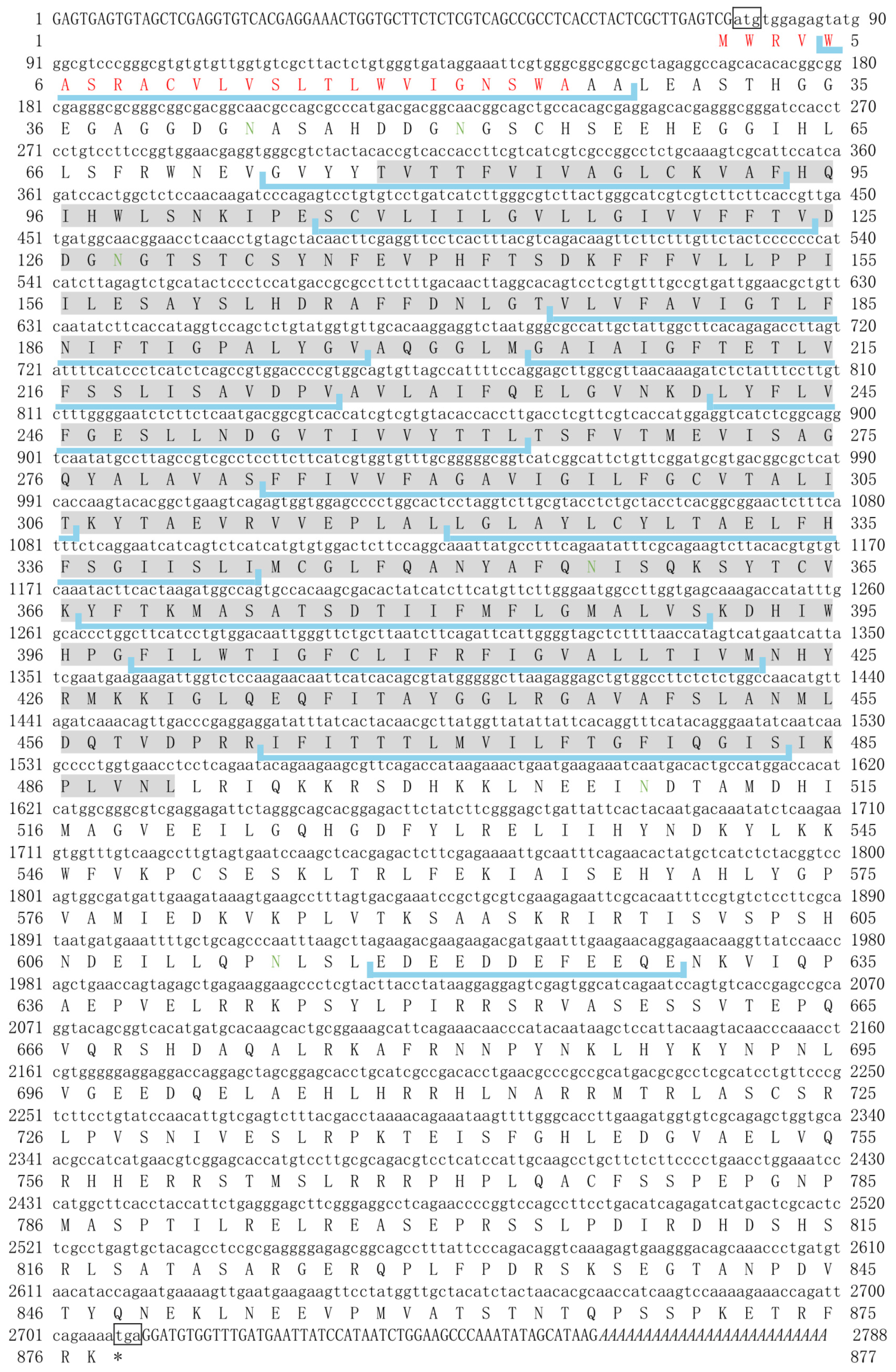
3.1. PmNHE Sequence Display and Bioinformatics Analysis

The full-length PmNHE cDNA was obtained by splicing the open reading frame and 5′/3′ non-coding region sequence sequencing results. PmNHE was 2788 bp long (GenBank accession No. MT164534), including an ORF of 2643 bp, a 5′-untranslated region (UTR) of 76 bp, and a 3′ UTR of 78 bp (Figure 1). The ORF of the PmNHE gene encoded 877 amino acids with a molecular weight of 97.97 KDa and a theoretical isoelectric point of 6.54. Bioinformatics prediction analysis showed that PmNHE had a variety of functional sites, contained 23 phosphorylation sites (20 serine sites and 3 threonine sites), 6 glycosylation sites (highlighted in green font), and 12 transmembrane domains (highlighted with light blue line segments). The sequence contains a sodium/proton exchanger domain (highlighted by gray shading) at 78-490 aa. The poly A structure is used in the 3′-UTR sequence (highlighted in italics).
The PmNHE (Genebank No. MT164534) start code (ATG) and termination code (TAA) are listed in a square box. The signal peptide sequence is highlighted in red font. The glycosylation site is highlighted in green font, and the Na+/H+-exchanger domain is expressed in gray. The Poly A structure is highlighted in italics.

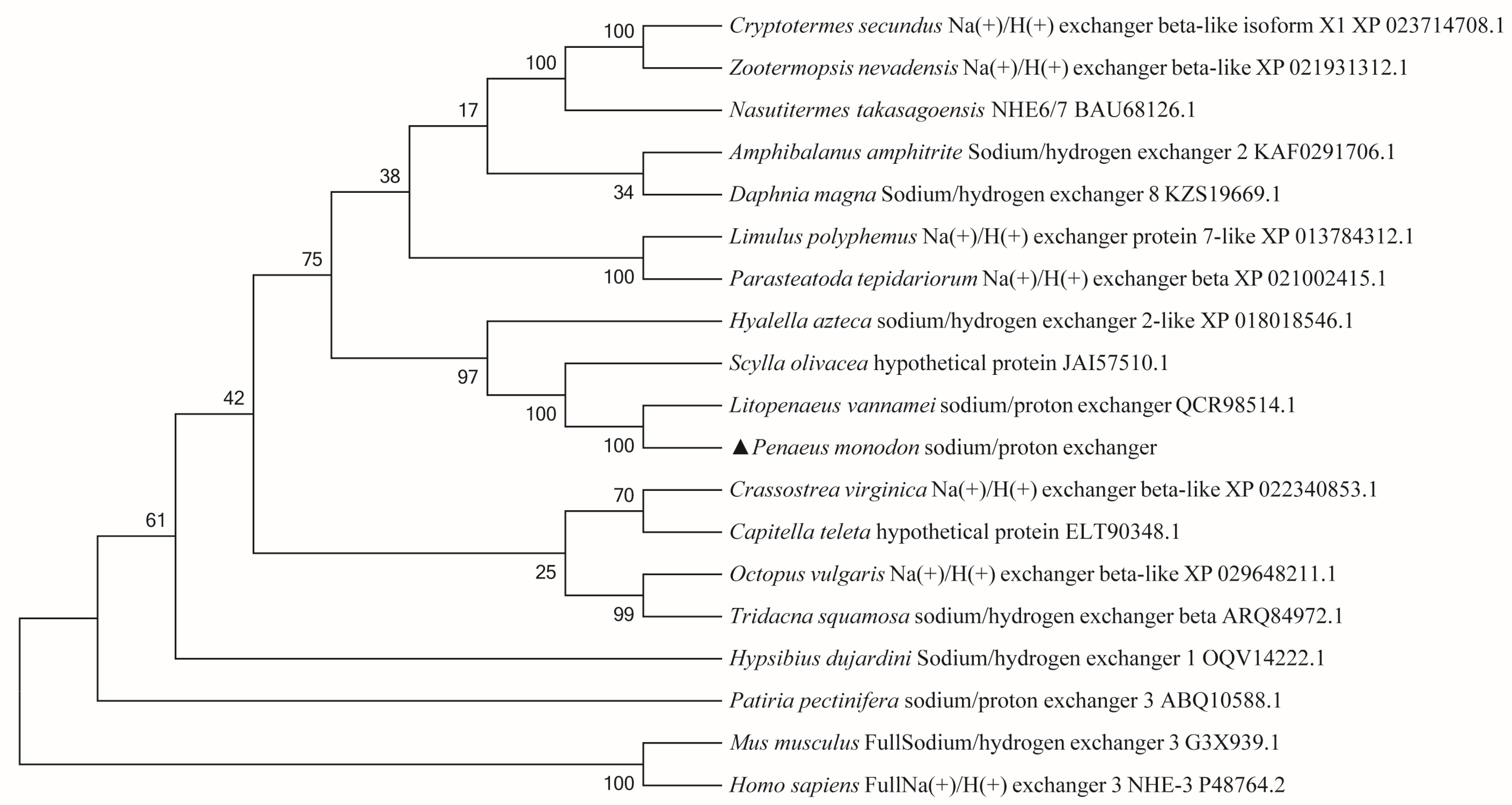
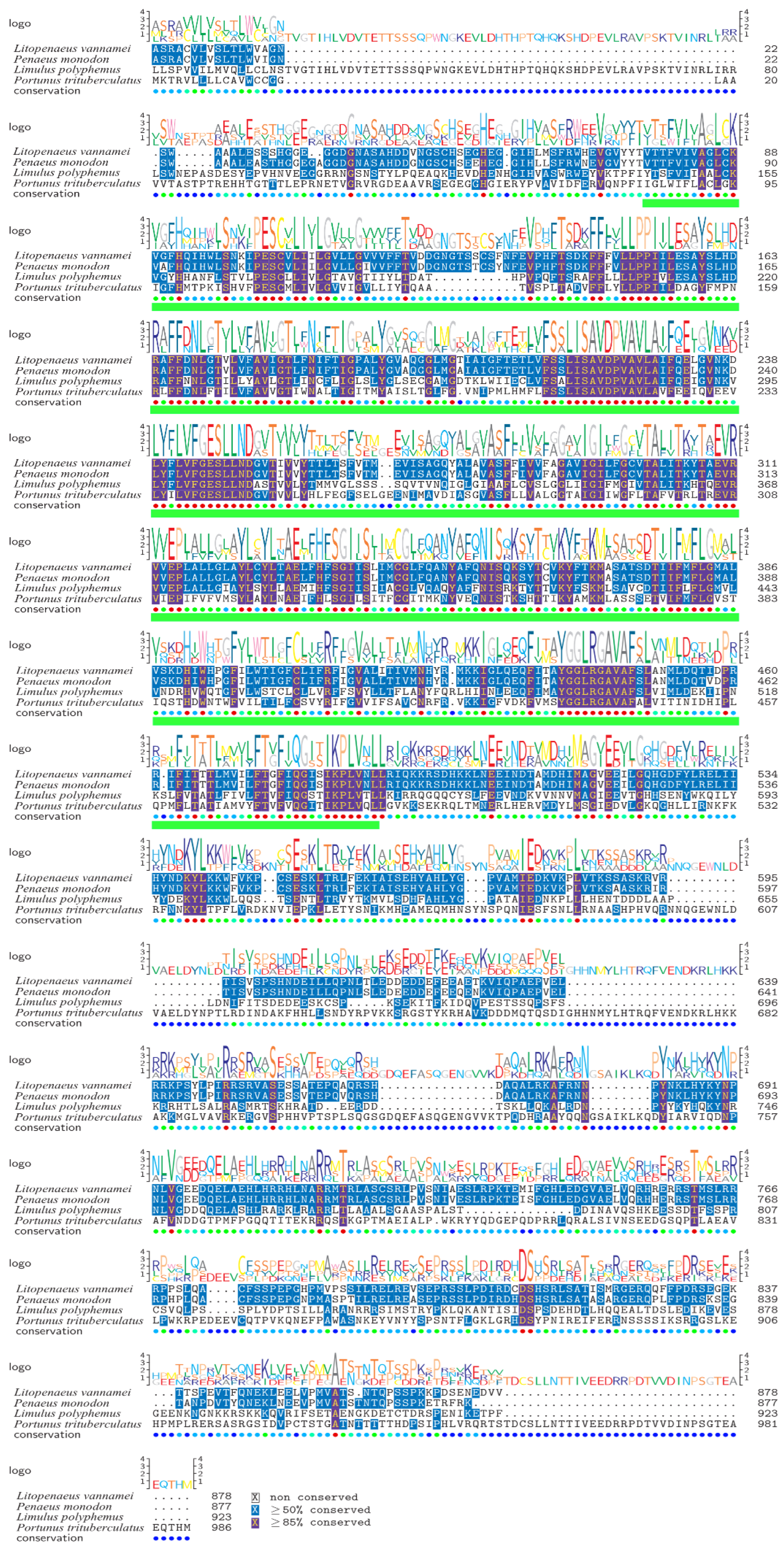
3.2. Phylogenetic Tree Analysis and Multiple Sequence Alignment

In NCBI, the amino acid sequence of the NHE gene of P. monodon was compared with other species, and it was found that it had a high homology with the amino acid sequence of the NHE protein of Penaeus vannamei.
The NJ evolutionary tree of PmNHE and this protein of other species was constructed by MEGA6.0 (Figure 2). The results show that the relationship between invertebrates was the closest, and the relationship between Penaeus vannamei and P. monodon was the closest and clustered together, and they clustered into a branch with Scylla olivacea and Hyalella azteca.
The results of amino acid sequence multiple alignment analysis (Figure 3) show that the amino acids of P. monodon NHE and other species have high homology, among which, PmNHE and Penaeus vannamei (qcr98514.1) have the highest similarity of 93.92%; the protein similarity with Limulus polyphemus (xp_013784312.1) was 42.71%; the protein similarity with Portunus trituberculatus (anv19765.1) was 35.77%.

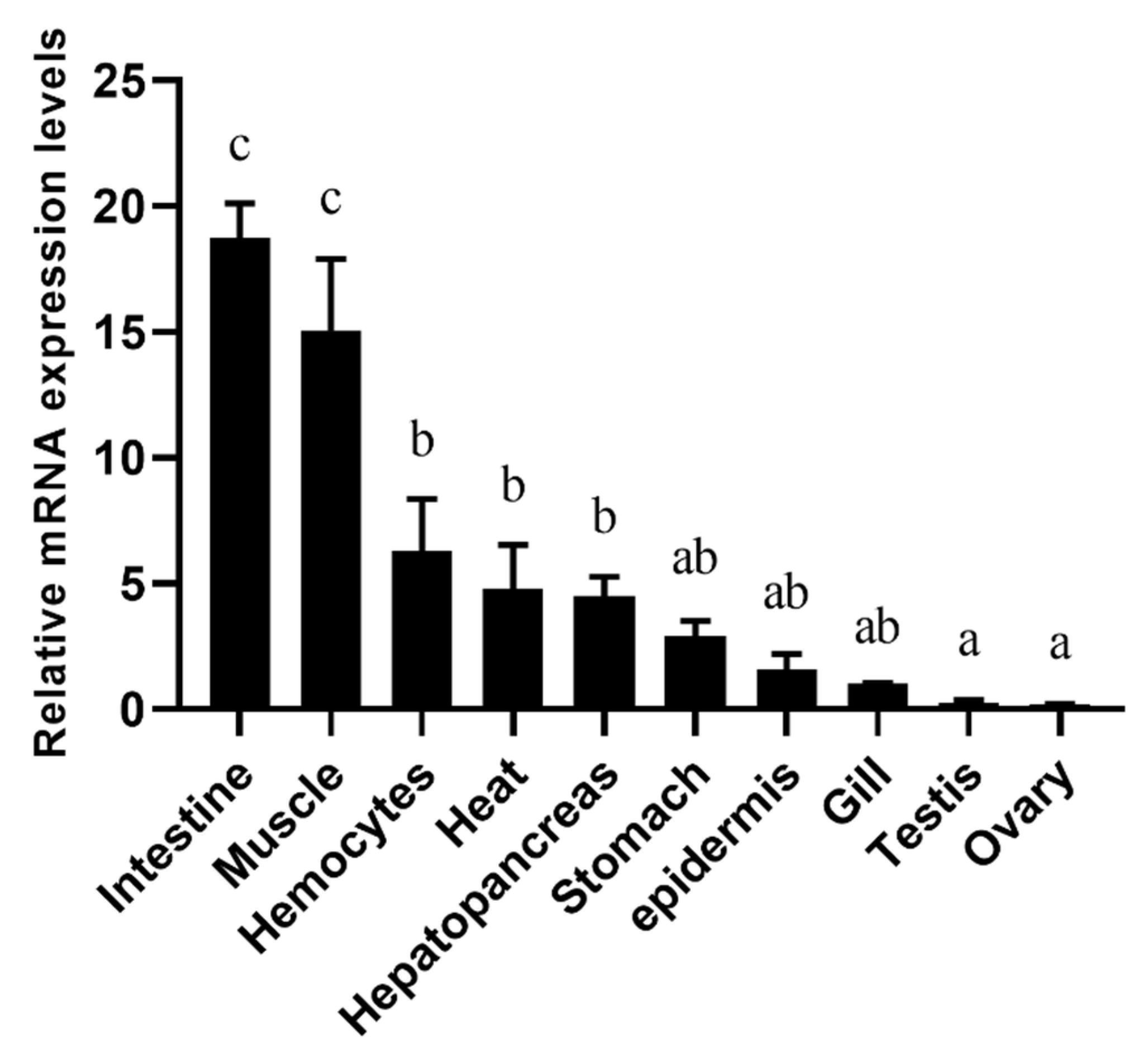
3.3. Expression Analysis of PmNHE mRNA in Different Tissues

In order to study the expression of the PmNHE gene in different tissues, ten tissues including the muscle, hepatopancreas, gill, epidermis, intestine, heart, hemolymph, stomach, ovary, and testis were mainly detected (Figure 4). The order of expression from high to low is intestine > muscle > hemolymph > heart > hepatopancreas > stomach > epidermis > gill > testis > ovary, and the intestine and muscle had the highest levels of PmNHE mRNA expression.

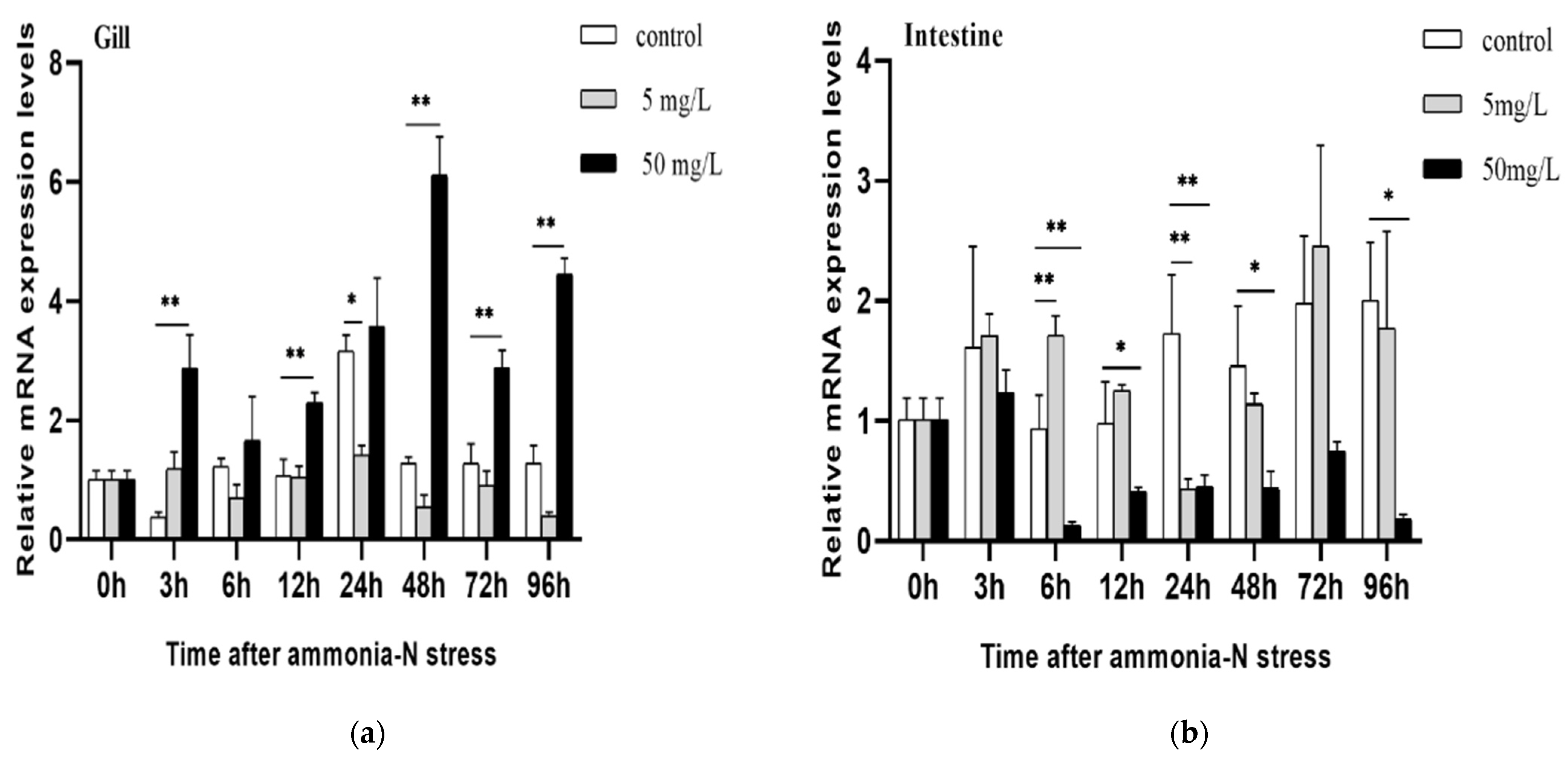
3.4. Expression Changes of PmNHE under Acute Ammonia Nitrogen Stress

The gills and intestines of P. monodon were subjected to two concentrations of ammonia nitrogen, and RT-qPCR was used to detect changes in PmNHE expression. The expression levels in the intestine and gills varied in both the safe and half-lethal concentrations of ammonia nitrogen when compared to the control group (Figure 5).
In the gill tissue, the fluctuation range of PmNHE in the safe concentration was small, and with the increase in stress time, the expression of the PmNHE gene was not significantly different from that of the control group. Under the stress of half-lethal ammonia nitrogen concentration, the expression of PmNHE at 3, 12, 48, 72, and 96 h was up-regulated compared with the control group (p < 0.01). In the intestine, PmNHE showed a trend of first decreasing and then increasing in the safe concentration, which was higher than that of the control group at 6 h and lower than that of the control group at 24 h. The expression of PmNHE under the half-lethal concentration stress was inhibited.

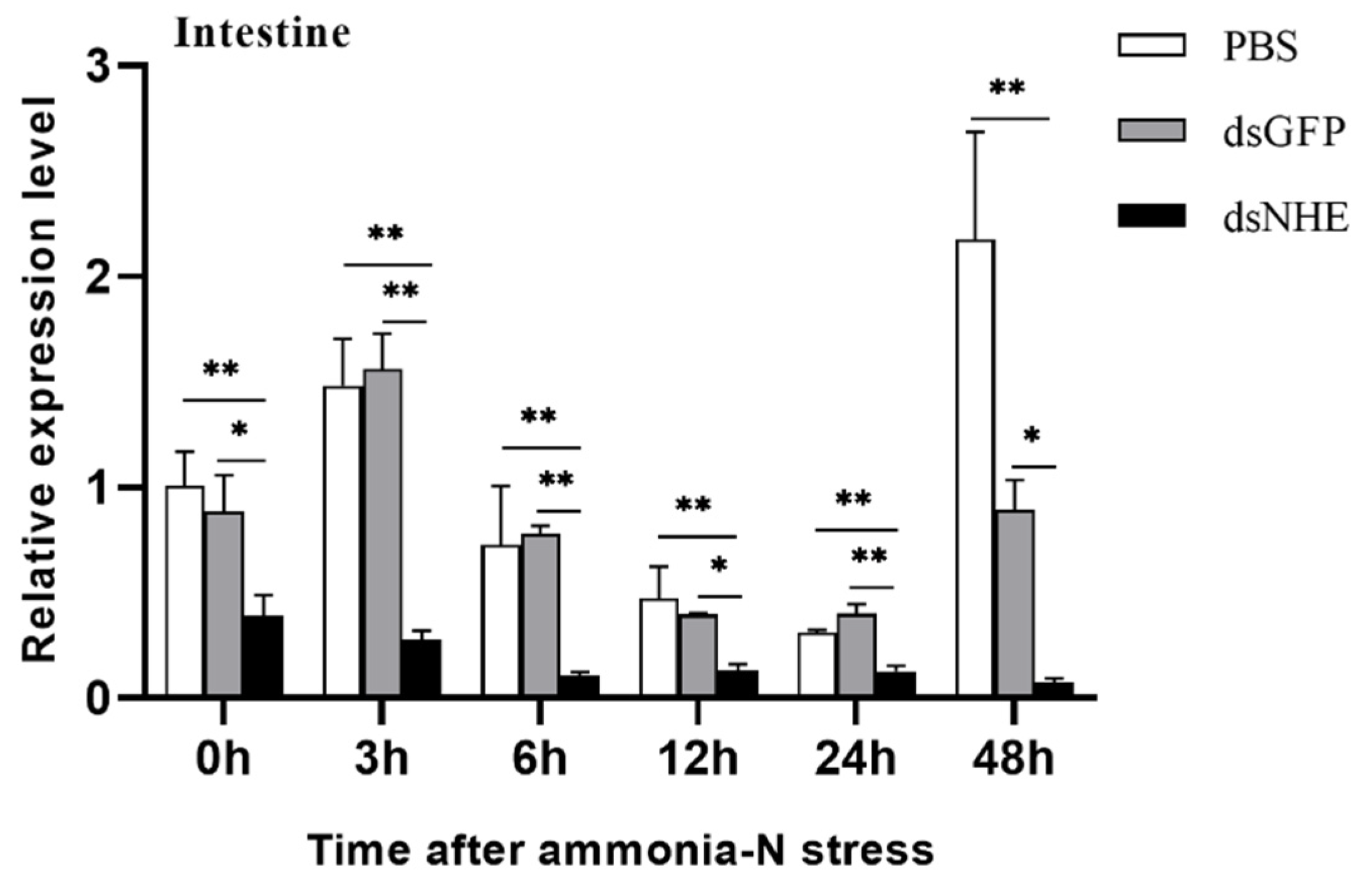
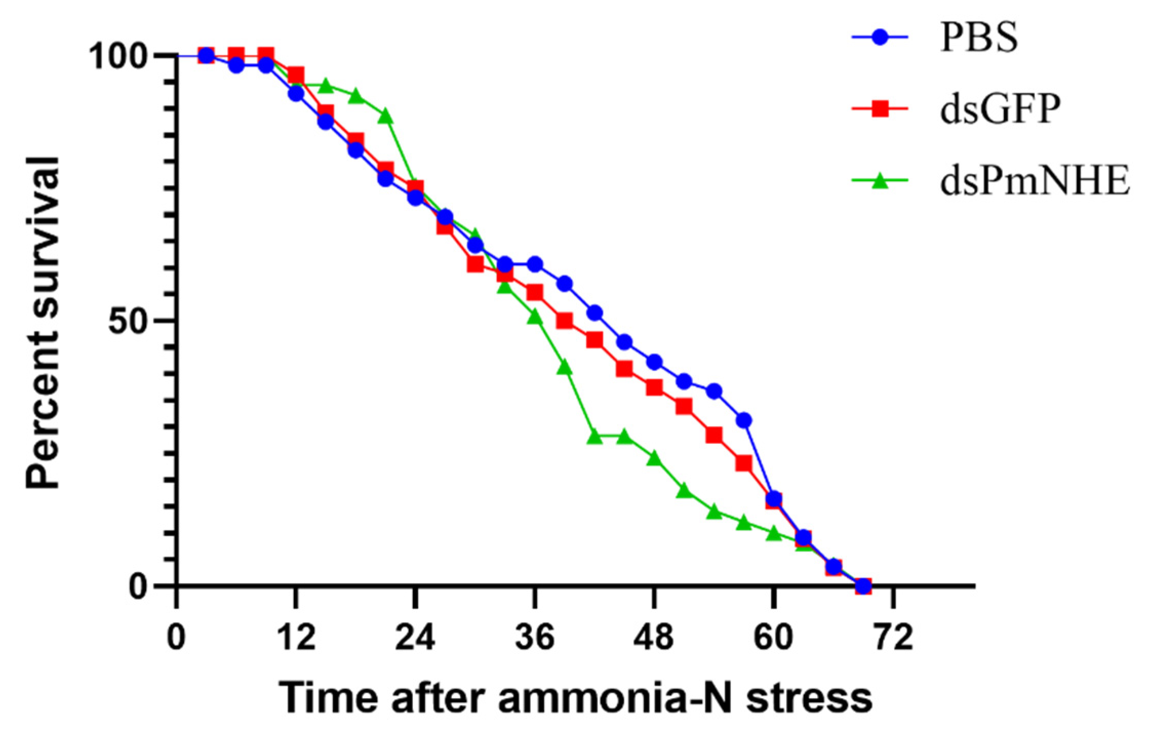
3.5. Mortality in High-Concentration Ammonia Nitrogen Environment after PmNHE Silencing

Under high-concentration ammonia nitrogen, the expression of PmNHE was significantly up-regulated in gills and down-regulated in the intestine. It is suggested that PmNHE may be involved in the mechanism of response to high-concentration ammonia nitrogen stress. In order to explore the role of PmNHE in the process of high-concentration ammonia nitrogen stress, in this study, RNA interference experiments were performed by injecting dsPmNHE synthesized in vitro, and 24 h after dsPmNHE injection, acute high-concentration ammonia nitrogen stress was applied to P. monodon. The results show that with phosphate-buffered saline (PBS) and dsGFP as controls, the expression of PmNHE was lower than that of the control group at 24 h after injection (Figure 6), and there was always a difference between the dsPmNHE injection group and the two control groups during the subsequent acute ammonia nitrogen stress experiment (p < 0.01). It showed that the whole experimental process had good interference efficiency.
The relative expression of PmNHE in the dsPmNHE and GFP groups at different time points under high-concentration ammonia-nitrogen stress was determined.
We compared the survival curves of the PmNHE silenced group and the control group under high levels of ammonia nitrogen stress to explore the relationship between the PmNHE gene and high-concentration ammonia nitrogen stress (Figure 7). The shrimp in the GFP group all died within 69 h, with an average survival time of 40.66 h, the shrimp in the PBS group all died within 69 h, with an average survival time of 42.32 h, the average survival time of the shrimp in the PmNHE-dsRNA group was 37.96 h. From the trend of the survival curve, it can be seen that the survival rate of shrimp in the silenced group was higher than that in the control group in the first 24 h under high-concentration ammonia nitrogen stress; after 36 h of stress, the death rate of shrimp in the PmNHE-silenced group began to increase, which was different from that of the two control groups.
dsPmNHE, GFP, and PBS group shrimp were simultaneously stressed under a higher concentration of ammonia nitrogen; the number of dead shrimp in each group was observed and recorded every 3 h.

4. Discussion

Numerous studies have shown that NHEs play an important role in the transport of various ions in animals [18]. In this research, the full-length cDNA encoding NHE was cloned from P. monodon for the first time. The amino acid sequence analysis of PmNHE by the SMART program found that PmNHE had 12 transmembrane domains, and the sequence contained a sodium/hydrogen ion exchanger domain (78–490 aa). Exploring this domain revealed that NHE transporters contained 10–12 transmembrane domains at the amino terminus and a larger cytoplasmic domain at the carboxy terminus. The transmembrane region M3–M12 had the same characteristics as other members of this family. The M6 and M7 transmembrane regions were highly conserved and therefore considered to be involved in the transport of sodium and hydrogen ions [19]. Phylogenetic tree analysis showed that the evolution relationship between PmNHE of P. monodon and NHE of Penaeus vannamei was the closest. The similarity between PmNHE and the NHE of Litopenaeus vannamei reached 93.92%, the similarity with the protein of Limulus americanus was 42.71%, and the similarity with the protein of Portunus trituberculatus was 35.77%; these data indicated that NHE sequences were species specific.
Quantitative analysis of various tissues showed that PmNHE was expressed in a variety of tissues, and the expression of PmNHE was the most abundant in the intestines of P. monodon, and the expression level in the muscle was second only to that of the intestinal tissues. It has been shown that the expression of NHE was the highest in the intestine of Penaeus vannamei, which was similar to our results [20]. Osmoregulation and ion regulation in crustaceans were mainly performed by gill tissues with multiple functions and the intestines, which was the excretory and digestive organ [21,22]. In addition, muscle has also been demonstrated to have an ion regulation function [23]. Therefore, we selected the gill and intestine as the research objects to explore their expression patterns under ammonia nitrogen stress.
The quantitative results show that the expression of PmNHE in the gill tissue of P. monodon was significantly up-regulated under a high concentration of ammonia nitrogen stress, which was similar to the result of the significant up-regulation of the NHE-2 gene in the gill tissue of Oncorhynchus mykiss when the concentration of ammonia nitrogen increased [24]. Other researchers found that it could block the excretion of ammonia nitrogen from their gills by inhibiting the NHE activity of Carcinus maenas, Cancer pagurus, and Astacus leptodactylus by non-specific inhibitor amiloride [7,8]. Therefore, we speculated that PmNHE also played a role in excreting ammonia in the gills of P. monodon, which may be in line with the “Na+/NH4+ exchange complex” model of ammonia excretion in freshwater fish. This model suggested that ammonia transport was carried out through Rh glycoproteins, and the hydration of CO2 and H+ by Na+/NH4+-exchange (NHE-2) and H+-ATPase led to the acidification of the gill boundary layer, thereby promoting ammonia transport [25]. Furthermore, we also conducted a quantitative analysis of the intestines, and the results show that the expression of PmNHE in the intestines of P. monodon under high levels of ammonia nitrogen stress was significantly inhibited compared with the control group. Silva et al. quantitatively analyzed the foregut and hindgut of Monopterus albus treated with high ammonia nitrogen and showed that the expression of NHE3 was increased in the foregut after 6 h of ammonia exposure, about 28 times that of the control group [25]. The expression of the NHE1 gene in the foregut and hindgut changed little over time. The expression of the NHE1 and NHE3 genes in the foregut and hindgut following chronic exposure was not different. They speculated that NHE3 in the foregut of Monopterus albus may have a significant impact on the active excretion of NH4+ under the high-concentration ammonia nitrogen environment. The high ammonia nitrogen environment made PmNHE up-regulated in the gill tissue of P. monodon and inhibited in the intestine. We speculated that it may be due to the differences between species; PmNHE in the gill tissue of P. monodon played an important role under ammonia nitrogen stress. To further explore the role of PmNHE in an environment with high levels of ammonia nitrogen, we compared the survival curves of the PmNHE silenced group and control group under high-concentration ammonia nitrogen stress. We found that after high levels of ammonia nitrogen stress, the death curves of shrimp in the GFP and PBS control groups were essentially consistent, and the average death time was similar. The average survival time of the shrimp in the PmNHE-dsRNA injection group was lower than that in the two control groups. This result indicates that PmNHE may have a significant impact on the high ammonia nitrogen environment.

5. Conclusions

In this research, the full-length PmNHE gene was cloned, the structure of the PmNHE gene was analyzed by bioinformatics, the expression changes of PmNHE in various tissues and in the gill and intestine of P. monodon under acute ammonia nitrogen stress were investigated, and the death curve under high levels of ammonia nitrogen stress was explored by RNAi technology. The results provide a basis for further exploring the role of the sodium/hydrogen ion transporter in P. monodon under ammonia nitrogen stress.

Author Contributions

S.J. (Shigui Jiang) and F.Z. conceived and designed the experiments. Y.L. performed the bioinformatics analysis and prepared the manuscript, the table, and the figures. H.F., J.H. and W.Z. conducted the experiment. L.Y., X.C., S.J. (Song Jiang) and Q.Y. collected the samples and performed sequencing. All authors have read and agreed to the published version of the manuscript.

Funding

This study was funded by the National Key R&D Program of China (2022YFD2401900); China Agriculture Research System (CARS-48); Hainan Yazhou Bay Seed Laboratory (Project of B21Y10701 and B21HJ0701); Research and Development Projects in Key Areas of Guangdong Province (2021B0202003); Central Public interest Scientific Institution Basal Research Fund, CAFS (2020TD30); Central Public Interest Scientific Institution Basal Research Fund, South China Sea Fisheries Research Institute, CAFS (2020ZD01,2021SD13); Hainan Provincial Natural Science Foundation of China (320QN359,322RC806,320LH008); Guangdong Basic and Applied Basic Research Foundation (2020A1515110200); Guangzhou Science and Technology Planning Project (202102020208); and Hainan Provincial Association for Science and Technology of Young Science and Technology Talents Innovation Plan Project (QCQTXM202206).

Institutional Review Board Statement

The use of all the shrimps in these experiments was approved by the Animal Care and Use Committee at the Chinese Academy of Fishery Sciences (CAFS), and we also applied the national and institutional guidelines for the care and use of laboratory animals at the CAFS.

Informed Consent Statement

Not applicable.

Data Availability Statement

Not applicable.

Conflicts of Interest

The authors declare no conflict of interest.

References

  1. Hamm, L.L.; Nakhoul, N.; Hering-Smith, K.S. Acid-Base Homeostasis. Clin. J. Am. Soc. Nephrol. 2015, 10, 2232–2242. [Google Scholar] [CrossRef] [PubMed] [Green Version]
  2. Weiner, I.D.; Verlander, J.W. Ammonia Transporters and Their Role in Acid-Base Balance. Physiol. Rev. 2017, 97, 465–494. [Google Scholar] [CrossRef] [PubMed]
  3. Aronson, P.S.; A Suhm, M.; Nee, J. Interaction of external H+ with the Na+-H+ exchanger in renal microvillus membrane vesicles. J. Biol. Chem. 1983, 258, 6767–6771. [Google Scholar] [CrossRef] [PubMed]
  4. Kinsella, J.L.; Aronson, P.S. Interaction of NH4+ and Li+ with the renal microvillus membrane Na+-H+ exchanger. Am. J. Physiol. Cell Physiol. 1981, 241, C220–C226. [Google Scholar] [CrossRef] [PubMed]
  5. Li, Y.D.; Zhou, F.L.; Huang, J.H.; Yang, L.S.; Jiang, S.; Yang, Q.B.; He, J.G.; Jiang, S.G. Transcriptome reveals involvement of immune defense, oxidative imbalance, and apoptosis in ammonia-stress response of the black tiger shrimp (Penaeus monodon). Fish Shellfish. Immunol. 2018, 83, 162–170. [Google Scholar] [CrossRef]
  6. Li, Y.; Zhou, F.; Yang, Q.; Jiang, S.; Huang, J.; Yang, L.; Ma, Z.; Jiang, S. Single-Cell Sequencing Reveals Types of Hepatopancreatic Cells and Haemocytes in Black Tiger Shrimp (Penaeus monodon) and Their Molecular Responses to Ammonia Stress. Front. Immunol. 2022, 13, 883043. [Google Scholar] [CrossRef]
  7. Lucu, Č.; Devescovi, M.; Siebers, D. Do amiloride and ouabain affect ammonia fluxes in perfused Carcinus gill epithelia? J. Exp. Zool. 1989, 249, 1–5. [Google Scholar] [CrossRef]
  8. Weihrauch, D.; Becker, W.; Postel, U.; Luck-Kopp, S.; Siebers, D. Potential of active excretion of ammonia in three different haline species of crabs. J. Comp. Physiol. B 1999, 169, 25–37. [Google Scholar] [CrossRef]
  9. Towle, D.W.; E Rushton, M.; Heidysch, D.; Magnani, J.J.; Rose, M.J.; Amstutz, A.; Jordan, M.K.; Shearer, D.W.; Wu, W.S. Sodium/proton antiporter in the euryhaline crab Carcinus maenas: Molecular cloning, expression and tissue distribution. J. Exp. Biol. 1997, 200, 1003–1014. [Google Scholar] [CrossRef]
  10. Li, Y.D.; Zhou, F.L.; Tang, Y.P.; Huang, J.H.; Yang, L.S.; Jiang, S.; Yang, Q.B.; Jiang, S.G. Variation in bacterial communities among stress-sensitive and stress-tolerant black tiger shrimp (Penaeus monodon) individuals. Aquac. Res. 2020, 52, 2146–2159. [Google Scholar] [CrossRef]
  11. Li, Y.D.; Zhou, F.L.; Huang, J.H.; Yang, L.S.; Jiang, S.; Yang, Q.B.; Jiang, S.G. Transcriptome and miRNA Profiles of Black Tiger Shrimp, Penaeus monodon, Under Different Salinity Conditions. Front. Mar. Sci. 2020, 7, 579381. [Google Scholar] [CrossRef]
  12. Fan, H.; Li, Y.; Yang, Q.; Jiang, S.; Yang, L.; Huang, J.; Jiang, S.; Zhou, F. Isolation and characterization of a MAPKK gene from Penaeus monodon in response to bacterial infection and low-salinity challenge. Aquac. Rep. 2021, 20, 100671. [Google Scholar] [CrossRef]
  13. Si, M.-R.; Li, Y.-D.; Jiang, S.-G.; Yang, Q.-B.; Jiang, S.; Yang, L.-S.; Huang, J.-H.; Chen, X.; Zhou, F.-L. A CSDE1/Unr gene from Penaeus monodon: Molecular characterization, expression and association with tolerance to low salt stress. Aquaculture 2022, 561, 738660. [Google Scholar] [CrossRef]
  14. Li, Y.; Yang, Q.; Su, T.; Zhou, F.; Yang, L.; Huang, J. The toxicity of ammonia-N on Penaeus monodon and immune parameters. J. Shanghai Ocean. Univ. 2012, 21, 358–362. [Google Scholar] [CrossRef]
  15. Zhou, K.; Zhou, F.; Huang, J.; Yang, Q.; Jiang, S.; Qiu, L.; Yang, L.; Zhu, C.; Jiang, S. Characterization and expression analysis of a chitinase gene (PmChi-4) from black tiger shrimp (Penaeus monodon) under pathogen infection and ambient ammonia nitrogen stress. Fish Shellfish. Immunol. 2017, 62, 31–40. [Google Scholar] [CrossRef] [PubMed]
  16. Li, Y.; Zhou, F.; Fan, H.; Jiang, S.; Yang, Q.; Huang, J.; Yang, L.; Chen, X.; Zhang, W.; Jiang, S. Molecular Technology for Isolation and Characterization of Mitogen-Activated Protein Kinase Kinase 4 from Penaeus monodon, and the Response to Bacterial Infection and Low-Salinity Challenge. J. Mar. Sci. Eng. 2022, 10, 1642. [Google Scholar] [CrossRef]
  17. Si, M.-R.; Li, Y.-D.; Jiang, S.-G.; Yang, Q.-B.; Jiang, S.; Yang, L.-S.; Huang, J.-H.; Chen, X.; Zhou, F.-L. Identification of multifunctionality of the PmE74 gene and development of SNPs associated with low salt tolerance in Penaeus monodon. Fish Shellfish Immunol. 2022, 128, 7–18. [Google Scholar] [CrossRef]
  18. Tseng, Y.; Yan, J.; Furukawa, F.; Hwang, P. Did Acidic Stress Resistance in Vertebrates Evolve as Na+/H+ Exchanger-Mediated Ammonia Excretion in Fish? BioEssays 2020, 42, e1900161. [Google Scholar] [CrossRef]
  19. Dibrov, P.; Fliegel, L. Comparative molecular analysis of Na+/H+ exchangers: A unified model for Na+/H+ antiport? FEBS Lett. 1998, 424, 1–5. [Google Scholar] [CrossRef] [Green Version]
  20. Li, H.; Ren, C.; Jiang, X.; Cheng, C.; Ruan, Y.; Zhang, X.; Huang, W.; Chen, T.; Hu, C.; Qiu, G.F. Na+/H+ exchanger (NHE) in Pacific white shrimp (Litopenaeus vannamei): Molecular cloning, transcriptional response to acidity stress, and physiological roles in pH homeostasis. PLoS ONE 2019, 14, e0212887. [Google Scholar] [CrossRef]
  21. Freire, C.A.; Onken, H.; McNamara, J.C. A structure–function analysis of ion transport in crustacean gills and excretory organs. Comp. Biochem. Physiol. Part A Mol. Integr. Physiol. 2008, 151, 272–304. [Google Scholar] [CrossRef] [PubMed]
  22. Chen, T.; Ren, C.; Wang, Y.; Gao, Y.; Wong, N.-K.; Zhang, L.; Hu, C. Crustacean cardioactive peptide (CCAP) of the Pacific white shrimp (Litopenaeus vannamei): Molecular characterization and its potential roles in osmoregulation and freshwater tolerance. Aquaculture 2016, 451, 405–412. [Google Scholar] [CrossRef]
  23. Mcfarland, W.; Lee, B.D. Osmotic and ionic concentrations of penaeidean shrimps of the Texas coast. Bull. Mar. Sci. 1963, 13, 391–417. [Google Scholar]
  24. Zimmer, A.M.; Nawata, C.M.; Wood, C.M. Physiological and molecular analysis of the interactive effects of feeding and high environmental ammonia on branchial ammonia excretion and Na+ uptake in freshwater rainbow trout. J. Comp. Physiol. B 2010, 180, 1191–1204. [Google Scholar] [CrossRef]
  25. Silva, J.M.; Coimbra, J.; Wilson, J. Weatherloach (Misgurnus anguillicaudatus) actively excretes ammonia through NHE. Comp. Biochem. Physiol. Part A Mol. Integr. Physiol. 2008, 150, S57. [Google Scholar] [CrossRef]
Figure 1. Nucleotide and deduced amino acid sequence of PmNHE. (* termination codon, tga).
Jmse 10 01897 g001
Figure 2. Phylogenetic analysis of PmNHE. (▲represents the PmNHE gene obtained in this study).
Jmse 10 01897 g002
Figure 3. Multiple alignment of amino acid sequences of NHE gene from Peenaeus monodon and other species. Sequence ID: Litopenaeus vannamei, QCR98514.1; Limulus Polyphemus, XP_013784312.1; Portunus trituberculatus, ANV19765.1.
Jmse 10 01897 g003
Figure 4. Relative expression of PmNHE in different tissues of Penaeus monodon. The values are x ± SD (n = 3). Bars with different lowercase letters indicate significant differences (p < 0.05).
Jmse 10 01897 g004
Figure 5. The mRNA expression levels of PmNHE in the gills and intestine at different time intervals of different-concentration ammonia nitrogen stress treatments. (a) Gill and (b) intestine under ammonia nitrogen stress. Data are shown as means ± SD (standard deviation) of three separate individuals. Significant differences are indicated with asterisks, * p < 0.05, ** p < 0.01.
Jmse 10 01897 g005
Figure 6. Silencing efficiency of PmNHE under ammonia-nitrogen stress. Data are shown as means ± SD (standard deviation) of three separate individuals. Significant differences are indicated with asterisks, * p < 0.05, ** p < 0.01.
Jmse 10 01897 g006
Figure 7. Mortality under ammonia-nitrogen stress after silencing of PmNHE.
Jmse 10 01897 g007
Table 1. Primers and sequences used in the study.
Primer NameNucleotide Sequence (5′ → 3′)Purpose
3′race GSPTACCTCTGCTACCTCACGGCGGAACT3′RACE
3′race NGSPGTAGAGCTGAGAAGGAAGCCCTCGTA3′RACE
5′race GSPTCCGTGCTGCCCTAGAATCTCC5′RACE
5′race NGSPTCCGCCGTGAGGTAGCAGAGGT5′RACE
qNHE-FTGGCTTCACAGAGACCTTRT-PCR
qNHE-RCACGCATCCGAACAGAATRT-PCR
qEF-1a-FAAGCCAGGTATGGTTGTCAACTTTRT-PCR
qEF-1a-RCGTGGTGCATCTCCACAGACTRT-PCR
dsNHE-FTAATACGACTCACTATAGGGGCACAAGGAGGTCTAATGGGdsRNA
dsNHE-RTAATACGACTCACTATAGGGGAGGTAGCAGAGGTACGCAAGdsRNA
dsGFP-FTAATACGACTCACTATAGGGATGGCTAGCAAAGGAGAAGAACTTTdsRNA
dsGFP-RTAATACGACTCACTATAGGGAACGGGAAAAGCATTGAACACCAdsRNA
Publisher’s Note: MDPI stays neutral with regard to jurisdictional claims in published maps and institutional affiliations.

