
Nanotechnology as a Promising Tool against Phytopathogens: A Futuristic Approach to Agriculture
 ,
,  ,
,  , ,
, ,  , , ,
, , ,
Abstract
:1. Introduction
2. Conventional Measures for Disease Control
3. Plant Pathogen Detection with Nanotechnology
3.1. Bio-Nanomaterials
3.2. Nanoparticle Bio-Barcode Assay
3.3. Nanopore DNA Sequencing
3.4. Nanodiagnostic Kit
3.5. Quantum Dots (QDs)
4. Nanomaterials: Sustainable Weapons against Phytopathogens
- (1)
- Damage to the membrane transporter and nutrient absorption systems.
- (2)
- Reactive oxygen species (ROS) production damages several cell organelles, including DNA, by inducing cellular and oxidative stress.
- (3)
- Toxic ion release causes changes in the permeability and activity of membrane proteins.
- (4)
- Cell death and genotoxicity are caused by the interrelationship of harmful ions produced by nanoparticles with DNA.
- (5)
- Energy production, membrane oxidation, and protein oxidation are all impacted by interference with metabolic processes. Depending on the size and dosage, the biocidal properties of nanoparticles are intended to provide distinctive and enhanced antibiotic activity [31].

4.1. Nano-Fungicides
4.2. Nanoparticles’ Impact on Bacteria
5. Application of Various Nanoparticles against Plant Pathogens
5.1. Antimicrobial Activity of Metal Nanoparticles

5.2. Antibacterial Activity of Metal Oxide Nanoparticles
6. Action of Nanoparticles against Plant Pathogens—The Mechanism
6.1. Formation of Reactive Oxygen Species (ROS)
6.2. Cell Membrane Damage
6.3. Liberation of Toxic Components
7. Environmental Risk of Nanoparticles
7.1. Nanoparticle–Soil Microorganism Interaction
7.2. Nanoparticles’ Effect on the Bacterial and Fungal Populations in Soil
8. Challenges and Limitations
9. Existing Commercial Limitations
10. Conclusions
Author Contributions
Funding
Institutional Review Board Statement
Data Availability Statement
Acknowledgments
Conflicts of Interest
Abbreviations
| Ag | Silver |
| Ag NW | Silver nanowire |
| Au | Gold |
| Au-MNPs | Magnetic gold nanoparticles |
| Au NP-ICTS | Gold nanoparticle-based immunochromatographic test strip |
| BBS | Bacterial brown stripe |
| BBSV | Broad bean stain virus |
| BBTV | Banana bunchy top virus |
| b-DNA | DNA with a bio-barcode |
| BYMV | Bean yellow mosaic virus |
| Cd | Cadmium |
| CdONP | Cadmium oxide nanoparticles |
| CdSe–PEI QD | Cadmium Selenium polyethylenimine–capped quantum dot |
| CdTe QD–CD | Cadmium telluride quantum dot |
| CeO2 | Cerium oxide |
| CMV | Cucumber mosaic virus |
| CNTs | Carbon nanotubes |
| CS | Chitosan |
| CTV | Citrs tristeza virus |
| Cu | Copper |
| CuO | Copper oxide |
| CV | Cyclic voltammetry |
| Dot-ELISA | Dot enzyme-linked immunosorbent assay |
| DPV | Differential pulse voltammetry |
| EIS | Electrochemical impedance spectroscopy |
| ELISA | Enzyme-linked immunosorbent assay |
| E-nose | Electronic nose |
| Fe3O4 | Iron oxide |
| FFT-SWV | Fast Fourier transform square wave voltammetry |
| FISH | Fluorescence in situ hybridization |
| FRET | Fluorescence resonance energy transfer |
| GCE | Glassy carbon electrode |
| GO | Graphene oxide |
| GO-Ag | Graphene oxide-Silver |
| GST | glutathione-S-transferase |
| ITO | Indium-tin oxide |
| LDH | Lactate dehydrogenase |
| LFA | Lateral flow assay |
| LOD | Limit of detection |
| LPNE | Lithographically patterned nanowire electrodeposition |
| LSPR | Surface plasmon resonance |
| LSV | Linear sweep voltammetry |
| mAbs | Monoclonal antibodies |
| MCMV | Maize chlorotic mottle virus |
| MD | Mosaic disease |
| MgO | Magnesium oxide |
| MIP | Molecularly imprinted polymer |
| MnO2 | Manganese dioxide |
| MTV | Tobacco mosaic virus |
| MWCNTs | Multiwalled carbon nanotubes |
| NGS | Next-generation sequencing |
| Ni | Nickel |
| NiO | Nickel oxide |
| NP | Nanoparticle |
| PCR | Polymerase chain reaction |
| Pd | Palladium |
| PGPR | Plant growth-promoting rhizobacteria |
| PPO | Polyphenol oxidase |
| PPY | Polypyrrole |
| PRSV | Papaya ringspot virus |
| PtNPs | Platinum nanoparticles |
| PVY | Potato virus Y |
| QDs | Quantum dots |
| RAPD | Random amplified polymorphic dna |
| rGO | Reduced graphene oxide |
| ROS | Reactive oxygen species |
| RTBV | Rice tungro bacilliform virus |
| RTSV | Rice tungro spherical virus |
| SAR | Systemic acquired resistance |
| Se | Selenium |
| SHRV | Sun hemp rosette virus |
| SiO2 | Silicon oxide |
| SPCE | Screen-printed carbon electrode |
| SPR | Surface plasmon resonance |
| SRAP | Sequence-related amplified polymorphism |
| ssDNA | Single-strain deoxyribonucleic acid |
| SWCNTs | Single-walled carbon nanotubes |
| TBSV | Tomato bushy stunt virus |
| TiO2 | Titanium oxide |
| TSP | Total soluble protein |
| TSWV | Tomato spotted wilt virus |
| TYLCV | Tomato yellow leaf curl virus |
| VOCs | Volatile organic compounds |
| WE SPCE | Working electrode screen-printed carbon electrode |
| YDV | Yellow dwarf virus |
| YMV | Yellow mosaic virus |
| ZnO | Zinc oxide |
References
- Chen, H.; Yada, R. Nanotechnologies in Agriculture: New Tools for Sustainable Development. Trends Food Sci. Technol. 2011, 22, 585–594. [Google Scholar] [CrossRef]
- Atiq, M.; Naeem, I.; Sahi, S.T.; Rajput, N.A.; Haider, E.; Usman, M.; Shahbaz, H.; Fatima, K.; Arif, E.; Qayyum, A. Nanoparticles: A Safe Way towards Fungal Diseases. Arch. Phytopathol. Plant Prot. 2020, 53, 781–792. [Google Scholar] [CrossRef]
- Kutawa, A.B.; Ahmad, K.; Ali, A.; Hussein, M.Z.; Abdul Wahab, M.A.; Adamu, A.; Ismaila, A.A.; Gunasena, M.T.; Rahman, M.Z.; Hossain, M.I. Trends in Nanotechnology and Its Potentialities to Control Plant Pathogenic Fungi: A Review. Biology 2021, 10, 881. [Google Scholar] [CrossRef] [PubMed]
- Tilman, D.; Balzer, C.; Hill, J.; Befort, B.L. Global Food Demand and the Sustainable Intensification of Agriculture. Proc. Natl. Acad. Sci. USA 2011, 108, 20260–20264. [Google Scholar] [CrossRef] [PubMed]
- Rajwade, J.M.; Chikte, R.G.; Paknikar, K.M. Nanomaterials: New Weapons in a Crusade against Phytopathogens. Appl. Microbiol. Biotechnol. 2020, 104, 1437–1461. [Google Scholar] [CrossRef]
- Panpatte, D.G.; Jhala, Y.K. Nanotechnology for Agriculture: Crop Production & Protection, 1st ed.; Springer: Singapore, 2019; ISBN 978-981-32-9374-8. [Google Scholar]
- Flood, J. The Importance of Plant Health to Food Security. Food Secur. 2010, 2, 215–231. [Google Scholar] [CrossRef]
- Stephenson, G.R. Pesticide Use and World Food Production: Risks and Benefits. In Environmental Fate and Effects of Pesticides; ACS Symposium Series; American Chemical Society: Washington, DC, USA, 2003; Volume 853, pp. 15–261. ISBN 9780841237223. [Google Scholar]
- Ghormade, V.; Deshpande, M.V.; Paknikar, K.M. Perspectives for Nano-Biotechnology Enabled Protection and Nutrition of Plants. Biotechnol. Adv. 2011, 29, 792–803. [Google Scholar] [CrossRef]
- Sangeetha, J.; Mundaragi, A.; Thangadurai, D.; Maxim, S.S.; Pandhari, R.M.; Alabhai, J.M. Nanobiotechnology for Agricultural Productivity, Food Security and Environmental Sustainability. In Nanotechnology for Agriculture: Crop Production & Protection; Panpatte, D.G., Jhala, Y.K., Eds.; Springer: Singapore, 2019; pp. 1–23. ISBN 978-981-32-9374-8. [Google Scholar]
- US Environmental Protection Agency. Nanotechnology White Paper; Report EPA 100/B-07/001; US Environmental Protection Agency: Washington, DC, USA, 2007; Volume 1.
- Das, K.; Jhan, P.K.; Das, S.C.; Aminuzzaman, F.M.; Benjamin, Y.A. Nanotechnology: Past, Present and Future Prospects in Crop Protection. In Technology in Agriculture; IntechOpen: London, UK, 2021; pp. 1–22. [Google Scholar]
- Nair, R.; Varghese, S.H.; Nair, B.G.; Maekawa, T.; Yoshida, Y.; Kumar, D.S. Nanoparticulate Material Delivery to Plants. Plant Sci. 2010, 179, 154–163. [Google Scholar] [CrossRef]
- Majeed, Z.H.; Taha, M.R. A Review of Stabilization of Soils by Using Nanomaterials. Aust. J. Basic Appl. Sci. 2013, 7, 576–581. [Google Scholar]
- Mukhopadhyay, S.S. Nanotechnology in Agriculture: Prospects and Constraints. Nanotechnol. Sci. Appl. 2014, 7, 63–71. [Google Scholar] [CrossRef]
- Deshpande, P.; Dapkekar, A.; Oak, M.D.; Paknikar, K.M.; Rajwade, J.M. Zinc Complexed Chitosan/TPP Nanoparticles: A Promising Micronutrient Nanocarrier Suited for Foliar Application. Carbohydr. Polym. 2017, 165, 394–401. [Google Scholar] [CrossRef] [PubMed]
- Baruah, S.; Dutta, J. Nanotechnology Applications in Pollution Sensing and Degradation in Agriculture: A Review. Environ. Chem. Lett. 2009, 7, 191–204. [Google Scholar] [CrossRef]
- Lisa, M.; Chouhan, R.S.; Vinayaka, A.C.; Manonmani, H.K.; Thakur, M.S. Gold Nanoparticles Based Dipstick Immunoassay for the Rapid Detection of Dichlorodiphenyltrichloroethane: An Organochlorine Pesticide. Biosens. Bioelectron. 2009, 25, 224–227. [Google Scholar] [CrossRef] [PubMed]
- Gan, N.; Yang, X.; Xie, D.; Wu, Y.; Wen, W. A Disposable Organophosphorus Pesticides Enzyme Biosensor Based on Magnetic Composite Nano-Particles Modified Screen Printed Carbon Electrode. Sensors 2010, 10, 625–638. [Google Scholar] [CrossRef] [PubMed]
- De, A.; Bose, R.; Kumar, A.; Mozumdar, S. Targeted Delivery of Pesticides Using Biodegradable Polymeric Nanoparticles; Springer: New Delhi, India, 2014; ISBN 978-81-322-1689-6. [Google Scholar]
- Ragaei, M.; Sabry, A. Nanotechnology for Insect Pest Control. Int. J. Sci. Envirion. Technol. 2014, 3, 528–545. [Google Scholar]
- Grillo, R.; dos Santos, N.Z.P.; Maruyama, C.R.; Rosa, A.H.; de Lima, R.; Fraceto, L.F. Poly(ε-Caprolactone)Nanocapsules as Carrier Systems for Herbicides: Physico-Chemical Characterization and Genotoxicity Evaluation. J. Hazard. Mater. 2012, 231–232, 1–9. [Google Scholar] [CrossRef]
- Guan, H.; Chi, D.; Yu, J.; Li, X. A Novel Photodegradable Insecticide: Preparation, Characterization and Properties Evaluation of Nano-Imidacloprid. Pestic. Biochem. Physiol. 2008, 92, 83–91. [Google Scholar] [CrossRef]
- Duhan, J.S.; Kumar, R.; Kumar, N.; Kaur, P.; Nehra, K.; Duhan, S. Nanotechnology: The New Perspective in Precision Agriculture. Biotechnol. Rep. 2017, 15, 11–23. [Google Scholar] [CrossRef]
- Kim, D.-Y.; Kadam, A.; Shinde, S.; Saratale, R.G.; Patra, J.; Ghodake, G. Recent Developments in Nanotechnology Transforming the Agricultural Sector: A Transition Replete with Opportunities. J. Sci. Food Agric. 2018, 98, 849–864. [Google Scholar] [CrossRef]
- Priester, J.H.; Ge, Y.; Mielke, R.E.; Horst, A.M.; Moritz, S.C.; Espinosa, K.; Gelb, J.; Walker, S.L.; Nisbet, R.M.; An, Y.-J.; et al. Soybean Susceptibility to Manufactured Nanomaterials with Evidence for Food Quality and Soil Fertility Interruption. Proc. Natl. Acad. Sci. USA 2012, 109, E2451–E2456. [Google Scholar] [CrossRef]
- Parisi, C.; Vigani, M.; Rodríguez-Cerezo, E. Agricultural Nanotechnologies: What Are the Current Possibilities? Nano Today 2015, 10, 124–127. [Google Scholar] [CrossRef]
- Rico, C.M.; Peralta-Videa, J.R.; Gardea-Torresdey, J.L. Chemistry, Biochemistry of Nanoparticles, and Their Role in Antioxidant Defense System in Plants. In Nanotechnology and Plant Sciences: Nanoparticles and Their Impact on Plants; Siddiqui, M.H., Al-Whaibi, M.H., Mohammad, F., Eds.; Springer International Publishing: Cham, Switzerland, 2015; pp. 1–17. ISBN 978-3-319-14502-0. [Google Scholar]
- Wang, P.C.; Zhao, S.; Yang, B.Y.; Wang, Q.H.; Kuang, H.X. Anti-Diabetic Polysaccharides from Natural Sources: A Review. Carbohydr. Polym. 2016, 148, 86–97. [Google Scholar] [CrossRef] [PubMed]
- Sarwat, R.; Shirin, G.; Keshtgar, M.; Seifalian, A.M. Semiconductor Quantum Dots as Fluorescent Probes for in Vitro and in Vivo Bio-Molecular and Cellular Imaging. Nano Rev. 2010, 1, 1–41. [Google Scholar] [CrossRef]
- Siddiqui, M.H.; Al-Whaibi, M.H.; Firoz, M.; Al-Khaishany, M.Y. Role of Nanoparticles in Plants. In Nanootechnology and Plant Sciences: Nanoparticles and Their Impact on Plants; Siddiqui, M.H., Al-Whaibi, M.H., Mohammad, F., Eds.; Springer International Publishing: Cham, Switzerland, 2015; pp. 19–35. ISBN 978-3-319-14502-0. [Google Scholar]
- Shoala, T. Dual Role of Nanoparticles in Plant Growth and Phytopathogen Management. In Nanotechnology in Plant Growth Promotion and Protection; Ingle, A.P., Ed.; John Wiley & Sons Ltd.: Hoboken, NJ, USA, 2021; pp. 203–219. [Google Scholar]
- Vincelli, P.C. Genetially Engineered Crops: Emerging Opportunities. Agric. Nat. Resour. 2016, 122. Available online: https://uknowledge.uky.edu/anr_reports/122 (accessed on 13 September 2023).
- Dong, O.X.; Ronald, P.C. Genetic Engineering for Disease Resistance in Plants: Recent Progress and Future Perspectives. Plant Physiol. 2019, 180, 26–38. [Google Scholar] [CrossRef]
- Sun, L.; Ke, F.; Nie, Z.; Wang, P.; Xu, J. Citrus Genetic Engineering for Disease Resistance: Past, Present and Future. Int. J. Mol. Sci. 2019, 20, 5256. [Google Scholar] [CrossRef]
- Wally, O.; Punja, Z.K. Genetic Engineering for Increasing Fungal and Bacterial Disease Resistance in Crop Plants. GM Crops 2010, 1, 199–206. [Google Scholar] [CrossRef]
- van Esse, H.P.; Reuber, T.L.; van der Does, D. Genetic Modification to Improve Disease Resistance in Crops. New Phytol. 2020, 225, 70–86. [Google Scholar] [CrossRef]
- Gleiter, H. Nanostructured Materials: Basic Concepts and Microstructure. Acta Mater. 2000, 48, 1–29. [Google Scholar] [CrossRef]
- Hamid, A.; Saleem, S. Role of Nanoparticles in Management of Plant Pathogens and Scope in Plant Transgenics for Imparting Disease Resistance. Plant Prot. Sci. 2022, 58, 173–184. [Google Scholar] [CrossRef]
- Khandelwal, N.; Barbole, R.S.; Banerjee, S.S.; Chate, G.P.; Biradar, A.V.; Khandare, J.J.; Giri, A.P. Budding Trends in Integrated Pest Management Using Advanced Micro- and Nano-Materials: Challenges and Perspectives. J. Environ. Manag. 2016, 184, 157–169. [Google Scholar] [CrossRef] [PubMed]
- Hayles, J.; Johnson, L.; Worthley, C.; Losic, D. Nanopesticides: A Review of Current Research and Perspectives. In New Pesticides and Soil Sensors; Grumezescu, A.M., Ed.; Academic Press: Cambridge, MA, USA, 2017; pp. 193–225. ISBN 978-0-12-804299-1. [Google Scholar]
- Worrall, E.A.; Hamid, A.; Mody, K.T.; Mitter, N.; Pappu, H.R. Nanotechnology for Plant Disease Management. Agronomy 2018, 8, 285. [Google Scholar] [CrossRef]
- Jahagirdar, S.; Ravikumar, M.R.; Siddaramaiah, A.L. Traditional Methods in The Management of Plant Diseases—A Review. Agric. Rev. 2003, 24, 142–146. [Google Scholar]
- McManus, P.S.; Stockwell, V.O.; Sundin, G.W.; Jones, A.L. Antibiotic Use in Plant Agriculture. Annu. Rev. Phytopathol. 2002, 40, 443–465. [Google Scholar] [CrossRef]
- Lamichhane, J.R.; Osdaghi, E.; Behlau, F.; Köhl, J.; Jones, J.B.; Aubertot, J.-N. Thirteen Decades of Antimicrobial Copper Compounds Applied in Agriculture. A Review. Agron. Sustain. Dev. 2018, 38, 28. [Google Scholar] [CrossRef]
- Kannan, V.R.; Bastas, K.K. Agro-Traditional Practices of Plant Pathogens Control. In Sustainable Approaches to Controlling Plant Pathogenic Bacteria; Taylor & Francis: Oxfordshire, UK, 2015; p. 12. [Google Scholar]
- Fang, Y.; Ramasamy, R.P. Current and Prospective Methods for Plant Disease Detection. Biosensors 2015, 5, 537–561. [Google Scholar] [CrossRef]
- Strange, R.N.; Scott, P.R. Plant Disease: A Threat to Global Food Security. Annu. Rev. Phytopathol. 2005, 43, 83–116. [Google Scholar] [CrossRef]
- Oluwaseun, A.C.; Phazang, P.; Sarin, N.B. Biosensing Technologies for the Detection of Pathogens—A Prospective Way for Rapid Analysis; IntechOpen: London, UK, 2018. [Google Scholar]
- Mark, D.; John, D.; John, D.T. QPCR Analysis Apparatus. U.S. Patent US2015/0165440A1, 18 June 2015. [Google Scholar]
- Shivashakarappa, K.; Reddy, V.; Tupakula, V.K.; Farnian, A.; Vuppula, A.; Gunnaiah, R. Nanotechnology for the Detection of Plant Pathogens. Plant Nano Biol. 2022, 2, 100018. [Google Scholar] [CrossRef]
- Hussain, T. Nanotechnology: Diagnosis of Plant Diseases. Agri. Res. Technol. 2017, 10, 555777. [Google Scholar] [CrossRef]
- Vinayaka, A.C.; Thakur, M.S. Photoabsorption and Resonance Energy Transfer Phenomenon in CdTe-Protein Bioconjugates: An Insight into QD-Biomolecular Interactions. Bioconjug. Chem. 2011, 22, 968–975. [Google Scholar] [CrossRef]
- Tran, T.T.; Clark, K.; Ma, W.; Mulchandani, A. Detection of a Secreted Protein Biomarker for Citrus Huanglongbing Using a Single-Walled Carbon Nanotubes-Based Chemiresistive Biosensor. Biosens. Bioelectron. 2020, 147, 111766. [Google Scholar] [CrossRef] [PubMed]
- Sahayaraj, K.; Roobadevi, M.; Rajesh, S.; Azizi, S. Vernonia Cinerea (L.) Less. Silver Nanocomposite and Its Antibacterial Activity against a Cotton Pathogen. Res. Chem. Intermed. 2015, 41, 5495–5507. [Google Scholar] [CrossRef]
- Berto, M.; Vecchi, E.; Baiamonte, L.; Condò, C.; Sensi, M.; Di Lauro, M.; Sola, M.; De Stradis, A.; Biscarini, F.; Minafra, A.; et al. Label Free Detection of Plant Viruses with Organic Transistor Biosensors. Sens. Actuators B Chem. 2019, 281, 150–156. [Google Scholar] [CrossRef]
- Greenshields, M.W.C.C.; Cunha, B.B.; Coville, N.J.; Pimentel, I.C.; Zawadneak, M.A.C.; Dobrovolski, S.; Souza, M.T.; Hümmelgen, I.A. Fungi Active Microbial Metabolism Detection of Rhizopus sp. and Aspergillus sp. Section Nigri on Strawberry Using a Set of Chemical Sensors Based on Carbon Nanostructures. Chemosensors 2016, 4, 19. [Google Scholar] [CrossRef]
- Huang, X.; Xu, J.; Ji, H.F.; Li, G.; Chen, H. Quartz Crystal Microbalance Based Biosensor for Rapid and Sensitive Detection of Maize Chlorotic Mottle Virus. Anal. Methods 2014, 6, 4530–4536. [Google Scholar] [CrossRef]
- Li, Z.; Liu, Y.; Hossain, O.; Paul, R.; Yao, S.; Wu, S.; Ristaino, J.B.; Zhu, Y.; Wei, Q. Real-Time Monitoring of Plant Stresses via Chemiresistive Profiling of Leaf Volatiles by a Wearable Sensor. Matter 2021, 4, 2553–2570. [Google Scholar] [CrossRef]
- Khater, M.; de la Escosura-Muñiz, A.; Quesada-González, D.; Merkoçi, A. Electrochemical Detection of Plant Virus Using Gold Nanoparticle-Modified Electrodes. Anal. Chim. Acta 2019, 1046, 123–131. [Google Scholar] [CrossRef]
- Khater, M.; La Escosura-Muñiz, A.D.; Altet, L.; Merkoçi, A. In Situ Plant Virus Nucleic Acid Isothermal Amplification Detection on Gold Nanoparticle-Modified Electrodes. Anal. Chem. 2019, 91, 4790–4796. [Google Scholar] [CrossRef]
- Fang, Y.; Umasankar, Y.; Ramasamy, R.P. Electrochemical Detection of P-Ethylguaiacol, a Fungi Infected Fruit Volatile Using Metal Oxide Nanoparticles. Analyst 2014, 139, 3804–3810. [Google Scholar] [CrossRef]
- Zhao, Y.; Liu, L.; Kong, D.; Kuang, H.; Wang, L.; Xu, C. Dual Amplified Electrochemical Immunosensor for Highly Sensitive Detection of Pantoea Stewartii Sbusp. Stewartii. ACS Appl. Mater. Interfaces 2014, 6, 21178–21183. [Google Scholar] [CrossRef]
- Rana, K.; Mittal, J.; Narang, J.; Mishra, A.; Pudake, R.N. Graphene Based Electrochemical Dna Biosensor for Detection of False Smut of Rice (Ustilaginoidea Virens). Plant Pathol. J. 2021, 37, 291–298. [Google Scholar] [CrossRef] [PubMed]
- Chaudhary, M.; Verma, S.; Kumar, A.; Basavaraj, Y.B.; Tiwari, P.; Singh, S.; Chauhan, S.K.; Kumar, P.; Singh, S.P. Graphene Oxide Based Electrochemical Immunosensor for Rapid Detection of Groundnut Bud Necrosis Orthotospovirus in Agricultural Crops. Talanta 2021, 235, 222717. [Google Scholar] [CrossRef] [PubMed]
- Lau, H.Y.; Wu, H.; Wee, E.J.H.; Trau, M.; Wang, Y.; Botella, J.R. Specific and Sensitive Isothermal Electrochemical Biosensor for Plant Pathogen DNA Detection with Colloidal Gold Nanoparticles as Probes. Sci. Rep. 2017, 7, 38896. [Google Scholar] [CrossRef] [PubMed]
- Chartuprayoon, N.; Rheem, Y.; Ng, J.C.K.; Nam, J.; Chen, W.; Myung, N.V. Polypyrrole Nanoribbon Based Chemiresistive Immunosensors for Viral Plant Pathogen Detection. Anal. Methods 2013, 5, 3497–3502. [Google Scholar] [CrossRef]
- Uda, M.N.A.; Hasfalina, C.M.; Samsuzana, A.A.; Faridah, S.; Rafidah, A.R.; Hashim, U.; Ariffin, S.A.B.; Gopinath, S.C.B. Determination of Set Potential Voltages for Cucumber Mosaic Virus Detection Using Screen Printed Carbon Electrode. AIP Conf. Proc. 2017, 1808, 020056. [Google Scholar] [CrossRef]
- Uda, M.N.A.; Adam, T.; Hasfalina, C.M.; Faridah, S.; Zamri, I.; Hashim, U.; Ariffin, S.A.B. Reviewed Immunosensor Format Using Nanomaterial for Tungro Virus Detection. Adv. Mater. Res. 2014, 832, 410–414. [Google Scholar] [CrossRef]
- Wang, H.; Wang, Y.; Hou, X.; Xiong, B. Bioelectronic Nose Based on Single-Stranded DNA and Single-Walled Carbon Nanotube to Identify a Major Plant Volatile Organic Compound (P-Ethylphenol) Released by Phytophthora Cactorum Infected Strawberries. Nanomaterials 2020, 10, 479. [Google Scholar] [CrossRef]
- Freitas, T.A.; Proença, C.A.; Baldo, T.A.; Mater’on, E.M.; Wong, A.; Magnani, R.F.R.; Faria, C. Ultrasensitive Immunoassay for Detection of Citrus Tristeza Virus in Citrus Sample Using Disposable Microfluidic Electrochemical Device. Talanta 2019, 205, 120110. [Google Scholar] [CrossRef]
- Peng, H.; Chen, I.A. Rapid Colorimetric Detection of Bacterial Species through the Capture of Gold Nanoparticles by Chimeric Phages. ACS Nano 2019, 13, 1244–1252. [Google Scholar] [CrossRef]
- Miranda, B.S.; Linares, E.M.; Thalhammer, S.; Kubota, L.T. Development of a Disposable and Highly Sensitive Paper-Based Immunosensor for Early Diagnosis of Asian Soybean Rust. Biosens. Bioelectron. 2013, 45, 123–128. [Google Scholar] [CrossRef]
- Razmi, A.; Golestanipour, A.; Nikkhah, M.; Bagheri, A.; Shamsbakhsh, M.; Malekzadeh-Shafaroudi, S. Localized Surface Plasmon Resonance Biosensing of Tomato Yellow Leaf Curl Virus. J. Virol. Methods 2019, 267, 1–7. [Google Scholar] [CrossRef] [PubMed]
- Wei, S.; Sun, Y.; Xi, G.; Zhang, H.; Xiao, M.; Yin, R. Development of a Single-Tube Nested PCR-Lateral Flow Biosensor Assay for Rapid and Accurate Detection of Alternaria Panax Whetz. PLoS ONE 2018, 13, e0206462. [Google Scholar] [CrossRef] [PubMed]
- Zhan, F.; Wang, T.; Iradukunda, L.; Zhan, J. A Gold Nanoparticle-Based Lateral Flow Biosensor for Sensitive Visual Detection of the Potato Late Blight Pathogen, Phytophthora Infestans. Anal. Chim. Acta 2018, 1036, 153–161. [Google Scholar] [CrossRef] [PubMed]
- Panferov, V.G.; Safenkova, I.V.; Byzova, N.A.; Varitsev, Y.A.; Zherdev, A.V.; Dzantiev, B.B. Silver-Enhanced Lateral Flow Immunoassay for Highly-Sensitive Detection of Potato Leafroll Virus. Food Agric. Immunol. 2018, 29, 445–457. [Google Scholar] [CrossRef]
- Cardoso, R.M.; Pereira, T.S.; Facure, M.H.M.; dos Santos, D.M.; Mercante, L.A.; Mattoso, L.H.C.; Correa, D.S. Current Progress in Plant Pathogen Detection Enabled by Nanomaterials-Based (Bio)Sensors. Sens. Actuators Rep. 2022, 4, 100068. [Google Scholar] [CrossRef]
- Sahayaraj, K. Bionanomaterials: Synthesis and Applications. In Proceedings of the First National Seminar on New Materials Research and Nanotechnology (NSNMRN’2012), Government Arts College, Ooty, Tamil Nadu, India, 12–14 September 2012; pp. 24–29. [Google Scholar]
- Li, Z.; Paul, R.; Ba Tis, T.; Saville, A.C.; Hansel, J.C.; Yu, T.; Ristaino, J.B.; Wei, Q. Non-Invasive Plant Disease Diagnostics Enabled by Smartphone-Based Fingerprinting of Leaf Volatiles. Nat. Plants 2019, 5, 856–866. [Google Scholar] [CrossRef]
- Schroeder, V.; Savagatrup, S.; He, M.; Lin, S.; Swager, T.M. Carbon Nanotube Chemical Sensors. Chem. Rev. 2019, 119, 599–663. [Google Scholar] [CrossRef]
- Chang, W.; Liu, W.; Liu, Y.; Zhan, F.; Chen, H.; Lei, H.; Liu, Y. Colorimetric Detection of Nucleic Acid Sequences in Plant Pathogens Based on CRISPR/Cas9 Triggered Signal Amplification. Microchim. Acta 2019, 186, 243. [Google Scholar] [CrossRef]
- Haji-Hashemi, H.; Norouzi, P.; Safarnejad, M.R.; Larijani, B.; Habibi, M.M.; Raeisi, H.; Ganjali, M.R. Sensitive Electrochemical Immunosensor for Citrus Bacterial Canker Disease Detection Using Fast Fourier Transformation Square-Wave Voltammetry Method. J. Electroanal. Chem. 2018, 820, 111–117. [Google Scholar] [CrossRef]
- Dickert, F.L.; Hayden, O.; Bindeus, R.; Mann, K.J.; Blaas, D.; Waigmann, E. Bioimprinted QCM Sensors for Virus Detection-Screening of Plant Sap. Anal. Bioanal. Chem. 2004, 378, 1929–1934. [Google Scholar] [CrossRef]
- Zheng, L.; Tao, Y.; Paul, R.; Fan, J.; Yang, Y.; Wei, Q. Agricultural Nanodiagnostics for Plant Diseases: Recent Advances and Challenges. Nanoscale Adv. 2020, 2, 3083. [Google Scholar] [CrossRef]
- Bao, Y.P.; Wei, T.-F.; Lefebvre, P.A.; An, H.; He, L.; Kunkel, G.T.; Müller, U.R. Detection of Protein Analytes via Nanoparticle-Based Bio Bar Code Technology. Anal. Chem. 2006, 78, 2055–2059. [Google Scholar] [CrossRef] [PubMed]
- Xu, J. Fungal DNA Barcoding. Genome 2016, 59, 913–932. [Google Scholar] [CrossRef]
- Sun, K.; Liu, Y.; Zhou, X.; Yin, C.; Zhang, P.; Yang, Q.; Mao, L.; Shentu, X.; Yu, X. Nanopore Sequencing Technology and Its Application in Plant Virus Diagnostics. Front. Microbiol. 2022, 13, 939666. [Google Scholar] [CrossRef]
- Chalupowicz, L.; Dombrovsky, A.; Gaba, V.; Luria, N.; Reuven, M.; Beerman, A.; Lachman, O.; Dror, O.; Nissan, G.; Manulis-Sasson, S. Diagnosis of Plant Diseases Using the Nanopore Sequencing Platform. Plant Pathol. 2019, 68, 229–238. [Google Scholar] [CrossRef]
- Bronzato Badial, A.; Sherman, D.; Stone, A.; Gopakumar, A.; Wilson, V.; Schneider, W.; King, J. Nanopore Sequencing as a Surveillance Tool for Plant Pathogens in Plant and Insect Tissues. Plant Dis. 2018, 102, 1648–1652. [Google Scholar] [CrossRef] [PubMed]
- Filloux, D.; Fernandez, E.; Loire, E.; Claude, L.; Galzi, S.; Candresse, T.; Winter, S.; Jeeva, M.L.; Makeshkumar, T.; Martin, D.P.; et al. Nanopore-Based Detection and Characterization of Yam Viruses. Sci. Rep. 2018, 8, 17879. [Google Scholar] [CrossRef]
- Khiyami, M.A.; Almoammar, H.; Awad, Y.M.; Alghuthaymi, A.; Abd-Elsalam, A. Plant Pathogen Nanodiagnostic Techniques: Forthcoming Changes? Biotechnol. Biotechnol. Equip. 2014, 28, 775–785. [Google Scholar] [CrossRef]
- Kashyap, P.L.; Kumar, S.; Srivastava, A.K. Nanodiagnostics for Plant Pathogens. Environ. Chem. Lett. 2017, 15, 7–13. [Google Scholar] [CrossRef]
- Pimentel, D. Invasive Plants: Their Role in Species Extinctions and Economic Losses to Agriculture in the USA. In Management of Invasive Weeds; Inderjit, Ed.; Springer: Dordrecht, The Netherlands, 2009; pp. 1–7. ISBN 978-1-4020-9202-2. [Google Scholar]
- Nezhad, A.S. Future of Portable Devices for Plant Pathogen Diagnosis. Lab Chip 2014, 14, 2887–2904. [Google Scholar] [CrossRef]
- Lattanzio, V.M.T.; Nivarlet, N.; Lippolis, V.; Della Gatta, S.; Huet, A.-C.; Delahaut, P.; Granier, B.; Visconti, A. Multiplex Dipstick Immunoassay for Semi-Quantitative Determination of Fusarium Mycotoxins in Cereals. Anal. Chim. Acta 2012, 718, 99–108. [Google Scholar] [CrossRef] [PubMed]
- Zhang, C.; Guo, M.; Dong, J.; Liu, L.; Zhou, X.; Wu, J. Visual and Super-Sensitive Detection of Maize Chlorotic Mottle Virus by Dot-ELISA and Au Nanoparticle-Based Immunochromatographic Test Strip. Viruses 2023, 15, 1607. [Google Scholar] [CrossRef] [PubMed]
- Edmundson, M.C.; Capeness, M.; Horsfall, L. Exploring the Potential of Metallic Nanoparticles within Synthetic Biology. N. Biotechnol. 2014, 31, 572–578. [Google Scholar] [CrossRef] [PubMed]
- Knudsen, B.R.; Jepsen, M.L.; Ho, Y.-P. Quantum Dot-Based Nanosensors for Diagnosis via Enzyme Activity Measurement. Expert Rev. Mol. Diagn. 2013, 13, 367–375. [Google Scholar] [CrossRef] [PubMed]
- Hong, S.; Lee, C. The Current Status and Future Outlook of Quantum Dot-Based Biosensors for Plant Virus Detection. Plant Pathol. J. 2018, 34, 85. [Google Scholar] [CrossRef]
- Algar, W.R.; Tavares, A.J.; Krull, U.J. Beyond Labels: A Review of the Application of Quantum Dots as Integrated Components of Assays, Bioprobes, and Biosensors Utilizing Optical Transduction. Anal. Chim. Acta 2010, 673, 1–25. [Google Scholar] [CrossRef]
- Jamieson, T.; Bakhshi, R.; Petrova, D.; Pocock, R.; Imani, M.; Seifalian, A.M. Biological Applications of Quantum Dots. Biomaterials 2007, 28, 4717–4732. [Google Scholar] [CrossRef]
- Kairdolf, B.A.; Smith, A.M.; Stokes, T.H.; Wang, M.D.; Young, A.N.; Nie, S. Semiconductor Quantum Dots for Bioimaging and Biodiagnostic Applications. Annu. Rev. Anal. Chem. 2013, 6, 143–162. [Google Scholar] [CrossRef]
- Wegner, K.D.; Hildebrandt, N. Quantum Dots: Bright and Versatile in Vitro and in Vivo Fluorescence Imaging Biosensors. Chem. Soc. Rev. 2015, 44, 4792–4834. [Google Scholar] [CrossRef]
- Ishikawa-Ankerhold, H.C.; Ankerhold, R.; Drummen, G.P.C. Advanced Fluorescence Microscopy Techniques––FRAP, FLIP, FLAP, FRET and FLIM. Molecules 2012, 17, 4047–4132. [Google Scholar] [CrossRef]
- López-Soriano, P.; Noguera, P.; Gorris, M.T.; Puchades, R.; Maquieira, Á.; Marco-Noales, E.; López, M.M. Lateral Flow Immunoassay for On-Site Detection of Xanthomonas Arboricola Pv. Pruni in Symptomatic Field Samples. PLoS ONE 2017, 12, e0176201. [Google Scholar] [CrossRef]
- Safarpour, H.; Safarnejad, M.R.; Tabatabaei, M.; Mohsenifar, A.; Rad, F.; Basirat, M.; Shahryari, F.; Hasanzadeh, F. Development of a Quantum Dots FRET-Based Biosensor for Efficient Detection of Polymyxa Betae. Can. J. Plant Pathol. 2012, 34, 507–515. [Google Scholar] [CrossRef]
- Safarnejad, M.R.; Samiee, F.; Tabatabie, M.; Mohsenifar, A. Development of Quantum Dot-Based Nanobiosensors against Citrus Tristeza Virus (CTV). Sens. Transducers 2017, 213, 54–60. [Google Scholar]
- Duan, N.; Wu, S.; Dai, S.; Miao, T.; Chen, J.; Wang, Z. Simultaneous Detection of Pathogenic Bacteria Using an Aptamer Based Biosensor and Dual Fluorescence Resonance Energy Transfer from Quantum Dots to Carbon Nanoparticles. Microchim. Acta 2015, 182, 917–923. [Google Scholar] [CrossRef]
- Shojaei, T.R.; Salleh, M.A.M.; Sijam, K.; Rahim, R.A.; Mohsenifar, A.; Safarnejad, R.; Tabatabaei, M. Fluorometric Immunoassay for Detecting the Plant Virus Citrus Tristeza Using Carbon Nanoparticles Acting as Quenchers and Antibodies Labeled with CdTe Quantum Dots. Microchim. Acta 2016, 183, 2277–2287. [Google Scholar] [CrossRef]
- Ocsoy, I.; Paret, M.L.; Ocsoy, M.A.; Kunwar, S.; Chen, T.; You, M.; Tan, W. Nanotechnology in Plant Disease Management: DNA-Directed Silver Nanoparticles on Graphene Oxide as an Antibacterial against Xanthomonas Perforans. ACS Nano 2013, 7, 8972–8980. [Google Scholar] [CrossRef]
- Fernando, S.; Gunasekara, T.; Holton, J. Antimicrobial Nanoparticles: Applications and Mechanisms of Action. Sri Lankan J. Infect. Dis. 2018, 8, 2–11. [Google Scholar] [CrossRef]
- Hoseinnejad, M.; Jafari, S.M.; Katouzian, I. Inorganic and Metal Nanoparticles and Their Antimicrobial Activity in Food Packaging Applications. Crit. Rev. Microbiol. 2018, 44, 161–181. [Google Scholar] [CrossRef]
- Karimi, E. Antimicrobial Activities of Nanoparticles. In Nanotechnology for Agriculture: Crop Production & Protection; Springer: Singapore, 2019; pp. 171–206. ISBN 978-981-32-9373-1. [Google Scholar]
- Lemire, J.A.; Harrison, J.J.; Turner, R.J. Antimicrobial Activity of Metals: Mechanisms, Molecular Targets and Applications. Nat. Rev. Microbiol. 2013, 11, 371–384. [Google Scholar] [CrossRef]
- Lauterwasser, C. Small Sizes That Matter: Opportunities and Risks of Nanotechnologies; Report Incooperation with the OECD; International Futures Programme—OECD: Paris, France, 2006. [Google Scholar]
- Kah, M.; Hofmann, T. Nanopesticide Research: Current Trends and Future Priorities. Environ. Int. 2014, 63, 224–235. [Google Scholar] [CrossRef]
- Bergeson, L.L. Nanosilver Pesticide Products: What Does the Future Hold? Environ. Qual. Manag. 2010, 19, 73–82. [Google Scholar] [CrossRef]
- Bernardes, P.C.; de Andrade, N.J.; Soares, N. de F.F. Nanotechnology in the Food Industry. Biosci. J. 2014, 30, 1919–1932. [Google Scholar]
- Chuan, L.; He, P.; Pampolino, M.F.; Johnston, A.M.; Jin, J.; Xu, X.; Zhao, S.; Qiu, S.; Zhou, W. Establishing a Scientific Basis for Fertilizer Recommendations for Wheat in China: Yield Response and Agronomic Efficiency. Field Crops Res. 2013, 140, 1–8. [Google Scholar] [CrossRef]
- Campos, E.V.; Proença, P.L.; Oliveira, J.L.; Melville, C.C.; Vechia, J.F.; Andrade, D.J.; Fraceto, L.F. Chitosan Nanoparticles Functionalized with -Cyclodextrin: A Promising Carrier for Botanical Pesticides. Sci. Rep. 2018, 8, 2067. [Google Scholar] [CrossRef]
- Wang, P.; Lombi, E.; Zhao, F.; Kopittke, P. Nanotechnology: A New Opportunity in Plant Sciences. Trends Plant Sci. 2016, 21, 699–712. [Google Scholar] [CrossRef]
- Rajkumari, N.P.; Roy, P.; Siddika, S.; Adhikary, K.; Goswami, P. Enhancing Anti-Inflammatory and Antibacterial Activity of Curcumin by Nano Composing with Curcumin Reduced Copper Nano for the Treatment of Bacterial Infection. Mater. Sci. Eng. B Solid-State Mater. Adv. Technol. 2023, 292, 116416. [Google Scholar] [CrossRef]
- Imran, M.; Jha, S.K.; Hasan, N.; Insaf, A.; Shrestha, J.; Shrestha, J.; Devkota, H.P.; Khan, S.; Panth, N.; Warkiani, M.E.; et al. Overcoming Multidrug Resistance of Antibiotics via Nanodelivery Systems. Pharmaceutics 2022, 14, 586. [Google Scholar] [CrossRef]
- Gunawan, C.; Faiz, M.B.; Mann, R.; Ting, S.R.S.; Sotiriou, G.A.; Marquis, C.P.; Amal, R. Nanosilver Targets the Bacterial Cell Envelope: The Link with Generation of Reactive Oxygen Radicals. ACS Appl. Mater. Interfaces 2020, 12, 5557–5568. [Google Scholar] [CrossRef]
- Hedwig, K. Einführung in Die Methoden Der Pflanzlichen Antibiotikaforschung; Deutsche Akademie der Landwirtschaftswissenschaften zu Berlin, Wissenschaftliche Abhandlungen Nr 13; Akademie: Berlin, Germany, 1956. [Google Scholar]
- Sundin, G.W.; Wang, N. Antibiotic Resistance in Plant-Pathogenic Bacteria. Annu. Rev. Phytopathol. 2018, 56, 161–180. [Google Scholar] [CrossRef]
- Hsueh, P.-R. New Delhi Metallo-ß-Lactamase-1 (NDM-1): An Emerging Threat among Enterobacteriaceae. J. Formos. Med. Assoc. 2010, 109, 685–687. [Google Scholar] [CrossRef]
- Wang, C.; Zhao, M.; Li, J.; Yu, J.; Sun, S.; Ge, S.; Guo, X.; Xie, F.; Jiang, B.; Wujcik, E.K.; et al. Silver Nanoparticles/Graphene Oxide Decorated Carbon Fiber Synergistic Reinforcement in Epoxy-Based Composites. Polymer 2017, 131, 263–271. [Google Scholar] [CrossRef]
- Wang, L.L.; Hu, C.; Shao, L.Q. The Antimicrobial Activity of Nanoparticles: Present Situation and Prospects for the Future. Int. J. Nanomed. 2017, 12, 1227–1249. [Google Scholar] [CrossRef] [PubMed]
- Sharma, V.K.; Sayes, C.M.; Guo, B.; Pillai, S.; Parsons, J.G.; Wang, C.; Yan, B.; Ma, X. Interactions between Silver Nanoparticles and Other Metal Nanoparticles under Environmentally Relevant Conditions: A Review. Sci. Total Environ. 2019, 653, 1042–1051. [Google Scholar] [CrossRef] [PubMed]
- Mishra, S.; Singh, B.R.; Singh, A.; Keswani, C.; Naqvi, A.H.; Singh, H.B. Biofabricated Silver Nanoparticles Act as a Strong Fungicide against Bipolaris Sorokiniana Causing Spot Blotch Disease in Wheat. PLoS ONE 2014, 9, e97881. [Google Scholar] [CrossRef]
- Khan, M.R.; Ahamad, F.; Rizvi, T.F. Effect of Nanoparticles on Plant Pathogens. In Advances in Phytonanotechnology; Academic Press: Cambridge, MA, USA, 2019; pp. 215–240. [Google Scholar] [CrossRef]
- Kim, S.W.; Jung, J.H.; Lamsal, K.; Kim, Y.S.; Min, J.S.; Lee, Y.S. Antifungal Effects of Silver Nanoparticles (AgNPs) against Various Plant Pathogenic Fungi. Mycobiology 2012, 40, 53–58. [Google Scholar] [CrossRef]
- Bryaskova, R.; Pencheva, D.; Nikolov, S.; Kantardjiev, T. Synthesis and Comparative Study on the Antimicrobial Activity of Hybrid Materials Based on Silver Nanoparticles (AgNps) Stabilized by Polyvinylpyrrolidone (PVP). J. Chem. Biol. 2011, 4, 185–191. [Google Scholar] [CrossRef]
- Hernández-Díaz, J.A.; Garza-García, J.J.O.; Zamudio-Ojeda, A.; León-Morales, J.M.; López-Velázquez, J.C.; García-Morales, S. Plant-Mediated Synthesis of Nanoparticles and Their Antimicrobial Activity against Phytopathogens. J. Sci. Food Agric. 2021, 101, 1270–1287. [Google Scholar] [CrossRef]
- Vanti, G.L.; Nargund, V.B.; Basavesha, K.N.; Vanarchi, R.; Kurjogi, M.; Mulla, S.I.; Tubaki, S.; Patil, R.R. Synthesis of Gossypium Hirsutum-Derived Silver Nanoparticles and Their Antibacterial Efficacy against Plant Pathogens. Appl. Organomet. Chem. 2019, 33, e4630. [Google Scholar] [CrossRef]
- Abdelkhalek, A.; Yassin, Y.; Abdel-Megeed, A.; Abd-Elsalam, K.A.; Moawad, H.; Behiry, S.I. Rhizobium Leguminosarum Bv. Viciae-Mediated Silver Nanoparticles for Controlling Bean Yellow Mosaic Virus (BYMV) Infection in Faba Bean Plants. Plants 2023, 12, 45. [Google Scholar] [CrossRef]
- Vargas-Hernandez, M.; Macias-Bobadilla, I.; Guevara-Gonzalez, R.G.; Rico-Garcia, E.; Ocampo-Velazquez, R.V.; Avila-Juarez, L.; Torres-Pacheco, I. Nanoparticles as Potential Antivirals in Agriculture. Agriculture 2020, 10, 444. [Google Scholar] [CrossRef]
- Mahfouz, A.Y.; Daigham, G.E.; Radwan, A.M.; Mohamed, A.A. Eco-Friendly and Superficial Approach for Synthesis of Silver Nanoparticles Using Aqueous Extract of Nigella Sativa and Piper Nigrum L Seeds for Evaluation of Their Antibacterial, Antiviral, and Anticancer Activities a Focus Study on Its Impact on Seed Ge. Egypt. Pharm. J. 2020, 19, 401–413. [Google Scholar] [CrossRef]
- Jain, D. Green Synthesis of Silver Nanoparticles and Their Application in Plant Virus Inhibition. J. Mycol. Plant Pathol. 2014, 44, 21–24. [Google Scholar]
- Elbeshehy, E.K.F.; Elazzazy, A.M.; Aggelis, G. Silver Nanoparticles Synthesis Mediated by New Isolates of Bacillus spp., Nanoparticle Characterization and Their Activity against Bean Yellow Mosaic Virus and Human Pathogens. Front. Microbiol. 2015, 6, 453. [Google Scholar] [CrossRef]
- El-Dougdoug, N.K.; Bondok, A.M.; El-Dougdoug, K.A. Evaluation of Silver Nanoparticles as Antiviral Agent Against ToMV and PVY in Tomato Plants. Middle East J. Appl. Sci. 2018, 8, 100–111. [Google Scholar]
- Siddiqi, K.S.; Husen, A.; Rao, R.A.K. A Review on Biosynthesis of Silver Nanoparticles and Their Biocidal Properties. J. Nanobiotechnology 2018, 16, 14. [Google Scholar] [CrossRef] [PubMed]
- Rai, A.; Prabhune, A.; Perry, C.C. Antibiotic Mediated Synthesis of Gold Nanoparticles with Potent Antimicrobial Activity and Their Application in Antimicrobial Coatings. J. Mater. Chem. 2010, 20, 6789–6798. [Google Scholar] [CrossRef]
- Rao, Y.; Inwati, G.K.; Singh, M. Green Synthesis of Capped Gold Nanoparticles and Their Effect on Gram-Positive and Gram-Negative Bacteria. Futur. Sci. 2017, 3, FSO239. [Google Scholar] [CrossRef]
- Payne, J.N.; Waghwani, H.K.; Connor, M.G.; Hamilton, W.; Tockstein, S.; Moolani, H.; Chavda, F.; Badwaik, V.; Lawrenz, M.B.; Dakshinamurthy, R. Novel Synthesis of Kanamycin Conjugated Gold Nanoparticles with Potent Antibacterial Activity. Front. Microbiol. 2016, 7, 607. [Google Scholar] [CrossRef]
- Vanti, G.; Masaphy, S.; Kurjogi, M.; Chakrasali, S.; Nargund, V. Synthesis and Application of Chitosan–Copper Nanoparticles on Damping off Causing Plant Pathogenic Fungi. Int. J. Biol Macromol. 2020, 156, 1387–1395. [Google Scholar] [CrossRef]
- Ouda, S. Antifungal Activity of Silver and Copper Nanoparticles on Two Plant Pathogens, Alternaria Alternata and Botrytis Cinerea. Res. J. Microbiol. 2014, 9, 34–42. [Google Scholar] [CrossRef]
- Hassan, S.E.-D.; Fouda, A.; Radwan, A.A.; Salem, S.S.; Barghoth, M.G.; Awad, M.A.; Abdo, A.M.; El-Gamal, M.S. Endophytic Actinomycetes Streptomyces spp. Mediated Biosynthesis of Copper Oxide Nanoparticles as a Promising Tool for Biotechnological Applications. JBIC J. Biol. Inorg. Chem. 2019, 24, 377–393. [Google Scholar] [CrossRef] [PubMed]
- Rajesh, K.M.; Ajitha, B.; Reddy, Y.A.K.; Suneetha, Y.; Reddy, P.S. Assisted Green Synthesis of Copper Nanoparticles Using Syzygium Aromaticum Bud Extract: Physical, Optical and Antimicrobial Properties. Optik 2018, 154, 593–600. [Google Scholar] [CrossRef]
- Shende, S.; Ingle, A.; Gade, A.; Rai, M. Green Synthesis of Copper Nanoparticles by Citrus Medica Linn. (Idilimbu) Juice and Its Antimicrobial Activity. World J. Microbiol. Biotechnol. 2015, 31, 865–873. [Google Scholar] [CrossRef] [PubMed]
- Hassan, S.E.D.; Fouda, A.; Saied, E.; Farag, M.M.S.; Eid, A.M.; Barghoth, M.G.; Awad, M.A.; Hamza, M.F.; Awad, M.F. Rhizopus Oryzae-Mediated Green Synthesis of Magnesium Oxide Nanoparticles (Mgo-Nps): A Promising Tool for Antimicrobial, Mosquitocidal Action, and Tanning Effluent Treatment. J. Fungi 2021, 7, 372. [Google Scholar] [CrossRef]
- Saied, E.; Eid, A.M.; Hassan, S.E.D.; Salem, S.S.; Radwan, A.A.; Halawa, M.; Saleh, F.M.; Saad, H.A.; Saied, E.M.; Fouda, A. The Catalytic Activity of Biosynthesized Magnesium Oxide Nanoparticles (Mgo-Nps) for Inhibiting the Growth of Pathogenic Microbes, Tanning Effluent Treatment, and Chromium Ion Removal. Catalysts 2021, 11, 821. [Google Scholar] [CrossRef]
- Gunti, L.; Dass, R.S.; Kalagatur, N.K. Phytofabrication of Selenium Nanoparticles from Emblica Officinalis Fruit Extract and Exploring Its Biopotential Applications: Antioxidant, Antimicrobial, and Biocompatibility. Front. Microbiol. 2019, 10, 391. [Google Scholar] [CrossRef]
- Tran, P.A.; O’Brien-Simpson, N.; Reynolds, E.C.; Pantarat, N.; Biswas, D.P.; O’Connor, A.J. Low Cytotoxic Trace Element Selenium Nanoparticles and Their Differential Antimicrobial Properties against S. Aureus and E. Coli. Nanotechnology 2015, 27, 045101. [Google Scholar] [CrossRef]
- Huang, T.; Holden, J.A.; Heath, D.E.; O’Brien-Simpson, N.M.; O’Connor, A.J. Engineering Highly Effective Antimicrobial Selenium Nanoparticles through Control of Particle Size. Nanoscale 2019, 11, 14937–14951. [Google Scholar] [CrossRef]
- Rajput, V.D.; Minkina, T.; Feizi, M.; Kumari, A.; Khan, M.; Mandzhieva, S.; Sushkova, S.; El-Ramady, H.; Verma, K.K.; Singh, A.; et al. Effects of Silicon and Silicon-Based Nanoparticles on Rhizosphere Microbiome, Plant Stress and Growth. Biology 2021, 10, 791. [Google Scholar] [CrossRef]
- Jan, H.; Gul, R.; Andleeb, A.; Ullah, S.; Shah, M.; Khanum, M.; Ullah, I.; Hano, C.; Abbasi, B.H. A Detailed Review on Biosynthesis of Platinum Nanoparticles (PtNPs), Their Potential Antimicrobial and Biomedical Applications. J. Saudi Chem. Soc. 2021, 25, 101297. [Google Scholar] [CrossRef]
- Ma, S.; Izutani, N.; Imazato, S.; Chen, J.; Kiba, W.; Yoshikawa, R.; Takeda, K.; Kitagawa, H.; Ebisu, S. Assessment of Bactericidal Effects of Quaternary Ammonium–Based Antibacterial Monomers in Combination with Colloidal Platinum Nanoparticles. Dent. Mater. J. 2012, 31, 150–156. [Google Scholar] [CrossRef] [PubMed]
- Zhao, Y.; Ye, C.; Liu, W.; Chen, R.; Jiang, X. Tuning the Composition of AuPt Bimetallic Nanoparticles for Antibacterial Application. Angew. Chemie Int. Ed. 2014, 53, 8127–8131. [Google Scholar] [CrossRef] [PubMed]
- Jeng, H.A.; Swanson, J. Toxicity of Metal Oxide Nanoparticles in Mammalian Cells. J. Environ. Sci. Health Part A Toxic Hazard. Subst. Environ. Eng. 2006, 41, 2699–2711. [Google Scholar] [CrossRef] [PubMed]
- Ghotekar, S. Green Synthesis of Fluorescent CdO Nanoparticles Using Leucaena Leucocephala L. Extract and Their Biological Activities. J. Bacteriol. Mycol. 2017, 5, 372–376. [Google Scholar] [CrossRef]
- Kumar, A.; Choudhary, A.; Kaur, H.; Guha, S.; Mehta, S.; Husen, A. Potential Applications of Engineered Nanoparticles in Plant Disease Management: A Critical Update. Chemosphere 2022, 295, 133798. [Google Scholar] [CrossRef] [PubMed]
- Elmer, W.; White, J.C. The Future of Nanotechnology in Plant Pathology. Annu. Rev. Phytopathol. 2018, 56, 111–133. [Google Scholar] [CrossRef] [PubMed]
- El Sawy, M.M.; Elsharkawy, M.M.; Mohamed Abass, J.; Hassan Kasem, M. Antiviral Activity of 2-Nitromethyl Phenol, Zinc Nanoparticles and Seaweed Extract Against Cucumber Mosaic Virus (CMV) in Eggplant. J. Virol. Antivir. Res. 2017, 6, 1000173. [Google Scholar] [CrossRef]
- Agarwal, H.; Venkat Kumar, S.; Rajeshkumar, S. A Review on Green Synthesis of Zinc Oxide Nanoparticles–An Eco-Friendly Approach. Resour. Technol. 2017, 3, 406–413. [Google Scholar] [CrossRef]
- Mondal, K.K.; Mani, C. Investigation of the Antibacterial Properties of Nanocopper against Xanthomonas Axonopodis Pv. Punicae, the Incitant of Pomegranate Bacterial Blight. Ann. Microbiol. 2012, 62, 889–893. [Google Scholar] [CrossRef]
- Chen, J.; Mao, S.; Xu, Z.; Ding, W. Various Antibacterial Mechanisms of Biosynthesized Copper Oxide Nanoparticles against Soilborne Ralstonia Solanacearum. RSC Adv. 2019, 9, 3788–3799. [Google Scholar] [CrossRef]
- Mehrdad, K.; Heli, H.; Jahani, P.M.; Azizi, H.; Nobre, M.A.L. Copper/Copper Oxide Nanoparticles Synthesis Using Stachys Lavandulifolia and Its Antibacterial Activity. IET Nanobiotechnol. 2017, 11, 709–713. [Google Scholar] [CrossRef]
- Subhapriya, S.; Gomathipriya, P. Green Synthesis of Titanium Dioxide (TiO2) Nanoparticles by Trigonella Foenum-Graecum Extract and Its Antimicrobial Properties. Microb. Pathog. 2018, 116, 215–220. [Google Scholar] [CrossRef]
- Thakur, B.K.; Kumar, A.; Kumar, D. Green Synthesis of Titanium Dioxide Nanoparticles Using Azadirachta Indica Leaf Extract and Evaluation of Their Antibacterial Activity. S. Afr. J. Bot. 2019, 124, 223–227. [Google Scholar] [CrossRef]
- Rajput, V.; Minkina, T.; Sushkova, S.; Behal, A.; Maksimov, A.; Blicharska, E.; Ghazaryan, K.; Movsesyan, H.; Barsova, N. ZnO and CuO Nanoparticles: A Threat to Soil Organisms, Plants, and Human Health. Environ. Geochem. Health 2020, 42, 147–158. [Google Scholar] [CrossRef] [PubMed]
- Goswami, P.; Yadav, S.; Mathur, J. Positive and Negative Effects of Nanoparticles on Plants and Their Applications in Agriculture. Plant Sci. Today 2019, 6, 232–242. [Google Scholar] [CrossRef]
- Li, Z.; Askim, J.R.; Suslick, K.S. The Optoelectronic Nose: Colorimetric and Fluorometric Sensor Arrays. Chem. Rev. 2019, 119, 231–292. [Google Scholar] [CrossRef]
- Yang, L.; Watts, D.J. Particle Surface Characteristics May Play an Important Role in Phytotoxicity of Alumina Nanoparticles. Toxicol. Lett. 2005, 158, 122–132. [Google Scholar] [CrossRef]
- Liu, W.; Li, C.; Sun, X.; Pan, W.; Yu, G.; Wang, J. Highly Crystalline Carbon Dots from Fresh Tomato: UV Emission and Quantum Confinement. Nanotechnology 2017, 28, 485705. [Google Scholar] [CrossRef]
- Bhau, B.S.; Phukon, P.; Ahmed, R.; Gogoi, B.; Borah, B.; Baruah, J.; Sharma, D.K.; Wann, S.B. A Novel Tool of Nanotechnology: Nanoparticle Mediated Control of Nematode Infection in Plants. In Microbial Inoculants in Sustainable Agricultural Productivity; Springer: New Delhi, India, 2016; pp. 253–270. [Google Scholar] [CrossRef]
- Burklew, C.E.; Ashlock, J.; Winfrey, W.B.; Zhang, B. Effects of Aluminum Oxide Nanoparticles on the Growth, Development, and Microrna Expression of Tobacco (Nicotiana Tabacum). PLoS ONE 2012, 7, e34783. [Google Scholar] [CrossRef]
- Devi, H.S.; Boda, M.A.; Shah, M.A.; Parveen, S.; Wani, A.H. Green Synthesis of Iron Oxide Nanoparticles Using Platanus Orientalis Leaf Extract for Antifungal Activity. Green Process. Synth. 2019, 8, 38–45. [Google Scholar] [CrossRef]
- Parveen, S.; Wani, A.H.; Shah, M.A.; Devi, H.S.; Bhat, M.Y.; Koka, J.A. Preparation, Characterization and Antifungal Activity of Iron Oxide Nanoparticles. Microb. Pathog. 2018, 115, 287–292. [Google Scholar] [CrossRef]
- Ansari, S.A.; Oves, M.; Satar, R.; Khan, A.; Ahmad, S.I.; Jafri, M.A.; Zaidi, S.K.; Alqahtani, M.H. Antibacterial Activity of Iron Oxide Nanoparticles Synthesized by Co-Precipitation Technology against Bacillus Cereus and Klebsiella Pneumoniae. Polish J. Chem. Technol. 2017, 19, 110–115. [Google Scholar] [CrossRef]
- Alkhatib, R.; Alkhatib, B.; Abdo, N.; Al-Eitan, L.; Creamer, R. Physio-Biochemical and Ultrastructural Impact of (Fe3O4) Nanoparticles on Tobacco. BMC Plant Biol. 2019, 19, 253. [Google Scholar] [CrossRef] [PubMed]
- Satti, S.H.; Raja, N.I.; Javed, B.; Akram, A.; Mashwani, Z.-U.-R.; Ahmad, M.S.; Ikram, M. Titanium Dioxide Nanoparticles Elicited Agro-Morphological and Physicochemical Modifications in Wheat Plants to Control Bipolaris Sorokiniana. PLoS ONE 2021, 16, e0246880. [Google Scholar] [CrossRef]
- Cai, L.; Chen, J.; Liu, Z.; Wang, H.; Yang, H.; Ding, W. Magnesium Oxide Nanoparticles: Effective Agricultural Antibacterial Agent Against Ralstonia Solanacearum. Front. Microbiol. 2018, 9, 790. [Google Scholar] [CrossRef] [PubMed]
- Lizzi, D.; Mattiello, A.; Piani, B.; Gava, E.; Fellet, G.; Marchiol, L. Single and Repeated Applications of Cerium Oxide Nanoparticles Differently Affect the Growth and Biomass Accumulation of Silene Flos-Cuculi L. (Caryophyllaceae). Nanomaterials 2021, 11, 229. [Google Scholar] [CrossRef] [PubMed]
- Ghazy, N.A.; Abd El-Hafez, O.A.; El-Bakery, A.M.; El-Geddawy, D.I.H. Impact of Silver Nanoparticles and Two Biological Treatments to Control Soft Rot Disease in Sugar Beet (Beta Vulgaris L). Egypt. J. Biol. Pest Control 2021, 31, 3. [Google Scholar] [CrossRef]
- Kaur, P.; Thakur, R.; Duhan, J.S.; Chaudhury, A. Management of Wilt Disease of Chickpea in Vivo by Silver Nanoparticles Biosynthesized by Rhizospheric Microflora of Chickpea (Cicer Arietinum). J. Chem. Technol. Biotechnol. 2018, 93, 3233–3243. [Google Scholar] [CrossRef]
- Ahmed, T.; Shahid, M.; Noman, M.; Niazi, M.B.K.; Mahmood, F.; Manzoor, I.; Zhang, Y.; Li, B.; Yang, Y.; Yan, C.; et al. Silver Nanoparticles Synthesized by Using Bacillus Cereus SZT1 Ameliorated the Damage of Bacterial Leaf Blight Pathogen in Rice. Pathogens 2020, 9, 160. [Google Scholar] [CrossRef]
- Masum, M.M.I.; Siddiqa, M.M.; Ali, K.A.; Zhang, Y.; Abdallah, Y.; Ibrahim, E.; Qiu, W.; Yan, C.; Li, B. Biogenic Synthesis of Silver Nanoparticles Using Phyllanthus Emblica Fruit Extract and Its Inhibitory Action Against the Pathogen Acidovorax Oryzae Strain RS-2 of Rice Bacterial Brown Stripe. Front. Microbiol. 2019, 10, 820. [Google Scholar] [CrossRef]
- Paulkumar, K.; Gnanajobitha, G.; Vanaja, M.; Rajeshkumar, S.; Malarkodi, C.; Pandian, K.; Annadurai, G. Piper Nigrum Leaf and Stem Assisted Green Synthesis of Silver Nanoparticles and Evaluation of Its Antibacterial Activity against Agricultural Plant Pathogens. Sci. World J. 2014, 2014, 829894. [Google Scholar] [CrossRef] [PubMed]
- Ali, K.A.; Yao, R.; Wu, W.; Masum, M.M.I.; Luo, J.; Wang, Y.; Zhang, Y.; An, Q.; Sun, G.; Li, B. Biosynthesis of Silver Nanoparticle from Pomelo (Citrus Maxima) and Their Antibacterial Activity against Acidovorax Oryzae RS-2. Mater. Res. Express 2020, 7, 15097. [Google Scholar] [CrossRef]
- Santiago, T.R.; Bonatto, C.C.; Rossato, M.; Lopes, C.A.P.; Lopes, C.A.; Mizubuti, G.E.S.; Silva, L.P. Green Synthesis of Silver Nanoparticles Using Tomato Leaf Extract and Their Entrapment in Chitosan Nanoparticles to Control Bacterial Wilt. J. Sci. Food Agric. 2019, 99, 4248–4259. [Google Scholar] [CrossRef] [PubMed]
- Abdallah, Y.; Ogunyemi, S.O.; Abdelazez, A.; Zhang, M.; Hong, X.; Ibrahim, E.; Hossain, A.; Fouad, H.; Li, B.; Chen, J. The Green Synthesis of MgO Nano-Flowers Using Rosmarinus Officinalis L. (Rosemary) and the Antibacterial Activities against Xanthomonas Oryzae Pv. Oryzae. Biomed Res. Int. 2019, 2019, 5620989. [Google Scholar] [CrossRef] [PubMed]
- Hossain, A.; Hong, X.; Ibrahim, E.; Li, B.; Sun, G.; Meng, Y.; Wang, Y.; An, Q. Green Synthesis of Silver Nanoparticles with Culture Supernatant of a Bacterium Pseudomonas Rhodesiae and Their Antibacterial Activity against Soft Rot Pathogen Dickeya Dadantii. Molecules 2019, 24, 2303. [Google Scholar] [CrossRef]
- Hossain, A.; Abdallah, Y.; Ali, M.A.; Masum, M.M.I.; Li, B.; Sun, G.; Meng, Y.; Wang, Y.; An, Q. Lemon-Fruit-Based Green Synthesis of Zinc Oxide Nanoparticles and Titanium Dioxide Nanoparticles against Soft Rot Bacterial Pathogen Dickeya Dadantii. Biomolecules 2019, 9, 863. [Google Scholar] [CrossRef]
- Ogunyemi, S.O.; Abdallah, Y.; Zhang, M.; Fouad, H.; Hong, X.; Ibrahim, E.; Masum, M.M.I.; Hossain, A.; Mo, J.; Li, B. Green Synthesis of Zinc Oxide Nanoparticles Using Different Plant Extracts and Their Antibacterial Activity against Xanthomonas Oryzae Pv. Oryzae. Artif. Cells Nanomed. Biotechnol. 2019, 47, 341–352. [Google Scholar] [CrossRef]
- Desai, P.; Jha, A.; Markande, A.; Patel, J. Silver Nanoparticles as a Fungicide against Soil–Borne Sclerotium Rolfsii: A Case Study for Wheat Plants. In Biobased Nanotechnology for Green Applications; Nanotechnology in the Life Sciences; Sarma, H., Joshi, S.J., Prasad, R., Jampilek, J., Eds.; Springer: Cham, Switzerland, 2021. [Google Scholar]
- El-Moslamy, S.H.; Elkady, M.F.; Rezk, A.H.; Abdel-Fattah, Y.R. Applying Taguchi Design and Large-Scale Strategy for Mycosynthesis of Nano-Silver from Endophytic Trichoderma Harzianum SYA.F4 and Its Application against Phytopathogens. Sci. Rep. 2017, 7, 45297. [Google Scholar] [CrossRef]
- Guilger, M.; Pasquoto-Stigliani, T.; Bilesky-Jose, N.; Grillo, R.; Abhilash, P.C.; Fraceto, L.F.; Lima, R. de Biogenic Silver Nanoparticles Based on Trichoderma Harzianum: Synthesis, Characterization, Toxicity Evaluation and Biological Activity. Sci. Rep. 2017, 7, 44421. [Google Scholar] [CrossRef]
- Chiranjeevi, N.; Anil Kumar, P.; Jayalakshmi, R.S.; Hari Prasad, K.V.; Prasad, T.N.V.K.V. Bio Efficacy of Biogenic Silver Nanoparticles against Rice Sheath Blight Causing Pathogen Rhizoctonia Solani Kuhn. Int. J. Curr. Microbiol. Appl. Sci. 2018, 7, 4148–4160. [Google Scholar] [CrossRef]
- Elgorban, A.M.; Aref, S.M.; Seham, S.M.; Elhindi, K.M.; Bahkali, A.H.; Sayed, S.R.; Manal, M.A. Extracellular Synthesis of Silver Nanoparticles Using Aspergillus Versicolor and Evaluation of Their Activity on Plant Pathogenic Fungi. Mycosphere 2016, 7, 844–852. [Google Scholar] [CrossRef]
- El-Aziz, A.R.M.; Al-Othman, M.R.; Mahmoud, M.; Metwaly, H.A. Biosynthesis of Silver Nanoparticles Using Fusarium Solani and Its Impact on Grain Borne Fungi. Dig. J. Nanomater. Biostruct. 2015, 10, 655–662. [Google Scholar]
- Ali, M.; Kim, B.; Belfield, K.D.; Norman, D.; Brennan, M.; Ali, G.S. Inhibition of Phytophthora Parasitica and P. Capsici by Silver Nanoparticles Synthesized Using Aqueous Extract of Artemisia Absinthium. Phytopathology 2015, 105, 1183–1190. [Google Scholar] [CrossRef] [PubMed]
- Sahayaraj, K.; Rajesh, S.; Rathi, J.A.M.; Kumar, V. Green Preparation of Seaweed-Based Silver Nano-Liquid for Cotton Pathogenic Fungi Management. IET Nanobiotechnol. 2019, 13, 219–225. [Google Scholar] [CrossRef]
- Ashraf, H.; Anjum, T.; Riaz, S.; Naseem, S. Microwave-Assisted Green Synthesis and Characterization of Silver Nanoparticles Using Melia Azedarach for the Management of Fusarium Wilt in Tomato. Front. Microbiol. 2020, 11, 238. [Google Scholar] [CrossRef]
- Jayaseelan, C.; Ramkumar, R.; Rahuman, A.A.; Perumal, P. Green Synthesis of Gold Nanoparticles Using Seed Aqueous Extract of Abelmoschus Esculentus and Its Antifungal Activity. Ind. Crops Prod. 2013, 45, 423–429. [Google Scholar] [CrossRef]
- Kheiri, A.; Moosawi Jorf, S.A.; Malihipour, A.; Saremi, H.; Nikkhah, M. Application of Chitosan and Chitosan Nanoparticles for the Control of Fusarium Head Blight of Wheat (Fusarium Graminearum) in Vitro and Greenhouse. Int. J. Biol. Macromol. 2016, 93, 1261–1272. [Google Scholar] [CrossRef]
- Hassan, S.E.-D.; Salem, S.S.; Fouda, A.; Awad, M.A.; El-Gamal, M.S.; Abdo, A.M. New Approach for Antimicrobial Activity and Bio-Control of Various Pathogens by Biosynthesized Copper Nanoparticles Using Endophytic Actinomycetes. J. Radiat. Res. Appl. Sci. 2018, 11, 262–270. [Google Scholar] [CrossRef]
- Ponmurugan, P.; Manjukarunambika, K.; Elango, V.; Gnanamangai, B.M. Antifungal Activity of Biosynthesised Copper Nanoparticles Evaluated against Red Root-Rot Disease in Tea Plants. J. Exp. Nanosci. 2016, 11, 1019–1031. [Google Scholar] [CrossRef]
- Al-Zubaidi, S.; Al-Ayafi, A.; Abdelkader, H. Biosynthesis, Characterization and Antifungal Activity of Silver Nanoparticles by Aspergillus Niger Isolate. J. Nanotechnol. Res. 2019, 1, 23–36. [Google Scholar] [CrossRef]
- Shen, Y.; Borgatta, J.; Ma, C.; Elmer, W.; Hamers, R.J.; White, J.C. Copper Nanomaterial Morphology and Composition Control Foliar Transfer through the Cuticle and Mediate Resistance to Root Fungal Disease in Tomato (Solanum Lycopersicum). J. Agric. Food Chem. 2020, 68, 11327–11338. [Google Scholar] [CrossRef] [PubMed]
- Ogunyemi, S.O.; Zhang, F.; Abdallah, Y.; Zhang, M.; Wang, Y.; Sun, G.; Qiu, W.; Li, B. Biosynthesis and Characterization of Magnesium Oxide and Manganese Dioxide Nanoparticles Using Matricaria Chamomilla L. Extract and Its Inhibitory Effect on Acidovorax Oryzae Strain RS-2. Artif. Cells Nanomed. Biotechnol. 2019, 47, 2230–2239. [Google Scholar] [CrossRef] [PubMed]
- Jagana, D.; Hegde, Y.; Lella, R. Green Nanoparticles–A Novel Approach for the Management of Banana Anthracnose Caused by Colletotrichum Musae. Int. J. Curr. Microbiol. Appl. Sci. 2017, 6, 1749–1756. [Google Scholar] [CrossRef]
- Ikram, M.; Raja, N.I.; Javed, B.; Mashwani, Z.-R.; Hussain, M.; Hussain, M.; Ehsan, M.; Rafique, N.; Malik, K.; Sultana, T.; et al. Foliar Applications of Bio-Fabricated Selenium Nanoparticles to Improve the Growth of Wheat Plants under Drought Stress. Green Process. Synth. 2020, 9, 706–714. [Google Scholar] [CrossRef]
- Joshi, S.M.; De Britto, S.; Jogaiah, S.; Ito, S.-I. Mycogenic Selenium Nanoparticles as Potential New Generation Broad Spectrum Antifungal Molecules. Biomolecules 2019, 9, 419. [Google Scholar] [CrossRef] [PubMed]
- Suriyaprabha, R.; Karunakaran, G.; Kavitha, K.; Yuvakkumar, R.; Rajendran, V.; Kannan, N. Application of Silica Nanoparticles in Maize to Enhance Fungal Resistance. IET Nanobiotechnol. 2014, 8, 133–137. [Google Scholar] [CrossRef]
- Spadola, G.; Sanna, V.; Bartoli, J.; Carcelli, M.; Pelosi, G.; Bisceglie, F.; Restivo, F.M.; Degola, F.; Rogolino, D. Thiosemicarbazone Nano-Formulation for the Control of Aspergillus Flavus. Environ. Sci. Pollut. Res. Int. 2020, 27, 20125–20135. [Google Scholar] [CrossRef]
- Zand, A.; Mikaeili, T.A.R.; Heir, A. Co–Application of Biochar and Titanium Dioxide Nanoparticles to Promote Remediation of Antimony from Soil by Sorghum Bicolor: Metal Uptake and Plant Response. Heliyon 2020, 6, e04669. [Google Scholar] [CrossRef]
- González-Merino, A.; Hernández Juárez, A.; Betancourt, R.; Ochoa-Fuentes, Y.; Valdez-Aguilar, L.; Limón-Corona, M. Antifungal Activity of Zinc Oxide Nanoparticles in Fusarium Oxysporum-Solanum Lycopersicum Pathosystem under Controlled Conditions. J. Phytopathol. 2021, 169, 533–544. [Google Scholar] [CrossRef]
- Rajiv, P.; Rajeshwari, S.; Venckatesh, R. Bio-Fabrication of Zinc Oxide Nanoparticles Using Leaf Extract of Parthenium Hysterophorus L. and Its Size-Dependent Antifungal Activity against Plant Fungal Pathogens. Spectrochim. Acta A. Mol. Biomol. Spectrosc. 2013, 112, 384–387. [Google Scholar] [CrossRef]
- Elamawi, R.M.; Al-harbi, R.E.; Hendi, A.A. Biosynthesis and Characterization of Silver Nanoparticles Using Trichoderma Longibrachiatum and Their Effect on Phytopathogenic Fungi. Egypt. J. Biol. Pest Control 2018, 28, 28. [Google Scholar] [CrossRef]
- Chao, C.H.; Chen, C.T.; Kang, C.Y.; Li, J.T.; Huang, L.H.; Yeh, S.D. Characterization of Melon Yellow Spot Virus Infecting Cucumber (Cucumis Sativus L.) in Taiwan. Plant Pathol. Bull. 2010, 19, 41–52. [Google Scholar]
- Shafie, R.M.; Salama, A.M.; Farroh, K.Y. Silver Nanoparticles Activity against Tomato Spotted Wilt Virus. Middle East J. Agric. Res. 2018, 7, 1251–1267. [Google Scholar]
- El-shazly, M.A.; Attia, Y.A.; Kabil, F.F.; Anis, E.; Hazman, M. Inhibitory Effects of Salicylic Acid and Silver Nanoparticles on Potato Virus Y-Infected Potato Plants in Egypt. Middle East J. Agric. Res. 2017, 6, 835–848. [Google Scholar]
- Alkubaisi, N.A.; Aref, N.M.A. Dispersed Gold Nanoparticles Potentially Ruin Gold Barley Yellow Dwarf Virus and Eliminate Virus Infectivity Hazards. Appl. Nanosci. 2017, 7, 31–40. [Google Scholar] [CrossRef]
- Aref, N.; Alkubaisi, N.; Marraiki, N.; Hindi, A. Multi-Functional Effects of Gold Nano-Particles Inducing Plant Virus Resistance Crops. In Proceedings of the 5th Annual World Congress of Industrial Biotechnology—2012, Xi’an, China, 18–20 May 2012. [Google Scholar]
- Eugene, K.; Zholobak, N. Antiviral Activity of Cerium Dioxide Nanoparticles on Tobacco Mosaic Virus Model. In Proceedings of the Topical Issues of New Drugs Development, Kharkiv, Ukraine, 21 April 2016. [Google Scholar]
- Adeel, M.; Farooq, T.; White, J.C.; Hao, Y.; He, Z.; Rui, Y. Carbon-Based Nanomaterials Suppress Tobacco Mosaic Virus (TMV) Infection and Induce Resistance in Nicotiana Benthamiana. J. Hazard. Mater. 2021, 404, 124167. [Google Scholar] [CrossRef] [PubMed]
- Cai, L.; Liu, C.; Fan, G.; Liu, C.; Sun, X. Preventing Viral Disease by ZnONPs through Directly Deactivating TMV and Activating Plant Immunity in Nicotiana Benthamiana. Environ. Sci. Nano 2019, 6, 3653–3669. [Google Scholar] [CrossRef]
- Elazzazy, A.M.; Elbeshehy, E.K.F.; Betiha, M.A. In Vitro Assessment of Activity of Graphene Silver Composite Sheets against Multidrug-Resistant Bacteria and Tomato Bushy Stunt Virus. Trop. J. Pharm. Res. 2017, 16, 2705–2711. [Google Scholar] [CrossRef]
- Hamed, D.A.S.; Elsharkawy, M.M. A New Strategy to Control Cucumber Mosaic Virus Using Fabricated NiO-Nanostructures. J. Biotechnol. 2019, 306, 134–141. [Google Scholar] [CrossRef]
- Wang, Y.; Sun, C.; Xu, C.; Wang, Z.; Zhao, M.; Wang, C.; Liu, L.; Chen, F. Preliminary Experiments on Nano-Silver against Tobacco Mosaic Virus and Its Mechanism. Tob. Sci. Technol. 2016, 49, 22–30. [Google Scholar] [CrossRef]
- Elsharkawy, M.M.; Derbalah, A. Antiviral Activity of Titanium Dioxide Nanostructures as a Control Strategy for Broad Bean Strain Virus in Faba Bean. Pest Manag. Sci. 2019, 75, 828–834. [Google Scholar] [CrossRef] [PubMed]
- Feng, Q.; Wu, J.; Chen, G.; Cui, F.; Kim, T.; Kim, J. A Mechanistic Study of the Antibacterial Effect of Silver Ions on Escherichia Coli and Staphylococcus Aureus. J. Biomed. Mater. Res. 2000, 52, 662–668. [Google Scholar] [CrossRef] [PubMed]
- Jung, K.W.; Cheong, K.H.; Woo, K.K.; Sook, S.; Hyun, K.S.; Ho, P.Y. Antibacterial Activity and Mechanism of Action of the Silver Ion in Staphylococcus Aureus and Escherichia Coli. Appl. Environ. Microbiol. 2008, 74, 2171–2178. [Google Scholar] [CrossRef]
- Lok, C.-N.; Ho, C.-M.; Chen, R.; He, Q.-Y.; Yu, W.-Y.; Sun, H.; Tam, P.K.-H.; Chiu, J.-F.; Che, C.-M. Proteomic Analysis of the Mode of Antibacterial Action of Silver Nanoparticles. J. Proteome Res. 2006, 5, 916–924. [Google Scholar] [CrossRef]
- Yang, W.; Shen, C.; Ji, Q.; An, H.; Wang, J.; Liu, Q.; Zhang, Z. Food Storage Material Silver Nanoparticles Interfere with DNA Replication Fidelity and Bind with DNA. Nanotechnology 2009, 20, 85102. [Google Scholar] [CrossRef] [PubMed]
- Carlson, C.; Hussain, S.M.; Schrand, A.M.; Braydich-Stolle, K.L.; Hess, K.L.; Jones, R.L.; Schlager, J.J. Unique Cellular Interaction of Silver Nanoparticles: Size-Dependent Generation of Reactive Oxygen Species. J. Phys. Chem. B 2008, 112, 13608–13619. [Google Scholar] [CrossRef]
- Gonzalez, L.; Lison, D.; Kirsch-Volders, M. Genotoxicity of Engineered Nanomaterials: A Critical Review. Nanotoxicology 2008, 2, 252–273. [Google Scholar] [CrossRef]
- Wang, S.; Lu, W.; Tovmachenko, O.; Rai, U.S.; Yu, H.; Ray, P.C. Challenge in Understanding Size and Shape Dependent Toxicity of Gold Nanomaterials in Human Skin Keratinocytes. Chem. Phys. Lett. 2008, 463, 145–149. [Google Scholar] [CrossRef]
- Wang, Y.; Aker, W.G.; Hwang, H.; Yedjou, C.G.; Yu, H.; Tchounwou, P.B. A Study of the Mechanism of in Vitro Cytotoxicity of Metal Oxide Nanoparticles Using Catfish Primary Hepatocytes and Human HepG2 Cells. Sci. Total Environ. 2011, 409, 4753–4762. [Google Scholar] [CrossRef]
- Sohaebuddin, S.K.; Thevenot, P.T.; Baker, D.; Eaton, J.W.; Tang, L. Nanomaterial Cytotoxicity Is Composition, Size, and Cell Type Dependent. Part. Fibre Toxicol. 2010, 7, 22. [Google Scholar] [CrossRef]
- Özel, R.E.; Alkasir, R.S.J.; Ray, K.; Wallace, K.N.; Andreescu, S. Comparative Evaluation of Intestinal Nitric Oxide in Embryonic Zebrafish Exposed to Metal Oxide Nanoparticles. Small 2013, 9, 4250–4261. [Google Scholar] [CrossRef] [PubMed]
- Miao, Y.; Zhao, S.; Gao, Y.; Wang, R.; Wu, Q.; Wu, H.; Luo, T. Curcumin Pretreatment Attenuates Inflammation and Mitochondrial Dysfunction in Experimental Stroke: The Possible Role of Sirt1 Signaling. Brain Res. Bull. 2016, 121, 9–15. [Google Scholar] [CrossRef] [PubMed]
- Zhu, X.; Hondroulis, E.; Liu, W.; Li, C. Biosensing Approaches for Rapid Genotoxicity and Cytotoxicity Assays upon Nanomaterial Exposure. Small 2013, 9, 1821–1830. [Google Scholar] [CrossRef] [PubMed]
- Fu, P.P.; Xia, Q.; Hwang, H.-M.; Ray, P.C.; Yu, H. Mechanisms of Nanotoxicity: Generation of Reactive Oxygen Species. J. Food Drug Anal. 2014, 22, 64–75. [Google Scholar] [CrossRef]
- Valko, M.; Rhodes, C.J.; Moncol, J.; Izakovic, M.; Mazur, M. Free Radicals, Metals and Antioxidants in Oxidative Stress-Induced Cancer. Chem. Biol. Interact. 2006, 160, 1–40. [Google Scholar] [CrossRef]
- Wardman, P.; Candeias, L.P. Fenton Chemistry: An Introduction. Radiat. Res. 1996, 145, 523–531. [Google Scholar] [CrossRef]
- Winnik, F.M.; Maysinger, D. Quantum Dot Cytotoxicity and Ways to Reduce It. Acc. Chem. Res. 2013, 46, 672–680. [Google Scholar] [CrossRef]
- Akhtar, M.J.; Ahamed, M.; Kumar, S.; Siddiqui, H.; Patil, G.; Ashquin, M.; Ahmad, I. Nanotoxicity of Pure Silica Mediated through Oxidant Generation Rather than Glutathione Depletion in Human Lung Epithelial Cells. Toxicology 2010, 276, 95–102. [Google Scholar] [CrossRef]
- Raffi, M.; Hussain, F.; Bhatti, T.; Akhter, J.; Hameed, A.; Hasan, M. Antibacterial Characterization of Silver Nanoparticles against E. Coli ATCC-15224. J. Mater. Sci. Technol. 2008, 24, 192–196. [Google Scholar]
- Adetunji, C.O.; Ugbenyen, M.A. Mechanism of Action of Nanopesticide Derived from Microorganism for the Alleviation of Abiotic and Biotic Stress Affecting Crop Productivity. In Nanotechnology for Agriculture: Crop Production & Protection; Panpatte, D.G., Jhala, Y.K., Eds.; Springer: Singapore, 2019; pp. 119–142. ISBN 978-981-32-9374-8. [Google Scholar]
- Kloepfer, A.J.; Mielke, E.R.; Nadeau, L.J. Uptake of CdSe and CdSe/ZnS Quantum Dots into Bacteria via Purine-Dependent Mechanisms. Appl. Environ. Microbiol. 2005, 71, 2548–2557. [Google Scholar] [CrossRef]
- Klaine, S.J.; Alvarez, P.J.J.; Batley, G.E.; Fernandes, T.F.; Handy, R.D.; Lyon, D.Y.; Mahendra, S.; McLaughlin, M.J.; Lead, J.R. Nanomaterials in the Environment: Behavior, Fate, Bioavailability, and Effects. Environ. Toxicol. Chem. 2008, 27, 1825–1851. [Google Scholar] [CrossRef] [PubMed]
- Dizaj, S.M.; Lotfipour, F.; Barzegar-Jalali, M.; Zarrintan, M.H.; Adibkia, K. Antimicrobial Activity of the Metals and Metal Oxide Nanoparticles. Mater. Sci. Eng. C 2014, 44, 278–284. [Google Scholar] [CrossRef] [PubMed]
- Khezerlou, A.; Alizadeh-Sani, M.; Azizi-Lalabadi, M.; Ehsani, A. Nanoparticles and Their Antimicrobial Properties against Pathogens Including Bacteria, Fungi, Parasites and Viruses. Microb. Pathog. 2018, 123, 505–526. [Google Scholar] [CrossRef] [PubMed]
- Bordes, P.; Pollet, E.; Avérous, L. Nano-Biocomposites: Biodegradable Polyester/Nanoclay Systems. Prog. Polym. Sci. 2009, 34, 125–155. [Google Scholar] [CrossRef]
- Bouwmeester, H.; Dekkers, S.; Noordam, M.Y.; Hagens, W.I.; Bulder, A.S.; de Heer, C.; ten Voorde, S.E.C.G.; Wijnhoven, S.W.P.; Marvin, H.J.P.; Sips, A.J.A.M. Review of Health Safety Aspects of Nanotechnologies in Food Production. Regul. Toxicol. Pharmacol. 2009, 53, 52–62. [Google Scholar] [CrossRef]
- Yan, J.; Huang, K.; Wang, Y.; Liu, S. Study on Anti-Pollution Nano-Preparation of Dimethomorph and Its Performance. Chin. Sci. Bull. 2005, 50, 108–112. [Google Scholar] [CrossRef]
- Khan, M.R.; Rizvi, T.F. Nanotechnology: Scope and Application in Plant Disease Management. Plant Pathol. J. 2014, 13, 214–231. [Google Scholar] [CrossRef]
- Chhipa, H. Nanofertilizers and Nanopesticides for Agriculture. Environ. Chem. Lett. 2017, 15, 15–22. [Google Scholar] [CrossRef]
- Yearla, S.R.; Padmasree, K. Exploitation of Subabul Stem Lignin as a Matrix in Controlled Release Agrochemical Nanoformulations: A Case Study with Herbicide Diuron. Environ. Sci. Pollut. Res. 2016, 23, 18085–18098. [Google Scholar] [CrossRef]
- Kumar, S.; Nehra, M.; Dilbaghi, N.; Marrazza, G.; Hassan, A.A.; Kim, K.-H. Nano-Based Smart Pesticide Formulations: Emerging Opportunities for Agriculture. J. Control. Release 2019, 294, 131–153. [Google Scholar] [CrossRef]
- Adisa, I.O.; Pullagurala, V.L.R.; Peralta-Videa, J.R.; Dimkpa, C.O.; Elmer, W.H.; Gardea-Torresdey, J.L.; White, J.C. Recent Advances in Nano-Enabled Fertilizers and Pesticides: A Critical Review of Mechanisms of Action. Environ. Sci. Nano 2019, 6, 2002–2030. [Google Scholar] [CrossRef]
- Pallas, G.; Peijnenburg, W.J.G.M.; Guinée, J.B.; Heijungs, R.; Vijver, M.G. Green and Clean: Reviewing the Justification of Claims for Nanomaterials from a Sustainability Point of View. Sustainability 2018, 10, 689. [Google Scholar] [CrossRef]
- Khan, M.; Khan, M.S.A.; Borah, K.K.; Goswami, Y.; Hakeem, K.R.; Chakrabartty, I. The Potential Exposure and Hazards of Metal-Based Nanoparticles on Plants and Environment, with Special Emphasis on ZnO NPs, TiO2 NPs, and AgNPs: A Review. Environ. Adv. 2021, 6, 100128. [Google Scholar] [CrossRef]
- Saxena, P.; Sangela, V.; Ranjan, S.; Dutta, V.; Dasgupta, N.; Phulwaria, M.; Rathore, D.S. Harish Aquatic Nanotoxicology: Impact of Carbon Nanomaterials on Algal Flora. Energy Ecol. Environ. 2020, 5, 240–252. [Google Scholar] [CrossRef]
- Tahir, K.; Nazir, S.; Ahmad, A.; Li, B.; Khan, A.U.; Khan, Z.U.H.; Khan, F.U.; Khan, Q.U.; Khan, A.; Rahman, A.U. Facile and Green Synthesis of Phytochemicals Capped Platinum Nanoparticles and In Vitro Their Superior Antibacterial Activity. J. Photochem. Photobiol. B Biol. 2017, 166, 246–251. [Google Scholar] [CrossRef] [PubMed]
- Hsin, Y.H.; Chen, C.F.; Huang, S.; Shih, T.S.; Lai, P.S.; Chueh, P.J. The Apoptotic Effect of Nanosilver Is Mediated by a ROS- and JNK-Dependent Mechanism Involving the Mitochondrial Pathway in NIH3T3 Cells. Toxicol. Lett. 2008, 179, 130–139. [Google Scholar] [CrossRef] [PubMed]
- Ameen, F.; Alsamhary, K.; Alabdullatif, J.A.; ALNadhari, S. A Review on Metal-Based Nanoparticles and Their Toxicity to Beneficial Soil Bacteria and Fungi. Ecotoxicol. Environ. Saf. 2021, 213, 112027. [Google Scholar] [CrossRef]
- Zhang, P.; Guo, Z.; Zhang, Z.; Fu, H.; White, J.C.; Lynch, I. Nanomaterial Transformation in the Soil–Plant System: Implications for Food Safety and Application in Agriculture. Small 2020, 16, e2000705. [Google Scholar] [CrossRef]
- Fayiga, A. Nanoparticles in Biosolids: Effect on Soil Health and Crop Growth. Peertechz J. Environ. Sci. Toxicol. 2017, 2, 59–67. [Google Scholar] [CrossRef]
- Jacoby, R.; Peukert, M.; Succurro, A.; Koprivova, A.; Kopriva, S. The Role of Soil Microorganisms in Plant Mineral Nutrition—Current Knowledge and Future Directions. Front. Plant Sci. 2017, 8, 1617. [Google Scholar] [CrossRef]
- Lambers, H.; Mougel, C.; Jaillard, B.; Hinsinger, P. Plant-Microbe-Soil Interactions in the Rhizosphere: An Evolutionary Perspective. Plant Soil 2009, 321, 83–115. [Google Scholar] [CrossRef]
- Pajuelo, E.; Rodríguez-Llorente, I.D.; Lafuente, A.; Caviedes, M.Á. Legume–Rhizobium Symbioses as a Tool for Bioremediation of Heavy Metal Polluted Soils BT–Biomanagement of Metal-Contaminated Soils; Khan, M.S., Zaidi, A., Goel, R., Musarrat, J., Eds.; Springer: Dordrecht, The Netherlands, 2011; pp. 95–123. ISBN 978-94-007-1914-9. [Google Scholar]
- Saccá, M.L.; Barra Caracciolo, A.; Di Lenola, M.; Grenni, P. Ecosystem Services Provided by Soil Microorganisms BT–Soil Biological Communities and Ecosystem Resilience; Lukac, M., Grenni, P., Gamboni, M., Eds.; Springer International Publishing: Cham, Switzerland, 2017; pp. 9–24. [Google Scholar]
- Govindasamy, V.; Senthilkumar, M.; Magheshwaran, V.; Kumar, U.; Bose, P.; Sharma, V.; Annapurna, K. Bacillus and Paenibacillus spp.: Potential PGPR for Sustainable Agriculture BT–Plant Growth and Health Promoting Bacteria; Maheshwari, D.K., Ed.; Springer: Berlin/Heidelberg, Germany, 2011; pp. 333–364. ISBN 978-3-642-13612-2. [Google Scholar]
- Zablotowicz, R.M.; Tipping, E.M.; Lifshitz, R.; Kloepper, J.W. Plant Growth Promotion Mediated by Bacterial Rhizosphere Colonizers BT—The Rhizosphere and Plant Growth. In Papers Presented at a Symposium Held 8–11 May 1989, at the Beltsville Agricultural Research Center (BARC), Beltsville, Maryland; Keister, D.L., Cregan, P.B., Eds.; Springer: Dordrecht, The Netherlands, 1991; pp. 315–326. ISBN 978-94-011-3336-4. [Google Scholar]
- Jha, C.K.; Patel, D.; Rajendran, N.; Saraf, M. Combinatorial Assessment on Dominance and Informative Diversity of PGPR from Rhizosphere of Jatropha Curcas L. J. Basic Microbiol. 2010, 50, 211–217. [Google Scholar] [CrossRef] [PubMed]
- Siddikee, M.A.; Chauhan, P.S.; Anandham, R.; Han, G.H.; Sa, T. Isolation, Characterization, and Use for Plant Growth Promotion under Salt Stress, of ACC Deaminase-Producing Halotolerant Bacteria Derived from Coastal Soil. J. Microbiol. Biotechnol. 2010, 20, 1577–1584. [Google Scholar] [CrossRef] [PubMed]
- Mesa-Marín, J.; Del-Saz, N.F.; Rodríguez-Llorente, I.D.; Redondo-Gomez, S.; Pajuelo, E.; Ribas-Carb’o, M.; Mateos-Naranjo, E. PGPR Reduce Root Respiration and Oxidative Stress Enhancing Spartina Maritima Root Growth and Heavy Metal Rhizoaccumulation. Front. Plant Sci. 2018, 9, 1500. [Google Scholar] [CrossRef]
- Cullen, L.G.; Tilston, E.L.; Mitchell, G.R.; Collins, C.D.; Shaw, L.J. Assessing the Impact of Nano- and Micro-Scale Zerovalent Iron Particles on Soil Microbial Activities: Particle Reactivity Interferes with Assay Conditions and Interpretation of Genuine Microbial Effects. Chemosphere 2011, 82, 1675–1682. [Google Scholar] [CrossRef]
- Gosling, P.; Hodge, A.; Goodlass, G.; Bending, G.D. Arbuscular Mycorrhizal Fungi and Organic Farming. Agric. Ecosyst. Environ. 2006, 113, 17–35. [Google Scholar] [CrossRef]
- Azcón, R.; Perálvarez, M.d.C.; Biró, B.; Roldán, A.; Ruíz-Lozano, J.M. Antioxidant Activities and Metal Acquisition in Mycorrhizal Plants Growing in a Heavy-Metal Multicontaminated Soil Amended with Treated Lignocellulosic Agrowaste. Appl. Soil Ecol. 2009, 41, 168–177. [Google Scholar] [CrossRef]
- Dawidziuk, A.; Popiel, D.; Kaczmarek, J.; Strakowska, J.; Jedryczka, M. Optimal Trichoderma Strains for Control of Stem Canker of Brassicas: Molecular Basis of Biocontrol Properties and Azole Resistance. BioControl 2016, 61, 755–768. [Google Scholar] [CrossRef]
- Guzmán-Guzmán, P.; Porras-Troncoso, M.D.; Olmedo-Monfil, V.; Herrera-Estrella, A. Trichoderma Species: Versatile Plant Symbionts. Phytopathology 2019, 109, 6–16. [Google Scholar] [CrossRef]
- Simonin, M.; Richaume, A. Impact of Engineered Nanoparticles on the Activity, Abundance, and Diversity of Soil Microbial Communities: A Review. Environ. Sci. Pollut. Res. 2015, 22, 13710–13723. [Google Scholar] [CrossRef]
- Hänsch, M.; Emmerling, H. Effects of Silver Nanoparticles on the Microbiota and Enzyme Activity in Soil. J. Plant Nutr. Soil Sci. 2010, 173, 554–558. [Google Scholar] [CrossRef]
- Ge, Y.; Schime, L.J.; Holden, P. Identification of Soil Bacteria Susceptible to TiO2 and ZnO Nanoparticles. Appl Env. Microbiol 2012, 78, 6749–6758. [Google Scholar] [CrossRef]
- Shen, Z.; Chen, Z.; Hou, Z.; Li, T.; Lu, X. Ecotoxicological Effect of Zinc Oxide Nanoparticles on Soil Microorganisms. Front. Environ. Sci. Eng. 2015, 9, 912–918. [Google Scholar] [CrossRef]
- Jośko, I.; Oleszczuk, P.; Dobrzyńska, J.; Futa, B.; Joniec, J.; Dobrowolski, R. Long-Term Effect of ZnO and CuO Nanoparticles on Soil Microbial Community in Different Types of Soil. Geoderma 2019, 352, 204–212. [Google Scholar] [CrossRef]
- Shahrokh, S.; Hosseinkhani, B.; Emtiazi, G. The Impact of Silver Nanoparticles on Bacterial Aerobic Nitrate Reduction Process. J. Bioproces Biotech. 2014, 3, 1000162. [Google Scholar] [CrossRef]
- Wang, X.; Cai, A.; Wen, X.; Jing, D.; Qi, H.; Yuan, H. Graphene Oxide-Fe3O4 Nanocomposites as High-Performance Antifungal Agents against Plasmopara Viticola. Sci. China Mater. 2017, 60, 258–268. [Google Scholar] [CrossRef]
- Zhang, L.; Wu, L.; Si, Y.; Shu, K. Size-Dependent Cytotoxicity of Silver Nanoparticles to Azotobacter Vinelandii: Growth Inhibition, Cell Injury, Oxidative Stress and Internalization. PLoS ONE 2018, 13, e0209020. [Google Scholar] [CrossRef] [PubMed]
- Abd-Alla, M.H.; Nafady, N.A.; Khalaf, D.M. Assessment of Silver Nanoparticles Contamination on Faba Bean-Rhizobium Leguminosarum Bv. Viciae-Glomus Aggregatum Symbiosis: Implications for Induction of Autophagy Process in Root Nodule. Agric. Ecosyst. Environ. 2016, 218, 163–177. [Google Scholar] [CrossRef]
- Noori, A.; White, J.C.; Newman, L.A. Mycorrhizal Fungi Influence on Silver Uptake and Membrane Protein Gene Expression Following Silver Nanoparticle Exposure. J. Nanoparticle Res. 2017, 19, 66. [Google Scholar] [CrossRef]
- Feng, Y.; Cui, X.; He, S.; Dong, G.; Chen, M.; Wang, J.; Lin, X. The Role of Metal Nanoparticles in Influencing Arbuscular Mycorrhizal Fungi Effects on Plant Growth. Environ. Sci. Technol. 2013, 47, 9496–9504. [Google Scholar] [CrossRef]
- He, S.; Feng, Y.; Ni, J.; Sun, Y.; Xue, L.; Feng, Y.; Yu, Y.; Lin, X.; Yang, L. Different Responses of Soil Microbial Metabolic Activity to Silver and Iron Oxide Nanoparticles. Chemosphere 2016, 147, 195–202. [Google Scholar] [CrossRef] [PubMed]
- Vandevoort, A.R.; Arai, Y. Macroscopic Observation of Soil Nitrification Kinetics Impacted by Copper Nanoparticles: Implications for Micronutrient Nanofertilizer. Nanomaterials 2018, 8, 927. [Google Scholar] [CrossRef] [PubMed]
- Simonin, M.; Cantarel, A.A.M.; Crouzet, A.; Gervaix, J.; Martins, J.M.F.; Richaume, A. Negative Effects of Copper Oxide Nanoparticles on Carbon and Nitrogen Cycle Microbial Activities in Contrasting Agricultural Soils and in Presence of Plants. Front. Microbiol. 2018, 9, 3102. [Google Scholar] [CrossRef] [PubMed]
- Chen, Z.; Gao, S.H.; Jin, M.; Sun, S.; Lu, J.; Yang, P.; Bond, P.L.; Yuan, Z.; Guo, J. Physiological and Transcriptomic Analyses Reveal CuO Nanoparticle Inhibition of Anabolic and Catabolic Activities of Sulfate-Reducing Bacterium. Environ. Int. 2019, 125, 65–74. [Google Scholar] [CrossRef]
- Chaves-Lopez, C.; Nguyen, H.N.; Oliveira, R.C.; Nadres, E.T.; Paparella, A.; Rodrigues, D.F. A Morphological, Enzymatic and Metabolic Approach to Elucidate Apoptotic-like Cell Death in Fungi Exposed to h- and α-Molybdenum Trioxide Nanoparticles. Nanoscale 2018, 10, 20702–20716. [Google Scholar] [CrossRef]
- Ma, Q.; Zhang, Q.; Yang, S.; Yilihamu, A.; Shi, M.; Ouyang, B.; Guan, X.; Yang, S.T. Toxicity of Nanodiamonds to White Rot Fungi Phanerochaete Chrysosporium through Oxidative Stress. Colloids Surf. B Biointerfaces 2020, 187, 110658. [Google Scholar] [CrossRef]
- Asadishad, B.; Chahal, S.; Akbari, A.; Cianciarelli, V.; Azodi, M.; Ghoshal, S.; Tufenkji, N. Amendment of Agricultural Soil with Metal Nanoparticles: Effects on Soil Enzyme Activity and Microbial Community Composition. Environ. Sci. Technol. 2018, 52, 1908–1918. [Google Scholar] [CrossRef]
- Huang, J.; Cao, C.; Li, R.; Guan, W. Effects of Silver Nanoparticles on Soil Ammonia-Oxidizing Microorganisms Under Temperatures of 25 and 5 °C. Pedosphere 2018, 28, 607–616. [Google Scholar] [CrossRef]
- Priyanka, K.P.; Harikumar, V.S.; Balakrishna, K.M.; Varghese, T. Inhibitory Effect of TiO2 NPs on Symbiotic Arbuscular Mycorrhizal Fungi in Plant Roots. IET Nanobiotechnology 2017, 11, 66–70. [Google Scholar] [CrossRef]
- Jiang, C.; Xu, X.; Megharaj, M.; Naidu, R.; Chen, Z. Inhibition or Promotion of Biodegradation of Nitrate by Paracoccus sp. in the Presence of Nanoscale Zero-Valent Iron. Sci. Total Environ. 2015, 530–531, 241–246. [Google Scholar] [CrossRef]
- Yuan, Z.; Li, J.; Cui, L.; Xu, B.; Zhang, H.; Yu, C.P. Interaction of Silver Nanoparticles with Pure Nitrifying Bacteria. Chemosphere 2013, 90, 1404–1411. [Google Scholar] [CrossRef] [PubMed]
- Du, J.; Zhang, Y.; Yin, Y.; Zhang, J.; Ma, H.; Li, K.; Wan, N. Do Environmental Concentrations of Zinc Oxide Nanoparticle Pose Ecotoxicological Risk to Aquatic Fungi Associated with Leaf Litter Decomposition? Water Res. 2020, 178, 115840. [Google Scholar] [CrossRef] [PubMed]
- Arciniegas-Grijalba, P.A.; Patiño-Portela, M.C.; Mosquera-Sánchez, L.P.; Guerra Sierra, B.E.; Muñoz-Florez, J.E.; Erazo-Castillo, L.A.; Rodríguez-Páez, J.E. ZnO-Based Nanofungicides: Synthesis, Characterization and Their Effect on the Coffee Fungi Mycena Citricolor and Colletotrichum sp. Mater. Sci. Eng. C 2019, 98, 808–825. [Google Scholar] [CrossRef] [PubMed]
- Li, S.; Liu, X.Q.; Wang, F.Y.; Miao, Y.F. Effects of ZnO Nanoparticles, ZnSO4 and Arbuscular Mycorrhizal Fungus on the Growth of Maize. Huanjing Kexue/Environ. Sci. 2015, 36, 4615–4622. [Google Scholar] [CrossRef]
- Wang, F.; Liu, X.; Shi, Z.; Tong, R.; Adams, C.A.; Shi, X. Arbuscular Mycorrhizae Alleviate Negative Effects of Zinc Oxide Nanoparticle and Zinc Accumulation in Maize Plants—A Soil Microcosm Experiment. Chemosphere 2016, 147, 88–97. [Google Scholar] [CrossRef]
- Kookana, R.S.; Boxall, A.B.A.; Reeves, P.T.; Ashauer, R.; Beulke, S.; Chaudhry, Q.; Cornelis, G.; Fernandes, T.F.; Gan, J.; Kah, M. Nanopesticides: Guiding Principles for Regulatory Evaluation of Environmental Risks. J. Agric. Food Chem. 2014, 62, 4227–4240. [Google Scholar] [CrossRef]
- Mushtaq, W.; Shakeel, A.; Fazili, M.A.; Chakrabartty, I.; Sevindik, M. Pros and Cons of Nanotechnology. In Nanobiotechnology in Agriculture; Hakeem, K., Pirzadah, T., Eds.; Springer: Cham, Switzerland, 2020; pp. 207–222. [Google Scholar]
- Chakrabartty, I. Plant-Based Nanoparticles and Their Applications. In Diverse Applications of Nanotechnology in the Biological Sciences; Hakeem, K.R., Kamli, M., Sabir, J.S.M., Alharby, H.F., Eds.; Apple Academic Press: Cambridge, MA, USA, 2022; pp. 327–340. [Google Scholar]
- Arts, J.H.E.; Hadi, M.; Keene, A.M.; Kreiling, R.; Lyon, D.; Maier, M.; Michel, K.; Petry, T.; Sauer, U.G.; Warheit, D. A Critical Appraisal of Existing Concepts for the Grouping of Nanomaterials. Regul. Toxicol. Pharmacol. 2014, 70, 492–506. [Google Scholar] [CrossRef]
- Mishra, S.; Keswani, C.; Abhilash, P.C.; Fraceto, L.F.; Singh, H.B. Integrated Approach of Agri-Nanotechnology: Challenges and Future Trends. Front. Plant Sci. 2017, 8, 471. [Google Scholar] [CrossRef]
- Chakrabartty, I. Plant Metabolic Engineering for a Futuristic Economy. In Metabolic Engineering in Plants; Springer: Berlin/Heidelberg, Germany, 2022; pp. 391–404. ISBN 9789811672620. [Google Scholar]
- Zhang, F.; He, M.; Zhang, C.; Lin, D.; Zhang, J. Combined toxic effects of dioxin-like PCB77 with Fe-based nanoparticles in earthworm Eisenia fetida. Sci. Total Environ. 2021, 766, 144347. [Google Scholar] [CrossRef]
- Xu, L.; Liu, Y.; Bai, R.; Chen, C. Applications and Toxicological Issues Surrounding Nanotechnology in the Food Industry. Pure Appl. Chem. 2010, 82, 349–372. [Google Scholar] [CrossRef]


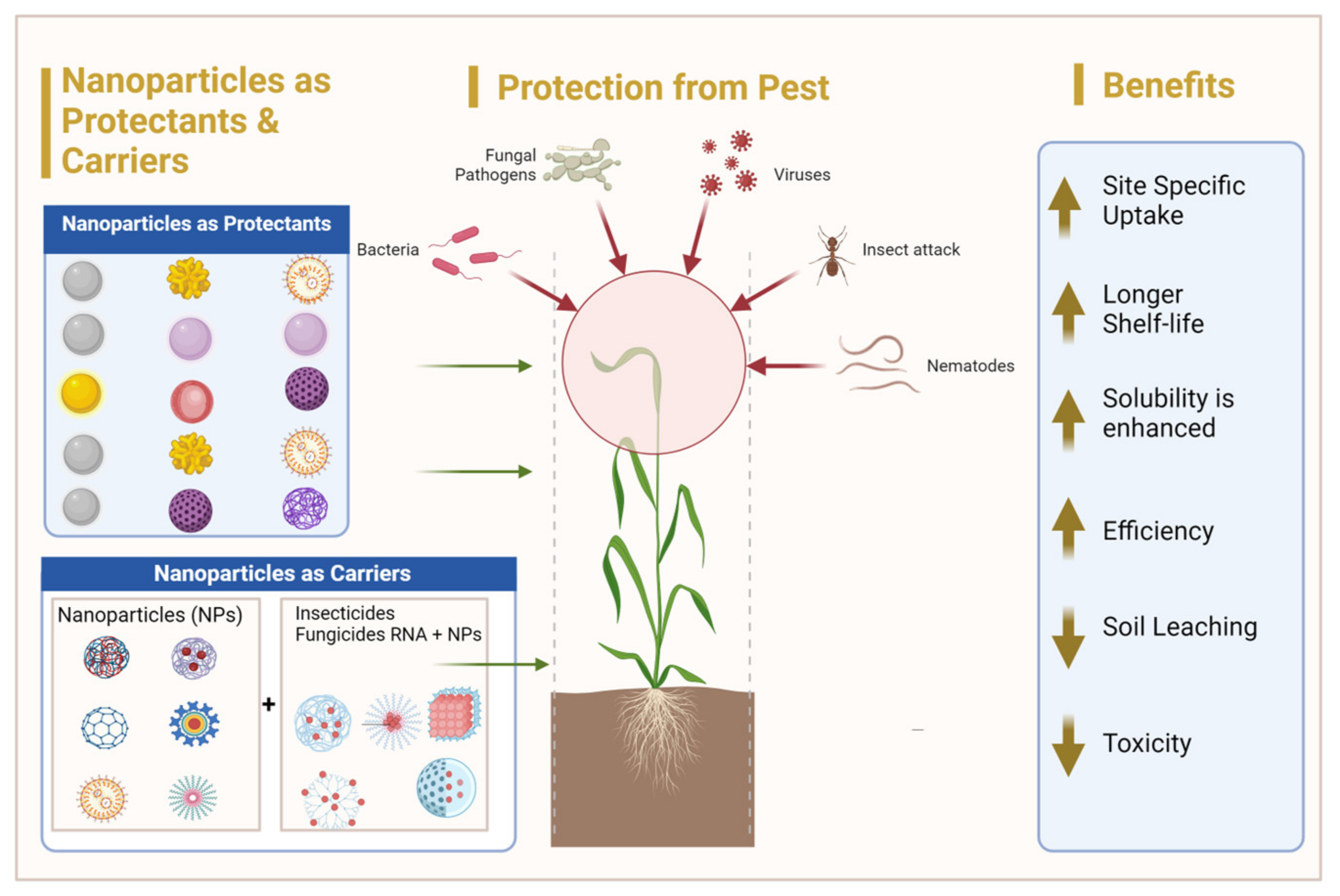
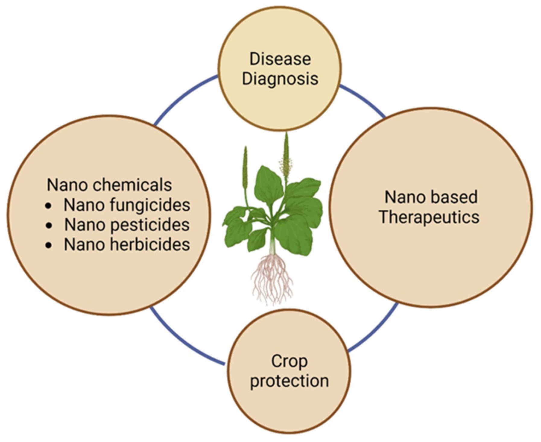
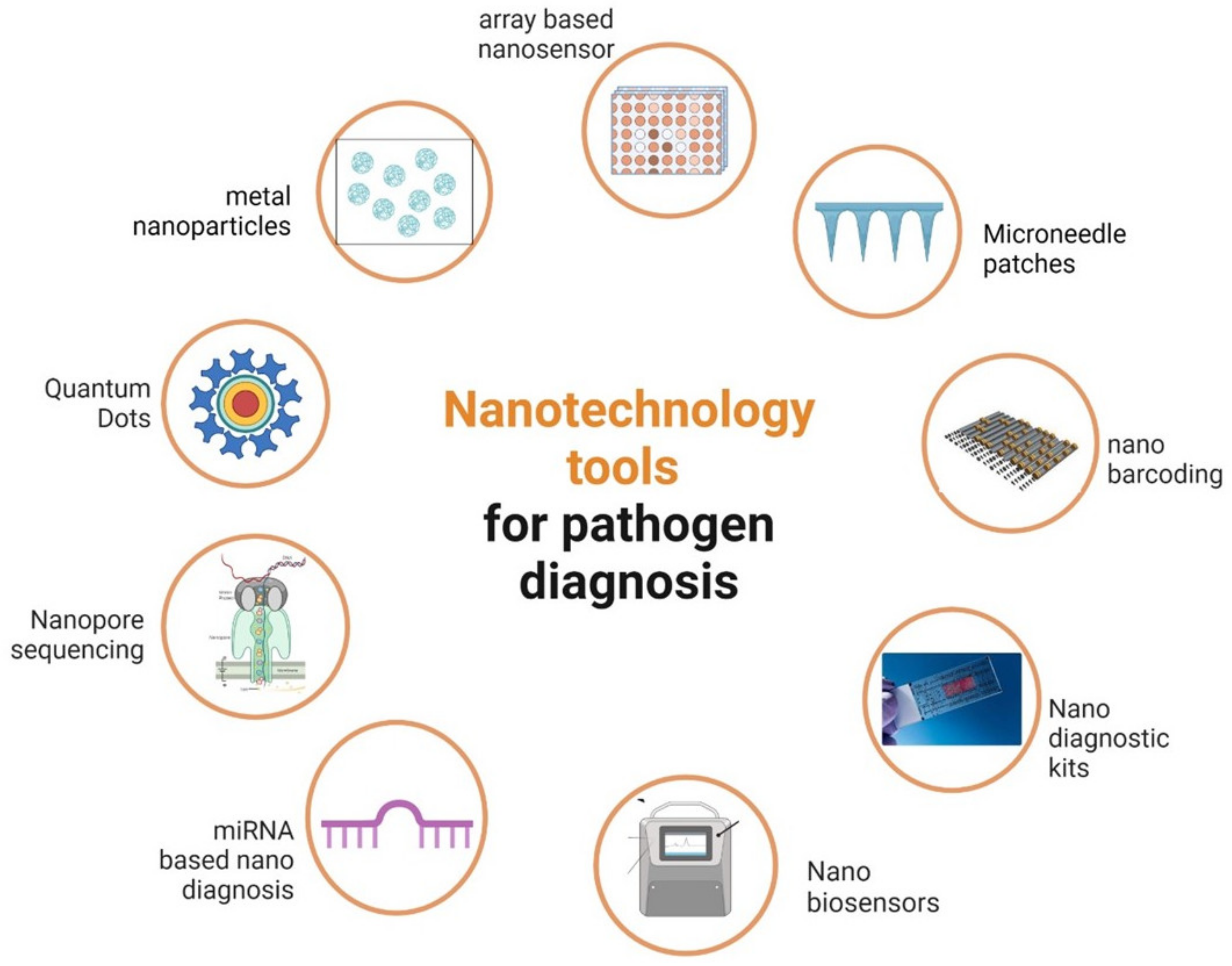
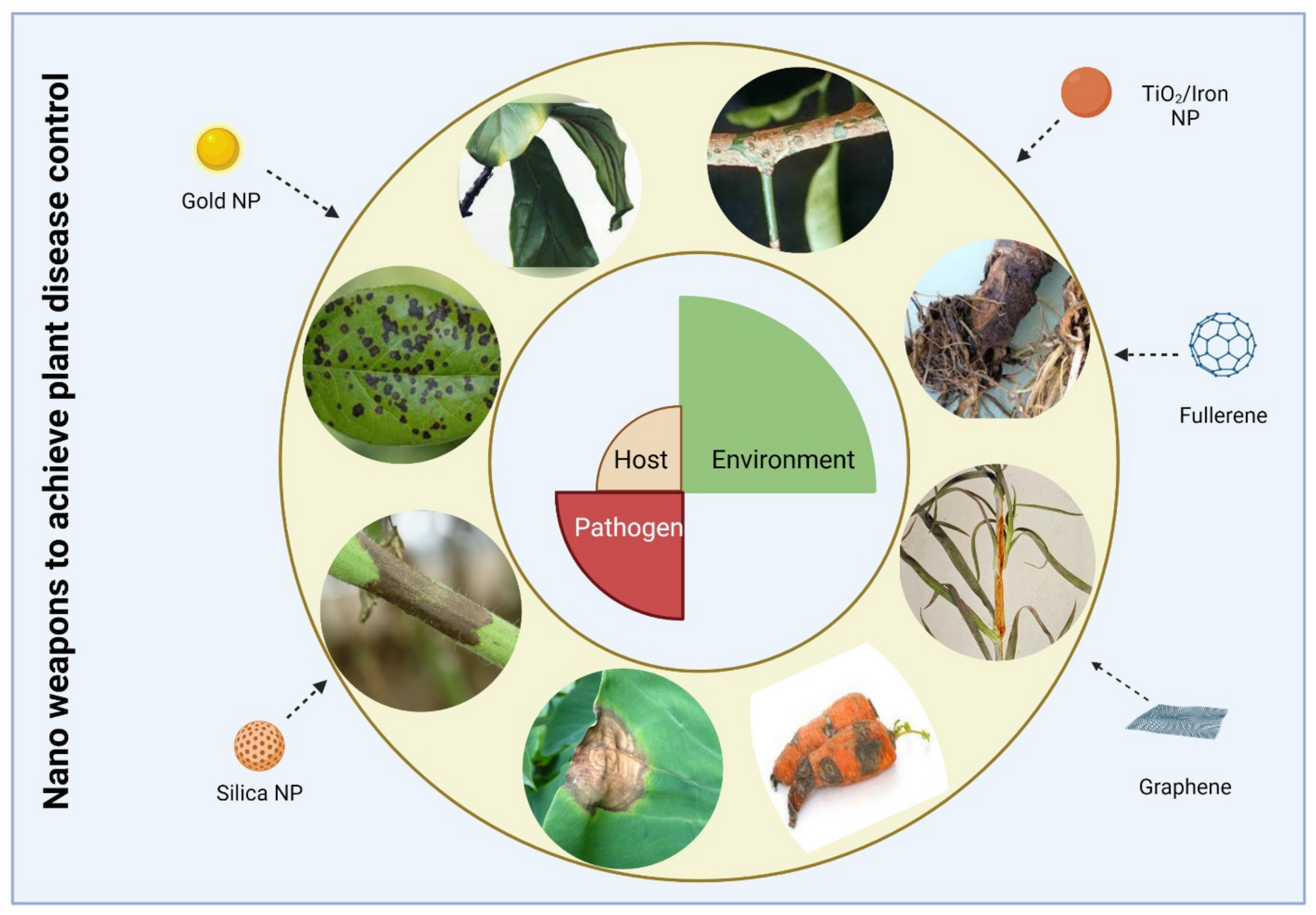
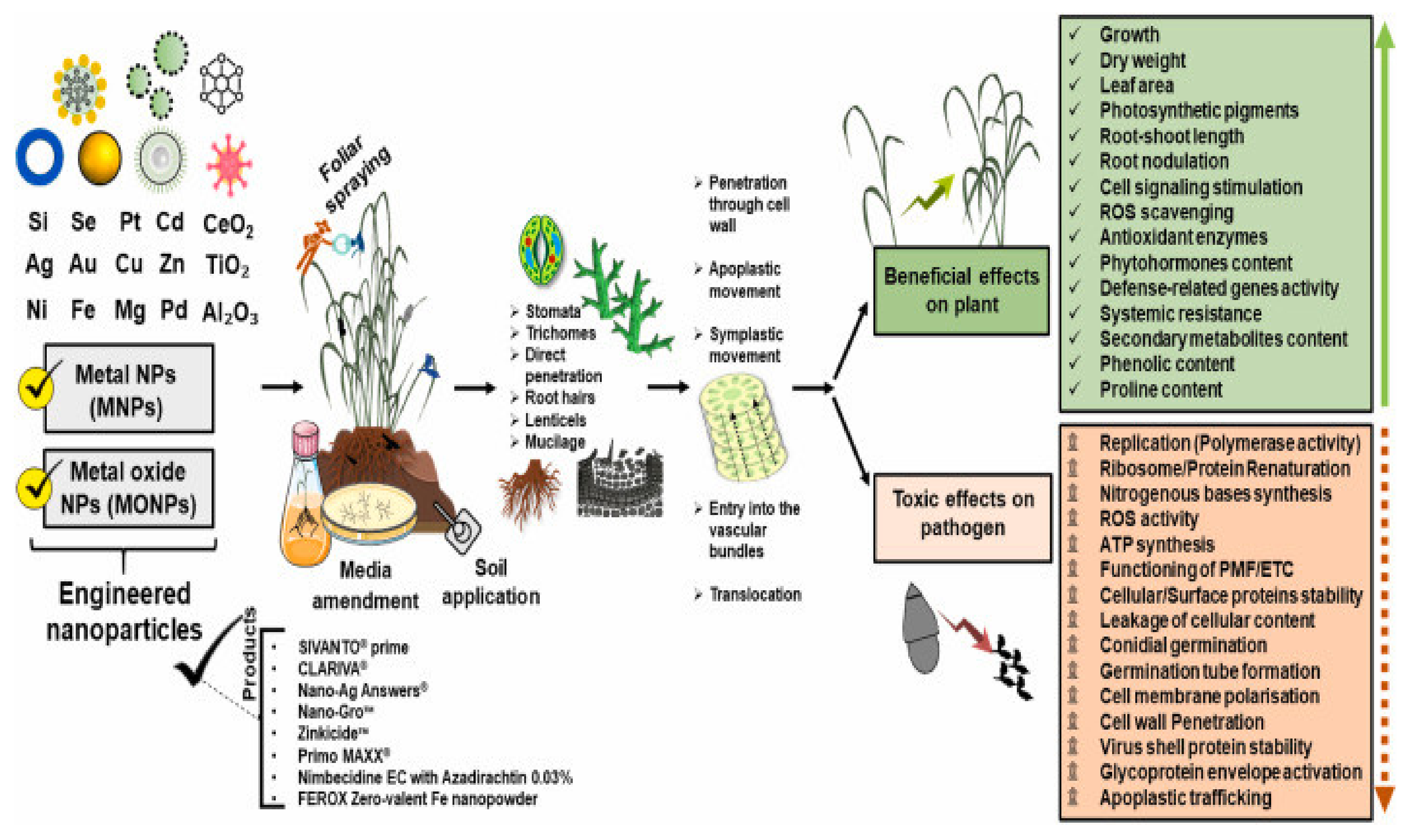
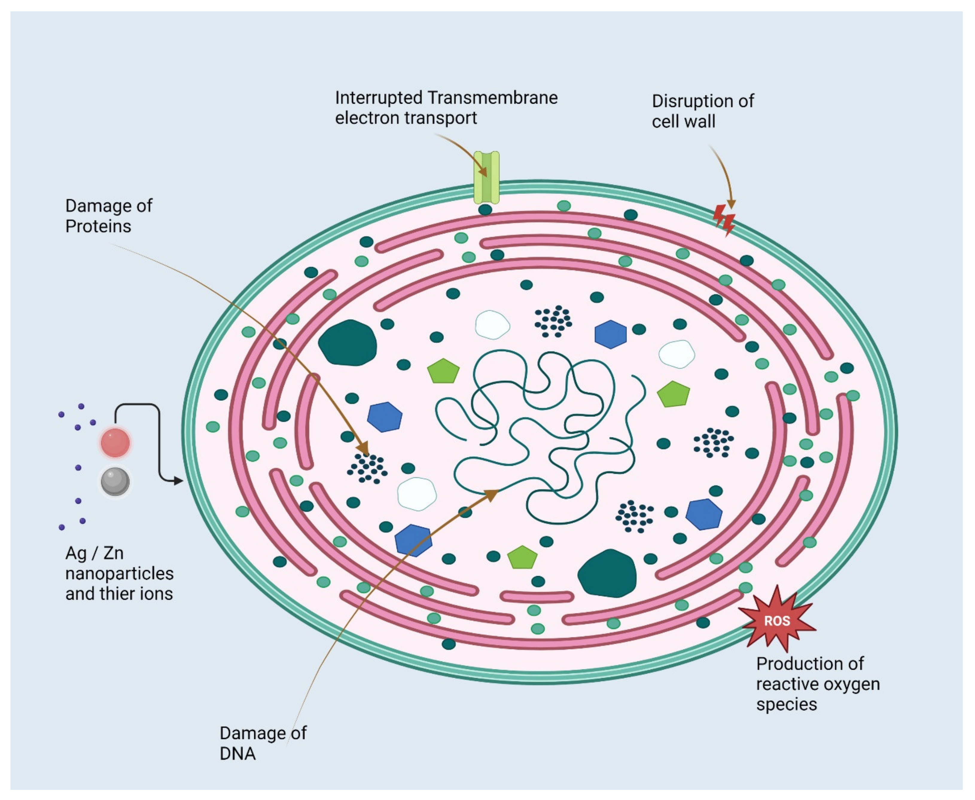
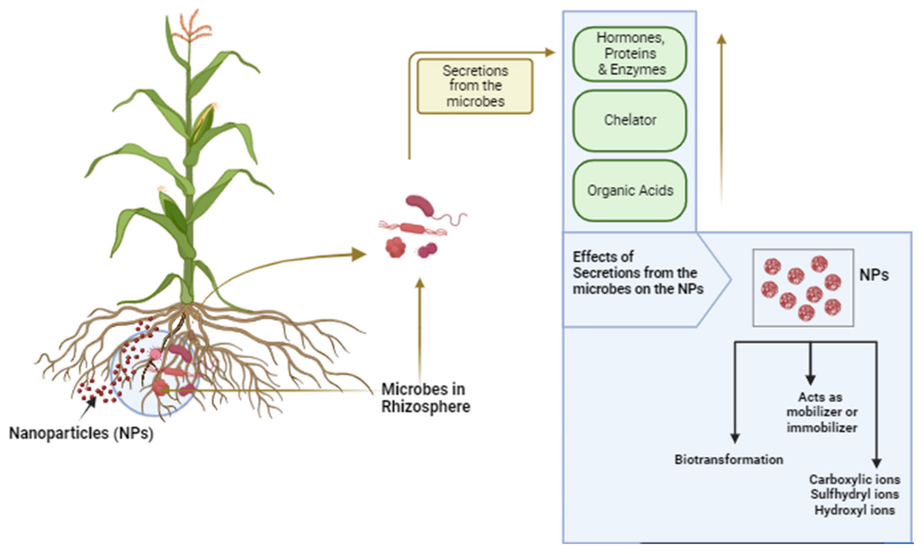
| Nanomaterial/ Substrate | Disease/Causal Organism | Target Plants | Bio-Recognition | Detection, LOD/Accuracy | References |
|---|---|---|---|---|---|
| SWCNTs/Gold microelectrodes on a Si/SiO2 wafer | Sec-delivered effector 1 (SDE1) | Citrus | Anti-SDE1 polyclonal | FET/LOD: 5 nM | [53] |
| Au and pentacene films/Gold gate electrode | PPV | Stone fruit trees | Anti-Plum Pox Virus polyclonal | EGOFET/LOD: 180 pg mL−1 | [54] |
| SWCNTs/Silicon wafer covered with SiO2 | p-Ethylphenol released by Phytophthora | Strawberry | ssDNA | E-nose 0.13% of Pethylphenol | [55] |
| N- and B-doped MWCNTs/Interdigitated Electroless nickel immersion gold electrodes | VOCs exhaled by Aspergillus and Rhizopus fungi | Strawberry | - | E-nose | [56] |
| rGO and Au NPs/ Kirigami-based structure with AgNW electrodes | VOCs exhaled by Phytophthora infestans infection | Tomato | Chemiresistive sensor array/ >97% accuracy | [57] | |
| Au NPs/GCE | Xanthomonas axonopodis | Citrus | Anti-PthA | FET-SWV/LOD: 0.01 nM | [58] |
| Au NPs/SPCE | CTV | Citrus | Thiolated ssDNA | EIS/LOD: 100 nM | [59] |
| Au NPs/SPCE | CTV detection | - | Thiolated primer | EIS L/OD: 1 pg μL−1 | [60] |
| TiO2 and SnO2 nanoparticles/SPCE | p-ethylguaiacol, volatile compound due to Phytophthora cactorum fungus infection | CV and DPV/LOD: 35–62 nmol L−1 | [61] | ||
| Au NPs/GCE | PSS | Stewartia | HRP | LSV/7.8 × 103 cfu mL−1 | [62] |
| GO/Paper electrodes | False smut caused by Ustilaginoidea virens | Rice | ssDNA | CV and LSV/10 fmol L−1 | [63] |
| GO/ITO | GBNV | Groundnut | anti-GBNV | DPV/LOD: 5.7 ng mL−1 | [64] |
| Au NPs/SPCE | Detection of plant pathogen DNA | - | Recombinase polymerase amplification | DPV/214 pmol L−1 | [65] |
| PPY nanoribbon/Gold microelectrode | Cucumber mosaic virus (CMV) | Cucumber | anti-CMV IgG | Chemiresistive Microelectrode/LOD 10 ng ml−1 | [66] |
| Au NPs/SPCE | CMV | Cucumber | - | Chronoamperometry | [67] |
| Au NPs/SPCE | Rice tungro disease | Rice | anti- RTBV/ RTSV | Cyclic voltammtry | [68] |
| Au NPs/Carbon ink 8-WE SPCE | CTV | Sweet orange trees | anti-bodies Ab1 and Ab2 | Amperometry/LOD: 0.3 fg mL−1 | [69] |
| Au NPs | P. infestans | Tomato | (Cys)-capped | C/0.4 ppm | [70] |
| Au NPs | Xanthomonas campestris | Brassica | Colorimetric/102 CFU mL−1 | [71] | |
| Fluorescent nanoparticles | Phakopsora Pachyrhizi | Soybean | IgG antibodies | fluorescence 2.2 ng mL−1 | [72] |
| Au NPs reverse primer (20-mer) | Yellow leaf curl virus | Tomato | Reverse primer (20- mer) | LSPR/5 ng μL−1 | [73] |
| Au NPs-SA | Alternaria panax Whetz | Ginseng | Mouse anti-Fam antibody and BSA-Biotin | LFA/0.01 pg μL−1 | [74] |
| Au NPs | Phytophthora infestan | Potato | Streptavidinbiotinylated T and C | LFA/0.01 pg μL−1 | [75] |
| Au NPs and silver | Leafroll virus | Potato | Anti-PLRV antibodies | LFA 0.2 ng ml−1 | [76] |
| Carbon nanoparticles | X. arboricola pv. Pruni | Stone fruits and almond | Polyclonal antibodies 2626.1-WC | LFA 104 CFU mL−1 | [77] |
| Nanoparticles | Causal Organism | Disease | Target Plants | Sources |
|---|---|---|---|---|
| Bacteria | ||||
| Ag | Pectobacterium carotovorum | Soft rot | Beta vulgaris | [186] |
| Ag | Xanthomonas oryzae pv. oryzae | Bacterial leaf streak | Oryza sativa | [187] |
| Ag | X. oryzae pv. oryzae | Rice bacterial blight | O. sativa | [188] |
| Ag | Acidovorax oryzae | BBS of rice | O. sativa | [189] |
| Ag | Erwinia cacticida | Soft rot erwinias | Citrullus lanatus | [190] |
| Ag | A. oryzae | BBS of rice | O. sativa | [191] |
| Ag | Ralstonia solanacearum | Bacterial wilt | N. tabacum | [192] |
| MgO | Xanthomonas oryzae | Bacterial blight disease in rice | O. sativa | [193] |
| TiO2 | Dickeya dadantii | Bacterial root rot | Ipomoea batatas | [194] |
| TiO2 | Dickeya dadantii | Soft rot | Ipomoea batatas | [195] |
| ZnO | Xanthomonas oryzae | Bacterial leaf blight diseases of rice | O. sativa | [196] |
| Fungi | ||||
| Ag | Sclerotium rolfsii | Collar rot | Cicer arietinum | [197] |
| Ag | Fusarium oxysporum | Fusarium wilt | Cicer arietinum | [187] |
| Ag | Phytophthora arenaria | Crown and root rot | Solanum lycopersicum | [198] |
| Ag | Sclerotinia sclerotiorum | Head rot | Brassica oleracea var. capitata | [199] |
| Ag | Rhizoctonia solani | Sheath blight | O. sativa | [200] |
| Ag | S. sclerotiorum | Gray mold | Fragaria ananassa | [201] |
| Ag | Rhizopus stolonifer | Discoloration of seed coat | Hordeum vulgare | [202] |
| Ag | Phytophthora parasitica | Black shank disease | Citrus limon | [203] |
| Ag | F. oxysporum | Fusarium wilt | Gossypium hirsutism | [204] |
| Ag | F. oxysporum | Black mold | S. lycopersicum | [205] |
| Au | Puccinia graminis | Wheat stem rust | T. aestivum | [206] |
| CS | F. graminearum | Seedling root rot | Triticum aestivum | [207] |
| Cu | Aspergillus niger | Black mold | Allium cepa | [208] |
| Cu | Poria hypolateritia | Red root | Camellia sinensis | [209] |
| Cu | Penicillin digitatum | Green rot | Citrus sinensis | [210] |
| Cu | Botrytis cinerea | Gray mold | Vitis vinefera | [148] |
| Cu | A. niger | Leaf rot | Eugenia caryophyllata | [150] |
| CuO | Pythium ultimum | Pink rot | Solanum tuberosum | [149] |
| CuO | Rhizoctonia solani | Root rot | Solanum lycopersicum | [211] |
| MgO | A. oryzae | BBS | O. sativa | [212] |
| MnO2 | A. oryzae | BBS | O. sativa | [212] |
| Ni | Colletotrichum musae | Crown rot | Musa acuminata | [213] |
| Se | Bipolaris sorokiniana | Black point disease | T. aestivum | [214] |
| Se | C. capsici | Die back rot | Capsicum annuum | [215] |
| SiO2 | Aspergillus flavus | Ear rot | Zea mays | [216] |
| Thiosemicarbazone NPs | A. flavus | Aspergillus ear rot | Zea mays | [217] |
| TiO2 | Bipolaris sorghicola | Target leaf spot | Sorghum bicolor | [218] |
| TiO2 | B. sorokiniana | Spot blotch disease | T. aestivum | [183] |
| TiO2 | B. sorokiniana | Spot blotch disease | T. aestivum | [183] |
| ZnO | F. oxysporum | Vascular wilt | Solanum lycopersicum | [219] |
| ZnO | F. culmorum | Fusarium wilt | Hordeum vulgare | [220] |
| Viruses | ||||
| Ag | SHRV | Chlorotic spot disease | Cyamopsis tetragonoloba | [140] |
| Ag | BYMV | MD | Vicia faba | [141] |
| Ag | Pyricularia grisea | Rice blast | O. sativa | [221] |
| Ag | Cucumis sativus | Melon yellow spot | Raphanus sativus | [222] |
| Ag | TSWV | Spotted wilt disease | S. tuberosum | [223] |
| Ag | PVY | Potato tuber necrotic ringspot disease | S. tuberosum | [224] |
| Au | BYMV | MD | Hordeum vulgare | [225] |
| Au | BYMV | Yellow mosaic disease | Hordeum vulgare | [226] |
| CeO2 | MTV | MD | Datura stramonium and N. tabacum | [227] |
| CNTs | MTV | MD | N. benthamiana | [228] |
| Fe3O4 | MTV | MD | N. benthamiana | [229] |
| GO-Ag | TBSV | Bushy stunt disease | Lactuca sativa | [230] |
| NiO | CMV | MD | Cucumis sativus | [231] |
| Schiff-based silver NPs | MTV | MD | N. benthamiana | [232] |
| SiO2 | MTV | MD | Nicotiana tabacum | [229] |
| SiO2 | TYLCV | Tomato leaf curl disease | S. lycopersicum | [224] |
| TiO2 | BBSV | Mottle/mosaic disease | Vicia faba | [233] |
| ZnO | MTV | MD | N. benthamiana | [229] |
| Nanoparticles | Concentration | Microbes | Types of Microbes | Impact | Sources |
|---|---|---|---|---|---|
| Ag | NR | Av | Bacteria | Biological nitrogen fixation was inhibited, ROS were produced, and cell number was reduced after exposure to NPs treated with Ag (size: 10 to 50 nm). | [294] |
| Ag NPs | 800 μg/kg sandy soil-loam mixture | Ga Faba bean | Fungi | Decreased glomalin levels, mycorrhizal responsiveness, and mycorrhizal colonization | [295] |
| Ag NPs | 12–36 mg/kg soil | AMF-Tomato | Fungi | Ag NPs reduce AMF colonization in a dose-dependent fashion. | [296] |
| Ag NPs and Fe2O3 NPs | 0.01–1 mg/kg Ag NPs and 0.032–3.2 mg/kg Fe2O3 NPs | Tr (Mycorrhizal clover) | Fungi | Mycorrhizal clover biomass drastically decreased, as did AMF’s ability to absorb nutrients from its roots and its glomalin concentration. | [297] |
| Ag NPs and FeO NPs | Ag NPs (0.1–10 mg/kg) | Soil-microbial activity | Bacteria | FeO NPs had a favorable impact on the soil’s microbial metabolism and nitrification capacity because they decreased the amount of ammonia-oxidizing bacteria in the soil and their abundance. | [298] |
| CuO NPs | Nitrifying soil microbes | Bacteria | CuO NPs reduced the rate of nitrification. | [299] | |
| CuO NPs | NR | Soil microorganisms engaged in C and N cycles | Bacteria | The amount of microbial activity related to C and N cycling was greatly reduced. CuO NPs’ toxicity to microorganisms was not reduced by the experimental plant varieties, including wheat. | [300] |
| CuO NPs | Dv (Sulfate reducing bacterium) | Bacteria | Sulfate reduction was inhibited by CuO NPs’ catabolic and anabolic activity, while respiratory and electron transport genes were suppressed. | [301] | |
| MoO3 | 10 and 200 mg/L | Af and An | Fungi | Apoptosis was caused by metabolic alterations, changes in hyphae shape, and nuclear condensation, all brought on by exposure to NPs. | [302] |
| Nanodiamonds | 0.01–1 mg/mL | Pc | Fungi | Hyphal death, cell wall degradation, loss of cytoplasm, and oxidative stress-induced laccase and manganese peroxidase inactivation | [303] |
| Cirate-Ag NPs, bare-ZnO NPs, bare-CuO NPs, and bare-TiO2 NPs | 100 mg/kg Ag NPs | Enzyme activity and the make-up of soil microbes | Bacteria | Particulate or dissolved application of Ag NPs inhibited specific soil enzymes, and the greater dose of Ag NPs altered the soil’s microbial population. Enzyme activity was marginally suppressed by TiO2 NPs. | [304] |
| PVP-coated Ag NPs | 10 and 100 μg/g Ag NPs | Ammonia-oxidizing bacteria | Bacteria | Soil nitrification and urease activities were drastically reduced. | [305] |
| TiO2 NPs | 8, 16, and 33 mg/kg sandy soil | a AMF-Rice group | Fungi | Rice symbiosis with AMF is inhibited. | [306] |
| ZVI NPs | 50 mg/L | Paracoccus sp. | Bacteria | Increased cell proliferation and NO3 biodegradation; decreased cell density in a dose-dependent manner. | [307] |
| ZVS NPs: PVA and Na2 ATP-doped | NR | Ne | Bacteria | Cell wall damage, nuclear fragmentation, oxidized NH3 capping, and size dependence | [308] |
| ZnO NPs | 30, 300, and 3000 ng/L | Marine and freshwater microcosms | Fungi | Affects the structure and activity of fungal communities and microorganisms negatively. | [309] |
| ZnO NPs | 6, 9, and 12 mmol/L | Mc and Colletotrichum sp. | Fungi | Fungal growth is inhibited by as much as 97% for M. citricolor and as much as 93% for Colletotrichum sp. | [310] |
| ZnO NPs | 500 mg/kg soil | Fm—Maize | Fungi | Negative impact on the AMF association | [311] |
| ZnO NPs | 800–3200 mg/kg loamy soil | Gv—Maize | Fungi | ZnO NPs disrupted the symbiosis of AMFs. | [312] |
Disclaimer/Publisher’s Note: The statements, opinions and data contained in all publications are solely those of the individual author(s) and contributor(s) and not of MDPI and/or the editor(s). MDPI and/or the editor(s) disclaim responsibility for any injury to people or property resulting from any ideas, methods, instructions or products referred to in the content. |
© 2023 by the authors. Licensee MDPI, Basel, Switzerland. This article is an open access article distributed under the terms and conditions of the Creative Commons Attribution (CC BY) license (https://creativecommons.org/licenses/by/4.0/).
Share and Cite
Ray, M.K.; Mishra, A.K.; Mohanta, Y.K.; Mahanta, S.; Chakrabartty, I.; Kungwani, N.A.; Avula, S.K.; Panda, J.; Pudake, R.N. Nanotechnology as a Promising Tool against Phytopathogens: A Futuristic Approach to Agriculture. Agriculture 2023, 13, 1856. https://doi.org/10.3390/agriculture13091856
Ray MK, Mishra AK, Mohanta YK, Mahanta S, Chakrabartty I, Kungwani NA, Avula SK, Panda J, Pudake RN. Nanotechnology as a Promising Tool against Phytopathogens: A Futuristic Approach to Agriculture. Agriculture. 2023; 13(9):1856. https://doi.org/10.3390/agriculture13091856
Chicago/Turabian StyleRay, Manjit Kumar, Awdhesh Kumar Mishra, Yugal Kishore Mohanta, Saurov Mahanta, Ishani Chakrabartty, Neelam Amit Kungwani, Satya Kumar Avula, Jibanjyoti Panda, and Ramesh Namdeo Pudake. 2023. "Nanotechnology as a Promising Tool against Phytopathogens: A Futuristic Approach to Agriculture" Agriculture 13, no. 9: 1856. https://doi.org/10.3390/agriculture13091856