Article

Use of Gas Chromatography and SPME Extraction for the Differentiation between Healthy and Paenibacillus larvae Infected Colonies of Bee Brood—Preliminary Research

1
Institute of Biology and Biotechnology, College of Natural Sciences, University of Rzeszow, Pigonia 1, 35-310 Rzeszow, Poland
2
Interdisciplinary Center for Preclinical and Clinical Research, University of Rzeszow, Werynia 2, 36-100 Kolbuszowa, Poland
3
Department of Ecotoxicology, Faculty of Biotechnology, University of Rzeszów, Rejtana 16c, 35-959 Rzeszow, Poland
4
Interdisciplinary Centre for Computational Modelling, College of Natural Sciences, University of Rzeszow, Pigonia 1, 35-310 Rzeszow, Poland
5
Institute of Material Engineering, College of Natural Sciences, University of Rzeszow, Pigonia 1, 35-310 Rzeszow, Poland
6
Institute of Agricultural Sciences, College of Natural Sciences, University of Rzeszow, Zelwerowicza 8b, 35-601 Rzeszow, Poland
*
Author to whom correspondence should be addressed.
Agriculture 2023, 13(2), 487; https://doi.org/10.3390/agriculture13020487
Submission received: 3 December 2022 / Revised: 14 February 2023 / Accepted: 15 February 2023 / Published: 18 February 2023
(This article belongs to the Special Issue The Pollinators in Agricultural Ecosystems)

Abstract

:
Paenibacillus larvae is a deadly pathogen for bee brood, which can lead to the death of entire colonies. The presence of specific volatile organic compounds (VOCs) in the hive may be related to the occurrence of this bacterium in brood. Compositions of those volatile fractions present in healthy brood from control colonies and the brood without symptoms of infection collected from the colonies infected by P. larvae were compared using gas chromatography coupled with mass spectrometry (GC-MS) and solid phase microextraction (SPME). Among the seven compounds detected and quantified, the relative concentrations of 3-carene and limonene significantly differentiated the brood from healthy and infected colonies. Based on the ratio analysis, the samples were differentiated in terms of the number of emitted VOCs.

Graphical Abstract

1. Introduction

Honey bee (Apis mellifera) colonies, unlike bumblebees [1] or wasps [2], live year-round. Thanks to this, bees can pollinate many species of early-flowering plants [3], including those used for economic purposes, from early spring, as soon as the thermal conditions outside the hive permit it. Although bumblebees are able to fly at much lower temperatures than bees [4], their small numbers in spring make the honey bee, not the bumblebee that tolerates low temperatures better, the dominant pollinator in this period. However, wintering in swarms has its drawbacks. One of them is that a virus, a parasite or a pathogen that has already entered colonies (whether as a result of the activity of the bees themselves, e.g., robberies of families, or as a result of incorrect beekeeping) remains in the bee colony forever (often until its death), because it has the ability to constantly spread between individuals [5]. Chronic diseases of bees include an infection whose causative agent is the spore-forming gram-positive bacterium Paenibacillus larvae. This small (2.5–5 µm by 0.5–0.8 µm), rod-shaped bacterium is able to form extremely tenacious endospores, which are the only infectious form of this organism [6]. This pathogen, which reproduces only in brood, even in brood-free periods such as winter, can stay in the nest for a long time—both in the digestive tract of bees and in the honey that bees feed on. This means that it will be able to infect brood when it reappears in the family [7].
P. larvae are responsible for the occurrence of American foulbrood disease (AFB) [6,8]. Taking into account the intensity of clinical symptoms of AFB infection in bee colonies and the longevity of spores, in some countries, including Poland, the lack of effective, currently acceptable methods of treatment (according to the European Commission Directive (37/2010 [9]), the use of antibiotics for the control of P. larvae infection in A. mellifera colonies is prohibited) and the economic losses related to the elimination of infected colonies from use, P. larvae is classified as a highly threatening organism for animal biosecurity [7].
Larvae become infected by swallowing spores that contaminate their food. During the first 12–36 h after hatching larvae are most susceptible to infection. The spores can come from young workers tending the brood. They can remain in their gastrointestinal tract for a long time, maintaining their full potential for infection [6]. They can also come from honey stored in the hive [10]. After the spores enter the digestive system of the brood, P. larvae bacteria begin to multiply, colonizing practically the entire middle intestine and, in the process of multiplication, displacing other genotypes of microorganisms inhabiting them. P. larvae have been found to produce several peptide antibiotics either through genes encoding for the synthesis of ribosomal peptides or through giant gene clusters encoding for enzyme complexes. As a result, active antimicrobial peptides or polyketides are synthesized in a non-ribosomal manner [11,12,13]. For the enterobacterial repetitive intergenic consensus type I and II (ERIC I and II) genotypes present in Europe, the time needed to kill all bee larvae in the colony is approx. 13 and approx. 7 days, respectively [7]. As indicated by Gliński and Jarosz [14] P. larvae is able to produce compounds with antibacterial activity. Once the intestine is full of P. larvae, thanks to their highly active proteases and chitin-degrading enzymes [15,16,17], they penetrate the epithelium of the middle intestine and cause an infection that kills the insect [18]. The very breach of the intestinal epithelium is equivalent to the death of the brood. Bacteria retain their activity long after the animal dies, transforming its body into a semi-liquid brown colloid with a very characteristic smell. This colloid, composed of a mixture of bacteria and P. larvae spores, is practically absent from other microorganisms usually involved in metabolizing dead organic matter [19]. Along with the drying process, more and more spores capable of infecting subsequent larvae appear in the colloid [20]. If the bees that keep the nest clean remove the dead brood in a colloidal form, containing a small amount of spores, there is a much lower chance of rapid AFB development in the colony than in the case of removing the dead, dried brood from under the caps containing only the infectious form of P. larvae. The dead brood removal rate is dependent on both the P. larvae genotype [21] (in the case of ERIC II, currently the most popular in Poland, 80 to 95% of the larvae die before the cells are sealed, which allows for their rapid removal by workers, in the case of ERIC I, 40 to 60% of the larvae die in the capped cells, which favors the formation of a significant number of sporangia [7]) and the genetically encoded hygienic behavior in bees [22], and the probability of survival of a colony infected with AFB increases with the growing number of genetically distinct males from which the mother obtained semen [23].
More and more studies indicate that many natural plant components are lethal to P. larvae (although these are mostly in vitro tests) [24,25,26]. Monocultures can restrict bees’ access to such compounds [27]. In addition, small reserves of pollen in the hive, i.e., a source of protein necessary for the development of young brood, and the placement of bee colonies in flowering monocultures for more than two or three weeks means that the embryogenesis of young specimens is often based on pollen from one species, which may cause deficiencies in certain components and the worse condition of the developing brood [28,29], which may reduce brood immunity, and thus may favor the development of pathogens such as P. larvae in bees intestines, especially in areas with a large number of bees (in Poland it is 2.21–10.8 bee colonies per km2, on average 5.57 [30]), where the transmission of the bacteria between bees occurs not only vertically from older to younger individuals within one colony, but also horizontally between colonies [31].
The presence of many compounds taken up by bees with food is manifested by the natural secretion of volatile organic compounds (VOCs) in the bee. There may be compounds with an antipathogenic effect, also against P. larvae [28]. The presence of AFB can also be determined by the VOC test. The markers of this disease are the presence of propionic and valeric acid [32,33], 2-nonanone ketone [32], isovaleric acid, butyric acid [33,34], 2-methyl-butanoic acid, hexanoic acid [33] 2-nonanone, dimethyl disulfide, dimethyl trisulfide, 2-undecanone [35], isobutenylcarbinol, acetamide, isobutyramide, butyramide, isovaleramide, caproamide, benzeneacetamide, anteisovaleric acid, caproic acid, methyl 3-methyl-2-oxopentanoate, 2,5-dimethylpyrazine, γ-caprolactone, dl-pantolactone and unknown: C5H10O2, C5H10O2, C5H11NO, and C6H11NO2 [34].
In the conducted research we tried to establish the composition of VOCs released by put to death by freezing A. mellifera brood collected from control and infected colonies. The aim of the study, therefore, was to check whether the composition and relative concentrations of respective VOCs released by brood collected from control colonies differ statistically significantly from those released by brood collected from colonies infected with AFB. The brood used for research from the infected colonies showed no signs of infection.

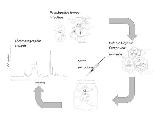
2. Material and Methods

2.1. Sampling

A total of 14 brood samples were collected from colonies infected with P. larvae (in each case clinical signs of infection, such as dead larvae with a specific brown color, a viscous consistency, and a characteristic smell of carpenter’s glue, arranged on the bottom of the cells, were found) and from 11 uninfected colonies (control ones). Only live larvae were collected each time. The collected brood was placed in a transport refrigerator (−4 °C), and within 40 min of collection, at the temperature of −20 °C. They were stored in these conditions until the extraction. The infected colonies (in each case the colonies were infected with the genotype ERIC II, dominant in Poland) came from the apiaries located in Kamionka (Ropczycko-Sędziszowski district), Domatków and Lipnica (Kolbuszowa district) and the city of Rzeszow (Rzeszow district), while the control colonies (in which no P. larvae was found) from the apiaries located in Werynia and Nowa Wieś (Kolbuszowa district), Nowiny (Stalowa Wola district) and Ostrów (Ropczycko-Sędziszowski district) (in each case, the Podkarpackie Voivodship). All apiaries were located in agricultural and forest areas composed of small areas of varied crops. The apiaries were at least 6 km apart (determined with the use of Google Maps [36]), which, assuming that 75% of bees forage within 1 km of the hive [37], significantly reduces the possibility of using the same food source by colonies. The colonies used for the study had however free contact with colonies from other apiaries and with wild bees (only in the case of registered bee colonies in the Podkarpackie District in 2019, there were 9.24 of them per 1 km2, which indicates the presence of too many A. mellifera families in this area; Central Statistical Office, 2021 [38]).
The samples were obtained in the period of May to September 2020, which ensured the dietary diversity of bees. Only the strong colonies with a large (>50,000) number of individuals on each frame in the nesting area and a large supply of food accumulated by bees in the hives (“strong” colonies were indicated by both veterinarians and social representatives of the beekeeping club during the valuation of apiaries intended for liquidation) were selected for the study (in the case of colonies with AFB infection, the strong colonies with clinical signs of P. larvae infection were selected). For formal and legal reasons (also related to the necessity to exterminate infected colonies and the ensuing payment of compensation for apiary owners) the presence or lack of P. larvae in the brood was confirmed, according to the GIWpr 02010-23/2016 method [39], each time in an accredited laboratory of the Veterinary Hygiene Department in Krosno (Podkarpackie Province, Poland). The presence of P. larvae was confirmed by Veterinary Services (potentially healthy third and fourth instar larvae were collected both from healthy colonies and from the colonies with clinical signs of infection). In the case of Eric II infection at this stage of development, the potential signs of AFB should already be visible [40]. No such changes were observed in any larvae from the colonies infected with AFB. Since we relied on the information obtained from Veterinary Inspections, and additionally we do not have a microbiologist on the team, we did not re-evaluate the samples to be analyzed by microbiological tests for P. larvae. We assessed the absence of P. larvae infection by chromatographic analysis. We did not detect the presence of AFB markers reported by other researchers [32,33,34,35], which indicates that the brood was not infected or the amount of bacteria present in it turned out to be too small to cause the symptoms of infection detectable with the methods we used. We also found no visual signs of infection which, in case of infection with ERIC II, should be visible. This means that there were probably no individuals infected with the bacterium in the prepared samples or the bacterium was present in the broods’ digestive tract below the limit of detection (<LOD) the methods we used. Oleic acid was also not found in the samples, which indicates the presence of a dead, decaying brood [32] that could occur after the death of brood before freezing due to a lethal bacterial infection of brood.
We did not analyze the volatile fraction secreted by the entire colony or the broods with clear signs of infection for the content of chemical markers of P. larvae’s presence.

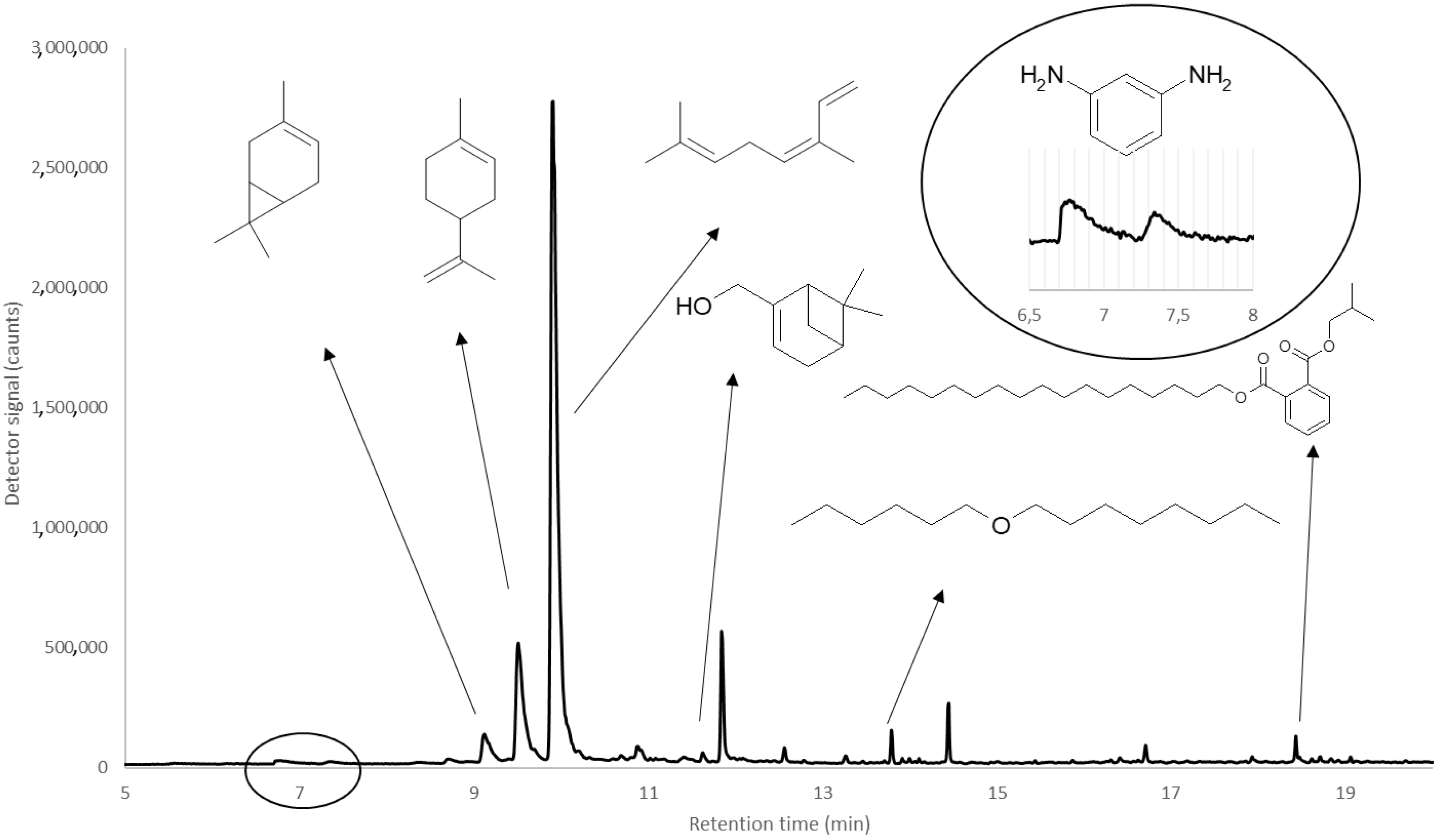
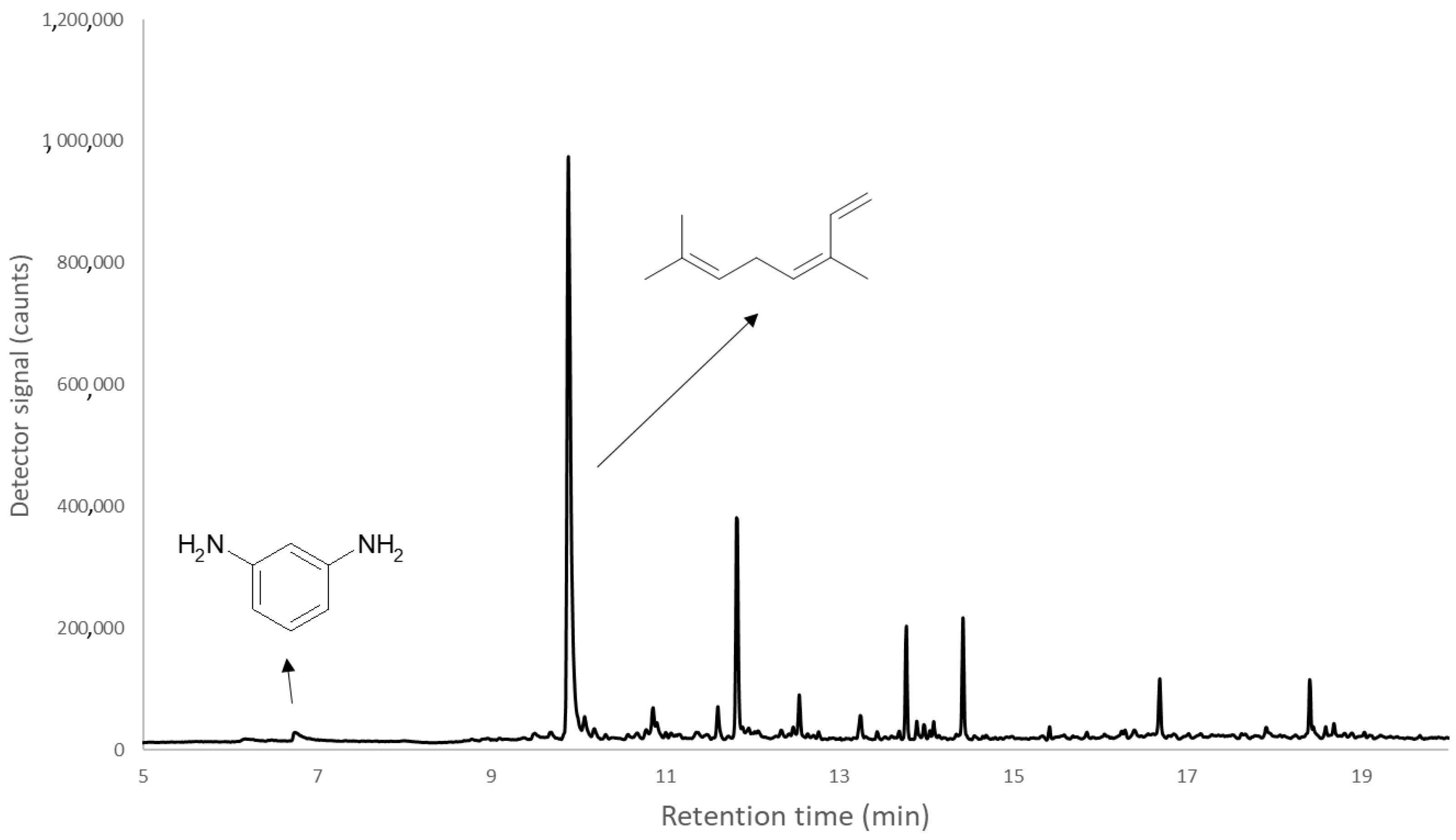
2.2. Extraction and Chromatographic Analysis of VOCs Emitted by Brood

The brood (25 individuals in each sample) were placed in a sealed 40 mL Erlenmeyer glass flask made of borosilicate glass (Glassco, Manglai, India) where the VOCs were released and then isolated and concentrated, on a surface of 100 µm of polydimethylsiloxane (PDMS) by Supelco Ltd. (Bellefonte, PA, USA) for 30 min at 45 °C (the flask with the sample and the SPME fiber placed on the sample was put in a water bath). The fiber used in research is dedicated to the analysis of samples with a low content of analytes (low pressure of VOCs). The same SPME fiber was used for all analyses. Then, the SPME fiber was introduced into a GC-MS dispenser (Varian 450 GC coupled with 240 MS, Palo Alto, California, USA. Software: Varian MS Data Review) at 250 °C for 5 min for desorption and determination.
The separation of VOCs was carried out on a capillary column with dimensions of 30 m × 0.25 mm × 0.25 μm with a moderately polar stationary phase HP-5 (Agilent Technologies, Inc., San-ta Clara, CA, USA), in the following temperature program: 50 °C and 5 min isotherm initially, then the temperature was increased at a rate of 10 °C per min to 300 °C, and finally, a 10 min isotherm was performed. The analysis was carried out for 35 min. The flow rate of the carrier gas (He) was 1 cm3 per min. The qualitative and quantitative composition of the VOCs profiles was established.
The content of identified compounds was expressed in % per standardized area. The relative share of the individual components of the VOCs profile containing between LOD and limit of quantification (LOQ) were accepted as trace levels (T). The LOQ is marked by Signal-to-noise ratio (slop sensitivity S/N 20) and based on minimum peak widths (4 s). The LOD was determined on the basis of peak size reject level (2000 counts) and probability factor (more than 90%) from NIST 08 database during the compound identification process. The compounds were identified on the basis of the MS spectrum library NIST 08 and calculated Kováts indexes (Table 1).

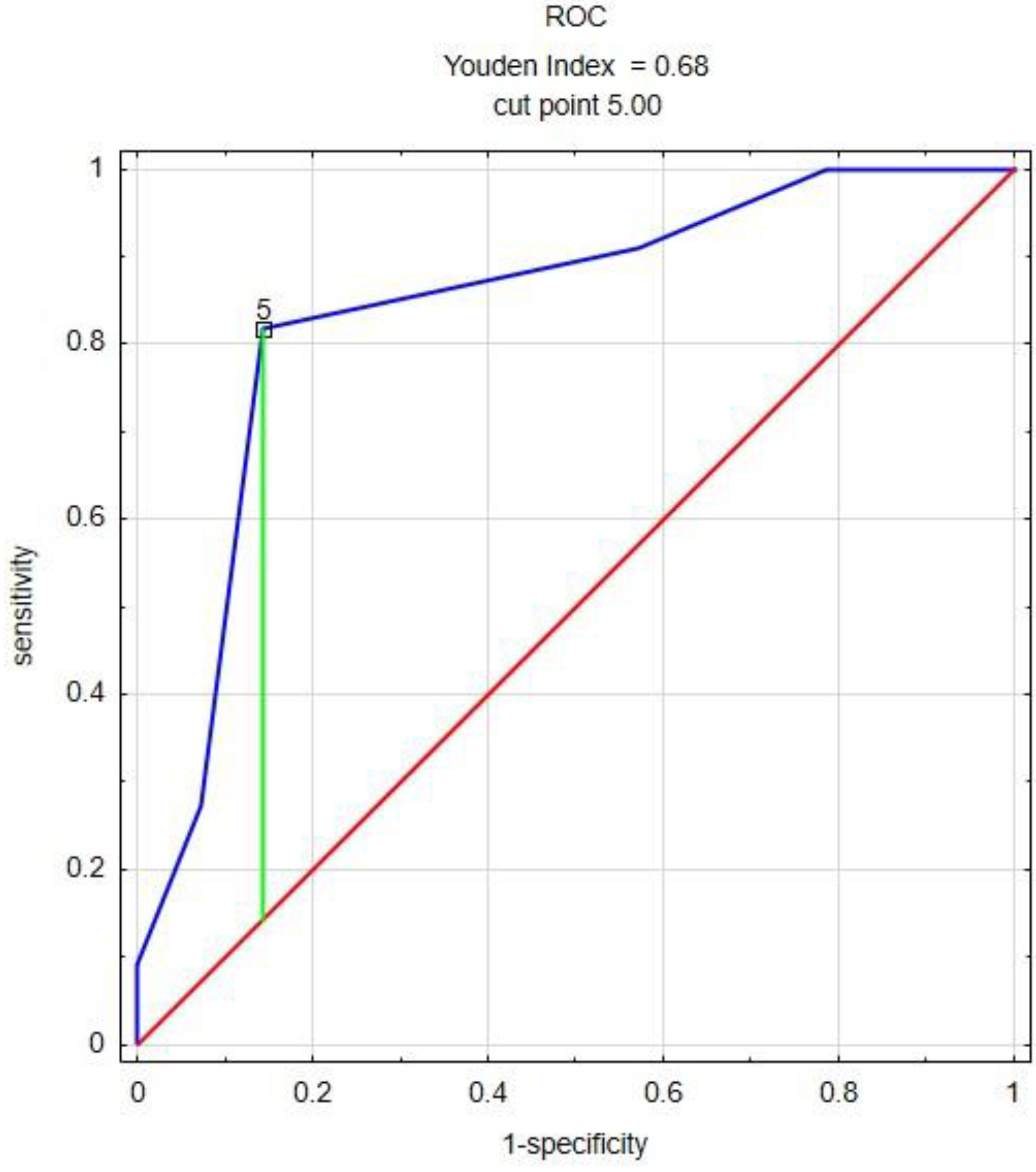
2.3. Statistical Analysis of the Obtained Results

The obtained results were analyzed statistically. Statistical analysis was performed with Statistica TIBCO 13.3 software (TIBCO Software Inc., Palo Alto, CA, USA) and R programme (version 3.6.1). The statistical significance of the differences between the quantitative feature in groups characterized by a binary variable (based on the presence or absence of VOCs) was assessed using the Mann-Whitney U test, receiver operating characteristic (ROC)—cut point analysis and the Od Ratio. The relationship between the qualitative characteristics was examined using the χ2 test, with corrections associated with, for example, the tables of dimension 2 × 2 (conditional probability was also used). Multiple correspondence analysis (MCA) was used to check the interdependence of qualitative variables.
The results with p-value lower than 0.05 were considered statistically significant.

3. Results

In the tested samples, seven volatile compounds were detected, 6 of them could be quantified and qualitatively determined, and one only qualitatively (Table 2, Figure 1 and Figure 2).
Since there are many missing percentage results in various preparations, in order to be able to draw conclusions with appropriate statistical power, the presence or absence of compounds was taken as binary values (qualitative analysis) (Table 3). The % data were classified as binary variables by creating a percentage table representing the odds of an event occurring provided that the VOC was present in the sample. The obtained results indicate that in the tested samples the presence of 3-carene and limonene statistically significantly differentiate samples from the colonies with known absence and confirmed presence of P. larvae infection.
Using the MCA analysis and conditional probability, we showed that in the absence of 3-carene or limonene (although in this case, these compounds have always co-existed) in the sample, the chance of AFB infection in the colony is 90.9%, in the presence of any of them, only 28.6%. These compounds in samples from control colonies were always in combination with hexyl octyl ether (although its presence was also found in many samples from colonies infected with P. larvae).
In order to test whether the amount of preparations used had a statistically significant effect on the occurrence or absence of foulbrood, a new ranking feature was created, being the sum of the preparations used in the sample. Finally, for the Mann-Whitney U test and the Ratio analysis, brood samples were divided for the evaluation of differences in total VOC content in brood samples from control colonies and those infected with P. larvae. The Mann–Whitney U test showed a significant difference in the number of VOCs between both types of samples (p = 0.003). Using the presence of AFB in the colonies from which the samples were taken as a stimulant, the cut-point = 5 (ROC analysis) was determined. The ratio analysis (infected brood: median = 4 VOCs, q1 = 3, q3 = 4; uninfected brood: median = 5 VOCs, q1 = 5, q3 = 6) indicates that in the case of 5 or more VOCs in the brood, the chance of AFB occurrence is 5.2 times lower than in the case if there were a smaller number of these compounds (p = 0.003).
3-Carene, limonene, ocimene, 2-pinen-10-ol, hexyl octyl ether, phthalic acid and isobutyl octadecyl ester were described with quantitative parameters (the results based on relative concentration [%] of VOCs) (Table 4). In order to check whether their relative concentrations were statistically significantly different, the Mann–Whitney U test was used.
As a result of this analysis, it was found that only two of the tested compounds statistically significantly differentiated the control and the groups infected with P. larvae (Table 5, Figure 3). These were the same compounds that distinguished the samples in the qualitative analysis: 3-carene and limonene. In both cases, higher values were found in the control group.
Finally, to perform the Od Ratio analysis, brood samples were divided into groups according to 3-carene and limonene content (group one: 3-carene <2.617 and limonene <9.5; group two: 3-carene ≥2.617 and limonene ≥9.5). For both VOC components, one case was found in group 3-carene <2.617 and limonene <9.5, respectively.

4. Discussion

The bee feeding range is 9.5 km from the nest [41]. However, if possible, bees forage in the immediate vicinity of the hives [37], which may impoverish the brood diet and thus negatively affect its health and development [42,43,44,45]. The youngest worker bee larvae are fed with royal jelly, which is a product of the hypopharyngeal glands of young adult worker bees, but its composition is also influenced by the current diet of worker bees producing this royal jelly, including pollen grains present in the royal jelly [46] and other components that may release VOCs. Royal jelly also contains active microorganisms such as bacteria: Clostridium botulinum, Bacillus cereus, Bacillus wakoensis and Micrococcus luteus, and fungi: Aspergillus niger, Penicillium sp. and Saccharomyces cerevisiae [47], that later colonize the intestinal flora of the brood. The digestive tract of bee larvae contains a microbiome consisting of a relatively small and cohesive set of bacteria [48]. Despite this, the limited access to microorganisms and brood diet (not only young but also older, eating mainly the food processed by workers) may affect the fact that only 7 VOCs were detected in the brood samples at >LOD concentration. This was significantly less than our adult bee study, where we found 21 VOCs (unpublished data). Another reason for the small number of VOCs in the samples may be the small number of samples (25 individuals/sample) and the fact that the tests were carried out on dead organisms, in which the course of natural metabolic processes and gas exchange was stopped. Probably for this reason, we did not detect any of the natural semiochemicals emitted by the brood in the samples to communicate with other hive inhabitants, such as palmitic acid, linoleic acid, linolenic acid, stearic acid, and oleic acid, [49], methyl palmitate, methyl oleate, methyl stearate, methyl linoleate, methyl linolenate, ethyl palmitate, ethyl oleate, ethyl stearate, ethyl linoleate, ethyl linolenate and E-β-ocymen [50,51], including those related to the activation of hygienic behavior in worker bees, such as (Z)-10-tritriacontene (Z10-C33), (Z)-8-hentriacontene (Z8-C31), (Z)-8-heptadecene (Z8- C17) and (Z)-6-pentadecene (Z6-C15) [52].
The results of the binary analysis confirm that tested samples containing more VOCs (≥5) have over 5 times greater chance for no infection with P. larvae than those with a smaller number of compounds. This may indicate a protective effect of either the VOCs themselves or the various microorganisms that secrete them. This is in line with previous research that indicates that if the brood’s diet is more varied, brood develops better [28,29]. The undifferentiated composition of the nectar and pollen brought by bees to the hives may result in the lack of exogenous chemical compounds that could potentially limit the development of P. larvae in A. mellifera colonies. Such antibacterial compounds, apart from royal jelly, are also present in propolis (a bee product used to seal the nest, characterized by antibacterial properties), which is lined with the cells in which the brood develops [28,53,54]. The greater the diversity of the diet, the greater chance of the presence of beneficial bacteria in the colony brought along with the feed to the colonies [55], including bacteria inhabiting the gastrointestinal tract and protecting it against infection with pathogens [56,57].
Sabaté et al. [58], indicate that compounds with bactericidal activity may additively and synergistically interact with each other, increasing their effectiveness in combating P. larvae, and the chances of such interactions will grow with an increase in the number of interacting compounds. It is possible that similar interactions occurred in these studies. In order to clearly state this, however, more detailed analyzes are required, in which a diverse selection of compounds and their concentrations would be used.
Of the seven compounds detected and assessed qualitatively (Table 2) and quantitatively (Table 4), it can be said that only limonene and 3-carene differentiate brood from foulbrood-infected colonies and from uninfected (control) brood. The presence of these compounds was detected in 10 out of 11 uninfected P. larvae samples and in only 4 out of 14 infected samples.
Limonene, a monocyclic monoterpene, just like m-pinene, is commonly found in nature and is produced by over 300 plants. Hence, it is a component of many essential oils of some plants (including citrus, fennel, caraway, walnut, pistachio, and celery) [59]. It is also produced by numerous microorganisms, including: Calothrix spp., Carnobacterium divergens, Nannocystis exedens, Phormidium spp., Plectonema spp., Pseudomonas fragi, Serratia proteamaculans, Streptomyces caviscabies, Tolypothrix distorta, Aspergillus flavus, Penicillium brevicompactum, Penicillium clavigerum, Penicillium commune, Trichoderma pseudokoningii and Saccharomyces cerevisiae [60] (given after other authors). Limonene, as we have already mentioned, occurs naturally in bacteria present in royal jelly [47]. This compound is also produced by bees themselves, as it is a component of bee venom [61]. This compound has been shown to have antifungal and antibacterial properties [62]. In vitro studies have shown that also the development of P. larvae is inhibited by limonene [63,64].
3-Carene, a dicyclic monoterpene, is a VOC released by some plants that gives them a specific smell. However, the occurrence of 3-carene in plants is very rare. In greater amounts, it is found only in Juniperus oxycedrus [65]. This may indicate the zoonotic origin of the compound detected in the samples. However, we did not find any information about its production by bees or brood. It is found in some species of termites, in which it acts as an alarm pheromone [66]. It is also attractive to bark beetles, such as Dendroctonus brevicomis and Ips paraconfusus, which use it to biosynthesize their own pheromones [67]. It can also be produced by microorganisms, such as Penicillium roqueforti [60]. In in vitro studies, the compounds such as limonene show activity against gram-positive and gram-negative bacteria [68,69].
We also detected compounds in the samples that could be of anthropogenic origin. Hexyl octyl ether is one of them. It has been shown that it is released into the environment under the influence of high temperatures [70]. At the same time, it has been shown that it is also naturally produced by Pseudomonas sp. bacteria (in the composition of VOCs with a bactericidal effect) [71] or also by Enterococcus faecalis [72]. It is also one of the metabolites in the process of natural fermentation [73]. Hexyl octyl ether is also found in plants, such as Ellettaria cardamomum [74], Strobilanthes crispus [75], Catha edulis [76], is very common also in Poland Solanum sp. [77] or Spinach oleracea [78].
Phthalates are used in a wide variety of products and applications with no chemical bound resulting in their migration to the environment [79]. Phthalic acid esters have been used as a plasticizer to improve the flexibility, adhesion, and solubility of polymers. They are present in cosmetics, pharmaceutical coatings, medical devices, food containers, paints, floors, and wall coverings. As a result, they are common in the environment from which they are taken up by the root system of plants [80]. In many studies describing the detection of phthalic acid, isobutyl octadecyl ester in plants, no anthropogenic origin of this compound is indicated [76,81,82,83,84]. As indicated by Shaheed et al. [81], Bhagat and Bhuktar [82] and Sharma [84] plants in which they detected the presence of phthalic acid, isobutyl octadecyl ester, were characterized by anti-microbial activity.
Based on the above information, it is difficult to determine both the route of transfer and the origin of the hexyl octyl ether and phthalic acid, isobutyl octadecyl ester found in the colony.
m-Phenylenediamine is produced by the reduction of metadinitrobenzene or nitroaniline with iron and hydrochloric acid. This compound is used to make diisocyanates for the production of polyurethane foams and other resins and polymers, used as a corrosion inhibitor, curing agent, photographic developer, and intermediate for other chemical products, also used to make dyes, to develop dyes, to dye hair, and to cure epoxy resins [85,86]. We have not found any information regarding the natural origin of m-phenylenediamine. Since it is a common ingredient in hair dyes, we can assume that it found its way into the colony as a result of bringing drinking water and cooling the nest by bees from reservoirs and watercourses contaminated with this ingredient. It could also be found in brood bodies indirectly as a result of contact of worker bees with microplastics or comes from parts of hives constructed from colored plastic [86].
Our research shows that VOCs can qualitatively and quantitatively differentiate between the brood from control colonies and the brood from colonies with the presence of P. larvae. At the same time, the chosen methodology does not allow one to draw unambiguous conclusions. Obtaining more specific data indicating whether the obtained results are related to the mere presence of specific compounds at a certain concentration, interactions between them, or other environmental factors requires further extensive laboratory research.

5. Conclusions

Using the SPME technique, the composition of VOCs released by bee brood collected from control colonies and infected with the P. larvae was determined.
Seven VOCs were found to be released from the samples tested: m-phenylenediamine, 3-carene, limonene, ocimene, 2-pinen-10-ol, hexyl octyl ether, and phthalic acid isobutyl octadecyl ester.
Statistical analysis of the obtained results of the chemical analyses showed that both the mere presence (above LOD) and the relatively high concentrations (above LOQ) of 3-carene and limonene statistically significantly distinguished uninfected brood from the brood infected with P. larvae.
The number of VOCs in the sample statistically differentiated bee brood from healthy colonies from those infected with P. larvae.

Author Contributions

The conception and design of the study—M.B. and B.P. Preparation of samples for analysis—M.B., A.K. (Aleksandra Kwiatek) and D.K. Chromatographic analysis—M.B. Analysis and interpretation of data—M.B., B.P. and L.Z. Drafting the article—B.P., A.K. (Anna Koziorowska) and S.S. Final approval of the version to be submitted—B.P., A.K. (Anna Koziorowska), S.S. and M.B. All authors have read and agreed to the published version of the manuscript.

Funding

The research was supported by the statutory fund of the College of Natural Sciences of the University of Rzeszow, Poland. Part of the research was supported by the project Interdisciplinary Center for Preclinical and Clinical Research (project number RPPK.01.01.00-18-0001/18).

Institutional Review Board Statement

The conducted research is not related to either human or animal use.

Data Availability Statement

The datasets generated during and/or analyzed during the current study are available from the corresponding or the first author on reasonable request.

Acknowledgments

We would like to thank Marek Olszowy from the District Veterinary Inspectorate in Kolbuszowa, Karolina Mróz from the District Veterinary Inspectorate in Rzeszow and Katarzyna Oleś-Bizoń from the District Veterinary Inspectorate in Ropczyce.

Conflicts of Interest

The authors declare no conflict of interest.

References

  1. Alford, D.V. A study of the hibernation of Bumblebees (Hymenoptera: Bombidae) in Southern England. J. Anim. Ecol. 1969, 38, 149–170. [Google Scholar] [CrossRef]
  2. Richards, O.W. The biology of the social wasps (Hymenoptera, Vespidae). Biol. Rev. 1971, 46, 483–528. [Google Scholar] [CrossRef]
  3. Herrera, C.M. Floral biology, microclimate, and pollination by ectothermic bees in an early-blooming herb. Ecology 1995, 76, 218–228. [Google Scholar] [CrossRef]
  4. Wahengbam, J.; Raut, A.M.; Satinder, P.; Banu, A.N. Role of bumble bee in pollination. Ann. Biol. 2019, 35, 290–295. [Google Scholar]
  5. Topolska, G.; Gajda, A.; Imińska, U. Atlas of Bee Diseases; PWRiL: Warsaw, Poland, 2018. (In Polish) [Google Scholar]
  6. Genersch, E. Paenibacillus larvae and American Foulbrood—long since known and still surprising. J. Verbr. Lebensm. 2008, 3, 429–434. [Google Scholar] [CrossRef]
  7. Schulz, M.; Łoś, A.; Skowronek, P.; Staniszewski, A.; Iwański, B.; Wójcik, Ł.; Strachecka, A. American foulbrood—A dangerous bacterial honey bee disease. J. Anim. Sci. Biol. Bioecon. 2018, 36, 35–47. [Google Scholar]
  8. Ebeling, J.; Knispel, H.; Hertlein, G.; Fünfhaus, A.; Genersch, E. Biology of Paenibacillus larvae, a deadly pathogen of honey bee larvae. Appl. Microbiol. Biotechnol. 2016, 100, 7387–7395. [Google Scholar] [CrossRef]
  9. No 37/2010; Commission Regulation (EU) No 37/2010 of 22 December 2009 on pharmacologically active substances and their classification regarding maximum residue limits in foodstuffs of animal origin (Text with EEA relevance). Commission Regulation (EU): Brussels, Belgium, 2009.
  10. Lindström, A.; Korpela, S.; Fries, I. The distribution of Paenibacillus larvae spores in adult bees and honey and larval mortality, following the addition of American foulbrood diseased brood or spore-contaminated honey in honey bee (Apis mellifera) colonies. J. Invertebr. Pathol. 2008, 99, 82–86. [Google Scholar] [CrossRef]
  11. Garcia-Gonzalez, E.; Müller, S.; Ensle, P.; Süssmuth, R.D.; Genersch, E. Elucidation of sevadicin, a novel nonribosomal peptide secondary metabolite produced by the honey bee pathogenic bacterium Paenibacillus larvae. Environ. Microbiol. 2014, 16, 1297–1309. [Google Scholar] [CrossRef] [PubMed]
  12. Sood, S.; Steinmetz, H.; Beims, B.; Mohr, K.I.; Stadler, M.; Djukic, M.; von der Ohe, W.; Steinert, M.; Daniel, R.; Müller, R. Panilarvins: Iturin family lipopeptides from the honey bee pathogen Paenibacillus larvae. Chem. BioChem. 2014, 15, 1947–1955. [Google Scholar]
  13. Müller, S.; Garcia-Gonzalez, E.; Genersch, E.; Süssmuth, R. Involvement of secondary metabolites in the pathogenesis of the American foulbrood of honey bees caused by Paenibacillus larvae. Nat. Prod. Rep. 2015, 32, 765–778. [Google Scholar] [CrossRef] [Green Version]
  14. Gliński, Z.; Jarosz, J. Does Bacillus larvae produce an antibacterial substance in infected honey bee larvae? Apidologie 1992, 23, 193–201. [Google Scholar] [CrossRef] [Green Version]
  15. Garcia-Gonzalez, E.; Poppinga, L.; Fünfhaus, A.; Hertlein, G.; Hedtke, K.; Jakubowska, A.; Genersch, E. Paenibacillus larvae chitindegrading protein PlCBP49 is a key virulence factor in American foulbrood of honey bees. PLoS Pathog. 2014, 10, e1004284. [Google Scholar] [CrossRef]
  16. Holst, E.C. A simple field test for American foulbrood. Amer. Bee 1946, 86, 34. [Google Scholar]
  17. Holst, E.C.; Sturtevant, A.P. Relation of proteolytic enzymes to phase of life cycle of Bacillus larvae, and two new culture media for this organism. J. Bacteriol. 1940, 40, 723–731. [Google Scholar] [CrossRef] [Green Version]
  18. Yue, D.; Nordhoff, M.; Wieler, L.H.; Genersch, E. Fluorescence in situ hybridization (FISH) analysis of the interactions between honeybee larvae and Paenibacillus larvae; the causative agent of American foulbrood of honeybees (Apis mellifera). Environ. Microbiol. 2008, 10, 1612–1620. [Google Scholar] [CrossRef]
  19. Bailey, L.; Ball, B.V. Honey Bee Pathology; Academic Press: New York, NY, USA, 1991. [Google Scholar]
  20. Genersch, E. American foulbrood in honeybees and its causative agent, Paenibacillus larvae. J. Invertebr. Pathol. 2010, 103, 10–19. [Google Scholar] [CrossRef]
  21. Ashiralieva, A.; Genersch, E. Reclassification; genotypes; and virulence of Paenibacillus larvae; the etiological agent of American foulbrood in honeybees—A review. Apidologie 2006, 37, 411–420. [Google Scholar] [CrossRef] [Green Version]
  22. Sherman, P.W.; Seeley, T.D.; Reeve, H.K. Parasites, pathogens, and polyandry in social Hymenoptera. Am. Nat. 1988, 131, 602–610. [Google Scholar] [CrossRef]
  23. Palmer, K.A.; Oldroyd, B.P. Evidence for intra-colonial genetic variance in resistance to American foulbrood of honey bees (Apis mellifera): Further support for the parasite/pathogen hypothesis for the evolution of polyandry. Naturwissenschaften 2003, 90, 265–268. [Google Scholar]
  24. Alonso-Salces, R.M.; Cugnata, N.M.; Guaspari, E.; Pellegrini, M.; Aubone, I.; De Piano, F.G.; Antúnez, K.; Fuselli, S.R. Natural strategies for the control of Paenibacillus larvae; the causative agent of American foulbrood in honey bees: A review. Apidologie 2017, 48, 387–400. [Google Scholar] [CrossRef]
  25. Dostálková, S.; Urajová, P.; Činčárová, D.; Vránová, T.; Hrouzek, P.; Petřivalský, M.; Lukeš, M.; Čapková-Frydrychová, R.; Danihlík, J. Fatty acids and their derivatives from Chlorella vulgaris extracts exhibit in vitro antimicrobial activity against the honey bee pathogen Paenibacillus larvae. J. Apic. Res. 2021, 1–13. [Google Scholar] [CrossRef]
  26. Tutun, H.; Koç, N.; Kart, A. Plant essential oils used against some bee diseases. Turk. J. Agric. Food Sci. Technol. 2018, 6, 34–45. [Google Scholar] [CrossRef] [Green Version]
  27. St Clair, A.L.; Zhang, G.; Dolezal, A.G.; O’Neal, M.E.; Toth, A.L. Diversified farming in a monoculture landscape: Effects on honey bee health and wild bee communities. Environ. Entomol. 2020, 49, 753–764. [Google Scholar] [CrossRef] [PubMed]
  28. Girard, M.; Chagnon, M.; Fournier, V. Pollen diversity collected by honey bees in the vicinity of Vaccinium spp. crops and its importance for colony development. Botany 2012, 90, 545–555. [Google Scholar] [CrossRef]
  29. Brodschneider, R.; Crailsheim, K. Nutrition and health in honey bees. Apidologie 2010, 41, 278–294. [Google Scholar] [CrossRef]
  30. Kobylińska, M. Regional diversity of beekeeping in Poland. Pol. Stat. 2021, 66, 25–38. (In Polish) [Google Scholar]
  31. Genersch, E.; Ashiralieva, A.; Fries, I. Strain-and genotype-specific differences in virulence of Paenibacillus larvae subsp. larvae, a bacterial pathogen causing American foulbrood disease in honeybees. Appl. Environ. Microbiol. 2005, 71, 7551–7555. [Google Scholar] [CrossRef] [Green Version]
  32. Lee, S.; Lim, S.; Choi, Y.S.; Lee, M.L.; Kwon, H.W. Volatile disease markers of American foulbrood-infected larvae in Apis mellifera. J. Insect Physiol. 2020, 122, 104040. [Google Scholar] [CrossRef]
  33. Gochnauer, T.A.; Shearer, D.A. Volatile acids from honeybee larvae infected with Bacillus larvae and from a culture of the organism. J. Apicul. Res. 1981, 20, 104–109. [Google Scholar] [CrossRef]
  34. Bikaun, J.M.; Bates, T.; Bollen, M.; Flematti, G.R.; Melonek, J.; Praveen, P.; Grassl, J. Volatile biomarkers for non-invasive detection of American foulbrood, a threat to honey bee pollination services. Sci. Total. Environ. 2022, 845, 157123. [Google Scholar] [CrossRef] [PubMed]
  35. Liolios, V.; Kanelis, D.; Tananaki, C.; Rodopoulou, M.A. A comparative study of healthy and american foulbrood-infected bee brood (Apis mellifera L.) through the investigation of volatile compounds. Agriculture 2022, 12, 812. [Google Scholar] [CrossRef]
  36. Google Maps. Available online: www.google.pl/maps/ (accessed on 20 October 2022).
  37. Apiculture Factsheet. Ministry of Agriculture, Food and Fisheries. Available online: https://www2.gov.bc.ca/gov/content/industry/agriculture-seafood/animals-and-crops/animal-production/bees (accessed on 20 January 2023).
  38. Central Statistical Office; The Polish Statistician. 2021. Available online: https://ws.stat.gov.pl/WS/2021/2/gus_ws_2021_02.pdf.7 (accessed on 11 November 2022).
  39. Instruction of the Chief Veterinary Officer. Instruction of the Chief Veterinary Officer No. GIWpr 02010-23/2016; Publishing House of the Chief Veterinary Inspectorate: Warsaw, Poland, 2016. (In Polish) [Google Scholar]
  40. Hansen, H.; Brodsgaard, C.J. American foulbrood: A review of its biology; diagnosis and control. Bee World 1999, 80, 195–201. [Google Scholar] [CrossRef]
  41. Beekman, M.; Ratnieks, F. Long-range foraging by the honey bee; Apis mellifera L. Funct. Ecol. 2000, 14, 490–496. [Google Scholar] [CrossRef] [Green Version]
  42. Crailsheim, K.; Stolberg, E. Influence of diet: Age and colony condition upon intestinal proteolytic activity and size of the hypopharyngeal glands in the honeybee (Apis mellifera L.). J. Insect Physiol. 1989, 35, 595–602. [Google Scholar] [CrossRef]
  43. Omar, E.; Abd-Ella, A.A.; Khodairy, M.M.; Moosbeckhofer, R.; Crailsheim, K.; Brodschneider, R. Influence of different pollen diets on the development of hypopharyngeal glands and size of acid gland sacs in caged honey bees (Apis mellifera). Apidologie 2017, 48, 425–436. [Google Scholar] [CrossRef] [Green Version]
  44. Barascou, L.; Sene, D.; Barraud, A.; Michez, D.; Lefebvre, V.; Medrzycki, P.; Di Prisco, G.; Strobl, V.; Yañez, O.; Neumann, P.; et al. Pollen nutrition fosters honeybee tolerance to pesticides. R. Soc. Open Sci. 2021, 8, 210818. [Google Scholar] [CrossRef]
  45. Lan, J.; Ding, G.; Ma, W.; Jiang, Y.; Huang, J. Pollen source affects development and behavioral preferences in honey bees. Insects 2021, 12, 130. [Google Scholar] [CrossRef]
  46. Piana, M.L.; Paola, B.; Livia, P.O.; Sara, P. Pollen analysis of royal jelly: Contribution to analytical methods and characterization. Apiacta 2006, 41, 28–43. [Google Scholar]
  47. Ashour, Z.A.; Ali, M.A.M.; Abdelmegeed, S.M.; Amin, K.M. Detection of microorganisms (bacteria, fungi and yeasts) in royal jelly. Arab Univ. J. Agric. Sci. 2019, 26, 2533–2539. [Google Scholar] [CrossRef]
  48. Hroncova, Z.; Havlik, J.; Killer, J.; Doskocil, I.; Tyl, J.; Kamler, M.; Titera, D.; Hakl, J.; Mrazek, J.; Bunesova, V.; et al. Variation in honey bee gut microbial diversity affected by ontogenetic stage, age and geographic location. PLoS ONE 2015, 10, e0118707. [Google Scholar] [CrossRef] [PubMed] [Green Version]
  49. Slessor, K.N.; Winston, M.L.; Le Conte, Y. Pheromone communication in the honeybee (Apis mellifera L.). J. Chem. Ecol. 2005, 31, 2731–2745. [Google Scholar] [CrossRef] [PubMed]
  50. Le Conte, Y.; Mohammedi, A.; Robinson, G.E. Primer effects of a brood pheromone on honeybee behavioural development. Proc. R. Soc. Biol. Sci. 2001, 268, 163–168. [Google Scholar] [CrossRef] [PubMed] [Green Version]
  51. Le Conte, Y.; Arnold, G.; Trouiller, J.; Masson, C. Identification of a brood pheromone in honeybees. Naturwissenschaften 1990, 81, 462–465. [Google Scholar] [CrossRef]
  52. Wagoner, K.M.; Millar, J.G.; Schal, C.; Rueppell, O. Cuticular pheromones stimulate hygienic behavior in the honey bee (Apis mellifera). Sci. Rep. 2020, 10, 7132. [Google Scholar] [CrossRef]
  53. Mihai, C.M.; Mărghitaş, L.A.; Dezmirean, D.S.; Chirilă, F.; Moritz, R.F.A.; Schlüns, H. Interactions among flavonoids of propolis affect antibacterial activity against the honeybee pathogen Paenibacillus larvae. J. Invertebr. Pathol. 2012, 110, 68–72. [Google Scholar] [CrossRef]
  54. Wilson, M.B.; Brinkman, D.; Spivak, M.; Gardner, G.; Cohen, J.D. Regional variation in composition and antimicrobial activity of US propolis against Paenibacillus larvae and Ascosphaera apis. J. Invertebr. Pathol. 2015, 124, 44–50. [Google Scholar] [CrossRef]
  55. Jaouani, I.; Abbassi, M.S.; Alessandria, V.; Bouraoui, J.; Ben Salem, R.; Kilani, H.; Mansouri, R.; Messadi, L.; Cocolin, L. High inhibition of Paenibacillus larvae and Listeria monocytogenes by Enterococcus isolated from different sources in Tunisia and identification of their bacteriocin genes. Lett. Appl. Microbiol. 2014, 59, 17–25. [Google Scholar] [CrossRef]
  56. Kešnerová, L.; Emery, O.; Troilo, M.; Liberti, J.; Erkosar, B.; Engel, P. Gut microbiota structure differs between honeybees in winter and summer. ISME J. 2020, 14, 801–814. [Google Scholar] [CrossRef] [Green Version]
  57. Castelli, L.; Branchiccela, B.; Garrido, M.; Invernizzi, C.; Porrini, M.; Romero, H.; Santos, E.; Zunino, P.; Antúnez, K. Impact of nutritional stress on honeybee gut microbiota immunity and Nosema ceranae infection. Microb. Ecol. 2020, 80, 908–919. [Google Scholar] [CrossRef]
  58. Sabaté, D.C.; Gonzaléz, M.J.; Porrini, M.P.; Eguaras, M.J.; Audisio, M.C.; Marioli, J.M. Synergistic effect of surfactin from Bacillus subtilis C4 and Achyrocline satureioides extracts on the viability of Paenibacillus larvae. World J. Microbiol. Biotechnol. 2012, 28, 1415–1422. [Google Scholar] [CrossRef] [PubMed]
  59. Trytek, M.; Kowalski, P.; Fiedurek, J. Presence; biological role and biotransformation of limonene. Biotechnologia. 2003, 2, 206–217. (In Polish) [Google Scholar]
  60. Effmert, U.; Kalderás, J.; Warnke, R.; Piechulla, B. Volatile mediated interactions between bacteria and fungi in the soil. J. Chem. Ecol. 2012, 38, 665–703. [Google Scholar] [CrossRef] [PubMed]
  61. Abd El-Wahed, A.A.; Farag, M.A.; Eraqi, W.A.; Mersal, G.A.M.; Zhao, C.; Khalifa, S.A.M.; El-Seedi, H.R. Unravelling the beehive air volatiles profile as analysed via solid-phase microextraction (SPME) and chemometrics. J. King Saud Univ. Sci. 2021, 33, 101449. [Google Scholar] [CrossRef]
  62. Espina, L.; Gelaw, T.K.; de Lamo-Castellví, S.; Pagán, R.; García-Gonzalo, D. Mechanism of bacterial inactivation by (+)-Limonene and its potential use in food preservation combined processes. PLoS ONE 2013, 8, e56769. [Google Scholar] [CrossRef] [Green Version]
  63. Pașca, C.; Matei, J.A.; Diaconeasa, Z.; Rotaru, A.; Erler, S.; Dezmirean, D.S. Biologically active extracts from different medicinal plants tested as potential additives against bee pathogens. Antibiotics 2021, 10, 960. [Google Scholar] [CrossRef]
  64. Ansari, M.J.; Al-Ghamdi, A.; Usmani, S.; Al-Waili, N.; Nuru, A.; Sharma, D.; Khan, K.A.; Kaur, M.; Omer, M. In vitro evaluation of the effects of some plant essential oils on Paenibacillus larvae; the causative agent of American foulbrood. Biotechnol. Biotechnol. Equip. 2016, 30, 49–55. [Google Scholar] [CrossRef]
  65. Cavaleiro, C.; Pinto, E.; Gonçalves, M.J.; Salgueiro, L. Antifungal activity of Juniperus essential oils against dermatophyte: Aspergillus and Candida strains. J. Appl. Microbiol. 2006, 100, 1333–1338. [Google Scholar] [CrossRef]
  66. Valterová, I.; Vrkoč, J.; Norin, T. The enantiomeric composition of monoterpene hydrocarbons in the defensive secretions of Nasutitermes termites (Isoptera): Inter- and intraspecific variations. Chemoecology 1993, 4, 120–123. [Google Scholar] [CrossRef]
  67. Harborne, J.B. Preferencje Żywieniowe Owadów. In Ekologia Biochemiczna; Harborne, J.B., Ed.; PWN: Warszawa, Poland, 1997; pp. 154–188. (In Polish) [Google Scholar]
  68. da Silveira, S.M.; Luciano, F.B.; Fronza, N.; Cunha, A.; Scheuermann, G.N.; Vieira, C.R.W. Chemical composition and antibacterial activity of Laurus nobilis essential oil towards foodborne pathogens and its application in fresh Tuscan sausage stored at 7 °C. LWT Food Sci. Technol. 2014, 59, 86–93. [Google Scholar] [CrossRef]
  69. Merghni, A.; Marzouki, H.; Hentati, H.; Aouni, M.; Mastouri, M. Antibacterial and antibiofilm activities of Laurus nobilis L. essential oil against Staphylococcus aureus strains associated with oral infections. Curr. Res. Transl. Med. 2016, 64, 29–34. [Google Scholar] [CrossRef] [PubMed]
  70. Yiin, L.M.; Millette, J.R.; Vette, A.; Ilacqua, V.; Quan, C.; Gorczynski, J.; Kendall, M.; Chen, L.C.; Weisel, C.P.; Buckley, B.; et al. Comparisons of the dust/smoke particulate that settled inside the surrounding buildings and outside on the streets of southern New York City after the collapse of the World Trade Center, September 11, 2001. J. Air Waste Manag. Assoc. 2004, 54, 515–528. [Google Scholar] [CrossRef] [PubMed]
  71. Safara, S.; Harighi, B.; Bahramnejad, B.; Ahmadi, S. Antibacterial activity of endophytic bacteria against sugar beet root rot agent by volatile organic compound production and induction of systemic resistance. Front. Microbiol. 2022, 2, 921762. [Google Scholar] [CrossRef] [PubMed]
  72. Boontawan, A.; Boontawan, P. Isolation and characterization of Jatropha oil-degradation by Enterococcus faecalis and Burkholderia cenocepacia W-1 under anaerobic condition. Afr. J. Biotechnol. 2011, 10, 13841–13851. [Google Scholar]
  73. Spada, F.P.; de Alencar, S.M.; Purgatto, E. Comprehensive chocolate aroma characterization in beverages containing jackfruit seed flours and cocoa powder. Future Foods. 2022, 6, 100158. [Google Scholar] [CrossRef]
  74. El-Bastawesy, A.M.; Mohamed, R.H. Evaluation of cardamom oil role as antimicrobial anticarcenogenic and anti- inflammatory agents. Egypt. J. Agric. Res. 2005, 83, 789–809. [Google Scholar] [CrossRef]
  75. Nurraihana, H.; Norfarizan-Hanoon, N.A. Phytochemistry, pharmacology and toxicology properties of Strobilanthes crispus. Int. Food Res. J. 2013, 20, 2045–2056. [Google Scholar]
  76. Hailu, Y.M.; Atlabachew, M.; Chandravanshi, B.S.; Redi-Abshiro, M. Composition of essential oil and antioxidant activity of Khat (Catha edulis Forsk), Ethiopia. Chem. Int. 2017, 3, 25–31. [Google Scholar]
  77. El-Shaboury, G.; Haroun, S.; Shaker, K.; Badrorcid, A. Systematics implications of GC-MS analysis of secondary metabolites in the ethanol extract of Solanum species from South West Saudi Arabia. Egypt. J. Bot. 2017, 57, 429–444. [Google Scholar]
  78. Chowdhury, M.T.I.; Solaiman, A.H.M.; Zeba, N.; Hasanuzzaman, M.; Mustafiz, S.S.B.; Nahar, K. Determining the status of chlorinated pesticide residue in some leafy vegetables. J. Ecobiotechnol. 2009, 1, 41–45. [Google Scholar]
  79. Clewell, R.A.; Kremer, J.J.; Williams, C.C.; Campbell, J.L.; Andersen, M.E.; Borghoff, S.J. Tissue exposures to free and glucuronidated monobutylyphthalate in the pregnant and fetal rat following exposure to di-n-butylphthalate: Evaluation with a PBPK model. Toxicol. Sci. 2008, 103, 241–259. [Google Scholar] [CrossRef] [PubMed] [Green Version]
  80. Omidpanah, S.; Saeidnia, S.; Saeedi, M.; Hadjiakhondi, A.; Manayi, A. Phthalate contamination of some plants and herbal products. Bol. Latinoam. Caribe Plantas Med. Aromat. 2018, 17, 61–67. [Google Scholar]
  81. Shaheed, K.A.; Al Garaawi, N.I.; Alsultany, A.K.; Abbas, Z.H.; Khshayyish, I.K.; Al Khazali, M.T. Analysis of bioactive phytochemical compound of (Cyperus iria L.) By using gas chromatography –mass spectrometry. IOP Conf. Ser. Earth Environ. Sci. 2019, 388, 012064. [Google Scholar] [CrossRef]
  82. Bhagat, A.P.; Bhuktar, A.S. Phytochemical and pharmacognostic investigation on Cardiospermum halicacabum L. (Sapindaceae). J. Pharmacogn. Phytochem. 2020, 9, 1691–1700. [Google Scholar]
  83. Joshi, U.P.; Wagh, R.D. GC-MS Analysis of phytochemical compounds present in the bark extracts of Ehretia laevis Roxb. Int. J. Res. Dev. Pharm. Life Sci. 2018, 7, 3150–3154. [Google Scholar] [CrossRef]
  84. Sharma, N.; Bahuguna, V.; Rawat, R.; Lalremruati, F.; Singh, A. Complete plant regeneration of Valeriana wallichii DC. on auxin enriched medium and phytochemical analysis. Plant Sci. Today 2020, 7, 542–550. [Google Scholar] [CrossRef]
  85. m-Phenylenediamne Compound Summary. Available online: https://pubchem.ncbi.nlm.nih.gov/compound/m-Phenylenediamine. (accessed on 12 February 2023).
  86. Anderson, F.A. Final report on the safety assessment of m-phenylenediamine and m-phenylenediamine sulfate. Int. J. Toxicol. 1997, 16, 59–115. [Google Scholar] [CrossRef]
Figure 1. Chromatograms of the VOCs isolated from sample from control colony. The unlabelled peaks are typical degradation products of silane from polydimethylsiloxane fibre used in solid phase microextraction.
Agriculture 13 00487 g001
Figure 2. Chromatograms of the VOCs isolated from sample from infected with P. larvae colony. The unlabelled peaks are typical degradation products of silane from polydimethylsiloxane fibre used in solid phase microextraction.
Agriculture 13 00487 g002
Figure 3. Statistical evaluation of the differences between concentrations of VOCs emitted by brood taken from healthy and infected with P. larvae colonies. ROC analysis. Number of VOCs in brood. Green line: change in sensitivity, blue line: change in specificity, red line: division of a square for field counting.
Agriculture 13 00487 g003
Table 1. Common names of detected VOCs and their Kováts retention indices.
Compound Namem-Phenylene
Diamine
3-CareneLimoneneOcimene2-Pinen-10-olHexyl Octyl etherPhthalic Acid, Isobutyl Octadecyl Ester
Molecular weight (g/mol)108136136136152214474
Calculated Kováts retention index *212101610341052110513251520
Reference Kováts retention index200–2201004–10171031–10561043–109711061302-
* Reference Kováts retention index from: https://www.pherobase.com/ (accessed on 11 November 2022).
Table 2. Relative concentrations [%] of VOCs in emission brood samples taken from control (-) and infected (+) colonies. In both types of samples, the brood showed no clinical signs of P. larvae infection.
Order NumberP. larvae
Infection
m-Phenylene
Diamine
3-CareneLimoneneOcimene2-Pinen-10-olHexyl Octyl EtherPhthalic Acid,
Isobutyl
Octadecyl Ester
1-<LOD *6.54319.46859.3510.8242.075<LOD
2-<LOD8.80045.525<LOD<LOD5.2523.375
3-<LOD6.13115.36359.9260.8171.535<LOD
4-<LOD7.90817.47067.9810.6911.212<LOD
5-<LOD5.22813.23373.0660.8360.771<LOD
6-<LOD<LOD<LOD14.288<LOD12.728Trace
7-<LOD6.66816.21349.3621.5032.6481.524
8-<LOD5.81018.13332.9591.1502.834Trace
9-Trace **2.6179.50053.5770.2900.7730.480
10-<LOD5.43218.00046.589<LOD4.280Trace
11-<LOD7.06939.6259.021<LOD4.661Trace
12+Trace9.70253.232<LOD<LOD3.9332.079
13+<LOD3.65012.82744.8541.9714.781Trace
14+<LOD1.18819.50250.843<LOD2.848<LOD
15+<LOD8.68747.592<LOD<LOD2.9543.418
16+<LOD<LOD<LOD19.330<LOD2.9446.667
17+<LOD<LOD<LOD81.0000.7532.115Trace
18+Trace<LOD<LOD42.099<LOD4.1812.949
19+Trace<LOD<LOD71.1450.639<LODTrace
20+<LOD<LOD<LOD59.698Trace5.891<LOD
21+<LOD<LOD<LOD87.2400.767<LOD<LOD
22+<LOD<LOD<LOD77.311Trace<LOD<LOD
23+Trace<LOD<LOD46.6230.672<LOD<LOD
24+<LOD<LOD<LOD23.191<LOD12.923<LOD
25+<LOD<LOD<LOD53.884Trace2.699Trace
Table 3. Statistical evaluation of the differences between concentrations of VOCs emitted by brood taken from healthy and infected with P. larvae colonies. In both types of samples, the brood showed no clinical signs of P. larvae infection. Qualitative analysis.
Compoundp-Value for χ2 Test *
m-phenylenediamine0.481
3-Carene0.007 **
Limonene0.007 **
Ocimene0.823
2-pinen-10-ol0.934
hexyl octyl ether0.167
phthalic acid, isobutyl octadecyl ester0.934
* χ2 test with Yates correction applicable to 2 by 2 tables, ** statistically significant differences.
Table 4. Statistical evaluation of the differences between concentrations of VOCs emitted by brood taken from healthy and infected with P. larvae colonies. In both types of samples, the brood showed no clinical signs of P. larvae infection. Results of the U Mann-Whitney test with stimulant.
CompoundU Mann–Whitney StatisticspStimulant
3-Carene2.720.007 *n **
Limonene2.200.028 *n
Ocimene−0.360.722-
2-Pinen-10-ol1.100.270-
Hexyl octyl ether0.030.978-
Phthalic acid, isobutyl octadecyl ester−0.030.977-
* statistically significant differences, ** samples from colonies free of P. larvae (n).
Table 5. Statistical evaluation of the differences between concentrations of VOCs emitted by brood taken from healthy and infected with P. larvae colonies. In both types of samples, the brood showed no clinical signs of P. larvae infection. Results for the ROC analysis.
CompoundCut PointAUC *SE **AUC Lower 95%AUC Upper 95%p
3-Carene2.6170.8120.0980.6191.0000.0015
Limonene9.50.7530.1080.5410.9650.0191
* area under the ROC curve (AUC), ** standard error (SE) of AUC estimation.
Disclaimer/Publisher’s Note: The statements, opinions and data contained in all publications are solely those of the individual author(s) and contributor(s) and not of MDPI and/or the editor(s). MDPI and/or the editor(s) disclaim responsibility for any injury to people or property resulting from any ideas, methods, instructions or products referred to in the content.

