Article

Temperature Requirements for the Colony Growth and Conidial Germination of Selected Isolates of Entomopathogenic Fungi of the Cordyceps and Paecilomyces Genera

by
Cezary Tkaczuk
and
Anna Majchrowska-Safaryan
*
Faculty of Agricultural Sciences, Institute of Agriculture and Horticulture, University of Siedlce, 08-110 Siedlce, Poland
*
Author to whom correspondence should be addressed.
Agriculture 2023, 13(10), 1989; https://doi.org/10.3390/agriculture13101989
Submission received: 4 September 2023 / Revised: 10 October 2023 / Accepted: 11 October 2023 / Published: 13 October 2023
(This article belongs to the Special Issue Integrated Management of Crop Diseases and Pests)

Abstract

:
The aim of the study was to determine the effect of temperature on the colony growth and conidia germination of selected species of entomopathogenic fungi in the genus Cordyceps (C. farinosa, C. fumosorosea and C. coleopterorum) and one isolate of Paecilomyces suffultus. In the first part of the experiment, selected isolates were grown on Sabouraud (SDA) medium at six temperatures: 5, 10, 15, 20, 25 and 30 °C. Colony growth was observed every 3 days until day 18, by measuring the colony diameter. In the second part of the experiment, slides with an SDA medium and spores were placed in incubators with the above temperature and observations on conidia germination were carried out after 24 and 48 h. The results revealed that the thermal optimum for colony growth of the fungal isolates was within the temperature range of 15 °C and 25 °C. The optimum temperature for the growth of P. suffultus colonies was 15 °C, with 20 °C for C. farinosa and C. coleopterorum. The highest thermal requirements were demonstrated by the C. fumosorosea, which developed best at 25 °C. Cordyceps farinosa and C. fumosorosea developed in a wider temperature range, from 5 °C to 30 °C. In contrast, growth of C. coleopterorum and P. suffultus colonies was observed only at temperatures between 10 °C and 25 °C. After 24 h, spore germination of the fungal species was most intense at 25 °C. After both 24 and 48 h, the temperature of 5 °C stopped the spore germination of all fungal species, and in the case of C. farinosa and C. fumosorosea no germination was also found at 30 °C. This study on the effect of temperature on the growth and spore germination of the species C. coleopterorum and P. suffultus is the first research of its type. The fungal isolates tested in this work in terms of thermal requirements have shown high pathogenicity in relation to selected plant pests in previous studies, which indicates their potential usefulness in IPM programs.

1. Introduction

Being a very important component of the environment, entomopathogenic fungi (EPF) infect many species of arthropods, causing disruption of physiological processes in the host body [1,2,3]. Fungi absorb nutrients, produce immunosuppressive toxins [4], damage the host cells and finally kill the host. The fungus grows out of the cuticle of the dead insect releasing conidiophores to infect other host individuals [5,6,7]. These features make EPF seem to be the most promising group of pathogens that can be used in the biological control of harmful insects [8,9].
The ability to infect healthy individuals in the host population is one of the key elements in a pathogen life cycle and is highly dependent on abiotic factors. Temperature affects the relationship between EPF and the host thus determining the success of plant pest control to a large extent [10,11,12], but thermal requirements often vary even within one species [13,14]. Research on the thermal requirements of EPF is mainly limited to determining the minimum and maximum temperature at which the pathogen is able to function [15,16]. For example, most isolates of EPF of the Hypocreales (Ascomycota) order grow in the temperature range from 8 °C to 30–32 °C, with their optima varying from 20 °C to 30 °C. In turn, the temperature that limits their development is 5 °C and 35 °C [17,18,19]. Entomopathogenic fungi tolerance to temperature extremes is considered to be particularly important as it affects their persistence and efficacy, as well as their shelf life during storage and transportation [10,20,21].
Hypocralean fungi are commonly found in the soil environment and some of them, including species of the Cordyceps (=Paecilomyces/Isaria Pers.) genus, are highly parasitic on insects [22,23]. Based on recent phylogenetic studies, most of the Paecilomyces Bainier species have been reassigned to the Isaria genus [24,25,26,27]. According to the current revision presented by Kepler et al. [28], isolates previously belonging to I. fumosorosea, I. farinosa and I. coleopterorum have been included in the Cordyceps genus. Cordyceps farinosa (Holmsk.) Kepler, B. Shrestha and Spatafora is frequently responsible for natural epizootics among butterflies overwintering as pupae, especially in forest litter and in soil [29,30]. While C. farinosa currently is of minor importance in research and as a biocontrol agent, C. fumosorosea (Wize) Kepler, B. Shrestha and Spatafora is regarded as a species complex, and its various strains are successfully used for the biocontrol of several pest insects, mainly whiteflies [8,22,31,32].
The aim of the study was to determine the effect of temperature on conidia germination and colony growth of three selected species of EPF in Cordyceps genus and one species in the Paecilomyces genus. Two of the tested fungi are generalists (C. farinosa and C. fumosorosea) whereas C. coleopterorum (Samson and H.C. Evans) Kepler, B. Shrestha and Spatafora and P. suffultus (Petch) Samson are rarely found under natural conditions on insects and are specialized in terms of their host. The former almost exclusively infects the larvae of beetles from the firefly (Lampyridae) family. The latter seems to be a specialized fungal pathogen of larvae of the bibionid fly in the Bibionidae family [33]. This type of research made it possible to compare the thermal requirements of fungal species with a diverse spectrum of infected host insects.

2. Materials and Methods

2.1. Fungal Isolates

The effect of temperature on entomopathogenic fungal colony growth and spore germination was investigated under laboratory conditions. Three selected species of the Cordyceps genus and one species of the Paecilomyces genus were used in the research. The cultures of the fungi used in this experiment were deposited in the fungal collection of the Institute of Agriculture and Horticulture, The University of Siedlce, Poland, and stored on an SDA medium at 4 °C. The origin of fungal isolates is presented in Table 1. They were identified macroscopically using standard keys [33,34], and their systematic affiliation was also confirmed by molecular identification. The ITS marker, proposed as a universal DNA code marker for fungi, was chosen for identification [35]. Molecular identification of isolates was carried out in the mycological laboratory of the Biological and Chemical Research Centre of the University of Warsaw, using Qiagen and Blirt tools (DNA isolation kits, a PCR kit and cleaning kit). The PCR reaction was carried out according to the procedure provided by Kovač et al. [36]. Sanger sequencing was used with single ITS2, ITS3, ITS4 and ITS5 primers and a BigDye Terminator Cycle Sequencing Ready Reaction Kit (Applied Biosystems, Waltham, MA, USA) containing fluorescently labeled dideoxynucleotide triphosphates (ddNTPs), deoxynucleotide triphosphates (dNTPs), Taq FS polymerase and buffer. After sequencing, product cleaning was performed by molecular filtration on columns with Sephadex G-50, and the reading of the result was entrusted to Genomed (Warsaw, Poland). The sequences were compared using the BLASTN 2.2.2 algorithm [37], while a collection of sequences was available in the NCBI databases.

2.2. Effect of Temperature on Colony Growth

Sabouraud Dextrose Agar ‘SDA’ (bioMérieux) was used as culture medium with 5 g casein enzymatic hydrolysate, 5 g hydrolyzed animal tissues, 40 g glucose and 15 g agar. The solution was sterilized using a steam pressure autoclave at 121 °C under a pressure of 1 atmosphere after which it was transferred to sterile, plastic Petri dishes (90 mm). In the first part of the experiment, selected fungal isolates were grown on an SDA medium at 21 °C ± 1 °C. A fragment of mycelium from 10-day-old cultures was sampled with a preparation needle, to be inoculated centrally to the SDA solid medium. Dishes with inoculated isolates were placed in incubators, without access to light, in six temperatures: 5, 10, 15, 20, 25 and 30 °C. Colony growth was observed every 3 days until day 18. Surface radial growth (mm/day) was recorded daily using two cardinal diameters, through two orthogonal axes, previously drawn on the bottom of each dish as a reference. Five replications were performed for each temperature.

2.3. Effect of Temperature on Conidial Germination

Spores from 3-week-old colonies were transferred with a scalpel to an aqueous solution. Under the microscope at 400× magnification, the titer (concentration) of spores in the solution was determined using the Fuchs–Rosenthal chamber. The spore solution was diluted to 1.0 × 10⁷ conidia/mL, which made it easier to observe the germinating spores. The number of spores in the field of view did not exceed 20–30. Approximately 1 mL of spore solution was pipetted onto the Sabouraud medium, prepared as described in Section 2.2. Slides with the medium and spores were placed in incubators with the above temperature ranges. Germination observations were carried out after 24 and 48 h. A drop of lactophenol was added to the medium with spores, and it was covered with a cover glass. Then, in the field of view the number of germinating spores per 100 observed conidia was counted. For each combination, i.e., fungus isolate—temperature, three replications were performed.

2.4. Statistical Analyses

The results were statistically processed using the Statistica 13.3 program of TIBCO Software Inc. (Palo Alto, CA, USA) ANOVA, a univariate analysis of variance (temperature, type of culture medium), and Tukey’s post hoc test were performed. The means were combined into homogeneous groups at a significance level of α = 0.05. Comparison of means for colony growth rates in mm/day was inferred from the significance test for means.

3. Results

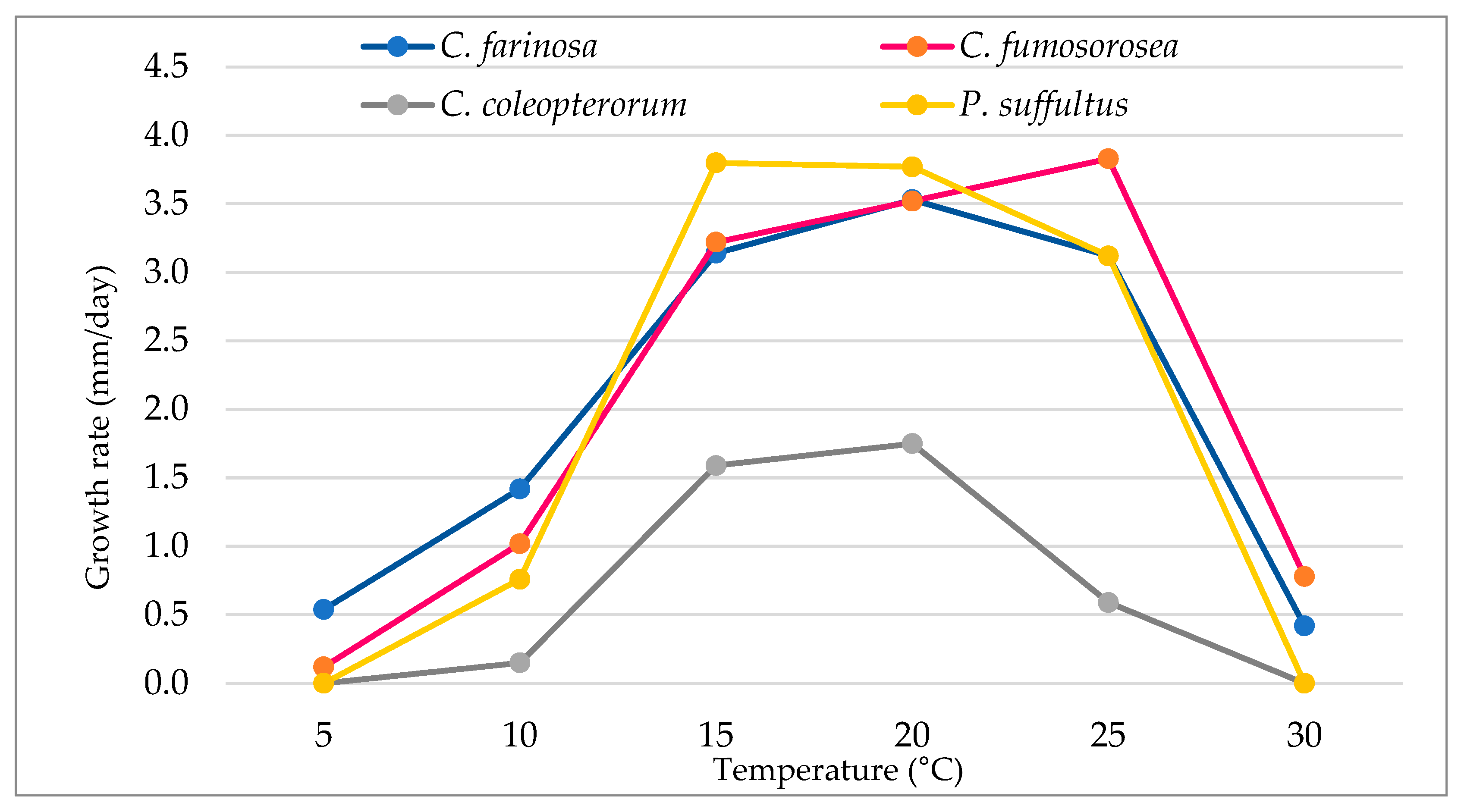
3.1. Effects of Temperature on Mycelial Growth Rate

The thermal optimum for the colony growth of the isolates of EPF of the Cordyceps and Paecilomyces genera was in the temperature range between 15 °C and 25 °C, with the values varying according to individual species. The colony growth of the C. farinosa strain was observed in all temperature ranges (Table 2). The smallest size of the fungal colonies was recorded at 30 °C. The optimum for their growth was 20 °C, at which temperature their diameter was 63.7 mm. The fastest growth rate of 3.5 mm/day was observed also at 20 °C (Figure 1).
Even though the colonies of the C. fumosorosea fungus grew in all temperatures, their best development was at 25 °C (69.0 mm). Of all the isolates, colonies of this fungus reached the largest sizes. The diameter of C. fumosorosea cultures was the smallest at 5 °C, with an average of 2.12 mm. The colony sizes at 10 and 30 °C did not differ much, with 18.4 and 14.5 mm, respectively. The daily growth of C. fumosorosea isolate across the temperature ranges of 15 to 25 °C was similar, with the highest value of 3.83 mm/day recorded at 25 °C (Figure 1).
Colony growth of two other species, i.e., C. coleopterorum and P. suffultus, was recorded in all temperature ranges except 5 and 30 °C (Table 2). The temperature optimum for C. coleopterorum growth was 20 °C, at which its colonies reached a size of 31.5 mm. Its cultures growing at 20 °C were over 50% smaller than the colonies of other species, and five times smaller at 25 °C. The daily growth rate of this fungus, at the optimum temperature, was the lowest of all isolates and amounted to 1.75 mm per day. The analysis of the significance test for the average growth rates of the C. coleopterorum strain at particular temperatures showed significant differences in relation to the other tested isolates (Figure 1). Colonies of the P. suffultus fungus reached the smallest sizes at 10 °C, with an average of 13.7 mm. Its best growth was at 15 °C, at which temperature P. suffultus colonies reached 68.5 mm on the 18th day. At 20 °C, the growth was only slightly slower. The fastest rate of daily growth of P. suffultus isolate was recorded at 15 and 20 °C, with 3.80 and 3.77 mm/day, respectively (Figure 1). The univariate analysis of variance indicated that their individual temperature ranges were a statistically significant factor affecting the colony size of the Cordyceps and Paecilomyces isolates (Table 2).

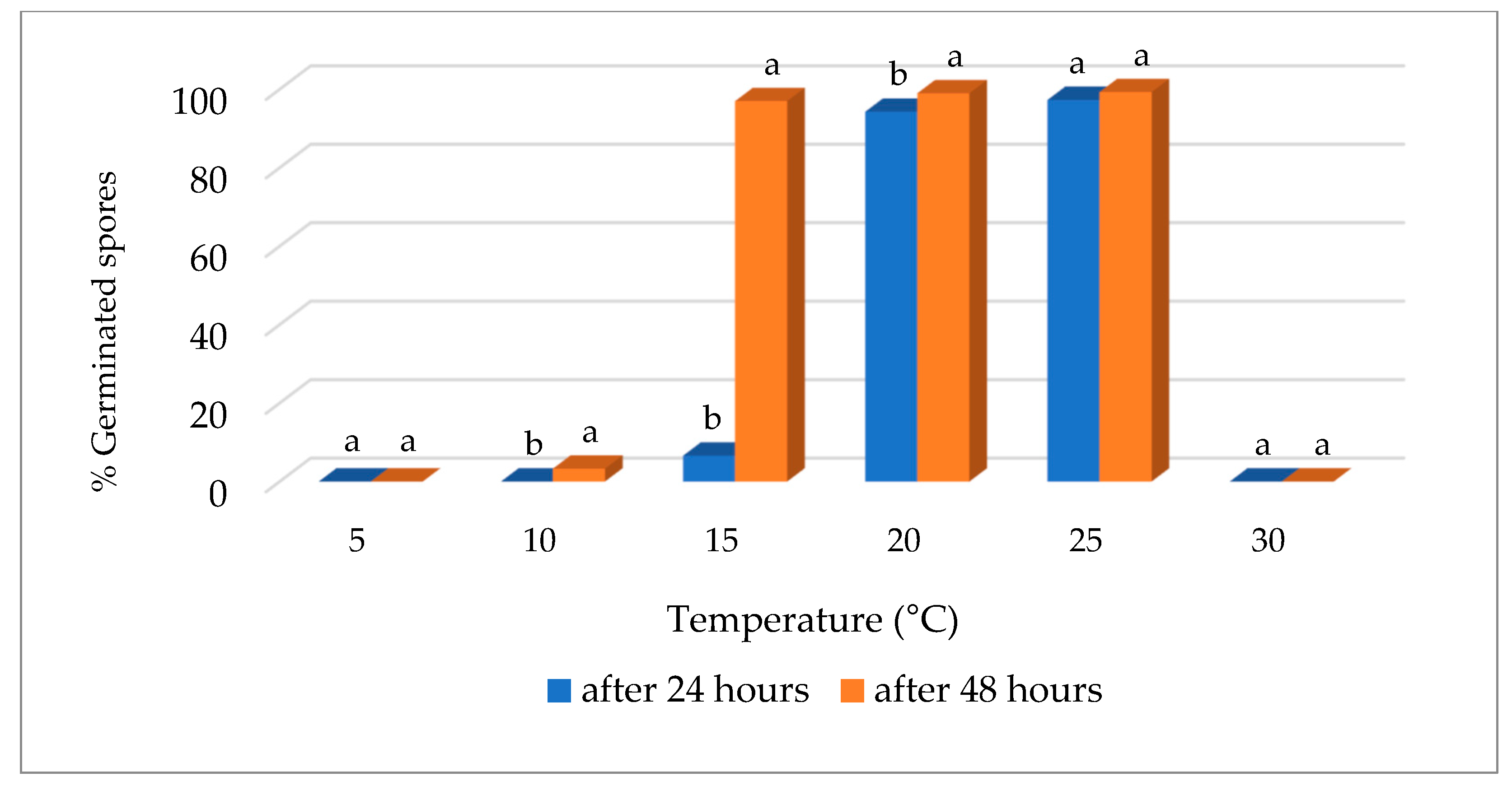
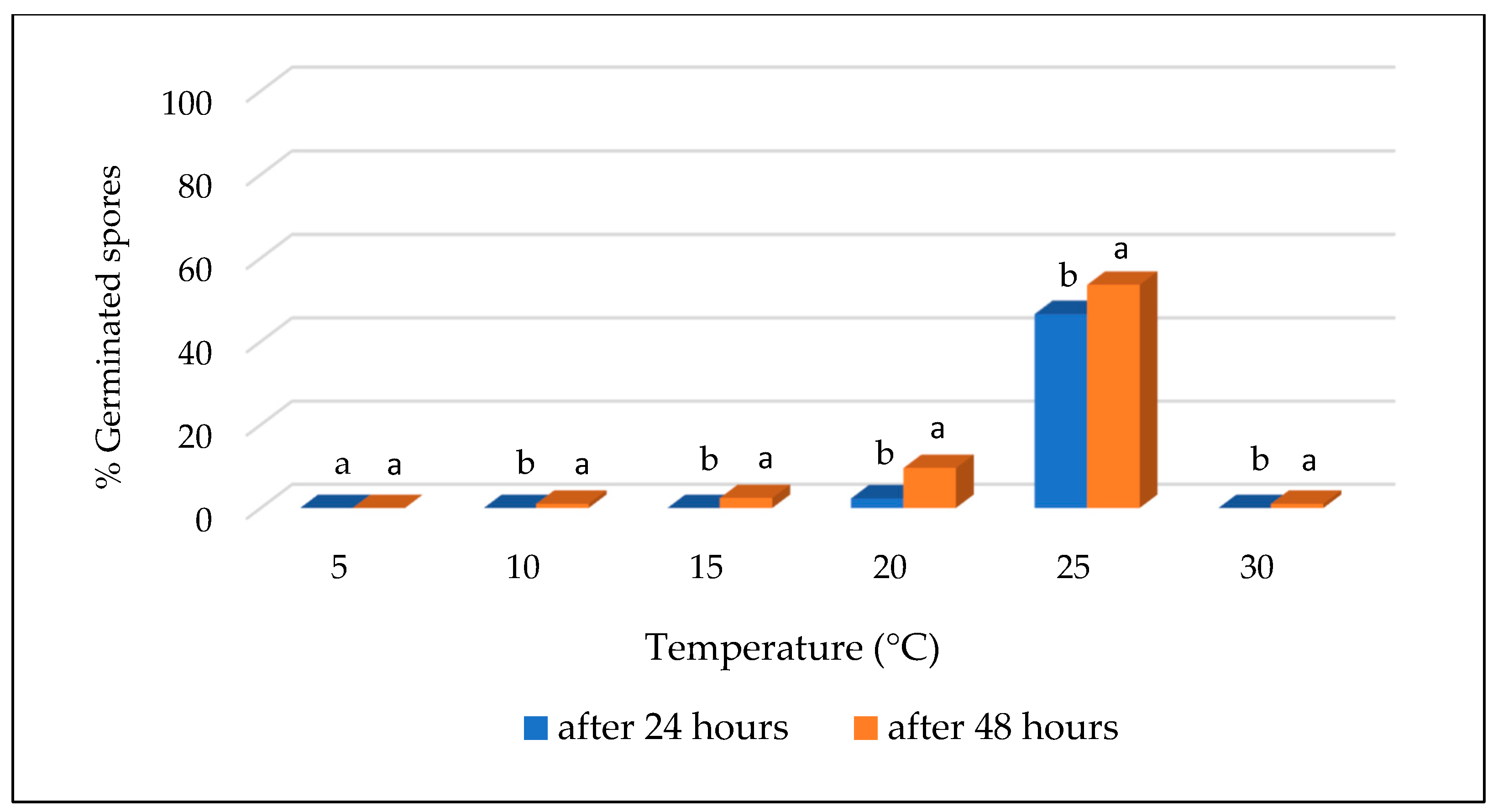
3.2. Effect of Temperature on Conidial Germination

This study showed that temperature and the length of the period in which spores are attached to the medium have a various effect on their germination. The proportion of germinated spores after 48 h on the culture medium was generally higher than after 24 h (Figure 2, Figure 3, Figure 4 and Figure 5).
After both 24 and 48 h, the temperature of 5 °C stopped the germination of spores of all fungal species. In the case of C. farinosa and C. fumosorosea, no germination was found at 30 °C.
After 24 h, spore germination of the fungal species was the most intense at 25 °C. A significant share of germinated spores at this temperature was observed in the case of C. farinosa, C. fumosorosea and P. suffultus (95.3, 97.3 and 94%, respectively).
Spores of C. coleopterorum also germinated most intensively at the 25 °C temperature with 46.6% spores germinated, and compared to other species this value was lower by over 50%. At 10 °C, no spore germination of any fungi was observed after 24 h, and spores of the C. coleopterorum isolate did not germinate at 15 °C either. Spores of the P. suffultus fungus had the greatest tolerance to elevated temperatures. Spore germination for this species was observed to be 38.6% of germinated spores after 24 h at 30 °C, and after 48 h, up to 93%.
After 48 h, the optimum temperature for germination of C. farinosa and P. suffultus spores was 20 °C, and for C. fumosorosea it was 25 °C, but this conclusion was not confirmed in a statistically significant way. Cordyceps coleopterorum spores also germinated best at 25 °C, at which temperature the proportion of germinated spores was 53.6% after 48 h.
According to statistical analysis, the time of contact (24 or 48 h) with the culture medium was a factor significantly affecting the percentage of germinated spores in separate temperature ranges. However, that did not apply to 5 °C and 30 °C, with a complete absence of germination, and in the case of C. farinosa, C. fumosorosea and P. suffultus species also at temperature 25 °C.

4. Discussion

In the development and life of all organisms, temperature is a fundamental factor. Consequently, it affects the growth and development of EPF that cause insect infections. These fungi seem to be the most promising group of pathogens that can be used in the biological control of harmful insects. However, it is necessary to select a strain with a wide spectrum of temperature tolerance [2,38,39,40,41]. Knowledge of the temperature tolerance of EPF is particularly important for the commercial production of biopesticides. This allows fungi to acclimate to thermal conditions to maintain their high pathogenicity.
Ascomycete fungi of the genera Metarhizium, Beauveria, and Cordyceps grow well on solid substrates at temperatures between 24 °C and 30 °C [22,42,43,44]. The favorable temperature ranges for the development of M. anisopliae, B. bassiana, M. rileyi, and C. fumosorosea are 24–30 °C, 22–26 °C, 20–30 °C, and 22–30 °C, respectively [19,22]. In turn, the temperature that limits the development of these species is 5 °C and 35 °C [17,45]. The species of fungi investigated in the present research had different temperature requirements for their growth. The optimum temperature for the growth of P. suffultus colonies was 15 °C, with 20 °C for C. farinosa and C. coleopterorum. The highest thermal requirements were demonstrated by the C. fumosorosea species, which developed best at 25 °C. The temperature ranges in which the fungi grew were also different. C. farinosa and C. fumosorosea developed in a wider temperature range, from 5 °C to 30 °C. In turn, growth of C. coleopterorum and P. suffultus colonies was observed only at temperatures between 10 °C and 25 °C.
In the literature, the most research concerns the thermal requirements of two species of the Cordyceps genus, i.e., C. farinosa and C. fumosorosea, that are pathogenic to different insects in different climatic zones. In terms of temperature ranges for their growth, both fungal species are mesophilic, i.e., they grow at moderate temperatures. For C. farinosa, the general temperature range for its growth is between 2–5 and 30–32 °C [46,47,48,49], and the optimum temperature for its germination and growth is 20 °C, ranging from 19 to 22.5 °C [47,48,49,50]. Doberski [46] found that one strain of C. farinosa caused infection of large elm bark beetle Scolytus scolytus larvae even at 28 °C. For other strains, no activity was observed at 0 and 5 °C (Machowicz-Stefaniak) [47]. According to Brown and Smith [51], colonies of C. farinosa grew slowly at 10–14 °C, with a higher growth rate at 24 °C. At 27 °C, its slowdown was observed, with a complete lack of growth at 37 °C. In turn, Ayysamy and Baskaran [52] report that the optimum temperature for colony growth of the C. farinosa strain they studied was 25 °C, with smaller colonies formed at 20 °C and the smallest at 30 °C. The germination process of C. farinosa spores was best at 20 °C and 25 °C. In our study, we found that after 24 h, spore germination of the investigated fungal species was the most intense at 25 °C. According to Tian et al. [53], the temperature of 26 °C was most favorable for the conidial germination of C. fumosorosea and for the infection of Bemisia tabaci Genadius 2nd instar nymphs. Rojas et al. [54] found that all Brazilian isolates of C. fumosorosea showed a fast reduction in germination during the first 30 min of exposure to 45 °C.
Questions still remain about the temperature optima of strains isolated from Mediterranean or tropical areas. Generally, C. fumosorosea prefers higher temperatures than C. farinosa, but there are strong differences in the temperature range among strains. Several isolates of C. fumosorosea from France grow between 11 and 30 °C [55], and Miętkiewski et al. [48] report that growth of C. fumosorosea is between 5 and 32 °C, with the optimum at 25 °C. Similar values relating to the optimal temperature for its colony growth are provided by Sosnowska and Ratajkiewicz [56]. Yeo et al. [38] reported that growth of several C. fumosorosea isolates was observed between 10 and 25 °C, with often the highest values, compared with other species, at 10 °C.
Some researchers pay attention to the close relationship between the geographical origin of a fungus strain and its thermal requirements. Vidal et al. [19] investigated the effect of temperature on the growth of isolates of C. fumosorosea isolated from various hosts (mainly from B. tabaci and Lepidoptera larvae) and originated from the southern USA, Europe, Pakistan, Naples, and India. In common with other authors, they found a wide variation among strains in terms of their thermal requirements. Strains from Europe grew at a temperature ranging from 8 to 30 °C, and their optimal was 20 °C, 20–25 °C or 25 °C. The temperature range in which strains from the south of the USA developed (from both humid and subtropical regions) and from western Asia (humid tropical climate) was wider and ranged from 8 to 35 °C, with their temperature optima of 25 °C, 25–28 °C or 28 °C. It was also found that isolates of C. fumosorosea originating from India were the most tolerant towards a high temperature, i.e., to 32 °C and 35 °C. In turn, the research of Bouamama et al. [57] provides evidence that INRA32, a European isolate of C. fumosorosea, was better adapted to temperate conditions than to both humid subtropical and arid ones. Similarly, Fargues et al. [45] showed that quiescent conidia of C. fumosorosea isolates from temperate regions were more susceptible to simulated sunlight (wavelengths ranging from 295 to 1100 nm at a UV-B irradiance of 0.3 Wm2) than isolates from warm regions. This intraspecific variability of the tolerance to environmental conditions could be related to C. fumosorosea genetic diversity [58,59,60,61,62,63].
It is worth mentioning that the C. fumosorosea isolate tested in this study showed high pathogenicity in laboratory conditions against the turnip moth Agrotis segetum Den. et Schiff. larvae, causing up to 89.6% mortality in the soil, while isolates of B. bassiana and M. anisopliae caused only 60% and 48% larval mortality, respectively [64]. The C. fumosorosea isolate tested in this study also showed high pathogenicity against the apple ermine Hyponomeuta malinellus Zeller (Lepidoptera) larvae and mountain-ash sawfly Pristiphora geniculata Hartig larvae, causing up to 97.0 and 90.0% mortality, respectively. Slightly lower larval mortality of the above-mentioned pests was caused by the isolate of the fungus C. farinosa tested in our research −89.8% and 88.5%, respectively [64]. The above data show a great potential of the individual fungal strains tested by us as a basis for the production of bioinsecticides, which may be used in the future in IPM of some pests, especially from the order of Lepidoptera.
As mentioned earlier, in addition to the generalist strains of C. farinosa and C. fumosorosea, which infect many insect species around the world, two other species were also included in the present research on the thermal requirements of EPF. They are rarely found in natural conditions on insects and appear to be specialized when it comes to their potential hosts. This type of research made it possible to compare the thermal requirements of the individual fungal strains with a wide and very narrow spectrum of infected host insects.
The P. suffultus isolate studied in the present research was obtained from the Bibionidae larvae, quite extensively infected by this fungus, in a litter from a typical oak-hornbeam forest in the Białowieża National Park, Poland. This fungus was first described under the name Cylindrodendrum suffultum by Petch [65] and was isolated from the pupa of a fly of the Psychodidae family in England. This species appears to be a specialized pathogen of Diptera order insects, especially the larvae of a fly on which it was found in Germany and France (Bałazy, unpublished data) but P. suffultus was for the first time described as an insect pathogen in Poland by Sosnowska et al. [66] in Białowieża National Park. In the available literature, there is no information about the thermal requirements of this fungal species. As the present research shows, this fungus achieved optimal growth at 15 °C, which is the lowest compared to the other species, while its spores showed the greatest tolerance to an elevated temperature. After 24 h of contact with the substrate at 30 °C, the highest percentage of its germinated spores was found. It should be mentioned here that the strain of P. suffultus tested in our research showed a very high pathogenicity in laboratory conditions in relation to the larvae of the marchfly (Bibio hortulanus L.), causing up to 87.0% mortality [67].
The isolate of C. coleopterorum used in this research was isolated from the larvae of an unmarked species of the Lampyridae family (Table 1), and its occurrence in Poland was confirmed for the first time by Tkaczuk [64]. This species was first found in Africa, also on the larvae of the Lampyridae beetle family, and was originally described by Evans [33]. In Germany and France, this fungus is considered to be a pathogen of fireflies [68]. The information about the thermal requirements of C. coleopterorum was provided only by Samson (1974), who observed that colonies of this fungus on malt extract agar grew slowly and reached a diameter of about 2.5 cm after 14 days at 25 °C.

5. Conclusions

Research on the thermal requirements of fungal species and isolates pathogenic towards insects or mites is also of great practical importance because it makes it possible to select strains with a wide spectrum of thermal tolerance, maintaining a high efficiency in field conditions of biopesticides based on them.
The results revealed that the thermal optimum for the colony growth of the investigated isolates of entomopathogenic fungi from the Cordyceps and Paecilomyces genus was in the temperature range between 15 °C and 25 °C. The present studies on the effect of temperature on the growth and germination of spores of such species as C. coleopterorum and P. suffultus are the first research of this type.
Further studies should focus on the use of EPF in pest biological control and on understanding their pathogenicity towards harmful insects. This is very important, especially because the fungal isolates tested in this work in terms of thermal requirements have shown high pathogenicity in relation to selected plant pests in previous studies, which indicates their potential usefulness in IPM programs.

Author Contributions

Conceptualization, C.T. and A.M.-S.; writing—original draft preparation, C.T. and A.M.-S.; writing—review and editing, C.T. and A.M.-S. All authors have read and agreed to the published version of the manuscript.

Funding

The results of the research carried out under research theme No. 159/23/B were financed by a science grant from the Ministry of Education and Science.

Institutional Review Board Statement

Not applicable.

Data Availability Statement

The data presented in this study are available on request from the corresponding author.

Conflicts of Interest

The authors declare no conflict of interest.

References

  1. Hajek, A.E.; St. Leger, R.J. Interactions between fungal pathogens and insect host. Annu. Rev. Entomol. 1994, 39, 39–293. [Google Scholar] [CrossRef]
  2. Chandler, D.; Sunderland, K.D.; Ball, B.V.; Davidson, G. Prospective biological control agents of Varroa destructor n. sp., an important pest of the European honeybee, Apis mellifera. Biocontrol Sci. Technol. 2001, 11, 429–448. [Google Scholar] [CrossRef]
  3. Quesada-Moraga, E.; Navas-Cortés, J.A.; Maranhao, E.A.; Ortiz-Urquiza, A.; Santiago-Alvarez, C. Factors affecting the occurrence and distribution of entomopathogenic fungi in natural and cultivated soils. Mycol. Res. 2007, 111, 947–966. [Google Scholar] [CrossRef] [PubMed]
  4. Vilcinskas, A.; Gotz, P. Parasitic fungi and their interactions with the insect immune system. Adv. Parasitol. 1999, 43, 267–313. [Google Scholar] [CrossRef]
  5. Xiong, Q.; Xiea, Y.; Zhua, Y.; Xuea, J.; Li, J.; Fan, R. Morphological and ultrastructural characterization of Carposina sasakii larvae (Lepidoptera: Carposinidae) infected by Beauveria bassiana (Ascomycota: Hypocreales: Clavicipitaceae). Micron 2013, 44, 303–311. [Google Scholar] [CrossRef] [PubMed]
  6. Wang, C.; Wang, S. Insect pathogenic fungi: Genomics, molecular interactions, and genetic improvements. Annu. Rev. Entomol. 2017, 62, 73–90. [Google Scholar] [CrossRef] [PubMed]
  7. Wei, G.; Lai, Y.; Wang, G.; Chen, H.; Li, F.; Wang, S. Insect pathogenic fungus interacts with the gut microbiota to accelerate mosquito mortality. Proc. Natl. Acad. Sci. USA 2017, 114, 5994–5999. [Google Scholar] [CrossRef]
  8. Gonzalez, F.; Tkaczuk, C.; Dinu, M.M.; Fiedler, Ż.; Vidal, S.; Zchori-Fein, E.; Messelink, G.J. New opportunities for the integration of microorganisms into biological pest control systems in greenhouse crops. J. Pest Sci. 2016, 89, 89–295. [Google Scholar] [CrossRef]
  9. Wu, S.; Toews, M.D.; Oliveira-Hofman, C.; Behle, R.W.; Simmons, A.M.; Shapiro-Ilan, D.I. Environmental tolerance of entomopathogenic fungi: A new strain of Cordyceps javanica isolated from a whitefly epizootic versus commercial fungal strains. Insects 2020, 11, 711. [Google Scholar] [CrossRef]
  10. Fargues, J.; Luz, C. Effects of fluctuating moisture and temperature regimes on the infection potential of Beauveria bassiana for Rhodinus prolixus. J. Invertebr. Pathol. 2000, 75, 202–211. [Google Scholar] [CrossRef]
  11. Wu, S.; Youngman, R.R.; Kok, L.T.; Laub, C.A.; Pfeiffer, D.G. Interaction between entomopathogenic nematodes and entomopathogenic fungi applied to third instar southern masked chafer white grubs, Cyclocephala lurida (Coleoptera: Scarabaeidae), under laboratory and greenhouse conditions. Biol. Control 2014, 76, 65–73. [Google Scholar] [CrossRef]
  12. Hajek, A.E.; Meyling, N.V. Fungi. In Ecology of Invertebrate Diseases; Hajek, A.E., Shapiro-Ilan, D.I., Eds.; Wiley: Oxford, UK, 2017; pp. 327–377. [Google Scholar]
  13. Devi, K.U.; Sridevi, V.; Mohan, C.M.; Padmavathi, J. Effect of high temperature and water stress on in vitro germination and growth in isolates of the entomopathogenic fungus Beauveria bassiana (Bals. ) Vuillemin. J. Invertebr. Pathol. 2005, 88, 181–189. [Google Scholar] [CrossRef]
  14. Alexandre, T.M.; Alves, L.F.A.; Neves, P.M.O.J.; Alves, S.B. Effect of temperature and poultry litter on Beauveria bassiana (Bals.) Vuill. and Metarhizium anisopliae (Metsch) virulence against the lesser mealworm Alphitobius diaperinus (Panzer) (Coleoptera: Tenebrionidae. Neotrop. Entomol. 2006, 35, 75–82. [Google Scholar] [CrossRef]
  15. Polar, P.; Aquino de Muro, M.; Kairo, M.T.K.; Moore, D.; Pegram, R.; John, S.A.; Roach-Benn, C. Thermal characteristics of Metarhizium anisopliae isolates important for the development of biological pesticides for the control of cattle ticks. Vet. Parasitol. 2005, 134, 159–167. [Google Scholar] [CrossRef]
  16. Ibarra-Corteś, K.H.; Guzmán-Franco, A.W.; González-Fernández, H.; Suarez-Espinosa, J.; Baverstock, J. Selection of a fungal isolate for the control of the pink hibiscus mealybug Maconellicoccus hirsutus. Pest. Manag. Sci. 2013, 69, 874–882. [Google Scholar] [CrossRef] [PubMed]
  17. Ferron, P.; Fargues, J.; Riba, G. Fungi as microbial insecticides against pests. In Handbook of Applied Mycology. Volume 2: Humans, Animals, and Insects; Arora, D.K., Ajello, L., Mukerji, K.G., Eds.; Marcel Dekker, Inc.: New York, NY, USA, 1991; pp. 665–706. [Google Scholar]
  18. Fargues, J.; Goettel, M.S.; Smits, N.; Ouedraog, A. Effect of temperature on vegetative growth of Beauveria bassiana isolates from different origins. Mycologia 1997, 89, 383. [Google Scholar] [CrossRef]
  19. Vidal, C.; Fargues, J.; Lacey, L.A. Intraspecific variability of Paecilomyces fumosoroseus: Effect of temperature on vegetative growth. J. Invertebr. Pathol. 1997, 70, 18–26. [Google Scholar] [CrossRef]
  20. Jackson, M.A.; Jaronski, S.T. Production of microsclerotia of the fungal entomopathogen Metarhizium anisopliae and their potential for use as a biocontrol agent for soil-inhabiting Insects. Mycol. Res. 2009, 113, 842–850. [Google Scholar] [CrossRef]
  21. Jaronski, S.T. Ecological factors in the inundative use of fungal entomopathogens. BioControl 2010, 55, 159–185. [Google Scholar] [CrossRef]
  22. Zimmermann, G. The entomopathogenic fungi Isaria farinosa (formerly Paecilomyces farinosus) and the Isaria fumosorosea species complex (formerly Paecilomyces fumosoroseus): Biology, ecology and use in biological control. Biocontrol Sci. Technol. 2008, 18, 865–901. [Google Scholar] [CrossRef]
  23. Ropek, D.; Krysa, A.; Rola, A.; Frączek, K. Antagonistic effect of Trichoderma viride on entomopathogenic fungi Beauveria bassiana, Isaria fumosorosea and Metarhizium anisopliae in vitro. Pol. J. Agron. 2014, 16, 57–63. [Google Scholar]
  24. Luangsa-ard, J.J.; Hywel-Jones, N.L.; Samson, R.A. The order level polyphyletic nature of Paecilomyces sensu lato as revealed through 18S-generated rRNA phylogeny. Mycologia 2004, 96, 773–780. [Google Scholar] [CrossRef] [PubMed]
  25. Luangsa-ard, J.J.; Hywel-Jones, N.L.; Manoch, L.; Samson, R.A. On the relationships of Paecilomyces sect. Isarioidea species. Mycol. Res. 2005, 109, 581–589. [Google Scholar] [CrossRef] [PubMed]
  26. Gams, W.; Hodge, K.T.; Samson, R.A.; Korf, R.P.; Seifert, K.A. Proposal to conserve the name Isaria (anamorphic fungi) with a conserved type. Taxon 2005, 54, 537. [Google Scholar] [CrossRef]
  27. Hodge, K.T.; Gams, W.; Samson, R.A.; Korf, R.P.; Seifert, K.A. Lectotypification and status of Isaria Pers. Taxon 2005, 54, 485–489. [Google Scholar] [CrossRef]
  28. Kepler, R.M.; Luangsa-Ard, J.J.; Hywel-Jones, N.L.; Quandt, C.A.; Sung, G.H.; Rehner, S.A.; Aime, M.C.; Henkel, T.W.; Sanjuan, T.; Zare, R. A phylogenetically-based nomenclature for Cordycipitaceae (Hypocreales). IMA Fungus 2017, 8, 335–353. [Google Scholar] [CrossRef]
  29. Vänninen, I. Distribution and occurrence of four entomopathogenic fungi in Finland: Effect of geographical location, habitat type and soil type. Mycol. Res. 1996, 100, 93–101. [Google Scholar] [CrossRef]
  30. Chandler, D.; Hay, D.; Reid, A.P. Sampling and occurrence of entomopathogenic fungi and nematodes in UK soils. Appl. Soil. Ecol. 1997, 5, 133–141. [Google Scholar] [CrossRef]
  31. Lacey, L.A.; Wraight, S.P.; Kirk, A.A. Entomopathogenic fungi for control of Bemisia spp. In Classical Biological Control of Bemisia tabaci in the USA: A Review of Interagency Research and Implementation; Progress in Biological Control; Gould, J.K., Hoelmer, K., Goolsby, J., Eds.; Springer: Dordrecht, The Netherlands, 2008; Volume 4, p. 3369. [Google Scholar]
  32. Kim, W.G.; Seok, S.J.; Weon, H.Y.; Lee, H.H.; Lee, C.J.; Kim, Y.S. Isolation and identification of entomopathogenic fungi collected from mountains and Islands in Korea. Kor. J. Mycol. 2010, 38, 99–104. [Google Scholar] [CrossRef]
  33. Samson, R.A. Paecilomyces and some allied Hyphomycetes. Stud. Mycol. 1974, 6, 32–41. [Google Scholar]
  34. Humber, R.A. Manual of Techniques in Invertebrate Pathology. In Identification of Entomopathogenic Fung; Lacey, L.A., Ed.; Academic Press: London, UK, 2012; pp. 151–187. [Google Scholar]
  35. Schoch, C.L.; Seifert, K.A.; Huhndorf, S.; Robert, V.; Spouge, J.L.; Levesque, C.A.; Chen, W.; Fungal Barcoding Consortium. Nuclear ribosomal internal transcribed spacer (ITS) region as a universal DNA barcode marker for Fungi. Proc. Natl. Acad. Sci. USA 2012, 109, 6241–6246. [Google Scholar] [CrossRef]
  36. Kovač, M.; Gorczak, M.; Wrzosek, M.; Tkaczuk, C.; Pernek, M. Identification of entomopathogenic fungi as naturally occurring enemies of the invasive oak lace bug, Corythucha arcuata (Say) (Hemiptera: Tingidae). Insects 2020, 11, 679. [Google Scholar] [CrossRef] [PubMed]
  37. Altschul, S.F.; Madden, T.L.; Schäffer, A.A.; Zhang, J.; Zhang, Z.; Miller, W.; Lipman, D.J. Gapped BLAST and PSI-BLAST: A new generation of protein database search programs. Nucleic Acids Res. 1997, 25, 3389–3402. [Google Scholar] [CrossRef] [PubMed]
  38. Yeo, H.; Pell, J.K.; Alderson, P.G.; Clark, S.J.; Pye, B.J. Laboratory evaluation of temperature effects on the germination and growth of entomopathogenic fungi and on their pathogenicity to two aphid species. Pest. Manag. Sci. 2003, 59, 56165. [Google Scholar] [CrossRef]
  39. Piątkowski, J.; Krzyżewska, A. Influence of some physical factors on the growth and sporulation of entomopathogenic fungi. Acta Myc. 2007, 42, 255–265. [Google Scholar] [CrossRef]
  40. Sapna, M.; Peeyush, K.; Anushree, M. Effect of temperature and humidity on pathogenicity of native Beauveria bassiana isolate against Musca domestica L. J. Parasit. Dis. 2015, 39, 697–704. [Google Scholar] [CrossRef]
  41. Christos, G.; Athanassiou, N.G.; Kavallieratos, C.I.R.; Demetrius, C.K. Influence of temperature and relative humidity on the insecticidal efficacy of Metarhizium anisopliae against larvae of Ephestia kuehniella (Lepidoptera: Pyralidae) on wheat. J. Insect Sci. 2017, 17, 22. [Google Scholar] [CrossRef]
  42. Zimmermann, G. Review on safety of the entomopathogenic fungi Beauveria bassiana and Beauveria brongniartii. Biocontrol Sci. Technol. 2007, 17, 553–596. [Google Scholar] [CrossRef]
  43. Zimmermann, G. Review on safety of the entomopathogenic fungus Metarhizium anisopliae. Biocontrol Sci. Technol. 2007, 17, 879–920. [Google Scholar] [CrossRef]
  44. Jaronski, S.T. Mass production of entomopathogenic fungi—State of the art. In Mass Production of Beneficial Organisms; Academic Press: London, UK, 2023; pp. 317–357. [Google Scholar] [CrossRef]
  45. Fargues, J.; Goettel, M.S.; Smits, N.; Ouedraogo, A.; Vidal, C.; Lacey, L.A.; Lomer, C.J.; Rougier, M. Variability in susceptibility to simulated sunlight of conidia among isolates of entomopathogenic Hyphomycetes. Mycopathologia 1997, 135, 171–181. [Google Scholar] [CrossRef]
  46. Doberski, J.W. Comparative laboratory studies on three fungal pathogens of the elm bark beetle Scolytus scolytus: Effect of temperature and humidity on infection by Beauveria bassiana, Metarhizium anisopliae and Paecilomyces farinosus. J. Invertebr. Pathol. 1981, 37, 195–200. [Google Scholar] [CrossRef]
  47. Machowicz-Stefaniak, Z. Studies on the effect of temperature on the growth and development of the fungus Paecilomyces farinosus (Dicks, ex Fr.) Brown et Smith (Moniliales, Deuteromycetes). Rocz. Nauk. Roln. 1988, 18, 121–130. [Google Scholar]
  48. Mietkiewski, R.; Tkaczuk, C.; Żurek, M.; Van der Geest, L.P.S. Temperature requirements of four entomopathogenic fungi. Acta Myc. 1994, 29, 109–120. [Google Scholar] [CrossRef]
  49. Hallsworth, J.E.; Magan, N. 1999. Water and temperature relations of growth of the entomogenous fungi Beauveria bassiana, Metarhizium anisopliae, and Paecilomyces farinosus. J. Invertebr. Pathol. 1999, 74, 261–266. [Google Scholar] [CrossRef]
  50. Huang, B.; Fan, M.Z.; Li, Z.Z.; Zhou, Q.; Wang, Z.X. Biological characteristics of different strains of Paecilomyces farinosus. J. Anhui Agric. 2005, 32, 8–11. [Google Scholar]
  51. Brown, H.S.; Smith, G. The genus Paecilomyces Bainier and its perfect stage Byssochlamys Westling. Trans. Br. Mycol. 1957, 40, 17–89. [Google Scholar] [CrossRef]
  52. Ayyasamy, R.; Baskaran, P. Effect of temperature and relative humidity on radial growth and sporulation of Paecilomyces farinosus. J. Food Agric. Environ. 2005, 3, 137–138. [Google Scholar] [CrossRef]
  53. Tian, J.; Hao, C.; Liang, L.; Ma, R.Y. Effects of temperature and relative humidity on conidial germination of Isaria fumosorosea (Hypocreales: Cordycipitaceae) IF-1106 and pathogenicity of the fungus against Bemisia tabaci (Homoptera: Aleyrodidae). Mycosystema 2014, 33, 668–679. [Google Scholar] [CrossRef]
  54. Rojas, V.M.A.; Iwanicki, N.S.A.; D’Alessandro, C.P.; Fatoretto, M.B.; Demétrio, C.G.B.; Delalibera, I., Jr. Characterization of Brazilian Cordyceps fumosorosea isolates: Conidial production, tolerance to ultraviolet-B radiation, and elevated temperature. J. Invertebr. Pathol. 2023, 197, 107888. [Google Scholar] [CrossRef]
  55. Fargues, J.; Maniania, N.K.; Delmas, J.C.; Smits, N. Influence de la temperature sur la croissance in vitro dhyphomycetes entomopathogenes. Agronomie 1992, 12, 557–564. [Google Scholar] [CrossRef]
  56. Sosnowska, D.; Ratajkiewicz, H. Assessment of the potential of local entomopathogenic fungi isolates as biocontrol agents. Prog. Plant Prot. 2021, 61, 121–127. [Google Scholar] [CrossRef]
  57. Bouamama, N.; Vidal, C.; Fargues, J. Effects of fluctuating moisture and temperature regimes on the persistence of quiescent conidia of Isaria fumosorosea. J. Invertebr. Pathol. 2010, 105, 139–144. [Google Scholar] [CrossRef] [PubMed]
  58. Tigano-Milani, M.S.; Carneiro, R.G.; De Faria, M.R.; Frazao, H.S.; McCoy, C.W. Isozyme characterization and pathogenicity of Paecilomyces fumosoroseus and P. lilacinus to Diabrotica speciosa (Coleoptera: Chrysomelidae) and Meloidogyne javanica (Nematoda: Tylenchidae). Biol. Control. 1995, 5, 378382. [Google Scholar] [CrossRef]
  59. Cantone, F.A.; Vandenberg, J.D. Intraspecific diversity in Paecilomyces fumosoroseus. Mycol. Res. 1998, 102, 209–215. [Google Scholar] [CrossRef]
  60. Fargues, J.; Bon, M.C.; Manguin, S.; Couteaudier, I. Genetic ariability amvong Paecilomyces fumosoroseus isolates from various geographical and host insect origins based on the rDNA-ITS regions. Mycol. Res. 2002, 106, 1066–1074. [Google Scholar] [CrossRef]
  61. Fargues, J.; Bon, M.C. Influence of temperature preferences of two Paecilomyces fumosoroseus lineages on their co-infection pattern. J. Invertebr. Pathol. 2004, 87, 94–104. [Google Scholar] [CrossRef]
  62. Gauthier, N.; Dalleau-Clouet, C.; Fargues, J.; Bon, M.C. Microsatellite variability in the entomopathogenic fungus Paecilomyces fumosoroseus: Genetic diversity and population structure. Mycologia 2007, 99, 693–704. [Google Scholar] [CrossRef]
  63. Islam, W.; Adnan, M.; Shabbir, A.; Naveed, H.; Abubakar, Y.S.; Qasim, M.; Tayyab, M.; Noman, A.; Nisar, M.S.; Khan, K.A.; et al. Insect-fungal-interactions: A detailed review on entomopathogenic fungi pathogenicity to combat insect pests. Microb. Pathog. 2021, 159, 105122. [Google Scholar] [CrossRef]
  64. Tkaczuk, C. Occurrence and Infective Potential of Entomopathogenic Fungi in Soils of Agrocenoses and Seminatural Habitats in the Agricultural Landscape; Scientific Dissertation No. 94; Wydawnictwo Akademii Podlaskiej: Siedlce, Poland, 2008; p. 160. (In Polish) [Google Scholar]
  65. Petch, T. Notes on entomogenous fungi. Trans. Br. mycol. Soc. 1944, 27, 81–93. [Google Scholar] [CrossRef]
  66. Sosnowska, D.; Balazy, S.; Prishchepa, L.; Mikulskaya, N. Biodiversity of arthropod pathogens in the Bialowieża Forest. J. Plant Prot. Res. 2004, 44, 313–321. [Google Scholar]
  67. Żurek, M.; Miętkiewski, R.; Tkaczuk, C. The susceptibility of Bibio hortulanus L. larvae to entomopathogenic fungi in soil. In Invertebrate Pathogen in Biological Control: Presen and Future; IOBC: Bari, Italy, 2005; p. 165. [Google Scholar]
  68. Bałazy, S. Znaczenie obszarów chronionych dla zachowania zasobów grzybów entomopatogenicznych. Kosmos 2004, 53, 5–16. [Google Scholar]
Figure 1. Daily growth rate of entomopathogenic fungal species at temperatures ranging from 5 to 30 °C.
Agriculture 13 01989 g001
Figure 2. Germination of C. farinosa spores after 24 and 48 h at temperatures ranging from 5 to 30 °C. ab—Means between hours at particular temperatures with the same lowercase letters are not significant at α = 0.05, Tukey’s HSD.
Agriculture 13 01989 g002
Figure 3. Germination of C. fumosorosea spores after 24 and 48 h at temperatures ranging from 5 to 30 °C. ab—Means between hours at particular temperatures with the same lowercase letters are not significant at α = 0.05, Tukey’s HSD.
Agriculture 13 01989 g003
Figure 4. Germination of C. coleopterorum spores after 24 and 48 h at temperatures ranging from 5 to 30 °C. ab—Means between hours at particular temperatures with the same lowercase letters are not significant at α = 0.05, Tukey’s HSD.
Agriculture 13 01989 g004
Figure 5. Germination of P. suffultus spores after 24 and 48 h at temperatures ranging from 5 to 30 °C. ab—Means between hours at particular temperatures with the same lowercase letters are not significant at α = 0.05, Tukey’s HSD.
Agriculture 13 01989 g005
Table 1. Characteristics of entomopathogenic fungal isolates.
Fungal Species and Strain NumberIsolate Origin
Cordyceps farinosa
(P04-UPH)
From the pupa of an unmarked butterfly species, found in oak-hornbeam forest litter of the Białowieża National Park, Poland
Cordyceps fumosorosea
(P03-UPH)
From Galleria mellonella larvae from the soil of an arable field in Pietrusy (Mazowieckie Voivodeship), Poland
Cordyceps coleopterorum
(P07-UPH)
From the larvae of a beetle from the firefly family (Lampyridae), found in the litter of an oak-hornbeam forest in the Białowieża National Park, Poland
Paecilomyces suffultus
(P06-UPH)
From the larvae of the Bibio sp., found in the litter of a typical oak-hornbeam forest in the Białowieża National Park, Poland
Table 2. Mean colony diameter [mm] of entomopathogenic fungal species growing at different temperature on day 18.
Fungi SpeciesTemperature
5 °C10 °C15 °C20 °C25 °C30 °C
Cordyceps farinosa9.75 ± 0.81 a25.7 ± 1.9 a56.6 ± 1.8 b63.7 ± 3.5 a56.3 ± 2.5 b7.5 ± 1.3 b
Cordyceps fumosorosea2.12 ± 0.32 b18.4 ± 1.3 b58.0 ± 2.1 b63.3 ± 1.6 a69.0 ± 1.8 a14.5 ± 1.1 a
Cordyceps coleopterorum0.0 ± 0.0 c2.75 ± 0.35 d28.7 ± 1.4 c31.5 ± 0.5 b10.7 ± 0.95 c0.0 ± 0.0 c
Paecilomyces suffultus0.0 ± 0.0 c13.7 ± 1.33 c68.5 ± 0.76 a68.0 ± 1.0 a56.2 ± 1.4 b0.0 ± 0.0 c
p-value0.000.000.000.000.000.00
F-value368.04824.683502.794226.461642.692488.00
abcd—Means within columns with the same lowercase letters are not significant at α = 0.05, Tukey’s HSD; ±—Standard deviation (SD).
Disclaimer/Publisher’s Note: The statements, opinions and data contained in all publications are solely those of the individual author(s) and contributor(s) and not of MDPI and/or the editor(s). MDPI and/or the editor(s) disclaim responsibility for any injury to people or property resulting from any ideas, methods, instructions or products referred to in the content.

