Article

Morphological, Cytological, and Molecular-Based Genetic Stability Analysis of In Vitro-Propagated Plants from Newly Induced Aneuploids in Caladium

Spice Crops Research Institute, College of Horticulture and Gardening, Yangtze University, Jingzhou 434025, China
*
Author to whom correspondence should be addressed.
These authors contributed equally to this work.
Agriculture 2022, 12(10), 1708; https://doi.org/10.3390/agriculture12101708
Submission received: 1 October 2022 / Revised: 12 October 2022 / Accepted: 14 October 2022 / Published: 17 October 2022

Abstract

:
Aneuploids are valuable materials of genetic diversity for genetic analysis and improvement in diverse plant species, which can be propagated mainly via in vitro culture methods. However, somaclonal variation is common in tissue culture-derived plants including euploid caladium. In the present study, the genetic stability of in vitro-propagated plants from the leaf cultures of two types of caladium (Caladium × hortulanum Birdsey) aneuploids obtained previously was analyzed morphologically, cytologically, and molecularly. Out of the randomly selected 23 and 8 plants regenerated from the diploid aneuploid SVT9 (2n = 2x − 2 = 28) and the tetraploid aneuploid SVT14 (2n = 4x − 6 = 54), respectively, 5 plants from the SVT9 and 3 plants from the SVT14 exhibited morphological differences from their corresponding parent. Stomatal analysis indicated that both the SVT9-derived variants and the SVT14-originated plants showed significant differences in stomatal guard cell length and width. In addition, the variants from the SVT14 were observed to have rounder and thicker leaves with larger stomatal guard cells and significantly reduced stomatal density compared with the regenerants of the SVT9. Amongst the established plants from the SVT9, two morphological variants containing 3.14–3.58% less mean fluorescence intensity (MFI) lost one chromosome, and four variants containing 4.55–11.02% more MFI gained one or two chromosomes. As for the plants regenerated from the SVT14, one variant with significantly higher MFI gained two chromosomes and three plants having significantly lower MFI resulted in losing four chromosomes. Three, out of the twelve, simple sequence repeat (SSR) markers identified DNA band profile changes in four variants from the SVT9, whereas no polymorphism was detected among the SVT14 and its regenerants. These results indicated that a relatively high frequency of somaclonal variation occurred in the in vitro-propagated plants from caladium aneuploids, especially for the tetraploid aneuploid caladium. Newly produced aneuploid plants are highly valuable germplasm for future genetic improvement and research in caladium.

1. Introduction

Aneuploidy refers to individuals with imbalanced variations in chromosome number from the basic chromosome sets characterized by each species in the somatic cells, including gains or losses of one or multiple individual chromosomes [1]. As a general rule, aneuploidy often causes detrimental side effects on the growth and development of organisms because of genomic imbalance resulting from the incomplete chromosome sets [2]. Accordingly, artificially produced aneuploid plant species often exhibit severe defective phenotypic consequences, such as poor growth, developmental delay, poor adaptation, and even loss of viability [1,2,3]. Nevertheless, it was confirmed that some aneuploid plant species demonstrate great resistance to the deleterious effects of aneuploidy and can grow to generate adult plants under field or controlled conditions, especially for polyploid species [4,5]. Until now, the occurrence of aneuploidy has been frequently found in the progenies of open-pollinated polyploids [3] and biological breeding programs, such as interploid hybridization [6], remote hybridization [7], in vitro chromosome doubling [8,9], and in vitro culture as well [10,11]. Aneuploidy is a large-effect genetic variation and may induce numerous heritable phenotypic variations and biological consequences in cells and organisms [1,12]. In some cases, the addition or deletion of individual chromosomes is even part of plant evolution [13]. As compared with their euploid progenitors, some aneuploid plant lines exhibit distinctive and desirable phenotypic characteristics, such as dwarfing and novel leaf characteristics [3,14,15]. Aneuploids, therefore, could hold great potential for genetic improvement and research in plants.
In general, aneuploid plants usually cannot be stably preserved through conventional sexual reproduction due to genetic instability of sexual offspring and reduced fertility and seed set [16,17]. Considering the great value of plant aneuploids in genetic research and crop breeding, it is essential to establish an efficient and reliable approach to stably maintaining them. Recently, in vitro clonal propagation has been considered as an efficient alternative for the stable preservation and propagation of aneuploid lines in plant species [18,19]. However, somaclonal variation is commonly observed in cultured cells or regenerated plants, as has been detected on the morphological, cytological, physiological, and molecular levels in recent years [10,20]. In general, heritable changes resulting from somaclonal variation could increase genetic variability and produce quality materials for crop genetic improvement. Nevertheless, maintaining the genetic fidelity is problematic when in vitro propagation is used in the large-scale propagation of commercial cultivars, germplasm conservation of endangered plant species, or preservation of distinctive materials like aneuploids. In recent decades, the occurrence and mechanisms of somaclonal variation have received constant attention for many in vitro culture programs [10,20].
Caladiums (Caladium × hortulanum Birdsey) are cultivated as important ornamental aroids due in large part to their long-lasting, colorful, and variably shaped foliage [21,22]. In search of new and novel cultivars for containers and landscapes in caladium, traditional cross-breeding between commercial cultivars and breeding lines has always been the main breeding approach [21,22,23]. Up to now, considerable efforts have been devoted to developing the caladium cultivars with enhanced colors or coloration patterns, tuber yield potential, resistance to diseases and pests, and tolerance to sunburns or chilling stress [21,22]. Nevertheless, this conventional breeding strategy has encountered difficulties such as the increasing scarcity of novel leaf characters in germplasm after continuously intensive sexual hybridization and selection [21]. Therefore, it is necessary to explore other efficient and reliable breeding methods to create novel sources of genetic variability for caladium breeding.
In caladium, recent reports demonstrated that a considerably high frequency of somaclonal variation occurred in the regenerated plants, especially when culturing in the medium with 2,4-dichlorophenoxyacetic acid (2,4-D) [24,25] or adopting long-term cultured callus as initial explants [11]. The variants showed a remarkable level of morphological differences either in leaf coloration patterns, leaf length, leaf width, plant height, or leaf thickness [11,24,25,26], which possess immense potential for caladium cultivar improvement and genetic studies. In the light of recent studies on the leaf color variation in caladium tissue culture, the instability of chromosome number, nuclear DNA content, photosynthetic pigment concentration, and DNA banding pattern might be responsible for the novel features of leaf morphology [8,9,11,25,26]. However, the systematic genetic stability of the asexual offspring of aneuploids in the Caladium species remains largely unexplored, resulting in some limitations in the application of these somaclonal variants to continued genetic improvement and study.
In a prior study, aneuploidization and polyploidization were observed in the plants regenerated from the callus of ‘Red Flash’ caladium, and several types of aneuploid variants were found to have improved ornamental values [11]. However, taking advantage of these aneuploids for subsequent breeding programs seems to be unfeasible, as the number of the individuals is limited, and several variation types have only 1–3 plants. Therefore, the preservation and proliferation of these promising caladium aneuploids is a prerequisite for future genetic and breeding studies. Generally, commercial propagation of caladium plants is mainly achieved by tuber division [21,22], though it is a slow and inefficient process, especially when the donor tubers are limited such as somaclonal or induced variants. Despite unpredictable and undesirable somaclonal variation, an in vitro culture technique is considered a promising alternative for the large-scale multiplication or conservation of plants. There are many studies on in vitro culture of and somaclonal variation in the euploids in caladium [11,24,25,26], while related studies are still not available for aneuploids at present. In this study, plants were regenerated in vitro from the young leaf segments of two somaclonal variation types (SVT), namely, the SVT9 (2n = 2x − 2 = 28) and the SVT14 (2n = 4x − 6 = 54), obtained previously in caladium tissue culture [11], and the regenerants were subjected to morphological observation, chromosome counting, flow cytometric assessment, stomatal analysis, and molecular characterization, with the aim to ascertain the genetic stability of the in vitro-propagated plantlets from caladium aneuploids.

2. Materials and Methods

2.1. Plant Materials

Two aneuploid plant lines, namely, the SVT9 (2n = 2x − 2 = 28) and the SVT14 (2n = 4x − 6 = 54), obtained previously from callus culture of ‘Red Flash’ caladium [11], were used as the donor plants. Both of them grew normally and had improved ornamental values such as attractive leaf color patterns or compact plant architecture.

2.2. In Vitro Culture of the Aneuploid Variants

Plant regeneration was conducted through in vitro culture of immature leaves of the plants forced from the tubers of the two aneuploids via one-step culture without medium renewal, as described previously [9]. Briefly, immature leaves were excised and washed thoroughly, their surface disinfected in 70% (v/v) alcohol for 15 s and then treated with 0.1% (w/v) HgCl2 for 12 min, and lastly rinsed with sterilized water five times. After disinfection, leaves were removed from the leaf margins and trimmed into leaf segments of about 0.5 cm × 0.5 cm, and then inoculated in glass jars filled with 50 mL fresh plantlet induction medium consisting of 4.43 g.L−1 commercial Murashige and Skoog (MS) [27] medium (PhytoTechnology Laboratories), 0.5 mg·L−1 NAA, 1.0 mg·L−1 6-BA, 40 g·L−1 (w/v) sucrose, and 0.75% (w/v) agar (pH 5.8). All cultures were maintained in a culture room at 25 ± 1 °C in the dark with the purpose of callus induction at the beginning, and then cultured under light conditions with a photoperiod of 14 h and photosynthetic active radiation of about 65 μmol m−2·s−1.

2.3. Plant Acclimatization and Establishment

After about 5 months of incubation, well-rooted plantlets were taken from glass jars, washed carefully under running water to remove any agar residues, and then transplanted to 32-cell trays (6 × 6 × 11 cm per cell) filled with the soilless potting mix purchased from Jiangsu Peilei Biotechnology Co., Ltd., Xuzhou, China. Then, the transplanted plantlets were kept in an artificial climate chamber at 28/20 °C (day/night) with a photoperiod of 14 h, a light level of 120 μmol m−2·s−1, and a relative humidity of 80 to 85%, and watered as required. After four weeks of acclimatization to ex vitro conditions, a total of 23 and 8 regenerated plantlets from the SVT9 and the SVT14, respectively, were selected randomly and transplanted to plastic pots (14.5 cm in diameter and 17.0 cm in depth) filled with the potting mix and incubated in a greenhouse under similar conditions to those mentioned above. To facilitate identification of genetic uniformity, the wild-type SVT9 and the SVT14—both forced from tubers—were also cultured under similar conditions and used as a control. Four months later, the plants exhibited stable morphological characteristics and were subjected to genetic stability analysis.

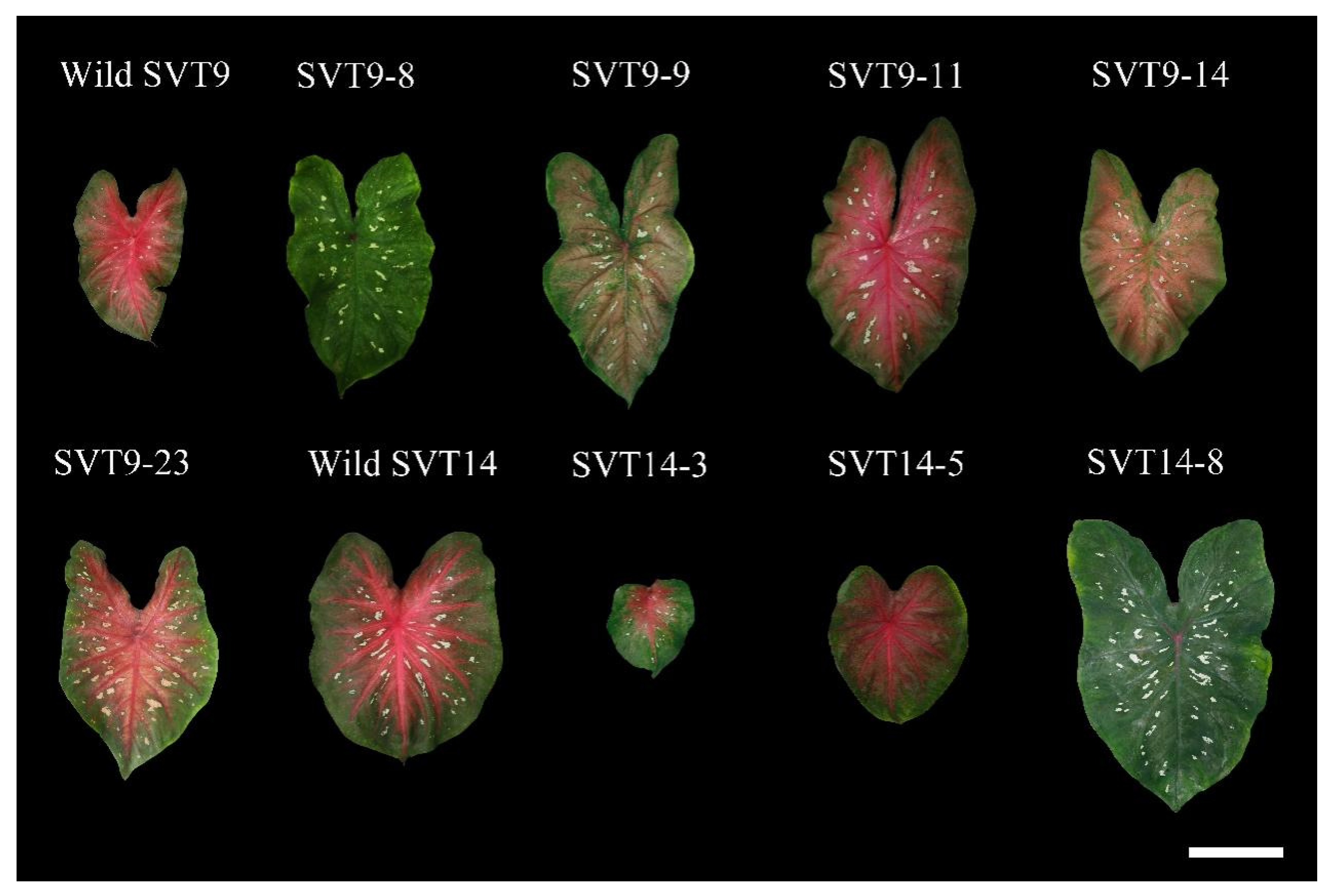
2.4. Visual Observation and Morphological Characterization

Morphological characteristics including plant height, leaf shape, and leaf color patterns were visually observed and compared. The plants with obviously morphological variation from their parental counterpart were flagged and monitored closely. By using an electronic digital caliper, leaf thickness was measured at three locations on the leaf lamina between major leaf veins, and petiole diameter was taken at the middle part of the petiole. Two or three mature leaves per plant were measured for leaf length, leaf width, leaf thickness, and petiole diameter.

2.5. Stomatal Assessment

To determine stomatal size and stomatal density, nail polish imprints obtained from the lower epidermis of the fully expanded caladium leaves were measured as described by Cai et al. [8]. The imprints were viewed and photographed under a Nikon Eclipse Ni-U microscope (Nikon, Tokyo, Japan) equipped with a Nikon DS-Ri2 camera at a magnification of 200 times. Stomatal measurement was carried out using NIS Elements D software, and three mature leaves were selected randomly from each plant and five independent counts made on each leaf.

2.6. Flow Cytometry Analysis

The relative nuclear DNA content was determined by using a CytoFLEX flow cytometer (Beckman Coulter, Suzhou, China) equipped with a 488 nm argon laser using the protocol proposed by Zhang et al. [9]. After main veins were removed, about 50 mg of fresh leaf tissue was rapidly chopped using a sharp razor blade in 350 μL of cold woody plant buffer (WPB), as described by Loureiro et al. [28] for releasing nuclei. The resulting crude solution was filtered through a 40 μm nylon mesh into a 5 mL sample loading tube to eliminate cell debris, and then 300 μL of fluorochrome stain (50 μg mL−1) composed of propidium iodide (PI) and RNase A were added to the tube. After incubation in the dark for 30 min for staining of the nuclei, the nuclear suspension was shaken gently for 5 s prior to sample analysis on the flow cytometer. Three flow cytometry runs were performed for each plant sample, and a minimum of 3000 nuclei were counted for each run, with only a coefficient of variance (CV) less than 5% accepted for the 2C peaks in the samples.

2.7. Chromosome Counting

To calibrate relative nuclear DNA content from flow cytometry analysis, a root tip squash method for counting chromosomes at mitotic metaphase was performed. Metaphase chromosomes were prepared according to Cao et al. [25], and at least 10 highly resolved cells with well-spread chromosomes were visualized and imaged under oil immersion at 1000 times magnification under a bright field of the Nikon Eclipse Ni-U microscope.

2.8. SSR Analysis of Nuclear Genome

Evaluation of genetic homogeneity was performed using 12 pairs of caladium-specific simple sequence repeat (SSR) primers designed by Gong and Deng [29], namely, CaM5, CaM16, CaM21, CaM35, CaM42, CaM62, CaM78, CaM87, CaM96, CaM101, CaM106, and CaM113, which were synthesized by Sangon Biotech (Shanghai, China). Total nuclear DNA was extracted from about 100 mg young leaf tissue of the plant samples using the modified cetyltrimethylammonium bromide (CTAB) method [30]. The quantity and quality of the DNA were determined with a NanoDrop One C spectrophotometer (Thermo Fisher Scientific, Madison, WI, USA) and 0.8% agarose gel electrophoresis, and then the DNA was diluted to a concentration of 25 ng/μL. SSR-PCR was performed using 20 μL reaction mixtures consisting of 2 μL of temple DNA, 10 μL 2 × Taq PCR StarMix with loading dye (GenStar, Beijing, China), 0.4 μL of each reverse and forward primer (10 μM), and 7.2 μL ddH2O on a T100 thermocycler (BIO-RAD, Singapore) following the amplification procedure described by Cao and Deng (2020). The amplified products were separated on 20 cm long and 1 mm thick non-denaturing 8% acrylamide gels through electrophoresis in 1× TBE buffer at a constant voltage of 100 V, 50 W, and 50 mA for 1.0–1.5 h. After electrophoresis, the gels were silver stained according to Bassam et al. [31].

2.9. Data Analysis

The resulting data were expressed as the mean value ± standard deviation and were analyzed by one-way analysis of variance in SPSS 23.0 software, and statistically significant differences were assessed according to Duncan’s multiple range test at p < 0.05.

3. Results

3.1. Plant Regeneration and Establishment

Callus initiation, shoot and root emergence, and plantlet regeneration from the leaf segments of the two aneuploid stock plants via one-step culture protocol are shown in Figure 1. After about 10 days of culture, it was found that the edges of the excised leaf pieces were slowly curled upward (Figure 1A). After one month of culture in the dark, a small amount of calli with a light yellow color and compact texture were observed around the cut surfaces of the explants (Figure 1B). New shoots and/or roots began to emerge directly from the induced calli 3 months after culture in the light (Figure 1C), and lots of well-developed plantlets with vigorous roots were recovered and were ready to be transferred to the acclimatization stage within 5 months of culture on the plantlet induction medium (Figure 1D). After three weeks of acclimatization, a survival rate of over 90% was recorded for the regenerated plants from both of the two aneuploids, and a total of 231 and 79 plants were obtained from the SVT9 and the SVT14, respectively. Lastly, a total of 23 and 8 plantlets were selected randomly from the regenerated populations of the SVT9 and the SVT14, respectively, and grown in plastic pots for subsequent analysis, and each plant was individually labeled with its donor parent followed by a sequential number, e.g., SVT9-1.

3.2. Comparisons of the Leaf Color Patterns and the Plant Performance among the Regenerated Plants

Approximately 4 months after transplanting, morphological differences were observed on some established plants. Amongst the randomly selected 23 and 8 individuals regenerated from the SVT9 and the SVT14, respectively, 5 plants from the SVT9 (SVT9-8, SVT9-9, SVT9-11, SVT9-14, and SVT9-23) and 3 plants originated from the SVT14 (SVT14-3, SVT14-5, and SVT14-8) showed a remarkable level of morphological variation in leaf color patterns compared with their corresponding parent plant (Figure 2 and Table 1). Namely, the morphological variation frequency reached as high as 21.7% and 37.5% among the randomly chosen plants regenerated from the SVT9 and the SVT14, respectively. In addition, all the morphological variants from the two aneuploids demonstrated some morphological differences from each other (Figure 2 and Figure 3 and Table 1), and the remaining plants exhibited similar morphological characteristics to their parental plant.
Clear differences were observed in the leaf color patterns (Figure 2 and Table 1) and plant performance (Figure 3) among the morphological variants and their donor plants. The wild SVT9 possessed heart-shaped (fancy) leaves with clear red main veins, small white spots between veins, and green leaf margins. As to the morphological variants from the SVT9, the leaves of SVT9-8, SVT9-9, and SVT9-14 had green main veins and whiter or pinkish-red spots, which was drastically different from the wild type. Additionally, the three plants shared most of the leaf color characteristics, though they could be easily distinguished from each other by the interveinal area color, with green (SVT9-8), purple-red (SVT9-9), or pinkish-red (SVT9-14) colors, respectively. The leaves of both SVT9-11 and SVT9-23 were bigger and had whiter or pinkish-red and much larger spots compared with the wild SVT9. The tetraploid aneuploid SVT14 plants grew well, and each developed several round fancy leaves with red main veins, red interveinal areas, a number of white spots, and green leaf margins. For the three variants (SVT14-3, SVT14-5, and SVT14-8) from the SVT14, all of them shared rounder and thicker leaves than the established plants from the SVT9. The main veins of both SVT14-3 and SVT14-5 showed a similar color to the SVT14, whereas most parts of the main veins of SVT14-8 appeared green in color. SVT14-3 grew very slowly and developed few leaves. Unlike the wild-type SVT14 and the other morphological variants, no spots were seen on the leaves of SVT14-5.

3.3. Morphological and Stomatal Characteristics among Morphological Variants

The morphological characteristics and stomatal parameters of the morphological variants and their parents were measured as shown in Table 2 and Table 3. Compared to the others, the resulting variants of the wild SVT9 were much higher with significantly longer and wider leaves. Considerable growth differences were also found among the three variants from the SVT14. SVT14-3 grew particularly slowly and produced only two significantly smaller leaves (9.90 cm in length and 8.01 cm in width) during a 4-month period in the greenhouse, whereas the plant height of SVT14-8 reached the highest value of 51.5 cm with significantly larger leaves. Within all the plants selected, there was a wide range in the ratio of leaf length to leaf width, which varied from a minimum of 1.49 (SVT9-23) to a maximum of 1.96 (SVT9-8) for the SVT9-regenerated morphological variants and varied from 1.16 (SVT14-5) to 1.47 (SVT14-8) for the regenerants from the SVT14. No significant differences of leaf thickness were found among the SVT9 and its variants nor among the SVT14 and its regenerants. Furthermore, compared with the diploid aneuploid SVT9 and its variants, all of the morphological variants from the SVT14 except SVT14-8 were characterized with significantly lower leaf length/width ratio and significantly thicker leaf blades.
As to stomatal guard cells, the variants from the SVT9 varied from 27.02 μm (SVT9-9) to 33.51 μm in length (SVT9-23) and from 17.07 μm (SVT9-11) to 18.91 μm (SVT9-23) in width, indicating that SVT9-23 had larger stomatal size than other variants from the SVT9. Significant differences in stomatal size were also found among the morphological variants from the SVT14, and both the length (34.90 μm) and width (23.81 μm) of the stomatal guard cell in SVT14-5 reached the highest values. In terms of stomatal density, no significant differences were found among the morphological variants from the SVT14, while it significantly differed among the SVT9-regenerated variants. Further, the wild SVT14 and its variants showed increased stomatal size and lower stomatal density when compared with the SVT9 group.

3.4. Relative DNA Content Variation

As shown in Table 4, substantial changes in mean fluorescence intensity (MFI) were recorded among the selected plants and their parent counterparts. In terms of the plants derived from the wild SVT9, their MFI ranged from 348594.2/2C (SVT9-8) to 401378.1/2C (SVT9-4), which was equivalent to 3.58% lower to 11.02% higher than that of their donor parent. Among the randomly selected 23 plants from the SVT9, the five morphological variants (SVT9-8, SVT9-9, SVT9-11, SVT9-14, and SVT9-23) and SVT9-4 (with no obvious morphological changes) were all found to have significant differences in MFI from the wild parent. Among the established plants from the SVT14, extensive variations in MFI from 615617.3/2C (SVT14-5) to 726124.5/2C (SVT14-1) were recorded, which was a 9.30% reduction to a 6.99% increase compared with the wild SVT14, respectively. Amongst the established eight plants from the SVT14, all four morphological variants (SVT14-1, SVT14-3, SVT14-5, and SVT14-8) showed significantly different MFI compared with the wild type, which means the MFI of 50% of the regenerated plants changed significantly from their original parent, a tetraploid aneuploid. Taken together, these results indicated that a higher percentage of plants regenerated in vitro from tetraploid aneuploids might tend to significantly change the relative DNA content compared with the diploid aneuploids in caladium.

3.5. Chromosome Number Changes

Detailed information on the chromosome number of the two wild-type aneuploids and their tissue-cultured plants are shown in Table 4 and Figure 4. By chromosome counting, the wild SVT9 and the wild SVT14 were found to have 2n = 2x − 2 = 28 and 2n = 4x − 6 = 54 chromosomes, respectively (Table 4; Figure 4A,H), consistent with the previous study by Chen et al. [11]. For the in vitro-produced plants from the leaf segments of the wild SVT9, the chromosome number ranged from 27 to 30. Two morphological variants, namely, SVT9-8 (Figure 4B) and SVT9-14 (Figure 4C), lost one chromosome and had 2n = 2x − 3 = 27 chromosomes, which might be explained by their significantly decreased MFI compared with the wild type. Two regenerated plants from the SVT9 (SVT9-9 and SVT9-11) with significantly increased MFI gained one extra chromosome (2n = 2x − 1 = 29) (Figure 4D,E). Intriguingly, the chromosome number was 2n = 2x = 30 in both the SVT9-14 (Figure 4F) and SVT9-23 (Figure 4G), which was the same in the wild ‘Red Flash’ caladium (2n = 2x = 30) (Cao et al., 2016), the donor sources of the SVT9. The rest of the plants with no significantly different changes in MFI had a similar chromosome number to the wild SVT9. In respect of the plants produced from the SVT14 in vitro, the chromosome number of four plants, namely, SVT14-2, SVT14-4, SVT14-6, and SVT14-7, was similar to the wild-type SVT14 (2n = 4x − 6 = 54) (Figure 4H), which was consistent with their measured MFI which had no significant changes. The SVT14-1 with significantly higher MFI gained two more chromosomes (2n = 4x − 4 = 56) (Figure 4I), and three plants, namely, SVT14-3 (Figure 4J), SVT14-5 (Figure 4K), and SVT14-8 (Figure 4L), with significantly lower MFI, lost four chromosomes (2n = 4x − 10 = 50).

3.6. SSR Identification of the Resulting Plants

SSR analysis was carried out using 12 pairs of caladium-specific primers to assess the genetic fidelity of the randomly selected 23 and 8 plantlets regenerated from the SVT9 and the SVT14 in vitro, respectively. For each pair of primers, the number of clear and reproducible DNA bands varied from one to four on 8% non-denaturing acrylamide gels (Figure 5). Out of the 12 markers adopted, 3 primers (CaM16, CaM42, and CaM101) detected banding pattern changes in the randomly selected plants from the SVT9. In detail, three plants (SVT9-8, SVT9-14, and SVT9-19) lost one of the two bands amplified by CaM16 (Figure 5A), and one plant (SVT9-23) showed one and two new alleles with primers CaM42 (Figure 5B) and CaM101 (Figure 5C), respectively, indicating genomic instability of the plants regenerated from the diploid aneuploid SVT9. Nevertheless, no polymorphism was detected among the SVT14 and its regenerants when employing all 12 pairs of SSR primers (Figure 5A–C).

4. Discussion

In caladium, a much higher proportion of aneuploid lines were found in the regenerated plants in vitro in recent cytological studies, and some of them grew normally with improved ornamental values [8,9,11,25,26]. Therefore, it is necessary to establish an ideal and efficient protocol to maintain these valuable sources of genetic variation for future breeding programs and genetic research activities. The stable conservation of aneuploid plant species mainly relies on in vitro clonal propagation [18,19]. However, the occurrence of somaclonal variation is one of the major problems during in vitro culture, as it may cause some unwanted changes in the agronomic traits of the donor genotypes [10,20]. Morphological and genetic changes originating from somaclonal variation can be identified at morphological, cytological, physiological, biochemical, and molecular levels [10]. Hence, this study comprehensively evaluated the genetic stability of the in vitro-propagated plants from two types of caladium aneuploids, i.e., the SVT9 (2n = 2x − 2 = 28) and the SVT14 (2n = 4x − 6 = 54) [11], by using morphological, cytological, and molecular techniques.
Out of 23 and 8 randomly selected plants from the SVT9 and the SVT14, respectively, 5 plants from the SVT9 and 3 plants from SVT14 exhibited obvious phenotypical differences from their parent (Figure 2 and Figure 3 and Table 1), indicating that 21.7% and 37.5% of the morphological variation occurred in the regenerated plants from the SVT9 and SVT14, respectively. The morphological variation frequency of the plants regenerated from the two aneuploids in this study was higher in both compared with the report on the plants from the immature leaves of ‘Red Flash’ caladium by Cao et al. [25] in which a variation frequency of only 6.9% and 12.5% was found when cultured on the medium with or without 2,4-D, respectively. This may be attributed to the higher genetic instability of aneuploids that can result in a much higher morphological variation frequency during plant tissue culture compared with euploids. A relatively high frequency of somaclonal variation was also reported in rice aneuploids, where a total of 26 lines were observed with morphological differences among 114 asexually propagated rice lines [19].
To avoid mistakes in identifying somaclonal variation relying on morphological characterization, flow cytometry analysis and chromosome counting were carried out in this study to confirm the ploidy level and the chromosome number of all the selected plants. Previous reports found that changes in the nuclear DNA content and chromosome number are common among tissue-cultured somaclones [11,25,26]. In this study, 6 out of 23 plants from the SVT9 and 4 out of 8 plants from the SVT14 also showed considerable variation in MFI (Table 4) and chromosome number (Figure 4 and Table 4), especially for the regenerants from the SVT14, a tetraploid aneuploid. It is worth noting that both SVT9-4 and SVT14-1 gained two extra chromosomes, whereas no obvious morphological differences from their corresponding parent were observed, which may be attributed to subtle and indistinguishable changes in the morphological traits of the two variants. The results of the chromosome counting were generally consistent with the measured MFI of these plants, except for several plants like SVT9-4 and SVT9-23. The MFI of SVT9-4 and SVT9-23 increased by 11.02% and 4.81% compared to their donor parent, respectively (Table 4), while both of them gained two extra chromosomes in somatic cells (Figure 4F,G). Cao et al. suggested that the gain or loss of one chromosome in the ‘Tapestry’ caladium genome might lead to a 3.3% increase or reduction in nuclear DNA content, respectively [26]. Therefore, the results of the chromosome counting and DNA content analysis lead us to believe that the SVT9-4 gained one or more chromosomes with a larger size compared with SVT9-23. Additional methods such as fluorescence in situ hybridization (FISH) are needed to visualize the extra chromosome(s) and the abnormal mitotic behavior in caladium aneuploids for the purpose of confirming these speculations.
In caladium, leaf thickness and shape, stomatal guard cell length, and stomatal density could be used as indicators for assessing ploidy level [8,9,11]. In this study, the tetraploid aneuploid SVT14 and its regenerated variants developed rounder and thicker leaves with larger stomatal guard cells and significantly reduced stomatal density compared with the diploid aneuploid SVT9 and its variants (Table 2 and Table 3). Significant differences in stomatal guard cell length and width were found both among the variants from the SVT9 and the regenerants from the SVT14 (Table 3), which suggests that changes in chromosome number have significant effects on stomatal size in caladium. Similar results were also observed in the induced diploid caladium aneuploids created by Zhang et al. [9] and the caladium somaclonal variants produced by Chen et al. [11].
In addition to morphological and cytological studies, SSR analysis is an extensively used and effective tool that enables rapid and accurate evaluation of genetic stability in somaclonal variation [10,26]. In this study, out of the 12 markers used, 3 SSR markers detected changes in the DNA bands (or alleles) in four plants from the SVT9 consisting of SVT9-8, SVT9-14, SVT9-19, and SVT9-23 (Figure 5). Both SVT9-8 and SVT9-14, with one chromosome missing, lost a similar smaller allele using the SSR markers CaM16 (Figure 5A) and CaM42 (Figure 5B), suggesting that the two markers may locate on the same chromosome. One and two new alleles were identified in SVT9-23 at the CaM42 (Figure 5B) and CaM101 loci (Figure 5C), respectively, which indicates that nucleotide changes or genetic combinations might emerge in SVT9-23 during tissue culture. However, no polymorphism was found among the wild SVT14 (a tetraploid aneuploid) and its regenerants using the 12 pairs of SSR primers in our study (Figure 5C). Polyploids contain multiple copies of the same genes, and thus we speculate that the regenerants from the SVT14 were only subjected to the changes in chromosome number, while their gene sequences and chromosome structure did not change. No or a low level of polymorphism was also observed among induced polyploid lines like Solanum tuberosum L. [32] and Chrysanthemum lavandulifolium [33] by using DNA molecular markers. Generally, one type of molecular marker alone may be insufficient to evaluate the genetic stability of micropropagated plants. Polyploidization can induce epigenetic changes in whole genomes in plant species [32,33], and therefore epigenetic studies, like the methylation-sensitive amplified polymorphism technique, should be adopted to assess the genetic stability of these aneuploid regenerants in the future.
Main vein color is considered to be controlled by a single nuclear locus (V) with three alleles, namely, Vr (red), Vw (white), and Vg (green), with a dominance order of Vr > Vw > Vg [34]. Changes in main vein color have been observed in somaclonal variants [11,25] and antimitotic agent-induced variants in caladium [8,9]. In this study, green main veins were observed in SVT9-8, SVT9-14, and SVT14-8 (Figure 2 and Figure 3 and Table 1). The wild SVT9 and SVT14 were both characterized by red main veins; thus, the occurrence of green main veins in the three variants might result from the mutation of Vr and Vw to Vg or from the loss of the Vr and Vw alleles. In addition, Deng et al. reported that leaf spots are controlled by a single locus with two alleles in caladium, namely, the dominant allele S and recessive allele s [35]. The SVT14-5 lost four chromosomes and had no leaf spots (Figure 2), which might be caused by a mutation from Ss to ss or SS to ss. These assumptions need more detailed evidence to support them.
To minimize the incidence of variation in tissue culture-derived plants, strategies such as using young tissue or organs as the explant source, optimization of plant growth regulators, and reducing the culture period have been employed during tissue culture [10]. Previous reports found that a high frequency of leaf color variation was associated with mature leaf explants or relatively high concentrations of auxins used, especially for 2,4-D [24,25]. In this study, immature leaves were cultured on a medium containing 0.5 mg·L−1 NAA and 1.0 mg·L−1 6-BA for reducing somaclonal variation, though quite a high frequency of somaclonal variation was found in the established plants, especially for the tetraploid aneuploid SVT14. Recently, regenerated plants through direct somatic embryogenesis were found to have a similar DNA content as that of their parent in caladium [36]. Therefore, in vitro propagation via direct somatic embryogenesis might be a feasible approach to maintaining the genetic stability of aneuploid caladiums.
In general, chromosome imbalance often has deleterious effects on the growth and development of plant species [1,37]. Nevertheless, some aneuploid plant lines can survive and even exhibit distinctive and desirable morphological traits [3,15]. For instance, the ‘White Wing’ caladium that gained four extra chromosomes (2n = 2x = 34) compared with its parents was found to have improved ornamental values [15], implying that aneuploid lines hold great potential for cultivar improvement in caladium. Previous studies have produced and identified a number of promising aneuploid plant lines with a normal growth habit and preferable esthetic values in caladium [8,9,11,25,26]. In this study, three variants from the SVT9 and four variants from SVT14 were also identified to be aneuploid, and most of these variants grew normally and vigorously. Amongst these variants, several aneuploids, like SVT9-11 and SVT14-5, exhibited enhanced ornamental values such as attractive leaf color patterns, increased leaf number, and compact plant architecture. Aneuploidy may result in alterations in global gene expression and phenotypic characteristics, thus providing potential germplasm for genetic analysis, chromosome engineering, and cultivar improvement in plant species [38]. Combining our results and previous reports, it can be concluded that caladium can tolerate various types of aneuploidy and should be regarded as a model plant for aneuploidy breeding and related genetic research.

5. Conclusions

This study provided insights into the genetic stability of in vitro-propagated plants from two types of caladium aneuploids at different ploidy levels. The results have shown that a relatively high frequency of somaclonal variation occurred in the plants regenerated from caladium aneuploids in vitro, especially for the tetraploid aneuploid SVT14. Cytological changes including chromosome loss or gain resulted in considerable changes in leaf color patterns, plant performance, and stomatal characteristics. SSR analysis detected changes in the DNA bands in some variants from the diploid aneuploid SVT9 but not among the wild SVT14 and its regenerants. The identified new aneuploid lines could provide valuable germplasm for future genetic improvement and studies in caladium.

Author Contributions

Conceptualization, S.Y., Y.W. and X.C.; data curation, S.Y. and X.Z.; formal analysis, S.Y. and Y.W.; funding acquisition, X.C. and Y.L.; investigation, Y.Z., D.J. and L.H.; methodology, S.Y. and Y.W.; resources, X.C. and Y.L.; writing—original draft, S.Y. and Y.W.; writing—review and editing, X.C. All authors have read and agreed to the published version of the manuscript.

Funding

This research was funded in part by the Scientific Research Project of Hubei Education Department, under grant no. B2018024; and Key Research and Development Project of Hubei Province, under grant no. 2021BBA096.

Institutional Review Board Statement

Not applicable.

Informed Consent Statement

Informed consent was obtained from all subjects involved in the present study.

Data Availability Statement

The data presented in this study are available on request from the corresponding authors.

Conflicts of Interest

The authors declare no conflict of interest.

References

  1. Henry, I.M.; Dilkes, B.P.; Miller, E.S.; Burkart-Waco, D.; Comai, L. Phenotypic consequences of aneuploidy in Arabidopsis thaliana. Genetics 2010, 186, 1231–1245. [Google Scholar] [CrossRef] [Green Version]
  2. Birchler, J.A. Aneuploidy in plants and flies: The origin of studies of genomic imbalance. Semin. Cell Dev. Biol. 2013, 24, 315–319. [Google Scholar] [CrossRef] [PubMed]
  3. Dang, J.; Wu, T.; Liang, G.; Wu, D.; Guo, Q. Identification and characterization of a loquat aneuploid with novel leaf phenotypes. HortScience 2019, 54, 804–808. [Google Scholar] [CrossRef] [Green Version]
  4. Siegel, J.J.; Amon, A. New insights into the troubles of aneuploidy. Annu. Rev. Cell Dev. Biol. 2012, 28, 189–214. [Google Scholar] [CrossRef] [PubMed] [Green Version]
  5. Gao, R.; Wang, H.; Dong, B.; Yang, X.; Chen, S.; Jiang, J.; Chen, F. Morphological, genome and gene expression changes in newly induced autopolyploid Chrysanthemum lavandulifolium (Fisch. ex Trautv.) Makino. Int. J. Mol. Sci. 2016, 17, 1690. [Google Scholar] [CrossRef] [Green Version]
  6. Cai, B.; Wang, T.; Yue, F.; Harun, A.; Zhu, B.; Qian, W.; Li, Z. Production and cytology of Brassica autoallohexaploids with two and four copies of two subgenomes. Theor. Appl. Genet. 2022, 135, 2641–2653. [Google Scholar] [CrossRef]
  7. Evtushenko, E.V.; Lipikhina, Y.A.; Stepochkin, P.I.; Vershinin, A.V. Cytogenetic and molecular characteristics of rye genome in octoploid triticale (× Triticosecale Wittmack). Comp. Cytogenet. 2019, 13, 423–434. [Google Scholar] [CrossRef] [PubMed]
  8. Cai, X.; Cao, Z.; Xu, S.; Deng, Z. Induction, regeneration and characterization of tetraploids and variants in ‘Tapestry’ caladium. Plant Cell Tissue Organ Cult. 2015, 120, 689–700. [Google Scholar] [CrossRef]
  9. Zhang, Y.S.; Chen, J.J.; Cao, Y.M.; Duan, J.X.; Cai, X.D. Induction of tetraploids in ‘Red Flash’ caladium using colchicine and oryzalin: Morphological, cytological, photosynthetic and chilling tolerance analysis. Sci. Hortic. 2020, 272, 109524. [Google Scholar] [CrossRef]
  10. Bairu, M.W.; Aremu, A.O.; Van Staden, J. Somaclonal variation in plants: Causes and detection methods. Plant. Growth. Regul. 2011, 63, 147–173. [Google Scholar] [CrossRef]
  11. Chen, J.J.; Zhang, Y.S.; Duan, J.X.; Cao, Y.M.; Cai, X.D. Morphological, cytological, and pigment analysis of leaf color variants regenerated from long-term subcultured caladium callus. In Vitro Cell Dev. Biol. Plant 2021, 57, 60–71. [Google Scholar] [CrossRef]
  12. Wu, Y.; Sun, Y.; Sun, S.; Li, G.; Wang, J.; Wang, B.; Lin, X.; Huang, M.; Gong, Z.; Sanguinet, K.A.; et al. Aneuploidization under segmental allotetraploidy in rice and its phenotypic manifestation. Theor. Appl. Genet. 2018, 131, 1273–1285. [Google Scholar] [CrossRef] [Green Version]
  13. Husband, B.C. Chromosomal variation in plant evolution. Am. J. Bot. 2004, 91, 621–625. [Google Scholar] [CrossRef]
  14. Henry, I.M.; Dilkes, B.P.; Young, K.; Watson, B.; Wu, H.; Comai, L. Aneuploidy and genetic variation in the Arabidopsis thaliana triploid response. Genetics 2005, 170, 1979–1988. [Google Scholar] [CrossRef] [PubMed] [Green Version]
  15. Parrish, S.B.; Deng, Z. Discovery and characterization of novel fertile triploids and a new chromosome number in Caladium (Caladium × hortulanum). HortScience 2022, 57, 1078–1085. [Google Scholar] [CrossRef]
  16. Ehdaie, B.; Waines, J.G. Chromosomal location of genes influencing plant characters and evapotranspiration efficiency in bread wheat. Euphytica 1997, 96, 363–375. [Google Scholar] [CrossRef]
  17. Bhatia, R.; Sharma, K.; Parkash, C.; Pramanik, A.; Singh, D.; Singh, S.; Dey, S.S. Microspore derived population developed from an inter-specific hybrid (Brassica oleracea × B. carinata) through a modified protocol provides insight into B genome derived black rot resistance and inter-genomic interaction. Plant Cell Tissue Organ Cult. 2021, 145, 417–434. [Google Scholar]
  18. Meghwal, P.R.; Singh, S.K.; Sharma, H.C. Micropropagation of aneuploid guava. Indian J. Hortic. 2003, 60, 29–33. [Google Scholar]
  19. Gong, Z.; Xue, C.; Zhou, Y.; Zhang, M.; Liu, X.; Shi, G.; Gu, M. Molecular cytological characterization of somatic variation in rice aneuploids. Plant Mol. Biol. Rep. 2013, 31, 1242–1248. [Google Scholar] [CrossRef]
  20. Lin, W.; Xiao, X.; Sun, W.; Liu, S.; Wu, Q.; Yao, Y.; Zhang, H.; Zhang, X. Genome-wide identification and expression analysis of cytosine DNA methyltransferase genes related to somaclonal variation in Pineapple (Ananas comosus L.). Agronomy 2022, 12, 1039. [Google Scholar] [CrossRef]
  21. Deng, Z. Caladium genetics and breeding: Recent advances. Floric. Ornam. Biotechnol. 2012, 6, 53–61. [Google Scholar]
  22. Wilfret, G.J. Caladium. In The Physiology of Flower Bulbs; Elsevier: Amsterdam, The Netherlands, 1993; pp. 239–247. [Google Scholar]
  23. Cao, Z.; Deng, Z.; Mclaughlin, M. Interspecific genome size and chromosome number variation shed new light on species classification and evolution in Caladium. J. Am. Soc. Hortic. Sci. 2014, 139, 449–459. [Google Scholar] [CrossRef] [Green Version]
  24. Ahmed, E.U.; Hayashi, T.; Yazawa, S. Auxins increase the occurrence of leaf-colour variants in Caladium regenerated from leaf explants. Sci. Hortic. 2004, 100, 153–159. [Google Scholar] [CrossRef]
  25. Cao, Z.; Sui, S.; Cai, X.; Yang, Q.; Deng, Z. Somaclonal variation in ‘Red Flash’ caladium: Morphological, cytological and molecular characterization. Plant Cell Tissue Organ Cult. 2016, 126, 269–279. [Google Scholar] [CrossRef] [Green Version]
  26. Cao, Z.; Deng, Z. Morphological, cytological and molecular marker analyses of ‘Tapestry’ caladium variants reveal diverse genetic changes and enable association of leaf coloration pattern loci with molecular markers. Plant Cell Tissue Organ Cult. 2020, 143, 363–375. [Google Scholar] [CrossRef]
  27. Murashige, T.; Skoog, F. A revised medium for rapid growth and bioassays with tobacco tissue cultures. Physiol. Plant. 1962, 15, 473–479. [Google Scholar] [CrossRef]
  28. Loureiro, J.; Rodriguez, E.; Doležel, J.; Santos, C. Two new nuclear isolation buffers for plant DNA flow cytometry: A test with 37 species. Ann. Bot. 2007, 100, 875–888. [Google Scholar] [CrossRef] [PubMed] [Green Version]
  29. Gong, L.; Deng, Z. Development and characterization of microsatellite markers for caladiums (Caladium Vent.). Plant Breed. 2011, 130, 591–595. [Google Scholar] [CrossRef]
  30. Fulton, T.M.; Chunzoongse, J.; Tanksley, S.D. Microprep protocol for extraction of DNA from tomato and other herbaceous plants. Plant Mol. Biol. Rep. 1995, 13, 207–209. [Google Scholar] [CrossRef]
  31. Bassam, B.J.; Caetano-Anollés, G.; Gresshoff, P.M. Fast and sensitive silver staining of DNA in polyacrylamide gels. Anal. Biochem. 1991, 196, 80–83. [Google Scholar] [CrossRef]
  32. Marfil, C.F.; Duarte, P.F.; Masuelli, R.W. Phenotypic and epigenetic variation induced in newly synthesized allopolyploids and autopolyploids of potato. Sci. Hortic. 2018, 234, 101–109. [Google Scholar] [CrossRef]
  33. Gao, L.; Diarso, M.; Zhang, A.; Zhang, H.; Dong, Y.; Liu, L.; Liu, B. Heritable alteration of DNA methylation induced by whole-chromosome aneuploidy in wheat. New Phytol. 2016, 209, 364–375. [Google Scholar] [CrossRef] [Green Version]
  34. Deng, Z.; Harbaugh, B.K. Independent inheritance of leaf shape and main vein color in caladium. J. Am. Soc. Hortic. Sci. 2006, 131, 53–58. [Google Scholar] [CrossRef]
  35. Deng, Z.; Goktepe, F.; Harbaugh, B.K. Inheritance of leaf spots and their genetic relationships with leaf shape and vein color in caladium. J. Am. Soc. Hortic. Sci. 2008, 133, 78–83. [Google Scholar] [CrossRef] [Green Version]
  36. Syeed, R.; Mujib, A.; Malik, M.Q.; Gulzar, B.; Zafar, N.; Mamgain, J.; Ejaz, B. Direct somatic embryogenesis and flow cytometric assessment of ploidy stability in regenerants of Caladium × hortulanum ‘Fancy’. J. Appl. Genet. 2022, 63, 199–211. [Google Scholar] [CrossRef] [PubMed]
  37. Sabooni, N.; Gharaghani, A. Induced polyploidy deeply influences reproductive life cycles, related phytochemical features, and phytohormonal activities in blackberry species. Front. Plant. Sci. 2022, 13, 938284. [Google Scholar] [CrossRef]
  38. Huettel, B.; Kreil, D.P.; Matzke, M.; Matzke, A.J.M. Effects of aneuploidy on genome structure, expression, and interphase organization in Arabidopsis thaliana. PLoS Genet. 2008, 4, e1000226. [Google Scholar] [CrossRef]
Figure 1. In vitro plant regeneration from leaf segments of two aneuploids of ‘Red Flash’ caladium (Caladium × hortulanum Birdsey), namely, the SVT9 and the SVT14. (A) Leaf segments of the SVT9 cultured on MS basal medium supplemented with 0.5 mg·L1 NAA and 1.0 mg·L1 6-BA for 10 days; (B) initiated callus from the edge of the excised leaf pieces of the SVT14 after one month of culture; (C) shoots or roots regenerated from the excised leaf pieces of the SVT9 after 3 months’ culture; (D) plantlets regenerated from the excised leaf pieces of the SVT9 after 5 months’ culture. Bars = 1 cm.
Agriculture 12 01708 g001
Figure 2. Typical leaves of the variants regenerated from the SVT9 and the SVT14 in vitro and their corresponding wild-type parent. The photos were taken 4 months after the plants were grown in plastic pots filled with a commercial potting mix and grown in a greenhouse. Scale bar: 5 cm.
Agriculture 12 01708 g002
Figure 3. Top views of the variants regenerated from the SVT9 and the SVT14 in vitro and their corresponding wild-type parent after transplantation to the pots for 4 months. Scale bar: 5 cm.
Agriculture 12 01708 g003
Figure 4. Micrographs of somatic chromosomes in the root tips of the randomly selected plants regenerated from the SVT9 and the SVT14 in vitro and their corresponding parent. (A) Wild SVT9 (2n = 2x − 2 = 28); (B) SVT9-8 (2n = 2x − 3 = 27); (C) SVT9-14 (2n = 2x − 3 = 27); (D) SVT9-9 (2n = 2x − 1 = 29); (E) SVT9-11 (2n = 2x − 1 = 29); (F) SVT9-4 (2n = 2x = 30); (G) SVT9-23 (2n = 2x = 30); (H) wild SVT14 (2n = 4x − 6 = 54); (I) SVT14-1 (2n = 4x − 4 = 56); (J) SVT14-3 (2n = 4x − 10 = 50); (K) SVT14-5 (2n = 4x − 10 = 50); (L) SVT14-8 (2n = 4x − 10 = 50). Bars = 10 μm.
Agriculture 12 01708 g004
Figure 5. SSR banding pattern of the randomly selected 23 and 8 plants regenerated from the SVT9 and the SVT14, respectively. (A) Banding patterns of SSR marker CaM16; (B) banding patterns of SSR marker CaM42; (C) banding patterns of SSR marker CaM101. Red arrows indicate the presence or absence of SSR bands in the in vitro-regenerated plants.
Agriculture 12 01708 g005
Table 1. Main foliar characteristics of the variants regenerated from two types of caladium aneuploids (SVT9 and SVT14) in vitro and their corresponding parent grown in plastic pots for 4 months.
Plant CodeLeaf ShapeMain VeinsInterveinal AreasLeaf MarginsLeaf Spots
Wild SVT9FancyRedRedGreenWhite
SVT9-8FancyGreenGreenLight greenWhite
SVT9-9FancyGreenPurple-redLight greenWhite
SVT9-11FancyRedRedGreenWhite and pinkish-red
SVT9-14FancyGreenPinkish-redGreenPinkish-red
SVT9-23FancyRedRedGreenPinkish-red
Wild SVT14FancyRedRedGreenWhite
SVT14-3FancyRedRedLight greenWhite
SVT14-5FancyRedRedGreenNo spots
SVT14-8FancyGreenGreenLight greenWhite
Table 2. Morphological characteristics of the in vitro-regenerated caladium variants and their corresponding wild type established in this study.
Plant CodePlant Height (cm)Leaf Number per PlantLeaf Length (cm)Leaf Width (cm)Leaf Length/Width RatioLeaf Thickness (mm)
Wild SVT926.2618.67 ± 0.15 d11.23 ± 0.42 f1.66 ± 0.05 b0.26 ± 0.00 b
SVT9-834.2525.40 ± 5.99 bc12.90 ± 0.26 e1.96 ± 0.42 a0.26 ± 0.02 b
SVT9-945.6527.77 ± 1.33 ab16.23 ± 0.72 c1.71 ± 0.12 ab0.27 ± 0.02 b
SVT9-1137.4627.00 ± 1.08 bc16.73 ± 0.91 c1.61 ± 0.12 b0.25 ± 0.02 b
SVT9-1430.5623.63 ± 0.91 bc14.30 ± 0.85 d1.65 ± 0.04 b0.25 ± 0.02 b
SVT9-2330.2424.87 ± 1.05 bc16.70 ± 0.53 c1.49 ± 0.04 bc0.27 ± 0.02 b
Wild SVT1433.4523.43 ± 0.55 c19.00 ± 0.36 b1.23 ± 0.02 cd0.35 ± 0.02 a
SVT14-318.629.90 ± 1.98 e8.01 ± 1.56 g1.24 ± 0.01 cd0.35 ± 0.01 a
SVT14-522.7417.07 ± 0.64 d14.70 ± 0.95 d1.16 ± 0.03 d0.33 ± 0.01 a
SVT14-851.5331.50 ± 1.01 a21.13 ± 1.05 a1.47 ± 0.12 bc0.27 ± 0.02 b
Means (±standard deviation) within a column followed by different letters are significantly different at 5% level according to Duncan’s multiple range test.
Table 3. Stomatal differences among the in vitro-regenerated caladium variants and their corresponding wild type.
Plant CodeStomatal Guard Cell Length (μm)Stomatal Guard Cell Width (μm)Stomatal Density (no./mm2)
Wild SVT927.88 ± 2.69 cd19.08 ± 1.82 bc112.12 ± 4.34 b
SVT9-828.40 ± 2.33 bcd17.98 ± 2.05 bc96.21 ± 9.56 c
SVT9-927.02 ± 0.94 d18.57 ± 1.65 bc129.49 ± 10.92 a
SVT9-1129.16 ± 1.77 bc17.07 ± 1.97 c122.97 ± 5.01 ab
SVT9-1424.51 ± 0.87 e17.48 ± 2.19 c88.25 ± 3.74 c
SVT9-2333.51 ± 2.85 a18.91 ± 4.69 bc122.97 ± 13.67 ab
Wild SVT1433.67 ± 2.86 a20.65 ± 4.05 b68.10 ± 13.05 d
SVT14-330.24 ± 1.76 b20.74 ± 1.21 b68.72 ± 7.23 d
SVT14-534.90 ± 1.62 a23.81 ± 4.17 a67.27 ± 6.42 d
SVT14-834.65 ± 1.99 a20.77 ± 1.65 b70.17 ± 12.36 d
Means within a column followed by the different letters are significantly different according to Duncan’s range test at p < 0.05.
Table 4. Relative DNA content and chromosome count of the aneuploid caladium plants and their regenerants.
Plant CodeMean Fluorescence Intensity (MFI)MFI Change Compared to the Corresponding Wild Type (%)Chromosome Number Observed in Somatic Cells (2n)
Wild SVT9361,547.4 ± 275.92x − 2 = 28
SVT9-1369,554.8 ± 3243.8 ns+2.212x − 2 = 28
SVT9-2360,779.3 ± 5688.5 ns−0.212x − 2 = 28
SVT9-3363,844.3 ± 3293.5 ns+0.642x − 2 = 28
SVT9-4401,378.1 ± 2886.3 *+11.022x = 30
SVT9-5364,728.6 ± 4364.3 ns+0.882x − 2 = 28
SVT9-6366,356.2 ± 1425.4 ns+1.332x − 2 = 28
SVT9-7356,318.6 ± 5712.4 ns−1.452x − 2 = 28
SVT9-8348,594.2 ± 993.8 *−3.582x − 3 = 27
SVT9-9378,007.6 ± 4449.3 *+4.552x − 1 = 29
SVT9-10364,349.8 ± 2577.7 ns+0.782x − 2 = 28
SVT9-11380,383.7 ± 2924.5 *+5.212x − 1 = 29
SVT9-12369,176.3 ± 8997.0 ns+2.112x − 2 = 28
SVT9-13371,075.8 ± 4186.6 ns+2.642x − 2 = 28
SVT9-14350,182.6 ± 555.9 *−3.142x − 3 = 27
SVT9-15368,263.1 ± 2776.1 ns+1.862x − 2 = 28
SVT9-16370,699.7 ± 2459.0 ns+2.532x − 2 = 28
SVT9-17364,147.3 ± 6699.7 ns+0.722x − 2 = 28
SVT9-18359,826.5 ± 1295.5 ns−0.482x − 2 = 28
SVT9-19372,395.5 ± 532.5 ns+3.002x − 2 = 28
SVT9-20370,988.0 ± 3242.0 ns+2.612x − 2 = 28
SVT9-21371,636.7 ± 2274.3 ns+2.792x − 2 = 28
SVT9-22362,934.1 ± 16,053.1 ns+0.382x − 2 = 28
SVT9-23378,928.2 ± 1686.4 *+4.812x = 30
Wild SVT14678,715.4 ± 8091.94x − 6 = 54
SVT14-1726,124.5 ± 2163.8 *+6.994x − 4 = 56
SVT14-2689,727.1 ± 15,919.7 ns+1.624x − 6 = 54
SVT14-3624,862.1 ± 2061.8 *−7.934x − 10 = 50
SVT14-4673,317.1 ± 10,184.6 ns−0.804x − 6 = 54
SVT14-5615,617.3 ± 5015.4 *−9.304x − 10 = 50
SVT14-6684,457.9 ± 8992.9 ns+0.854x − 6 = 54
SVT14-7668,038.6 ± 2779.5 ns−1.574x − 6 = 54
SVT14-8640,112.3 ± 3472.5 *−5.694x − 10 = 50
MFI change compared to the corresponding wild type = [(MFI of the regenerated plant − MFI of its corresponding wild type) ÷ MFI of the corresponding wild type] × 100%. ns and * stand for, respectively, no significant and significant difference between the regenerated plant and its donor parent in mean fluorescence intensity according to Duncan’s range test at p < 0.05.
Publisher’s Note: MDPI stays neutral with regard to jurisdictional claims in published maps and institutional affiliations.

