Diagnostic Performance of [18F]Fluorocholine and [68Ga]Ga-PSMA PET/CT in Prostate Cancer: A Comparative Study

 , ,
, ,
Abstract
:1. Introduction
2. Materials and Methods
2.1. Patients
2.2. Study Agents
2.3. PET/CT Imaging and Evaluation
2.4. Statistics
3. Results
3.1. Study Cohort
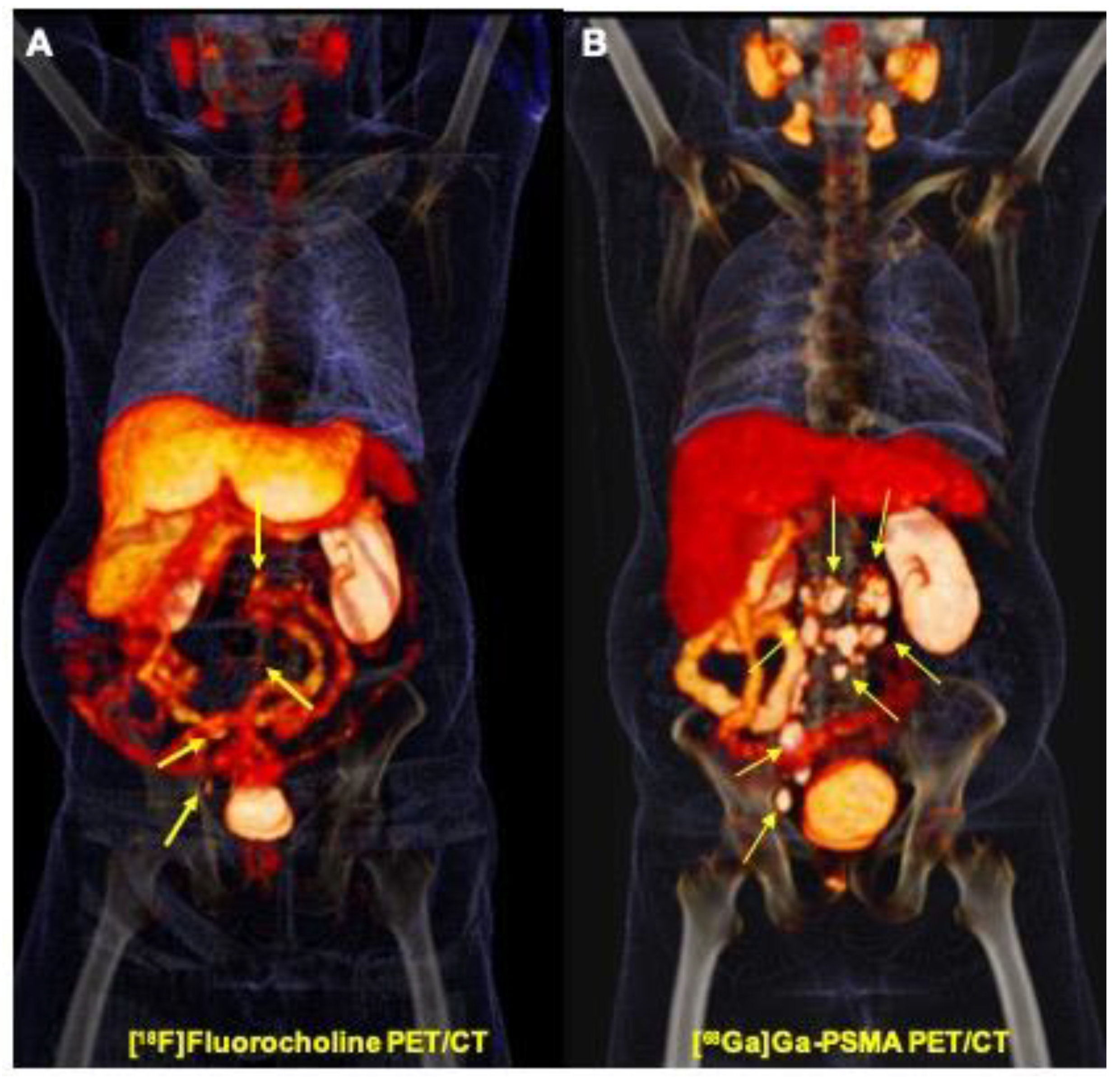
3.2. Lesion Based Findings
3.3. Prostate, Prostate Bed, and Seminal Vesicles
3.4. Regional and Distant Lymph nodes
3.5. Skeletal Lesions
3.6. Organ Metastases
3.7. Patient Based Findings
3.7.1. TNM Staging
3.7.2. Discordant Lesions
3.7.3. Correlation of PET Positivity and PSA Value
4. Discussion
5. Conclusions
Author Contributions
Funding
Conflicts of Interest
References
- Bray, F.; Ferlay, J.; Soerjomataram, I.; Siegel, R.L.; Torre, L.A.; Jemal, A. Global cancer statistics 2018: GLOBOCAN estimates of incidence and mortality worldwide for 36 cancers in 185 countries. CA A Cancer J. Clin. 2018, 68, 394–424. [Google Scholar] [CrossRef] [Green Version]
- Dulaney, C.R.; Osula, D.O.; Yang, E.S.; Rais-Bahrami, S. Prostate Radiotherapy in the Era of Advanced Imaging and Precision Medicine. Prostate Cancer 2016, 2016, 1–10. [Google Scholar] [CrossRef] [PubMed] [Green Version]
- Bouchelouche, K.; Turkbey, B.; Choyke, P.L. Advances in imaging modalities in prostate cancer. Curr. Opin. Oncol. 2015, 27, 224–231. [Google Scholar] [CrossRef] [PubMed]
- Uchio, E.M.; Aslan, M.; Wells, C.K.; Calderone, J.; Concato, J. Impact of Biochemical Recurrence in Prostate Cancer Among US Veterans. Arch. Intern. Med. 2010, 170, 1390–1395. [Google Scholar] [CrossRef] [PubMed] [Green Version]
- Ward, J.F.; Moul, J.W. Rising prostate-specific antigen after primary prostate cancer therapy. Nat. Clin. Pr. Urol. 2005, 2, 174–182. [Google Scholar] [CrossRef] [PubMed]
- Li, R.; Ravizzini, G.C.; Gorin, M.A.; Maurer, T.; Eiber, M.; Cooperberg, M.R.; Alemozzaffar, M.; Tollefson, M.K.; Delacroix, S.E.; Chapin, B.F. The use of PET/CT in prostate cancer. Prostate Cancer Prostatic Dis. 2017, 21, 4–21. [Google Scholar] [CrossRef]
- Fendler, W.P.; Calais, J.; Eiber, M.; Flavell, R.R.; Mishoe, A.; Feng, F.Y.; Nguyen, H.G.; Reiter, R.E.; Rettig, M.B.; Okamoto, S.; et al. Assessment of 68Ga-PSMA-11 PET Accuracy in Localizing Recurrent Prostate Cancer: A Prospective Single-Arm Clinical Trial. JAMA Oncol. 2019, 5, 856–863. [Google Scholar] [CrossRef] [Green Version]
- Mottet, N.; Bellmunt, J.; Bolla, M.; Briers, E.; Cumberbatch, M.G.; De Santis, M.; Fossati, N.; Gross, T.; Henry, A.M.; Joniau, S.; et al. EAU-ESTRO-SIOG Guidelines on Prostate Cancer. Part 1: Screening, Diagnosis, and Local Treatment with Curative Intent. Eur. Urol. 2017, 71, 618–629. [Google Scholar] [CrossRef]
- Kosuri, S.; Akhtar, N.H.; Smith, M.; Osborne, J.; Tagawa, S.T. Review of Salvage Therapy for Biochemically Recurrent Prostate Cancer: The Role of Imaging and Rationale for Systemic Salvage Targeted Anti-Prostate-Specific Membrane Antigen Radioimmunotherapy. Adv. Urol. 2012, 2012, 1–8. [Google Scholar] [CrossRef]
- Hovels, A.M.; Heesakkers, R.; Adang, E.; Jager, G.; Strum, S.; Hoogeveen, Y.; Severens, J.; Barentsz, J.O. The diagnostic accuracy of CT and MRI in the staging of pelvic lymph nodes in patients with prostate cancer: A meta-analysis. Clin. Radiol. 2008, 63, 387–395. [Google Scholar] [CrossRef] [PubMed]
- Horiuchi-Suzuki, K.; Konno, A.; Ueda, M.; Fukuda, Y.; Nishio, S.; Hashimoto, K.; Saji, H. Skeletal affinity of Tc(V)-DMS is bone cell mediated and pH dependent. Eur. J. Nucl. Med. Mol. Imaging 2004, 31, 388–398. [Google Scholar] [CrossRef] [PubMed]
- Picchio, M.; Messa, C.; Landoni, C.; Gianolli, L.; Sironi, S.; Brioschi, M.; Matarrese, M.; Matei, D.; De Cobelli, F.; Del Maschio, A.; et al. Value of [11C]choline-Positron Emission Tomography for Re-Staging Prostate Cancer: A Comparison With [18F]fluorodeoxyglucose-Positron Emission Tomography. J. Urol. 2003, 169, 1337–1340. [Google Scholar] [CrossRef]
- Beauregard, J.-M.; Beaulieu, A. How we read FCH-PET/CT for prostate cancer. Cancer Imaging 2016, 16, 41. [Google Scholar] [CrossRef] [Green Version]
- Beheshti, M.; Haim, S.; Zakavi, R.; Steinmair, M.; Waldenberger, P.; Kunit, T.; Nader, M.; Langsteger, W.; Loidl, W. Impact of [18F]Fluorocholine PET/CT in prostate cancer patients with biochemical recurrence: Influence of androgen deprivation therapy and correlation with PSA kinetics. J. Nucl. Med. 2013, 54, 833–840. [Google Scholar] [CrossRef] [PubMed] [Green Version]
- Beheshti, M.; Imamovic, L.; Broinger, G.; Vali, R.; Waldenberger, P.; Stoiber, F.; Nader, M.; Gruy, B.; Janetschek, G.; Langsteger, W. 18 F Choline PET/CT in the Preoperative Staging of Prostate Cancer in Patients with Intermediate or High Risk of Extracapsular Disease: A Prospective Study of 130 Patients 1. Radiology 2010, 254, 925–933. [Google Scholar] [CrossRef] [PubMed] [Green Version]
- Fanti, S.; Minozzi, S.; Castellucci, P.; Balduzzi, S.; Herrmann, K.; Krause, B.J.; Oyen, W.J.; Chiti, A. PET/CT with 11C-choline for evaluation of prostate cancer patients with biochemical recurrence: Meta-analysis and critical review of available data. Eur. J. Nucl. Med. Mol. Imaging 2015, 43, 55–69. [Google Scholar] [CrossRef] [PubMed]
- Mamede, M.; Ceci, F.; Castellucci, P.; Schiavina, R.; Fuccio, C.; Nanni, C.; Brunocilla, E.; Fantini, L.; Costa, S.; Ferretti, A. The Role of 11C-Choline PET Imaging in the Early Detection of Recurrence in Surgically Treated Prostate Cancer Patients With Very Low PSA Level <0.5 ng/mL. Clin. Nucl. Med. 2013, 38, e342-5. [Google Scholar]
- Lütje, S.; Heskamp, S.; Cornelissen, A.S.; Poeppel, T.D.; Broek, S.A.M.W.V.D.; Rosenbaum-Krumme, S.; Bockisch, A.; Gotthardt, M.; Rijpkema, M.; Boerman, O.C. PSMA Ligands for Radionuclide Imaging and Therapy of Prostate Cancer: Clinical Status. Theranostics 2015, 5, 1388–1401. [Google Scholar] [CrossRef] [Green Version]
- Heck, M.M.; Maurer, T.; Souvatzoglou, M.; Beer, A.J.; Ruffani, A.; Haller, B.; Graner, F.-P.; Kübler, H.; Haberhorn, U.; Eisenhut, M.; et al. Evaluation of Hybrid 68Ga-PSMA Ligand PET/CT in 248 Patients with Biochemical Recurrence After Radical Prostatectomy. J. Nucl. Med. 2015, 56, 668–674. [Google Scholar]
- Beheshti, M.; Manafi-Farid, R.; Geinitz, H.; Vali, R.; Loidl, W.; Mottaghy, F.M.; Langsteger, W. Multi-phasic 68Ga-PSMA PET/CT in detection of early recurrence in prostate cancer patients with PSA <1 ng/mL: A prospective study of 135 cases. J. Nucl. Med. 2020. [Google Scholar] [CrossRef]
- Calais, J.; Fendler, W.P.; Eiber, M.; Gartmann, J.; Chu, F.-I.; Nickols, N.G.; Reiter, R.E.; Rettig, M.B.; Marks, L.S.; Ahlering, T.E.; et al. Impact of 68Ga-PSMA-11 PET/CT on the Management of Prostate Cancer Patients with Biochemical Recurrence. J. Nucl. Med. 2017, 59, 434–441. [Google Scholar] [CrossRef] [Green Version]
- Bluemel, C.; Krebs, M.; Polat, B.; Linke, F.; Eiber, M.; Samnick, S.; Lapa, C.; Lassmann, M.; Riedmiller, H.; Czernin, J.; et al. 68Ga-PSMA-PET/CT in Patients With Biochemical Prostate Cancer Recurrence and Negative 18F-Choline-PET/CT. Clin. Nucl. Med. 2016, 41, 515–521. [Google Scholar] [CrossRef] [Green Version]
- Beheshti, M.; Paymani, Z.; Brilhante, J.; Geinitz, H.; Gehring, D.; Leopoldseder, T.; Wouters, L.; Pirich, C.; Loidl, W.; Langsteger, W. Optimal time-point for 68Ga-PSMA-11 PET/CT imaging in assessment of prostate cancer: Feasibility of sterile cold-kit tracer preparation? Eur. J. Nucl. Med. Mol. Imaging 2018, 45, 1188–1196. [Google Scholar] [CrossRef] [PubMed]
- Schwenck, J.; Rempp, H.; Reischl, G.; Kruck, S.; Stenzl, A.; Nikolaou, K.; Pfannenberg, C.; La Fougère, C. Comparison of 68Ga-labelled PSMA-11 and 11C-choline in the detection of prostate cancer metastases by PET/CT. Eur. J. Nucl. Med. Mol. Imaging 2016, 44, 92–101. [Google Scholar] [CrossRef] [PubMed]
- Treglia, G.; Mestre, R.P.; Ferrari, M.; Bosetti, D.G.; Pascale, M.; Oikonomou, E.; De Dosso, S.; Jermini, F.; O Prior, J.; Roggero, E.; et al. Radiolabelled choline versus PSMA PET/CT in prostate cancer restaging: A meta-analysis. Am. J. Nucl. Med. Mol. Imaging 2019, 9, 127–139. [Google Scholar] [PubMed]
- Alonso, O.; Dos Santos, G.; García Fontes, M.; Balter, H.; Engler, H. 68Ga-PSMA and 11C-cho-line comparison using a tri-modality PET/CT-MRI (3.0 T) system with a dedicated shuttle. Eur. J. Hybrid Imaging 2018, 2, 9. [Google Scholar] [CrossRef]
- Cantiello, F.; Crocerossa, F.; Russo, G.I.; Gangemi, V.; Ferro, M.; Vartolomei, M.D.; Lucarelli, G.; Mirabelli, M.; Scafuro, C.; Ucciero, G.; et al. Comparison between 64Cu-PSMA-617 PET/CT and 18F-Choline PET/CT imaging in early diagnosis of prostate cancer biochemical recurrence. Clin. Genitourin. Cancer 2018, 16, 385–391. [Google Scholar] [CrossRef]
- Afshar-Oromieh, A.; Zechmann, C.M.; Malcher, A.; Eder, M.; Eisenhut, M.; Linhart, H.G.; Holland-Letz, T.; Hadaschik, B.A.; Giesel, F.L.; Debus, J.; et al. Comparison of PET imaging with a 68Ga-labelled PSMA ligand and 18F-choline-based PET/CT for the diagnosis of recurrent prostate cancer. Eur. J. Nucl. Med. Mol. Imaging 2013, 41, 11–20. [Google Scholar] [CrossRef] [Green Version]
- Chondrogiannis, S.; Marzola, M.C.; Ferretti, A.; Grassetto, G.; Maffione, A.M.; Rampin, L.; Fanti, S.; Giammarile, F.; Rubello, D. Is the detection rate of 18F-choline PET/CT influenced by androgen-deprivation therapy? Eur. J. Nucl. Med. Mol. Imaging 2014, 41, 1293–1300. [Google Scholar] [CrossRef]
- Leitsmann, C.; Thelen, P.; Schmid, M.; Meller, J.; Sahlmann, C.; Meller, B.; Lutz, T.; Arne, S. Enhancing PSMA-uptake with androgen deprivation therapy—a new way to detect prostate cancer metastases? Int. Braz. J. Urol. 2019, 45, 459–467. [Google Scholar] [CrossRef] [Green Version]
- Vaz, S.C.; Hadaschik, B.; Gabriel, M.; Herrmann, K.; Eiber, M.; Costa, D. Influence of androgen deprivation therapy on PSMA expression and PSMA-ligand PET imaging of prostate cancer patients. Eur. J. Nucl. Med. Mol. Imaging 2019, 47, 9–15. [Google Scholar] [CrossRef] [PubMed] [Green Version]
- Afshar-Oromieh, A.; Debus, N.; Uhrig, M.; Hope, T.A.; Evans, M.J.; Holland-Letz, T.; Giesel, F.L.; Kopka, K.; Hadaschik, B.; Kratochwil, C.; et al. Impact of long-term and rogen deprivation therapy on PSMA ligand PET/CT in patients with castration-sensitive prostate cancer. Eur. J. Nucl. Med. Mol. Imaging 2018, 45, 2045–2054. [Google Scholar] [CrossRef] [PubMed] [Green Version]
- Pianou, N.K.; Stavrou, P.Z.; Vlontzou, E.; Rondogianni, P.; Exarhos, D.N.; E Datseris, I. More advantages in detecting bone and soft tissue metastases from prostate cancer using 18F-PSMA PET/CT. Hell. J. Nucl. Med. 2019, 22, 6–9. [Google Scholar] [PubMed]
- Moghul, M.; Somani, B.K.; Lane, T.; Vasdev, N.; Chaplin, B.; Peedell, C.; Kandaswamy, G.V.; Rai, B.P. Detection rates of recurrent prostate cancer: 68Gallium (Ga)-labelled prostate-specific membrane antigen versus choline PET/CT scans. A systematic review. Ther. Adv. Urol. 2019, 11. [Google Scholar] [CrossRef] [PubMed]
- Leyton, J.; Smith, G.; Zhao, Y.; Perumal, M.; Nguyen, Q.; Robins, E.; Arstad, E.; Aboagye, E.O. [18F]Fluromethyl-[1,2-2H4]-choline: A novel radiotracer for imaging choline metabolism in tumorsby positron emission tomography. Cancer Res. 2009, 69, 7721–7728. [Google Scholar] [CrossRef] [PubMed] [Green Version]
- Damjanovic, J.; Janssen, J.-C.; Prasad, V.; Diederichs, G.; Walter, T.; Brenner, W.; Makowski, M.R. 68Ga-PSMA-PET/CT for the evaluation of liver metastases in patients with prostate cancer. Cancer Imaging 2019, 19, 37. [Google Scholar] [CrossRef] [PubMed]
- Schwenck, J.; Olthof, S.-C.; Pfannenberg, C.; Reischl, G.; Wegener, D.; Marzec, J.; Bedke, J.; Stenzl, A.; Nikolaou, K.; La Fougère, C.; et al. Intention-to-Treat Analysis of 68Ga-PSMA and 11C-Choline PET/CT Versus CT for Prostate Cancer Recurrence After Surgery. J. Nucl. Med. 2019, 60, 1359–1365. [Google Scholar] [CrossRef] [Green Version]


| Modality | Anatomic Category | Overall Lesion Number | |||
|---|---|---|---|---|---|
| Prostate, Prostate Bed and Seminal Vesicles | Skeletal | Regional and Distant Lymph Nodes | Soft Tissue (Hepatic) | ||
| [68Ga]Ga-PSMA PET/CT positive only | 1 (6.3%) | 9 (15.1%) | 17 (24.6%) | 3 (100%) | 30 (20.3%) |
| [18F]Fluorocholine PET/CT positive only | 0 | 8 (13.3%) | 5 (7.3%) | 0 | 13 (8.8%) |
| Positive on both modalities | 15 (93.7%) | 43 (71.6%) | 47 (68.1%) | 0 | 105 (70.9%) |
| Total | 16 | 60 | 69 | 3 | 148 |
| Anatomic Category | |||
|---|---|---|---|
| Modality | Prostate, Prostate Bed and Seminal Vesicles SUV Max (Mean ± SD) | Skeletal SUV Max (Mean ± SD) | Regional and Distant Lymph Nodes SUV Max (Mean ± SD) |
| [68Ga]Ga-PSMA PET/CT | 14.6 ± 8.4 | 23.4 ± 16.9 | 20.3 ± 13.2 |
| [18F]Fluorocholine PET/CT | 6.9 ± 3.4 | 10.8 ± 3.4 | 7.8 ± 4.1 |
| p-value | 0.001 | <0.001 | <0.001 |
| Patient Number | Referring Cause | PSA Level | Stage by [68Ga]Ga-PSMA PET/CT | Stage by [18F]Fluorocholine PET/CT |
|---|---|---|---|---|
| 20 | restaging | 0.07 | N1M1b | N1 (false) |
| 3 | restaging | 0.35 | N1, stage IV | Complete remission (false) |
| 6 | staging | 21.4 | T2c, stage IIB | N1, stage IV (false inflammatory node) |
| 7 | restaging | 2.51 | N1 (false) | N1M1b |
| Patient Number | Clinical History | Imaging Findings | Note |
|---|---|---|---|
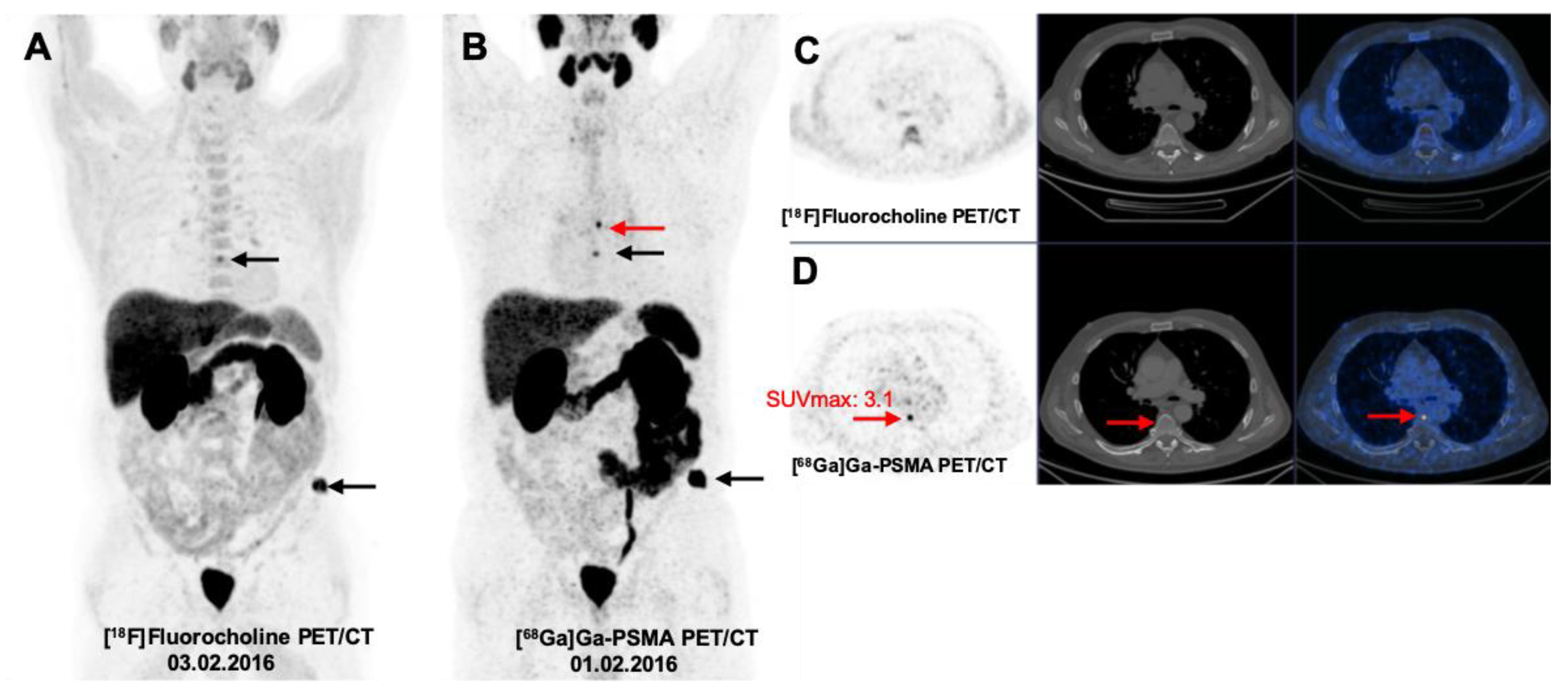
| 13 | Referred for Staging PSA = 61 Gleason score = 7 castration resistant (hormone therapy, chemotherapy, local radiation). | In Prostate bed and skeleton the [68Ga]Ga-PSMA PET/CT was the superior imaging modality. While variable pattern of imaging superiority was found in lymph nodes lesions, [18F]Fluorocholine PET/CT was prominent in some prominent nodes, even up to 60 mm in size. | Castration refractory prostate cancer and elevated PSA value not responding to conventional treatments; [18F]Fluorocholine PET/CT superiority may belong to dedifferentiating nodal metastases. |
| 6 | Referred for Staging PSA = 21.42 Gleason Score = 8 (TURP performed. After two months PSA reaches 0.17 in one year follow up). | In Prostate gland, [68Ga]Ga-PSMA PET/CT was the superior modality. No skeletal lesion detected. There is a solitary [18F]Fluorocholine positive node in right para-rectal space (measuring 5 mm, node SUV max 2.19). | PSA reached its nadir (0.17) after TURP and the node was assumed inflammatory pelvic node. By considering borderline radioactivity SUV max 2.19, we may avoid misinterpretation. |
| Patient Number | Clinical History | Imaging Findings | Note |
|---|---|---|---|
| 17 | Referred for Staging PSA = 81.17 Gleason Score = 7 | In Prostate bed and lymph nodes [68Ga]Ga-PSMA PET/CT was the superior imaging modality; While variable pattern of imaging superiority was found in skeletal lesions, [18F]Fluorocholine PET/CT was prominent in some skeletal lesions including small and negligible lesions in L2 vertebra(measured 10 mm) and left 8th rib (measured 8 mm) | [18F]Fluorocholine PET/CT superiority was only in few, small and sub-centimetric lesions may be due to significant [68Ga]Ga-PSMA uptake by larger metastases. |
| 29 | Referred for RestagingPSA = 19.3 Gleason Score = 8 Progressive disease | While variable pattern of imaging superiority was found in skeletal lesions, [18F]Fluorocholine PET/CT was prominent in some skeletal lesions including small and negligible lesions measuring up to 17 mm. | [18F]Fluorocholine PET/CT superiority was only in few, small and sub-centimetric lesions may be due to significant [68Ga]Ga-PSMA uptake by larger metastases. |
| 5 | Referred for Restaging PSA = 2078 Gleason score = 9 Castration resistant progressive disease (Xtandi, Zytiga, chemotherapy with Zyklen and Taxotere) | No prostate lesion or lymphatic involvement detected in the patient. Multiple bone lesions were noticed; all prominent in number and SUV (twice) in [18F]Fluorocholine PET/CT. | Castration refractory prostate cancer and elevated PSA value (2078) not responding to conventional treatments; [18F]Fluorocholine PET/CT superiority may be due to dedifferentiating metastases. |
| 7 | Referred for Restaging PSA = 2.51ng/mL previous PSA one month before was 5ng/mL Gleason score = 9 | No prostate bed or soft tissue involvement detected by the imaging modalities, The two lymph nodes detected were prominent in [68Ga]Ga-PSMA scan. Bone lesions (7 mm in T11 and 12 mm in T6 vertebra) were exclusively detected by [18F]Fluorocholine PET/CT and confirmed by MRI. | A pattern could not be supposed explaining small vertebral metastases exclusively found on [18F]Fluorocholine PET/CT (confirmed by MRI). |
© 2020 by the authors. Licensee MDPI, Basel, Switzerland. This article is an open access article distributed under the terms and conditions of the Creative Commons Attribution (CC BY) license (http://creativecommons.org/licenses/by/4.0/).
Share and Cite
Paymani, Z.; Rohringer, T.; Vali, R.; Loidl, W.; Alemohammad, N.; Geinitz, H.; Langsteger, W.; Beheshti, M. Diagnostic Performance of [18F]Fluorocholine and [68Ga]Ga-PSMA PET/CT in Prostate Cancer: A Comparative Study. J. Clin. Med. 2020, 9, 2308. https://doi.org/10.3390/jcm9072308
Paymani Z, Rohringer T, Vali R, Loidl W, Alemohammad N, Geinitz H, Langsteger W, Beheshti M. Diagnostic Performance of [18F]Fluorocholine and [68Ga]Ga-PSMA PET/CT in Prostate Cancer: A Comparative Study. Journal of Clinical Medicine. 2020; 9(7):2308. https://doi.org/10.3390/jcm9072308
Chicago/Turabian StylePaymani, Zeinab, Taryn Rohringer, Reza Vali, Wolfgang Loidl, Nafiseh Alemohammad, Hans Geinitz, Werner Langsteger, and Mohsen Beheshti. 2020. "Diagnostic Performance of [18F]Fluorocholine and [68Ga]Ga-PSMA PET/CT in Prostate Cancer: A Comparative Study" Journal of Clinical Medicine 9, no. 7: 2308. https://doi.org/10.3390/jcm9072308