

The Influence of Different Multipolar Mapping Catheter Types on Procedural Outcomes in Patients Undergoing Pulmonary Vein Isolation for Atrial Fibrillation
 , ,
, ,
Abstract
:1. Introduction
2. Materials and Methods
2.1. Study Patients
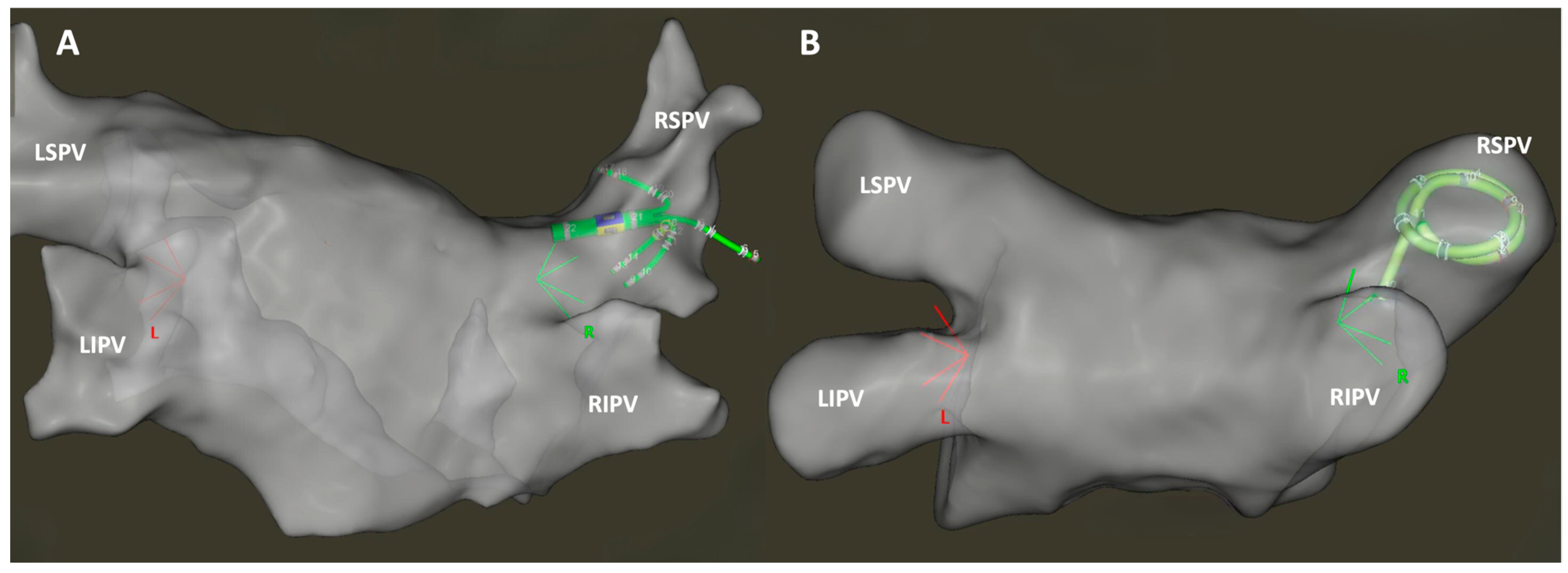
2.2. Study Protocol
2.3. Procedural Outcomes
2.4. Statistical Analysis
3. Results
4. Discussion
5. Study Limitations
6. Conclusions
Author Contributions
Funding
Institutional Review Board Statement
Informed Consent Statement
Data Availability Statement
Acknowledgments
Conflicts of Interest
References
- Hindricks, G.; Potpara, T.; Dagres, N.; Arbelo, E.; Bax, J.J.; Blomström-Lundqvist, C.; Boriani, G.; Castella, M.; Dan, G.-A.; Dilaveris, P.E.; et al. 2020 ESC Guidelines for the Diagnosis and Management of Atrial Fibrillation Developed in Collaboration with the European Association for Cardio-Thoracic Surgery (EACTS). Eur. Heart J. 2021, 42, 373–498. [Google Scholar] [CrossRef]
- Chen, C.; Zhou, X.; Zhu, M.; Chen, S.; Chen, J.; Cai, H.; Dai, J.; Xu, X.; Mao, W. Catheter Ablation versus Medical Therapy for Patients with Persistent Atrial Fibrillation: A Systematic Review and Meta-Analysis of Evidence from Randomized Controlled Trials. J. Interv. Card. Electrophysiol. 2018, 52, 9–18. [Google Scholar] [CrossRef] [PubMed]
- Morillo, C.A.; Verma, A.; Connolly, S.J.; Kuck, K.H.; Nair, G.M.; Champagne, J.; Sterns, L.D.; Beresh, H.; Healey, J.S.; Natale, A. Radiofrequency Ablation vs. Antiarrhythmic Drugs as First-Line Treatment of Paroxysmal Atrial Fibrillation (RAAFT-2) a Randomized Trial. JAMA J. Am. Med. Assoc. 2014, 311, 692–699. [Google Scholar] [CrossRef]
- Mark, D.B.; Anstrom, K.J.; Sheng, S.; Piccini, J.P.; Baloch, K.N.; Monahan, K.H.; Daniels, M.R.; Bahnson, T.D.; Poole, J.E.; Rosenberg, Y.; et al. Effect of Catheter Ablation vs. Medical Therapy on Quality of Life among Patients with Atrial Fibrillation: The CABANA Randomized Clinical Trial. JAMA J. Am. Med. Assoc. 2019, 321, 1275–1285. [Google Scholar] [CrossRef]
- Calkins, H.; Hindricks, G.; Cappato, R.; Kim, Y.-H.; Saad, E.B.; Aguinaga, L.; Akar, J.G.; Badhwar, V.; Brugada, J.; Camm, J.; et al. 2017 HRS/EHRA/ECAS/APHRS/SOLAECE Expert Consensus Statement on Catheter and Surgical Ablation of Atrial Fibrillation. Heart Rhythm 2017, 14, e275–e444. [Google Scholar] [CrossRef]
- Di Biase, L.; Diaz, J.C.; Zhang, X.-D.; Romero, J. Pulsed Field Catheter Ablation in Atrial Fibrillation. Trends Cardiovasc. Med. 2022, 32, 378–387. [Google Scholar] [CrossRef] [PubMed]
- Reddy, V.Y.; Dukkipati, S.R.; Neuzil, P.; Anic, A.; Petru, J.; Funasako, M.; Cochet, H.; Minami, K.; Breskovic, T.; Sikiric, I.; et al. Pulsed Field Ablation of Paroxysmal Atrial Fibrillation. JACC Clin. Electrophysiol. 2021, 7, 614–627. [Google Scholar] [CrossRef] [PubMed]
- Rottner, L.; Bellmann, B.; Lin, T.; Reissmann, B.; Tönnis, T.; Schleberger, R.; Nies, M.; Jungen, C.; Dinshaw, L.; Klatt, N.; et al. Catheter Ablation of Atrial Fibrillation: State of the Art and Future Perspectives. Cardiol. Ther. 2020, 9, 45–58. [Google Scholar] [CrossRef]
- Koruth, J.S.; Heist, E.K.; Danik, S.; Barrett, C.D.; Kabra, R.; Blendea, D.; Ruskin, J.; Mansour, M. Accuracy of Left Atrial Anatomical Maps Acquired with a Multielectrode Catheter during Catheter Ablation for Atrial Fibrillation. J. Interv. Card. Electrophysiol. 2011, 32, 45–51. [Google Scholar] [CrossRef]
- Anter, E.; Tschabrunn, C.M.; Josephson, M.E. High-Resolution Mapping of Scar-Related Atrial Arrhythmias Using Smaller Electrodes with Closer Interelectrode Spacing. Circ. Arrhythm. Electrophysiol. 2015, 8, 537–545. [Google Scholar] [CrossRef]
- Jones, D.G.; McCready, J.W.; Kaba, R.A.; Ahsan, S.Y.; Lyne, J.C.; Wang, J.; Segal, O.R.; Markides, V.; Lambiase, P.D.; Wong, T.; et al. A Multi-Purpose Spiral High-Density Mapping Catheter: Initial Clinical Experience in Complex Atrial Arrhythmias. J. Interv. Card. Electrophysiol. 2011, 31, 225–235. [Google Scholar] [CrossRef]
- Sommer, P.; Sciacca, V.; Anselmino, M.; Tilz, R.; Bourier, F.; Lehrmann, H.; Bulava, A. Practical Guidance to Reduce Radiation Exposure in Electrophysiology Applying Ultra Low-Dose Protocols: A European Heart Rhythm Association Review. Europace 2023, 25, euad191. [Google Scholar] [CrossRef] [PubMed]
- Debreceni, D.; Janosi, K.; Bocz, B.; Turcsan, M.; Lukacs, R.; Simor, T.; Antolič, B.; Vamos, M.; Komocsi, A.; Kupo, P. Zero Fluoroscopy Catheter Ablation for Atrial Fibrillation: A Systematic Review and Meta-Analysis. Front. Cardiovasc. Med. 2023, 10, 1178783. [Google Scholar] [CrossRef] [PubMed]
- Kim, Y.; Chen, S.; Ernst, S.; Guzman, C.E.; Han, S.; Kalarus, Z.; Labadet, C.; Lin, Y.; Lo, L.; Nogami, A.; et al. 2019 APHRS Expert Consensus Statement on Three-dimensional Mapping Systems for Tachycardia Developed in Collaboration with HRS, EHRA, and LAHRS. J. Arrhythm 2020, 36, 215–270. [Google Scholar] [CrossRef] [PubMed]
- Patel, A.M.; d’Avila, A.; Neuzil, P.; Kim, M.S.J.; Mela, T.; Singh, J.P.; Ruskin, J.N.; Reddy, V.Y. Atrial Tachycardia After Ablation of Persistent Atrial Fibrillation. Circ. Arrhythm Electrophysiol. 2008, 1, 14–22. [Google Scholar] [CrossRef] [PubMed]
- Liang, J.J.; Elafros, M.A.; Muser, D.; Pathak, R.K.; Santangeli, P.; Supple, G.E.; Schaller, R.D.; Frankel, D.S.; Dixit, S. Comparison of Left Atrial Bipolar Voltage and Scar Using Multielectrode Fast Automated Mapping versus Point-by-Point Contact Electroanatomic Mapping in Patients With Atrial Fibrillation Undergoing Repeat Ablation. J. Cardiovasc. Electrophysiol. 2017, 28, 280–288. [Google Scholar] [CrossRef] [PubMed]
- Bun, S.-S.; Delassi, T.; Latcu, D.G.; El Jamili, M.; Ayari, A.; Errahmouni, A.; Berte, B.; Saoudi, N. A Comparison between Multipolar Mapping and Conventional Mapping of Atrial Tachycardias in the Context of Atrial Fibrillation Ablation. Arch. Cardiovasc. Dis. 2018, 111, 33–40. [Google Scholar] [CrossRef] [PubMed]
- Sroubek, J.; Rottmann, M.; Barkagan, M.; Leshem, E.; Shapira-Daniels, A.; Brem, E.; Fuentes-Ortega, C.; Malinaric, J.; Basu, S.; Bar-Tal, M.; et al. A Novel Octaray Multielectrode Catheter for High-resolution Atrial Mapping: Electrogram Characterization and Utility for Mapping Ablation Gaps. J. Cardiovasc. Electrophysiol. 2019, 30, 749–757. [Google Scholar] [CrossRef]
- Huo, Y.; Gaspar, T.; Schönbauer, R.; Wójcik, M.; Fiedler, L.; Roithinger, F.X.; Martinek, M.; Pürerfellner, H.; Kirstein, B.; Richter, U.; et al. Low-Voltage Myocardium-Guided Ablation Trial of Persistent Atrial Fibrillation. NEJM Evid. 2022, 1, EVIDoa2200141. [Google Scholar] [CrossRef]
- Vogler, J.; Willems, S.; Sultan, A.; Schreiber, D.; Lüker, J.; Servatius, H.; Schäffer, B.; Moser, J.; Hoffmann, B.A.; Steven, D. Pulmonary Vein Isolation Versus Defragmentation. J. Am. Coll. Cardiol. 2015, 66, 2743–2752. [Google Scholar] [CrossRef]
- Kistler, P.M.; Chieng, D.; Sugumar, H.; Ling, L.-H.; Segan, L.; Azzopardi, S.; Al-Kaisey, A.; Parameswaran, R.; Anderson, R.D.; Hawson, J.; et al. Effect of Catheter Ablation Using Pulmonary Vein Isolation With vs Without Posterior Left Atrial Wall Isolation on Atrial Arrhythmia Recurrence in Patients with Persistent Atrial Fibrillation. JAMA 2023, 329, 127–135. [Google Scholar] [CrossRef] [PubMed]
- Koneru, J.N. Ablating Persistent Atrial Fibrillation—Still Learning While Burning! NEJM Evid. 2022, 1, EVIDe2200246. [Google Scholar] [CrossRef] [PubMed]
- Chieng, D.; Sugumar, H.; Ling, L.-H.; Segan, L.; Azzopardi, S.; Prabhu, S.; Al-Kaisey, A.; Voskoboinik, A.; Parameswaran, R.; Morton, J.B.; et al. Catheter Ablation for Persistent Atrial Fibrillation: A Multicenter Randomized Trial of Pulmonary Vein Isolation (PVI) versus PVI with Posterior Left Atrial Wall Isolation (PWI)—The CAPLA Study. Am. Heart J. 2022, 243, 210–220. [Google Scholar] [CrossRef] [PubMed]
- Marrouche, N.F.; Wazni, O.; McGann, C.; Greene, T.; Dean, J.M.; Dagher, L.; Kholmovski, E.; Mansour, M.; Marchlinski, F.; Wilber, D.; et al. Effect of MRI-Guided Fibrosis Ablation vs Conventional Catheter Ablation on Atrial Arrhythmia Recurrence in Patients with Persistent Atrial Fibrillation. JAMA 2022, 327, 2296. [Google Scholar] [CrossRef]
- Jia, H.; Wang, W.; Yu, B. Efficacy and Safety of Low Voltage Area Ablation for Atrial Fibrillation: A Systematic Review and Meta-Analysis. J. Interv. Card. Electrophysiol. 2022, 66, 1519–1527. [Google Scholar] [CrossRef]


| Group Lasso (n = 35) | Group PentaRay (n = 35) | p Value | |
|---|---|---|---|
| Male, n (%) | 28 (80) | 26 (74) | 0.57 |
| Age, y | 68.6 (58.7; 71.5) | 66.5 (50.6; 73.5) | 0.88 |
| Hypertension, n (%) | 28 (80) | 28 (80) | 1.0 |
| Heart failure, n (%) | 5 (14.3) | 6 (17.1) | 0.74 |
| Coronary artery disease, n (%) | 5 (14.3) | 8 (22.9) | 0.36 |
| Diabetes mellitus, n (%) | 8 (22.9) | 7 (20.0) | 0.77 |
| Chronic kidney disease, n (%) | 6 (17.1) | 7 (20.0) | 0.76 |
| Prior stroke/TIA, n (%) | 1 (2.9) | 5 (14.3) | 0.09 |
| Left atrial diameter, mm | 54.5 ± 8.1 | 52.9 ± 7.8 | 0.18 |
| Group Lasso (n = 35) | Group PentaRay (n = 35) | p Value | |
|---|---|---|---|
| Procedure time, min | 80.2 ± 17.7 | 75.7 ± 14.8 | 0.13 |
| Time from access to start of mapping, min | 31.2 ± 7.0 | 28.9 ± 6.8 | 0.80 |
| Mapping time, min | 8 (6; 13) | 9 (6.5; 10.5) | 0.73 |
| Time between first and last ablation, min | 32 (30; 36) | 33 (26; 40) | 0.52 |
| Validation time, min | 3 (2; 4) | 3 (1; 5) | 0.46 |
| First pass rate, % | 89% | 91% | 0.71 |
| Left atrial dwelling time, min | 46 (37; 53) | 45 (36.5; 53) | 0.56 |
| Total ablation time, s | 1187 (1063; 1534) | 1150.5 (1053; 1393) | 0.49 |
| Number of ablations, n | 78 (73; 93) | 83 (71.3; 92.8) | 0.60 |
| Total ablation energy, J | 52,300 (47,265; 66,804) | 49,666 (46,395; 56,502) | 0.35 |
| Fluoroscopy time, s | 150 ± 71 | 143 ± 56 | 0.14 |
| Fluoroscopy dose, mGy | 6.7 ± 4.0 | 7.4 ± 4.4 | 0.90 |
| Complications, n | 0 | 0 | NA |
Disclaimer/Publisher’s Note: The statements, opinions and data contained in all publications are solely those of the individual author(s) and contributor(s) and not of MDPI and/or the editor(s). MDPI and/or the editor(s) disclaim responsibility for any injury to people or property resulting from any ideas, methods, instructions or products referred to in the content. |
© 2024 by the authors. Licensee MDPI, Basel, Switzerland. This article is an open access article distributed under the terms and conditions of the Creative Commons Attribution (CC BY) license (https://creativecommons.org/licenses/by/4.0/).
Share and Cite
Janosi, K.-F.; Debreceni, D.; Bocz, B.; Torma, D.; Keseru, M.; Simor, T.; Kupo, P. The Influence of Different Multipolar Mapping Catheter Types on Procedural Outcomes in Patients Undergoing Pulmonary Vein Isolation for Atrial Fibrillation. J. Clin. Med. 2024, 13, 1029. https://doi.org/10.3390/jcm13041029
Janosi K-F, Debreceni D, Bocz B, Torma D, Keseru M, Simor T, Kupo P. The Influence of Different Multipolar Mapping Catheter Types on Procedural Outcomes in Patients Undergoing Pulmonary Vein Isolation for Atrial Fibrillation. Journal of Clinical Medicine. 2024; 13(4):1029. https://doi.org/10.3390/jcm13041029
Chicago/Turabian StyleJanosi, Kristof-Ferenc, Dorottya Debreceni, Botond Bocz, Dalma Torma, Mark Keseru, Tamas Simor, and Peter Kupo. 2024. "The Influence of Different Multipolar Mapping Catheter Types on Procedural Outcomes in Patients Undergoing Pulmonary Vein Isolation for Atrial Fibrillation" Journal of Clinical Medicine 13, no. 4: 1029. https://doi.org/10.3390/jcm13041029