
Pathogenesis, Diagnosis and Management of Squamous Cell Carcinoma and Pseudoepithelial Hyperplasia Secondary to Red Ink Tattoo: A Case Series and Review
 ,
, 
Abstract
:1. Introduction
2. Materials and Methods
3. Results
3.1. Case Reports
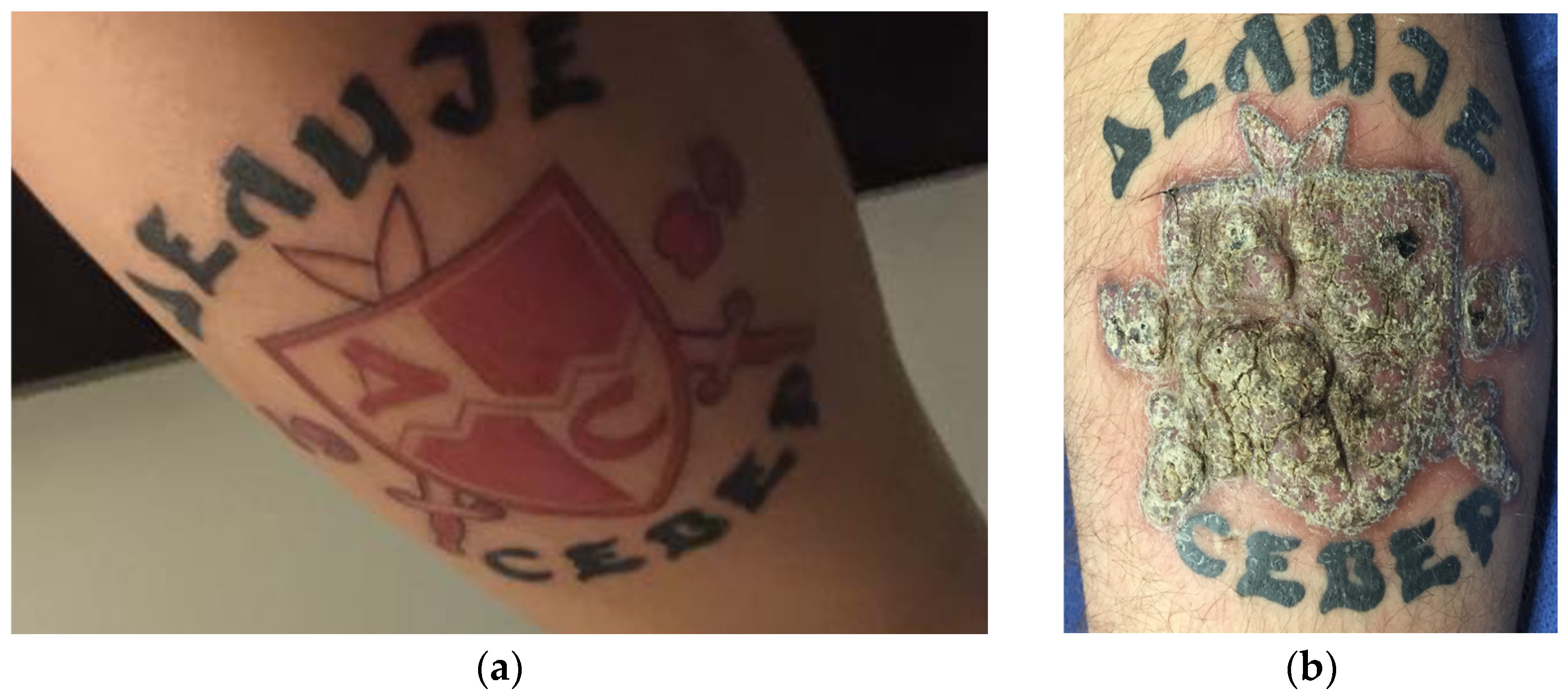
3.1.1. Case 1
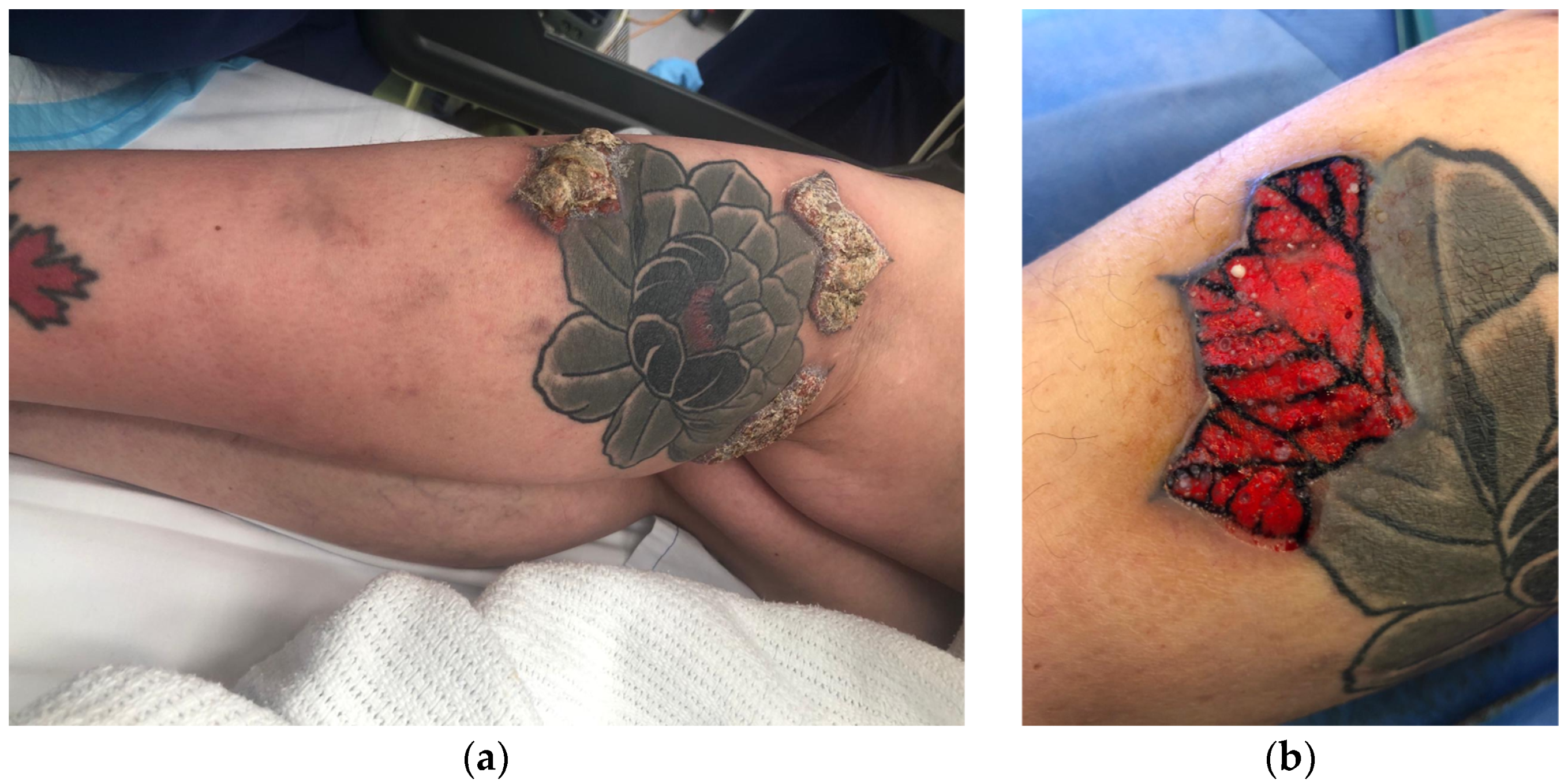
3.1.2. Case 2
3.1.3. Case 3
3.1.4. Case 4
4. Discussion
4.1. Pathogenesis of Cutaneous Reactions Secondary to Red Ink Tattoo
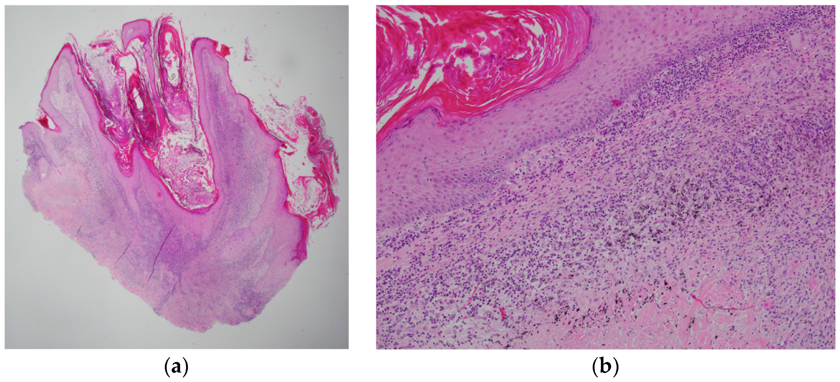
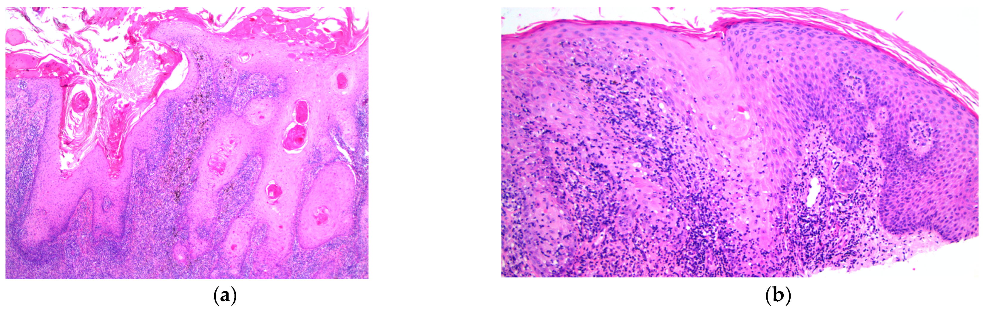
4.2. Common Histopathological Features
4.3. Diagnostic Approaches for PEH and SCC
4.4. Efficacious Treatment
5. Conclusions
Author Contributions
Funding
Informed Consent Statement
Data Availability Statement
Conflicts of Interest
References
- Weber, P.; Tschandl, P.; Sinz, C.; Kittler, H. Dermatoscopy of Neoplastic Skin Lesions: Recent Advances, Updates, and Revisions. Curr. Treat. Options Oncol. 2018, 19, 56. [Google Scholar] [CrossRef] [Green Version]
- Badavanis, G.; Constantinou, P.; Pasmatzi, E.; Monastirli, A.; Tsambaos, D. Late-onset pseudoepitheliomatous hyperplasia developing within a red ink tattoo. Dermatol. Online J. 2019, 25. [Google Scholar] [CrossRef]
- Swigost, A.J.; Peltola, J.; Jacobson-Dunlop, E.; Goldfarb, N. Tattoo-related squamous proliferations: A spectrum of reactive hyperplasia. Clin. Exp. Dermatol. 2018, 43, 728–732. [Google Scholar] [CrossRef] [PubMed]
- Kluger, N.; Cribier, B. Keratoacanthomas and Squamous Cell Carcinomas on Tattoos: A Review of 42 Cases. Dermatology 2021, 237, 309–312. [Google Scholar] [CrossRef] [PubMed]
- Heywood, W.; Patrick, K.; Smith, A.M.; Simpson, J.M.; Pitts, M.K.; Richters, J.; Shelley, J.M. Who gets tattoos? Demographic and behavioral correlates of ever being tattooed in a representative sample of men and women. Ann. Epidemiol. 2012, 22, 51–56. [Google Scholar] [CrossRef] [PubMed]
- Sarma, D.P.; Dentlinger, R.B.; Forystek, A.M.; Stevens, T.; Huerter, C. Poorly differentiated squamous cell carcinoma arising in tattooed skin. Case Rep. Med. 2010, 2010, 431813. [Google Scholar] [CrossRef] [Green Version]
- Shrout, M.; DeCoster, R.; Wermeling, R.; Vasconez, H.C. Risk Factors for Squamous Cell Carcinoma: A Case for Red Pigment in Tattoos. Am. Surg. 2019, 85, e77–e78. [Google Scholar] [CrossRef]
- Tammaro, A.; Narcisi, A.; Cortesi, G.; Abruzzese, C.; Socciarelli, F.; Pulcini, F.; De Marco, G.; Persechino, S. A case of pseudoepitheliomatous hyperplasia to tattoos. J. Eur. Acad. Dermatol. Venereol. 2015, 29, 1439–1440. [Google Scholar] [CrossRef]
- De Roeck, A.; Joujoux, J.M.; Fournier, F.; Dandurand, M.; Meunier, L.; Stoebner, P.E. Florid pseudoepitheliomatous hyperplasia related to tattoo: A case report. Int. Wound J. 2013, 10, 539–541. [Google Scholar] [CrossRef]
- Weber, P.J.; Johnson, B.L.; Dzubow, L.M. Pseudoepitheliomatous hyperplasia following Mohs micrographic surgery. J. Dermatol. Surg. Oncol. 1989, 15, 557–560. [Google Scholar] [CrossRef]
- Tammaro, A.; Abruzzese, C.; Narcisi, A.; Cortesi, G.; Fontana, E.; Persechino, S.; Salmaso, R.; Alaibac, M.S.A. Localised pseudoepitheliomatous hyperplasia: Unusual cutaneous reaction pattern to tattoo. Int. Wound J. 2016, 13, 294. [Google Scholar] [CrossRef]
- Kiss, F.; May, K.; Piguet, V. Image gallery: Pseudoepitheliomatous hyperplasia, a rare tattoo reaction. Br. J. Dermatol. 2016, 175, e112. [Google Scholar] [CrossRef] [Green Version]
- Tammaro, A.; Raffa, S.; Petrigliano, N.; Zollo, V.; Gelormini, E.; Moliterni, E.; Magri, F.; Parisella, F.; Persechino, S. Marked pseudoepitheliomatous hyperplasia secondary to a red-pigmented tattoo: A case report. J. Eur. Acad. Dermatol. Venereol. 2018, 32, e272–e273. [Google Scholar] [CrossRef]
- Sherif, S.; Blakeway, E.; Fenn, C.; German, A.; Laws, P. A case of squamous cell carcinoma developing within a red-ink tattoo. J. Cutan. Med. Surg. 2017, 21, 61–63. [Google Scholar] [CrossRef]
- Agarwal, P.; Jagati, A.; Mehta, R.; Vadher, P.; Rathod, S.; Bodar, P. Histopathological evaluation of cutaneous reactions to tattoos: Study at a tertiary care center. J. Cutan. Pathol. 2021, 48, 870–876. [Google Scholar] [CrossRef]
- Swigost, A.; Farah, R.S.; Canova, E.; Goldfarb, N. Multiple squamous neoplasms arising in a red tattoo after laser tattoo removal. Dermatol. Surg. 2020, 46, 970–973. [Google Scholar] [CrossRef]
- Tammaro, A.; Chello, C.; Magri, F.; Gelormini, E.; Parisella, F.R.; Daniele, A.; Signati, F.; Persechino, S.; Raffa, S. Atypical koebner phenomenon on a tattoo. J. Cosmet. Dermatol. 2020, 19, 1205–1207. [Google Scholar] [CrossRef]
- Laux, P.; Tralau, T.; Tentschert, J.; Blume, A.; Al Dahouk, S.; Bäumler, W.; Bernstein, E.; Bocca, B.; Alimonti, A.; Colebrook, H. A medical-toxicological view of tattooing. Lancet 2016, 387, 395–402. [Google Scholar] [CrossRef]
- Australian Government Department of Health National Industrial Chemicals Notification and Assessment Scheme (NICNAS). Characterisation of tattoo inks used in Australia. Available online: https://www.industrialchemicals.gov.au/sites/default/files/2020-05/Characterisation%20of%20tattoo%20inks%20used%20in%20Australia%20%5BPDF%20456KB%5D.pdf (accessed on 13 February 2023).
- Balfour, E.; Olhoffer, I.; Leffell, D.; Handerson, T. Massive pseudoepitheliomatous hyperplasia: An unusual reaction to a tattoo. Am. J. Dermatopathol. 2003, 25, 338–340. [Google Scholar] [CrossRef]
- Pitarch, G.; Martínez-Menchón, T.; Martínez-Aparicio, A.; Sánchez-Carazo, J.L.; Muñoz, D.; Fortea, J.M. Squamous cell carcinoma over tattoos. J. Am. Acad. Dermatol. 2007, 56, 1072–1073. [Google Scholar] [CrossRef]
- Paprottka, F.J.; Krezdorn, N.; Narwan, M.; Turk, M.; Sorg, H.; Noah, E.M.; Hebebrand, D. Trendy tattoos—Maybe a serious health risk? Aesthetic Plast. Surg. 2018, 42, 310–321. [Google Scholar] [CrossRef] [PubMed]
- Lerche, C.M.; Heerfordt, I.M.; Serup, J.; Poulsen, T.; Wulf, H.C. Red tattoos, ultraviolet radiation and skin cancer in mice. Exp. Dermatol. 2017, 26, 1091–1096. [Google Scholar] [CrossRef] [PubMed]
- Kluger, N. Self-reported tattoo reactions in a cohort of 448 French tattooists. Int. J. Dermatol. 2016, 55, 764–768. [Google Scholar] [CrossRef] [PubMed]
- Kaur, R.R.; Kirby, W.; Maibach, H. Cutaneous allergic reactions to tattoo ink. J. Cosmet. Dermatol. 2009, 8, 295–300. [Google Scholar] [CrossRef]
- Godinho, M.M.; Aguinaga, F.; Grynszpan, R.; Lima, V.M.; Azulay, D.R.; Cuzzi, T.; Ramos-e-Silva, M.; Manela-Azulay, M. Granulomatous reaction to red tattoo pigment treated with allopurinol. J. Cosmet. Dermatol. 2015, 14, 241–245. [Google Scholar] [CrossRef]
- Wollina, U.; Gruner, M.; Schönlebe, J. Granulomatous tattoo reaction and erythema nodosum in a young woman: Common cause or coincidence? J. Cosmet. Dermatol. 2008, 7, 84–88. [Google Scholar] [CrossRef]
- Kazlouskaya, V.; Junkins-Hopkins, J.M. Pseudoepitheliomatous hyperplasia in a red pigment tattoo: A separate entity or hypertrophic lichen planus-like reaction? J. Clin. Aesthetic. Dermatol. 2015, 8, 48. [Google Scholar]
- Kuo, W.E.; Richwine, E.E.; Sheehan, D.J. Pseudolymphomatous and lichenoid reaction to a red tattoo: A case report. Cutis 2011, 87, 89–92. [Google Scholar]
- Marchesi, A.; Parodi, P.C.; Brioschi, M.; Marchesi, M.; Bruni, B.; Cangi, M.G.; Vaienti, L. Tattoo ink-related cutaneous pseudolymphoma: A rare but significant complication. Case report and review of the literature. Aesthetic Plast. Surg. 2014, 38, 471–478. [Google Scholar] [CrossRef]
- Clarke, P. Nonmelanoma skin cancers: Treatment options. Aust. Fam. Physician 2012, 41, 476–480. [Google Scholar]
- Kheradmand, S.; Perry, B.M.; Bohlke, A. Pseudoepitheliomatous Hyperplasia Arising From Purple Tattoo Pigment. Cutis 2020, 105, E22–E24. [Google Scholar]
- Cipollaro, V. Keratoacanthoma developing in a tattoo. Cutis 1973, 11, 809. [Google Scholar]
- Chakrabarti, S.; Chakrabarti, P.R.; Agrawal, D.; Somanath, S. Pseudoepitheliomatous hyperplasia: A clinical entity mistaken for squamous cell carcinoma. J. Cutan. Aesthetic Surg. 2014, 7, 232. [Google Scholar] [CrossRef]
- Lee, Y.S.; Teh, M. p53 expression in pseudoepitheliomatous hyperplasia, keratoacanthoma, and squamous cell carcinoma of skin. Cancer 1994, 73, 2317–2323. [Google Scholar] [CrossRef]
- Su, A.; Ra, S.; Li, X.; Zhou, J.; Binder, S. Differentiating cutaneous squamous cell carcinoma and pseudoepitheliomatous hyperplasia by multiplex qRT-PCR. Mod. Pathol. 2013, 26, 1433–1437. [Google Scholar] [CrossRef] [Green Version]
- Gajanan, G.; Bohra, C.; Nanjappa, S.; Greene, J.N. Chronic infections leading to squamous cell carcinoma from chronic inflammation: A case report and review of all causes. Infect. Dis. Clin. Pract. 2016, 24, 133–137. [Google Scholar] [CrossRef]
- Schmidt, H.; Christensen, H. Red poster paint tattoo granuloma. Arch. Dermatol. 1978, 114, 965–966. [Google Scholar] [CrossRef]
- Mlakar, B. Successful removal of hyperkeratotic-lichenoid reaction to red ink tattoo with preservation of the whole tattoo using a skin grafting knife. Acta. Dermatovenerol. Alp. Pannonica. Adriat. 2015, 24, 81–82. [Google Scholar]
- Kluger, N.; Durand, L.; Minier-Thoumin, C.; Plantie, F.; Cotten, H.; Berteloot, E.; Blatiere, V.; Dereure, O. Pseudoepitheliomatous epidermal hyperplasia in tattoos: Report of three cases. Am. J. Clin. Dermatol. 2008, 9, 337–340. [Google Scholar] [CrossRef]
- De Argila, D.; Chaves, A.; Moreno, J. Erbium: Yag laser therapy of lichenoid red tattoo reaction. J. Eur. Acad. Dermatol. Venereol. 2004, 18, 332–333. [Google Scholar] [CrossRef]
- Antony, F.; Harland, C. Red ink tattoo reactions: Successful treatment with the Q-switched 532 nm Nd: YAG laser. Br. J. Dermatol. 2003, 149, 94–98. [Google Scholar] [PubMed]
- Lemperle, G.; Gauthier-Hazan, N.; Wolters, M.; Eisemann-Klein, M.; Zimmermann, U.; Duffy, D.M. Foreign body granulomas after all injectable dermal fillers: Part 1. Possible causes. Plast. Reconstr. Surg. 2009, 123, 1842–1863. [Google Scholar] [CrossRef] [PubMed] [Green Version]
- Ortiz, A.; Yamauchi, P.S. Rapidly growing squamous cell carcinoma from permanent makeup tattoo. J. Am. Acad. Dermatol. 2009, 60, 1073–1074. [Google Scholar] [CrossRef] [PubMed]
- McQuarrie, D.G. Squamous-cell carcinoma arising in a tattoo. Minn. Med. 1966, 49, 799–801. [Google Scholar]


Disclaimer/Publisher’s Note: The statements, opinions and data contained in all publications are solely those of the individual author(s) and contributor(s) and not of MDPI and/or the editor(s). MDPI and/or the editor(s) disclaim responsibility for any injury to people or property resulting from any ideas, methods, instructions or products referred to in the content. |
© 2023 by the authors. Licensee MDPI, Basel, Switzerland. This article is an open access article distributed under the terms and conditions of the Creative Commons Attribution (CC BY) license (https://creativecommons.org/licenses/by/4.0/).
Share and Cite
Rahbarinejad, Y.; Guio-Aguilar, P.; Vu, A.N.; Lo, M.; McTigue, C.; Nirenberg, A.; Rozen, W.M. Pathogenesis, Diagnosis and Management of Squamous Cell Carcinoma and Pseudoepithelial Hyperplasia Secondary to Red Ink Tattoo: A Case Series and Review. J. Clin. Med. 2023, 12, 2424. https://doi.org/10.3390/jcm12062424
Rahbarinejad Y, Guio-Aguilar P, Vu AN, Lo M, McTigue C, Nirenberg A, Rozen WM. Pathogenesis, Diagnosis and Management of Squamous Cell Carcinoma and Pseudoepithelial Hyperplasia Secondary to Red Ink Tattoo: A Case Series and Review. Journal of Clinical Medicine. 2023; 12(6):2424. https://doi.org/10.3390/jcm12062424
Chicago/Turabian StyleRahbarinejad, Yasmina, Pedro Guio-Aguilar, Anh Ngoc Vu, Michael Lo, Christine McTigue, Alex Nirenberg, and Warren M. Rozen. 2023. "Pathogenesis, Diagnosis and Management of Squamous Cell Carcinoma and Pseudoepithelial Hyperplasia Secondary to Red Ink Tattoo: A Case Series and Review" Journal of Clinical Medicine 12, no. 6: 2424. https://doi.org/10.3390/jcm12062424