
Transcatheter Aortic Valve Replacement in Degenerated Perceval Bioprosthesis: Clinical and Technical Aspects in 32 Cases
 ,
,  , , and
Abstract
:1. Introduction
2. Materials and Methods
2.1. Patients
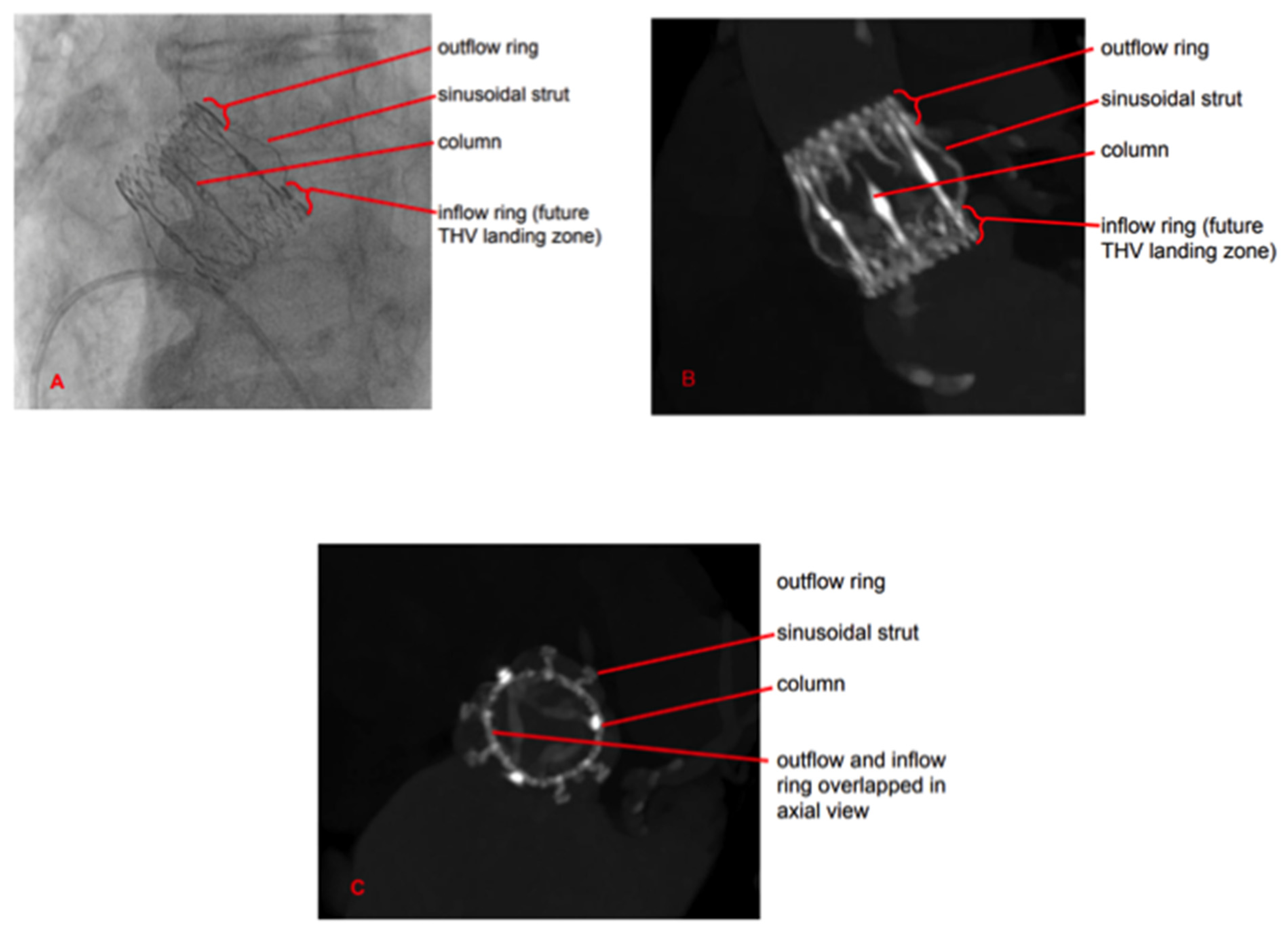
2.2. Technology
2.3. VIV-TAVR Planning and Technical Procedures
2.4. Statistical Analysis
3. Results
4. Discussion
Author Contributions
Funding
Institutional Review Board Statement
Informed Consent Statement
Data Availability Statement
Conflicts of Interest
References
- Nishimura, R.A.; Otto, C.M.; Bonow, R.O.; Carabello, B.A.; ErwinIII, J.P.; Guyton, R.A.; O’Gara, P.T.; Ruiz, C.E.; Skubas, N.J.; Sorajja, P.; et al. American College of Cardiology/American Heart Association Task Force on Practice Guidelines. 2014 AHA/ACC guideline for the management of patients with valvular heart disease: Executive summary: A report of the American College of Cardiology/American Heart Association Task Force on Practice Guidelines. J. Am. Coll. Cardiol. 2014, 63, 2438–2488. [Google Scholar] [PubMed]
- Vahanian, A.; Alfieri, O.; Andreotti, F.; Antunes, M.J.; Barón-Esquivias, G.; Baumgartner, H.; Borger, M.A.; Carrel, T.P.; De Bonis, M.; Evangelista, A.; et al. Guidelines on the management of valvular heart disease (version 2012): The Joint Task Force on the Management of Valvular Heart Disease of the European Society of Cardiology (ESC) and the European Association for Cardio-Thoracic Surgery (EACTS). Eur. J. Cardiothorac. Surg. 2012, 42, S1–S44. [Google Scholar] [PubMed]
- Goodney, P.P.; O’Connor, G.T.; Wennberg, D.E.; Birkmeyer, J.D. Do hospitals with low mortality rates in coronary artery bypass also perform well in valve replacement? Ann. Thorac. Surg. 2003, 76, 1131–1136. [Google Scholar] [CrossRef] [PubMed]
- Brown, J.M.; O’Brien, S.M.; Wu, C.; Sikora, J.A.; Griffith, B.P.; Gammie, J.S. Isolated aortic valve replacement in North America comprising 108,687 patients in 10 years: Changes in risks, valve types, and outcomes in the Society of Thoracic Surgeons National Database. J. Thorac. Cardiovasc. Surg. 2009, 137, 82–90. [Google Scholar] [CrossRef]
- Fischlein, T.; Meuris, B.; Hakim-Meibodi, K.; Misfeld, M.; Carrel, T.; Zembala, M.; Gaggianesi, S.; Madonna, F.; Laborde, F.; Asch, F.; et al. CAVALIER Trial Investigators. The sutureless aortic valve at 1 year: A large multicenter cohort study. J. Thorac. Cardiovasc. Surg. 2016, 151, 1617–1626.e4. [Google Scholar] [CrossRef]
- Fischlein, T.; Pfeiffer, S.; Pollari, F.; Sirch, J.; Vogt, F.; Santarpino, G. Sutureless Valve Implantation via Mini J-Sternotomy: A Single Center Experience with 2 Years Mean Follow-up. Thorac. Cardiovasc. Surg. 2015, 63, 467–471. [Google Scholar] [CrossRef]
- Zannis, K.; Joffre, J.; Czitrom, D.; Folliguet, T.; Noghin, M.; Lansac, M.N.E.; Mitchell-Heggs, L.; Debauchez, M.; Laborde, F. Aortic valve replacement with the perceval S bioprosthesis: Single-center experience in 143 patients. J. Heart Valve Dis. 2014, 23, 795–802. [Google Scholar]
- Shrestha, M.; Fischlein, T.; Meuris, B.; Flameng, W.; Carrel, T.; Madonna, F.; Misfeld, M.; Folliguet, T.; Haverich, A.; Laborde, F. European multicentre experience with the sutureless Perceval valve: Clinical and haemodynamic outcomes up to 5 years in over 700 patients. Eur. J. Cardiothorac. Surg. 2016, 49, 234–241. [Google Scholar] [CrossRef]
- Concistrè, G.; Bianchi, G.; Margaryan, R.; Zancanaro, E.; Chiaramonti, F.; Kallushi, E.; Gasbarri, T.; Murzi, M.; Varone, E.; Simeoni, S.; et al. Ten-year experience with sutureless Perceval bioprosthesis: Single-centre analysis in 1157 implants. J. Cardiovasc. Med. 2023, 24, 506–513. [Google Scholar] [CrossRef]
- Concistré, G.; Baghai, M.; Santarpino, G.; Royse, A.; Scherner, M.; Troise, G.; Glauber, M.; Solinas, M. Clinical and hemodynamic outcomes of the Perceval sutureless aortic valve from a real-world registry. Interdiscip. Cardiovasc. Thorac. Surg. 2023, 36, ivad103. [Google Scholar] [CrossRef]
- Belluschi, I.; Buzzatti, N.; Blasio, A.; Romano, V.; De Bonis, M.; Castiglioni, A.; Montorfano, M.; Alfieri, O. Self-expandable valve-in-valve treatment for failing sutureless aortic bioprosthesis. J. Card. Surg. 2020, 35, 477–479. [Google Scholar] [CrossRef] [PubMed]
- Suleiman, T.; Tanseco, K.; Arunothayaraj, S.; Michail, M.; Cockburn, J.; Hadjivassilev, S.; Hildick-Smith, D. Valve-in-Valve Transcatheter Aortic Valve Implantation for the Failing Surgical Perceval Bioprosthesis. Cardiovasc. Revasc. Med. 2022, 40, 148–153. [Google Scholar] [CrossRef] [PubMed]
- Landes, U.; Dvir, D.; Schoels, W.; Tron, C.; Ensminger, S.; Simonato, M.; Schäfer, U.; Bunc, M.; Aldea, G.S.; Cerillo, A.; et al. Transcatheter aortic valve-in-valve implantation in degenerative rapid deployment bioprostheses. EuroIntervention 2019, 15, 37–43. [Google Scholar] [CrossRef]
- Kay, R.T.; Linjawi, H.; Butler, C.; Mathew, A.; Muhll, I.V.; Khandekar, S.; Tyrrell, B.D.; Nagendran, J.; Taylor, D.; Welsh, R.C. Transcatheter Aortic Valve Implantation in a Failed Perceval Sutureless Valve, Complicated by Aortic Annular Rupture. CJC Open 2022, 4, 577–580. [Google Scholar] [CrossRef] [PubMed]
- Ellouze, M.; Mazine, A.; Carrier, M.; Bouchard, D. Sutureless and Transcatheter Aortic Valve Replacement: When Rivals Become Allies. Semin. Thorac. Cardiovasc. Surg. 2020, 32, 427–430. [Google Scholar] [CrossRef]
- Akins, C.W.; Miller, D.C.; Turina, M.I.; Kouchoukos, N.T.; Blackstone, E.H.; Grunkemeier, G.L.; Takkenberg, J.J.; David, T.E.; Butchart, E.G.; Adams, D.H.; et al. Guidelines for reporting mortality and morbidity after cardiac valve interventions. J. Thorac. Cardiovasc. Surg. 2008, 135, 732–738. [Google Scholar] [CrossRef]
- Généreux, P.; Head, S.J.; Hahn, R.; Daneault, B.; Kodali, S.; Williams, M.R.; van Mieghem, N.M.; Alu, M.C.; Serruys, P.W.; Kappetein, A.P.; et al. Paravalvular leak after transcatheter aortic valve replacement: The new Achilles’ heel? A comprehensive review of the literature. J. Am. Coll. Cardiol. 2013, 61, 1125–1136. [Google Scholar] [CrossRef]
- Margaryan, R.; Kallushi, E.; Gilmanov, D.; Micelli, A.; Murzi, M.; Solinas, M.; Cerillo, A.G.; Glauber, M. Sutureless Aortic Valve Prosthesis Sizing: Estimation and Prediction Using Multidetector-Row Computed Tomography. Innovations 2015, 10, 230–235. [Google Scholar] [CrossRef]
- Cerillo, A.G.; Amoretti, F.; Mariani, M.; Cigala, E.; Murzi, M.; Gasbarri, T.; Solinas, M.; Chiappino, D. Increased Gradients After Aortic Valve Replacement With the Perceval Valve: The Role of Oversizing. Ann. Thorac. Surg. 2018, 106, 121–128. [Google Scholar] [CrossRef]
- Dvir, D.; Webb, J.G.; Bleiziffer, S.; Pasic, M.; Waksman, R.; Kodali, S.; Barbanti, M.; Latib, A.; Schaefer, U.; Rodés-Cabau, J.; et al. Valve-in-Valve International Data Registry Investigators. Transcatheter aortic valve implantation in failed bioprosthetic surgical valves. JAMA 2014, 312, 162–170. [Google Scholar] [CrossRef]
- Majmundar, M.; Doshi, R.; Kumar, A.; Johnston, D.; Brockett, J.; Kanaa’n, A.; Lahorra, J.L.; Svensson, L.S.; Krishnaswamy, A.; Reed, G.R.; et al. Valve-in-valve transcatheter aortic valve implantation versus repeat surgical aortic valve replacement in patients with a failed aortic bioprosthesis. EuroIntervention 2022, 17, 1227–1237. [Google Scholar] [CrossRef] [PubMed]
- Allen, K.B.; Chhatriwalla, A.K.; Saxon, J.T.; Huded, C.P.; Sathananthan, J.; Nguyen, T.C.; Whisenant, B.; Webb, J.G. Bioprosthetic valve fracture: A practical guide. Ann. Cardiothorac. Surg. 2021, 10, 564–570. [Google Scholar] [CrossRef] [PubMed]
- Bagga, S.; Chhatriwalla, A.K. Updates on bioprosthetic valve fracture. Expert Rev. Cardiovasc. Ther. 2023, 21, 409–422. [Google Scholar] [CrossRef] [PubMed]
- O’Donnell, J.P.; O’Sullivan, C.J. Bioprosthetic Aortic Valve Fracture During Valve-in-valve Transcatheter Aortic Valve Implantation. Interv. Cardiol. 2019, 14, 147–151. [Google Scholar] [CrossRef] [PubMed]
- Dvir, D.; Leipsic, J.; Blanke, P.; Ribeiro, H.B.; Kornowski, R.; Pichard, A.; Rodés-Cabau, J.; Wood, D.A.; Stub, D.; Ben-Dor, I. Coronary obstruction in transcatheter aortic valve-in-valve implantation: Preprocedural evaluation, device selection, protection, and treatment. Circ. Cardiovasc. Interv. 2015, 8, e002079. [Google Scholar] [CrossRef]
- Tarantini, G.; Dvir, D.; Tang, G.H.L. Transcatheter aortic valve implantation in degenerated surgical aortic valves. EuroIntervention 2021, 17, 709–719. [Google Scholar] [CrossRef]
- Johnston, D.R.; Soltesz, E.G.; Vakil, N.; Rajeswaran, J.; Roselli, E.E.; Sabik, J.F.; Smedira, N.G.; Svensson, L.G.; Lytle, B.W.; Blackstone, E.H. Long-term durability of bioprosthetic aortic valves: Implications from 12,569 implants. Ann. Thorac. Surg. 2015, 99, 1239–1247. [Google Scholar] [CrossRef]


| Variables | Overall n = 32 |
|---|---|
| Age, mean ± SD | 79.6 ± 7.2 |
| Sex, female, (%) | 20 (62.5) |
| BSA, median (IQR), m2 | 1.6 (1.16–2.08) |
| Hypertension, (%) | 26 (81) |
| Diabetes mellitus, (%) | 9 (28) |
| COPD, (%) | 11 (34) |
| Hyperlipidemia, (%) | 21 (66) |
| Chronic renal failure, (%) | 3 (9) |
| Peripheral vascular disease, (%) | 4 (12.5) |
| EuroSCORE II, mean ± SD, (%) | 9.5 ± 6.4 |
| STS score, mean ± SD, (%) | 8.7 ± 4.2 |
| Variables | Overall n = 32 | |
|---|---|---|
| Size degenerated Perceval | ||
| S (n = 5) | (16%) | |
| M (n = 11) | (34%) | |
| L (n = 14) | (44%) | |
| XL (n = 2) | (6%) | |
| Isolated VIV-TAVR procedures | ||
| 30 | (94%) | |
| Combined procedures | 2 | (6%) |
| 1 | (3) |
| 1 | (3) |
| Surgical approaches | ||
| Transfemoral | 30 | (94%) |
| Transapical | 2 | (6%) |
| Size TAVR | ||
| 20 mm | 6 | (19%) |
| 23 mm | 14 | (44%) |
| 26 mm | 11 | (34%) |
| 29 mm | 1 | (3) |
| Variables | Overall n = 32 |
|---|---|
| |
| 0 |
| 4.6 ± 2.4 |
| 2 (6) |
| 0 |
| |
| 1.25 ± 0.44 |
| 0 |
| 1 |
| Variables | Preoperative | At Discharge | At Follow-Up |
|---|---|---|---|
| LVEF, mean ± SD, (%) | 56.2 ± 8.8 | 53.9 ± 4.3 | 53.2 ± 8 |
| Mean pressure gradient, mean ± SD, (mmHg) | 54.4 ± 15.4 | 16.8 ± 5.8 | 53.1 ± 18.3 |
| Type of Perceval degeneration |
| ||
| |||
|
| Variables | At Discharge | At Follow-Up |
|---|---|---|
| LVEF, mean ± SD, (%) | 52.8 ± 7.1 | 56.2 ± 5.4 |
| Mean pressure gradient, mean ± SD, (mmHg) | 19.1 ± 7.9 | 18.7 ± 5.3 |
| Paravalvular leakage | ||
| Trivial | 1 | 1 |
| Moderate | 0 | 1 |
Disclaimer/Publisher’s Note: The statements, opinions and data contained in all publications are solely those of the individual author(s) and contributor(s) and not of MDPI and/or the editor(s). MDPI and/or the editor(s) disclaim responsibility for any injury to people or property resulting from any ideas, methods, instructions or products referred to in the content. |
© 2023 by the authors. Licensee MDPI, Basel, Switzerland. This article is an open access article distributed under the terms and conditions of the Creative Commons Attribution (CC BY) license (https://creativecommons.org/licenses/by/4.0/).
Share and Cite
Concistrè, G.; Gasbarri, T.; Ravani, M.; Al Jabri, A.; Trianni, G.; Bianchi, G.; Margaryan, R.; Chiaramonti, F.; Murzi, M.; Kallushi, E.; et al. Transcatheter Aortic Valve Replacement in Degenerated Perceval Bioprosthesis: Clinical and Technical Aspects in 32 Cases. J. Clin. Med. 2023, 12, 6265. https://doi.org/10.3390/jcm12196265
Concistrè G, Gasbarri T, Ravani M, Al Jabri A, Trianni G, Bianchi G, Margaryan R, Chiaramonti F, Murzi M, Kallushi E, et al. Transcatheter Aortic Valve Replacement in Degenerated Perceval Bioprosthesis: Clinical and Technical Aspects in 32 Cases. Journal of Clinical Medicine. 2023; 12(19):6265. https://doi.org/10.3390/jcm12196265
Chicago/Turabian StyleConcistrè, Giovanni, Tommaso Gasbarri, Marcello Ravani, Anees Al Jabri, Giuseppe Trianni, Giacomo Bianchi, Rafik Margaryan, Francesca Chiaramonti, Michele Murzi, Enkel Kallushi, and et al. 2023. "Transcatheter Aortic Valve Replacement in Degenerated Perceval Bioprosthesis: Clinical and Technical Aspects in 32 Cases" Journal of Clinical Medicine 12, no. 19: 6265. https://doi.org/10.3390/jcm12196265