Review

Decoding Sepsis-Induced Disseminated Intravascular Coagulation: A Comprehensive Review of Existing and Emerging Therapies

by
Ahsanullah Unar
1,
Lorenzo Bertolino
1,
Fabian Patauner
1,
Raffaella Gallo
1 and
Emanuele Durante-Mangoni
1,2,*
1
Department of Precision Medicine, University of Campania ‘L. Vanvitelli’, 80138 Naples, Italy
2
Unit of Infectious and Transplant Medicine, AORN Ospedali dei Colli-Monaldi Hospital, 80131 Naples, Italy
*
Author to whom correspondence should be addressed.
J. Clin. Med. 2023, 12(19), 6128; https://doi.org/10.3390/jcm12196128
Submission received: 31 July 2023 / Revised: 9 September 2023 / Accepted: 19 September 2023 / Published: 22 September 2023
(This article belongs to the Section Infectious Diseases)

Abstract

:
Disseminated intravascular coagulation (DIC) is a recurrent complication of sepsis. Since DIC not only promotes organ dysfunction but also represents a strong prognostic factor, it is important to diagnose DIC as early as possible. When coagulation is activated, fibrinolysis is inhibited, blood thinners are consumed, and a condition is created that promotes blood clotting, making it more difficult for the body to remove fibrin or prevent it from being deposited in the blood vessels. This leads to microvascular thrombosis, which plays a role in organ dysfunction. Despite efforts to understand the underlying mechanisms of sepsis-induced DIC, healthcare providers worldwide still face challenges in effectively treating this condition. In this review, we provide an in-depth analysis of the available strategies for sepsis-induced DIC, considering their effectiveness, limitations, and potential for future advances. Corticosteroids (CS), recombinant thrombomodulin (rTM), vitamin C, fibrinolytic therapy, and platelet transfusion are among the treatments discussed in the review. In addition, we are specifically addressing immunomodulatory therapy (IMT) by investigating treatments such as granulocyte colony-stimulating factor (G-CSF), granulocyte-macrophage colony-stimulating factor (GM-CSF), interferon gamma (IFN-γ), and mesenchymal stem cell therapy (MSC). Finally, we also examined how these therapies might affect COVID-19 cases, which often present with sepsis-induced DIC. The review suggests that targeted experiments with randomization are needed to verify the effectiveness of these treatments and to discover novel approaches to treat sepsis-induced DIC. By increasing our knowledge of sepsis-induced DIC, we can develop targeted treatments that have the potential to save lives and improve outcomes.

1. Introduction

Disseminated intravascular coagulation (DIC) is a pathological disease that often manifests as a complication in patients with sepsis. Sepsis is a systemic inflammatory response caused by infection and is a major public health concern worldwide [1]. To understand the evolution of the sepsis concept, Table 1 provides an overview of the differences between the traditional approach based on systemic inflammatory response syndrome (SIRS) and the sepsis-3 definition, which emphasizes organ dysfunction or risk of death [1,2,3,4,5,6]. Coagulation disorders that can lead to the development of DIC are often observed in sepsis. DIC is a disease that results in microvascular coagulation, decreased organ perfusion, organ failure, and an increased risk of death. The incidence rate of DIC is estimated at 2.5 cases per 1000 people, with an 8.7% increase over the two decades [1,3]. Sepsis disrupts the blood coagulation process and leads to disruption of hemostasis; however, among these, DIC represents the most serious complication. Approximately 50–70% of patients suffer from DIC. In approximately 35% of cases, it manifests itself overtly. The diagnosis of DIC typically involves the assessment of coagulation markers but lacks sufficient specificity. Therefore, it is crucial to distinguish DIC from diseases characterized by platelet count [7,8]. Unfortunately, several patients who develop thrombocytopenia from a variety of causes are often initially misdiagnosed as having disseminated DIC. This misdiagnosis can result in these patients not receiving the treatment they need. The coagulation process is closely intertwined with the system and is linked to other inflammatory responses [9,10]. The term immune thrombosis refers to the interaction between coagulation and innate immunity [11]. Traditionally, it has been assumed that coagulation activation is triggered by a tissue factor on monocytes and macrophages that is induced by microorganisms and their components, so-called pathogen-associated molecular patterns (PAMPs) [12].Tissue factor (TF) is a potent initiator of coagulation [13] and induces proinflammatory responses through the activation of protease-activated receptors (PARs) [13,14]. Phosphatidylserine on the cell membrane has been identified as an important coagulation activator [15]. Apart from these PAMPs, it has also been found that damage-associated molecular patterns (DAMPs) released by injured cells, such as B. cell-free DNA histones and high mobility group box one protein (HMGB1), contribute to the initiation of coagulation [9]. Extracellular neutrophil traps (NETs), composed of DNA fibers, nuclear proteins, and antimicrobial peptides, have been found to enhance thrombogenicity [9].In addition to activation of coagulation, suppression of fibrinolysis is an important feature of sepsis DIC. PAI-1 released from damaged endothelial cells inhibits fibrinolysis and leads to the development of a thrombotic phenotype associated with coagulopathy (Figure 1) [16,17].
This paper explains the need for efficient treatments for sepsis-induced DIC and presents different theories on the mechanisms linking sepsis and DIC. It examines various therapies, such as corticosteroids, recombinant thrombomodulin, vitamin C, fibrinolytic therapy, platelet transfusion, and immunomodulatory therapies, and examines their efficacy, limitations, and potential for future advances. The review also examines the impact of these therapies on severe COVID-19 cases. The paper underscores the importance of high-quality, randomized controlled trials to validate these therapies and demonstrates the potential for developing new therapeutic targets. Overall, the review aims to guide the development of more targeted and effective treatments to reduce the global health burden of sepsis and DIC.

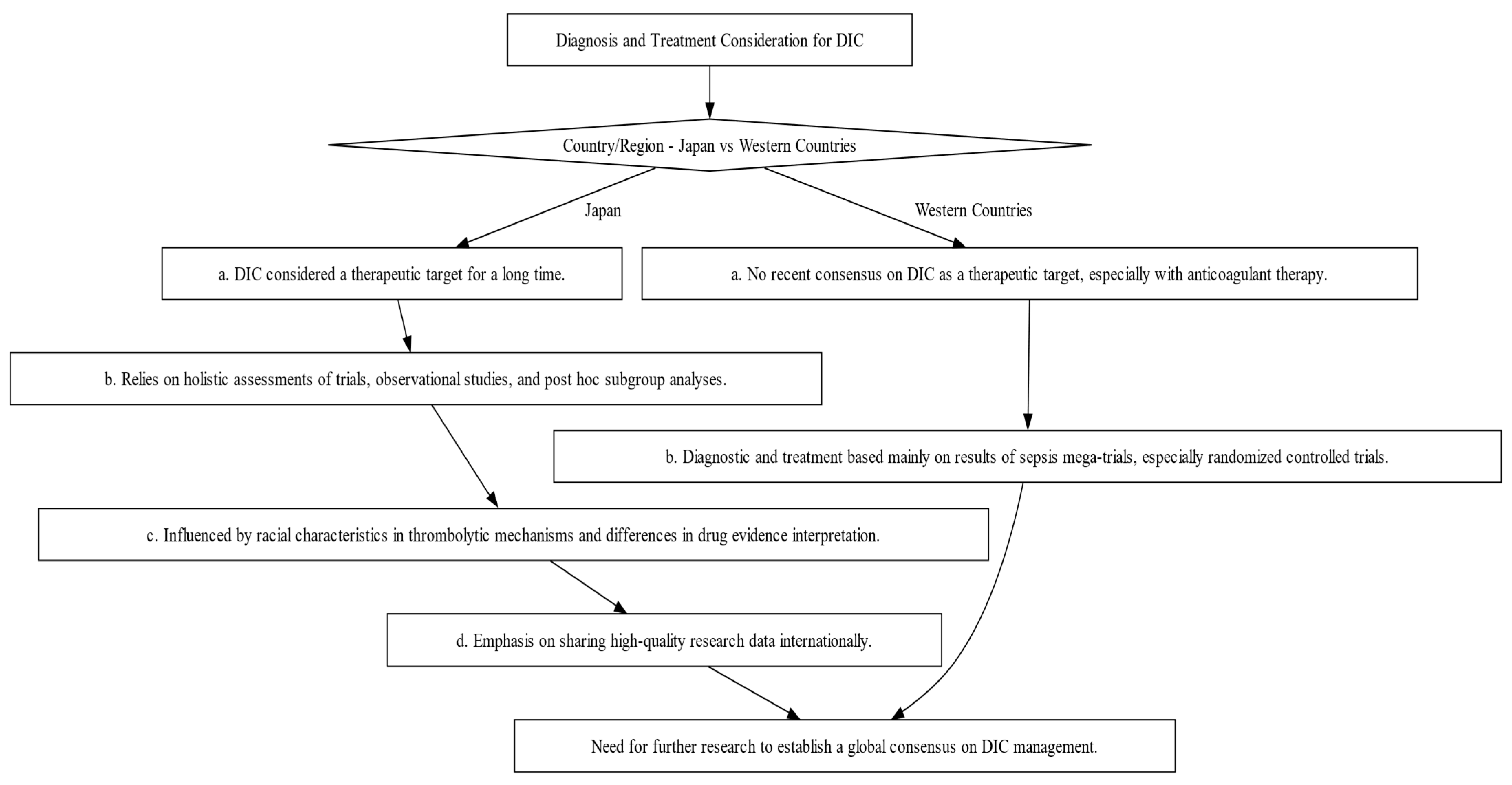
2. Comparative Analysis of DIC Diagnosis and Treatment: Eastern vs. Western Approaches

The diagnosis and management of DIC manifest distinct variations between Japan and Western countries (Figure 2). These variations are shaped by multiple factors, including differing understandings of thrombolytic mechanisms and the types of evidence deemed valid for therapeutic decision-making. In Japan, clinicians adopt a holistic approach, integrating a wide array of research methodologies, ranging from clinical trials and subgroup analyses to observational studies, to inform treatment protocols [19,20]. Conversely, Western medical practice primarily relies on large-scale studies that focus on sepsis, often employing randomized controlled trials (RCTs) as the research design [19]. In this section, we shed light on these distinctions and their implications and as well as highlight the primary commonalities and distinctions in the clinical guidelines for managing DIC as laid out by BCSH (British Committee for Standards in Haematology), JSTH (Japanese Society of Thrombosis and Hemostasis), and SISET (Italian Society for Thrombosis and Hemostasis (Figure 3) [19,20,21]. The International Society on Thrombosis and Haemostasis (ISTH) has established specific criteria for the diagnosis of overt DIC, which include parameters such as low platelet count and prolonged prothrombin time. In contrast, Japan introduced an alternative approach in 2006 called the Japanese Society of Acute Medicine (JAAM) criteria, which emphasizes laboratory tests and clinical data for an accurate diagnosis.
A comparative study by Gando et al. found that the JAAM criteria have higher sensitivity compared to the ISTH criteria. Sensitivity here means that JAAM criteria are better able to correctly identify DIC cases. In their study, the JAAM criteria diagnosed DIC in 46.8% of cases, while the ISTH criteria identified it in only 18.1%. It is important that all cases identified according to ISTH criteria were also recorded according to JAAM criteria. When looking at 28-day mortality rates, both criteria showed similar results, with 31.8% for JAAM and 30.1% for ISTH [22,23]. This suggests that patients excluded by ISTH criteria may suffer from DIC, highlighting the value of JAAM criteria due to their integrative approach. However, the landscape changed with the introduction of the Sepsis-3 definition, which includes the Systemic Inflammatory Response Syndrome (SIRS) score, making the JAAM criteria somewhat less relevant. In response, a new set of criteria called sepsis-induced coagulopathy (SIC) was developed in 2017 to support early DIC diagnosis in sepsis patients. It considers both sepsis and clotting problems, such as a low platelet count. In diagnosing and managing DIC, physicians rely on laboratory findings, including low platelet count, elevated D-dimers, and abnormal clotting times, alongside clinical assessment [24,25]. These indicators inform the ISTH scoring system for overt DIC diagnosis [2,3]. Key tests include Complete Blood Count (CBC), Partial Thromboplastin Time (PTT), Prothrombin Time (PT) assay, fibrinogen, and D-dimer assays. D-dimer and Fibrin Degradation Product (FDP) tests offer robust diagnostic value [4]. A comprehensive DIC panel includes D-dimer and FDP for swift diagnosis and antithrombin for severity assessment and prognosis [24,25,26]. Table 2 provides a detailed comparison of the diagnostic criteria used by the ISTH for both open DIC and SIC and the criteria used by the JAAM for DIC. The criteria are divided into low-risk, medium-risk, and high-risk categories, each of which has a specific rating [21,22,23,27,28,29,30].
Managing DIC is a multifaceted challenge, and the primary goal is to address the underlying infection. However, there’s limited solid evidence supporting the use of anticoagulant therapy alongside antibiotics and source control. Several large-scale randomized controlled trials (RCTs) involving different anticoagulants have failed to provide conclusive evidence of their effectiveness. Nonetheless, guidelines recommend anticoagulant therapy for patients with severe sepsis-associated DIC, particularly if they have coagulation problems. These recommendations have been validated by the ISTH DIC Scientific Standardization Committee [1,21,27,31]. A hypothesis known as the “East Asian paradox” suggests that unique genetic or environmental factors specific to East Asian populations might contribute to the observed differences in DIC diagnosis and management. The choice of anticoagulant therapy for sepsis-induced DIC depends on the condition’s severity and the specific diagnostic criteria in use. Japanese researchers are encouraged to share their clinical research data internationally to improve our understanding of DIC and its management [30]. Overall, there is a major discrepancy between diagnostic and treatment options in Japan and Western countries, and further research is needed to establish an international consensus on DIC as a therapeutic target. Investigations should also focus on the coagulofibrinolytic system in sepsis and the impact of racial characteristics on thrombolytic mechanisms.

3. Can Sepsis-Induced DIC Patients Benefit from Corticosteroids?

Corticosteroids, commonly referred to as CS, continue to be a subject of debate within the community regarding their use in treating sepsis, septic shock, and DIC. There is no consensus due to conflicting results from studies. Gibbison et al. [32] Annane et al. [33] Salluh et al. [34] share perceptions about the possible advantages of CS in addressing sepsis-induced DIC. However, they also highlight the need for research considering the nature of the current evidence. There is obviously a need for more high-quality randomized controlled trials based on these two comprehensive reviews, reflecting the existing uncertainty in this area. In contrast, Rochwerg et al. [35] reported that sepsis patients treated with CS may have reduced mortality. However, they also noted the low reliability of these results, consistent with the urge for more detailed investigation expressed in earlier studies. In addition, according to Gibbison et al. [32], there is quite a positive response to CS on coagulation factors. Despite the possibility of a positive result, the researchers emphasized the experimental nature of their findings and the need for future studies to confirm them. A positive perspective was provided by Gazzaniga et al. [36], who suggested a potential reduction in the risk of SIC with the use of CS. However, like the other studies, they acknowledged the moderate quality of their evidence, further underscoring the need for higher-quality studies. Finally, Ni et al. [37] and Liang et al. [38] found potential benefits of CS in reducing mortality in patients with septic shock and improving outcomes in patients with sepsis-induced coagulopathy. Despite these encouraging results, both studies recommended further investigation and careful interpretation of the results due to possible confounding factors.
Considering these diverse studies, it becomes clear that while there are hints of potential benefits associated with CS in sepsis and septic shock treatment, the current evidence is still inconclusive, with questions about the certainty and quality of the findings. The consensus among all authors is the pressing need for further rigorous and high-quality research to substantiate these preliminary findings, assess the potential risks and benefits more robustly, and clarify the role of CS in the treatment of sepsis and sepsis-induced coagulopathy.
Moreover, future research should consider the factors that might explain the discrepancies in these studies’ results, such as variations in dosage, timing of administration, patient population, patient endotypes, and study design (Table 3). Until a clear consensus appears from more definitive research, clinicians should make decisions on the use of CS on a case-by-case basis, considering each patient’s individual circumstances, the potential benefits and risks of corticosteroid use, and the existing guidelines.

4. Evidence from Studies: Rtm’s Effectiveness in Sepsis-Induced DIC

In the context of sepsis-induced DIC, thrombomodulin alfa (rTM), a recombinant form of human thrombomodulin, has been investigated as a potential therapeutic intervention. The mechanism of action of rTM involves its binding to thrombin, leading to the activation of protein C, an anticoagulant that plays a role in the downregulation of the coagulation cascade [8]. Assorted studies were conducted to investigate the efficacy of rTM in the treatment of DIC produced by sepsis. Valeriani et al. [52] conducted a meta-analysis that included data from randomized controlled trials. The research showed a notable decrease in overall mortality among patients who received treatment with rTM compared to the control group [52]. The results of this study indicate that the use of rTM reduced 28-day mortality rates while mitigating bleeding problems. Bleeding complications are often seen as important adverse events in the context of clotting therapy [43,52]. The research performed by Toshiaki Iba and his colleagues provides more evidence to support the effectiveness of rTM as a therapeutic intervention. In the phase III trial, it was shown that although the overall decrease in death rate did not reach statistical significance, a notable decrease in mortality was observed specifically among patients who presented with SIC at the beginning of the study. This implies that rTM may have enhanced efficacy in patients who already demonstrate systemic inflammatory response syndrome (SIRS) at the initiation of therapy [88]. Despite the encouraging results, more assessment is needed for various aspects of rTM use, including the optimal dose, time of application, effectiveness in different patient subgroups, and discussions on cost-efficiency (Table 3). The review of these inquiries will be of paramount importance in fully exploiting the potential of rTM as a therapeutic intervention for sepsis-induced DIC [53].
In general, the existing body of literature, encompassing the studies conducted by Yang et al., Yamakawa et al., and Toshiaki Iba, indicates a potentially advantageous function of rTM in the management of DIC generated by sepsis. Nevertheless, further study is required to enhance the application protocols of rTM, obtain a comprehensive understanding of its long-term effects, and assess its cost-effectiveness. Such efforts will facilitate the translation of these scientific discoveries into advantageous consequences for patients [88].

5. Vitamin C in Sepsis and DIC: Promise or Paradox?

The investigation and discussion of vitamin C’s role and benefit in sepsis and DIC treatment has attracted significant medical attention. The putative antioxidant, anti-inflammatory, and anticoagulant properties of vitamin C point to its likely benefits in mitigating organ failure and death rates among people afflicted with these ailments [56,57,58]. However, the existing body of scientific literature presents a multifaceted and contradictory image, emphasizing the need for further research. According to a systematic review and meta-analysis conducted by researchers [59], the administration of vitamin C did not provide a statistically significant reduction in the mortality rate, length of stay in the intensive care unit (ICU), or duration of mechanical breathing among patients with sepsis. Nevertheless, the study showed a discernible inclination toward diminishing the length of vasopressor medication, hence implying that vitamin C may influence the disease’s progression. A recent scientific publication [61] highlighted the possible benefits of vitamin C in the treatment of sepsis, especially focusing on its antioxidant properties and ability to regulate the immunological response. Nevertheless, the authors also placed significant emphasis on the equivocal nature of the existing data and advocated for more studies to ascertain the most effective dosage and timing of vitamin C delivery in individuals with sepsis [61]. Research conducted on the use of high-dose vitamin C (HDVC) therapy in the treatment of sepsis and DIC has shown varied outcomes. A comprehensive investigation revealed that the administration of intravenous vitamin C (IVVC) resulted in a notable enhancement of the delta Sequential Organ Failure Assessment (SOFA) score and a decrease in the duration of vasopressor utilization among individuals diagnosed with sepsis or septic shock. However, no significant correlation was observed between the use of IVVC and a reduction in short-term death rates [57]. Nevertheless, the Vitamin C Therapy for Routine Care in Septic Shock (ViCTOR) trial concluded that the intravenous administration of a regimen consisting of vitamin C, thiamine, and hydrocortisone did not lead to a significant decrease in overall mortality in patients afflicted with septic shock. However, it was noted that the time needed to reverse the onset of septic shock was markedly reduced in the patient group that received this combination therapy [60]. These results were consistent with recent randomized controlled trials (RCTs) that also reported no mortality benefit with combination therapy [89,90,91,92,93,94]. Another study revealed an intriguing finding. It showed that adult patients dealing with sepsis and undergoing vasopressor therapy within the ICU, when treated with intravenous vitamin C, faced an increased risk. In contrast to those who were administered a placebo, those who received active treatment had an increased probability of experiencing either fatality or persistent organ failure. The heightened risk endured for a substantial duration, namely, until the 28th day following the initiation of the therapy [54]. The divergent results highlight the intricate nature of the involvement of vitamin C in the treatment of sepsis and DIC. While several studies have shown potential advantages, others have discovered no substantial effect or even possible negative consequences [54,55]. This underscores the need for more rigorous, randomized, controlled studies to clarify the therapeutic efficacy of vitamin C in these specific disorders. It is advisable for clinicians to use prudence when considering administering vitamin C for sepsis treatment and DIC, pending the availability of further conclusive data. These findings are significant for researchers in recognizing the complex nature of vitamin C’s role in sepsis. Consequently, there is a need for studies that not only examine its influence on death rates and organ dysfunction but also delve into its impact on the development of the disease and the quality of life experienced by patients. It is important to make efforts to standardize dosage regimens to improve the effectiveness of treatment (Table 3).
In short, the current corpus of literature provides a nuanced and incongruous portrayal of the involvement of vitamin C in the treatment of sepsis and DIC. The continuous discourse among academics and doctors highlights the divergent findings, hence emphasizing the necessity for more exploration. Based on the available information, healthcare professionals are recommended to exercise prudence when considering the administration of vitamin C in cases of sepsis and DIC. The determination of whether to use this intervention should be based on the specific attributes of each patient and the ongoing accumulation of scientific knowledge in this area. The intricacy of biological systems and the difficulties in converting possible therapeutic pathways into practical therapies are highlighted by this argument, particularly for preclinical readers.

6. Fibrinolytic Therapy in Sepsis-Induced DIC: A Potential Game-Changer?

The investigation of fibrinolytic treatment, particularly the use of fibrinolytic drugs such as tissue plasminogen activator (tPA), is a subject of research in the context of sepsis-induced DIC. While the administration of fibrinolytic treatment has a potential hazard of inducing bleeding, it also offers promising advantages by mitigating clot formation and enhancing organ perfusion. Numerous investigations have been conducted to examine the use of fibrinolytic medicines in DIC generated by sepsis. Schouten et al. [66] studied the administration of tPA in a baboon model of DIC induced by sepsis. The authors noted that the administration of tPA resulted in an amplification of fibrinolysis, a reduction in microvascular thrombosis, and an improvement in organ perfusion [66]. The authors [65,67] assessed the use of low-dose tPA in patients who developed DIC due to sepsis. Improvements in coagulation abnormalities were observed because of how to manage low-dose tPA treatment in patients with DIC induced by sepsis, as specified by the study results. The observed enhancements included a reduction in the levels of fibrin degradation products and an augmentation in platelet counts. Significantly, the administration of low-dose tPA did not yield any considerable complications related to bleeding. However, it is imperative to recognize that the application of fibrinolytic therapy in sepsis-induced DIC remains a topic of debate due to the simultaneous risk of hemorrhage. Achieving a delicate equilibrium between the inherent hazards associated with bleeding problems and the prospective advantages necessitates prudent patient selection and diligent monitoring.
In the realm of DIC triggered by sepsis, a thorough examination was undertaken by numerous esteemed researchers. This comprehensive review sought to analyze the existing information pertaining to fibrinolytic treatment [62,63,64,65]. In the realm of scholarly inquiry, a group of diligent researchers undertook a recent systematic analysis to meticulously scrutinize the existing data pertaining to the utilization of fibrinolytic treatment in instances characterized by DIC provoked by sepsis. The findings of the analysis suggest that although fibrinolytic therapy may exhibit improvements in certain laboratory markers linked to coagulation and fibrinolysis, its impact on clinical endpoints, such as mortality, remains inconclusive [62,63,64,65]. The necessity for further study is posited by the authors to ascertain the efficacy of fibrinolytic treatment in augmenting patient outcomes in instances of sepsis-induced DIC. Further investigation is warranted concerning the utilization of fibrinolytic therapy, specifically tPA, in the setting of DIC precipitated by sepsis. Despite the potential for bleeding, the administration of fibrinolytic therapy possesses the capability to attenuate the formation of blood clots and augment the perfusion of vital organs. Favorable effects on fibrinolysis and coagulation problems have been associated with the use of fibrinolytic drugs, as indicated by previous research. At present, the definitive impact of these agents on clinical outcomes remains to be conclusively determined. The optimization of fibrinolytic therapy for sepsis-induced DIC necessitates additional inquiry (Table 3).

7. Platelet Transfusion in Sepsis-Induced DIC: Navigating Controversy and Conflicting Evidence

In this study, we aim to fully examine the contentious issue surrounding the use of platelet transfusion in the context of sepsis-induced DIC. Therapeutic decision-making can be obscured by contradictory advice within the extensive range of guidelines. In accordance with certain recommendations, it is advisable to consider the possibility of platelet transfusion for patients who are afflicted with sepsis-induced DIC and manifest severe thrombocytopenia or are currently undergoing active bleeding [71]. However, it is vital to recognize the lack of evidence on the effectiveness of platelet transfusion in this context. The existing body of research relies on expert opinion rather than robust scientific trials. A notable investigation in the same domain of the TOPPS trial was conducted by Estcourt et al. [69]. The study’s objective was to evaluate the impact of platelet transfusion on the mortality rate among critically sick patients with thrombocytopenia. The research findings indicated that the administration of platelet transfusion did not have any statistically significant effects on reducing death rates or enhancing patient outcomes [69]. A further investigation was conducted, wherein attention was directed toward individuals suffering from septic shock and DIC [68]. This study’s main objective was to assess the potential association between platelet transfusion and mortality. In congruence with the TOPPS trial, this study demonstrated a lack of mortality reduction in relation to platelet transfusions [68].
This narrative is confirmed by the study conducted by He et al. [70] and Yatabe et al. [72]. The results of their study stipulate that the administration of platelet transfusion did not lead to a reduction in mortality rates among people diagnosed with sepsis. Conversely, it is plausible that this could reduce the duration of stay in the intensive care unit and the hospital, suggesting the potential for unfavorable outcomes [70,72]. In total, the tests collectively demonstrate the lack of empirical evidence of the effectiveness of platelet transfusion in cases of sepsis-induced DIC. Presenting the innovative outlook, Syed Muzaffar et al. offer a contrasting perspective to the studies [73]. The focal point of their research lies in the examination of the pivotal role played by coagulation disorder in the development of sepsis, thereby illuminating the range of coagulopathy induced by sepsis. A more individualized strategy for patient care can be enabled through the proposed triad of coagulation profiles utilizing the Coagulation Index (CI), as put forth by the authors. The authors also underscored the limitations of conventional coagulation assays (CCAs) and drew attention to the utility of thromboelastography (TEG) in the assessment of coagulation profiles [95,96]. The decision to administer platelet transfusion for sepsis-induced DIC should depend on certain contextual factors, considering the patient’s clinical condition, bleeding tendency, and overall trade-off between the advantages and disadvantages of platelet transfusion. The importance of close monitoring of platelet counts and clinical indications in making this decision cannot be overstated. While some guidelines suggest giving platelet transfusion for severe thrombocytopenia or active bleeding, there is also research available, such as the TOPPS test [69] and the study by Andrew et al. [68], that does not demonstrate a significant reduction in mortality or improvement in outcomes from platelet transfusions in this scenario.
Valuable contributions to the existing knowledge of platelet transfusions and coagulation patterns in individuals with sepsis are made by the research conducted by He et al. [70] and Muzaffar et al. [73]. The significance of their study lies in the emphasis on the necessity of implementing meticulous randomized controlled trials and pursuing additional research endeavors to acquire a more comprehensive comprehension of the intricate involvement of platelet transfusion in sepsis-induced DIC. Efforts in future research should be directed toward the identification of distinct subpopulations of patients who may potentially derive advantages from platelet transfusion. Furthermore, the evaluation of their influence on clinical outcomes, encompassing mortality rates and the incidence of hemorrhagic complications, should be undertaken (Table 3).
The ongoing debate and uncertainty surrounding the role of platelet transfusion in the context of sepsis-induced DIC can be attributed to conflicting research findings and divergent viewpoints among experts. There is an ongoing debate within the medical community regarding the pressing necessity for comprehensive research aimed at enhancing our comprehension and expediting the application of clinical therapies in a streamlined fashion. The administration of platelet transfusion in sepsis-induced DIC is currently the subject of a lively debate. This debate stems from the absence of compelling evidence highlighting a noteworthy decrease in mortality rates or enhancement in clinical outcomes linked to this intervention [68,69,70]. Within the realm of clinical decision-making, the utmost significance lies in the prioritization of a customized approach, which is contingent upon meticulous monitoring and diligent evaluation of the unique clinical trajectory exhibited by each patient. To formulate personalized recommendations that align with the specific circumstances of each patient, physicians are required to meticulously assess and modify the intricate equilibrium between potential hazards and expected advantages associated with a transfusion. Valuable insight into the complex elements of the coagulopathy associated with sepsis has been provided by recent developments in scientific studies. The findings of this study lend support to the concept that a uniform treatment strategy may not be the most advantageous but rather emphasize the importance of customizing medical interventions to suit the unique characteristics of each patient (Table 3). In this context, the significance of TEG becomes apparent, as exemplified by its noteworthy application as a diagnostic instrument [73]. Considering the existing dearth of definitive and all-encompassing data, it is important for medical practitioners to exercise judiciousness in contemplating the utilization of platelet transfusion for managing sepsis-induced DIC.

8. Immunomodulatory Therapy: G-CSF, GM-CSF, IFN-γ, MSCs in Sepsis, DIC, and Their Implications for Clinical Practice & Severe COVID-19

IMT in the context of sepsis-induced DIC is currently an area of active research [74,88]. Various agents, including granulocyte colony-stimulating factor (G-CSF), granulocyte-macrophage colony-stimulating factor (GM-CSF), and interferon-gamma (IFN-γ), have been investigated for their potential role in sepsis-induced DIC [97,98]. Nevertheless, the precise elucidation of their specific influence on sepsis-induced DIC remains to be definitively established [97,98]. Granulocyte colony-stimulating factor (G-CSF), a hematological growth factor, stimulates the production and mobilization of neutrophils. Conversely, it is worth noting that granulocyte-macrophage colony-stimulating factor (GM-CSF) exerts its influence on both neutrophils and monocytes/macrophages [99]. The drugs under consideration have been the focal point of scientific inquiry, with the objective of examining their capacity to enhance immune functionality and enhance outcomes in instances of DIC precipitated by sepsis [74,75]. The impact of G-CSF treatment on individuals diagnosed with sepsis and DIC was examined by the authors [76,77]. The study’s findings elucidated that the administration of G-CSF led to a noteworthy increase in neutrophil counts and amelioration of coagulation parameters in individuals afflicted with sepsis-induced DIC [76,77]. In the therapeutic management of DIC induced by sepsis, IFN-γ, an immunomodulatory drug, has exhibited promising potential. The impact of IFN-γ treatment on patients with sepsis-induced DIC was examined by Iba T. et al. [53] in their study. The study’s findings revealed that the administration of IFN-γ yielded enhancements in coagulation abnormalities, a decrease in the duration of DIC, and a potential decline in fatality rates [53]. However, further inquiry is imperative to attain a holistic understanding of the role played by immunomodulatory drugs in the context of DIC induced by sepsis. Included in this analysis are the exploration of the mechanisms of action, the determination of the best dosage, the identification of the ideal timing for administration, and the refinement of the criteria for selecting patients who may potentially derive therapeutic benefits from such treatment. Conducting randomized controlled studies [6,8,53,72,99,100,101,102,103] is imperative for assessing the effectiveness, safety, and clinical outcomes associated with the utilization of immunomodulatory drugs in this specific context.
Significant interest has been generated in the treatment of sepsis, transplant medicine, and autoimmune diseases through the utilization of mesenchymal stem cell (MSC) therapy. Although the precise cellular and molecular mechanisms underlying MSC-mediated immunomodulation have not yet been fully elucidated, preclinical studies have demonstrated promising results. The immunomodulatory effects of MSCs are exerted via the secretion of cytokines, a process that can be influenced by both the local microenvironment and inflammatory cytokines [78,79].
Additionally, even apoptotic, metabolically inactivated, or fragmented MSCs possess immunomodulatory potential [79,104]. However, the lack of standardization in the isolation, culture, and characterization of MSCs complicates data comparison. MSCs can be derived from various adult and neonatal tissues, each exhibiting unique features in vitro and in vivo [79,80,81] Notably, freshly thawed MSCs appear to have reduced immunomodulatory capacity compared to continuously cultured MSCs [105]. The local microenvironment plays a critical role in shaping MSC-mediated immunomodulation, further adding to its complexity [82,83]. MSCs exert their immunomodulatory effects through a combination of cell contact-dependent mechanisms and the release of soluble factors [79,83,84,85,106,107,108,109,110,111]. MSCs have a significant impact on various immune cells, particularly anti-inflammatory monocytes/macrophages and regulatory T cells (Tregs) [84,85,86]. Interestingly, MSC viability does not appear to be a prerequisite for some of their immunomodulatory effects, as apoptotic MSCs have demonstrated beneficial effects in animal models [112]. Exploring the use of dead or fragmented MSCs may provide more predictable immunomodulatory effects and facilitate better comparison across studies [112].
A case report by Galic et al. presented the successful management of a 14-month-old [93]. The treatment included a combination of antibiotics, plasmapheresis, dialysis, methylprednisolone, mycophenolate mofetil, and eculizumab [87]. Eculizumab therapy was considered in rare cases of sepsis with massive complement consumption after resolving life-threatening multiorgan failure [87]. These studies and reports have important implications for future clinical practice. IMT, including the use of G-CSF, GM-CSF, IFN-γ, and MSCs, shows promise in improving outcomes in sepsis-induced DIC. However, further research is needed to determine the optimal use, dosing, timing, and impact on clinical outcomes of these therapies [8,53,72,75,78,80,81,82,98,99,100,101,103,104,107,111,113,114,115,116,117,118,119,120,121,122,123,124,125]. The need for cautious administration and withdrawal of the drug is underscored by the potential utilization of eculizumab in instances of sepsis accompanied by complement consumption. In addition, the investigation of extracellular vesicles derived from mesenchymal stem cells (MSC-EVs) as a therapy without the need for cell transplantation presents a hopeful alternative to treatments based on MSCs [87].
By elucidating the intricate relationship between severe COVID-19 and sepsis, which arises from viral infection, one can glean profound insights into the nature of the disease and subsequently enhance the trajectory of future investigations pertaining to therapeutic interventions and preventive strategies [126]. Consideration should be given to the inclusion of IMT in the treatment regimen for severe cases of COVID-19 in conjunction with etiological and supportive interventions [126,127]. The primary objective in the context of COVID-19 immunotherapy should revolve around the mitigation of exaggerated inflammatory responses while simultaneously preserving a controlled level of inflammation and facilitating the restoration of profound immunosuppression [126,127]. In severe cases of COVID-19, it is imperative to conduct additional research to substantiate the safety, efficacy, timing, and dosing of IMT (Table 3) [126].
In conclusion, IMT and MSC-based treatments hold promise for improving outcomes in sepsis-induced DIC and other conditions. However, further research is necessary to fully understand their mechanisms of action, optimize their use in clinical practice, and ensure their safety and efficacy. The exploration of MSC-EVs as a cell-free therapy and the consideration of sepsis-like features in severe COVID-19 provide valuable insights for future studies and therapeutic developments.

9. The Controversy Surrounding Anticoagulant Therapy for Sepsis and DIC Management: An In-Depth Analysis

Blood clotting problems are common in sepsis and DIC, which can lead to organ failure and death. The effectiveness of using anticoagulant therapy to treat SIC and DIC remains a controversial topic among researchers [27,128]. A comprehensive analysis by Freeman et al. found no clear survival benefits from anticoagulant therapy in the general sepsis population and even suggested a higher risk of bleeding complications [129]. Some studies examined the concomitant use of low-dose heparin for the prevention of deep vein thrombosis and evaluated various outcomes, including all-cause mortality, at different time points [130].

9.1. Summary of Previous Research

This section reviews previous studies to provide a comprehensive overview of the effectiveness of anticoagulant therapy in sepsis treatment (Figure 4). When reviewing [27,53,131] various studies [6,8,132,133,134], it becomes clear that the results derived from these studies were based predominantly on short-term observations. It is important to recognize that these short-term results may not necessarily reflect the long-term dynamics of sepsis in humans. Therefore, when considering the use of anticoagulant therapy, it is essential to strike a balance between the potential benefits and associated side effects. Furthermore, the authors of these studies have wisely identified certain critical gaps in the existing research landscape. A notable omission concerns the lack of consideration of the cost-effectiveness of anticoagulant therapies, particularly in settings outside Japan (Table 4) [131]. This is very worrying as the economic impact of such therapies is often overlooked. Furthermore, the authors highlighted several issues that contribute to the heterogeneity observed in different studies. These issues include issues such as the risk of bias, the criteria for defining bleeding complications, and differences in the types of anticoagulants used in individual research efforts [27,128,129,131,135,136]. It is important to understand these factors because they can have a significant impact on the interpretation of study results. A review of previous research highlights the need for a more nuanced and comprehensive approach when considering anticoagulant therapy for sepsis treatment [131,137,138]. The aim is to examine not only the immediate effects but also to consider the possible long-term effects, cost-effectiveness, and factors that contribute to the heterogeneity of the studies. Such considerations are critical to advancing our understanding of the utility of anticoagulant therapy in the context of sepsis treatment.

9.2. Study Hypothesis, Significance, and Unanswered Questions

In this section, we address the hypothesis, significance, and open questions surrounding the use of anticoagulant therapy in sepsis treatment (Table 3 and Table 4 and Figure 2). The present study examines the potential benefits of anticoagulant therapy in patients with SIC or DIC, with a focus on specific subgroups of individuals. However, it is important to recognize that despite some studies indicating favorable outcomes, the totality of the evidence does not provide clear support for the universal use of anticoagulant treatment in the broader sepsis population [27,30,137]. Therefore, the question of whether anticoagulant therapy is generally beneficial for sepsis patients remains a controversial topic in the scientific community. Anticoagulation plays a central role in the body’s innate defense mechanisms against infections [139,140]. However, widespread, and indiscriminate use is discouraged due to the increased risk of bleeding. In addition, there are concerns about possible impairment of pathogen clearance due to excessive coagulation [139]. A constant challenge in this area is, therefore, the establishment of precise criteria for patient selection. This challenge is critical to ensure that anticoagulation therapy is administered to those who can derive the greatest benefit from it. The debate surrounding the use of anticoagulation therapy in sepsis patients highlights the complexity of this medical intervention. As researchers continue to study and elucidate the potential benefits, the need for a differentiated approach to patient selection and treatment remains a primary focus of future research efforts [141,142,143].

9.3. Antithrombin and Combination Therapy

This section focuses on the role of antithrombin, an important endogenous anticoagulant, and explores the potential of combination therapy (Table 4 and Table 5) [144]. Antithrombin has been the subject of extensive investigation in the context of sepsis-induced coagulopathy. However, it is important to emphasize that relying on a single study alone to make a final decision about effectiveness is not conclusive. A notable phase 4 study conducted in patients with septic DIC provided results worth considering. This study found no improvement in 28-day mortality associated with the use of antithrombin in this population [128,131,137,140,141,142,143,145,146,147]. Interestingly, there is growing interest in the concept of combination therapy. This approach is promising because both the antithrombin GAG system and the thrombomodulin protein C system exhibit significant downregulation and function independently in sepsis-induced coagulopathy [146,148,149]. It is important to note that while clinical trials have not yet been completed in this specific context, preliminary investigations into combining these therapeutic approaches have shown potential. These early studies suggest that combination therapy, compared to individual therapeutic interventions, may be key to ameliorating organ damage and ultimately improving survival rates [130]. These results highlight the evolving landscape of sepsis treatment and highlight the need for further research to refine therapeutic strategies and improve patient outcomes in severe sepsis and coagulation disorders.

9.4. Optimal Therapeutic Targets and Considerations in Anticoagulant Therapy

In the domain of anticoagulant therapy, the choice of therapeutic targets is contingent upon clinical variables and the specific anticoagulant employed. Notably, patients with mechanical heart valves necessitate a targeted International Normalized Ratio (INR) range between 2.5 and 4.9, with a recommended target INR of 3.0 to 4.0 [153], whereas those undergoing vitamin K antagonist therapy are advised to achieve a target INR of 2.0 to 3.0 [154]. However, the optimal therapeutic targets for antithrombin (AT) and recombinant thrombomodulin (rTM) therapies remain less well-defined. Furthermore, the utilization of target-specific oral anticoagulants (TSOACs) such as dabigatran, apixaban, and rivaroxaban necessitates meticulous patient assessment to determine their suitability in the clinical context [155,156]. Among the array of anticoagulant options available, unfractionated heparin offers rapid onset and a brief half-life, low-molecular-weight heparin presents a longer half-life and subcutaneous administration [157,158], while fondaparinux is a synthetic pentasaccharide with selective antithrombin binding and factor Xa inhibition. Warfarin, functioning as a vitamin K antagonist, inhibits the synthesis of vitamin K-dependent clotting factors, and direct thrombin inhibitors (e.g., dabigatran, argatroban, desirudin, bivalirudin) directly obstruct thrombin activity [158,159,160]. Factor Xa inhibitors (e.g., rivaroxaban, apixaban, edoxaban, betrixaban) belong to a distinct category, inhibiting factor Xa. The choice of anticoagulant therapy hinges upon the patient’s clinical presentation, the indication for therapy, and the clinical expertise of the healthcare provider, all within the framework of established treatment guidelines. The primary concern associated with anticoagulant therapy is the risk of bleeding, which can manifest in various anatomical locations and pose significant clinical challenges. Additionally, patients may experience a spectrum of potential side effects, including gastrointestinal symptoms, dizziness, headaches, dermatological issues, hair loss, jaundice, and skin hemorrhage. Regular blood tests to monitor clotting time are requisite for patients receiving warfarin to assess bleeding risk. In cases where signs of excessive bleeding, such as hematuria or prolonged nosebleeds, become evident, prompt medical attention is imperative. Consequently, comprehensive discussions between patients and their healthcare providers regarding potential side effects should precede the initiation of anticoagulant therapy to facilitate informed decision-making and optimize patient outcomes [140,161].

9.5. Biomarkers, Machine Learning, and Emerging Trends in Sepsis Diagnosis and DIC Management

Recent research [30,140,162,163] focused on identifying biomarkers for sepsis diagnosis to address the complex heterogeneity of sepsis and its diverse clinical presentation, encompassing various clinical signs and symptoms. Biomarkers like C-reactive protein (CRP) [164], procalcitonin (PCT) [165], interleukin-6 (IL-6) [166], CD64 [165,167], presepsin [165], and soluble TREM-1 (sTREM-1) [168] have displayed promise in sepsis diagnosis. However, their practical application is challenged by the multifaceted nature of sepsis. Finding the ideal sepsis-specific biomarker remains a challenge, with multiple biomarkers potentially working in concert to identify critically ill patients in need of closer monitoring. Host response biomarkers play a critical role in diagnosis, early organ dysfunction recognition, risk assessment, prognosis, and patient management, including antibiotic stewardship [168].
Machine learning has emerged as a potent tool for biomarker discovery, enabling the identification of novel diagnostic and prognostic biomarkers by analyzing extensive datasets with intricate relationships [169,170,171]. Machine learning algorithms like Random Forest, Support Vector Machines (SVM), Artificial Neural Networks (ANN), Logistic Regression, and Decision Trees have been employed to sift through vast datasets, revealing patterns and relationships not readily discernible through conventional analysis [163,165,170,172]. Ensuring high-quality and representative training data for machine learning algorithms is imperative [165]. Additionally, the clinical relevance and interpretability of machine learning models are critical considerations, necessitating validation with independent datasets to ensure their applicability in patient care [168]. The choice of a specific machine learning algorithm depends on the research question and dataset characteristics [167,171]. As research in sepsis biomarkers and machine learning progresses, these innovative approaches hold promise for enhancing sepsis diagnosis and patient care.

9.6. Challenges and Future Perspectives

This section reviews the complexities, challenges, and potential future directions associated with anticoagulant therapy in the context of septic coagulopathy (Table 5). Anticoagulant therapy, although promising, has significant disadvantages, including high cost and increased risk of bleeding [131]. It is important to recognize that anticoagulant therapy consistently shows a trend toward bleeding events compared to placebo, with bleeding occurring more than 1.7 times more frequently in the treatment group. Unfortunately, these bleeding events often negate the benefit of anticoagulants. In addition, it is important to understand that the risk of bleeding may vary depending on the inhibitors used, their concentration, how long they remain in the body and the associated disorders in the body’s blood clotting system. Therefore, it is critical to carefully select patients who are most likely to benefit from this treatment due to persistent increased bleeding risk. The challenges associated with the use of anticoagulant therapy as a treatment strategy become apparent when examining the problems faced by randomized controlled trials (RCTs) examining its use in sepsis. The treatment of coagulation problems in sepsis patients should be approached with great care and considered in the broader context of individualized patient care. The difficulties observed in RCTs investigating anticoagulation therapy in sepsis highlight the need for a more differentiated and personalized approach. As our understanding of sepsis and its coagulation aspects continues to improve, we can expect to see the development of more refined treatment approaches. These encouraging developments hold the potential to significantly improve the treatment of this complicated and challenging medical condition.

10. Conclusions

With limited viable treatment strategies, sepsis-related DIC continues to pose a complex and perilous challenge. The management of this condition remains complex and life-threatening. Clarification of the pathological mechanisms behind sepsis and DIC, coupled with advances in therapeutic interventions, can achieve favorable clinical results in affected individuals. Additional high-quality randomized controlled trials are needed to mitigate the efficacy and controversies surrounding the role of anticoagulant therapy, which encompasses the utilization of heparin, recombinant activated protein C (APC), and antithrombin (AT). Blood purification methods such as polymyxin B hemoperfusion and immunotherapy approaches such as reversing immune function with drugs or antibodies have shown some potential, but further investigation is needed to assess their efficacy and safety. The current state of research on sepsis treatment DIC underscores the need for multidisciplinary approaches and collaborations between clinicians, researchers, and healthcare organizations to improve patient outcomes. Amid the ongoing global pandemic, the integration of these therapies, notoriously IMT, is crucial for treating severe COVID-19 cases presenting as sepsis-induced DIC. Further studies are required to identify new therapeutic targets and improve our understanding of the underlying mechanisms of sepsis DIC. By continually developing our knowledge of sepsis DIC, we can work to expand effective and targeted therapies that can save lives and improve patient outcomes.

Author Contributions

All authors contributed equally to this work. All authors have read and agreed to the published version of the manuscript.

Funding

This research received no external funding.

Institutional Review Board Statement

Not applicable.

Informed Consent Statement

Not applicable.

Data Availability Statement

Not applicable.

Acknowledgments

Unar and Bertolino were supported through the PhD program in Translational Medicine at the Università degli Studi della Campania Luigi Vanvitelli.

Conflicts of Interest

The authors declare no conflict of interest.

References

  1. Singer, M.; Deutschman, C.S.; Seymour, C.W.; Shankar-Hari, M.; Annane, D.; Bauer, M.; Bellomo, R.; Bernard, G.R.; Chiche, J.D.; Coopersmith, C.M.; et al. The Third International Consensus Definitions for Sepsis and Septic Shock (Sepsis-3). JAMA 2016, 315, 801–810. [Google Scholar] [CrossRef]
  2. Tsantes, A.G.; Parastatidou, S.; Tsantes, E.A.; Bonova, E.; Tsante, K.A.; Mantzios, P.G.; Vaiopoulos, A.G.; Tsalas, S.; Konstantinidi, A.; Houhoula, D. Sepsis-Induced Coagulopathy: An Update on Pathophysiology, Biomarkers, and Current Guidelines. Life 2023, 13, 350. [Google Scholar] [CrossRef] [PubMed]
  3. Giustozzi, M.; Ehrlinder, H.; Bongiovanni, D.; Borovac, J.A.; Guerreiro, R.A.; Gąsecka, A.; Papakonstantinou, P.E.; Parker, W.A.E. Coagulopathy and Sepsis: Pathophysiology, Clinical Manifestations and Treatment. Blood Rev. 2021, 50, 100864. [Google Scholar] [CrossRef] [PubMed]
  4. Martin, G.S.; Mannino, D.M.; Eaton, S.; Moss, M. The Epidemiology of Sepsis in the United States from 1979 through 2000. N. Engl. J. Med. 2003, 348, 1546–1554. [Google Scholar] [CrossRef]
  5. Iba, T.; Di Nisio, M.; Thachil, J.; Wada, H.; Asakura, H.; Sato, K.; Saitoh, D. A Proposal of the Modification of Japanese Society on Thrombosis and Hemostasis (JSTH) Disseminated Intravascular Coagulation (DIC) Diagnostic Criteria for Sepsis-Associated DIC. J. Clin. Appl. Thromb. Hemost. 2018, 24, 439–445. [Google Scholar] [CrossRef]
  6. Iba, T.; Umemura, Y.; Watanabe, E.; Wada, T.; Hayashida, K.; Kushimoto, S. Diagnosis of Sepsis-induced Disseminated Intravascular Coagulation and Coagulopathy. J. Acute Med. Surg. 2019, 6, 223–232. [Google Scholar] [CrossRef] [PubMed]
  7. Wheeler, A.P.; Bernard, G.R. Treating Patients with Severe Sepsis. N. Engl. J. Med. 1999, 340, 207–214. [Google Scholar] [CrossRef] [PubMed]
  8. Iba, T.; Watanabe, E.; Umemura, Y.; Wada, T.; Hayashida, K.; Kushimoto, S.; Japanese Surviving Sepsis Campaign Guideline Working Group for Disseminated Intravascular Coagulation; Wada, H. Sepsis-Associated Disseminated Intravascular Coagulation and Its Differential Diagnoses. J. Intensive Care 2019, 7, 32. [Google Scholar] [CrossRef]
  9. Iba, T.; Levy, J.H. Haemostasis Inflammation and Thrombosis: Roles of Neutrophils, Platelets and Endothelial Cells and Their Interactions in Thrombus Formation during Sepsis. J. Thromb. Haemost. 2018, 16, 231–241. [Google Scholar] [CrossRef]
  10. Semeraro, N.; Ammollo, C.T.; Semeraro, F.; Colucci, M. Coagulopathy of Acute Sepsis. In Proceedings of the Seminars in Thrombosis and Hemostasis; Thieme Medical Publishers: Stuttgart, Germany, 2015; Volume 41, pp. 650–658. [Google Scholar]
  11. Engelmann, B.; Massberg, S. Thrombosis as an Intravascular Effector of Innate Immunity. Nat. Rev. Immunol. 2013, 13, 34–45. [Google Scholar] [CrossRef]
  12. Corrigan, J.J., Jr.; Ray, W.L.; May, N. Changes in the Blood Coagulation System Associated with Septicemia. N. Engl. J. Med. 1968, 279, 851–856. [Google Scholar] [CrossRef]
  13. Østerud, B.; Bjørklid, E. The Tissue Factor Pathway in Disseminated Intravascular Coagulation. In Proceedings of the Seminars in Thrombosis and Hemostasis; Thieme Medical Publishers, Inc.: Stuttgart, Germany, 2001; Volume 27, pp. 605–618. [Google Scholar]
  14. Nieman, M.T. Protease-Activated Receptors in Hemostasis. Blood J. Am. Soc. Hematol. 2016, 128, 169–177. [Google Scholar] [CrossRef]
  15. Ma, R.; Xie, R.; Yu, C.; Si, Y.; Wu, X.; Zhao, L.; Yao, Z.; Fang, S.; Chen, H.; Novakovic, V. Phosphatidylserine-Mediated Platelet Clearance by Endothelium Decreases Platelet Aggregates and Procoagulant Activity in Sepsis. Sci. Rep. 2017, 7, 4978. [Google Scholar] [CrossRef] [PubMed]
  16. Gando, S.; Levi, M.; Toh, C.-H. Disseminated Intravascular Coagulation. J. Nat. Rev. Dis. Primers 2016, 2, 1–16. [Google Scholar]
  17. Levi, M.; van der Poll, T. Coagulation and Sepsis. Thromb. Res. 2017, 149, 38–44. [Google Scholar] [CrossRef] [PubMed]
  18. Chris Nickson Sepsis Definitions and Diagnosis. Available online: https://litfl.com/sepsis-definitions-and-diagnosis/ (accessed on 3 November 2020).
  19. Wada, H.; Thachil, J.; Di Nisio, M.; Mathew, P.; Kurosawa, S.; Gando, S.; Kim, H.K.; Nielsen, J.D.; Dempfle, C.; Levi, M. Guidance for Diagnosis and Treatment of Disseminated Intravascular Coagulation from Harmonization of the Recommendations from Three Guidelines. J. Thromb. Haemost. 2013, 11, 761–767. [Google Scholar] [CrossRef]
  20. Di Nisio, M.; Baudo, F.; Cosmi, B.; D’Angelo, A.; De Gasperi, A.; Malato, A.; Schiavoni, M.; Squizzato, A. Diagnosis and Treatment of Disseminated Intravascular Coagulation: Guidelines of the Italian Society for Haemostasis and Thrombosis (SISET). J. Thromb. Res. 2012, 129, e177–e184. [Google Scholar] [CrossRef]
  21. Asakura, H.; Takahashi, H.; Uchiyama, T.; Eguchi, Y.; Okamoto, K.; Kawasugi, K.; Madoiwa, S.; Wada, H. Proposal for New Diagnostic Criteria for DIC from the Japanese Society on Thrombosis and Hemostasis. Thromb. J. 2016, 14, 42. [Google Scholar] [CrossRef]
  22. Gando, S.; Wada, H.; Thachil, J.; Scientific, T. Differentiating Disseminated Intravascular Coagulation (DIC) with the Fibrinolytic Phenotype from Coagulopathy of Trauma and Acute Coagulopathy of Trauma-Shock (COT/ACOTS). J. Thromb. Haemost. 2013, 11, 826–835. [Google Scholar] [CrossRef]
  23. Gando, S.; Hayakawa, M. Pathophysiology of Trauma-Induced Coagulopathy and Management of Critical Bleeding Requiring Massive Transfusion. In Proceedings of the Seminars in Thrombosis and Hemostasis; Thieme Medical Publishers: Stuttgart, Germany, 2015; pp. 155–165. [Google Scholar]
  24. Yu, M.; Nardella, A.; Pechet, L. Screening Tests of Disseminated Intravascular Coagulation: Guidelines for Rapid and Specific Laboratory Diagnosis. Crit. Care Med. 2000, 28, 1777–1780. [Google Scholar] [CrossRef]
  25. Disseminated Intravascular Coagulation—DIC. Choose the Right Test. Available online: https://arupconsult.com/content/disseminated-intravascular-coagulation (accessed on 9 September 2023).
  26. Disseminated Intravascular Coagulation (DIC). Causes & Symptoms. Available online: https://my.clevelandclinic.org/health/diseases/21836-disseminated-intravascular-coagulation-dic (accessed on 9 September 2023).
  27. Unar, A.; Bertolino, L.; Patauner, F.; Gallo, R.; Durante-Mangoni, E. Pathophysiology of Disseminated Intravascular Coagulation in Sepsis: A Clinically Focused Overview. Cells 2023, 12, 2120. [Google Scholar] [CrossRef]
  28. Iba, T.; Di Nisio, M.; Levy, J.H.; Kitamura, N.; Thachil, J. New Criteria for Sepsis-Induced Coagulopathy (SIC) Following the Revised Sepsis Definition: A Retrospective Analysis of a Nationwide Survey. BMJ Open 2017, 7, e017046. [Google Scholar] [CrossRef] [PubMed]
  29. Gando, S.; Iba, T.; Eguchi, Y.; Ohtomo, Y.; Okamoto, K.; Koseki, K.; Mayumi, T.; Murata, A.; Ikeda, T.; Ishikura, H. A Multicenter, Prospective Validation of Disseminated Intravascular Coagulation Diagnostic Criteria for Critically Ill Patients: Comparing Current Criteria. Crit. Care Med. 2006, 34, 625–631. [Google Scholar] [CrossRef] [PubMed]
  30. Ushio, N.; Wada, T.; Ono, Y.; Yamakawa, K. Sepsis-induced Disseminated Intravascular Coagulation: An International Estrangement of Disease Concept. Acute Med. Surg. 2023, 10, e00843. [Google Scholar] [CrossRef] [PubMed]
  31. Iba, T.; Helms, J.; Connors, J.M.; Levy, J.H. The Pathophysiology, Diagnosis, and Management of Sepsis-Associated Disseminated Intravascular Coagulation. J. Intensive Care 2023, 11, 24. [Google Scholar] [CrossRef] [PubMed]
  32. Gibbison, B.; López-López, J.A.; Higgins, J.P.T.; Miller, T.; Angelini, G.D.; Lightman, S.L.; Annane, D. Corticosteroids in Septic Shock: A Systematic Review and Network Meta-Analysis. J. Crit. Care 2017, 21, 78. [Google Scholar] [CrossRef]
  33. Annane, D.; Bellissant, E.; Bollaert, P.E.; Briegel, J.; Keh, D.; Kupfer, Y. Corticosteroids for Treating Severe Sepsis and Septic Shock. J. Cochrane Database Syst. Rev. 2004, 1, 7. [Google Scholar]
  34. Salluh, J.I.F.; Povoa, P. Corticosteroids in Severe Sepsis and Septic Shock: A Concise Review. J. Shock. 2017, 47, 47–51. [Google Scholar] [CrossRef]
  35. Rochwerg, B.; Oczkowski, S.J.; Siemieniuk, R.A.C.; Agoritsas, T.; Belley-Cote, E.; D’Aragon, F.; Duan, E.; English, S.; Gossack-Keenan, K.; Alghuroba, M. Corticosteroids in Sepsis: An Updated Systematic Review and Meta-Analysis. J. Crit. Care Med. 2018, 46, 1411–1420. [Google Scholar] [CrossRef]
  36. Gazzaniga, G.; Tavecchia, G.A.; Bravi, F.; Scavelli, F.; Travi, G.; Campo, G.; Vandenbriele, C.; Tritschler, T.; Sterne, J.A.C.; Murthy, S. The Effect of Antithrombotic Treatment on Mortality in Patients with Acute Infection: A Meta-Analysis of Randomized Clinical Trials. J. Int. J. Cardiol. 2023, 383, 75–81. [Google Scholar] [CrossRef]
  37. Ni, Y.-N.; Liu, Y.-M.; Wang, Y.-W.; Liang, B.-M.; Liang, Z.-A. Can Corticosteroids Reduce the Mortality of Patients with Severe Sepsis? A Systematic Review and Meta-Analysis. Am. J. Emerg. Med. 2019, 37, 1657–1664. [Google Scholar] [CrossRef] [PubMed]
  38. Liang, H.; Song, H.; Zhai, R.; Song, G.; Li, H.; Ding, X.; Kan, Q.; Sun, T. Corticosteroids for Treating Sepsis in Adult Patients: A Systematic Review and Meta-Analysis. J. Front. Immunol. 2021, 12, 709155. [Google Scholar] [CrossRef] [PubMed]
  39. Aikawa, N.; Shimazaki, S.; Yamamoto, Y.; Saito, H.; Maruyama, I.; Ohno, R.; Hirayama, A.; Aoki, Y.; Aoki, N. Thrombomodulin Alfa in the Treatment of Infectious Patients Complicated by Disseminated Intravascular Coagulation: Subanalysis from the Phase 3 Trial. J. Shock. 2011, 35, 349–354. [Google Scholar] [CrossRef]
  40. Liu, X.; Wang, X.; Liu, X.; Hao, D.; Jaladat, Y.; Lu, F.; Sun, T.; Lv, C. Low-dose Heparin as Treatment for Early Disseminated Intravascular Coagulation during Sepsis: A Prospective Clinical Study. J. Exp. Ther. Med. 2014, 7, 604–608. [Google Scholar] [CrossRef]
  41. Wada, H.; Matsumoto, T.; Yamashita, Y. Diagnosis and Treatment of Disseminated Intravascular Coagulation (DIC) According to Four DIC Guidelines. J. Intensive Care 2014, 2, 15. [Google Scholar] [CrossRef]
  42. Vincent, J.-L.; Francois, B.; Zabolotskikh, I.; Daga, M.K.; Lascarrou, J.-B.; Kirov, M.Y.; Pettilä, V.; Wittebole, X.; Meziani, F.; Mercier, E. Effect of a Recombinant Human Soluble Thrombomodulin on Mortality in Patients with Sepsis-Associated Coagulopathy: The SCARLET Randomized Clinical Trial. J. Jama 2019, 321, 1993–2002. [Google Scholar] [CrossRef] [PubMed]
  43. Yamakawa, K.; Murao, S.; Aihara, M. Recombinant Human Soluble Thrombomodulin in Sepsis-Induced Coagulopathy: An Updated Systematic Review and Meta-Analysis. J. Thromb. 2019, 119, 56–65. [Google Scholar] [CrossRef]
  44. Tagami, T.; Matsui, H.; Horiguchi, H.; Fushimi, K.; Yasunaga, H. Antithrombin and Mortality in Severe Pneumonia Patients with Sepsis-associated Disseminated Intravascular Coagulation: An Observational Nationwide Study. J. Thromb. Haemost. 2014, 12, 1470–1479. [Google Scholar] [CrossRef]
  45. Wiedermann, C.J. Antithrombin Concentrate Use in Disseminated Intravascular Coagulation of Sepsis: Meta-analyses Revisited. J. Thromb. Haemost. 2018, 16, 455–457. [Google Scholar] [CrossRef]
  46. Iba, T.; Gando, S.; Thachil, J. Anticoagulant Therapy for Sepsis-associated Disseminated Intravascular Coagulation: The View from Japan. J. Thromb. Haemost. 2014, 12, 1010–1019. [Google Scholar] [CrossRef]
  47. Dhainaut, J.; Yan, S.B.; Joyce, D.E.; Pettilä, V.; Basson, B.; Brandt, J.T.; Sundin, D.P.; Levi, M. Treatment Effects of Drotrecogin Alfa (Activated) in Patients with Severe Sepsis with or without Overt Disseminated Intravascular Coagulation 1. J. Thromb. Haemost. 2004, 2, 1924–1933. [Google Scholar] [CrossRef]
  48. Aoki, N.; Matsuda, T.; Saito, H.; Takatsuki, K.; Okajima, K.; Takahashi, H.; Takamatsu, J.; Asakura, H.; Ogawa, N. A Comparative Double-Blind Randomized Trial of Activated Protein C and Unfractionated Heparin in the Treatment of Disseminated Intravascular Coagulation. J. Int. J. Hematol. 2002, 75, 540–547. [Google Scholar] [CrossRef]
  49. Warren, B.L.; Eid, A.; Singer, P.; Pillay, S.S.; Carl, P.; Novak, I.; Chalupa, P.; Atherstone, A.; Pénzes, I.; Kübler, A. High-Dose Antithrombin III in Severe Sepsis: A Randomized Controlled Trial. JAMA 2001, 286, 1869–1878. [Google Scholar] [CrossRef] [PubMed]
  50. Nishida, O.; Ogura, H.; Egi, M.; Fujishima, S.; Hayashi, Y.; Iba, T.; Imaizumi, H.; Inoue, S.; Kakihana, Y.; Kotani, J. The Japanese Clinical Practice Guidelines for Management of Sepsis and Septic Shock 2016 (J-SSCG 2016). J. Intensive Care 2018, 6, 7. [Google Scholar] [CrossRef]
  51. Yao, Y.Y.; Lin, L.L.; Gu, H.Y.; Wu, J.Y.; Niu, Y.M.; Zhang, C. Are Corticosteroids Beneficial for Sepsis and Septic Shock? Based on Pooling Analysis of 16 Studies. Front. Pharmacol. 2019, 10, 714. [Google Scholar] [CrossRef]
  52. Valeriani, E.; Squizzato, A.; Gallo, A.; Porreca, E.; Vincent, J.; Iba, T.; Hagiwara, A.; Di Nisio, M. Efficacy and Safety of Recombinant Human Soluble Thrombomodulin in Patients with Sepsis-associated Coagulopathy: A Systematic Review and Meta-analysis. J. Thromb. Haemost. 2020, 18, 1618–1625. [Google Scholar] [CrossRef]
  53. Iba, T.; Levy, J.H. Sepsis-Induced Coagulopathy and Disseminated Intravascular Coagulation. J. Anesthesiol. 2020, 132, 1238–1245. [Google Scholar] [CrossRef] [PubMed]
  54. Lamontagne, F.; Masse, M.-H.; Menard, J.; Sprague, S.; Pinto, R.; Heyland, D.K.; Cook, D.J.; Battista, M.-C.; Day, A.G.; Guyatt, G.H. Intravenous Vitamin C in Adults with Sepsis in the Intensive Care Unit. N. Engl. J. Med. 2022, 386, 2387–2398. [Google Scholar] [CrossRef] [PubMed]
  55. Amrein, K.; Oudemans-van Straaten, H.M.; Berger, M.M. Vitamin Therapy in Critically Ill Patients: Focus on Thiamine, Vitamin C, and Vitamin D. J. Intensive Care Med. 2018, 44, 1940–1944. [Google Scholar] [CrossRef]
  56. Carr, A.C.; Maggini, S. Vitamin C and Immune Function. Nutrients 2017, 9, 1211. [Google Scholar] [CrossRef]
  57. Muhammad, M.; Jahangir, A.; Kassem, A.; Sattar, S.B.A.; Jahangir, A.; Sahra, S.; Niazi, M.R.K.; Mustafa, A.; Zia, Z.; Siddiqui, F.S. The Role and Efficacy of Vitamin C in Sepsis: A Systematic Review and Meta-Analysis. J. Adv. Respir. Med. 2022, 90, 281–299. [Google Scholar] [CrossRef] [PubMed]
  58. Truwit, J.D.; Hite, R.D.; Morris, P.E.; DeWilde, C.; Priday, A.; Fisher, B.; Thacker, L.R.; Natarajan, R.; Brophy, D.F.; Sculthorpe, R. Effect of Vitamin C Infusion on Organ Failure and Biomarkers of Inflammation and Vascular Injury in Patients with Sepsis and Severe Acute Respiratory Failure: The CITRIS-ALI Randomized Clinical Trial. J. JAMA 2019, 322, 1261–1270. [Google Scholar]
  59. Brown, J.; Robertson, C.; Sevilla, L.; Garza, J.; Rashid, H.; Benitez, A.C.; Shipotko, M.; Ali, Z. A Systematic Review and Meta-Analysis on Possible Role of Vitamin c in Sepsis. J. Cureus 2022, 14, e32886. [Google Scholar] [CrossRef] [PubMed]
  60. Ammar, M.A.; Ammar, A.A.; Condeni, M.S.; Bell, C.M. Vitamin C for Sepsis and Septic Shock. J. Am. J. Ther. 2021, 28, e649–e679. [Google Scholar] [CrossRef]
  61. Kashiouris, M.G.; L’Heureux, M.; Cable, C.A.; Fisher, B.J.; Leichtle, S.W.; Fowler, A.A. The Emerging Role of Vitamin C as a Treatment for Sepsis. J. Nutr. 2020, 12, 292. [Google Scholar] [CrossRef]
  62. Adelborg, K.; Larsen, J.B.; Hvas, A. Disseminated Intravascular Coagulation: Epidemiology, Biomarkers, and Management. J. Br. J. Haematol. 2021, 192, 803–818. [Google Scholar] [CrossRef]
  63. Carey, M.J.; Rodgers, G.M. Disseminated Intravascular Coagulation: Clinical and Laboratory Aspects. J. Am. J. Hematol. 1998, 59, 65–73. [Google Scholar] [CrossRef]
  64. Papageorgiou, C.; Jourdi, G.; Adjambri, E.; Walborn, A.; Patel, P.; Fareed, J.; Elalamy, I.; Hoppensteadt, D.; Gerotziafas, G.T. Disseminated Intravascular Coagulation: An Update on Pathogenesis, Diagnosis, and Therapeutic Strategies. J. Clin. Appl. Thromb. Hemost. 2018, 24, 8S–28S. [Google Scholar] [CrossRef]
  65. Wada, H.; Asakura, H.; Okamoto, K.; Iba, T.; Uchiyama, T.; Kawasugi, K.; Koga, S.; Mayumi, T.; Koike, K.; Gando, S. Expert Consensus for the Treatment of Disseminated Intravascular Coagulation in Japan. J. Thromb. Res. 2010, 125, 6–11. [Google Scholar] [CrossRef]
  66. Schouten, M.; van der Sluijs, K.F.; Gerlitz, B.; Grinnell, B.W.; Roelofs, J.J.T.H.; Levi, M.M.; van ’t Veer, C.; Poll, T.V.D. Activated Protein C Ameliorates Coagulopathy but Does Not Influence Outcome in Lethal H1N1 Influenza: A Controlled Laboratory Study. Crit. Care 2010, 14, R65. [Google Scholar] [CrossRef]
  67. Zeerleder, S.; Hack, C.E.; Wuillemin, W.A. Disseminated Intravascular Coagulation in Sepsis. J. Chest 2005, 128, 2864–2875. [Google Scholar] [CrossRef] [PubMed]
  68. Andrew, M.; Vegh, P.; Caco, C.; Kirpalani, H.; Jefferies, A.; Ohlsson, A.; Watts, J.; Saigal, S.; Milner, R.; Wang, E. A Randomized, Controlled Trial of Platelet Transfusions in Thrombocytopenic Premature Infants. J. Pediatr. 1993, 123, 285–291. [Google Scholar] [CrossRef] [PubMed]
  69. Estcourt, L.J.; Desborough, M.J.R.; Hopewell, S.; Doree, C.; Stanworth, S.J. Comparison of Different Platelet Transfusion Thresholds Prior to Insertion of Central Lines in Patients with Thrombocytopenia. J. Cochrane Database Syst. Rev. 2015, 2015, CD011771. [Google Scholar]
  70. He, S.; Fan, C.; Ma, J.; Tang, C.; Chen, Y. Platelet Transfusion in Patients with Sepsis and Thrombocytopenia: A Propensity Score-Matched Analysis Using a Large ICU Database. J. Front. Med. 2022, 9, 830177. [Google Scholar] [CrossRef]
  71. Rhodes, A.; Evans, L.E.; Alhazzani, W.; Levy, M.M.; Antonelli, M.; Ferrer, R.; Kumar, A.; Sevransky, J.E.; Sprung, C.L.; Nunnally, M.E.; et al. Surviving Sepsis Campaign: International Guidelines for Management of Sepsis and Septic Shock: 2016. Crit. Care Med. 2017, 45, 486–552. [Google Scholar] [CrossRef] [PubMed]
  72. Yatabe, T.; Inoue, S.; Sakamoto, S.; Sumi, Y.; Nishida, O.; Hayashida, K.; Hara, Y.; Fukuda, T.; Matsushima, A.; Matsuda, A. The Anticoagulant Treatment for Sepsis Induced Disseminated Intravascular Coagulation; Network Meta-Analysis. J. Thromb. Res. 2018, 171, 136–142. [Google Scholar] [CrossRef]
  73. Muzaffar, S.N.; Baronia, A.K.; Azim, A.; Verma, A.; Gurjar, M.; Poddar, B.; Singh, R.K. Thromboelastography for Evaluation of Coagulopathy in Nonbleeding Patients with Sepsis at Intensive Care Unit Admission. Indian J. Crit. Care Med. Off. Publ. Indian Soc. Crit. Care Med. 2017, 21, 268. [Google Scholar]
  74. Christaki, E.; Giamarellos-Bourboulis, E.J. The Beginning of Personalized Medicine in Sepsis: Small Steps to a Bright Future. J. Clin. Genet. 2014, 86, 56–61. [Google Scholar] [CrossRef]
  75. Kudo, D.; Hayakawa, M.; Ono, K.; Yamakawa, K. Impact of Non-Anticoagulant Therapy on Patients with Sepsis-Induced Disseminated Intravascular Coagulation: A Multicenter, Case-Control Study. J. Thromb. Res. 2018, 163, 22–29. [Google Scholar] [CrossRef]
  76. Mohammad, R.A. Use of Granulocyte Colony-Stimulating Factor in Patients with Severe Sepsis or Septic Shock. J. Am. J. Health-Syst. Pharm. 2010, 67, 1238–1245. [Google Scholar] [CrossRef]
  77. Mathias, B.; Szpila, B.E.; Moore, F.A.; Efron, P.A.; Moldawer, L.L. A Review of GM-CSF Therapy in Sepsis. J. Med. 2015, 94, e2044. [Google Scholar] [CrossRef]
  78. Le Blanc, K.; Frassoni, F.; Ball, L.; Locatelli, F.; Roelofs, H.; Lewis, I.; Lanino, E.; Sundberg, B.; Bernardo, M.E.; Remberger, M. Mesenchymal Stem Cells for Treatment of Steroid-Resistant, Severe, Acute Graft-versus-Host Disease: A Phase II Study. J. Lancet 2008, 371, 1579–1586. [Google Scholar] [CrossRef]
  79. Weiss, A.R.R.; Dahlke, M.H. Immunomodulation by Mesenchymal Stem Cells (MSCs): Mechanisms of Action of Living, Apoptotic, and Dead MSCs. J. Front. Immunol. 2019, 10, 1191. [Google Scholar] [CrossRef] [PubMed]
  80. Elahi, K.C.; Klein, G.; Avci-Adali, M.; Sievert, K.D.; MacNeil, S.; Aicher, W.K. Human Mesenchymal Stromal Cells from Different Sources Diverge in Their Expression of Cell Surface Proteins and Display Distinct Differentiation Patterns. J. Stem Cells Int. 2016, 2016, 5646384. [Google Scholar] [CrossRef] [PubMed]
  81. Hass, R.; Kasper, C.; Böhm, S.; Jacobs, R. Different Populations and Sources of Human Mesenchymal Stem Cells (MSC): A Comparison of Adult and Neonatal Tissue-Derived MSC. J. Cell Commun. Signal. 2011, 9, 12. [Google Scholar] [CrossRef] [PubMed]
  82. De Witte, S.F.H.; Lambert, E.E.; Merino, A.; Strini, T.; Douben, H.J.C.W.; O’Flynn, L.; Elliman, S.J.; De Klein, A.J.; Newsome, P.N.; Baan, C.C. Aging of Bone Marrow–and Umbilical Cord–Derived Mesenchymal Stromal Cells during Expansion. J. Cytotherapy 2017, 19, 798–807. [Google Scholar] [CrossRef]
  83. De Witte, S.F.H.; Merino, A.M.; Franquesa, M.; Strini, T.; Van Zoggel, J.A.A.; Korevaar, S.S.; Luk, F.; Gargesha, M.; O’Flynn, L.; Roy, D. Cytokine Treatment Optimises the Immunotherapeutic Effects of Umbilical Cord-Derived MSC for Treatment of Inflammatory Liver Disease. J. Stem Cell Res. Ther. 2017, 8, 140. [Google Scholar] [CrossRef]
  84. Eggenhofer, E.; Popp, F.C.; Mendicino, M.; Silber, P.; Van’T Hof, W.; Renner, P.; Hoogduijn, M.J.; Pinxteren, J.; van Rooijen, N.; Geissler, E.K. Heart Grafts Tolerized through Third-Party Multipotent Adult Progenitor Cells Can Be Retransplanted to Secondary Hosts with No Immunosuppression. J. Stem Cells Transl. Med. 2013, 2, 595–606. [Google Scholar] [CrossRef]
  85. Ge, W.; Jiang, J.; Arp, J.; Liu, W.; Garcia, B.; Wang, H.J.T. Regulatory T-Cell Generation and Kidney Allograft Tolerance Induced by Mesenchymal Stem Cells Associated with Indoleamine 2, 3-Dioxygenase Expression. J. Stem Cells Int. 2010, 90, 1312–1320. [Google Scholar] [CrossRef]
  86. Riquelme, P.; Haarer, J.; Kammler, A.; Walter, L.; Tomiuk, S.; Ahrens, N.; Wege, A.K.; Goecze, I.; Zecher, D.; Banas, B. TIGIT+ ITregs Elicited by Human Regulatory Macrophages Control T Cell Immunity. J. Nat. Commun. 2018, 9, 2858. [Google Scholar] [CrossRef]
  87. Takahashi, G.; Shibata, S.; Ishikura, H.; Miura, M.; Fukui, Y.; Inoue, Y.; Endo, S. Presepsin in the Prognosis of Infectious Diseases and Diagnosis of Infectious Disseminated Intravascular Coagulation: A Prospective, Multicentre, Observational Study. J. Eur. J. Anaesthesiol. EJA 2015, 32, 199–206. [Google Scholar] [CrossRef] [PubMed]
  88. Iba, T.; Levy, J.H.; Raj, A.; Warkentin, T.E. Advance in the Management of Sepsis-Induced Coagulopathy and Disseminated Intravascular Coagulation. J. Clin. Med. 2019, 8, 728. [Google Scholar] [CrossRef]
  89. Chang, P.; Liao, Y.; Guan, J.; Guo, Y.; Zhao, M.; Hu, J.; Zhou, J.; Wang, H.; Cen, Z.; Tang, Y. Combined Treatment with Hydrocortisone, Vitamin C, and Thiamine for Sepsis and Septic Shock: A Randomized Controlled Trial. J. Chest 2020, 158, 174–182. [Google Scholar] [CrossRef] [PubMed]
  90. Fujii, T.; Luethi, N.; Young, P.J.; Frei, D.R.; Eastwood, G.M.; French, C.J.; Deane, A.M.; Shehabi, Y.; Hajjar, L.A.; Oliveira, G.; et al. Effect of Vitamin C, Hydrocortisone, and Thiamine vs Hydrocortisone Alone on Time Alive and Free of Vasopressor Support Among Patients with Septic Shock: The VITAMINS Randomized Clinical Trial. JAMA 2020, 323, 423–431. [Google Scholar] [CrossRef]
  91. Hwang, Y.S.; Suzuki, S.; Seita, Y.; Ito, J.; Sakata, Y.; Aso, H.; Sato, K.; Hermann, B.P.; Sasaki, K. Reconstitution of Prospermatogonial Specification in Vitro from Human Induced Pluripotent Stem Cells. Nat. Commun. 2020, 11, 5656. [Google Scholar] [CrossRef] [PubMed]
  92. Iglesias, J.; Vassallo, A.V.; Patel, V.V.; Sullivan, J.B.; Cavanaugh, J.; Elbaga, Y. Outcomes of Metabolic Resuscitation Using Ascorbic Acid, Thiamine, and Glucocorticoids in the Early Treatment of Sepsis: The ORANGES Trial. J. Chest 2020, 158, 164–173. [Google Scholar] [CrossRef]
  93. Moskowitz, A.; Huang, D.T.; Hou, P.C.; Gong, J.; Doshi, P.B.; Grossestreuer, A.V.; Andersen, L.W.; Ngo, L.; Sherwin, R.L.; Berg, K.M.; et al. Effect of Ascorbic Acid, Corticosteroids, and Thiamine on Organ Injury in Septic Shock: The ACTS Randomized Clinical Trial. JAMA 2020, 324, 642–650. [Google Scholar] [CrossRef]
  94. Sevransky, J.E.; Rothman, R.E.; Hager, D.N.; Bernard, G.R.; Brown, S.M.; Buchman, T.G.; Busse, L.W.; Coopersmith, C.M.; DeWilde, C.; Ely, E.W.; et al. Effect of Vitamin C, Thiamine, and Hydrocortisone on Ventilator- and Vasopressor-Free Days in Patients with Sepsis: The VICTAS Randomized Clinical Trial. JAMA 2021, 325, 742–750. [Google Scholar] [CrossRef]
  95. Müller, M.C.; Meijers, J.C.M.; Vroom, M.B.; Juffermans, N.P. Utility of Thromboelastography and/or Thromboelastometry in Adults with Sepsis: A Systematic Review. J. Crit. Care 2014, 18, R30. [Google Scholar] [CrossRef]
  96. Bolliger, D.; Seeberger, M.D.; Tanaka, K.A. Principles and Practice of Thromboelastography in Clinical Coagulation Management and Transfusion Practice. Transfus. Med. Rev. 2012, 26, 1–13. [Google Scholar] [CrossRef]
  97. Jarczak, D.; Kluge, S.; Nierhaus, A. Sepsis—Pathophysiology and Therapeutic Concepts. J. Front. Med. 2021, 8, 609. [Google Scholar] [CrossRef]
  98. Peters van Ton, A.M.; Kox, M.; Abdo, W.F.; Pickkers, P. Precision Immunotherapy for Sepsis. J. Front. Immunol. 2018, 9, 1926. [Google Scholar] [CrossRef] [PubMed]
  99. Mithal, L.B.; Arshad, M.; Swigart, L.R.; Khanolkar, A.; Ahmed, A.; Coates, B.M. Mechanisms and Modulation of Sepsis-Induced Immune Dysfunction in Children. J. Pediatr. Res. 2022, 91, 447–453. [Google Scholar] [CrossRef] [PubMed]
  100. Giarratano, A. Sepsis-Induced Coagulopathy and Disseminated Intravascular Coagulation. J. AboutOpen 2022, 9, 58–60. [Google Scholar] [CrossRef]
  101. Inata, Y. Should We Treat Sepsis-Induced DIC with Anticoagulants? J. Intensive Care 2020, 8, 18. [Google Scholar] [CrossRef] [PubMed]
  102. Kudo, D.; Hayakawa, M.; Iijima, H.; Yamakawa, K.; Saito, S.; Uchino, S.; Iizuka, Y.; Sanui, M.; Takimoto, K.; Mayumi, T. The Treatment Intensity of Anticoagulant Therapy for Patients with Sepsis-Induced Disseminated Intravascular Coagulation and Outcomes: A Multicenter Cohort Study. J. Clin. Appl. Thromb. Hemost. 2019, 25, 1076029619839154. [Google Scholar] [CrossRef] [PubMed]
  103. Okamoto, K.; Tamura, T.; Sawatsubashi, Y. Sepsis and Disseminated Intravascular Coagulation. J. Intensive Care 2016, 4, 23. [Google Scholar] [CrossRef]
  104. Nasef, A.; Mathieu, N.; Chapel, A.; Frick, J.; François, S.; Mazurier, C.; Boutarfa, A.; Bouchet, S.; Gorin, N.-C.; Thierry, D. Immunosuppressive Effects of Mesenchymal Stem Cells: Involvement of HLA-G. J. Transplant. 2007, 84, 231–237. [Google Scholar] [CrossRef]
  105. Moll, G.; Geißler, S.; Catar, R.; Ignatowicz, L.; Hoogduijn, M.J.; Strunk, D.; Bieback, K.; Ringdén, O. Cryopreserved or Fresh Mesenchymal Stromal Cells: Only a Matter of Taste or Key to Unleash the Full Clinical Potential of MSC Therapy? J. Biobanking Cryopreserv. Stem Cells 2016, 951, 77–98. [Google Scholar]
  106. Luk, F.; Carreras-Planella, L.; Korevaar, S.S.; de Witte, S.F.H.; Borràs, F.E.; Betjes, M.G.H.; Baan, C.C.; Hoogduijn, M.J.; Franquesa, M. Inflammatory Conditions Dictate the Effect of Mesenchymal Stem or Stromal Cells on B Cell Function. J. Front. Immunol. 2017, 8, 1042. [Google Scholar] [CrossRef]
  107. Németh, K.; Leelahavanichkul, A.; Yuen, P.S.T.; Mayer, B.; Parmelee, A.; Doi, K.; Robey, P.G.; Leelahavanichkul, K.; Koller, B.H.; Brown, J.M. Bone Marrow Stromal Cells Attenuate Sepsis via Prostaglandin E2–Dependent Reprogramming of Host Macrophages to Increase Their Interleukin-10 Production. J. Nat. Med. 2009, 15, 42–49. [Google Scholar] [CrossRef]
  108. Obermajer, N.; Popp, F.C.; Soeder, Y.; Haarer, J.; Geissler, E.K.; Schlitt, H.J.; Dahlke, M.H. Conversion of Th17 into IL-17Aneg Regulatory T Cells: A Novel Mechanism in Prolonged Allograft Survival Promoted by Mesenchymal Stem Cell–Supported Minimized Immunosuppressive Therapy. J. Immunol. 2014, 193, 4988–4999. [Google Scholar] [CrossRef] [PubMed]
  109. Popp, F.C.; Eggenhofer, E.; Renner, P.; Slowik, P.; Lang, S.A.; Kaspar, H.; Geissler, E.K.; Piso, P.; Schlitt, H.J.; Dahlke, M.H. Mesenchymal Stem Cells Can Induce Long-Term Acceptance of Solid Organ Allografts in Synergy with Low-Dose Mycophenolate. J. Transplant. Immunol. 2008, 20, 55–60. [Google Scholar] [CrossRef] [PubMed]
  110. Spaggiari, G.M.; Capobianco, A.; Abdelrazik, H.; Becchetti, F.; Mingari, M.C.; Moretta, L. Mesenchymal Stem Cells Inhibit Natural Killer–Cell Proliferation, Cytotoxicity, and Cytokine Production: Role of Indoleamine 2, 3-Dioxygenase and Prostaglandin E2. J. Blood J. Am. Soc. Hematol. 2008, 111, 1327–1333. [Google Scholar] [CrossRef] [PubMed]
  111. Wu, Y.; Hoogduijn, M.J.; Baan, C.C.; Korevaar, S.S.; de Kuiper, R.; Yan, L.; Wang, L.; van Besouw, N.M. Adipose Tissue-Derived Mesenchymal Stem Cells Have a Heterogenic Cytokine Secretion Profile. J. Stem Cells Int. 2017, 2017, 4960831. [Google Scholar] [CrossRef] [PubMed]
  112. Chang, C.-L.; Leu, S.; Sung, H.-C.; Zhen, Y.-Y.; Cho, C.-L.; Chen, A.; Tsai, T.-H.; Chung, S.-Y.; Chai, H.-T.; Sun, C.-K. Impact of Apoptotic Adipose-Derived Mesenchymal Stem Cells on Attenuating Organ Damage and Reducing Mortality in Rat Sepsis Syndrome Induced by Cecal Puncture and Ligation. J. Transl. Med. 2012, 10, 1–14. [Google Scholar] [CrossRef]
  113. Baxter, M.A.; Wynn, R.F.; Jowitt, S.N.; Wraith, J.E.; Fairbairn, L.J.; Bellantuono, I. Study of Telomere Length Reveals Rapid Aging of Human Marrow Stromal Cells Following In Vitro Expansion. J. Stem Cells 2004, 22, 675–682. [Google Scholar] [CrossRef]
  114. Bartholomew, A.; Sturgeon, C.; Siatskas, M.; Ferrer, K.; McIntosh, K.; Patil, S.; Hardy, W.; Devine, S.; Ucker, D.; Deans, R. Mesenchymal Stem Cells Suppress Lymphocyte Proliferation in Vitro and Prolong Skin Graft Survival in Vivo. J. Exp. Hematol. 2002, 30, 42–48. [Google Scholar] [CrossRef]
  115. Bonab, M.M.; Alimoghaddam, K.; Talebian, F.; Ghaffari, S.H.; Ghavamzadeh, A.; Nikbin, B. Aging of Mesenchymal Stem Cell in Vitro. J. BMC Cell Biol. 2006, 7, 14. [Google Scholar] [CrossRef]
  116. Nadarajan, S.; Lambert, T.J.; Altendorfer, E.; Gao, J.; Blower, M.D.; Waters, J.C.; Colaiá Covo, M.P. Polo-like Kinase-Dependent Phosphorylation of the Synaptonemal Complex Protein SYP-4 Regulates Double-Strand Break Formation through a Negative Feedback Loop. Elife 2017, 6, e23437. [Google Scholar] [CrossRef]
  117. Deng, Y.; Zhang, Y.; Ye, L.; Zhang, T.; Cheng, J.; Chen, G.; Zhang, Q.; Yang, Y. Umbilical Cord-Derived Mesenchymal Stem Cells Instruct Monocytes towards an IL10-Producing Phenotype by Secreting IL6 and HGF. J. Sci. Rep. 2016, 6, 37566. [Google Scholar] [CrossRef] [PubMed]
  118. Forbes, G.M.; Sturm, M.J.; Leong, R.W.; Sparrow, M.P.; Segarajasingam, D.; Cummins, A.G.; Phillips, M.; Herrmann, R.P. A Phase 2 Study of Allogeneic Mesenchymal Stromal Cells for Luminal Crohn’s Disease Refractory to Biologic Therapy. J. Clin. Gastroenterol. Hepatol. 2014, 12, 64–71. [Google Scholar] [CrossRef] [PubMed]
  119. González, M.A.; Gonzalez–Rey, E.; Rico, L.; Büscher, D.; Delgado, M. Adipose-Derived Mesenchymal Stem Cells Alleviate Experimental Colitis by Inhibiting Inflammatory and Autoimmune Responses. J. Gastroenterol. 2009, 136, 978–989. [Google Scholar] [CrossRef] [PubMed]
  120. Hu, J.; Yu, X.; Wang, Z.; Wang, F.; Wang, L.; Gao, H.; Chen, Y.; Zhao, W.; Jia, Z.; Yan, S. Long Term Effects of the Implantation of Wharton’s Jelly-Derived Mesenchymal Stem Cells from the Umbilical Cord for Newly-Onset Type 1 Diabetes Mellitus. J. Endocr. J. 2013, 60, 347–357. [Google Scholar] [CrossRef]
  121. Iba, T.; Umemura, Y.; Wada, H.; Levy, J.H. Roles of Coagulation Abnormalities and Microthrombosis in Sepsis: Pathophysiology, Diagnosis, and Treatment. J. Arch. Med. Res. 2021, 52, 788–797. [Google Scholar] [CrossRef]
  122. Reinders, M.E.J.; de Fijter, J.W.; Roelofs, H.; Bajema, I.M.; de Vries, D.K.; Schaapherder, A.F.; Claas, F.H.J.; van Miert, P.P.M.C.; Roelen, D.L.; van Kooten, C. Autologous Bone Marrow-Derived Mesenchymal Stromal Cells for the Treatment of Allograft Rejection after Renal Transplantation: Results of a Phase I Study. J. Stem Cells Transl. Med. 2013, 2, 107–111. [Google Scholar] [CrossRef]
  123. Schellenberg, A.; Lin, Q.; Schüler, H.; Koch, C.M.; Joussen, S.; Denecke, B.; Walenda, G.; Pallua, N.; Suschek, C.V.; Zenke, M. Replicative Senescence of Mesenchymal Stem Cells Causes DNA-Methylation Changes Which Correlate with Repressive Histone Marks. J. Aging 2011, 3, 873. [Google Scholar] [CrossRef]
  124. Yin, J.Q.; Zhu, J.; Ankrum, J.A. Manufacturing of Primed Mesenchymal Stromal Cells for Therapy. Nat. Biomed. Eng. 2019, 3, 90–104. [Google Scholar] [CrossRef]
  125. Da Meirelles, L.S.; Chagastelles, P.C.; Nardi, N.B. Mesenchymal Stem Cells Reside in Virtually All Post-Natal Organs and Tissues. J. Cell Sci. 2006, 119, 2204–2213. [Google Scholar] [CrossRef]
  126. Lin, H.-Y. The Severe COVID-19: A Sepsis Induced by Viral Infection? And Its Immunomodulatory Therapy. J. Chin. J. Traumatol. 2020, 23, 190–195. [Google Scholar] [CrossRef]
  127. Unar, A.; Imtiaz, M.; Trung, T.T.; Rafiq, M.; Fatmi, M.Q.; Jafar, T.H. Structural and Functional Analyses of SARS-CoV-2 RNA-Dependent RNA Polymerase Protein and Complementary vs. Synthetic Drugs against COVID-19 and the Exploration of Binding Sites for Docking, Molecular Dynamics Simulation, and Density Functional Theory Studies. Curr. Bioinform. 2022, 17, 632–656. [Google Scholar]
  128. Murao, S.; Yamakawa, K. A Systematic Summary of Systematic Reviews on Anticoagulant Therapy in Sepsis. J. Clin. Med. 2019, 8, 1869. [Google Scholar] [CrossRef]
  129. Freeman, B.D.; Zehnbauer, B.A.; Buchman, T.G. A Meta-Analysis of Controlled Trials of Anticoagulant Therapies in Patients with Sepsis. Shock 2003, 20, 5–9. [Google Scholar] [CrossRef] [PubMed]
  130. Umemura, Y.; Yamakawa, K.; Ogura, H.; Yuhara, H.; Fujimi, S. Efficacy and Safety of Anticoagulant Therapy in Three Specific Populations with Sepsis: A Meta-analysis of Randomized Controlled Trials. J. Thromb. Haemost. 2016, 14, 518–530. [Google Scholar] [CrossRef] [PubMed]
  131. Thachil, J.; Iba, T. The Application of Anticoagulant Therapy to Sepsis. J. Intensive Care 2017, 5, 32. [Google Scholar] [CrossRef]
  132. van der Poll, T.; Opal, S.M. Should All Septic Patients Be given Systemic Anticoagulation? No. Intensive Care Med. 2017, 43, 455–457. [Google Scholar] [CrossRef]
  133. Iba, T.; Arakawa, M.; Di Nisio, M.; Gando, S.; Anan, H.; Sato, K.; Ueki, Y.; Levy, J.H.; Thachil, J. Newly Proposed Sepsis-Induced Coagulopathy Precedes International Society on Thrombosis and Haemostasis Overt-Disseminated Intravascular Coagulation and Predicts High Mortality. J. Intensive Care Med. 2020, 35, 643–649. [Google Scholar] [CrossRef]
  134. Egi, M.; Ogura, H.; Yatabe, T.; Atagi, K.; Inoue, S.; Iba, T.; Kakihana, Y.; Kawasaki, T.; Kushimoto, S.; Kuroda, Y.; et al. The Japanese Clinical Practice Guidelines for Management of Sepsis and Septic Shock 2020 (J-SSCG 2020). Acute Med. Surg. 2021, 8, 659. [Google Scholar] [CrossRef]
  135. Yoshimura, J.; Yamakawa, K.; Ogura, H.; Umemura, Y.; Takahashi, H.; Morikawa, M.; Inoue, Y.; Fujimi, S.; Tanaka, H.; Hamasaki, T.; et al. Benefit Profile of Recombinant Human Soluble Thrombomodulin in Sepsis-Induced Disseminated Intravascular Coagulation: A Multicenter Propensity Score Analysis. Crit. Care 2015, 19, 78. [Google Scholar] [CrossRef]
  136. Carnicelli, A.P.; Hong, H.; Connolly, S.J.; Eikelboom, J.; Giugliano, R.P.; Morrow, D.A.; Patel, M.R.; Wallentin, L.; Alexander, J.H.; Cecilia Bahit, M.; et al. Direct Oral Anticoagulants Versus Warfarin in Patients with Atrial Fibrillation: Patient-Level Network Meta-Analyses of Randomized Clinical Trials with Interaction Testing by Age and Sex. Circulation 2022, 145, 242–255. [Google Scholar] [CrossRef]
  137. Guo, J.-Y.; Lin, H.-Y. Why Anticoagulant Studies on Sepsis Fail Frequently–––Start with SCARLET. Chin. J. Traumatol. 2023, 26, 297–302. [Google Scholar] [CrossRef] [PubMed]
  138. Ranieri, V.M.; Thompson, B.T.; Barie, P.S.; Dhainaut, J.-F.; Douglas, I.S.; Finfer, S.; Gårdlund, B.; Marshall, J.C.; Rhodes, A.; Artigas, A.; et al. Drotrecogin Alfa (Activated) in Adults with Septic Shock. N. Engl. J. Med. 2012, 366, 2055–2064. [Google Scholar] [CrossRef] [PubMed]
  139. Antoniak, S. The Coagulation System in Host Defense. Res. Pract. Thromb. Haemost. 2018, 2, e12109. [Google Scholar] [CrossRef] [PubMed]
  140. Amaraneni, A.; Tadi, P.; Rettew, A.C. Anticoagulation Safety. StatPearls 2023. [Google Scholar]
  141. Kazmi, R.S.; Lwaleed, B.A. New Anticoagulants: How to Deal with Treatment Failure and Bleeding Complications. Br. J. Clin. Pharmacol. 2011, 72, 593–603. [Google Scholar] [CrossRef] [PubMed]
  142. McIlroy, G.; Smith, N.; Lokare, A.; Beale, K.; Kartsios, C. Treatment Failure in Patients Receiving Direct Oral Anticoagulants: Clinical Management and Outcomes from a Single-Center Review of 59 Consecutive Patients. Blood 2018, 132, 5058. [Google Scholar] [CrossRef]
  143. Galli, F.; Borghi, L.; Faioni, E.; Cavicchioli, M.; Ferrari Losi, J.; Vegni, E. Failure of the Anticoagulant Therapy and Psychological Distress: Still Far from a Bridge. Front. Psychol. 2018, 9, 1709. [Google Scholar] [CrossRef]
  144. Iba, T.; Nagaoka, I.; Boulat, M. The Anticoagulant Therapy for Sepsis-Associated Disseminated Intravascular Coagulation. Thromb. Res. 2013, 131, 383–389. [Google Scholar] [CrossRef]
  145. Schulman, S. How I Treat Recurrent Venous Thromboembolism in Patients Receiving Anticoagulant Therapy. Blood J. Am. Soc. Hematol. 2017, 129, 3285–3293. [Google Scholar] [CrossRef]
  146. Furugohri, T.; Sugiyama, N.; Morishima, Y.; Shibano, T. Antithrombin-Independent Thrombin Inhibitors, but Not Direct Factor Xa Inhibitors, Enhance Thrombin Generation in Plasma through Inhibition of Thrombin-Thrombomodulin-Protein C System. Thromb. Haemost. 2011, 106, 1076–1083. [Google Scholar]
  147. Obeagu, E.; Nwosu, D.; Obeagu, G. Antithrombin III: A Review. Int. J. Curr. Res. Biol. Med. 2022, 7, 20–27. [Google Scholar]
  148. Wu, C.-C.; Lan, H.-M.; Han, S.-T.; Chaou, C.-H.; Yeh, C.-F.; Liu, S.-H.; Li, C.-H.; Blaney, G.N.; Liu, Z.-Y.; Chen, K.-F. Comparison of Diagnostic Accuracy in Sepsis between Presepsin, Procalcitonin, and C-Reactive Protein: A Systematic Review and Meta-Analysis. J. Ann. Intensive Care 2017, 7, 91. [Google Scholar] [CrossRef] [PubMed]
  149. Kylat, R.I.; Ohlsson, A. Recombinant Human Activated Protein C for Severe Sepsis in Neonates. Cochrane Database Syst. Rev. 2012. [Google Scholar] [CrossRef]
  150. Bernard, G.R.; Vincent, J.-L.; Laterre, P.-F.; LaRosa, S.P.; Dhainaut, J.-F.; Lopez-Rodriguez, A.; Steingrub, J.S.; Garber, G.E.; Helterbrand, J.D.; Ely, E.W. Efficacy and Safety of Recombinant Human Activated Protein C for Severe Sepsis. N. Engl. J. Med. 2001, 344, 699–709. [Google Scholar] [CrossRef]
  151. Levi, M. The Coagulant Response in Sepsis. Clin. Chest Med. 2008, 29, 627–642. [Google Scholar] [CrossRef]
  152. Casserly, B.; Gerlach, H.; Phillips, G.S.; Marshall, J.C.; Lemeshow, S.; Levy, M.M. Evaluating the Use of Recombinant Human Activated Protein C in Adult Severe Sepsis: Results of the Surviving Sepsis Campaign. Crit. Care Med. 2012, 40, 1417–1426. [Google Scholar] [CrossRef]
  153. Cannegieter, S.C.; Rosendaal, F.R.; Wintzen, A.R.; Van der Meer, F.J.M.; Vandenbroucke, J.P.; Briet, E. Optimal Oral Anticoagulant Therapy in Patients with Mechanical Heart Valves. N. Engl. J. Med. 1995, 333, 11–17. [Google Scholar] [CrossRef]
  154. Holbrook, A.; Schulman, S.; Witt, D.M.; Vandvik, P.O.; Fish, J.; Kovacs, M.J.; Svensson, P.J.; Veenstra, D.L.; Crowther, M.; Guyatt, G.H. Evidence-Based Management of Anticoagulant Therapy: Antithrombotic Therapy and Prevention of Thrombosis: American College of Chest Physicians Evidence-Based Clinical Practice Guidelines. Chest 2012, 141, e152S–e184S. [Google Scholar] [CrossRef]
  155. Yokoyama, N.; Takaki, S.; Yokose, M.; Kuwabara, K.; Anzai, A.; Hamada, T.; Kashiwagi, S.; Okamura, K.; Sugawara, Y.; Goto, T. A Question Is “What Are the Optimal Targets for Anticoagulant Therapies?”. J. Intensive Care 2020, 8, 17. [Google Scholar] [CrossRef] [PubMed]
  156. Target-Specific Oral Anticoagulants. Available online: https://www.uspharmacist.com/article/targetspecific-oral-anticoagulants (accessed on 8 September 2023).
  157. Anticoagulant Drugs: Uses, Types, and Side Effects. Available online: https://www.medicalnewstoday.com/articles/anticoagulant-drugs#how-they-work (accessed on 8 September 2023).
  158. Umerah Co, Momodu II. Anticoagulation. In StatPearls [Internet]; StatPearls Publishing: Treasure Island, FL, USA, 2023. Available online: https://www.ncbi.nlm.nih.gov/books/NBK560651/ (accessed on 17 July 2023).
  159. Anticoagulants (Blood Thinners): What They Do, Types and Side Effects. Available online: https://my.clevelandclinic.org/health/treatments/22288-anticoagulants (accessed on 8 September 2023).
  160. Anticoagulant Medicines—NHS. Available online: https://www.nhs.uk/conditions/anticoagulants/ (accessed on 8 September 2023).
  161. Varadarajan, P. Anticoagulation. Cardiol. Board. Rev. 2023, 615–626. [Google Scholar] [CrossRef]
  162. Costello, R.A.; Nehring, S.M. Disseminated Intravascular Coagulation. In StatPearls [Internet]; StatPearls Publishing: Treasure Island, FL, USA, 2023. Available online: https://www.ncbi.nlm.nih.gov/books/NBK441834/ (accessed on 20 January 2023).
  163. Faix, J.D. Biomarkers of Sepsis. Crit. Rev. Clin. Lab. Sci. 2013, 50, 23. [Google Scholar] [CrossRef] [PubMed]
  164. Teggert, A.; Datta, H.; Ali, Z. Biomarkers for Point-of-Care Diagnosis of Sepsis. Micromachines 2020, 11, 286. [Google Scholar] [CrossRef] [PubMed]
  165. Biron, B.M.; Ayala, A.; Lomas-Neira, J.L. Biomarkers for Sepsis: What Is and What Might Be? Biomark. Insights 2015, 10, 7. [Google Scholar] [CrossRef]
  166. Levi, M. Pathogenesis and Diagnosis of Disseminated Intravascular Coagulation. Int. J. Lab. Hematol. 2018, 40, 15–20. [Google Scholar] [CrossRef]
  167. Barichello, T.; Generoso, J.S.; Singer, M.; Dal-Pizzol, F. Biomarkers for Sepsis: More than Just Fever and Leukocytosis—A Narrative Review. Crit. Care 2022, 26, 14. [Google Scholar] [CrossRef] [PubMed]
  168. Póvoa, P.; Coelho, L.; Dal-Pizzol, F.; Ferrer, R.; Huttner, A.; Conway Morris, A.; Nobre, V.; Ramirez, P.; Rouze, A.; Salluh, J.; et al. How to Use Biomarkers of Infection or Sepsis at the Bedside: Guide to Clinicians. Intensive Care Med. 2023, 49, 142–153. [Google Scholar] [CrossRef]
  169. Sardesai, A.U.; Tanak, A.S.; Krishnan, S.; Striegel, D.A.; Schully, K.L.; Clark, D.V.; Muthukumar, S.; Prasad, S. An Approach to Rapidly Assess Sepsis through Multi-Biomarker Host Response Using Machine Learning Algorithm. Sci. Rep. 2021, 11, 16905. [Google Scholar] [CrossRef]
  170. Komorowski, M.; Green, A.; Tatham, K.C.; Seymour, C.; Antcliffe, D. Sepsis Biomarkers and Diagnostic Tools with a Focus on Machine Learning. EBioMedicine 2022, 86, 104394. [Google Scholar] [CrossRef]
  171. Taneja, I.; Damhorst, G.L.; Lopez-Espina, C.; Zhao, S.D.; Zhu, R.; Khan, S.; White, K.; Kumar, J.; Vincent, A.; Yeh, L.; et al. Diagnostic and Prognostic Capabilities of a Biomarker and EMR-Based Machine Learning Algorithm for Sepsis. Clin. Transl. Sci. 2021, 14, 1578–1589. [Google Scholar] [CrossRef]
  172. Parkinson, E.; Liberatore, F.; Watkins, W.J.; Andrews, R.; Edkins, S.; Hibbert, J.; Strunk, T.; Currie, A.; Ghazal, P. Gene Filtering Strategies for Machine Learning Guided Biomarker Discovery Using Neonatal Sepsis RNA-Seq Data. Front. Genet. 2023, 14, 1158352. [Google Scholar] [CrossRef]
Figure 1. Illustration of the occurrence of excessive thrombin formation in DIC resulting in either bleeding or thrombosis. The specific outcome is determined by the predominant change disrupting the delicate balance between procoagulant and fibrinolytic effects. The dynamic interaction between procoagulant and fibrinolytic mechanisms in DIC plays a crucial role in determining the clinical manifestations of the disease. Therefore, it is imperative to implement timely and targeted therapeutic strategies to maximize patient outcomes.
Jcm 12 06128 g001
Figure 2. Decision-making Flowchart Depicting the Contrasts in Diagnosis and Treatment Approaches for DIC between Japan and Western Countries. This flowchart illustrates the divergent philosophies and methods for DIC diagnosis and treatment, emphasizing the influence of regional factors such as evidence interpretation and trial designs.
Jcm 12 06128 g002
Figure 3. Comparative Overview of DIC Guidelines: Commonalities and Distinctions. This figure illustrates the commonalities and distinctions between DIC guidelines from BCSH (British Committee for Standards in Haematology), JSTH (Japanese Society of Thrombosis and Hemostasis), and SISET (Italian Society for Thrombosis and Haemostasis). Shared principles encompass recognizing DIC as a systemic coagulation activation syndrome with microvascular thrombosis and organ dysfunction, prioritizing treatment of the underlying trigger, and discouraging specific interventions. In suspected DIC cases, all guidelines favor established diagnostic scores (International Society on Thrombosis and Haemostasis (ISTH), the Japanese Ministry of Health and Welfare (JMHW), and the Japanese Association for Acute Medicine (JAAM)). Differences include variations in treatment recommendations, the ISTH’s simple scoring system for overt DIC, JAAM’s focus on critically ill patients, SISET’s endorsement of diagnostic scores, and BCSH’s objective measurement using ISTH DIC scoring system, which is closely linked to clinical outcomes.
Jcm 12 06128 g003
Figure 4. Understanding coagulation disorders in sepsis. This flowchart provides a structured overview of the complexities and challenges associated with understanding and treating coagulation disorders in sepsis. It describes the complexity of coagulation disorders, the challenges of diagnosis and treatment, and the factors that complicate medical intervention in this area. The graphic also highlights the need for personalized treatment plans due to different patient risk profiles and concludes with a note on ongoing research aimed at unraveling these complexities.
Jcm 12 06128 g004
Table 1. A Comparative Analysis of Sepsis Definitions: Traditional SIRS-based vs. Sepsis 3 Approach [18].
FeaturePrevious Sepsis Definitions (SIRS-Based)Sepsis 3 Definition
DefinitionSepsis is SIRS + confirmed or presumed infections *Sepsis is life-threatening organ dysfunction due to a dysregulated host response to infection
Organ Dysfunction CriteriaBased on individual clinical criteria (e.g., temperature, heart rate, respiratory rate, WBC count)Organ dysfunction defined as an increase of 2 or more points in the Sequential Organ Failure Assessment (SOFA) score
Clinical CriteriaRelatively simple criteria (e.g., T > 38 C or <36 C, p > 90/min, RR > 20/min or PaCO2 < 32 mmHg, WBC > 12 or >10% immature band forms)qSOFA (HAT) **: Hypotension (SBP ≤ 100 mmHg), Altered mental status (any GCS < 15), Tachypnea (RR ≥ 22)
Classification of SeveritySepsis, Severe Sepsis, Septic ShockSepsis, Septic Shock (Severe Sepsis no longer exists)
Diagnostic AccuracyLack of sensitivity and specificity for diagnosing severe sepsisImproved predictive validity and accuracy in diagnosing sepsis
Use in ICU PatientsSIRS criteria lacked sensitivity for defining sepsis in ICU patientsSOFA score superior to SIRS in predicting mortality in ICU patients
Use in Non-ICU PatientsLess accurate in predicting hospital mortality outside the ICUSimilar predictive performance in non-ICU patients
Global ApplicabilityUsed globally, but lacks standardization and content validityDevelopment and validation conducted in high-income countries
Prognostic ValueLimited ability to predict patient outcomes and mortalityEnhanced ability to prognosticate patient outcomes and mortality risk
Emphasis on Infection TriggerInclusion of infection as a crucial component in sepsis diagnosisMaintains the importance of infection in defining sepsis
Endorsement by Professional Orgs.Various organizations endorsed previous definitionsNot universally endorsed by all organizations
T > Temperature, p > Pulse Rate, RR > Respiratory Rate, Pa-CO2 > Partial Pressure of Carbon Dioxide (Pa-CO2), WBC > White Blood Cell Count. qSOFA > quick Sequential Organ Failure Assessment, “HAT” represents the three components of qSOFA: H-Hypotension, A-Altered Mental Status. T–Tachypnea. * Sepsis is characterized by Systemic Inflammatory Response Syndrome (SIRS) accompanied by confirmed or presumed infections. ** qSOFA is a simplified bedside tool that aids healthcare providers in quickly assessing patients with suspected infection for signs of organ dysfunction. If a patient presents with two or more of the qSOFA criteria, it indicates a higher risk of sepsis-related complications and may prompt further evaluation and early intervention to improve patient outcomes. However, it is important to note that qSOFA is not intended to diagnose sepsis definitively but serves as a screening tool to identify patients who require closer monitoring and additional evaluation for possible sepsis.
Table 2. Comparative Evaluation of Diagnostic Criteria Across ISTH Overt DIC, JAAM DIC, and ISTH SIC Scoring Systems.
Parameter (Units)Diagnostic MethodLow-Risk Criteria (Score = 1)Moderate-Risk Criteria (Score = 2)High-Risk Criteria (Score = 3)Interpretative Notes
Platelet Count (×10⁹ per L)ISTH Overt DIC50–100N/A<80 or 50% drop in 24 h 1Lower counts indicate severe clotting issues
JAAM DIC<50N/AN/A-
ISTH SIC100–150<100N/A-
Fibrin Degradation Products (FDP)/D-dimer (μg/mL)ISTH Overt DICN/AModerate increase 2Strong increase 3Elevated levels suggest severe clotting issues
JAAM DIC10–25N/A≥25-
ISTH SICN/AN/AN/A-
Prothrombin Time (PT) (seconds or PT-INR)ISTH Overt DIC1.2–1.4 PT-INR3–6 s≥6 sLonger times signify clotting dysfunction
JAAM DIC1.2–1.4 PT-INRN/A>1.4 PT-INR-
ISTH SICN/AN/AN/A-
Fibrinogen Levels (g/mL)ISTH Overt DICN/AN/A<100Low levels indicate severe coagulation issues
JAAM DICN/AN/AN/A-
ISTH SICN/AN/AN/A-
SIRS ScoreISTH Overt DICN/AN/AN/A-
JAAM DIC>3N/AN/AElevated scores indicate systemic inflammation
ISTH SICN/AN/AN/A-
SOFA ScoreISTH Overt DICN/AN/AN/A-
JAAM DIC1N/AN/AScore assesses multi-organ dysfunction
ISTH SIC1≥2N/A-
1 A reduction of 50% in platelet count within 24 h is indicative of high risk for DIC as per ISTH guidelines. 2 A ‘Moderate increase’ in FDP/D-dimer generally refers to a 10–25% increase from baseline levels. 3 A ‘Strong increase’ in FDP/D-dimer generally refers to an increase greater than 25% from baseline levels. DIC: Disseminated Intravascular Coagulation, a severe disorder causing abnormal blood clotting. SIC: Sepsis-Induced Coagulopathy, a condition where blood clotting is triggered by infection. JAAM: Methodology developed by the Japanese Association for Acute Medicine. ISTH: Methodology established by the International Society on Thrombosis and Haemostasis. PT-INR: Prothrombin Time to International Normalized Ratio, a standardized measure of blood clotting time. Measured in seconds or as a PT to International Normalized Ratio (PT-INR). SIRS: Systemic Inflammatory Response Syndrome, an indicator of systemic inflammation. SOFA: Sequential Organ Failure Assessment, an evaluation of multi-organ functionality. Platelet Count: Measured in ×109 per liter (L); D-dimer Levels: Measured in micrograms per milliliter (μg/mL); Fibrinogen Levels: Measured in grams per milliliter (g/mL); “Score = 1” denotes Low-Risk Criteria, “Score = 2” denotes Moderate-Risk Criteria, and “Score = 3” denotes High-Risk Criteria. ‘N/A’ signifies that the criteria are not applicable under the particular diagnostic method.
Table 3. Summary of Therapies for Sepsis-Related DIC.
TherapyMechanism of ActionDosage and AdministrationEfficacyAdverse EffectsReferences
Unfractionated Heparin (UFH)AnticoagulantDosage: Based on weight, typically 80 units/kg bolus followed by 18 units/kg/hr infusionLimited high-quality evidence for use in sepsis-related DIC. Small trials show potential benefits in early-stage sepsis patients but not necessarily in sepsis DIC patientsBleeding risk[19,39,40,41]
Recombinant Soluble TM (rsTM)Alleviates DIC and reduces mortalityDosage: Varies, typically administered intravenouslyMore effective than UFH in alleviating DIC and reducing mortality in infectious DIC patientsNS *[39,40,41,42,43]
Activated Protein C (APC)Anticoagulant and anti-inflammatory agent; degrades extracellular histonesDosage: Varies, typically administered intravenouslyNo significant difference in response rates compared to UFH for DIC; reduces bleeding risk and mortalityBleeding risk[44,45,46,47,48,49]
High-dose Antithrombin (AT)Reduces mortality in DIC patients without significant bleeding eventsDosage: Varies, typically administered intravenouslyNo reduction in mortality in sepsis patients; increases bleeding riskIncreased bleeding risk[44,45,49,50]
CorticosteroidsUnclear mechanism; potential benefits in sepsis-induced DICDosage: Varies depending on the specific corticosteroid used and patient conditionContrasting findings, inconclusive evidence. Some studies suggest potential benefits, while others show no significant impact or potential harmPotential adverse effects: increased risk of infection, metabolic disturbances[32,33,34,35,36,38,51]
Thrombomodulin alfa (rTM)Binds to thrombin, activates protein C, downregulates coagulationDosage: Varies, typically administered intravenouslyReduction in overall mortality rates, minimized bleeding complicationsNS *[8,52,53]
Vitamin CPotential antioxidant, anti-inflammatory, and anticoagulant propertiesDosage: Varies, typically administered intravenouslyInconclusive evidence. Some studies show potential benefits in certain parameters, while others show no significant impact or potential harmNS *[54,55,56,57,58,59,60,61]
Fibrinolytic TherapyReduces clot formation, improves organ perfusionDosage: Varies depending on the specific fibrinolytic agent usedImpact on clinical outcomes inconclusive; some studies show improvements in coagulation parameters, while others show no significant effectBleeding risk[62,63,64,65,66,67]
Platelet TransfusionControversial; potential benefits in severe thrombocytopenia or active bleedingDosage: Varies depending on the patient’s platelet count and clinical conditionEvidence supporting efficacy is sparse; conflicting recommendationsPotential adverse effects: bleeding complications[68,69,70,71,72,73]
Granulocyte Colony-Stimulating Factor (G-CSF)Stimulates production and mobilization of neutrophilsDosage: Varies, typically administered subcutaneously or intravenouslyPotential benefits in improving coagulation parametersNS *[74,75,76,77]
Granulocyte-Macrophage Colony-Stimulating Factor (GM-CSF)Acts on neutrophils and monocytes/macrophagesDosage: Varies, typically administered subcutaneously or intravenouslyImpact on sepsis-induced DIC not yet clearly definedNS[74,75]
Interferon-gamma (IFN-γ)Improves coagulation abnormalities, shows a trend toward decreased mortality in sepsis-induced coagulopathy patientsDosage: Varies, typically administered intravenouslyImproved coagulation abnormalities, reduced DIC duration, potential decrease in mortalityNS[53]
Mesenchymal Stem Cells (MSCs)Immunomodulatory effects through cytokine secretionDosage: Varies, typically administered intravenouslyPromising results in preclinical studies, potential to improve outcomes in sepsis-induced DICNS *[78,79,80,81,82,83,84,85,86,87]
* NS stands for Not Specified.
Table 4. Comparative Analysis of Clinical Trials and Guidelines on Coagulation Interventions in Sepsis.
AspectPROWESS TrialKyberSept TrialOPTIMIST TrialSSCG 2021 GuidelinesShould We Target Coagulation Abnormalities?
Primary ObjectEvaluate activated protein C (drotrecogin alfa) for severe sepsisAssess high-dose antithrombin therapy in severe sepsisStudy tifacogin, a recombinant tissue factor pathway inhibitor, in sepsisUpdate clinical guidelines for sepsis and septic shockN/A
Key FindingsNo long-term benefit; risk of bleedingNo benefit; potential interaction with other anticoagulants like heparinNo benefit; complexity of sepsis notedEliminated pharmaceutical recommendations and omitted the term “DIC”Subject of ongoing debate
Reasons for FailureRisk-benefit profile questioned due to bleeding risksPossible interaction with other anticoagulants like heparinSepsis too complex for single-target therapyN/AComplexity and heterogeneity of sepsis
Scientific ImplicationsRaised questions about the role of anticoagulants in sepsis treatmentHighlighted the need to understand the interaction between anticoagulantsCalled for a broader understanding of sepsis beyond coagulation abnormalitiesIndicates shift in understanding of sepsis and coagulopathyRaised questions about the feasibility of targeting coagulation abnormalities
Considerations for Future ResearchNeed for trials with better risk stratificationExploration of combination therapiesNeed for multi-targeted approachesN/ARequires a more nuanced approach considering the multifaceted nature of sepsis
This table summarizes and compares the primary objectives, key findings, reasons for failure, scientific implications, and considerations for future research across the PROWESS Trial, KyberSept Trial, OPTIMIST Trial, SSCG 2021 Guidelines, and the debate surrounding the targeting of coagulation abnormalities in sepsis management.
Table 5. Comparative Analysis of Bleeding Risks in Different Anticoagulant Studies Involving Severe Sepsis and Septic DIC Patients.
Study Name & Reference IDInvestigational Agent & Target Patient PopulationStudy Design & Participant CountClassification of Bleeding Adverse EventsIncidence in Intervention Arm (%)Incidence in Control Arm (%)Statistical SignificanceRemarksRef.
PROWESS Recombinant Activated Protein C (rAPC); Patients with Severe SepsisRandomized Controlled Trial (RCT); N = 1690Any Type/MajorAny: 12.5%, Major: 3.5%Any: 12.1%, Major: 2.0%Any: p = 0.84, Major: p = 0.06Treatment and control groups are similar in bleeding rates[144,150]
PROWESS-SHOCK rAPC; Patients with Severe Sepsis and ShockRCT; N = 1666Any Type/MajorAny: 8.6%, Major: 1.2%Any: 4.8%, Major: 1.0%Any: p = 0.002, Major: p = 0.81Significant increase in any type of bleeding; no significant difference in major bleeding.[29,144]
KyberSept High-dose Antithrombin; Patients with Severe SepsisRCT; N = 2314Any Type/MajorAny: 22.0%, Major: 10.0%Any: 12.8%, Major: 5.7%RR: 1.71 (95% CI: 1.42–2.06), Major: RR: 1.75 (95% CI: 1.32–2.33)Significantly higher bleeding risk associated with antithrombin; caution advised.[49,144]
Iba et al. Supplemental-dose Antithrombin; Patients with Septic DICNon-Randomized Phase-4 Trial; N = 729Any Type/MajorAny: 6.52%, Major: 1.71%N/AN/AControl group absent; no comparative statistical analysis possible.[144,151]
Online Data Recombinant Thrombomodulin (rTM); Patients with Severe SepsisPhase-2B Trial; N = 741MajorMajor: 6.7%Major: 6.2%Not ReportedLack of significant statistical evaluation on bleeding.[144,152]
Post-marketing SurveyRecombinant Thrombomodulin (rTM); Patients with Septic DICPhase-4 Trial; N = 2516Any TypeAny: 5.4%N/AN/ASurvey study; No control group for comparison. Possible.[144,152]
Randomized Controlled Trial (RCT) is a robust study design featuring both an intervention and a control arm for unbiased comparison. Non-randomized and phase trials lack a control arm, limiting their comparative statistical validity. Statistical significance (p-value or Relative Risk with Confidence Interval) is used to assess whether the observed differences are likely to be due to the intervention rather than random variability. Abbreviations: rAPC: Recombinant Activated Protein C; rTM: Recombinant Thrombomodulin; DIC: Disseminated Intravascular Coagulation; RCT: Randomized Controlled Trial; RR: Relative Risk; CI: Confidence Interval.
Disclaimer/Publisher’s Note: The statements, opinions and data contained in all publications are solely those of the individual author(s) and contributor(s) and not of MDPI and/or the editor(s). MDPI and/or the editor(s) disclaim responsibility for any injury to people or property resulting from any ideas, methods, instructions or products referred to in the content.

