

Stapled Anastomosis for Side-to-Side Cavo-Cavostomy in Orthotopic Liver Transplantation
 , , and
Abstract
:1. Introduction
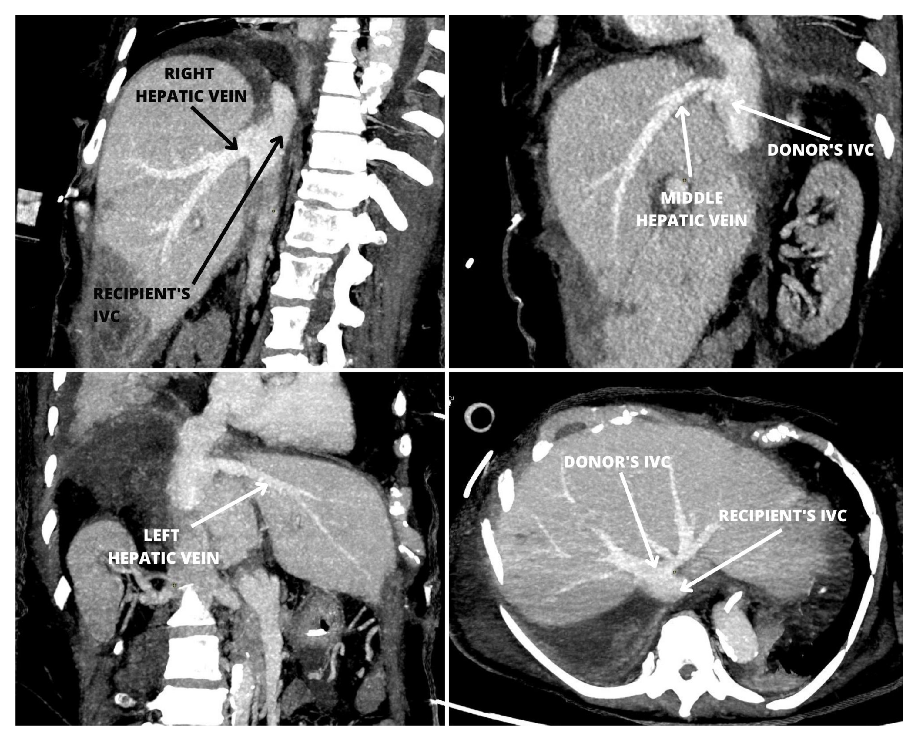
2. Case Report
3. Discussion
4. Conclusions
5. Future Directions
Author Contributions
Funding
Informed Consent Statement
Data Availability Statement
Conflicts of Interest
Abbreviations
| AKI | acute kidney injury |
| ALP | alkaline phosphatase |
| ALT | alanine transferase |
| AST | aspartate transaminase |
| GGTP | gamma glutamyl transpeptidase |
| HCV | hepatitis C virus |
| Hgb | hemoglobin |
| INF. | inferior |
| INR | international normalized ratio |
| IVC | inferior vena cava |
| MELD | Model for End-Stage Liver Disease |
| PLT | platelet count |
| POD | postoperative day |
| SUP. | superior |
| Tot. Bil. | total serum bilirubin |
| Vmax | maximal blood flow velocity |
| WBC | white blood cell count |
References
- Starzl, T.E.; Marchioro, T.L.; Vonkaulla, K.N.; Hermann, G.; Brittain, R.S.; Waddell, W.R. Homotransplantation of the liver in humans. Surg. Gynecol. Obstet. 1963, 117, 659–676. [Google Scholar] [PubMed]
- Calne, R.Y.; Williams, R. Liver transplantation in man. I. Observations on technique and organization in five cases. Br. Med. J. 1968, 5630, 535–540. [Google Scholar] [CrossRef] [PubMed] [Green Version]
- Belghiti, J.; Panis, Y.; Sauvanet, A.; Gayet, B.; Fékété, F. A new technique of side to side caval anastomosis during orthotopic hepatic transplantation without inferior vena caval occlusion. Surg. Gynecol. Obstet. 1992, 175, 270–272. [Google Scholar] [PubMed]
- Mehrabi, A.; Mood, Z.A.; Fonouni, H.; Kashfi, A.; Hillebrand, N.; Müller, S.A.; Encke, J.; Büchler, M.W.; Schmidt, J. A single-center experience of 500 liver transplants using the modified piggyback technique by Belghiti. Liver Transplant. 2009, 15, 466–474. [Google Scholar] [CrossRef] [PubMed]
- Shaker, T.M.; Eason, J.D.; Davidson, B.R.; Barth, R.N.; Pirenne, J.; Imventarza, O.; Spiro, M.; Raptis, D.A.; Fung, J.; ERS4OLT.org Working Group. Which cava anastomotic techniques are optimal regarding immediate and short-term outcomes after liver transplantation: A systematic review of the literature and expert panel recommendations. Clin. Transplant. 2022, 36, e14681. [Google Scholar] [CrossRef] [PubMed]
- Quintini, C.; Miller, C.M.; Hashimoto, K.; Philip, D.; Uso, T.D.; Aucejo, F.; Kelly, D.; Winans, C.; Eghtesad, B.; Vogt, D.; et al. Side-to-side cavocavostomy with an endovascular stapler: Rescue technique for severe hepatic vein and/or inferior vena cava outflow obstruction after liver transplantation using the piggyback technique. Liver Transplant. 2009, 15, 49–53. [Google Scholar] [CrossRef]
- Akbulut, S.; Wójcicki, M.; Kayaalp, C.; Yilmaz, S. Liver transplantation with piggyback anastomosis using a linear stapler: A case report. Transplant. Proc. 2013, 45, 1031–1033. [Google Scholar] [CrossRef]
- Voyles, C.R.; Vogel, S.B. Hepatic resection using stapling devices to control the hepatic veins. Am. J. Surg. 1989, 158, 459–460. [Google Scholar] [CrossRef]
- Schemmer, P.; Friess, H.; Dervenis, C.; Schmidt, J.; Weitz, J.; Uhl, W.; Büchler, M.W. The use of endo-GIA vascular staplers in liver surgery and their potential benefit: A review. Dig. Surg. 2007, 24, 300–305. [Google Scholar] [CrossRef]
- Raoof, M.; Aloia, T.A.; Vauthey, J.N.; Curley, S.A. Morbidity and mortality in 1174 patients undergoing hepatic parenchymal transection using a stapler device. Ann. Surg. Oncol. 2014, 21, 995–1001. [Google Scholar] [CrossRef]
- Fabian, M.A.; Herrin, M.K.; Baxter, J.; Ackermann, J.R. Extension of the right renal vein in cadaveric renal transplants with use of the vena cava and the TA-30 V3 surgical stapler. Surg. Gynecol. Obstet. 1991, 173, 233–234. [Google Scholar]
- Hussain, S.M.; Ghazanfar, A. Utilising endovascular stapling device to lengthen right renal vein using IVC-patch for deceased donor renal transplantation. Ann. R. Coll. Surg. Engl. 2022, 104, 153–155. [Google Scholar] [CrossRef]
- Grąt, M.; Kornasiewicz, O.; Lewandowski, Z.; Skalski, M.; Zieniewicz, K.; Pączek, L.; Krawczyk, M. The impact of surgical technique on the results of liver transplantation in patients with hepatocellular carcinoma. Ann. Transplant. 2013, 18, 448–459. [Google Scholar]
- Jochmans, I.; Fieuws, S.; Tieken, I.; Samuel, U.; Pirenne, J. The Impact of Implantation Time during Liver Transplantation on Outcome: A Eurotransplant Cohort Study. Transplant. Direct 2018, 4, e356. [Google Scholar] [CrossRef]
- Sutherasan, M.; Vorasittha, A.; Taesombat, W.; Nonthasoot, B.; Uthaithammarat, T.; Sirichindakul, P. Comparison of Three Inferior Vena Cava Reconstruction Techniques in Adult Orthotopic Liver Transplantation: Result from King Chulalongkorn Memorial Hospital, Thailand. Transplant. Proc. 2022, 54, 2224–2229. [Google Scholar] [CrossRef] [PubMed]
- Hannon, V.; Kothari, R.P.; Zhang, L.; Bokoch, M.P.; Hill, R.; Roll, G.R.; Mello, A.; Feiner, J.R.; Liu, K.D.; Niemann, C.U.; et al. The Association Between Vena Cava Implantation Technique and Acute Kidney Injury after Liver Transplantation. Transplantation 2020, 104, e308–e316. [Google Scholar] [CrossRef] [PubMed]
- Liu, C.; Tsai, H.L.; Loong, C.C.; Hsia, C.Y.; Chin, T.; Wei, C. ENDO-GIA staplers for side-to-side anastomosis of veins. Eur. J. Vasc. Endovasc. Surg. 2006, 32, 584–588. [Google Scholar] [CrossRef] [Green Version]
- Levi, D.M.; Pararas, N.; Tzakis, A.G.; Nishida, S.; Tryphonopoulos, P.; Gonzalez-Pinto, I.; Tekin, A.; Selvaggi, G.; Livingstone, A.S. Liver transplantation with preservation of the inferior vena cava: Lessons learned through 2000 cases. J. Am. Coll. Surg. 2012, 214, 691–698. [Google Scholar] [CrossRef]
- Arudchelvam, J.; Bartlett, A.; McCall, J.; Johnston, P.; Gane, E.; Munn, S. Hepatic venous outflow obstruction in piggyback liver transplantation: Single centre experience. ANZ J. Surg. 2017, 87, 182–185. [Google Scholar] [CrossRef]
- Wojcicki, M.; Post, M.; Pakosz-Golanowska, M.; Zeair, S.; Lubikowski, J.; Jarosz, K.; Czuprynska, M.; Milkiewicz, P. Vascular complications following adult piggyback liver transplantation with end-to-side cavo-cavostomy: A single-center experience. Transplant. Proc. 2009, 41, 3131–3134. [Google Scholar] [CrossRef] [PubMed]
- Silva, A.M.; Waisberg, D.R.; Fernandes, M.R.; Martino, R.B.; Rocha-Santos, V.; Pinheiro, R.S.; Nacif, L.S.; Arantes, R.M.; Ducatti, L.; Galvão, F.H.; et al. Hepatic Venous Outflow Obstruction in Adult Deceased Donor Liver Transplantation: Classic Piggyback Implantation Versus a Modified Technique that Widens the Ostium of the Recipient’s Left and Middle Hepatic Veins. Transplant. Proc. 2022, 54, 1316–1319. [Google Scholar] [CrossRef] [PubMed]
- Pitchaimuthu, M.; Roll, G.R.; Zia, Z.; Olliff, S.; Mehrzad, H.; Hodson, J.; Gunson, B.K.; Perera, M.T.; Isaac, J.R.; Muiesan, P.; et al. Long-term follow-up after endovascular treatment of hepatic venous outflow obstruction following liver transplantation. Transplant. Int. 2016, 29, 1106–1116. [Google Scholar] [CrossRef] [PubMed] [Green Version]
- Khorsandi, S.E.; Athale, A.; Vilca-Melendez, H.; Jassem, W.; Prachalias, A.; Srinivasan, P.; Rela, M.; Heaton, N. Presentation, diagnosis, and management of early hepatic venous outflow complications in whole cadaveric liver transplant. Liver Transplant. 2015, 21, 914–921. [Google Scholar] [CrossRef] [PubMed]
- Navarro, F.; Le Moine, M.C.; Fabre, J.M.; Belghiti, J.; Cherqui, D.; Adam, R.; Pruvot, F.R.; Letoublon, C.; Domergue, J. Specific vascular complications of orthotopic liver transplantation with preservation of the retrohepatic vena cava: Review of 1361 cases. Transplantation 1999, 68, 646–650. [Google Scholar] [CrossRef] [PubMed]
- Tashiro, H.; Ohdan, H.; Itamoto, T.; Ishifuro, M.; Hara, H.; Tokita, D.; Onoe, T.; Ishiyama, K.; Mitsuta, H.; Ide, K.; et al. Vascular closure staples for portal vein reconstruction in living-donor liver transplantation. Am. J. Surg. 2005, 190, 65–68. [Google Scholar] [CrossRef] [PubMed]
- Liu, K.; Yang, H.; Huang, G.; Shi, A.; Lu, Q.; Wang, S.; Qiao, W.; Wang, H.; Ke, M.; Ding, H.; et al. Adhesive anastomosis for organ transplantation. Bioact. Mater. 2021, 13, 260–268. [Google Scholar] [CrossRef] [PubMed]


| At Admission | POD 7 | POD 30 | POD 60 | |
|---|---|---|---|---|
| ALP (IU/L) | 69 | 325 | 147 | 87 |
| GGTP (IU/L) | 23 | 310 | 107 | 36 |
| AST (IU/L) | 18 | 37 | 27 | 20 |
| ALT (IU/L) | 18 | 151 | 52 | 29 |
| Tot. Bil (mg/dL) | 2.72 | 1.78 | 0.86 | 0.53 |
| INR | 1.10 | 1.13 | 1.11 | 1.11 |
| Albumin (g/dL) | 3.4 | 3.4 | 4.5 | 4.4 |
| PLT (103/L) | 167 | 246 | 237 | 164 |
| WBC (103/L) | 5.34 | 7.82 | 4.76 | 3.29 |
| Hgb (g/dL) | 9.3 | 8.8 | 9.9 | 9.9 |
| Creatinine (mg/dL) | 0.98 | 0.59 | 0.89 | 1.06 |
Disclaimer/Publisher’s Note: The statements, opinions and data contained in all publications are solely those of the individual author(s) and contributor(s) and not of MDPI and/or the editor(s). MDPI and/or the editor(s) disclaim responsibility for any injury to people or property resulting from any ideas, methods, instructions or products referred to in the content. |
© 2023 by the authors. Licensee MDPI, Basel, Switzerland. This article is an open access article distributed under the terms and conditions of the Creative Commons Attribution (CC BY) license (https://creativecommons.org/licenses/by/4.0/).
Share and Cite
Kruk, E.; Kalinowski, P.; Gibiński, K.; Dudek, K.; Skalski, M.; Przybysz, M.; Zhylko, A.; Nazarewski, Ł.; Morawski, M.; Grąt, M. Stapled Anastomosis for Side-to-Side Cavo-Cavostomy in Orthotopic Liver Transplantation. J. Clin. Med. 2023, 12, 5289. https://doi.org/10.3390/jcm12165289
Kruk E, Kalinowski P, Gibiński K, Dudek K, Skalski M, Przybysz M, Zhylko A, Nazarewski Ł, Morawski M, Grąt M. Stapled Anastomosis for Side-to-Side Cavo-Cavostomy in Orthotopic Liver Transplantation. Journal of Clinical Medicine. 2023; 12(16):5289. https://doi.org/10.3390/jcm12165289
Chicago/Turabian StyleKruk, Emilia, Piotr Kalinowski, Krzysztof Gibiński, Krzysztof Dudek, Michał Skalski, Marta Przybysz, Andriy Zhylko, Łukasz Nazarewski, Marcin Morawski, and Michał Grąt. 2023. "Stapled Anastomosis for Side-to-Side Cavo-Cavostomy in Orthotopic Liver Transplantation" Journal of Clinical Medicine 12, no. 16: 5289. https://doi.org/10.3390/jcm12165289