
Vascularized Growth Plate Transfer in Paediatric Ulna Non-Union: Operative Technique and Review of the Literature

Abstract
:1. Introduction
2. Case Report
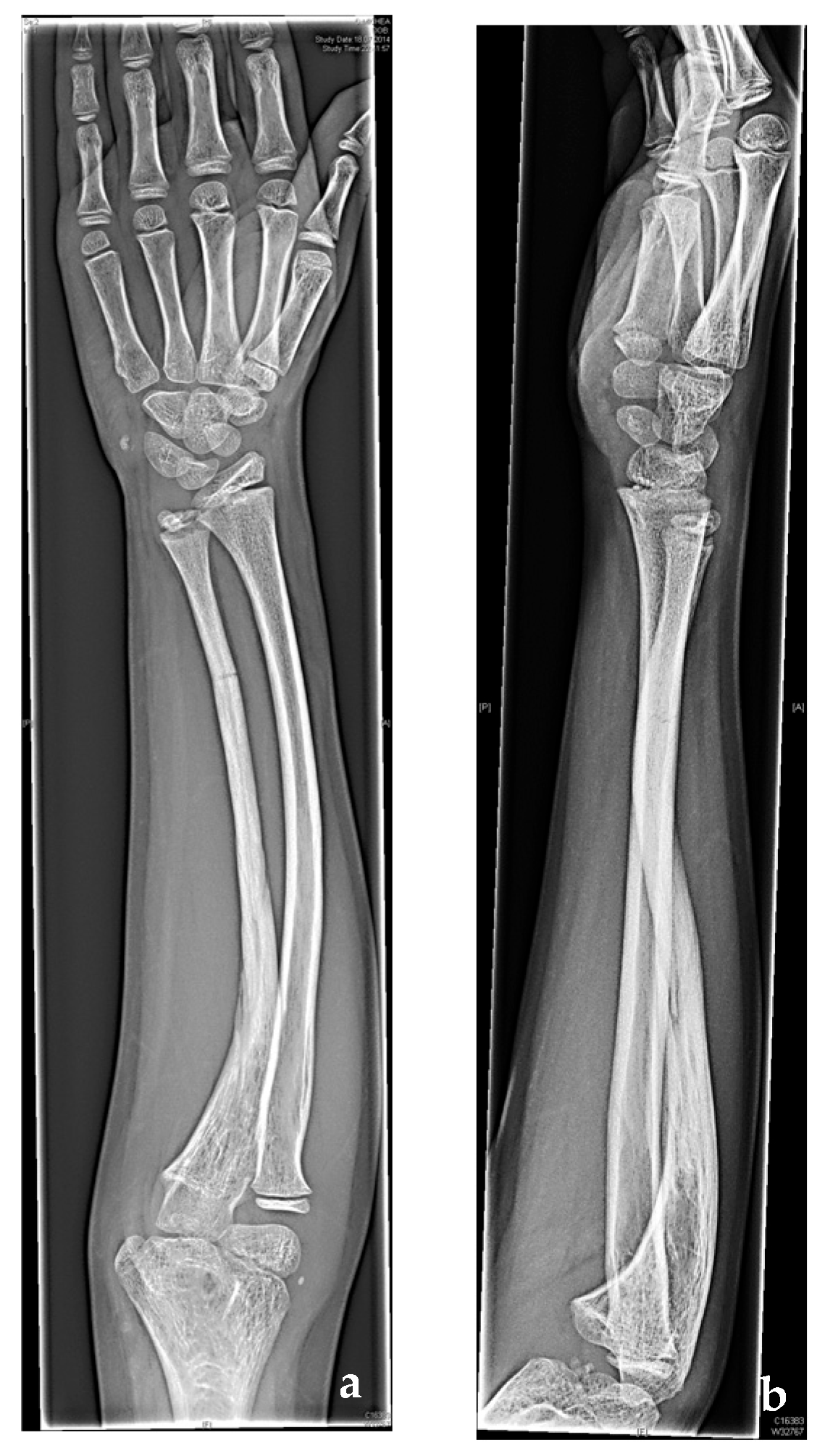
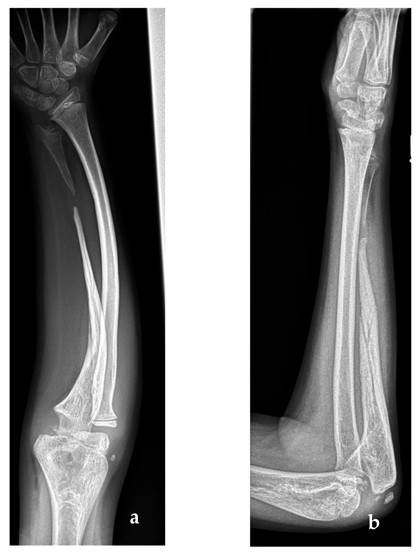
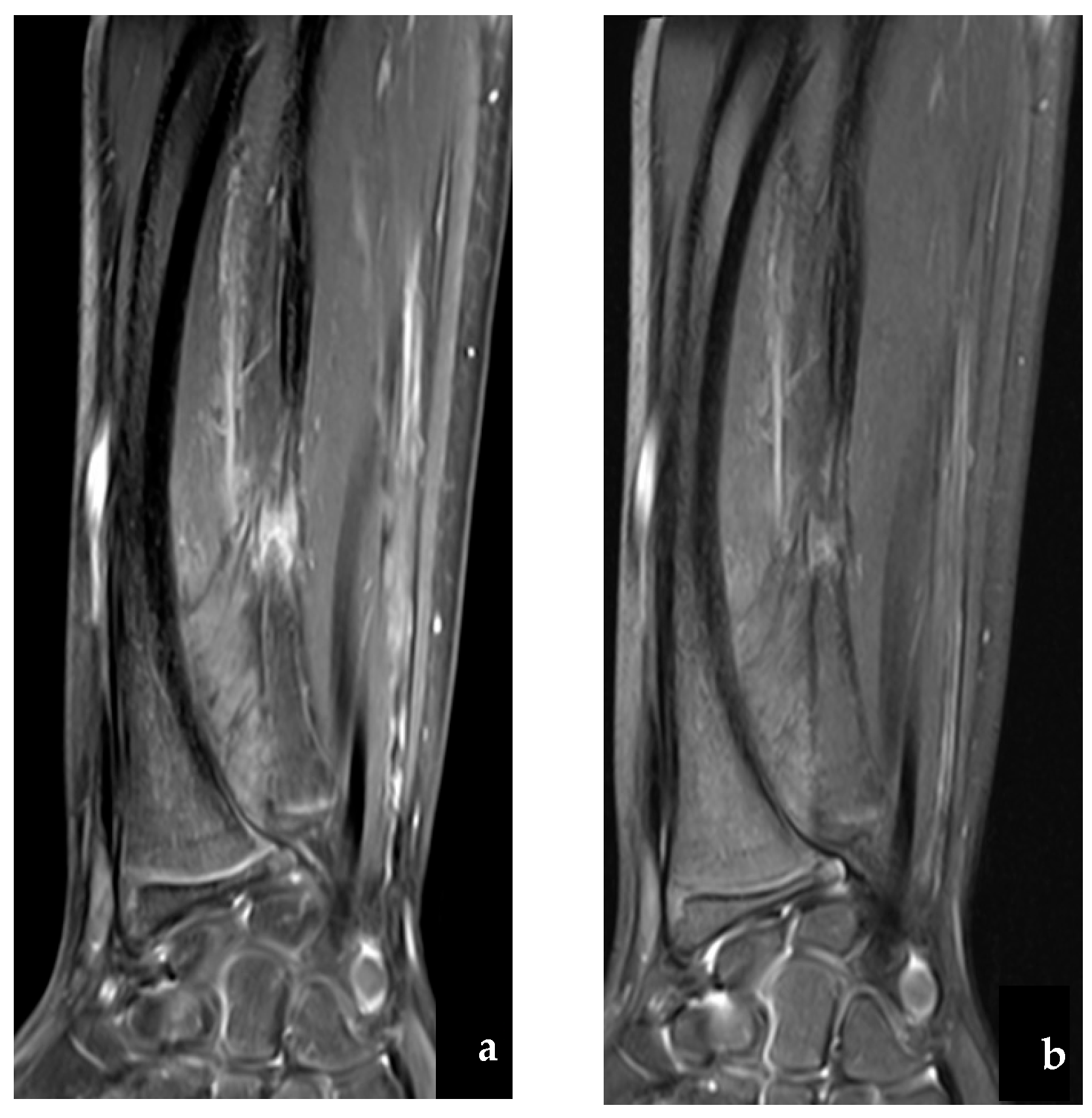
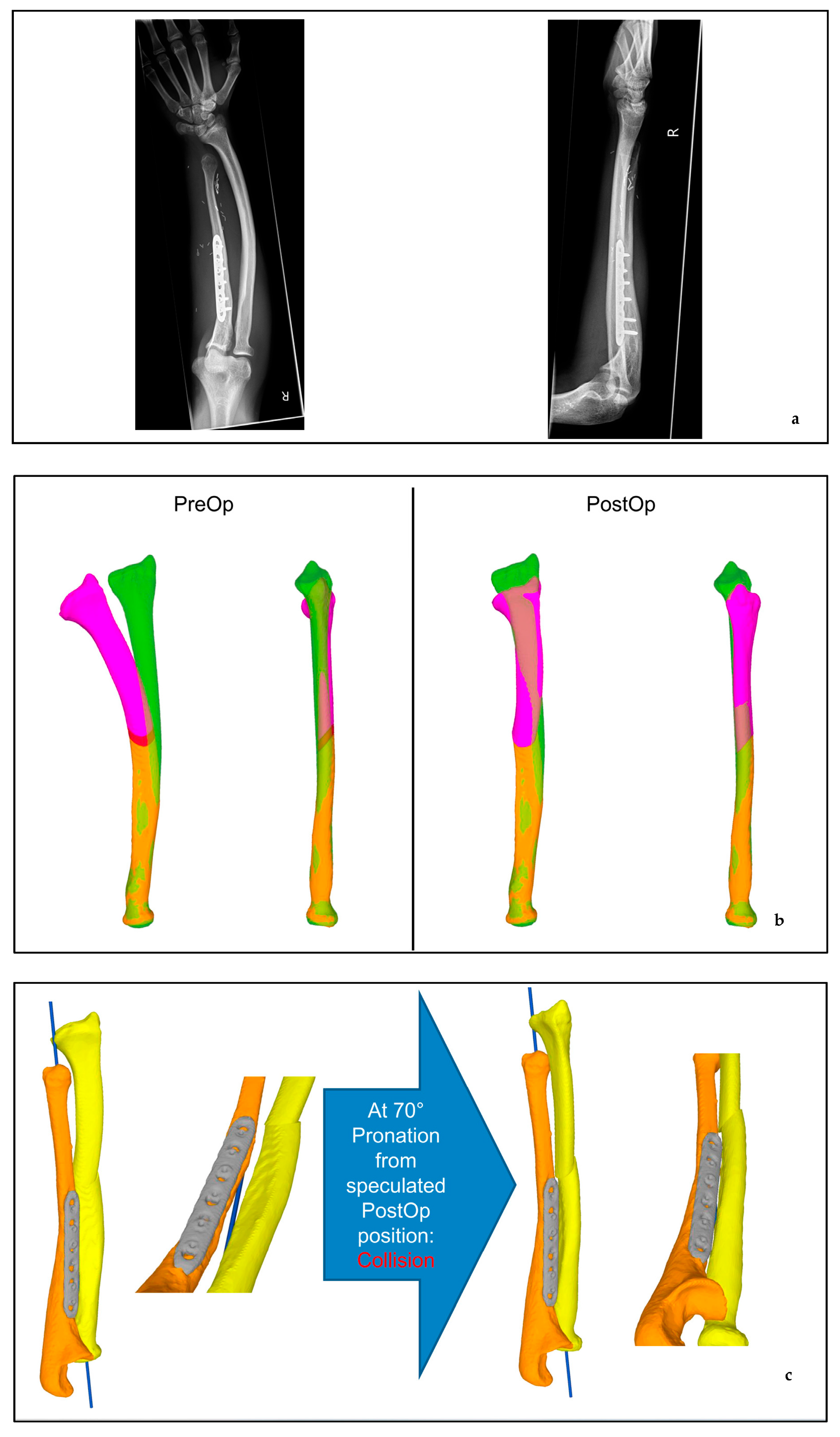
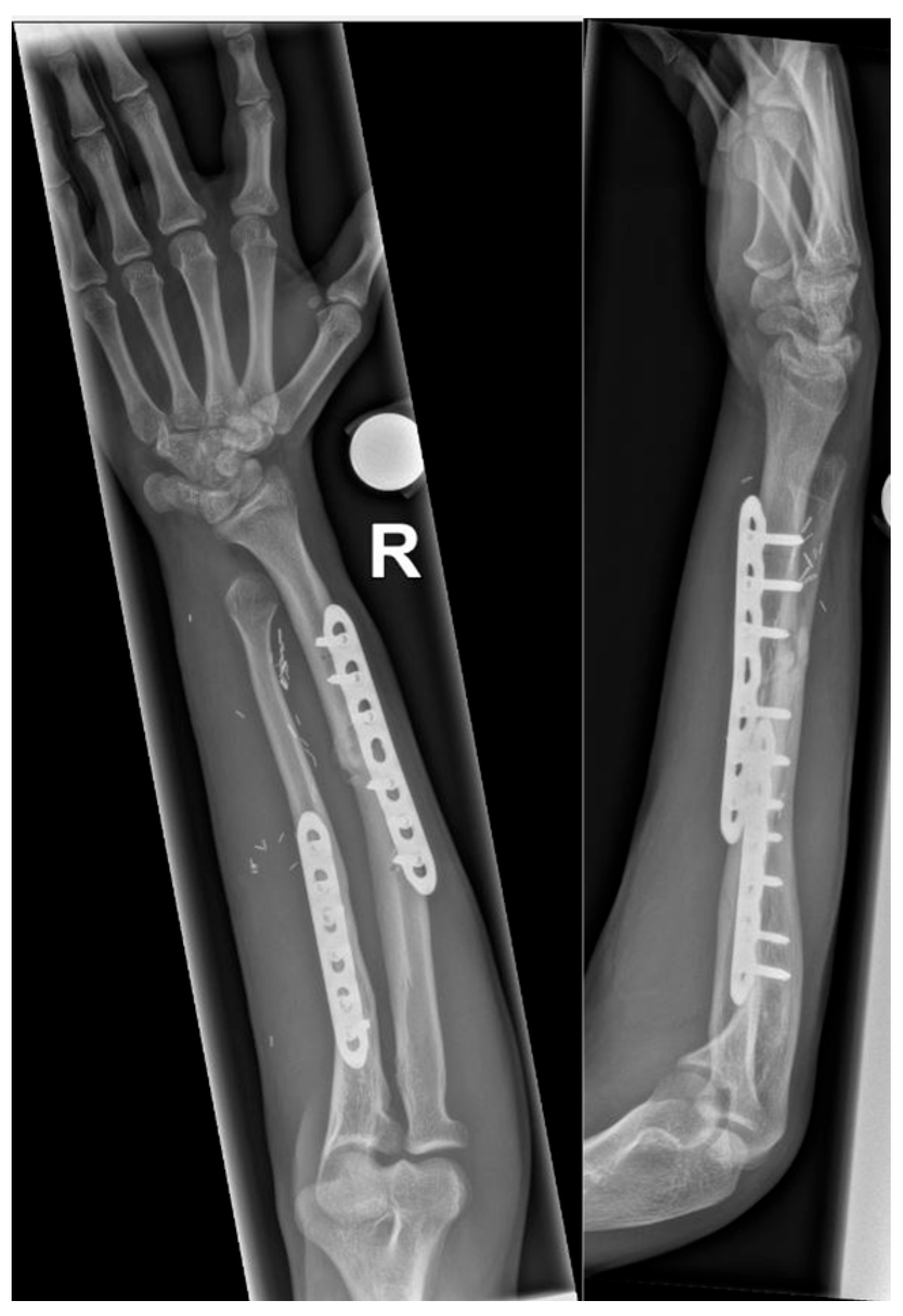
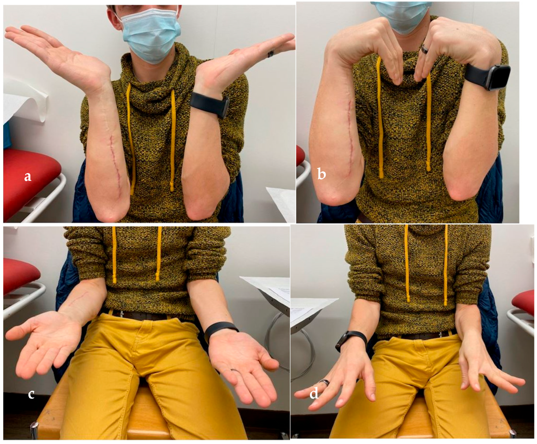
2.1. Case Report
2.2. Literature Review
2.2.1. Material and Methods
2.2.2. Results
3. Discussion
Author Contributions
Funding
Institutional Review Board Statement
Informed Consent Statement
Conflicts of Interest
References
- Bae, D.S.; Waters, P.M.; E Sampson, C. Use of Free Vascularized Fibular Graft for Congenital Ulnar Pseudarthrosis: Surgical decision making in the growing child. J. Pediatr. Orthop. 2005, 25, 755–762. [Google Scholar] [CrossRef] [PubMed]
- Aegerter, E.E. The possible relationship of neurofibromatosis, congenital pseudarthrosis, and fibrous dysplasia. J. Bone Joint Surg. 1950, 32, 618–626. [Google Scholar] [CrossRef] [PubMed]
- Crawford, A.H. Neurofibromatosis in the pediatric patient. Orthop. Clin. N. Am. 1978, 9, 11–23. [Google Scholar] [CrossRef]
- Charles, Y.-P.; Diméglio, A.; Chammas, M. Pseudarthrose congénitale de l’avant-bras. À propos de deux cas et revue de la littérature [Congenital pseudarthrosis of the forearm. Report of two cases and review of the literature]. Chir. Main 2009, 28, 26–32. [Google Scholar] [CrossRef]
- Ding, D.Y.; LaMartina, J.; Tai, C.; Pandya, N.K. Congenital Pseudoarthrosis of the Distal Radius Treated with Physeal-Sparing Double-Barrel Vascularized Free Fibula Transfer: A Case Report. Hand 2017, 12, NP140–NP144. [Google Scholar] [CrossRef]
- Mathoulin, C.; Gilbert, A.; Azze, R.G. Congenital pseudarthrosis of the forearm: Treatment of six cases with vascularized fibular graft and a review of the literature. Microsurgery 1993, 14, 252–259. [Google Scholar] [CrossRef]
- Bauer, A.S.; Singh, A.K.; Amanatullah, D.; Lerman, J.; James, M.A. Free Vascularized Fibular Transfer with Langenskiöld Procedure for the Treatment of Congenital Pseudarthrosis of the Forearm. Tech. Hand Up. Extremity Surg. 2013, 17, 144–150. [Google Scholar] [CrossRef]
- Nagaraju, K.D.; Vidyadhara, S.; Raja, D.; Rajasekaran, S. Congenital pseudarthrosis of the ulna. J. Pediatr. Orthop. B 2007, 16, 150–152. [Google Scholar] [CrossRef]
- Bell, D.F. Congenital forearm pseudarthrosis: Report of six cases and review of the literature. J. Pediatr. Orthop. 1989, 9, 438–443. [Google Scholar] [CrossRef]
- Witoonchart, K.; Uerpairojkit, C.; Leechavengvongs, S.; Thuvasethakul, P. Congenital pseudarthrosis of the forearm treated by free vascularized fibular graft: A report of three cases and a review of the literature. J. Hand Surg. 1999, 24, 1045–1055. [Google Scholar] [CrossRef]
- Ostrowski, D.M.; Eilert, R.E.; Waldstein, G. Congenital Pseudarthrosis of the Ulna: A Report of Two Cases and a Review of the Literature. J. Pediatr. Orthop. 1985, 5, 463–467. [Google Scholar] [CrossRef]
- Allieu, Y.; Gomis, R.; Yoshimura, M.; Dimeglio, A.; Bonnel, F. Congenital pseudarthrosis of the forearm—Two cases treated by free vascularized fibular graft. J. Hand Surg. 1981, 6, 475–481. [Google Scholar] [CrossRef]
- Innocenti, M.; Delcroix, L.; Manfrini, M.; Ceruso, M.; Capanna, R. Vascularized Proximal Fibular Epiphyseal Transfer for Distal Radial Reconstruction. J. Bone Jt. Surg. 2005, 87, 237–246. [Google Scholar] [CrossRef]
- Masterson, E.; Earley, M.J.; Stephens, M.M. Congenital pseudarthrosis of the ulna treated by free vascularized fibular graft: A case report and review of methods of treatment. J. Hand Surg. Br. 1993, 18, 285–288. [Google Scholar] [CrossRef] [PubMed]
- Cheng, J.C.Y.; Hung, L.K.; Bundoc, R.C. Congenital Pseudarthrosis of the Ulna. J. Hand Surg. 1994, 19, 238–243. [Google Scholar] [CrossRef]
- Lee, K.-S.; Lee, S.-H.; Ha, K.-H.; Lee, S.-J. Congenital pseudarthrosis of the ulna treated by free vascularized fibular graft—A case report. Hand Surg. 2000, 5, 61–67. [Google Scholar] [CrossRef]
- Suzuki, O.; Ishida, O.; Sunagawa, T.; Ichikawa, M.; Yasunaga, Y.; Ikuta, Y.; Ochi, M. Congenital Pseudoarthrosis of the Ulna Treated with a Free Vascularized Fibular Graft. Plast. Reconstr. Surg. 2005, 115, 1379–1384. [Google Scholar] [CrossRef]
- El Hage, S.; Ghanem, I.; Dagher, F.; Kharrat, K. Free Vascularized Fibular Flap for Congenital Ulnar Pseudarthrosis: A report of two cases and review of the literature. Ann. Plast. Surg. 2009, 62, 329–334. [Google Scholar] [CrossRef]
- Beris, A.E.; Lykissas, M.G.; Kostas-Agnantis, I.; Vasilakakos, T.; Vekris, M.D.; Korompilias, A.V. Congenital Pseudarthrosis of the Radius Treated with Gradual Distraction and Free Vascularized Fibular Graft: Case Report. J. Hand Surg. 2010, 35, 406–411. [Google Scholar] [CrossRef]
- Allieu, Y.; zu Reckendorf, G.M.; Chammas, M.; Gomis, R. Congenital pseudarthrosis of both forearm bones: Long-term results of two cases managed by free vascularized fibular graft. J. Hand Surg. 1999, 24, 604–608. [Google Scholar] [CrossRef]
- Siebelt, M.; de Vos-Jakobs, S.; Koenrades, N.; van Nieuwenhoven, C.A.; Oostenbrink, R.; Bramer, W.M.; Verhaar, J.A.; Bessems, G.J.; Kempink, D.R. Congenital Forearm Pseudarthrosis, a Systematic Review for a Treatment Algorithm on a Rare Condition. J. Pediatr. Orthop. 2020, 40, e367–e374. [Google Scholar] [CrossRef] [PubMed]
- Elfatairy, K.K.; Ehrlich, L.; Porrino, J.; Wang, A. Congenital pseudarthrosis of the forearm as a single manifestation of neurofibromatosis type 1 at birth: A case report. Clin. Imaging 2021, 78, 214–216. [Google Scholar] [CrossRef] [PubMed]
- Kohler, R.; Solla, F.; Pinson, S.; Romana, C.; Chau, E.; Dohin, B. Pseudarthrose congénitale de l’avant-bras associée à la neurofibromatose: À propos d’un cas et revue de la littérature. Rev. Chir. Orthopédique Réparatrice L’appareil Mot. 2005, 91, 773–781. [Google Scholar] [CrossRef] [PubMed]
- Taylor, G.I.; Miller, G.D.H.; Ham, F.J. The free vascularized bone graft: A clinical extension of microvascular techniques. Plast. Reconstr. Surg. 1975, 55, 533–544. [Google Scholar] [CrossRef] [PubMed]
- Langenskiöld, A. Pseudarthrosis of the fibula and progressive valgus defformity of the ankle in children: Treatment by fusion of the distal tibial and fibular metaphyses. Review of three cases. J. Bone Jt. Surg. 1967, 49, 463–470. [Google Scholar] [CrossRef]
- Cleveland, R.; Wilkinson, R.; Cleveland, V.G.A.R.W.R.; Keppler, J.S.; Conti, P.S.; Parisky, Y.R.; Sardi, A.; Hamm, R.; Hughes, K.; Esserman, L.; et al. Congenital pseudarthrosis of the radius. Am. J. Roentgenol. 1978, 130, 955–957. [Google Scholar] [CrossRef]
- Ali, M.S.; Hooper, G. Congenital pseudarthrosis of the ulna due to neurofibromatosis. J. Bone Jt. Surg. 1982, 64, 600–602. [Google Scholar] [CrossRef]
- McCullough, M.C.; Arkader, A.; Ariani, R.; Lightdale-Miric, N.; Tolo, V.; Stevanovic, M. Surgical Outcomes, Complications, and Long-Term Functionality for Free Vascularized Fibula Grafts in the Pediatric Population: A 17-Year Experience and Systematic Review of the Literature. J. Reconstr. Microsurg. 2020, 36, 386–396. [Google Scholar] [CrossRef]
- Ghert, M.; Colterjohn, N.; Manfrini, M. The Use of Free Vascularized Fibular Grafts in Skeletal Reconstruction for Bone Tumors in Children. J. Am. Acad. Orthop. Surg. 2007, 15, 577–587. [Google Scholar] [CrossRef]


| Author | Year | Ref. | Nr of Cases | Age | Neuro-Fibromatosis | Follow-Up | Complications | Further Surgeries | Union |
|---|---|---|---|---|---|---|---|---|---|
| Mathoulin | 1993 | [6] | 1 | 3 y | Yes | 144 mt | None | None | Yes |
| Masterson | 1993 | [14] | 2 | 5 y | Yes | 9 mt | None | None | Yes |
| Cheng | 1994 | [15] | 3 | 9 mt | Yes | 8 mt | None | None | Yes |
| Allieu | 1999 | [12] | 4 5 | 22 y 17 y | n/a n/a | 17 y 13 y | Yes Yes | None None | Yes Yes |
| Witoonchart | 1999 | [10] | 6 | 1 y | No | 48 mt | None | None | Yes |
| 7 | 5 y | Yes | 24 mt | Yes | Yes | Yes | |||
| Suzuki | 2005 | [17] | 8 | 4 y | n/a | 96 mt | Yes | Yes | Yes |
| 9 | 4 y | n/a | 96 mt | Yes | Yes | Yes | |||
| 10 | 1 y 7 mt | n/a | 60 mt | None | Yes | Yes | |||
| Bae | 2005 | [1] | 11 | 15 y | No | 81 mt | Yes | Yes | Yes |
| 12 | 3 y | Yes | 78 mt | Yes | Yes | Yes | |||
| 13 | 5 y 3 mt | Yes | 31 mt | None | None | Yes | |||
| 14 | 16 y | Yes | 42 mt | Yes | Yes | Yes | |||
| El Hage | 2009 | [18] | 15 | 18 mt | Yes | 72 mt | Yes | Yes | Yes |
| 16 | 12 y | Yes | 36 mt | Yes | Yes | Yes | |||
| Bauer | 2013 | [7] | 17 | 9 y 6 mt | Yes | 31 mt (range 23 to 83 mt) | Yes | Yes | Yes |
| 18 | 2 y 8 mt | Yes | None | None | Yes | ||||
| 19 | 11 y 6 mt | n/a | None | None | Yes | ||||
| 20 | 7 y | No | Yes | None | Yes | ||||
| 21 | 12 y 6 mt | Yes | None | None | Yes |
| Author | Ref. | Nr of Case | ROM Wrist/Elbow |
|---|---|---|---|
| Mathoulin | [6] | 1 | wrist: normal, elbow: normal, P/S: 90/0/50° |
| Masterson | [14] | 2 | wrist: full flexion, extension 25°, elbow: full ROM |
| P/S: 30/0/90° | |||
| Cheng | [15] | 3 | wrist: F/E: 60/0/30°, elbow: n/a, P/S: 60/0/50° |
| Allieu | [12] | 4 | wrist: F/E: 80/0/80°, elbow: normal function, P/S: 30/0/80° |
| 5 | wrist: F/E: 70/0/5°, elbow: n/a, P/S: Neutral position | ||
| Witoonchart | [10] | 6 | wrist: n/a, elbow: n/a, P/S: 10/0/80° |
| 7 | wrist: normal function, elbow: normal function | ||
| P/S: 10/080° | |||
| Suzuki | [17] | 8 | wrist: F/E: 25/0/45°, elbow:F/E: 130/0/0°, P/S: 20/0/60° |
| 9 | wrist: F/E: 60/0/65°, elbow: F/E 120/5/0°, P/S: 35/0/75° | ||
| 10 | wrist: F/E: 55/0/60°, elbow: F/E 125/10/0°, P/S: 80/0/80° | ||
| Bae | [1] | 11–14 | n/a |
| El Hage | [18] | 15 | wrist: n/a, elbow: F/E 130/0/0, P/S: 70/0/70 |
| 16 | wrist: n/a, elbow: F/E 120/0/0, P/S: 70/0/60° | ||
| Bauer | 17 | normal function | |
| 18 | normal function | ||
| 19 | normal function | ||
| 20 | normal function | ||
| 21 | P/S:45/0/85° |
Disclaimer/Publisher’s Note: The statements, opinions and data contained in all publications are solely those of the individual author(s) and contributor(s) and not of MDPI and/or the editor(s). MDPI and/or the editor(s) disclaim responsibility for any injury to people or property resulting from any ideas, methods, instructions or products referred to in the content. |
© 2023 by the authors. Licensee MDPI, Basel, Switzerland. This article is an open access article distributed under the terms and conditions of the Creative Commons Attribution (CC BY) license (https://creativecommons.org/licenses/by/4.0/).
Share and Cite
Grünberger, N.M.; Klein, A.; Barandun, M.; Schaefer, D.J.; Krieg, A.H.; Kaempfen, A. Vascularized Growth Plate Transfer in Paediatric Ulna Non-Union: Operative Technique and Review of the Literature. J. Clin. Med. 2023, 12, 4981. https://doi.org/10.3390/jcm12154981
Grünberger NM, Klein A, Barandun M, Schaefer DJ, Krieg AH, Kaempfen A. Vascularized Growth Plate Transfer in Paediatric Ulna Non-Union: Operative Technique and Review of the Literature. Journal of Clinical Medicine. 2023; 12(15):4981. https://doi.org/10.3390/jcm12154981
Chicago/Turabian StyleGrünberger, Nisha M., Amelie Klein, Marina Barandun, Dirk J. Schaefer, Andreas H. Krieg, and Alexandre Kaempfen. 2023. "Vascularized Growth Plate Transfer in Paediatric Ulna Non-Union: Operative Technique and Review of the Literature" Journal of Clinical Medicine 12, no. 15: 4981. https://doi.org/10.3390/jcm12154981