Article

The Effect of Cadmium on Sleep Parameters Assessed in Polysomnographic Studies: A Case–Control Study

1
Department of Internal Medicine, Occupational Diseases, Hypertension and Clinical Oncology, Wroclaw Medical University, 50-556 Wroclaw, Poland
2
Student Research Club No K133, Faculty of Medicine, Wroclaw Medical University, 50-367 Wroclaw, Poland
3
Department of Experimental Dentistry, Wroclaw Medical University, 50-425 Wroclaw, Poland
4
Division of Environmental Health and Occupational Medicine, Department of Population Health, Wroclaw Medical University, 50-345 Wroclaw, Poland
*
Author to whom correspondence should be addressed.
J. Clin. Med. 2023, 12(12), 3899; https://doi.org/10.3390/jcm12123899
Submission received: 11 May 2023 / Revised: 31 May 2023 / Accepted: 6 June 2023 / Published: 7 June 2023
(This article belongs to the Special Issue New Insights into Sleep Medicine)

Abstract

:
Cadmium is a heavy metal that accumulates in the body due to environmental and occupational exposure. The main form of environmental exposure to cadmium is related to cigarette smoking. The primary aim of this study was to evaluate the effect of cadmium on numerous sleep parameters with the use of polysomnography. The secondary aim of this study was to investigate if environmental exposure to cadmium is a risk factor for the intensity of sleep bruxism (SB). Methods: A total of 44 adults underwent a full night of polysomnographic examination. The polysomnograms were assessed according to guidelines set out by the American Academy of Sleep Medicine (AASM). The concentration of cadmium in the blood and urine was determined spectrophotometrically. Results: The polysomnographic examination confirmed that cadmium, age, male gender and smoking status are independent risk factors for an increase in the apnea–hypopnea index (AHI). Cadmium alters sleep architecture by favoring sleep fragmentation and decreasing the duration of the rapid eye movement (REM) phase of sleep. However, cadmium exposure is not a risk factor for the development of sleep bruxism. Conclusions: In summary, this study demonstrates that cadmium affects sleep architecture and is a risk factor for the development of obstructive sleep apnea; however, it does not affect sleep bruxism.

Graphical Abstract

1. Introduction

Cadmium is a widespread heavy metal that is present naturally in the Earth’s crust [1]. Its biological half-life within the body, after environmental exposure, is estimated to be about 20 years [2]. In the last century, the industrial sector has most frequently used cadmium in the coating of iron and steel to prevent corrosion. Nowadays, this sector utilizes cadmium in the manufacture of batteries, pigments, coatings, plating substances and stabilizers for plastic. However, beyond the manufacturing sector, the most common form of exposure to cadmium is by means of inhalation and ingestion. Occupational exposure to cadmium relates to processes involving the heating of cadmium-containing materials [1].
Tobacco plants are known for absorbing heavy metals from the soil through their roots, accumulating them within their leaves which are ultimately used in the production of cigarettes. The single major significant source of cadmium exposure for the general population is tobacco smoking [3,4]. It has been established that smokers tend to have elevated cadmium concentrations in the blood, compared to their non-smoking counterparts [5,6]. It is worth noting, though, that non-smokers are also exposed to high quantities of cadmium that is present in water and food. This is particularly true of populations consuming diets that are high in rice and wheat content [5].
The kidneys are the major site of cadmium accumulation in the body. This in turn leads to nephrotoxicity and renal tubular damage, causing numerous negative health outcomes [3,7]. Cadmium has also been found to be neurotoxic. The recent literature states that cadmium has a strictly dose-dependent effect on neurons: Cadmium can gradually cause cell injury, cell death and organ failure at high doses. On the other hand, it can modulate specific mechanisms at low doses without significantly harming cells [8]. Neurodegenerative disorders, such as Alzheimer’s disease, Parkinson’s disease, amyotrophic lateral sclerosis, multiple sclerosis and myalgic encephalomyelitis, have previously been linked with cadmium-dependent neurotoxicity [9].
The symptoms of poisoning depend on the level of cadmium in the blood and may result in acute and chronic intoxication. It has been determined that cadmium is a type I carcinogen and acts as a proinflammatory cytokine inducer, leading to a chronic inflammatory response [3]. Cadmium exposure has been linked to the development of numerous illnesses, including osteoporosis [10], depressive symptoms [11,12], smoking-induced cardiovascular diseases [13] and pulmonary diseases [3]. Recently, cadmium exposure from tobacco smoking has been linked with type 2 diabetes mellitus [14]. Direct prolonged exposure to cadmium is known to be a major etiological factor of itai-itai disease (otherwise known as “ouch-ouch” disease) in Japanese female residents living near cadmium-contaminated rivers. Severe spine and joint pain, softening of the bones and kidney failure were the main findings in these patients. Several studies were carried out after reports of itai-itai disease came to light, in order to determine the levels of cadmium exposure that are a health threat [7,15]. Due to its adverse effects on human health, cadmium usage has been restricted worldwide under numerous regulations [16].
Worldwide culture-associated smoking habits have brought the attention of the scientific community to the substances present in cigarettes, ultimately raising environmental and health concerns. No studies have been published investigating the possible influence of cadmium on sleep architecture (rapid eye movement (REM), non-rapid eye movement (nREM)), respiratory function (apnea–hypopnea index (AHI) and saturation parameters) and movement disorders (bruxism episode index (BEI)). Due to the lack of research in this regard, the primary aim of this study was to evaluate the influence of cadmium on sleep architecture, by utilizing polysomnographic examination. The secondary aim of this study was to establish if exposure to cadmium increases the likelihood of developing obstructive sleep apnea (OSA) and if cadmium itself is a risk factor for sleep bruxism (SB).

2. Materials and Methods

2.1. Participants

This was a prospective, observational study of a total of 44 adults. The patients were admitted to the Department of Internal Medicine and Occupational Diseases, Hypertension and Clinical Oncology at Wroclaw Medical University, Poland, in order to undergo polysomnographic examination. The patients were referred to the sleep laboratory for examination, due to a suspicion that they may have a sleep disorder. These 44 subjects were admitted to the department between December 2020 and May 2021. Inclusion criteria were as follows: age over 18 years, written informed consent to polysomnographic examination and urine/blood sample collection. Patients were excluded from the study if they had neurological disorders and/or neuropathic pain, active inflammation, confirmed active malignancy, present or past occupational exposure to cadmium, severe respiratory and cardiac insufficiency, treatments affecting muscle function and sleep structure, severe mental disorders, cognitive disability and a lack of compliance during the study. All study participants declared no occupational cadmium exposure throughout their lifetime. The study was approved by the Ethics Committee at Wroclaw Medical University (approval no. KB-790/2022). Voluntary written informed consent was obtained from all patients prior to commencing the study. This study made use of the Strengthening the Reporting of Observational Studies in Epidemiology (STROBE) checklist [17].

2.2. Polysomnography

All subjects (n = 44) underwent polysomnographic investigation with a NoxA1 (NOX Medical, Reykjavík, Iceland) device. The recording was conducted between 22:00 and 6:00, in accordance with the patient’s natural circadian rhythm. Electromyographic electrodes were placed in line with guidelines set out by the American Academy of Sleep Medicine (AASM) [18]. Audio and video recordings together with electrocardiographic, electroencephalographic, electro-oculographic and electromyographic recordings were included, along with body position detection and thoracic and abdominal breathing activity. To measure saturation (SpO2%), pulse and plethysmography data, a WristOx2 3150 pulse oximeter (Nonin Medical Inc., Plymouth, MN, USA) was used. The respiratory, sleep bruxism (SB) and sleep parameters were gathered. Sleep parameters were obtained and classified pursuant to standard AASM criteria: non-REM 1 sleep stage (N1), non-REM 2 sleep stage (N2), non-REM 3 sleep stage (N3), REM sleep stage, arousal index (ArI), sleep latency (SL), wake after sleep onset (WASO), sleep efficiency (SE), apnea–hypopnea index (AHI), oxygen desaturation index (ODI) and snoring. SB parameters were also assessed: bruxism episodes index (BEI), phasic bruxism, tonic bruxism and mixed bruxism. A constant burst episode sustained over 2 s was categorized as a tonic episode. An episode including three or more bursts lasting over 2 s was categorized as phasic, and an episode showing the characteristics of both tonic and phasic episodes was categorized as a mixed episode of SB. SB episodes were defined as the rhythmic movements of the masseter muscles that occurred at least three seconds after the previous muscle movement. EMG activity had to be at least twice the amplitude of the background EMG. The bruxism episodes index (BEI) was measured by counting the number of bruxism episodes per hour of sleep. Phenotypes of bruxism were distinguished as follows: phasic, tonic and mixed [18]. The number of bruxism episodes per hour of sleep was used to categorize sleep bruxism and assigned as irrelevant (BEI < 2), mild to moderate (BEI 2–4) and severe (BEI > 4) [19].

2.3. Sample Collection and Determination of Blood and Urine Cadmium Concentration

Fasting venous blood samples from the ulnar vein and urine samples were obtained at 7:00 a.m., after twelve hours of overnight fasting. These blood samples were collected into polyethylene terephthalate (PET) plastic tubes (Becton, Dickinson and Company; Franklin Lakes, NJ, USA) with K2EDTA. Blood samples were stored at −70 °C until subsequent analyses were performed. Blood and urine samples were analyzed at the Main Laboratory of Wroclaw Medical University. Laboratory tests were performed according to the standard laboratory protocols of Wroclaw Medical University Teaching Hospital. The blood concentration of cadmium was determined by atomic absorption spectrophotometry with the use of a SOLAAR M6 (Thermo Elemental Ltd., London, UK) in an electrographite cuvette at λ = 228.8 nm, equipped with a Zeeman background correction system, in a certified atomic absorption laboratory at the University Hospital in Wroclaw, Poland. Values were measured in micrograms per deciliter (mg/dL).
For the analysis, the Stoeppler and Brandt method was used [20]. For trace metal analysis, the samples were deproteinized with 65% nitric acid (Suprapur; Merck KGaA, Darmstadt, Germany). Using ClinCal Whole Blood Calibrators (Cat.) and comparing the absorbance of the sample to a standard curve, the concentration of cadmium in the sample was determined. No. 9943) and control tests were performed (ClinChek Entire Blood Controls, level I, II, III; Cat. No. 8840–8843; Recipe Chemicals + Instruments GmbH, Munich, Germany). Within the framework of the German External Quality Assessment Scheme (G-EQUAS), external laboratory control was carried out with the assistance of the Intercomparison Program for Toxicological Analyses in Biological Materials (Institute and Out-Patient Clinic for Occupational, Social and Environmental Medicine of the Friedrich-Alexander University, Erlangen, Germany). The described method had a detection limit of 0.082 g/L.

2.4. Statistics

Statistical analysis was carried out based on the statistical software “Dell Statistica 13” (Dell Inc., Tulsa, OK, USA). The distribution of variables was examined with the Lilliefors and W-Shapiro–Wilk tests. In comparative analyses, subgroups were compared based on the median, first quartile or third quartile of the grouping variable. In the case of quantitative independent variables with a normal distribution, the t-test was used for further statistical analysis. In the case of variables with a distribution other than normal, the Mann–Whitney U test was used for quantitative independent variables. For qualitative independent variables, the maximum likelihood chi-square test was utilized. To determine the relationship between the studied variables, correlation and regression analysis was performed. In the case of quantitative variables with a normal distribution, Pearson’s r correlation coefficients were marked. For quantitative variables with a non-normal distribution, on the other hand, Spearman’s r coefficients were used. The parameters of the model obtained in the multivariable stepwise regression analysis were estimated using the least squares method. Results at the level of p < 0.05 were considered to be statistically significant.

3. Results

The data that were obtained from the study group and subsequently evaluated are demonstrated in Table 1. Overall, 44 patients were included in this study. The minimum sample group size required to conduct this study was estimated using a sample size calculator. The following estimation input conditions were used: population size: 3,000,000 (population size of the Lower Silesian Voivodeship); fraction size: 0.1 (assumed OSA prevalence in the Polish population); confidence level: 95% (default value); and maximum error: 10% (one of the typical values). The minimum required sample group size of 35 people was achieved.
The average age of the study group was 47.45 ± 13.69 years old. In total, 38.63% (n = 17) of the participants were female, while 61.36% (n = 27) were male; 86.36% (n = 38) of patients were current smokers; and 29 (67.44%) of the 44 the patients were diagnosed as sleep bruxers. Obstructive sleep apnea (OSA) was present in 70.45% (n = 31) patients in the study group. The degree of OSA was determined in each subject: 20.45% (n = 9) of cases were mild, 22.72% (n = 10) moderate and 27.27% (n = 12) of subjects had severe OSA. The mean blood and urine cadmium concentration in the study group was 0.37 ± 0.39 µg/L and 0.78 ± 0.63 µg/g of creatinine, respectively.
Patients were divided into two groups in accordance with their median cadmium blood concentration, namely, a low cadmium concentration group (<0.25) and a high cadmium concentration group (>0.25). In the low cadmium group, the average cadmium concentration was 0.14 ± 0.07, while in the high cadmium group it was 0.57 ± 0.46. The average SpO2% and minimal SpO2% were decreased in both of these groups. Statistical significance was present in SpO2 < 90% duration, minimal SpO2, average SpO2, REM sleep stage, N2 sleep stage, WASO, bruxism episode index and phasic bruxism (Table 2).
The results for cadmium concentration in the urine were compared as shown in Table 3. Correlation analysis was also performed and is shown in Table 4.
The result of the regression analysis is presented in Table 5. Age, high urine cadmium concentration, male gender and smoking are independent risk factors for increased AHI values (Table 5). Cadmium has not been found to be an independent risk factor for sleep bruxism.

4. Discussion

4.1. Evidence for Role of Cadmium in OSA

To the best of our knowledge, this is the first study investigating the role of cadmium on sleep parameters. In this study, cadmium was found to be a risk factor for an increased apnea/hypopnea index (AHI). As a result, our study shows that environmental exposure to cadmium may increase the risk of developing OSA.
We also determined that blood and urine cadmium concentration was correlated with blood oxygen saturation parameters. This suggests that cadmium exposure may affect the level of oxygen saturation in subjects with sleep disorders. As far as we know, no previous studies have ever investigated this specific relationship. However, it is known that cigarette smoking itself, which is the main source of cadmium exposure, has a negative influence on respiratory parameters [21].
Past studies evaluating the serum levels of heavy metals in patients with obstructive sleep apnea (OSA) revealed that, among others, the serum level of cadmium was increased in the study group (compared to controls), possibly due to oxidative stress and inflammation [22]. One such study, conducted by Asker et al., was based on recorded polygraphic measurements. No sleep architecture parameters were obtained in this study. However, despite this, the results are in agreement with the outcomes of the present study. Cadmium is known for its oxidative stress and inflammatory properties [7]. There is also a strong correlation between inflammation, oxidative stress and OSA, which has been proven in previous studies [23].
Cadmium may increase inflammation in the tissues of the respiratory tract, favoring the narrowing of the upper airway during sleep and affecting respiratory control stability/loop gain, the respiratory arousal threshold and upper airway muscle function via neurotoxic activity. On the other hand, hypoxia in OSA may affect the absorption of cadmium, thus the cadmium–OSA relationship may be bidirectional. However, these hypotheses require further research.
The influence of cigarette smoking on OSA development has recently been a subject of interest, with varying results in numerous studies [24]. Still, other proven risk factors for OSA include male gender, older age, obesity and craniofacial deformities [25]. No past studies have investigated the potential mechanisms (arising from cadmium exposure) that lead to the development of sleep apnea. To this day, the mechanisms underlying cadmium-induced airway pathologies have not yet been fully understood due to the complex relationship between biochemical and clinical outcomes. However, to the best of our knowledge, our study is the first to demonstrate that cadmium exposure is an independent OSA risk factor.

4.2. The Influence of Cadmium on Sleep Bruxism (SB) Intensity

No studies thus far have investigated the influence of cadmium exposure on SB intensity. In one of our previous studies, our team demonstrated that smoking, which is the main source of cadmium in humans, is a risk factor for SB development [26]. Our current study shows that cadmium itself is not a risk factor for the development of SB. However, we did find that the intensity of SB was increased in patients with a higher cadmium serum concentration, compared to subjects with a lower cadmium concentration. This result is not standalone though, and it is important to remember that many factors can influence the intensity of bruxism in patients with cadmium exposure, such as insomnia, psychological status or caffeine consumption. Sleep bruxism and obstructive sleep apnea are also two strongly connected disorders that can even co-occur [27]. Besides its nicotine content, various toxic components are present in a single cigarette, each of which may affect the intensity of bruxism. A host of reactive oxygen and nitrogen species (ROS and RNS) may alter the intensity of bruxism and endothelial function. The endothelium regulates the inflammatory state in a human’s body and cigarette smoking is known to lead to endothelial dysfunction [28]. Thus, many proinflammatory and toxic compounds can affect the development of sleep bruxism but, as explained above, not cadmium.
In general, cigarettes act on the sympathetic nervous system by activating it [29]. On the contrary, though, in sleep bruxism the trigemino-cardiac reflex (TCR) plays an important role in the regulation of the autonomic nervous system. The activation of the TCR results in the suppression of sympathetic nervous activity, while parasympathetic activity is activated as a sort of defense mechanism [30]. As a result, the intensity of sleep bruxism is increased in smokers, however not due to the cadmium content, but rather due to other factors.

4.3. Evidence for the Role of Cadmium in Sleep Architecture Alterations

No research thus far has investigated the influence of cadmium on sleep architecture in humans. Only one study has explored the relationship between sleep and cadmium exposure in rats. However, this study only made use of an EEG investigation, which revealed that a high concentration of cadmium in drinking water led to an increase in non-REM sleep and a decrease in rhythms of locomotor activity [31]. Animal-based studies found that cadmium can influence sleep architecture and sleep duration, while acute cadmium exposure seemed to have an impact on the circadian rhythm [32,33,34,35]. Our results are consistent with previous investigations.
Past studies investigating the neurotoxic consequences of cadmium determined that cadmium exposure induces memory impairment and results in a decreased attention span in humans [36]. Interestingly, our present study demonstrates that cadmium exposure impacts the REM sleep stage by decreasing the duration of this phase of sleep. The REM stage of sleep participates in memory consolidation [37] and emotional processing [38]. These mechanisms can potentially be affected in patients exposed to cadmium. Further investigations are needed to explain this correlation.
We also found a positive correlation between cadmium content in blood/urine and the frequency of arousals during sleep. Higher concentrations of cadmium in the blood and urine were found among subjects with an increased arousal index. It is known that arousals favor sleep fragmentation and sleep architecture alterations [39]. It is worth noting that fragmented sleep is a known cardiovascular risk factor [40].
Our study group had environmental exposure to cadmium. The mean blood cadmium concentration was 0.37 ± 0.39 µg/g, while the concentration in the urine was 0.78 ± 0.63 µg/g of creatinine. In the general population, geometric cadmium levels in blood have been estimated to be around 0.315 ng/L, while in urine these levels are approximately 0.193 ng/g of creatinine [41]. In our study, these biomarkers were higher, and this result confirms environmental cadmium exposure in our patients.
In conclusion, the complexity of cadmium-dependent sleep architecture outcomes remains to be clarified and further studies on the various mechanisms that result in sleep pattern changes are needed. It may help to develop new methods of OSA treatments. However, in conclusion of our study, patients with OSA should avoid environmental and occupational cadmium exposure.

4.4. Study Strengths

Full-night audio–video polysomnography, which is the gold standard in sleep examination, was performed in the whole study group. In addition, both blood and urine samples were obtained and measured to determine cadmium concentration. Blood and urine are known to be exposure biomarkers [3]. However, blood cadmium is more likely to indicate recent cadmium exposure, while cadmium found in the urine has been linked to chronic or past exposure and may indicate the total cadmium body burden [42].

4.5. Study Limitations

The relatively small study group (n = 44) was the major limitation of this study. In addition, there was no adaptive night prior to conducting the PSG examination due to a limited capacity in the sleep laboratory, and due to restrictions associated with the organization of the hospital. This study was not intended to show an influence of cigarette smoking on sleep parameters, but to investigate blood and urine cadmium influence on sleep architecture. However, smoking may explain some changes in sleep architecture and further research is needed to determine this relation.

5. Conclusions

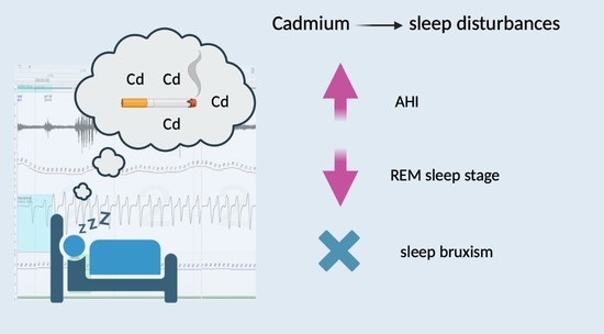
  • Cadmium is an independent factor for increased AHI, similarly to age, gender and smoking status.
  • Cadmium is not a risk factor for sleep bruxism.
  • Cadmium favors sleep disturbances, including sleep fragmentation, and results in the limitation of the duration of the REM sleep stage.

Author Contributions

Conceptualization, W.F. and H.M.; methodology, H.M.; software, P.G. and R.P.; investigation, W.F. and H.M.; resources, W.F.; writing—original draft preparation, W.F.; writing—review and editing, H.M., M.W. and G.L.; supervision, H.M., M.W. and G.M.; project administration, G.M.; funding acquisition, G.M. All authors have read and agreed to the published version of the manuscript.

Funding

This research was funded by Wroclaw Medical University (grant number SUBZ.A210.22.041).

Institutional Review Board Statement

The study was conducted in accordance with the Declaration of Helsinki and approved by the Bioethical Committee of Wroclaw Medical University (protocol code KB-790/2022).

Informed Consent Statement

Informed consent was obtained from all subjects involved in the study.

Data Availability Statement

The data supporting the findings of this study are available on request from the corresponding author and are not publicly available due to privacy or ethical restrictions.

Conflicts of Interest

The authors declare no conflict of interest.

References

  1. Agency for Toxic Substances and Disease Registry. Toxicological Profile for Cadmium. Available online: https://www.atsdr.cdc.gov/toxprofiles/tp5.pdf (accessed on 11 April 2023).
  2. Ishizaki, M.; Suwazono, Y.; Kido, T.; Nishijo, M.; Honda, R.; Kobayashi, E.; Nogawa, K.; Nakagawa, H. Estimation of Biological Half-Life of Urinary Cadmium in Inhabitants after Cessation of Environmental Cadmium Pollution Using a Mixed Linear Model. Food Addit. Contam. A Chem. Anal. Control Expo. Risk Assess. 2015, 32, 1273–1276. [Google Scholar] [CrossRef] [PubMed]
  3. Richter, P.; Faroon, O.; Pappas, R.S. Cadmium and Cadmium/Zinc Ratios and Tobacco-Related Morbidities. Int. J. Environ. Res. Public. Health 2017, 14, 1154. [Google Scholar] [CrossRef] [PubMed] [Green Version]
  4. Lee, J.W.; Kim, Y.; Kim, Y.; Yoo, H.S.; Kang, H.T. Cigarette Smoking in Men and Women and Electronic Cigarette Smoking in Men Are Associated with Higher Risk of Elevated Cadmium Level in the Blood. J. Korean Med. Sci. 2020, 35, e15. [Google Scholar] [CrossRef]
  5. Wang, C.C.; Zhang, Q.C.; Yan, C.A.; Tang, G.Y.; Zhang, M.Y.; Ma, L.Q.; Gu, R.H.; Xiang, P. Heavy metal(loid)s in agriculture soils, rice, and wheat across China: Status assessment and spatiotemporal analysis. Sci. Total Environ. 2023, 882, 163361. [Google Scholar] [CrossRef]
  6. Mérida-ortega, Á.; López-carrillo, L.; Rangel-moreno, K.; Ramirez, N.; Rothenberg, S.J. Tobacco Smoke Exposure and Urinary Cadmium in Women from Northern Mexico. Int. J. Environ. Res. Public Health 2021, 18, 12581. [Google Scholar] [CrossRef]
  7. Rani, A.; Kumar, A.; Lal, A.; Pant, M. Cellular Mechanisms of Cadmium-Induced Toxicity: A Review. Int. J. Environ. Health Res. 2014, 24, 378–399. [Google Scholar] [CrossRef] [PubMed]
  8. López, E.; Figueroa, S.; Oset-Gasque, M.J.; González, M.P. Apoptosis and Necrosis: Two Distinct Events Induced by Cadmium in Cortical Neurons in Culture. Br. J. Pharmacol. 2003, 138, 901–911. [Google Scholar] [CrossRef]
  9. Branca, J.J.V.; Morucci, G.; Pacini, A. Cadmium-Induced Neurotoxicity: Still Much Ado. Neural Regen. Res. 2018, 13, 1879. [Google Scholar] [CrossRef]
  10. Li, H.; Wallin, M.; Barregard, L.; Sallsten, G.; Lundh, T.; Ohlsson, C.; Mellström, D.; Andersson, E.M. Smoking-Induced Risk of Osteoporosis Is Partly Mediated by Cadmium from Tobacco Smoke: The MrOS Sweden Study. J. Bone Miner. Res. 2020, 35, 1424–1429. [Google Scholar] [CrossRef] [Green Version]
  11. Scinicariello, F.; Buser, M.C. Blood Cadmium and Depressive Symptoms in Young Adults (Aged 20–39 Years). Psychol. Med. 2015, 45, 807–815. [Google Scholar] [CrossRef] [Green Version]
  12. Buser, M.C.; Scinicariello, F. Cadmium, Lead, and Depressive Symptoms: Analysis of National Health and Nutrition Examination Survey 2011–2012. J. Clin. Psychiatry 2017, 78, e515–e521. [Google Scholar] [CrossRef] [PubMed]
  13. Li, H.; Fagerberg, B.; Sallsten, G.; Borné, Y.; Hedblad, B.; Engström, G.; Barregard, L.; Andersson, E.M. Smoking-Induced Risk of Future Cardiovascular Disease Is Partly Mediated by Cadmium in Tobacco: Malmö Diet and Cancer Cohort Study. Environ. Health 2019, 18, 56. [Google Scholar] [CrossRef] [PubMed] [Green Version]
  14. Salcedo-Bellido, I.; Gómez-Peña, C.; Pérez-Carrascosa, F.M.; Vrhovnik, P.; Mustieles, V.; Echeverría, R.; Fiket, Ž.; Pérez-Díaz, C.; Barrios-Rodríguez, R.; Jiménez-Moleón, J.J.; et al. Adipose Tissue Cadmium Concentrations as a Potential Risk Factor for Insulin Resistance and Future Type 2 Diabetes Mellitus in GraMo Adult Cohort. Sci. Total Environ. 2021, 780, 146359. [Google Scholar] [CrossRef] [PubMed]
  15. Nishijo, M.; Nakagawa, H.; Suwazono, Y.; Nogawa, K.; Kido, T. Causes of Death in Patients with Itai-Itai Disease Suffering from Severe Chronic Cadmium Poisoning: A Nested Case–Control Analysis of a Follow-up Study in Japan. BMJ Open 2017, 7, e015694. [Google Scholar] [CrossRef] [PubMed] [Green Version]
  16. Ferrari, P.; Arcella, D.; Heraud, F.; Cappé, S.; Fabiansson, S. Impact of Refining the Assessment of Dietary Exposure to Cadmium in the European Adult Population. Food Addit. Contam. A Chem. Anal. Control Expo. Risk Assess. 2013, 30, 687–697. [Google Scholar] [CrossRef] [PubMed]
  17. Von Elm, E.; Altman, D.G.; Egger, M.; Pocock, S.J.; Gøtzsche, P.C.; Vandenbroucke, J.P. The Strengthening the Reporting of Observational Studies in Epidemiology (STROBE) Statement: Guidelines for Reporting Observational Studies. J. Clin. Epidemiol. 2008, 61, 344–349. [Google Scholar] [CrossRef] [Green Version]
  18. Berry, R.B.; Budhiraja, R.; Gottlieb, D.J.; Gozal, D.; Iber, C.; Kapur, V.K.; Marcus, C.L.; Mehra, R.; Parthasarathy, S.; Quan, S.F.; et al. Rules for Scoring Respiratory Events in Sleep: Update of the 2007 AASM Manual for the Scoring of Sleep and Associated Events: Deliberations of the Sleep Apnea Definitions Task Force of the American Academy of Sleep Medicine. J. Clin. Sleep. Med. 2012, 8, 597. [Google Scholar] [CrossRef] [Green Version]
  19. Sateia, M.J. International Classification of Sleep Disorders-Third Edition. Chest 2014, 146, 1387–1394. [Google Scholar] [CrossRef]
  20. Stoeppler, M.; Brandt, K. Contributions to Automated Trace Analysis—Part V. Determination of Cadmium in Whole Blood and Urine by Electrothermal Atomic-Absorption Spectrophotometry. Fresenius’ Z. Anal. Chem. 2009, 300, 372–380. [Google Scholar] [CrossRef]
  21. Adatia, A.; Wahab, M.; Shahid, I.; Moinuddin, A.; Killian, K.J.; Satia, I. Effects of Cigarette Smoke Exposure on Pulmonary Physiology, Muscle Strength and Exercise Capacity in a Retrospective Cohort with 30,000 Subjects. PLoS ONE 2021, 16, e0250957. [Google Scholar] [CrossRef]
  22. Asker, S.; Asker, M.; Yeltekin, A.C.; Aslan, M.; Demir, H. Serum Levels of Trace Minerals and Heavy Metals in Severe Obstructive Sleep Apnea Patients: Correlates and Clinical Implications. Sleep Breath. 2015, 19, 547–552. [Google Scholar] [CrossRef] [PubMed]
  23. Orrù, G.; Storari, M.; Scano, A.; Piras, V.; Taibi, R.; Viscuso, D. Obstructive Sleep Apnea, Oxidative Stress, Inflammation and Endothelial Dysfunction—An Overview of Predictive Laboratory Biomarkers. Eur. Rev. Med. Pharmacol. Sci. 2020, 24, 6939–6948. [Google Scholar] [CrossRef] [PubMed]
  24. Pataka, A.; Kotoulas, S.; Kalamaras, G.; Tzinas, A.; Grigoriou, I.; Kasnaki, N.; Argyropoulou, P. Does Smoking Affect OSA? What about Smoking Cessation? J. Clin. Med. 2022, 11, 5164. [Google Scholar] [CrossRef]
  25. Jordan, A.S.; McSharry, D.G.; Malhotra, A. Adult Obstructive Sleep Apnoea. Lancet 2014, 383, 736. [Google Scholar] [CrossRef] [PubMed] [Green Version]
  26. Frosztega, W.; Wieckiewicz, M.; Nowacki, D.; Michalek-Zrabkowska, M.; Poreba, R.; Wojakowska, A.; Kanclerska, J.; Mazur, G.; Martynowicz, H. Polysomnographic Assessment of Effects of Tobacco Smoking and Alcohol Consumption on Sleep Bruxism Intensity. J. Clin. Med. 2022, 11, 7453. [Google Scholar] [CrossRef]
  27. Wieckiewicz, M.; Bogunia-Kubik, K.; Mazur, G.; Danel, D.; Smardz, J.; Wojakowska, A.; Poreba, R.; Dratwa, M.; Chaszczewska-Markowska, M.; Winocur, E.; et al. Genetic Basis of Sleep Bruxism and Sleep Apnea—Response to a Medical Puzzle. Sci. Rep. 2020, 10, 7497. [Google Scholar] [CrossRef]
  28. Münzel, T.; Hahad, O.; Kuntic, M.; Keaney, J.F.; Deanfield, J.E.; Daiber, A. Effects of Tobacco Cigarettes, e-Cigarettes, and Waterpipe Smoking on Endothelial Function and Clinical Outcomes. Eur. Heart J. 2020, 41, 4057. [Google Scholar] [CrossRef]
  29. Dimitriadis, K.; Narkiewicz, K.; Leontsinis, I.; Konstantinidis, D.; Mihas, C.; Andrikou, I.; Thomopoulos, C.; Tousoulis, D.; Tsioufis, K. Acute Effects of Electronic and Tobacco Cigarette Smoking on Sympathetic Nerve Activity and Blood Pressure in Humans. Int. J. Environ. Res. Public. Health 2022, 19, 3237. [Google Scholar] [CrossRef]
  30. Chowdhury, T.; Bindu, B.; Singh, G.P.; Schaller, B. Sleep Disorders: Is the Trigemino-Cardiac Reflex a Missing Link? Front. Neurol. 2017, 8, 63. [Google Scholar] [CrossRef] [Green Version]
  31. Unno, K.; Yamoto, K.; Takeuchi, K.; Kataoka, A.; Ozaki, T.; Mochizuki, T.; Honda, K.; Miura, N.; Ikeda, M. Acute Enhancement of Non-Rapid Eye Movement Sleep in Rats after Drinking Water Contaminated with Cadmium Chloride. J. Appl. Toxicol. 2014, 34, 205–213. [Google Scholar] [CrossRef]
  32. Hall, C.E.; Nasseth, D.; Hungerford, S. Augmented Depression and Reduced Excitability of the Central Nervous System (CNS) by Cadmium in the Rat. Pharmacol. Biochem. Behav. 1985, 22, 619–621. [Google Scholar] [CrossRef]
  33. Aristakesian, E.A.; Vataev, S.I.; Golubev, N.S.; Oganesian, G.A.; Slepian, E.I. The Effect of Cadmium on the EEG and the Representation of Different Forms of Wakefulness and Rest in the Frog Rana Temporaria in Its Diurnal Cycle. Zh. Evol. Biokhim. Fiziol. 1991, 27, 779–784. [Google Scholar] [PubMed]
  34. Vataev, S.I.; Mal’gina, N.A.; Oganesian, G.A. The Effect of Cadmium on the Structure of the Circadian Cycle of Waking-Sleep and on the EEG in Wistar Rats. Zh. Evol. Biokhim. Fiziol. 1994, 30, 408–419. [Google Scholar] [PubMed]
  35. Aristakesian, E.A.; Kiiashchenko, L.I.; Oganesian, G.A. The Effect of Cadmium on the Wakefulness—Sleep Cycle in Rats in Early Postnatal Ontogeny. Zh. Evol. Biokhim. Fiziol. 1996, 32, 402–408. [Google Scholar] [PubMed]
  36. Pacini, S.; Fiore, M.G.; Magherini, S.; Morucci, G.; Branca, J.J.V.; Gulisano, M.; Ruggiero, M. Could Cadmium Be Responsible for Some of the Neurological Signs and Symptoms of Myalgic Encephalomyelitis/Chronic Fatigue Syndrome. Med. Hypotheses 2012, 79, 403–407. [Google Scholar] [CrossRef]
  37. Boyce, R.; Williams, S.; Adamantidis, A. REM Sleep and Memory. Curr. Opin. Neurobiol. 2017, 44, 167–177. [Google Scholar] [CrossRef]
  38. Tempesta, D.; Socci, V.; De Gennaro, L.; Ferrara, M. Sleep and Emotional Processing. Sleep Med. Rev. 2018, 40, 183–195. [Google Scholar] [CrossRef]
  39. Thomas, R.J. Sleep Fragmentation and Arousals from Sleep-Time Scales, Associations, and Implications. Clin. Neurophysiol. 2006, 117, 707–711. [Google Scholar] [CrossRef]
  40. Korostovtseva, L.; Bochkarev, M.; Sviryaev, Y. Sleep and Cardiovascular Risk. Sleep Med. Clin. 2021, 16, 485–497. [Google Scholar] [CrossRef]
  41. Smereczański, N.M.; Brzóska, M.M. Current Levels of Environmental Exposure to Cadmium in Industrialized Countries as a Risk Factor for Kidney Damage in the General Population: A Comprehensive Review of Available Data. Int. J. Mol. Sci. 2023, 24, 8413. [Google Scholar] [CrossRef]
  42. Hoffmann, K.; Becker, K.; Friedrich, C.; Helm, D.; Krause, C.; Seifert, B. The German Environmental Survey 1990/1992 (GerES II): Cadmium in Blood, Urine and Hair of Adults and Children. J. Expo. Anal. Environ. Epidemiol. 2000, 10, 126–135. [Google Scholar] [CrossRef] [PubMed] [Green Version]
Table 1. Polysomnographic parameters in the study group.
ParameterAverageMedianMinimumMaximum
AHI (n/h)24.22 ± 25.3414.850.5086.20
ODI (n/h)23.45 ± 24.1113.650.0086.70
Snore (% of TST)25.79 ± 22.2926.750.0075.40
Average SpO2 (%)91.57 ± 4.5092.5574.6095.80
Minimal SpO2 (%)79.82 ± 10.4683.0051.0093.00
SpO2 duration <90% (%)16.01 ± 25.024.100.0086.60
SL (min)19.97 ± 15.7913.651.0064.60
WASO (min)59.03 ± 44.1144.507.50186.10
SE (%)82.80 ± 12.2485.2536.5097.40
N1 (% of TST)7.14 ± 7.743.400.3032.70
N2 (% of TST)52.03 ± 21.4550.3028.60181.00
N3 (% of TST)25.37 ± 20.5523.006.40146.50
REM (% of TST)21.31 ± 6.5321.700.0034.40
ArI (n/h)7.96 ± 13.573.750.1088.30
BEI (n/h)4.17 ± 3.293.300.0013.60
Phasic bruxism episode index (n/h)1.79 ± 2.181.400.0010.70
Tonic bruxism episode index (n/h)1.60 ± 1.391.100.005.50
Mixed bruxism episode index (n/h)0.81 ± 0.800.600.003.40
AI (n/h)11.96 ± 17.803.050.0069.20
OA (n/h)9.89 ± 15.602.000.0062.40
MA (n/h)0.88 ± 2.390.000.0014.20
CA (n/h)1.19 ± 2.770.350.0016.80
HI (n/h)12.26 ± 12.158.350.1050.60
Average pulse (bpm)62.47 ± 8.8361.3548.7094.40
Maximal pulse (bpm)94.95 ± 14.2896.5064.00140.00
Minimal pulse (bpm)46.29 ± 11.3748.004.6071.0
AHI: apnea–hypopnea index; ODI: oxygen desaturation index; SL: sleep latency; WASO: wake after sleep onset; SE: sleep efficiency; REM: rapid eye movement; TST: total sleep time; ArI: arousal index; BEI: bruxism episode index; AI: apnea index; OA: obstructive apneas; MA: mixed apneas; CA: central apneas; HI: hypopneas index.
Table 2. Blood cadmium concentration (micrograms/g) regarding median (Me), lower (Q1) and upper quartile (Q3).
ParamererAverage ≥ MeAverage < MepAverage ≥ Q1Average < Q1pAverage
<Q3
Average
≥Q3
p
Cadmium blood concentration (µg/L)0.57 ± 0.460.14 ± 0.070.0000.46 ± 0.420.09 ± 0.030.0060.20 ± 0.100.85 ± 0.540.000
AHI (n/h)30.49 ± 26.7217.35 ± 22.380.08627.92 ± 26.7013.14 ± 17.350.09420.13 ± 21.9936.50 ± 31.520.063
ODI (n/h)29.34 ± 25.6917.00 ± 20.970.09027.24 ± 25.5912.09 ± 14.630.07119.74 ± 20.4934.59 ± 31.190.076
Snore (%)27.00 ± 24.5824.46 ± 20.010.71027.48 ± 22.8920.73 ± 20.560.39122.15 ± 19.0436.72 ± 28.310.060
Average SpO2 (%)90.29 ± 5.5892.97 ± 2.330.04890.88 ± 4.9493.62 ± 1.720.08192.58 ± 2.8188.52 ± 6.940.008
Minimal SpO2 (%)76.04 ± 12.1383.95 ± 6.240.01077.76 ± 11.0386.00 ± 4.960.02282.94 ± 6.6270.45 ± 14.190.000
SpO2 duration <90% (min)20.03 ± 29.2411.60 ± 19.150.27019.30 ± 27.486.12 ± 11.470.13210.22 ± 16.1533.38 ± 37.610.006
SL (min)21.76 ± 18.7418.01 ± 11.910.43820.34 ± 16.8418.85 ± 12.740.78919.18 ± 13.1222.33 ± 22.640.573
WASO (min)64.73 ± 41.0752.79 ± 47.420.37667.04 ± 46.1835.01 ± 26.380.03556.71 ± 43.4565.99 ± 47.470.552
SE (%)82.17 ± 9.9083.49 ± 14.610.72682.29 ± 10.5784.32 ± 16.840.64083.21 ± 12.7781.55 ± 10.960.702
N1 (% of TST)6.77 ± 6.797.55 ± 8.820.7437.06 ± 7.837.38 ± 7.820.9077.45 ± 8.456.20 ± 5.290.647
N2 (% of TST)49.49 ± 9.0954.80 ± 29.720.41948.07 ± 8.8463.88 ± 39.020.03351.91 ± 24.3852.37 ± 8.750.951
N3 (% of TST)23.30 ± 10.3927.62 ± 27.910.49323.30 ± 9.3031.55 ± 38.510.25325.97 ± 23.1023.55 ± 10.160.740
REM (% of TST)20.41 ± 7.1622.29 ± 5.770.34821.54 ± 7.0420.61 ± 4.870.68722.46 ± 5.2517.85 ± 8.790.041
Bruxism episodes index (n/h)4.20 ± 3.254.14 ± 3.410.9554.64 ± 3.232.80 ± 3.200.1113.60 ± 2.936.03 ± 3.870.039
Phasic bruxism2.06 ± 2.381.50 ± 1.960.4072.02 ± 2.231.14 ± 1.960.2531.34 ± 1.653.27 ± 3.050.012
Tonic bruxism1.48 ± 1.041.73 ± 1.690.5591.74 ± 1.281.19 ± 1.660.2611.55 ± 1.471.75 ± 1.130.701
Mixed bruxism0.70 ± 0.630.94 ± 0.940.3230.93 ± 0.840.49 ± 0.560.1190.74 ± 0.831.05 ± 0.650.289
ArI (n/h)9.53 ± 18.086.23 ± 5.530.4278.30 ± 15.436.93 ± 5.320.7756.38 ± 5.8312.70 ± 25.500.184
AI (n/h)14.81 ± 19.438.84 ± 15.700.27213.77 ± 19.136.55 ± 12.190.2499.85 ± 15.6118.30 ± 22.910.176
OA (n/h)11.72 ± 16.397.89 ± 14.820.42211.26 ± 16.575.78 ± 11.970.3197.62 ± 12.7816.71 ± 21.340.094
MA (n/h)1.22 ± 3.140.50 ± 1.090.3301.02 ± 2.680.46 ± 1.220.5150.84 ± 2.560.99 ± 1.920.858
CA (n/h)1.88 ± 3.710.43 ± 0.510.0831.49 ± 3.150.28 ± 0.240.2161.38 ± 3.160.62 ± 0.780.439
HI (n/h)15.68 ± 14.268.51 ± 8.090.04914.15 ± 12.976.58 ± 7.010.07310.28 ± 10.3218.21 ± 15.560.060
Me: median; Q: quartile; AHI: apnea–hypopnea index; ODI: oxygen desaturation index; SL: sleep latency; WASO: wake after sleep onset; SE: sleep efficiency; REM: rapid eye movement; TST: total sleep time; ArI: arousal index; BEI: bruxism episode index; AI: apnea index; OA: obstructive apneas; MA: mixed apneas; CA: central apneas; HI: hypopneas index; statistically significant values are shown in bold (p < 0.05).
Table 3. Urine cadmium concentration (micrograms/g creatinine) regarding median, lower (Q1) and upper quartile (Q3).
ParameterAverage < MeAverage ≥ MepAverage < Q1Average ≥ Q1pAverage < Q3Average ≥ Q3p
Cadmium urine concentration (µg/g of creatinine)0.34 ± 0.111.22 ± 0.620.0000.24 ± 0.050.94 ± 0.630.0010.47 ± 0.231.70 ± 0.540.000
AHI (n/h)16.85 ± 20.3131.59 ± 28.090.05313.94 ± 18.0027.24 ± 26.590.14720.02 ± 21.2536.81 ± 32.920.056
ODI (n/h)15.76 ± 18.8231.14 ± 26.680.03313.13 ± 15.0026.49 ± 25.570.12519.49 ± 19.7735.34 ± 32.260.058
Snore (%)25.50 ± 21.8326.08 ± 23.250.93316.74 ± 22.3728.45 ± 21.890.14626.30 ± 21.2624.25 ± 26.200.794
Average SpO2 (%)93.13 ± 2.2090.00 ± 5.610.01993.57 ± 1.8890.98 ± 4.880.11092.58 ± 2.7588.53 ± 7.020.008
Minimal SpO2 (%)83.64 ± 7.1376.00 ± 11.940.01484.80 ± 5.3578.35 ± 11.180.08782.42 ± 7.1572.00 ± 14.720.003
SpO2 duration <90% (% of TST)9.09 ± 17.3022.93 ± 29.700.0669.26 ± 11.3317.99 ± 27.620.3389.70 ± 15.5534.94 ± 37.320.003
SL (min)18.64 ± 14.1821.30 ± 17.480.58226.03 ± 16.9018.19 ± 15.240.17019.42 ± 14.1621.62 ± 20.630.694
WASO (min)51.02 ± 41.3067.05 ± 46.300.23252.01 ± 41.6561.10 ± 45.200.57354.00 ± 42.3174.14 ± 48.000.193
SE (%)84.00 ± 14.2681.59 ± 10.010.51979.21 ± 18.1283.85 ± 10.030.29783.76 ± 12.9979.91 ± 9.580.372
N1 (% of TST)8.13 ± 9.036.15 ± 6.250.4027.73 ± 7.616.97 ± 7.880.7887.10 ± 8.067.27 ± 7.040.949
N2 (% of TST)54.99 ± 28.6649.06 ± 10.120.36662.24 ± 42.0949.02 ± 8.690.08752.56 ± 23.8350.42 ± 12.580.778
N3 (% of TST)26.85 ± 27.7323.88 ± 9.540.63733.69 ± 40.2622.92 ± 9.000.14726.11 ± 22.8023.15 ± 12.060.684
REM (% of TST)21.74 ± 5.0920.87 ± 7.800.66422.09 ± 6.5421.08 ± 6.600.67122.03 ± 4.8019.15 ± 10.100.208
Bruxism episode index (n/h)4.11 ± 3.924.22 ± 2.560.9143.65 ± 3.414.32 ± 3.290.5763.98 ± 3.414.79 ± 2.920.501
Phasic bruxism episode index (n/h)1.81 ± 2.741.77 ± 1.430.9451.44 ± 1.991.90 ± 22.250.5671.62 ± 2.322.35 ± 1.610.360
Tonic bruxism episode index (n/h) 1.55 ± 1.621.65 ± 1.120.8121.55 ± 1.871.62 ± 1.240.8981.61 ± 1.471.58 ± 1.120.959
Mixed bruxism episode index (n/h)0.77 ± 0.830.86 ± 0.770.7040.68 ± 0.660.85 ± 0.840.5490.78 ± 0.840.94 ± 0.660.573
ArI (n/h)6.99 ± 5.508.93 ± 18.570.6406.51 ± 5.688.39 ± 15.180.7066.29 ± 5.0112.97 ± 25.980.160
AI(n/h)8.26 ± 14.0315.66 ± 20.580.1717.09 ± 12.7113.39 ± 18.970.3319.95 ± 15.7317.99 ± 22.750.198
OAI (n/h)7.19 ± 13.2112.60 ± 17.570.2556.07 ± 12.5911.01 ± 16.380.3857.94 ± 12.9115.75 ± 21.520.153
MAI (n/h)0.52 ± 1.081.23 ± 3.210.3320.53 ± 1.260.98 ± 2.640.6080.89 ± 2.580.85 ± 1.820.960
CAI (n/h)0.55 ± 0.601.83 ± 3.810.1270.50 ± 0.471.39 ± 3.130.3801.12 ± 2.941.39 ± 2.310.781
HI (n/h)8.60 ± 7.9515.91 ± 14.530.0456.86 ± 7.2813.85 ± 12.900.11110.08 ± 8.4018.81 ± 18.600.037
Average pulse (bpm)59.56 ± 9.3065.37 ± 7.450.02759.58 ± 13.2263.31 ± 7.120.24461.00 ± 9.1566.86 ± 6.280.055
Maximal pulse (bpm)94.64 ± 11.0947.95 ± 11.640.08591.70 ± 20.0995.91 ± 12.300.41994.88 ± 15.5145.36 ± 12.060.952
Minimal pulse (bpm)44.62 ± 11.0947.95 ± 11.640.33641.16 ± 13.2347.79 ± 10.500.10546.59 ± 11.3045.36 ± 12.060.760
Me: median; Q: quartile; AHI: apnea–hypopnea index; ODI: oxygen desaturation index; SL: sleep latency; WASO: wake after sleep onset; SE: sleep efficiency; REM: rapid eye movement; TST: total sleep time; ArI: arousal index; BEI: bruxism episode index; AI: apnea index; OAI: obstructive apneas index; MAI: mixed apneas index; CAI: central apneas index; HI: hypopneas index; statistically significant values are shown in bold (p < 0.05).
Table 4. Correlation analysis.
VariableCadmium Blood Concentration (µg/L)pCadmium Urine Concentration [µg/g of Creatinine]p
AHI (n/h)0.44<0.050.36<0.05
ODI (n/h)0.44<0.050.38<0.05
Snore (%)0.33<0.05−0.02>0.05
Average SpO2 (%)−0.57<0.05−0.56<0.05
SpO2 duration <90% (% of TST)0.47<0.050.50<0.05
Average desaturation0.45<0.050.52<0.05
Minimal SpO2 (%)−0.54<0.05−0.53<0.05
Minimal pulse−0.46<0.050.07>0.05
REM (% of TST)−0.44<0.05−0.08>0.05
ArI (n/h)0.60<0.050.42<0.05
AI (n/h)0.29>0.050.32<0.05
OAI (n/h)0.33<0.050.35<0.05
HI (n/h)0.48<0.050.29>0.05
AHI: apnea–hypopnea index; ODI: oxygen desaturation index; REM: rapid eye movement; ArI: arousal index; AI: apnea index; OAI: obstructive apneas index; HI: hypopneas index; statistically significant values are shown in bold (p < 0.05).
Table 5. The regression model of obstructive sleep apnea predictors in the study population.
ParameterRCSEM of RCp
Intercept−37.02415.1560.0193
Age0.4800.2340.0438
Cadmium urine concentration (µg/g of creatinine)15.7955.9210. 0111
Male22.9557.1060.0025
Smoking11.7653.2260.0257
RC—regression coefficient; SEM of RC—standard error mean of RC; statistically significant values are shown in bold (p < 0.05).
Disclaimer/Publisher’s Note: The statements, opinions and data contained in all publications are solely those of the individual author(s) and contributor(s) and not of MDPI and/or the editor(s). MDPI and/or the editor(s) disclaim responsibility for any injury to people or property resulting from any ideas, methods, instructions or products referred to in the content.

