
Improvement in Dacryoendoscopic Visibility after Image Processing Using Comb-Removal and Image-Sharpening Algorithms

 ,
,  , and
, and 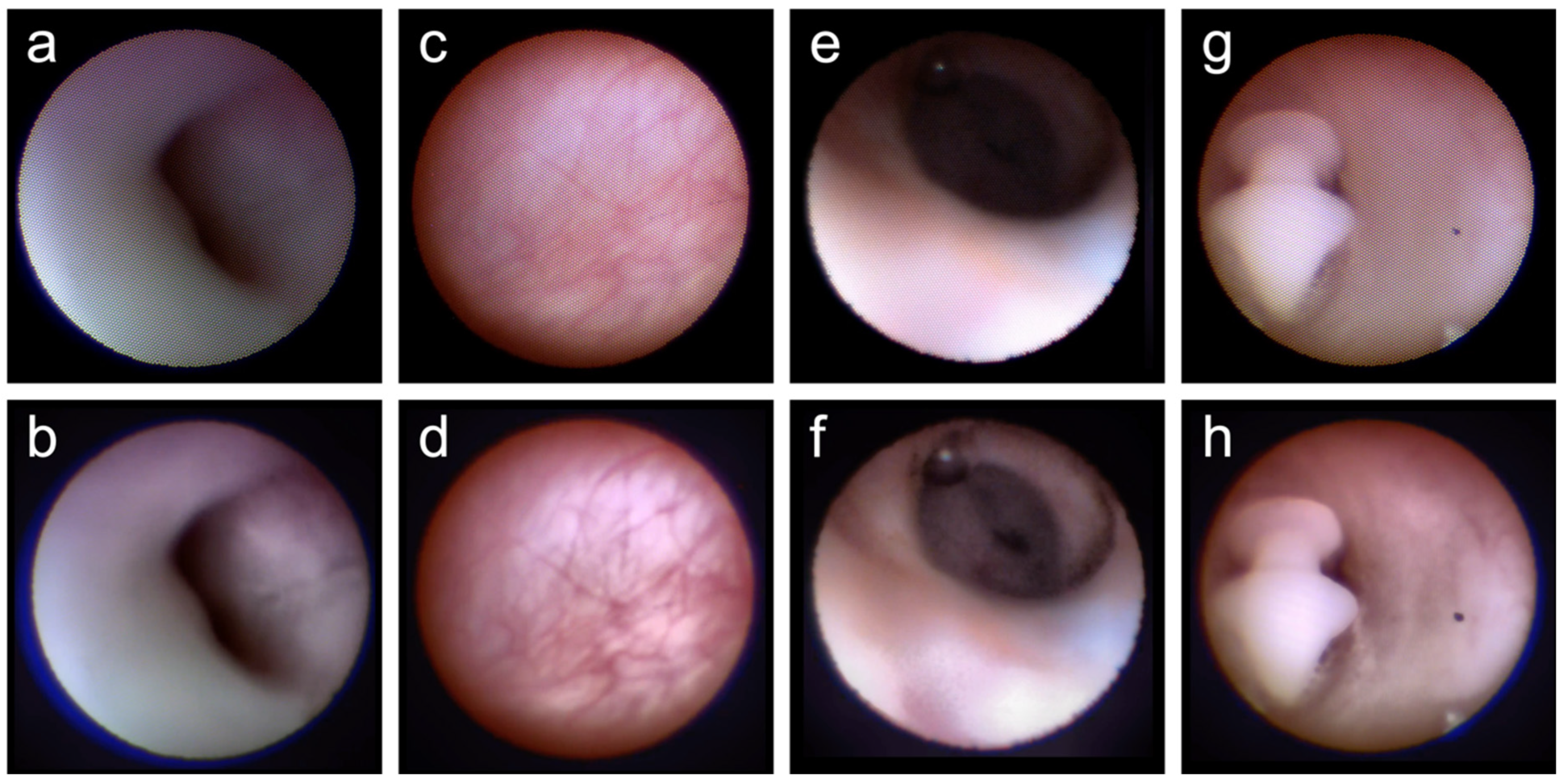
Abstract
:1. Introduction
2. Materials and Methods
2.1. Image Samples
2.2. Image Processing
2.3. Image Visibility Evaluation
2.4. In Vitro Experiments
3. Results
4. Discussion
5. Conclusions
Supplementary Materials
Author Contributions
Funding
Institutional Review Board Statement
Informed Consent Statement
Data Availability Statement
Acknowledgments
Conflicts of Interest
References
- Matsumura, N.; Suzuki, T.; Goto, S.; Fujita, T.; Yamane, S.; Maruyama-Inoue, M.; Kadonosono, K. Transcanalicular Endoscopic Primary Dacryoplasty for Congenital Nasolacrimal Duct Obstruction. Eye 2019, 33, 1008–1013. [Google Scholar] [CrossRef] [PubMed]
- Nakayama, T.; Watanabe, A.; Rajak, S.; Yamanaka, Y.; Sotozono, C. Congenital Nasolacrimal Duct Obstruction Continues Trend for Spontaneous Resolution Beyond First Year of Life. Br. J. Ophthalmol. 2020, 104, 1161–1163. [Google Scholar] [CrossRef] [PubMed]
- Ali, M.J. Metagenomics of the Lacrimal Sac in Primary Acquired Nasolacrimal Duct Obstruction: The Lacriome paper. Br. J. Ophthalmol. 2021. ahead of print. [Google Scholar] [CrossRef] [PubMed]
- Khorrami Kashi, A.; Keilani, C.; Nguyen, T.H.; Keller, P.; Elahi, S.; Piaton, J.M. Dacryolithiasis Diagnosis and Treatment: A 25-Year Experience Using Nasal Endoscopy. Br. J. Ophthalmol. 2021. ahead of print. [Google Scholar] [CrossRef]
- Hiraoka, T.; Hoshi, S.; Tasaki, K.; Oshika, T. Assessment of Conjunctival Flora in Eyes with Lacrimal Passage Obstruction Before and After Successful Dacryoendoscopic Recanalisation. Br. J. Ophthalmol. 2021, 105, 909–913. [Google Scholar] [CrossRef]
- Tasaki, K.; Hoshi, S.; Hiraoka, T.; Oshika, T. Deterioration of Contrast Sensitivity in Eyes with Epiphora due to Lacrimal Passage Obstruction. PLoS ONE 2020, 15, e0233295. [Google Scholar] [CrossRef]
- Hoshi, S.; Tasaki, K.; Hiraoka, T.; Oshika, T. Improvement in Contrast Sensitivity Function after Lacrimal Passage Intubation in Eyes with Epiphora. J. Clin. Med. 2020, 9, 2761. [Google Scholar] [CrossRef]
- Pakdel, F.; Soleimani, M.; Kasaei, A.; Ameli, K.; Pirmarzdashti, N.; Tari, A.S.; Ghasempour, M.; Banafsheafshan, A. Shifting to Very Early Endoscopic DCR in Acute Suppurative Dacryocystitis. Eye 2020, 34, 1648–1653. [Google Scholar] [CrossRef]
- Curragh, D.S.; Rajak, S.N.; Selva, D. Dacryoendoscopic-Assisted Nasolacrimal Intubation in an Australian Population. Clin. Exp. Ophthalmol. 2019, 47, 1209–1211. [Google Scholar] [CrossRef]
- Lee, S.M.; Lew, H. Transcanalicular endoscopic dacryoplasty in patients with primary acquired nasolacrimal duct obstruction. Graefes Arch. Clin. Exp. Ophthalmol. 2021, 259, 173–180. [Google Scholar] [CrossRef]
- Koh, S.; Ochi, S.; Inoue, Y. Lacrimal Drainage Function after Cheese Wiring of Lacrimal Passage Intubation. Graefes Arch. Clin. Exp. Ophthalmol. 2020, 258, 1087–1093. [Google Scholar] [CrossRef] [PubMed]
- Nowak, R.; Rekas, M.; Ali, M.J. Long-Term Outcomes of StopLoss™ Jones Tube (SLJT) and Minimally Invasive Conjunctivodacryocystorhinostomy. Graefes Arch. Clin. Exp. Ophthalmol. 2022, 260, 327–333. [Google Scholar] [CrossRef] [PubMed]
- Guo, Y.; Rokohl, A.C.; Kroth, K.; Li, S.; Lin, M.; Jia, R.; Heindl, L.M. Endoscopy-Guided Diode Laser-Assisted Transcaruncular StopLoss Jones Tube Implantation for Canalicular Obstructions in Primary Surgery. Graefes Arch. Clin. Exp. Ophthalmol. 2020, 258, 2809–2817. [Google Scholar] [CrossRef] [PubMed]
- Fiorino, M.G.; Quaranta-Leoni, C.; Quaranta-Leoni, F.M. Proximal Lacrimal Obstructions: A Review. Acta Ophthalmol. 2021, 99, 701–711. [Google Scholar] [CrossRef]
- Quaranta-Leoni, F.M.; Fiorino, M.G.; Serricchio, F.; Quaranta-Leoni, F. Management of Proximal Lacrimal Obstructions: A Rationale. Acta Ophthalmol. 2021, 99, e569–e575. [Google Scholar] [CrossRef]
- Sasaki, T.; Sounou, T.; Sugiyama, K. Dacryoendoscopic Surgery and Tube Insertion in Patients with Common Canalicular Obstruction and Ductal Stenosis as a Frequent Complication. Jpn. J. Ophthalmol. 2009, 53, 145–150. [Google Scholar] [CrossRef]
- Sasaki, T.; Sounou, T.; Tsuji, H.; Sugiyama, K. Air-Insufflated High-Definition Dacryoendoscopy Yields Significantly Better Image Quality than Conventional Dacryoendoscopy. Clin. Ophthalmol. 2017, 11, 1385–1391. [Google Scholar] [CrossRef] [Green Version]
- Singh, S.; Ali, M.J. A Review of Diagnostic and Therapeutic Dacryoendoscopy. Ophthalmic Plast. Reconstr. Surg. 2019, 35, 519–524. [Google Scholar] [CrossRef]
- Bae, S.H.; Park, J.; Lee, J.K. Comparison of Digital Subtraction Dacryocystography and Dacryoendoscopy in Patients with Epiphora. Eye 2021, 35, 877–882. [Google Scholar] [CrossRef]
- Fujimoto, M.; Ogino, K.; Matsuyama, H.; Miyazaki, C. Success Rates of Dacryoendoscopy-Guided Probing for Recalcitrant Congenital Nasolacrimal Duct Obstruction. Jpn. J. Ophthalmol. 2016, 60, 274–279. [Google Scholar] [CrossRef]
- Sasaki, T.; Nagata, Y.; Sugiyama, K. Nasolacrimal Duct Obstruction Classified by Dacryoendoscopy and Treated with Inferior Meatal Dacryorhinotomy. Part I: Positional Diagnosis of Primary Nasolacrimal Duct Obstruction with Dacryoendoscope. Am. J. Ophthalmol. 2005, 140, 1065–1069. [Google Scholar] [CrossRef] [PubMed]
- Sasaki, T.; Miyashita, H.; Miyanaga, T.; Yamamoto, K.; Sugiyama, K. Dacryoendoscopic Observation and Incidence of Canalicular Obstruction/Stenosis Associated with S-1, an Oral Anticancer Drug. Jpn. J. Ophthalmol. 2012, 56, 214–218. [Google Scholar] [CrossRef] [PubMed]
- Inomata, D.; Hoshi, S.; Alcântara, C.P.B.C.; Hiraoka, T.; Tasaki, K.; Oshika, T.; Matayoshi, S. Dacryoendoscopic Recanalization of Lacrimal Passage Obstruction/Stenosis After Radioiodine Therapy for Differentiated Thyroid Carcinoma. Am. J. Ophthalmol. Case Rep. 2022, 25, 101344. [Google Scholar] [CrossRef] [PubMed]
- Su, Y.; Zhang, L.; Li, L.; Fan, X.; Xiao, C. Surgical Procedure of Canaliculoplasty in the Treatment of Primary Canaliculitis Associated with Canalicular Dilatation. BMC Ophthalmol. 2020, 20, 245. [Google Scholar] [CrossRef]
- Mimura, M.; Ueki, M.; Oku, H.; Sato, B.; Ikeda, T. Evaluation of Granulation Tissue Formation in Lacrimal Duct Post Silicone Intubation and its Successful Management by Injection of Prednisolone Acetate Ointment into the Lacrimal Duct. Jpn. J. Ophthalmol. 2016, 60, 280–285. [Google Scholar] [CrossRef]
- Ali, M.J.; Singh, S.; Ganguly, A.; Naik, M.N. Dacryoendoscopy-Guided Transcanalicular Intralesional Interferon Alpha 2b for Canalicular Squamous Papillomas. Int. Ophthalmol. 2018, 38, 1343–1346. [Google Scholar] [CrossRef]
- Emmerich, K.H.; Meyer-Rüsenberg, H.W.; Simko, P. Endoscopy of the Lacrimal Ducts. Ophthalmologe 1997, 94, 732–735. [Google Scholar] [CrossRef]
- Singh, A.D.; Singh, A.; Whitmore, I.; Taylor, E. Endoscopic Visualisation of the Human Nasolacrimal System: An Experimental Study. Br. J. Ophthalmol. 1992, 76, 663–667. [Google Scholar] [CrossRef] [Green Version]
- Fein, W.; Daykhovsky, L.; Papaioannou, T.; Beeder, C.; Grundfest, W.S. Endoscopy of the Lacrimal Outflow System. Arch. Ophthalmol. 1992, 110, 1748–1750. [Google Scholar] [CrossRef]
- Suzuki, T. Dacryofiberscopy. Jpn. J. Ophthal. Surg. 2003, 16, 485–491. (In Japanese) [Google Scholar]
- Shinde, A.; Matham, M.V. Pixelate removal in an image fiber probe endoscope incorporating comb structure removal methods. J. Med. Imaging Health Inform. 2014, 4, 203–211. [Google Scholar] [CrossRef]
- Waterhouse, D.J.; Luthman, A.S.; Yoon, J.; Gordon, G.S.D.; Bohndiek, S.E. Quantitative Evaluation of Comb-Structure Correction Methods for Multispectral Fibrescopic Imaging. Sci. Rep. 2018, 8, 17801. [Google Scholar] [CrossRef] [PubMed] [Green Version]
- Sugimoto, M. New Sheath-Assisted Dacryoendoscopic Surgery. J. Eye 2007, 24, 1219–1222. [Google Scholar]
- Chen, D.; Ge, J.; Wang, L.; Gao, Q.; Ma, P.; Li, N.; Li, D.Q.; Wang, Z. A Simple and Evolutional Approach Proven to Recanalise the Nasolacrimal Duct Obstruction. Br. J. Ophthalmol. 2009, 93, 1438–1443. [Google Scholar] [CrossRef] [PubMed] [Green Version]
- Mihailovic, N.; Blumberg, A.F.; Rosenberger, F.; Brücher, V.C.; Lahme, L.; Eter, N.; Merté, R.L.; Alnawaiseh, M. Long-Term Outcome of Transcanalicular Microdrill Dacryoplasty: A Minimally Invasive Alternative for Dacryocystorhinostomy. Br. J. Ophthalmol. 2021, 105, 1480–1484. [Google Scholar] [CrossRef]
- Mimura, M.; Alameddine, R.M.; Korn, B.S.; Kikkawa, D.O.; Oku, H.; Sato, B.; Ikeda, T. Endoscopic Evaluation of Lacrimal Mucosa with Indigo Carmine Stain. Ophthalmic Plast. Reconstr. Surg. 2020, 36, 49–54. [Google Scholar] [CrossRef] [PubMed]
- Fujimoto, M.; Uji, A.; Ogino, K.; Akagi, T.; Yoshimura, N. Lacrimal Canaliculus Imaging Using Optical Coherence Tomography Dacryography. Sci. Rep. 2018, 8, 9808. [Google Scholar] [CrossRef] [Green Version]


Publisher’s Note: MDPI stays neutral with regard to jurisdictional claims in published maps and institutional affiliations. |
© 2022 by the authors. Licensee MDPI, Basel, Switzerland. This article is an open access article distributed under the terms and conditions of the Creative Commons Attribution (CC BY) license (https://creativecommons.org/licenses/by/4.0/).
Share and Cite
Hoshi, S.; Tasaki, K.; Maruo, K.; Ueno, Y.; Mori, H.; Morikawa, S.; Moriya, Y.; Takahashi, S.; Hiraoka, T.; Oshika, T. Improvement in Dacryoendoscopic Visibility after Image Processing Using Comb-Removal and Image-Sharpening Algorithms. J. Clin. Med. 2022, 11, 2073. https://doi.org/10.3390/jcm11082073
Hoshi S, Tasaki K, Maruo K, Ueno Y, Mori H, Morikawa S, Moriya Y, Takahashi S, Hiraoka T, Oshika T. Improvement in Dacryoendoscopic Visibility after Image Processing Using Comb-Removal and Image-Sharpening Algorithms. Journal of Clinical Medicine. 2022; 11(8):2073. https://doi.org/10.3390/jcm11082073
Chicago/Turabian StyleHoshi, Sujin, Kuniharu Tasaki, Kazushi Maruo, Yuta Ueno, Haruhiro Mori, Shohei Morikawa, Yuki Moriya, Shoko Takahashi, Takahiro Hiraoka, and Tetsuro Oshika. 2022. "Improvement in Dacryoendoscopic Visibility after Image Processing Using Comb-Removal and Image-Sharpening Algorithms" Journal of Clinical Medicine 11, no. 8: 2073. https://doi.org/10.3390/jcm11082073