Article

Core Lab Adjudication of the ACURATE neo2 Hemodynamic Performance Using Computed-Tomography-Corrected Left Ventricular Outflow Tract Area

1
Discipline of Cardiology, Saolta Group, Galway University Hospital, Health Service Executive and CORRIB Core Lab, University of Galway, H91 V4AY Galway, Ireland
2
Islamic Center of Cardiology, Al-Azhar University, Nasr City, Cairo 11651, Egypt
3
Department of Cardiology, Karolinska University Hospital, 17176 Stockholm, Sweden
4
Department of Cardiology and Cardiac Surgery, Kerckhoff Heart and Lung Centre, 61231 Bad Nauheim, Germany
5
Department of Cardiology, Heart Center Leipzig at University of Leipzig, 04289 Leipzig, Germany
6
The Heart Center—Rigshospitalet, 2100 Copenhagen, Denmark
7
Division of Cardiology, Department of Internal Medicine II, Medical University of Vienna, 1090 Vienna, Austria
8
CÚRAM, SFI Research Centre for Medical Devices, H91 W2TY Galway, Ireland
9
Department of Cardiothoracic Surgery, Anesthesia and Intensive Care, Skane University Hospital, Lund University, 22185 Lund, Sweden
10
Data Science Institute, University of Galway, H91 TK33 Galway, Ireland
*
Author to whom correspondence should be addressed.
J. Clin. Med. 2022, 11(20), 6103; https://doi.org/10.3390/jcm11206103
Submission received: 9 September 2022 / Revised: 28 September 2022 / Accepted: 13 October 2022 / Published: 17 October 2022
(This article belongs to the Special Issue Valvular Heart Disease: From Basic to Clinical Advances)

Abstract

:
(1) Background: Hemodynamic assessment of prosthetic heart valves using conventional 2D transthoracic Echocardiography-Doppler (2D-TTE) has limitations. Of those, left ventricular outflow tract (LVOT) area measurement is one of the major limitations of the continuity equation, which assumes a circular LVOT. (2) Methods: This study comprised 258 patients with severe aortic stenosis (AS), who were treated with the ACURATE neo2. The LVOT area and its dependent Doppler-derived parameters, including effective orifice area (EOA) and stroke volume (SV), in addition to their indexed values, were calculated from post-TAVI 2D-TTE. In addition, the 3D-LVOT area from pre-procedural MDCT scans was obtained and used to calculate corrected Doppler-derived parameters. The incidence rates of prosthesis patient mismatch (PPM) were compared between the 2D-TTE and MDCT-based methods (3) Results: The main results show that the 2D-TTE measured LVOT is significantly smaller than 3D-MDCT (350.4 ± 62.04 mm2 vs. 405.22 ± 81.32 mm2) (95% Credible interval (CrI) of differences: −55.15, −36.09), which resulted in smaller EOA (2.25 ± 0.59 vs. 2.58 ± 0.63 cm2) (Beta = −0.642 (95%CrI of differences: −0.85, −0.43), and lower SV (73.88 ± 21.41 vs. 84.47 ± 22.66 mL), (Beta = −7.29 (95% CrI: −14.45, −0.14)), respectively. PPM incidence appears more frequent with 2D-TTE- than 3D-MDCT-corrected measurements (based on the EOAi) 8.52% vs. 2.32%, respectively. In addition, significant differences regarding the EOA among the three valve sizes (S, M and L) were seen only with the MDCT, but not on 2D-TTE. (4) Conclusions: The corrected continuity equation by combining the 3D-LVOT area from MDCT with the TTE Doppler parameters might provide a more accurate assessment of hemodynamic parameters and PPM diagnosis in patients treated with TAVI. The ACURATE neo2 THV has a large EOA and low incidence of PPM using the 3D-corrected LVOT area than on 2D-TTE. These findings need further confirmation on long-term follow-up and in other studies.

1. Introduction

Transcatheter aortic valve implantation (TAVI) is a recommended interventional strategy in selected patients with severe aortic stenosis (AS) [1,2]. Transcatheter heart valve (THV) hemodynamic performance requires detailed and accurate assessment at multiple time points to determine the device’s success and detect prosthetic dysfunction. The first post-TAVI hemodynamic measurements are typically obtained before or early after hospital discharge, and are considered a baseline characterization of the implanted device (fingerprint) [3,4]; further follow-up can be compared with this baseline. Transthoracic echocardiography (TTE) is the gold standard imaging modality to assess the hemodynamic performance of THVs by measuring the peak velocity, transvalvular pressure gradients, effective orifice area (EOA), stroke volume, dimensionless velocity index (DVI) and the residual post-TAVI aortic regurgitation [3,4,5,6]. Several of these hemodynamic parameters are dependent on measuring the left ventricular outflow tract area (LVOT) area.
LVOT area calculation by the monoplane 2D-TTE is based on the measurement of a single diameter in mid-systole from the parasternal long-axis view, which resembles the small anteroposterior diameter and assumes a circular shape of the LVOT. Error in area estimation due to geometric assumptions will lead to an erroneous calculation of all derived parameters such as EOA and SV [7,8]. Accordingly, the accurate assessment of hemodynamic parameters mandates reducing or eliminating errors in LVOT area measurements.
The ACURATE neo2 (Boston Scientific, Marlborough, MA, USA), is a new generation of self-expandable, supra-annular THV, with favorable outcomes, including a significant reduction in the incidence of residual regurgitation in comparison to the earlier iteration ACURATE neo [9,10]. Data on the hemodynamic performance of the ACURATE neo2 are scarce and based only on 2D-TTE.
We hypothesized that measurements of LVOT area in a 3D fashion, from gated multi-phase reconstructed multidetector computed tomography (MDCT) scans, could result in different hemodynamic performance values, and thus, reclassification of the 2D-TEE-derived ACURATE neo2 THV outcome. In this study, we sought to assess the LVOT-dependent hemodynamic parameters such as EOA, stroke volume and their indexed values through the multimodal imaging approach, combining the more accurate 3D-MDCT-derived LVOT area and the 2D-TTE Doppler values. Reporting of these corrected values may give a true estimation of the hemodynamic performance of the ACURATE neo2.

2. Materials and Methods

This is a Core-Lab-led post hoc analysis of the Early neo2 Registry, a multi-center investigator-initiated European Registry of the first patients treated with the ACURATE neo2 THV Prosthesis in Europe after market approval (NCT04810195). This study is a retrospective analysis of patients with severe native AS or malfunctioning bioprosthetic surgical aortic valves who underwent TAVI with the ACURATE neo2 THV. We included patients with available pre-TAVI multi-phase MDCT scans and the comprehensive 2D-TTE assessment within seven days from the index procedure. All TTE and MDCT analyses were performed by three well-experienced senior cardiologists (AE, HE and MA).
The primary outcomes were the changes in hemodynamic classification of prosthesis patient mismatch (PPM) and the differences in the LVOT-dependent parameters (EOA, SV and their indexed values) between the 2D-TTE-derived continuity equation (CE) and the 3D-corrected CE by combining the MDCT-derived 3D-LVOT area and 2D-TTE Doppler measurements. The rate of PPM (moderate and severe PPM) between the two methods and the rate of reclassifications were reported.

2.1. Definition of Prosthesis Patient Mismatch (PPM)

Prosthesis patient mismatch (PPM) was identified as an EOA smaller than expected or the normal value, which led to inadequate cardiac output to meet the patient’s body demands, despite a normally functioning device without structural abnormality [2]. The indexed EOA (EOAi) is the main parameter used to assess the PPM according to the guideline’s recommendations [2,4].
  • For patients with BMI < 30 kg/cm2; PPM is:
    -
    Hemodynamically insignificant if the indexed EOA is >0.85 cm2/m2.
    -
    Moderate if between 0.66 and 0.85 cm2/m2.
    -
    Severe if ≤0.65 cm2/m2.
  • For obese patients with BMI ≥ 30 kg/m2; PPM is:
    -
    Hemodynamically insignificant if the indexed EOA is >0.70 cm2/m2.
    -
    Moderate if between 0.56 and 0.70 cm2/m2.
    -
    Severe if ≤0.55 cm2/m2.

2.2. Echocardiography

A comprehensive 2D-TTE assessment of post-TAVI patients was performed before hospital discharge or within seven days from the index procedure according to the recommended guidelines for evaluating prosthetic heart valves [3,6]. Echocardiographic analyses were performed according to the Core Lab Standard Operating Procedures (SOP) based on the most recent guidelines recommendations [3,4,5,6], using a dedicated workstation (TOMTEC ARENA, TOMTEC Imaging Systems GmbH, Unterschlessheim, Germany). Velocity time integral (VTI) of blood flow across the THV (VTIAV) was measured from the Continuous-wave Doppler (CWD) and that of the LVOT (VTILVOT) was measured from the pulsed-wave Doppler (PWD) of LVOT. Both measurements were obtained from the 3- or the 5-chamber apical views, if appropriate. The sample volume for the VTILVOT was typically positioned at the LV edge of the THV in systole. As recommended by guidelines, the external LVOT diameter was measured from the parasternal long-axis view in zoomed view below the prosthetic stent (inflow level) in mid-systole (Figure 1). LVOT area was calculated automatically with the formula (A = πr2) and used to calculate the EOA using the CE in addition to the calculation of SV across the LVOT using the flow equation (FlowLVOT (SVLVOT) = LVOT area × VTILVOT). All hemodynamic parameters values were indexed to the patient’s body surface area.

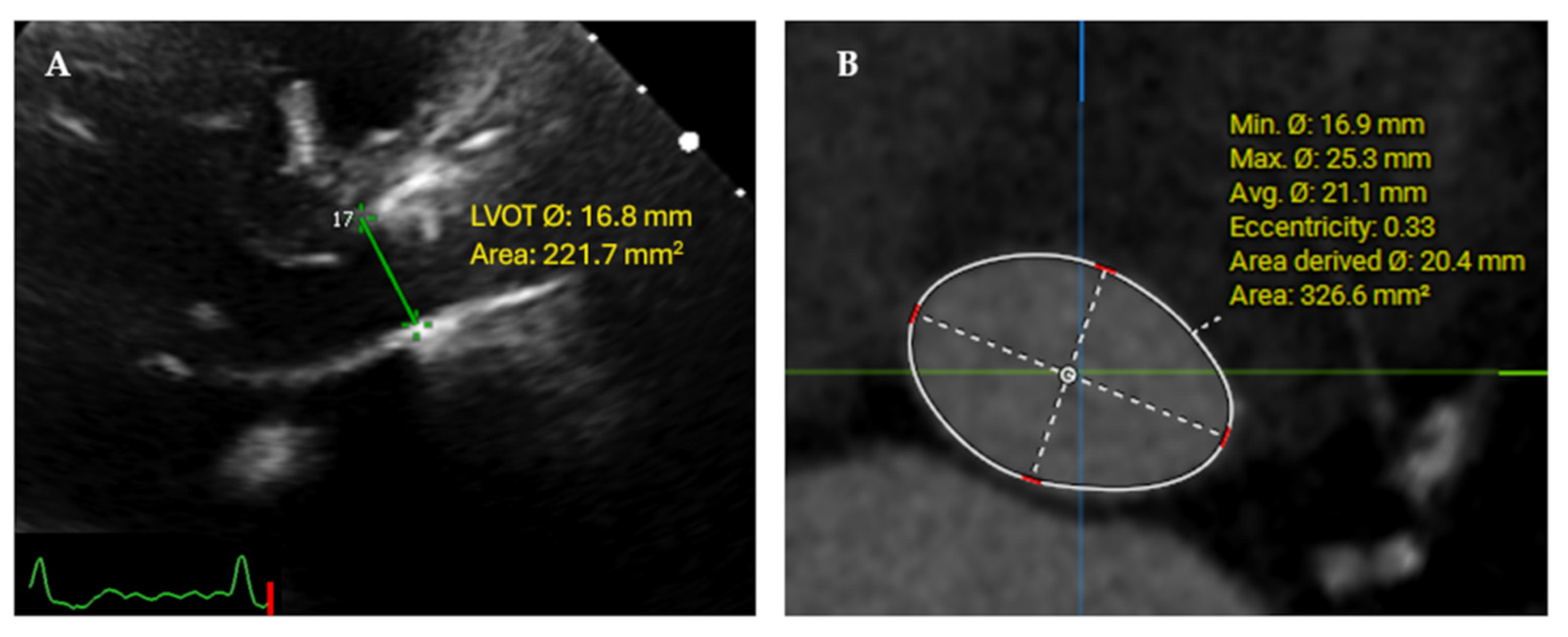
2.3. Multidetector Computerized Tomography (MDCT)

Pre-TAVI MDCT scans acquisition was performed according to each center’s protocol. Offline 3D multiplanar reconstruction and comprehensive analysis were performed according to the Core Lab SOP in accordance with the Society of Cardiac Computed Tomography (SCCT) guidelines [11]. The LVOT was measured at 5 mm below and perpendicular to the predefined native aortic annulus level from contrast enhanced MDCT scans, using a dedicated workstation (3mensio® Structural Heart 10.2, 3mensio Medical Imaging, B.V., The Netherlands). Direct planimetry of the LVOT area and the diameters were measured in the enface and zoomed view as vertical (Minimum = Dmin) and horizontal (Maximum = Dmax) on the mid-late systole (30–40% systolic phases) (Figure 1). The diameters were used to calculate the LVOT eccentricity index. The eccentricity index was calculated to define the shape of the LVOT (circular or elliptical) using the formula [1 − (Dmin/Dmax)] × 100. LVOT is considered circular when the eccentricity index was <10% [12].

2.4. Corrected Continuity and Flow Equations

The EOA and SV were calculated using the conventional 2D-TTE-derived parameters; post-TAVI EOA TTE = [(LVOT area TTE × PWD VTILVOT)/CWD VTIAV]. Post-TAVI SV (SVTTE) was calculated as SVTTE = [(LVOT area TTE × PWD VTI LVOT).
On the other hand, the corrected equations indicate the use of the MDCT-derived 3D-LVOT area (Direct planimetry from MPR views without geometric assumptions) to be used in the calculation of AV EOA and SV instead of the TTE-derived LVOT area (based on the assumption of circular LVOT shape). Therefore, the corrected parameters were calculated as follows.
Post-TAVI EOAMSCT = [(LVOT area MDCT × PWD VTILVOT)/CWD VTI AV]
Post-TAVI SVMSCT = [(LVOT area MDCT × PWD VTI LVOT). In addition, EOA and SV were indexed to patients’ BSA to calculate the indexed corrected parameters.
We compared the PPM rate between the 2D-TTE and the 3D-Corrected-MDCT CE-derived EOAi values.

2.5. Statistical Analysis

Results are presented as mean ± standard deviation (SD) or median with interquartile range (IQR), according to their distribution pattern. We used the Shapiro–Wilks test as well as QQ plot to assess the normality of continuous variables. Categorical data were presented as percentages and fractions of occurrence. Correlation and agreement between the LVOT area and LVOT area dependent parameters, obtained by different methods (2D TTE and MDCT), were determined using Pearson correlation, Spearman rank correlation and Bland–Altman analysis, respectively. Correlation and agreement between mean trans prosthetic PG, with the EOA and EOAi, calculated from TTE and MDCT.
Intra-observer and inter-observer (two independent blinded observers) reproducibility of LVOT area measured by TTE and MSCT was performed in a random set of 20 patients and evaluated using the intraclass correlation coefficient for absolute agreement. Good agreement was defined as >0.80. Mean transprosthetic PG was scatter-plotted for each imaging-technique-derived EOA and EOAi and fitted curves for data pairs were constructed.
The Bayesian mixed-effect model was used to account the cluster effects of measurements, while parameters obtained from two methods are nested within patients.
Bayesian mixed-effect models with gaussian and asymptotic Laplace priors based on the distribution pattern of the dependent variables were used to compare the quantitative parameters between the two groups. While gaussian and asymptotic Laplace were used for normal and skewed distribution, respectively. Furthermore, Bayesian mixed-effect models with Bernoulli (binary) and cumulative priors (ordinal) were used to compare the PPM rate between the two methods. We also used the mixed-effect Bayesian regression model to compare changes in the hemodynamic performance of ACURATE neo2 among small (23 mm), intermediate (25 mm) and large (27 mm) sizes after implantation.
The convergence of the Bayesian models was examined using R-hat, LOO, and posterior predictive plots. The R-hat < 1.1 indicates a suitable model of convergence. All statistical analyses were conducted using the ggplot2 and rstan packages in the R 4.1.1 environment.
The posterior Beta or Odds ratio (OR) was used to report the associations between variables of interest. The 95% credible interval (Crl) was used to examine the differences between the two groups, Crl includes zero value for continuous models and one for categorical models indicating non-significant associations.

3. Results

A total of 554 patients with severe AS were treated with TAVI using ACURATE neo2 between September 2020 and April 2021 and included in the Early neo2 Registry. We excluded patients with MDCT without mid–late systolic phases, patients who required valve in valve bailout therapy with a device other than ACURATE neo2, and patients without either post-TAVI 2D TTE study or pre-TAVI MDCT available in the Core Lab for the independent analysis. In total, 258 patients comprised the final cohort of this study. Mean age was 81.6 ± 6.1 years, 65% women with a median of the European System for Cardiac Operative Risk Evaluation (EuroSCORE) II of 3.34% [2.15, 3.5]. The baseline characteristics of the study population are shown in Table 1. The median duration between the pre-TAVI MDCT scan and the TAVI procedure was 13 days [2, 46]. The study cohort included eight patients (3.1%) with type I bicuspid AV, and six patients with TAVI in malfunctioning surgical AV prosthesis (TAVI in SAVR). Pre-TAVI MDCT scans analysis revealed a mean of the native annulus area of 430.2 ± 62.9 mm2, LVOT minimum and maximum diameters of 19.03 ± 2.55 mm, and 26.92 ± 2.43 mm, respectively with a measured LVOT area of 405.22 ± 81.32 mm2, and LVOT eccentricity index 29.21 ± 7.4% indicating that LVOT area was oval (Elliptical) in 257 (99.5%) cases (Table 1). All patients were treated via transfemoral vascular access, balloon pre-dilatation was performed in 81.8%, while post-dilatation was performed in 41.1% (Table 2).

3.1. Hemodynamic Outcomes (Conventional 2D TTE and MSCT-Corrected Parameters)

Post-procedural 2D-TTE assessment revealed LVEF of 58.9 ± 9.8%, AV maximum velocity 1.98 ± 0.44 m/s, trans-prosthetic mean pressure gradient 7.22 ± 3.11 mmHg and dimensionless velocity index (DVI 0.64 ± 0.13. Post-TAVI residual AR assessment revealed 59.7% of patients with none/trace AR, 36.4% had mild AR, 1.9% with moderate AR and none had severe AR (Table 3).
The mean LVOT diameter on 2D-TTE was 21.03 ± 1.9 mm and shows a significant difference between the LVOT dimensions obtained from the MDCT scan, Dmin 19.03 ± 2.55 mm (95% Crl of differences: 1.7, 2.31) and Dmax 26.92 ± 2.43 mm (95% Crl of differences: −6.2, −5.58).
The mean LVOT area obtained from TTE and MDCT were 350.4 ± 62.04 mm2 and 405.22 ± 81.32 mm2, respectively (95% CrI of differences: −55.15, −36.09), which resulted in a smaller EOA and lower SV (2.25 ± 0.59 vs. 2.58 ± 0.63 cm2) and (73.88 ± 21.41 vs. 84.47 ± 22.66 mL), (Beta = −0.642 (95%CrI of differences: −0.85, −0.43), (Beta = −7.29 (95% CrI: −14.45, −0.14)), respectively and consequently the indexed values (EOAi 1.20 ± 0.32 cm2/m2 vs. 1.41 ± 0.34 cm2/m2 (95% CrI of differences: −0.207, −0.136), SVi TTE 41 ± 12.6 mL/m2 vs. 46.14 ± 12 mL/m2 (95% CrI of differences: −0.207, −0.136) (Table 4).

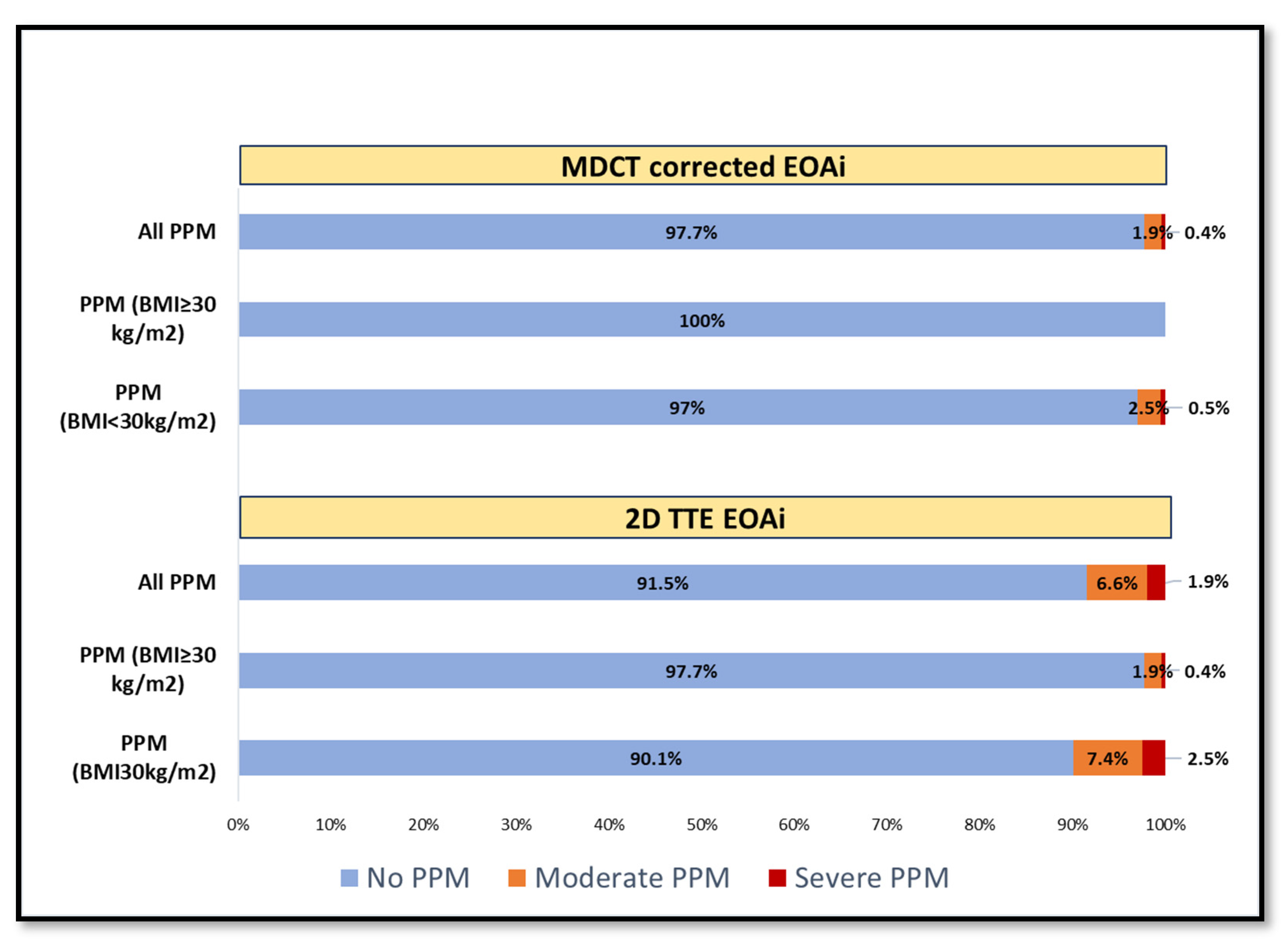
3.2. Prosthesis-Patient Mismatch (PPM) Incidence and Reclassification

The incidence of all (overall) PPM measured by conventional 2D-TTE (8.52%) was higher than MDCT-corrected formula (2.32%), OR = 8.36 (95% Crl: 2.42, 39.61), (Kappa w = 0.323, 95% confidence interval (CI): 0.13, 0.51). However, the differences remained statistically significant in the adjusted model by sex, age, and BMI variables (OR = 10.33; 95% CrI: 2.5, 67.34). The distributions of PPM frequency within BMI categories are shown in Figure 2.

3.3. Stroke Volume Index Changes in Patients with Low EF%

In 29 patients with EF < 50% (mean of EF was 40.47 ± 6.49%), the SVi changed significantly from 34.1 ± 11.4 mL/m2 by TTE to 39.3 ± 11 mL/m2 with MDCT LVOT-corrected calculation (Beta = 5.18; 95% CrI: 2.36, 8).

3.4. Inter Valve Size Differences in Hemodynamic Performance and Incidence of PPM

According to the results of the mixed-effects model adjusted for age, sex, BMI, and BSA, to determine the effect of other variables on MDCT and TTE, the detection ability of the interaction effects between methods and independent variables was tested (Table S1).
The EOA*ACURATE neo2 size interaction was statistically significant; thus, subgroup analysis according to ACURATE neo2 sizes indicated that mean differences in EOA between TTE and MDCT were obvious for the 23 mm (diff = 0.64, 95% CrI: 0.44, 0.85) compared with the 25 mm (diff = 0.208, 95% CrI: 0.03, 0.35) and 27 mm (diff = 0.26, 95% CrI: 0.11 0.44). The interaction effects between methods and the rest of the independent variables were insignificant. (Figures S4 and S5).
In the simple Bayesian logistic regression model, a higher risk of PPM was observed for ACURATE neo2 size 23 mm than ACURATE neo2 size 25 and 27 mm (OR = 3.57; 95% CrI: 1.12, 12.2) with 2D-TTE. With the MDCT, there was no association between the size of ACURATE neo2 and PPM (Table S1 and Figure S4).

3.5. Intra-Observer and Inter-Observer Reliability

An excellent agreement was observed for the intra-observer and Inter observer reliability regarding LVOT area measured by MDCT (ICC = 0.99 [95% CI; 0.98–0.99]) and (ICC = 0.98 [95% CI; 0.95 to 0.99]), respectively and was good regarding TTE (ICC = 0.87 [95% CI; 0.71, 0.95]) and (ICC = 0.85 [95% CI; 0.63, 0.94]) (Figure S2).

4. Discussion

This is the first study that systematically evaluates the new supra-annular ACURATE neo2 THV hemodynamic performance using the LVOT area derived from both the conventional 2D-TTE and pre-procedural MDCT scan 3D measurements. Both techniques were used to calculate all LVOT-dependent hemodynamic parameters (EOA, SV in addition to their indexed values) aiming to accurately report hemodynamic parameters outcome early at patients’ hospital discharge and to define the baseline hemodynamic performance of further follow up in comparison to the obtained values, especially for the diagnosis and severity of PPM.
The main findings of this report are as follows; (1) the calculation of LVOT area from 2D-TTE significantly underestimated the area in comparison to the MDCT measured 3D-LVOT area (350 vs. 405 mm2), and all LVOT-dependent parameters; (2) furthermore, the LVOT was oval in most cases (99.5%) with a mean eccentricity index of 29.2%; (3) recalculation of EOAi resulted in a significant reduction in PPM incidence among the included cohort (8.5% to 2.3%), (4) 3D-MDCT-corrected LVOT are measurements resulted in obtaining more concordance between EOA and other hemodynamic parameters; and (5) finally, the results also show a significant difference between the different sizes with the use of the corrected LVOT assessment in contrast to the conventional TTE assessment.
The fact of measuring a 3D structure using a 2D image usually carries the risk of inaccurate assessment. In LVOT area measurements, our results confirm significant underestimation of the LVOT area in agreement with multiple reports. Liu et al. compared LVOT area measurements using biplane versus single dimensions using TTE, and resulted in the LVOT with the biplane method being larger than the conventional method (420 vs. 373 mm2) [13]; in addition, Weber et al., have reported that the MDCT-derived LVOT area was larger than 2D-TTE (456.9 vs. 303.7 mm2) and resulted in larger EOA in patients with severe AS and reclassification of 30% of the included cohort from severe to moderate AS [14].
The concept of using accurately measured LVOT (3D-LVOT) to be included in the CE is a quite old seeking more accurate and reproducible results [15,16,17], but the application and the use of 3D-LVOT area (3D echocardiography, MDCT, or CMR) area combined with TTE Doppler (CWD and PWD) to obtain AV EOA and SV (corrected parameters) still uncommon practice. However, it could be used especially if discordance in the parameters was noticed either pre- or post-AV replacement or when PPM is suspected [15,16,17]. Multiple reports confirm the utility of the corrected calculation of EOA using the LVOT area measured from MDCT scan either pre- or post-AV replacement or even for the prediction of EOA, and mainly for the diagnosis of PPM [12,14,18]. The incidence of overall and/or severe PPM after TAVI was reported to be lower than SAVR [19,20], especially with self-expandable, supra-annular devices with larger EOA and lower gradients [21].
The incidence of PPM according to the MDCT-corrected EOAi resulted in a lower frequency of all PPM and BMI-adjusted PPM than 2D-TTE (8.5% vs. 2.3%). (Figure 2 and Table 4) the results agree with those of Fukui et al., who reported larger EOAi (1.57 vs. 1.1 cm2/m2) and reclassification of all PPM from 19.5% by TTE to 3.5% with MDCT 3D-LVOT correction for both SEV and BEV [12]. As larger devices are expected to provide larger EOA, a sub-analysis has been performed according to the implanted ACURATE neo2 size, revealed a significant difference in EOA between the medium, 25 mm, and large, 27 mm, sizes in comparison with the small, 23 mm, devices when the MDCT-corrected LVOT area was used instead of the 2D-TTE LVOT area, which showed a non-significant difference (Table S1, Figure S4).
This study recommends that LVOT area should be directly measured on a 3D imaging modality such as MDCT in all cases, if possible. Correlation of this study findings of misclassified PPM cases might offer an explanation on the lack of clinical correlation of PPM following TAVI in earlier publications. Those cases were most probably misclassified due to underestimation of LVOT area, and consequently EOA.

Study Limitations

Although this is the first study to provide comprehensive hemodynamic reassessment and describes the recalculation of EOA and SV after implantation of the ACURATE neo2 THV using the MDCT-derived LVOT area, some limitations exist. First, this is a retrospective study with small sample size. Second, long-term clinical outcomes of the PPM reclassification between the two methods are not available. Third, we used the same cutoff values established for the TTE assessment. Therefore, new cut-off values of PPM based on 3D-derived EOA should be derived from long-term outcome studies, and finally, the use of the pre-TAVI MDCT to measure the 3D-LVOT area, but we thought that with the short time interval between the pre-procedural MDCT and the TAVI procedure (13 days) and the post-procedural TTE, no significant changes in the LV mass will occur. Additionally, the low radial force of the ACURATE neo2 will not significantly affect the shape of the LVOT.

5. Conclusions

LVOT is eccentric in most patients undergoing TAVI, which might lead to erroneous estimation of hemodynamic performance of THV from 2D-TTE using the continuity equation. Using the directly measured LVOT area on a 3D MDCT scan, instead of 2D-TTE, in combination with the TTE Doppler might reduce these limitations, and could result in an accurate and reproducible assessment of continuity-equation-derived parameters. The correction of the LVOT area showed a lower rate of PPM diagnosis dependent on the EOAi, resulting in a better correlation with other hemodynamic parameters, such as mean gradient. Using the MDCT-corrected measurements, the ACURATE neo2 THV, a self-expandable supra-annular valve, provides a very low rate of PPM, a large EOA associated with a low trans-prosthetic pressure gradient.

Supplementary Materials

The following supporting information can be downloaded at: https://www.mdpi.com/article/10.3390/jcm11206103/s1, Figure S1: Bland–Altman plot showing the difference in calculated LVOT area, EOA, SV and EOAi using the 2D TTE- and MDCT-corrected method. Figure S2: MDCT LVOT measurement; Interobserver and Intra-observer reliability with excellent ICCs. Figure S3: Scatter plots of the mean transvalvular gradient and EOA-2D TTE-, EOAi-2D TTE-, MDCT-corrected EOA, MDCT-corrected EOAi. Figure S4: PPM (BMI adjusted) incidence, assessed by the 2D TTE- and MDCT-corrected methods and classified per ACURATEneo2 size. Figure S5: Inter valve size difference according to EOA, EOAi, SV and SVi per valve size; Table S1: Comparison among ACURATEneo2 sizes and their interactions with methods (2D TTE & MDCT); The results of adjusted multiple Bayesian regression model.

Author Contributions

Conceptualization, A.E. and O.S.; methodology, A.E. and O.S.; validation, A.E., O.S. and A.R.; formal analysis, A.E., M.A., H.E., S.K.M. and S.A.-J.; investigation, A.E., O.S., A.R., W.-K.K., C.U.M., N.S., M.A.-W., O.D.B., C.H. and H.B.; resources, A.R., W.-K.K. and M.A.-W.; data curation, A.E. and S.K.M.; writing—original draft preparation, A.E., S.A.-J. and O.S.; writing—review and editing, A.E., O.S., A.R., W.-K.K., M.A.-W., O.D.B., C.H. and A.S.; visualization, O.S.; supervision, O.S. All authors have read and agreed to the published version of the manuscript.

Funding

This research received support from Science Foundation of Ireland (Grant # 13/RC/2073_P2).

Institutional Review Board Statement

The study was conducted in accordance with the Declaration of Helsinki and approved by the Institutional Review Board (or Ethics Committee) of each participating site.

Informed Consent Statement

Informed consent was obtained from all subjects involved in the study.

Data Availability Statement

All data are available within the abstract.

Conflicts of Interest

Andreas Rück reports grants and non-financial support from Boston Scientific, during the conduct of the study; grants and personal fees from Boston Scientific, personal fees from Edwards Lifesciences, outside the submitted work. Won-Keun Kim reports proctor/speaker fees/advisory board participation for Abbott, Boston Scientific, Edwards Lifesciences, Medtronic, Meril Life Sciences, and Shockwave Medical. Mohammed Abdel-Wahab reports speakers’ honoraria and/or consulting fees from Boston Scientific and Medtronic paid to his institution on his behalf. Ole De Backer reports institutional research grants and consulting fees from Boston Scientific. Christopher U.Meduri reports advisory board participation and proctoring for Boston Scientific, consulting for Medtronic, and advisory board participation for Cardiovalve and Admedus (Anteris). Osama Soliman reports several institutional grants and non-financial support from industry and is the medical director of Computed Tomography, Echocardiography and Cardiovascular MRI Core Laboratory at the CORRIB Core Lab for multiple industry-sponsored trials, for which he receives no direct financial compensation. The other authors declare no conflict of interest.

References

  1. Vahanian, A.; Beyersdorf, F.; Praz, F.; Milojevic, M.; Baldus, S.; Bauersachs, J.; Capodanno, D.; Conradi, L.; De Bonis, M.; De Paulis, R.; et al. 2021 ESC/EACTS Guidelines for the management of valvular heart disease. Eur. Heart J. 2021, 43, 561–632. [Google Scholar] [CrossRef] [PubMed]
  2. Otto, C.M.; Nishimura, R.A.; Bonow, R.O.; Carabello, B.A.; Erwin, J.P., 3rd; Gentile, F.; Jneid, H.; Krieger, E.V.; Mack, M.; McLeod, C.; et al. 2020 ACC/AHA Guideline for the Management of Patients with Valvular Heart Disease: A Report of the American College of Cardiology/American Heart Association Joint Committee on Clinical Practice Guidelines. J. Am. Coll. Cardiol. 2021, 77, e25–e197. [Google Scholar] [CrossRef] [PubMed]
  3. Lancellotti, P.; Pibarot, P.; Chambers, J.; Edvardsen, T.; Delgado, V.; Dulgheru, R.; Pepi, M.; Cosyns, B.; Dweck, M.R.; Garbi, M.; et al. Recommendations for the imaging assessment of prosthetic heart valves: A report from the European Association of Cardiovascular Imaging endorsed by the Chinese Society of Echocardiography, the Inter-American Society of Echocardiography, and the Brazilian Department of Cardiovascular Imaging. Eur. Heart J. Cardiovasc. Imaging 2016, 17, 589–590. [Google Scholar] [CrossRef] [PubMed] [Green Version]
  4. VARC-3 WRITING COMMITTEE; Genereux, P.; Piazza, N.; Alu, M.C.; Nazif, T.; Hahn, R.T.; Pibarot, P.; Bax, J.J.; Leipsic, J.A.; Blanke, P.; et al. Valve Academic Research Consortium 3: Updated endpoint definitions for aortic valve clinical research. Eur. Heart J. 2021, 42, 1825–1857. [Google Scholar] [CrossRef]
  5. Zoghbi, W.A.; Chambers, J.B.; Dumesnil, J.G.; Foster, E.; Gottdiener, J.S.; Grayburn, P.A.; Khandheria, B.K.; Levine, R.A.; Marx, G.R.; Miller, F.A., Jr.; et al. Recommendations for evaluation of prosthetic valves with echocardiography and doppler ultrasound: A report From the American Society of Echocardiography’s Guidelines and Standards Committee and the Task Force on Prosthetic Valves, developed in conjunction with the American College of Cardiology Cardiovascular Imaging Committee, Cardiac Imaging Committee of the American Heart Association, the European Association of Echocardiography, a registered branch of the European Society of Cardiology, the Japanese Society of Echocardiography and the Canadian Society of Echocardiography, endorsed by the American College of Cardiology Foundation, American Heart Association, European Association of Echocardiography, a registered branch of the European Society of Cardiology, the Japanese Society of Echocardiography, and Canadian Society of Echocardiography. J. Am. Soc. Echocardiogr. 2009, 22, 975–1014. [Google Scholar] [CrossRef]
  6. Zoghbi, W.A.; Asch, F.M.; Bruce, C.; Gillam, L.D.; Grayburn, P.A.; Hahn, R.T.; Inglessis, I.; Islam, A.M.; Lerakis, S.; Little, S.H.; et al. Guidelines for the Evaluation of Valvular Regurgitation After Percutaneous Valve Repair or Replacement: A Report from the American Society of Echocardiography Developed in Collaboration with the Society for Cardiovascular Angiography and Interventions, Japanese Society of Echocardiography, and Society for Cardiovascular Magnetic Resonance. J. Am. Soc. Echocardiogr. 2019, 32, 431–475. [Google Scholar] [CrossRef]
  7. Baumgartner, H.; Hung, J.; Bermejo, J.; Chambers, J.B.; Edvardsen, T.; Goldstein, S.; Lancellotti, P.; LeFevre, M.; Miller, F., Jr.; Otto, C.M. Recommendations on the Echocardiographic Assessment of Aortic Valve Stenosis: A Focused Update from the European Association of Cardiovascular Imaging and the American Society of Echocardiography. J. Am. Soc. Echocardiogr. 2017, 30, 372–392. [Google Scholar] [CrossRef]
  8. Maes, F.; Pierard, S.; de Meester, C.; Boulif, J.; Amzulescu, M.; Vancraeynest, D.; Pouleur, A.C.; Pasquet, A.; Gerber, B.; Vanoverschelde, J.L. Impact of left ventricular outflow tract ellipticity on the grading of aortic stenosis in patients with normal ejection fraction. J. Cardiovasc. Magn. Reson. 2017, 19, 37. [Google Scholar] [CrossRef] [Green Version]
  9. Mollmann, H.; Holzhey, D.M.; Hilker, M.; Toggweiler, S.; Schafer, U.; Treede, H.; Joner, M.; Sondergaard, L.; Christen, T.; Allocco, D.J.; et al. The ACURATE neo2 valve system for transcatheter aortic valve implantation: 30-day and 1-year outcomes. Clin. Res. Cardiol. 2021, 110, 1912–1920. [Google Scholar] [CrossRef]
  10. Rück, A.; Kim, W.-K.; Kawashima, H.; Abdelshafy, M.; Elkoumy, A.; Elzomor, H.; Wang, R.; Meduri, C.U.; Verouhis, D.; Saleh, N.; et al. Paravalvular Aortic Regurgitation Severity Assessed by Quantitative Aortography: ACURATE neo2 versus ACURATE neo Transcatheter Aortic Valve Implantation. J. Clin. Med. 2021, 10, 4627. [Google Scholar] [CrossRef]
  11. Blanke, P.; Weir-McCall, J.R.; Achenbach, S.; Delgado, V.; Hausleiter, J.; Jilaihawi, H.; Marwan, M.; Norgaard, B.L.; Piazza, N.; Schoenhagen, P.; et al. Computed tomography imaging in the context of transcatheter aortic valve implantation (TAVI)/transcatheter aortic valve replacement (TAVR): An expert consensus document of the Society of Cardiovascular Computed Tomography. J. Cardiovasc. Comput. Tomogr. 2019, 13, 1–20. [Google Scholar] [CrossRef]
  12. Fukui, M.; Garcia, S.; Lesser, J.R.; Gossl, M.; Tang, L.; Caye, D.; Newell, M.; Hashimoto, G.; Lopes, B.B.C.; Stanberry, L.I.; et al. Prosthesis-patient mismatch defined by cardiac computed tomography versus echocardiography after transcatheter aortic valve replacement. J. Cardiovasc. Comput. Tomogr. 2021, 15, 403–411. [Google Scholar] [CrossRef] [PubMed]
  13. Liu, S.; Churchill, J.; Hua, L.; Zeng, X.; Rhoades, V.; Namasivayam, M.; Baliyan, V.; Ghoshhajra, B.B.; Dong, T.; Dal-Bianco, J.P.; et al. Direct Planimetry of Left Ventricular Outflow Tract Area by Simultaneous Biplane Imaging: Challenging the Need for a Circular Assumption of the Left Ventricular Outflow Tract in the Assessment of Aortic Stenosis. J. Am. Soc. Echocardiogr. 2020, 33, 461–468. [Google Scholar] [CrossRef] [PubMed]
  14. Weber, M.; Jaenisch, M.; Spilker, M.; Pingel, S.; Schueler, R.; Stundl, A.; Sedaghat, A.; Hammerstingl, C.; Mellert, F.; Grube, E.; et al. TAVR outcome after reclassification of aortic valve stenosis by using a hybrid continuity equation that combines computed tomography and echocardiography data. Catheter. Cardiovasc. Interv. 2020, 96, 958–967. [Google Scholar] [CrossRef] [PubMed] [Green Version]
  15. Doddamani, S.; Bello, R.; Friedman, M.A.; Banerjee, A.; Bowers, J.H., Jr.; Kim, B.; Vennalaganti, P.R.; Ostfeld, R.J.; Gordon, G.M.; Malhotra, D.; et al. Demonstration of left ventricular outflow tract eccentricity by real time 3D echocardiography: Implications for the determination of aortic valve area. Echocardiography 2007, 24, 860–866. [Google Scholar] [CrossRef]
  16. Poh, K.K.; Levine, R.A.; Solis, J.; Shen, L.; Flaherty, M.; Kang, Y.J.; Guerrero, J.L.; Hung, J. Assessing aortic valve area in aortic stenosis by continuity equation: A novel approach using real-time three-dimensional echocardiography. Eur. Heart J. 2008, 29, 2526–2535. [Google Scholar] [CrossRef]
  17. Gaspar, T.; Adawi, S.; Sachner, R.; Asmer, I.; Ganaeem, M.; Rubinshtein, R.; Shiran, A. Three-dimensional imaging of the left ventricular outflow tract: Impact on aortic valve area estimation by the continuity equation. J. Am. Soc. Echocardiogr. 2012, 25, 749–757. [Google Scholar] [CrossRef]
  18. Mooney, J.; Sellers, S.L.; Blanke, P.; Pibarot, P.; Hahn, R.T.; Dvir, D.; Douglas, P.S.; Weissman, N.J.; Kodali, S.K.; Thourani, V.H.; et al. CT-Defined Prosthesis-Patient Mismatch Downgrades Frequency and Severity, and Demonstrates No Association With Adverse Outcomes after Transcatheter Aortic Valve Replacement. JACC Cardiovasc. Interv. 2017, 10, 1578–1587. [Google Scholar] [CrossRef]
  19. He, S.; Fang, Z. Incidence, predictors, and outcome of prosthesis-patient mismatch after transcatheter aortic valve replacement: A meta-analysis. Medicine 2020, 99, e20717. [Google Scholar] [CrossRef]
  20. Miyasaka, M. Prosthesis-patient mismatch after transcatheter aortic valve implantation. Cardiovasc. Interv. Ther. 2022, 37, 615–625. [Google Scholar] [CrossRef]
  21. Leone, P.P.; Regazzoli, D.; Pagnesi, M.; Sanz-Sanchez, J.; Chiarito, M.; Cannata, F.; Van Mieghem, N.M.; Barbanti, M.; Tamburino, C.; Teles, R.; et al. Predictors and Clinical Impact of Prosthesis-Patient Mismatch after Self-Expandable TAVR in Small Annuli. JACC Cardiovasc. Interv. 2021, 14, 1218–1228. [Google Scholar] [CrossRef] [PubMed]
Figure 1. LVOT measurements: (A) 2D-TTE LVOT diameter measurements from the parasternal long axis zoomed view in mid-systole, (B) MDCT multiplanar reconstruction of the LVOT (5 mm below the annular plane, in mid-systolic phase 30%) with the minimum (anteroposterior) diameter and the maximum (medio-lateral) diameter with measured 3D-LVOT area, with a larger area calculated from the MDCT measured minimum and maximum diameters with eccentricity index of 0.33.
Jcm 11 06103 g001
Figure 2. Incidence of PPM (BMI adjusted) based on the EOAi assessed by 2D-TTE and MDCT-corrected method; (All PPM; moderate or severe (2.3% (MDCT-corrected) vs. 8.5% (2D-TTE).
Jcm 11 06103 g002
Table 1. Patients’ baseline characteristics, pre-procedural Echocardiography and MDCT scan.
Characteristicn = 258
Age81.6 (6.1)
Women168 (65%)
Body surface area, m21.8 [1.7–2.0]
Body mass index, kg/m2
     Body mass index < 30 kg/m2
     Body mass index ≥ 30 kg/m2
26 [23.7–29.3]
204 (79%)
54 (21%)
Euroscore II, %3.34 [2.15–3.5]
Hypertension212 (82.2%)
Diabetes mellitus Type I
Diabetes mellitus Type II
32 (12.4%)
49 (19%)
Baseline creatinine, mg/dL1.0 [0.8–1.3]
Prior Atrial fibrillation102 (39.5%)
Chronic lung obstructive disease39 (15.1%)
Prior stroke or TIA33 (12.8%)
Peripheral arterial disease30 (11.6)
Prior permanent pacemaker implantation26 (10.1%)
Previous cardiac surgery30 (11.6%)
Previous CABG15 (5.8%)
Previous PCI51 (19.8)
New York Heart Association (NYHA) class
    Class II
    Class III
    Class IV
86 (33.3%)
141 (54.7%)
25 (9.7%)
Valve-in-Valve procedure (TAVI-in-SAVR)6 (2.3%)
Preprocedural 2D-TTE characteristics
LV Ejection fraction, %60 [55–65]
Aortic valve maximum velocity, m/s4.29 (0.56)
Mean pressure gradient, mmHg43.6 [35–52]
Aortic valve effective orifice area, cm20.7 [0.6–0.8]
Moderate-severe aortic regurgitation28 (10.9%)
Moderate-severe mitral regurgitation38 (14.8%)
Moderate-severe tricuspid regurgitation21 (8.2%)
Pre-procedural MDCT characteristics
Bicuspid Aortic Valve (Type I)8 (3.1%)
Native aortic annulus area, mm2430.2 (62.9)
MDCT-derived LVOT measurements
     Minimum diameter, mm
     Maximum diameter, mm
     LVOT area, mm2
19.03 (2.55)
26.92 (2.43)
405.22 (81.32)
Values are either Median [IQR], Mean (±SD), and n (%).
Table 2. Procedural characteristics and In-hospital outcomes.
n (%)
Vascular access
     Transfemoral258 (100%)
Balloon pre-dilatation211 (81.8%)
ACURATEneo2 size
Small {23 mm}
Medium {25 mm}
Large {27 mm}

59 (22.9%)
101 (39.1%)
98 (38%)
Balloon post-dilatation106 (41.1%)
Valve embolization1 (0.4%)
Need for second valve implantation1 (0.4%)
Annular injury (rupture)0
Cardiac tamponade0
Procedural death0
Coronary obstruction0
New postoperative permanent pacemaker18 (7%)
Major vascular complications4 (1.6%)
Major bleeding4 (1.6%)
Life-threatening bleeding3 (1.2%)
In-hospital stroke7 (2.7%)
Conversion to surgery0
New dialysis0
All-cause mortality0
Values are presented as n (%).
Table 3. Post-procedural TTE-Doppler assessment.
TTE (n = 258)
LV ejection fraction, %58.9 (9.8)
AV maximum velocity, m/s1.98 (0.44)
AV mean pressure gradient, mmHg7.22 (3.11)
Dimensionless velocity index0.64 (0.13)
Systolic pulmonary artery pressure, mmHg36.8 [29.5–44.1]
Post-TAVI aortic regurgitation
         None/trace
         Mild
         Moderate
154 (59.7%)
94 (36.4%)
5 (1.9%)
Moderate–severe mitral regurgitation36 (15.2%)
Moderate–severe tricuspid regurgitation49 (24.5%)
Values are either Median [IQR], Mean [±SD] and n (%).
Table 4. LVOT-dependent hemodynamic parameters (TTE- vs. MDCT-derived LVOT area).
TTEMSCT95% CrI of Difference
LVOT diameter, mm
21.03 (1.9)
Minimum diameter 19.03 (2.55)[1.7, 2.31]
Maximum diameter 26.92 (2.43)[−6.2, −5.58]
LVOT area, mm2350.4 (62.04)405.22 (81.32)[−55.15, −36.09]
EOA, cm22.25 (0.59)2.58 (0.63)[−0.85, −0.43]
EOA index, cm2/m21.20 (0.32)1.41 (0.34)[−0.207, −0.136]
LVOT SV, mL73.88 (21.41)84.47 (22.66)[−14.45, −0.14]
LVOT SV index, mL/m241.0 (12.6)46.14 (12)[−0.207, −0.136]
Prosthesis-Patient Mismatch (PPM)
-
All PPM
-
BMI adjusted PPM
22 (8.52%)6 (2.32%)
  BMI < 30 kg/m2
       Moderate PPM15 (7.3%)5 (2.5%)
       Severe PPM5 (2.5%)1 (0.5%)
  BMI ≥ 30 kg/m2
       Moderate PPM2 (3.7%)0
       Severe PPM00
Values are either Median [IQR], Mean (±SD) and n (%); BMI = Body Mass Index.
Publisher’s Note: MDPI stays neutral with regard to jurisdictional claims in published maps and institutional affiliations.

