Article

Investigation of the Effect of Imatinib and Hydroxyurea Combination Therapy on Hematological Parameters and Gene Expression in Chronic Myeloid Leukemia (CML) Patients

1
Department of Medical Laboratory Sciences, Faculty of Allied Medical Sciences, Al-Ahliyya Amman University, Amman 19328, Jordan
2
Pharmacological and Diagnostic Research Center (PDRC), Al-Ahliyya Amman University, Amman 19328, Jordan
3
Department of Medical Laboratory Sciences, Faculty of Science, Al-Balqa Applied University, Al-Salt 19117, Jordan
4
Department of Biopharmaceutics and Clinical Pharmacy, Faculty of Pharmacy, Al-Ahliyya Amman University, Amman 19328, Jordan
5
Faculty of Pharmacy, Al-Zaytoonah University of Jordan, Amman 11733, Jordan
6
College of Medicine, QU Health, Qatar University, Doha P.O. Box 2713, Qatar
*
Authors to whom correspondence should be addressed.
J. Clin. Med. 2022, 11(17), 4954; https://doi.org/10.3390/jcm11174954
Submission received: 22 July 2022 / Revised: 8 August 2022 / Accepted: 9 August 2022 / Published: 24 August 2022
(This article belongs to the Special Issue Chronic Myeloid Leukemia: Clinical Diagnosis and Treatment)

Abstract

:
(1) Background: Chronic myeloid leukemia is defined as the neoplastic development of mostly myeloid cells in the bone marrow. Several treatments, including chemotherapy, radiation, hormone treatment, and immunological therapy, can be used to control this condition. The therapeutic impact on leukemic individuals varies, and the response to therapy varies between patients due to disease heterogeneity. The primary goal of this study is to compare the effects of single and Imatinib (IM) and Hydroxyurea (HU) combined treatment on hematological parameters and gene expression in CML patients. (2) Methods: This study was conducted on 51 patients, with chronic myeloid leukemia, who were admitted to Al-Basher hospital in Amman, Jordan, for follow-up. Their hematological parameters were checked and gene expression was measured for (BCL2, PP2A, CIP2A, and WT1). (3) Results: The BCL2 gene was found to be less expressed in both IM and (HU + IM) treatments as compared to the HU group alone, while PP2A gene expression was raised. Such a thing indicates that the outcome of the combined therapy method is not ideal, since PP2A activation causes CML cells to move toward the blast crisis stage. Furthermore, CIP2A gene expression revealed that IM and (HU + IM) had the same therapeutic effect and were more successful in CML patients than HU alone. With regards to the treatment effect on hematological parameters, notably in CML patients in later stages, the combination therapy (HU + IM) raised lymphocyte count, indicating a greater response to the treatment. When compared to single medicines, the combination treatment reduced the proportion of neutrophils to normal reference ranges. Platelet counts, on the other hand, dramatically decreased in both IM and (HU + IM). (4) Conclusion: Because the studied genes (BCL2, PP2A, CIP2A, and WT1) are participating in cell proliferation and death, the findings show that the examined genes are significant to understand the efficacy of various therapies. Furthermore, it was found that there was a clear effect of the clinic-based strategic treatment on hematological indicators such as WBCs, lymphocytes, neutrophils, and platelet counts.

1. Introduction

1.1. Cancer and Leukemia

Cancer is one of the major reasons of death, as, among every eight people worldwide, one dies due to one type of cancer [1]. These different types are characterized by a relatively uncontrolled cell proliferation that can invade body tissues, replace normal cells, and metastasize to distant organs [2]. Cancer occurs when aberrant cells divide uncontrollably and diffuse to other tissues in the body. The disease appears when genetic mutations disrupt the order of the cell proliferation process. Based on histological features, the disease is divided into five major types: carcinoma, sarcoma, myeloma, leukemia, and lymphoma [3].
Leukemia is a malignant leukocyte clonal proliferation that directly affects cell maturation and production in the bone marrow. This disease is generated from lymphoid or myeloid lineages and may be acute (precursor cell) or chronic (mature cell) subtypes. The onset of acute leukemia is quick, with fast progression, and a lethal end in a few weeks, compared with chronic leukemia [4]. This disease is also characterized by a frequent increase in the count of white blood cells (WBC), and the rate of proliferation in both mature and maturing cells of a particular lineage which leads to cell accumulation. Leukemia cells gradually replace most normal hematopoietic ones in the bone marrow, compromising normal bone marrow function. At the time of diagnosis, the symptoms of chronic leukemia are often obscure and varied [5].

1.2. Chronic Myeloid Leukemia (CML)

CML is a hematopoietic cancerous stem cell driven by the BCR-ABL fusion protein. The disease appears at an indolent chronic phase that can last for 3 to 5 years [6]. CML is known as chronic granulocytic leukemia (CGL), chronic myeloid leukemia, and chronic myelocytic leukemia. It is primarily characterized by the neoplastic growth of myeloid cells in the bone marrow. Erythrocytic and megakaryocytic lineages can expand, and the extramedullary granulocytic proliferation is localized in the spleen and liver, and that reflects disease progression [7]. CML is divided into chronic, rapid, and blast crises. Traditional chemotherapy is used to restore and maintain the normal division rate for months and up to several years. Comparatively, 70–90% of the treated patients in the chronic phase, with tyrosine kinase inhibitors (imatinib mesylate), experienced a 5-year progression-free survival [7].

1.3. Treatment of CML

Hydroxyurea (HU) is the first-line preferred option to be used for a patient with suspected CML and an increased WBC count (e.g., >80–100 × 109/L), to decrease the WBC count to normal levels [8]. The treatment protocol suggests giving the patients HU until confirming the presence or absence of the Philadelphia chromosome. In cases where the WBC is exceedingly high, allopurinol is taken to reduce the risk of some problems generated by tumor lysis. After the diagnosis is confirmed, the patient directly receives Imatinib, a tyrosine kinase inhibitor (TKI). Generally, it is not required to maintain the count of WBC at normal levels before using TKIs, and HU should be stopped once the treatment with TKI starts [8,9].
Imatinib (IM) was developed to target BCR-ABL1, which is constitutively active in chronic myeloid leukemia (CML) [9]. It is authorized by the FDA for the treatment of chronic myelogenous leukemia and gastrointestinal stromal tumors. In addition, this treatment has been studied and applied to a variety of malignant cancers [10]. Imatinib Mesylate has the chemical formula 4-4 ((4-methyl-1-piperazinyl) methyl)-N-(4-methyl-3-((4-(3-pyridinyl)-2-pyrimidinyl) amino) phenyl) -benzamide mono methane sulfonate (IMT). Its chemical formula is C29H31N7O.CH4O3S and the molecular weight is about 589.71 g mol−1 [11].
HU, also known as hydroxycarbamide, is a non-alkylating antineoplastic and antiviral drug that has been used to treat a broad range of health problems interrelated with hematology, oncology, contagious diseases, and dermatology. A study discovered that it might be utilized to treat a variety of solid tumors and myeloproliferative diseases [12]. HU is a white or almost white powder that is crystalline and hygroscopic. The chemical formula of this medication is CH4N2O2 and has a molecular weight of 76,055 g mol−1 [13].
Although newer and more efficient treatments have replaced HU in certain cases, it is still used in clinics as a proven, dependable, and well-tolerable small-molecule medication for a variety of neoplastic and non-neoplastic disorders [14]. It is being used to treat sickle cell anemia and chronic myeloproliferative diseases, and is classified by the World Health Organization as an essential therapy [15].

1.4. Genes Involved in CML

PP2A is an essential protein phosphatase that regulates most cellular processes by forming holoenzyme that control cell proliferation, survival, and differentiation. It provides more than 90% of the cell’s phosphatase activity along with PP1. PP2A is routinely inactivated in both AML and CML. Numerous studies currently focus on PP2A inactivation as a prerequisite for human cell transformation.
CIP2A protein, also known as a carcinogenic inhibitor of PP2A, is a protein that is found mainly in humans. In human cancers, CIP2A reduces PP2A’s tumor suppressor action. In particular, this has been shown that blocks PP2A activity towards the oncogenic transcription factor c-Myc, leading to prevent the proteolytic breakdown of c-Myc. Furthermore, CIP2A is necessary for malignant cellular proliferation and tumor development in vivo. Overexpression of CIP2A enhances Ras-elicited cell proliferation and changes immortalized human cells, which are consistent with CIP2A’s oncogenic activity (HEK-TERVs) [16].
B cell lymphoma 2 (BCL2) is a key participant in the eukaryotic cell’s genetic program that promotes survival by suppressing cell death. BCL2 protein overexpression has been observed in a variety of human malignancies, including leukemia, lymphomas, and carcinomas. The mechanism of BCL2 activation in follicular lymphomas and a subset of BCLs diffuse were discovered to be the translocation t(14,18)(q32;q21), which results in uncontrolled gene expression [17].
Wilms’ tumor gene (WT1) encodes a transcription factor that is essential for cell proliferation and differentiation. The WT1 gene is strongly expressed in leukemia and other forms of solid tumors. This gene is a useful tumor marker for detecting leukemia’s minimum residual illness; it was initially thought to be a tumor suppressor gene, but it was found to be an oncogene [18].
The major goal of this study is to evaluate the effect of single and combined chemotherapy of IM and HU on hematological parameters and gene expression in CML patients that distinguish the pathophysiology of this disease.
Our findings might give more insights into the effectiveness of treatment protocols followed in clinics, and whether these therapeutics are potent in controlling disease progression.

2. Materials and Methods

2.1. Ethical Approval

This study was performed in line with the principles of the Declaration of Helsinki. Approval was granted by the scientific research committee of Al-Ahliyya Amman University (IRB: AAU/3/8/2021–2022).

2.2. Consent to Participate

Informed consent was obtained from all 51 participants included in the study.

2.3. Patient Cohort Criteria

A total of 51 patients, males and females, who were diagnosed with CML, participated in this study. The inclusion criteria of the samples were as follows: patients diagnosed with CML according to the World Health Organization (WHO) 2008 classification: ages about 20 years old and above; patients after single-agent induction of chemotherapy (HU); and those who received both treatment agents (IM and HU) chemotherapy. All patients were treated with standard induction chemotherapy. The CML newly diagnosed patients (with no treatment), together with those who were previously diagnosed, were both treated with a short dose of HU and a long dose of IM.
The exclusion criteria for this studied cohort included the following: any missing clinical data related to the type of treatment other than IM or HU or a combination of IM and HU; patients less than 20 years old; and patients who were hospitalized.

2.4. Patients’ Collection Sample and Ethical Consideration

Venous blood samples of patient cohorts who meet the inclusion criteria were collected throughout August-November 2021 (Figure 1). Those patients were admitted to Al-Basher hospital in Amman, Jordan, for follow-up. The blood samples were immediately processed for both hematological and gene expression (BCL2, PP2A, CIP2A, and WT1). The study comprised blood samples of 51 patients diagnosed with CML and those who had already received treatment. The population size was chosen based on Statistics from the Jordanian Ministry of Health, regarding the prevalence of CML disease in Jordan.

2.5. Gene Expression

2.5.1. Extraction of Ribonucleic Acid (RNA) and Synthesis of Complementary Deoxyribonucleic Acid (cDNA)

Zymo Research’s Direct-zol RNA miniprep kit was used to extract the RNA, Lot No. ZRC206161/USA). cDNA was synthesized from 1 ng/mL of total RNA using a primeScript RT master mix (Takara- Lot No. AK22353A/San Jose, CA, USA), according to the manufacturer’s protocol.

2.5.2. Primer Design

In all genes, the sequence of their primers was designed using primer blast, except for CIP2A, which was taken from a scientifically published paper and used as a reference [19], as illustrated in Table 1. The designed primers were ordered by a commercial provider (IDT, Coralville, IA, USA). A strict set of criteria was employed for primer design to achieve optimum specificity and efficiency during PCR amplification.
After selecting the optimal annealing temperature, based on the information mentioned above, optimization steps were performed.

2.5.3. Gene Expression Analysis

According to the manufacturer’s instructions, quantitative real-time PCR (qPCR) was used to determine gene expression in leukemic patient cells using (SsoAdvanced Universal SYBR Green Supermix/ BIO-RAD, Hercules, CA, USA) (Lot No 1725274). As a result, the expression of these genes assessed by real-time quantitative PCR (qPCR) was standardized to that of β-actin. The CFX96 real-time PCR detection device was used to perform RT qPCR in 96-well plates (Bio-Rad, Hercules, CA, USA).
For each gene, two independent technical replicates were employed. The identical RNA preparation was used to amplify all cDNA samples in triplicate, and the mean value was used (i.e., two technical replicates). To prevent differences between runs, all samples for each analyzed gene were performed on the same plate. The CFX Manage version 1.6 software (Bio-Rad, Hercules, CA, USA) generated the baseline and Ct values automatically, using the default settings.
The thermal profile and details of RT-PCR programs for the four genes BCL2, PP2A, CIP2A, and WT1 were optimized based on the literature and manufacturer recommendations.

2.6. Statistical Analysis

This study was conducted on 51 chronic myeloid leukemia patients: 19 patients without treatment and 32 patients with different treatment protocols. For data analysis, GraphPad Prism version 5 statistical software was used.
The variance for each studied gene was calculated besides the standard deviation and the standard deviation of the reference gene, which was normalized to the exponential arithmetic mean of the ct value, in comparison with the reference genes. The patients were divided into four groups. The first group included patients without treatment (control); the second included patients with HU; the third was that of patients who were treated with IM therapy; and the fourth included patients under combination therapy (HU + IM). First, the Δct of the target gene, and that of the housekeeping one, were calculated, then the ΔΔct was calculated in addition. The two-way paired t-test was performed and used to calculate and consider the null hypothesis to determine whether there is a relationship between the genes or not.
A paired t-test was applied to assess the relationship between different hematological parameters in all patient categories. These parameters include: lymphocytes, neutrophils, basophils, platelets, and RDW-SD. The results were taken into consideration if the p-value was less than or equal to 0.05.
Correlation analysis was performed to detect the significant relationship between different gene expressions in the same patient group (UT, HU, IM, HU + IM).

3. Results

3.1. Patient Cohort Demographic Data

This study was conducted on 51 chronic myeloid leukemia patients, 15 males and 36 females. The patients’ ages ranged between 21 to 70 years. All of them met the inclusion criteria included in this study. The study participants consist of four groups: CML without treatment (control group) (n = 19, M = 4, F =15); CML patients with HU treatment (n = 12, M = 1, F = 11); CML patients with IM treatment (n = 8, M = 6, F = 2); and CML patients with combination therapy (HU + IM) (n = 12, M = 4, F = 8). HU + IM; HU, and IM. None of the patients have a family history of CML. The error bars indicate the standard error, n = 51.

3.2. Gene Expression

In this section, we focused on comparing the relative expression fold for each studied gene among different patient categories.

3.2.1. BCL2 Expression

The relative expression fold for the BCL2 gene was measured using RT-PCR. Both primer and housekeeping genes were run twice and the average was calculated, then the average of ∆∆Ct for each CML patient category (UT, HU, IM, HU + IM) was measured (Figure 2). All treated groups were statistically compared with the UT CML patients to observe the effect of treatment on gene expression. The results showed that the Bcl2 gene had low expression in both IM and HU + IM treatments, compared with other groups. This indicated that the apoptotic effect of the combination is the same as the treatment with a single agent (IM), which revealed that using the combination therapy has no significant effect compared with IM. Therefore, using IM alone is enough to have an impact on cancer cells.

3.2.2. PP2A Expression

The relative expression fold for the PP2A gene was measured using RT-PCR. Both primer and housekeeping genes were run twice and the average was calculated, then the average of ∆∆Ct for each CML patient category (UT, HU, IM, HU + IM) was also measured (Figure 3). All treated groups were statistically compared with the UT CML patients to observe the effect of treatment on gene expression.
This figure shows that the PP2A gene has a significant increase in the relative expression fold in the combination group, compared with other groups. This indicates that the outcome of combination therapy is not preferable in the treatment strategy, as the activation of Pp2a leads to an increase in the progression of CML cells into the blast crisis stage. Therefore, using HU as a single agent has a more potent effect on inhibiting the proliferation of cancerous cells. The findings confirmed that CML patients under HU have a lower relative expression fold of Pp2a, which improved the disease response to treatment.

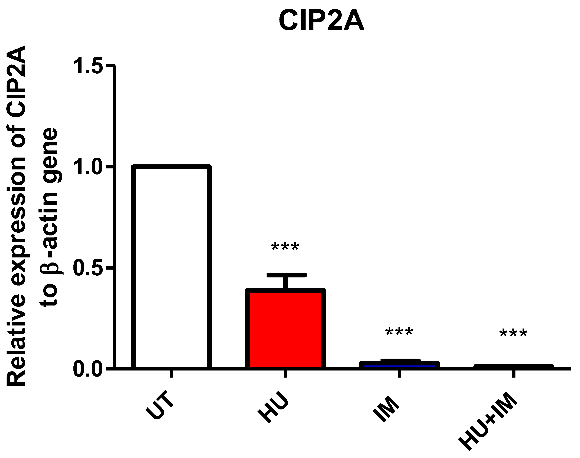
3.2.3. CIP2A Expression

The relative expression fold for the CIP2A gene was measured using RT-PCR. Both primer and housekeeping genes were run twice and the average was calculated, then the average of ∆∆Ct for each CML patient category (UT, HU, IM, HU + IM) was measured as well (Figure 4). All treated groups were statistically compared with the UT CML patients to observe the effect of treatment on gene expression.
The results indicated that both IM and HU + IM have the same treatment effect and are more potent in CML patients, compared with HU alone. These findings suggest that using IM or HU + IM is recommended to improve the patient outcome as they inhibit CIP2A gene expression involved, in regulating cell proliferation and increasing cell death.

3.2.4. WT1 Expression

The relative expression fold for the WT1 gene was measured using RT-PCR. Both primer and housekeeping genes were run twice and the average was calculated, then the average of ∆∆Ct for each CML patient category (UT, HU, IM, HU + IM) was measured as well (Figure 5). All treated groups were statistically compared with the UT CML patients to observe the effect of treatment on gene expression.
As the relative expression fold of WT1 can be considered a marker for cancer progression, decreasing its expression may, therefore, play a role in stabilizing the disease status in CML cases. Based on the above findings, utilizing different single-agent protocols (HU or IM) seems to be much more beneficial than combination therapy (HU + IM). The results of WT1 in the combination therapy are similar to those in the patients without treatment. This suggests finding out other combinations to reduce the WT1 expression, particularly in CML patients in later stages.

3.3. Hematological Parameters for Study Patients

Several hematological parameters are shown below (Figure 6). They were selected based on the significant change, whether an increase or decrease, in their values compared with the control (UT) group after receiving treatments (HU, IM, HU + IM). The average was calculated in all patient categories (UT, HU, IM, HU + IM) for each hematological parameter.
(A) The number of absolute lymphocytes was decreased with the combination therapy (HU+IM), indicating a better response to the treatment compared with other single-agent therapy. (B) The WBC count was decreased significantly in the combination and IM therapy, and the effect of both treatments on the WBC count was more than the treatment with HU alone. (C) The combination therapy decreased the percentage of neutrophils to the normal reference intervals compared with single agents. (D) The percentage of basophils was maintained in the normal range in both (IM, HU + IM). (E) RDW-CV values indicated normal cell population size after treatment, especially after treatment with combination therapy. (F) Platelet numbers were reduced, particularly in both (IM, HU + IM).

3.4. Correlation Analysis of Gene Expression

A correlation analysis was performed to understand the pattern of the relationship between expressions of the different genes discussed in this study (Table 2). Only the CML patients who received treatment were included in this section.
The correlation analysis revealed that there is an inverse relationship between all treatments of PP2A vs. CIP2A, BCL2 vs. PP2A, and WT1 vs. PP2A. There is also a positive relationship between CML patients who received IM treatment in BCL2 vs. CIP2A, WT1 vs. CIP2A, and WT1 vs. BCL2.

4. Discussion

Several types of research were published over the past few years that focused on understanding the potency of cancer therapies through observation of the expression of the targeted genes [20]. The results could give more insight into predicting disease prognosis and treatment response. The overexpression, or low expression, of some key genes is involved in inducing the proliferation of cancerous cells, in addition to preventing the rate of apoptosis [21].
Recently, pharmacological development and therapeutic methods have improved at an accelerating and remarkable rate. The factor that plays the most important role in this aspect is knowledge increase and the understanding of the pattern in which cancer cells divide and spread in the body [22]. This helps to produce more different types of drugs that are more targeted to get the maximum possible benefit, and minimize the cytotoxic effects after receiving treatment.
In this study, two types of therapies were selected, based on the treatment strategy that is used in Jordanian hospitals for patients with Chronic Myelogenous Leukemia (CML). The mechanism of action of HU is that it is an efficient ribonucleotide reductase (RNR) inhibitor that diminishes intracellular deoxynucleotide triphosphate pools and effects as an S-phase-specific agent with DNA synthesis suppression, which ultimately leads to cell cytotoxicity. The main reason for using this agent in leukemia patients is to reduce or prevent the growth of tumor cells in the body [23]. IM is used in a clinical setting to prevent a BCR-ABL protein from exerting its role in the oncogenic pathway in CML. IM directly inhibits the substrate such as; GRB2 (Growth Factor Receptor Bound Protein 2) from entering the tyrosine kinase site, leading to the prevention of tumor cell proliferation [24].
Various gene expression techniques have been established, including: Reporter genes; Northern blotting; RNA Seq; DNA microarray; and reverse transcription-polymerase chain reaction (RT-PCR). The latter was conducted in this study, as this method is simple, easy to use, rapid, and cost-effective, and a smaller number of samples can be used with enormous specificity and sensitivity [7].
The BCL-2 relative expression fold has clearly shown that using a single agent of IM or combination therapy decreased the expression, indicating that both treatment strategies have raised the apoptotic effect in tumor cells. This finding agrees with the previous study which reported that using combination therapy, such as ABT-199 and IM that targets both BCL-2 and BCR-ABL tyrosine kinase, has a significant impact on regulating and improving CML patient stability in chronic phase and blast crisis phases [25].
The combination therapy (HU + IM) has an adverse effect on the CML patient in the blast crisis phase, as the relative expression fold of PP2A was high in this category of patients. Thus, using single-agent HU or IM is more effective, with fewer side effects, particularly in CML patients at late stages. In the literature, a study was conducted on leukemic stem cells which showed that inhibiting both PP2A and BCR-ABL (IM) may be a viable therapeutic approach for targeting a drug-insensitive for these cells, that leads to preserving MRD in patients [26].
Since CIP2A can be used as a marker for the pathogenesis of chronic myelocytic leukemia, our results showed that there was an impact of treatment on the relative expression fold for the CIP2A gene. The results also demonstrated that receiving IM or combination therapy in CML patients has a positive effect on inducing cell death and on regulating cell proliferation, which was observed from the down-expression of this gene. In addition, CIP2A indicates that it may reveal therapy resistance of tyrosine kinase inhibitors (IM). Therefore, the reduction in the expression of this gene gives a good impression of the treatment response [27].
The WT1 gene is usually used for monitoring the progression of BCR-ABL positive chronic myeloid leukemia. Our results showed that the patient who received combination therapy (HU + IM) had a bad prognosis, compared with the patient who had never received any treatment (UT). Based on these findings, it is highly recommended to alter the treatment protocol for the patient group (HU + IM) and to use a single agent (HU or IM) instead of the combination therapy (HU + IM). El-Menoufy, M. A. and Ahmed, M. A. revealed that repeated evaluation of WT1 transcript levels in CML patients might be a valuable marker for predicting early and abrupt disease progression [28].
On the other hand, hematological parameters were affected to varying degrees (increasing or decreasing) after receiving treatment. Total white blood cells were maintained in the normal range in both (IM, HU + IM). After receiving a single agent of IM or combination therapy (HU + IM), the total number of white blood cell counts has reduced and maintained in the normal range. This correlates with a published research article that reported that patients with low WBC and/or early MRD response may be treated with autologous SCT or long-term TKI therapy [29].
The patients who received (HU + IM) had a significant induce in absolute lymphocyte count. This result demonstrated that following the administration of a therapy containing IM, the number of B lymphocytes is expected to be activated and this could be considered as a marker of CML patients for treatment response, specifically if combined with a cytogenetic response. This conclusion completely matches the previous findings, the bone marrow CD20+ lymphocytes were increased after receiving IM therapy, due to the long-term effect of inhibiting the BCR-ABL kinase. The number of lymphocytes in the patients who received HU was reduced, which corresponds to its mechanism of action [30].
Administration of IM or (HU + IM), induced the reduction of both neutrophil and basophil counts and almost maintained the normal reference intervals. After receiving IM, the basophil count decreased to below 3%. A published study on a 57-year-old woman with chronic myeloid leukemia who had significant severe basophilia, revealed that after a few days of stopping IM, a remarkable decrease in basophils occurred; such as thing may result in severe cutaneous responses [30]. Furthermore, a research study conducted on a 42-year-old woman, diagnosed with chronic-phase CML Philadelphia, confirmed that receiving seven full months of IM dose leads to a decreased number of neutrophils to normal values. Patients who experienced myelosuppression required a dosage reduction, as they have a lower likelihood of responding to IM [31].
The number of platelets was markedly decreased especially after the administration of combination therapy (HU + IM). This finding copes with the mechanism of the treatment strategy through targeting and reducing myeloid cell count and affecting their function. Decreasing platelet count might lead to several complications including hemorrhage, so using a single agent HU or IM seems to be safer than the combination therapy. Akay, O. M. et al. mentioned that a high rate of chronic myeloid leukemia patients suffer from different types of platelet dysfunction that are not associated with the use of imatinib mesylate. IM also plays a role in causing a significant decline in ristocetin-induced platelet aggregation [32].
The red blood cell distribution width (RDW-CV) was elevated in the control patient group, as an increase in the blast cells in the bone marrow directly affects the production of RBCs, causing anemia. After the WBC count has been maintained, the size of the RBC population was induced to the normal range to compensate for the lost cells. Researchers have shown that RDW has a critical role in the health status of CML-CP patients for predicting treatment responses and outcomes and could assist in treatment planning. As such, patients with low RDW have a much better treatment response and a 5-year event-free survival rate compared with patients with high RDW levels [33].
A correlation analysis has been performed (see Table 2) to measure how closely two variables are connected. A correlational investigation can provide three outcomes: a positive correlation, a negative correlation, or no correlation. A positive correlation is a two-variable association in which both variables move in the same direction. As a result, when one variable rises, the other increases, or when one variable reduces the other declines. A negative correlation is a link between two variables in which a rise in one causes a decrease in the other. When there is no link between two variables, the correlation is 0, which means that there is no correlation detected.
Our correlation analysis results revealed that all patients who received IM had a positive correlation between BCL-2 vs. CIP2A, WT1 vs. CIP2A, and WT1 vs. BCL-2. This indicates that the reduction of their relative expression folds has a positive impact on stimulating growth arrest and apoptosis in malignant cells. Whereas all the previously mentioned genes negatively correlated to the relative expression folds of PP2A expression. These findings cope with the literature as activating PP2A leads to dephosphorylate BCL2 and/or stimulates BCL2/p53 binding, which may demonstrate an effective and innovative strategy for the treatment of hematologic malignancies [34].

5. Conclusions

The results of the study on genes revealed that they are critical and involved in disease progression. In addition, the relative expression fold indicates the cell’s response to the treatment, as the selected genes are essential for cell proliferation and cell apoptosis. Our findings might be used as a guideline for selecting the appropriate treatment that fits patients in clinics. Also, the hematological parameters such as lymphocytes, neutrophils, and platelet count seemed to be abnormal in some CML patients who received particular treatments. These results recommend having some precautions after using cancer therapy, as it might impact the immune system or coagulation.

Author Contributions

J.F.A., O.M.A.-S. and M.I.M. conceptualized the manuscript. J.F.A., O.M.A.-S. and M.I.M. wrote the initial draft and revised subsequent drafts. K.M.A. designed the primers. M.A. support experiment optimization. J.A.-N. provide patient samples. L.H. provide PCR reagents. J.F.A. and M.I.M. reviewed and edited the manuscript before submission. E.K.A.-A. did the practical work in the lab. M.I.M. Funding acquisition. All authors have read and agreed to the published version of the manuscript.

Funding

Financial support was offered by Al-Ahliyya Amman University/Jordan. Open Access funding provided by the Qatar National Library (QNL).

Institutional Review Board Statement

This study was performed in line with the principles of the Declaration of Helsinki. Approval was granted by the scientific research committee of Al-Ahliyya Amman University (IRB: AAU/3/8/2021-2022).

Informed Consent Statement

Informed consent was obtained from all subjects involved in the study.

Data Availability Statement

The datasets generated during and/or analyzed during the current study are available at request from the corresponding author.

Conflicts of Interest

The authors declare no conflict of interest.

Abbreviations

2G TKISecond-generation tyrosine kinase inhibitors
APAccelerated phase
Ara-CCytosine arabinoside
ATPAdenosine triphosphate
B-ALLB acute Lymphoblastic Leukemia
BasoBasophils
BCL-2B-cell lymphoma 2
BCRB-cell receptor
BMBone marrow
BPBlastic phase
cDNAComplementary deoxyribonucleic acid
CGLChronic granulocytic leukemia
CIP2ACancerous inhibitor of protein phosphatase 2A
CMLChronic myeloid leukemia
C-MYCCellular-Myelocytomatosis
CPChronic phase
DNADeoxyribonucleic acid
E2F1E2 promoter binding factor 1
EDTAEthylenediamine tetra acetic acid
EosinoEosinophils
ERK1/2Extracellular signal-regulated kinases 1 and 2
FDAFood and Drug Administration
FISHFluorescence in Situ Hybridization
GRB2Growth Factor Receptor Bound Protein 2
HBHemoglobin
Hb FFetal hemoglobin
HFLCLymphocytes with strong fluorescence
HHTHereditary hemorrhagic telangiectasia
HUHydroxyurea
IMImatinib
JAK2Janus kinase 2
LymphLymphocyte
MCHMean corpuscular hemoglobin
MCHCMean corpuscular hemoglobin Concentration
MCVMean cell volume
MonoMonocytes
MPVMean platelet volume
MRDMinimal residual disease
mRNAMessenger ribonucleic acid
NCBNormal cord blood
NeutroNeutrophils
NRBCNucleated red blood cell
PADs’PP2A-activating drugs
PCRPolymerase chain reaction
PCTPlateletcrit
PCVPacked Cell Volume
PDGFRPlatelet-derived growth factor
PDGFRAPlatelet-derived growth factor receptor A
PDWPlatelet distribution width
PLTPlatelet
PP1Protein phosphatase 1
PP2Protein phosphatase 2A
RDW-CVRed blood cell distribution width -cell volume
RDW-SDRed blood cell distribution width—standard division
RNARibonucleic acid
RNA SeqRNA sequencing
RNRRibonucleotide reductase
RR M2 subunitRibonucleotide reductase regulatory subunit M2
RT-qPCRQuantitative reverse transcription—Polymerase chain reaction
RT-PCRReverse transcription- Polymerase chain reaction
SSCSide scatter light
STATSignal transducer and activator of transcription 5
TKITyrosine kinase inhibitors
TmMelting temperatures
UTUntreated
WBCWhite blood cell
WHOWorld health organization
WNR channelsWhite cell nucleated
WDF channelsWBC differential
WT1Wilms’ tumor protein

References

  1. Bray, F.; Ferlay, J.; Soerjomataram, I.; Siegel, R.L.; Torre, L.A.; Jemal, A. Global cancer statistics 2018: GLOBOCAN estimates incidence and mortality worldwide for 36 cancers in 185 countries. CA Cancer J. Clin. 2018, 68, 394–424. [Google Scholar] [CrossRef] [PubMed] [Green Version]
  2. Stratton, M.R.; Campbell, P.J.; Futreal, P.A. The cancer genome. Nature 2009, 458, 719–724. [Google Scholar] [CrossRef] [PubMed] [Green Version]
  3. Awad, A.A.A.R. Assessment of Complete Blood Count among Patients Receiving Chemotherapy at Tumor Therapy and Cancer Research Center in Shendi Town. 2018. Available online: http://localhost:8080/xmlui/handle/123456789/539 (accessed on 1 July 2022).
  4. Chennamadhavuni, A.; Lyengar, V.; Shimanovsky, A. Leukemia. StatPearls. 2021. Available online: https://www.ncbi.nlm.nih.gov/books/NBK560490/ (accessed on 1 July 2022).
  5. Bain, B.J. Rodak’s Hematology: Clinical Principles and Applications. By E.M. Keohane, L.J. Smith and J.M. Walenga (Eds), Elsevier, St Louis, 2016. Br. J. Haematol. 2017, 177, 662. [Google Scholar] [CrossRef] [Green Version]
  6. McKenzie, S.B.; Williams, J.L. Clinical Laboratory Hematology, 3rd ed.; Pearson PLC: London, UK, 2015. [Google Scholar]
  7. Mo, Y.; Wan, R.; Zhang, Q. Application of reverse transcription-PCR and real-time PCR in nanotoxicity research. Methods Mol. Biol. 2012, 926, 99–112. [Google Scholar] [CrossRef] [Green Version]
  8. Cortes, J.; Kantarjian, H. How I treat newly diagnosed chronic phase CML. Blood 2012, 120, 1390–1397. [Google Scholar] [CrossRef] [PubMed]
  9. Suttorp, M.; Bornhäuser, M.; Metzler, M.; Millot, F.; Schleyer, E. Pharmacology and pharmacokinetics of imatinib in pediatric patients. Expert Rev. Clin. Pharmacol. 2018, 11, 219–231. [Google Scholar] [CrossRef] [PubMed]
  10. Oertel, S.; Krempien, R.; Lindel, K.; Zabel, A.; Milker-Zabel, S.; Bischof, M.; Lipson, K.E.; Peschke, P.; Debus, J.; Abdollahi, A.; et al. Human glioblastoma and carcinoma xenograft tumors treated by combined radiation and imatinib (Gleevec®). Strahlenther. Und Onkol. 2006, 182, 400–407. [Google Scholar] [CrossRef]
  11. Delhi, I.G.; Battu, S.; Gandu, V.; Nenavathu, B.P. Simple spectrophotometric method for estimation of drugs using chloraminet and indigo caramine dye couple. Asian J. Biomed. Pharm. Sci. 2020, 10, 19–24. [Google Scholar] [CrossRef]
  12. Amraoui, N.; Meziane, M.; Gallouj, S.; Bono, W.; Abid, H.; Elmrini, A.; Mernissi, F.Z. Hydroxyurea-induced Amyopathic Dermatomyositis. J. Clin. Med. Res. 2021, 3, 1–7. [Google Scholar] [CrossRef]
  13. Santos, S.S.; Gomes, I.; Santos, D.; Vasconcelos Da Silva, C.; Cristina, E.; Cazedey, L. Qualitative Analysis of Hydroxyurea. Drug Anal. Res. 2020, 4, 18–21. [Google Scholar] [CrossRef]
  14. Saban, N.; Bujak, M. Hydroxyurea and hydroxamic acid derivatives as antitumor drugs. Cancer Chemother. Pharmacol. 2009, 64, 213–221. [Google Scholar] [CrossRef]
  15. Singh, A.; Xu, Y.J. The cell killing mechanisms of hydroxyurea. Genes 2016, 7, 99. [Google Scholar] [CrossRef] [PubMed] [Green Version]
  16. Junttila, M.R.; Puustinen, P.; Niemelä, M.; Ahola, R.; Arnold, H.; Böttzauw, T.; Ala-Aho, R.; Nielsen, C.; Ivaska, J.; Taya, Y.; et al. CIP2A Inhibits PP2A in human malignancies. Cell 2007, 130, 51–62. [Google Scholar] [CrossRef] [PubMed] [Green Version]
  17. Cimmino, A.; Calin, G.A.; Fabbri, M.; Iorio, M.V.; Ferracin, M.; Shimizu, M.; Wojcik, S.E.; Aqeilan, R.I.; Zupo, S.; Dono, M.; et al. miR-15 and miR-16 induce apoptosis by targeting BCL2. Proc. Natl. Acad. Sci. USA 2005, 102, 13944–13949. [Google Scholar] [CrossRef] [PubMed] [Green Version]
  18. Sugiyama, H. WT1 (Wilms’ tumor gene 1): Biology and cancer immunotherapy. Jpn. J. Clin. Oncol. 2010, 40, 377–387. [Google Scholar] [CrossRef] [PubMed] [Green Version]
  19. Liu, X.; Cao, W.; Qin, S.; Zhang, T.; Zheng, J.; Dong, Y.; Ming, P.; Cheng, Q.; Lu, Z.; Guo, Y.; et al. Overexpression of CIP2A is associated with poor prognosis in multiple myeloma. Cancerous inhibitor of protein phosphatase 2A (CIP2A), an endogenous protein phosphatase 2A (PP2A) inhibitor. Signal Transduct. Target. Ther. 2017, 2, 17013. [Google Scholar] [CrossRef] [PubMed]
  20. Gotwals, P.; Cameron, S.; Cipolletta, D.; Cremasco, V.; Crystal, A.; Hewes, B.; Mueller, B.; Quaratino, S.; Sabatos-Peyton, C.; Petruzzelli, L.; et al. Prospects for combining targeted and conventional cancer therapy with immunotherapy. Nat. Rev. Cancer 2017, 17, 286–301. [Google Scholar] [CrossRef]
  21. Longley, D.B.; Johnston, P.G. Molecular mechanisms of drug resistance. J. Pathol. 2005, 205, 275–292. [Google Scholar] [CrossRef] [PubMed]
  22. Knox, S.S. From “omics” to complex disease: A systems biology approach to gene-environment interactions in cancer. Cancer Cell Int. 2010, 10, 11. [Google Scholar] [CrossRef] [Green Version]
  23. Kapor, S.; Čokić, V.; Santibanez, J.F. Mechanisms of Hydroxyurea-Induced Cellular Senescence: An oxidative stress connection? Oxidative Med. Cell. Longev. 2021, 2021, 7753857. [Google Scholar] [CrossRef]
  24. Alvarez, R.H.; Kantarjian, H.; Cortes, J.E. The biology of chronic myelogenous leukemia: Implications for Imatinib therapy. Semin. Hematol. 2007, 44, 4–14. [Google Scholar] [CrossRef] [PubMed]
  25. Carter, B.Z.; Mak, P.Y.; Mu, H.; Zhou, H.; Mak, D.H.; Schober, W.; Leverson, J.D.; Zhang, B.; Bhatia, R.; Huang, X.; et al. Combined targeting of BCL-2 and BCR-ABL tyrosine kinase eradicates chronic myeloid leukemia stem cells. Sci. Transl. Med. 2016, 8, 355ra117. [Google Scholar] [CrossRef] [PubMed] [Green Version]
  26. Lai, D.; Chen, M.; Su, J.; Liu, X.; Rothe, K.; Hu, K.; Forrest, D.L.; Eaves, C.J.; Morin, G.B.; Jiang, X. PP2A inhibition sensitizes cancer stem cells to ABL tyrosine kinase inhibitors in BCR-ABL+ human leukemia. Sci. Transl. Med. 2018, 10, eaan8735. [Google Scholar] [CrossRef] [PubMed] [Green Version]
  27. Wang, J.; Huang, T.; Sun, J.; Yu, Y.; Liu, Z.; Li, W.; Jia, J.; Chen, C. CIP2A is overexpressed and involved in the pathogenesis of chronic myelocytic leukemia by interacting with breakpoint cluster region-Abelson leukemia virus. Med. Oncol. 2014, 31, 112. [Google Scholar] [CrossRef]
  28. El-Menoufy, M.A.; Ahmed, M.A. Wilms’ tumor gene 1 expression can predict sudden disease progression to blast crisis in patients with chronic myeloid leukemia receiving imatinib therapy. Egypt. J. Haematol. 2018, 43, 38. [Google Scholar] [CrossRef]
  29. Latagliata, R.; Breccia, M.; Carmosino, I.; Cannella, L.; De Cuia, R.; Diverio, D.; Frustaci, A.; Loglisci, G.; Mancini, M.; Santopietro, M.; et al. “Real-life” results of front-line treatment with Imatinib in older patients (≥65 years) with newly diagnosed chronic myelogenous leukemia. Leuk. Res. 2010, 34, 1472–1475. [Google Scholar] [CrossRef] [PubMed]
  30. Catellani, S.; Pierri, I.; Gobbi, M.; Poggi, A.; Zocchi, M.R. Imatinib treatment induces CD5+ B Lymphocytes and IgM natural antibodies with anti-leukemic reactivity in patients with chronic myelogenous leukemia. PLoS ONE 2011, 6, e18925. [Google Scholar] [CrossRef]
  31. Lazzaro, A.; Bernuzzi, P.; Trabacchi, E.; Vallisa, D.; Arcari, A.; Bertè, R.; Moroni, C.F.; Anselmi, E.; Cavanna, L. Imatinib-related neutropenia in chronic-phase chronic myelogenous leukaemia: Safety and efficacy of granulocyte-colony-stimulating factor (Filgrastim) Use. Blood 2005, 106, 4864. [Google Scholar] [CrossRef]
  32. Akay, O.M.; Mutlu, F.; Gülbaş, Z. Platelet dysfunction in patients with chronic myeloid leukemia: Does Imatinib mesylate improve it? Turk. J. Hematol. 2016, 33, 127. [Google Scholar] [CrossRef] [PubMed]
  33. Iriyama, N.; Hatta, Y.; Kobayashi, S.; Uchino, Y.; Miura, K.; Kurita, D.; Kodaira, H.; Takahashi, H.; Iizuka, Y.; Inoue, M.; et al. Higher red blood cell distribution width is an adverse prognostic factor in chronic-phase chronic myeloid leukemia patients treated with tyrosine kinase inhibitors. Anticancer Res. 2015, 35, 5473–5478. [Google Scholar]
  34. Perrotti, D.; Neviani, P. Protein phosphatase 2A (PP2A), a drugable tumor suppressor in Ph1(+) leukemias. Cancer Metastasis Rev. 2008, 27, 159–168. [Google Scholar] [CrossRef] [PubMed]
Figure 1. The flow chart illustrates our research strategy for obtaining the final sample.
Jcm 11 04954 g001
Figure 2. The relative expression of BCL2 gene to β-actin gene. Two-tailed paired t-test revealed a significant statistical difference between UT vs. HU, UT vs. IM and UT vs. HU + IM (*** p < 0.005). (mean ± SEM; n = 2).
Jcm 11 04954 g002
Figure 3. Relative expression of PP2A to β-actin gene. Two-tailed paired t-test showed a significant statistical difference between UT vs. HU (** p < 0.01), and UT vs. HU + IM (* p < 0.05). (mean ± SEM; n = 2).
Jcm 11 04954 g003
Figure 4. Relative expression of CIP2A to β-actin gene. Two-tailed paired t-test showed a significant statistical difference between UT vs. HU, UT vs. IM, and UT vs. HU + IM (*** p < 0.005). (mean ± SEM; n = 2).
Jcm 11 04954 g004
Figure 5. Relative expression of WT1 to β-actin gene. Two-tailed paired t-test showed a significant statistical difference between UT vs. HU and UT vs. IM (*** p < 0.005). (mean ± SEM; n = 2).
Jcm 11 04954 g005
Figure 6. Hematological parameter changes due to IM, HU, IM, and HU treatment of CML patients. (A) absolute lymphocyte count; (B) WBC’s; (C) neutrophils; (D) basophils; (E) RDW-SD; (F) platelets. Two-tailed paired t-test was performed and the statistical difference between the untreated and treated patients is shown in the figures (* p < 0.05, ** p < 0.01, *** p < 0.005).
Jcm 11 04954 g006
Table 1. Shows the sequence of nucleotides for the designed primers used in this study:
GeneForwardReverse
B-actinCGG GAC CTG ACT GAC TAC CTGA AGG TAG TTT CGT GGA TGC
Bcl2ATC GCC CTG TGG ATG ACT GAG TGCC AGG AGA AAT CAA ACA GAG GC
Cip2aTGC GGC ACT TGG AGG TAA TTT AGC TCT ACA AGG CAA CTC AAG C
Pp2aGGT GGT CTC TCG CCA TCT ATA G CTG GAT CTG ACC ACA GCA AGT C
Wt1GAA AAT AGG GGA TGG TCC AG CAA TGG ATT TCC TCA CCC AG
Table 2. Correlation between the studied genes under different treatments for CML patients.
Gene ExpressionTreatments
HUIMCombination
PP2A vs. CIP2A−0.18433−0.30453−0.20241
BCL2 vs. CIP2A−0.332060.991093−0.40355
BCL2 vs. PP2A−0.22445−0.35362−0.64193
WT1 vs. PP2A0.324249−0.54996−0.76978
WT1 vs. CIP2A−0.60940.9395110.509226
WT1 vs. BCL2−0.341970.9676960.579199
Publisher’s Note: MDPI stays neutral with regard to jurisdictional claims in published maps and institutional affiliations.

