
Novel Therapeutic Devices in Heart Failure

 , , ,
, , ,
Abstract
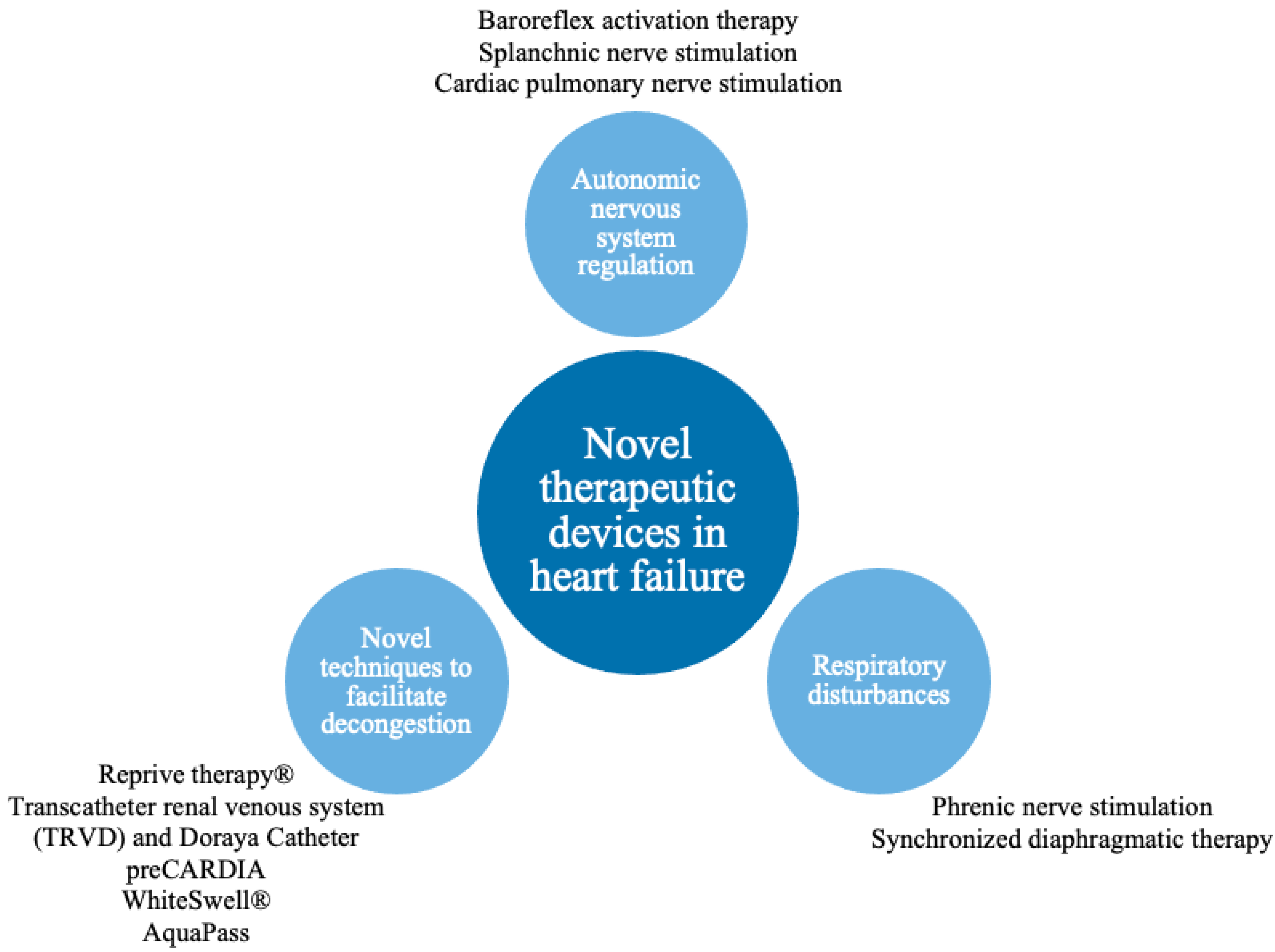
:1. Introduction
2. Targeting Autonomic Nervous System Regulation
2.1. Potential Pathophysiological Target
2.2. Baroreflex Activation Therapy
2.2.1. Existing Evidence
2.2.2. Weaknesses or Unexplained Issues
2.3. Vagus Nerve Stimulation
2.3.1. Existing Evidence
2.3.2. Weakness or Unexplained Issues
2.4. Splanchnic Nerve Modulation
2.4.1. Existing Evidence
2.4.2. Weakness or Unexplained Issues
2.5. Cardiac Pulmonary Nerve Stimulation
2.5.1. Existing Evidence
2.5.2. Weakness or Unexplained Issues
3. Respiratory Disturbances in Heart Failure
3.1. Potential Pathophysiological Target
3.2. Phrenic Nerve Stimulation
3.2.1. Existing Evidence
3.2.2. Weaknesses or Unexplained Issues
3.3. Synchronized Diaphragmatic Therapy
3.3.1. Existing Evidence
3.3.2. Weaknesses or Unexplained Issues
4. Novel Techniques to Facilitate Decongestion
4.1. Potential Pathophysiological Target
4.2. Reprieve Therapy®
4.2.1. Existing Evidence
4.2.2. Weaknesses or Unexplained Issues
4.3. Transcatheter Renal Venous Decongestion System (TRVD) and Doraya Catheter
4.3.1. Existing Evidence
4.3.2. Weaknesses or Unexplained Issues
4.4. preCARDIA
4.4.1. Existing Evidence
4.4.2. Weaknesses or Unexplained Issues
4.5. WhiteSwell®
4.5.1. Existing Evidence
4.5.2. Weaknesses or Unexplained Issues
4.6. AquaPass
Existing Evidence
5. Limitations
6. Conclusions
Author Contributions
Funding
Institutional Review Board Statement
Informed Consent Statement
Data Availability Statement
Conflicts of Interest
References
- McDonagh, T.A.; Metra, M.; Adamo, M.; Gardner, R.S.; Baumbach, A.; Böhm, M.; Burri, H.; Butler, J.; Čelutkienė, J.; Chioncel, O.; et al. 2021 ESC Guidelines for the diagnosis and treatment of acute and chronic heart failure. Eur. Heart J. 2021, 42, 3599–3726. [Google Scholar] [CrossRef] [PubMed]
- Zile, M.R.; Lindenfeld, J.; Weaver, F.A.; Zannad, F.; Galle, E.; Rogers, T.; Abraham, W.T. Baroreflex Activation Therapy in Patients With Heart Failure With Reduced Ejection Fraction. J. Am. Coll. Cardiol. 2020, 76, 1–13. [Google Scholar] [CrossRef] [PubMed]
- Dell’Oro, R.; Gronda, E.; Seravalle, G.; Costantino, G.; Alberti, L.P.; Baronio, B.; Staine, T.; Vanoli, E.; Mancia, G.; Grassi, G. Restoration of normal sympathetic neural function in heart failure following baroreflex activation therapy: Final 43-month study report. J. Hypertens. 2017, 35, 2532–2536. [Google Scholar] [CrossRef] [PubMed] [Green Version]
- Zannad, F.; De Ferrari, G.M.; Tuinenburg, A.E.; Wright, D.; Brugada, J.; Butter, C.; Klein, H.; Stolen, C.; Meyer, S.; Stein, K.M.; et al. Chronic vagal stimulation for the treatment of low ejection fraction heart failure: Results of the NEural Cardiac TherApy foR Heart Failure (NECTAR-HF) randomized controlled trial. Eur. Heart J. 2015, 36, 425–433. [Google Scholar] [CrossRef] [Green Version]
- Premchand, R.K.; Sharma, K.; Mittal, S.; Monteiro, R.; Dixit, S.; Libbus, I.; Dicarlo, L.A.; Ardell, J.L.; Rector, T.S.; Amurthur, B.; et al. Autonomic Regulation Therapy via Left or Right Cervical Vagus Nerve Stimulation in Patients With Chronic Heart Failure: Results of the ANTHEM-HF Trial. J. Card. Fail. 2014, 20, 808–816. [Google Scholar] [CrossRef] [PubMed] [Green Version]
- Fudim, M.; Ganesh, A.; Green, C.; Jones, W.S.; Blazing, M.A.; Devore, A.D.; Felker, G.M.; Kiefer, T.L.; Kong, D.F.; Boortz-Marx, R.L.; et al. Splanchnic nerve block for decompensated chronic heart failure: Splanchnic-HF. Eur. Heart J. 2018, 39, 4255–4256. [Google Scholar] [CrossRef]
- Fudim, M.; Jones, W.S.; Boortz-Marx, R.L.; Ganesh, A.; Green, C.L.; Hernandez, A.F.; Patel, M.R. Splanchnic Nerve Block for Acute Heart Failure. Circulation 2018, 138, 951–953. [Google Scholar] [CrossRef]
- Fudim, M.; Fail, P.S.; Litwin, S.E.; Shaburishvili, T.; Goyal, P.; Hummel, S.L.; Borlaug, B.A.; Mohan, R.C.; Patel, R.B.; Mitter, S.S.; et al. Endovascular ablation of the right greater splanchnic nerve in heart failure with preserved ejection fraction: Early results of the REBALANCE-HF trial roll-in cohort. Eur. J. Heart Fail. 2022. [Google Scholar] [CrossRef]
- Goedeke, S.; Emani, S.; Abraham, W.T.; Brandt, M.M.; Schaefer, J.A. Cardiac Pulmonary Nerve Stimulation (CPNSTM)): A Novel Treatment for Acute Decompensated Heart Failure. JACC Basic Transl. Sci. 2022, 7, 324–325. [Google Scholar] [CrossRef]
- Costanzo, M.R.; Ponikowski, P.; Coats, A.; Javaheri, S.; Augostini, R.; Goldberg, L.R.; Holcomb, R.; Kao, A.; Khayat, R.N.; Oldenburg, O.; et al. Phrenic nerve stimulation to treat patients with central sleep apnoea and heart failure. Eur. J. Heart Fail. 2018, 20, 1746–1754. [Google Scholar] [CrossRef] [Green Version]
- Costanzo, M.R.; Javaheri, S.; Ponikowski, P.; Oldenburg, O.; Augostini, R.; Goldberg, L.R.; Stellbrink, C.; Fox, H.; Schwartz, A.R.; Gupta, S.; et al. Transvenous Phrenic Nerve Stimulation for Treatment of Central Sleep Apnea: Five-Year Safety and Efficacy Outcomes. Nat. Sci. Sleep 2021, 13, 515–526. [Google Scholar] [CrossRef] [PubMed]
- Beeler, R.; Schoenenberger, A.W.; Bauerfeind, P.G.K.-H.; Kobza, R.; Bergner, M.; Mueller, X.; Schlaepfer, R.; Zuberbühler, M.; Erne, S.; Erne, P. Improvement of cardiac function with device-based diaphragmatic stimulation in chronic heart failure patients: The randomized, open-label, crossover Epiphrenic II Pilot Trial. Eur. J. Heart Fail. 2014, 16, 342–349. [Google Scholar] [CrossRef] [PubMed] [Green Version]
- Cleland, J.G.; Young, R.; Jorbenadze, A.; Shaburishvili, T.; Demyanchuk, V.; Buriak, R.; Todurov, B.; Rudenko, K.; Zuber, M.; Stämpfli, S.F.; et al. A First in Human Multi-center, Open Label, Prospective Study to Evaluate Safety, Usability and Performance of the VisONE System for Heart Failure with a Reduced Left Ventricular Ejection Fraction. J. Card. Fail. 2020, 26, S64. [Google Scholar] [CrossRef]
- Biegus, J.; Zymlinski, R.; Siwolowski, P.; Testani, J.; Szachniewicz, J.; Tycińska, A.; Banasiak, W.; Halpert, A.; Levin, H.; Ponikowski, P. Controlled decongestion by Reprieve therapy in acute heart failure: Results of the TARGET-1 and TARGET-2 studies. Eur. J. Heart Fail. 2019, 21, 1079–1087. [Google Scholar] [CrossRef] [PubMed]
- Zymliński, R.; Dierckx, R.; Biegus, J.; Vanderheyden, M.; Bartunek, J.; Ponikowski, P. Novel IVC Doraya Catheter Provides Congestion Relief in Patients With Acute Heart Failure. JACC Basic Transl. Sci. 2022, 7, 326–327. [Google Scholar] [CrossRef]
- Kapur, N.K.; Kiernan, M.S.; Gorgoshvili, I.; Yousefzai, R.; Vorovich, E.E.; Tedford, R.J.; Sauer, A.J.; Abraham, J.; Resor, C.D.; Kimmelstiel, C.D.; et al. Intermittent Occlusion of the Superior Vena Cava to Improve Hemodynamics in Patients With Acutely Decompensated Heart Failure: The VENUS-HF Early Feasibility Study. Circ. Heart Fail. 2022, 15, e008934. [Google Scholar] [CrossRef]
- Abraham, W.T.; Jonas, M.; Dongaonkar, R.M.; Geist, B.; Ueyama, Y.; Render, K.; Youngblood, B.; Muir, W.; Hamlin, R.; del Rio, C.L. Direct Interstitial Decongestion in an Animal Model of Acute-on-Chronic Ischemic Heart Failure. JACC Basic Transl. Sci. 2021, 6, 872–881. [Google Scholar] [CrossRef]
- Aronson, D.; Nitzan, Y.; Petcherski, S.; Bravo, E.; Habib, M.; Burkhoff, D.; Abraham, W.T. Enhancing sweat rate using a novel device for the treatment of congestion in heart failure. Eur. Heart J. 2021, 42, ehab724.1056. [Google Scholar] [CrossRef]
- Lesyuk, W.; Kriza, C.; Kolominsky-Rabas, P. Cost-of-illness studies in heart failure: A systematic review 2004–2016. BMC Cardiovasc. Disord. 2018, 18, 74. [Google Scholar] [CrossRef] [Green Version]
- McCorry, L.K. Physiology of the Autonomic Nervous System. Am. J. Pharm. Educ. 2007, 71, 78. [Google Scholar] [CrossRef] [Green Version]
- Florea, V.G.; Cohn, J.N. The Autonomic Nervous System and Heart Failure. Circ. Res. 2014, 114, 1815–1826. [Google Scholar] [CrossRef] [PubMed] [Green Version]
- Kishi, T. Heart failure as an autonomic nervous system dysfunction. J. Cardiol. 2012, 59, 117–122. [Google Scholar] [CrossRef] [PubMed] [Green Version]
- Victor, R.G. Carotid baroreflex activation therapy for resistant hypertension. Nat. Rev. Cardiol. 2015, 12, 451–463. [Google Scholar] [CrossRef] [PubMed]
- Babar, N.; Giedrimiene, D. Updates on Baroreflex Activation Therapy and Vagus Nerve Stimulation for Treatment of Heart Failure With Reduced Ejection Fraction. Cardiol. Res. 2022, 13, 11–17. [Google Scholar] [CrossRef]
- De Ferrari, G.M.; Crijns, H.J.; Borggrefe, M.; Milasinovic, G.; Smid, J.; Zabel, M.; Gavazzi, A.; Sanzo, A.; Dennert, R.; Kuschyk, J.; et al. Chronic vagus nerve stimulation: A new and promising therapeutic approach for chronic heart failure. Eur. Heart J. 2011, 32, 847–855. [Google Scholar] [CrossRef] [Green Version]
- Schwartz, P.J.; De Ferrari, G.M.; Sanzo, A.; Landolina, M.E.; Rordorf, R.; Raineri, C.; Campana, C.; Revera, M.; Ajmone-Marsan, N.; Tavazzi, L.; et al. Long term vagal stimulation in patients with advanced heart failure First experience in man. Eur. J. Heart Fail. 2008, 10, 884–891. [Google Scholar] [CrossRef]
- Gold, M.R.; Van Veldhuisen, D.J.; Hauptman, P.J.; Borggrefe, M.; Kubo, S.H.; Lieberman, R.A.; Milasinovic, G.; Berman, B.J.; Djordjevic, S.; Neelagaru, S.; et al. Vagus Nerve Stimulation for the Treatment of Heart Failure. J. Am. Coll. Cardiol. 2016, 68, 149–158. [Google Scholar] [CrossRef]
- Fudim, M.; Ponikowski, P.P.; Burkhoff, D.; Dunlap, M.E.; Sobotka, P.A.; Molinger, J.; Patel, M.R.; Felker, G.M.; Hernandez, A.F.; Litwin, S.E.; et al. Splanchnic nerve modulation in heart failure: Mechanistic overview, initial clinical experience, and safety considerations. Eur. J. Heart Fail. 2021, 23, 1076–1084. [Google Scholar] [CrossRef]
- Gajewski, P.; Fudim, M.; Kittipibul, V.; Engelman, Z.J.; Biegus, J.; Zymliński, R.; Ponikowski, P. Early Hemodynamic Changes following Surgical Ablation of the Right Greater Splanchnic Nerve for the Treatment of Heart Failure with Preserved Ejection Fraction. J. Clin. Med. 2022, 11, 1063. [Google Scholar] [CrossRef]
- Fudim, M.; Abraham, W.T.; von Bardeleben, R.S.; Lindenfeld, J.; Ponikowski, P.P.; Salah, H.M.; Khan, M.S.; Sievert, H.; Stone, G.W.; Anker, S.D.; et al. Device Therapy in Chronic Heart Failure: JACC State-of-the-Art Review. J. Am. Coll. Cardiol. 2021, 78, 931–956. [Google Scholar] [CrossRef]
- Chen, Y.-L.; Su, M.-C.; Liu, W.-H.; Wang, C.-C.; Lin, M.-C.; Chen, M.-C. Influence and Predicting Variables of Obstructive Sleep Apnea on Cardiac Function and Remodeling in Patients without Congestive Heart Failure. J. Clin. Sleep Med. 2014, 10, 57–64. [Google Scholar] [CrossRef] [PubMed] [Green Version]
- Wang, Y.; Schöbel, C.; Penzel, T. Management of Obstructive Sleep Apnea in Patients With Heart Failure. Front. Med. 2022, 9, 803388. [Google Scholar] [CrossRef] [PubMed]
- Costanzo, M.R.; Khayat, R.; Ponikowski, P.; Augostini, R.; Stellbrink, C.; Mianulli, M.; Abraham, W.T. Mechanisms and Clinical Consequences of Untreated Central Sleep Apnea in Heart Failure. J. Am. Coll. Cardiol. 2015, 65, 72–84. [Google Scholar] [CrossRef] [PubMed] [Green Version]
- Sanders, M.H. Article reviewed: A mechanism of central sleep apnea in patients with heart failure. Sleep Med. 2000, 1, 63–64. [Google Scholar] [CrossRef]
- Javaheri, S. A Mechanism of Central Sleep Apnea in Patients with Heart Failure. N. Engl. J. Med. 1999, 341, 949–954. [Google Scholar] [CrossRef]
- Carlisle, T.; Ward, N.R.; Atalla, A.; Cowie, M.; Simonds, A.K.; Morrell, M.J. Investigation of the link between fluid shift and airway collapsibility as a mechanism for obstructive sleep apnea in congestive heart failure. Physiol. Rep. 2017, 5, e12956. [Google Scholar] [CrossRef]
- Gottlieb, D.J.; Punjabi, N.M. Diagnosis and Management of Obstructive Sleep Apnea: A review. JAMA 2020, 323, 1389–1400. [Google Scholar] [CrossRef]
- Voigt, J.; Emani, S.; Gupta, S.; Germany, R.; Khayat, R. Meta-Analysis Comparing Outcomes of Therapies for Patients With Central Sleep Apnea and Heart Failure With Reduced Ejection Fraction. Am. J. Cardiol. 2020, 127, 73–83. [Google Scholar] [CrossRef]
- Yu, J.; Zhou, Z.; McEvoy, D.; Anderson, C.; Rodgers, A.; Perkovic, V.; Neal, B. Association of Positive Airway Pressure With Cardiovascular Events and Death in Adults With Sleep Apnea: A Systematic Review and Meta-analysis. JAMA J. Am. Med. Assoc. 2017, 318, 156–166. [Google Scholar] [CrossRef]
- Fudim, M.; Mirro, M.; Goldberg, L.R. Synchronized Diaphragmatic Stimulation for the Treatment of Symptomatic Heart Failure: A Novel Implantable Therapy Concept. JACC Basic Transl. Sci. 2022, 7, 322–323. [Google Scholar] [CrossRef]
- Costanzo, M.R.; Ponikowski, P.; Javaheri, S.; Augostini, R.; Goldberg, L.; Holcomb, R.; Kao, A.; Khayat, R.N.; Oldenburg, O.; Stellbrink, C.; et al. Transvenous neurostimulation for central sleep apnoea: A randomised controlled trial. Lancet 2016, 388, 974–982. [Google Scholar] [CrossRef]
- Ponikowski, P.; Javaheri, S.; Michalkiewicz, D.; Bart, B.A.; Czarnecka, D.; Jastrzebski, M.; Kusiak, A.; Augostini, R.; Jagielski, D.; Witkowski, T.; et al. Transvenous phrenic nerve stimulation for the treatment of central sleep apnoea in heart failure. Eur. Heart J. 2012, 33, 889–894. [Google Scholar] [CrossRef] [PubMed] [Green Version]
- Gheorghiade, M.; Filippatos, G. Reassessing treatment of acute heart failure syndromes: The ADHERE Registry. Eur. Heart J. Suppl. 2005, 7, B13–B19. [Google Scholar] [CrossRef] [Green Version]
- Costanzo, M.R. Novel Devices for the Cardiorenal Syndrome in Heart Failure. Curr. Treat. Options Cardiovasc. Med. 2020, 22, 23. [Google Scholar] [CrossRef]
- Urban, S.; Blaziak, M.; Biegus, J.; Zymlinski, R. Ultrafiltration in acute heart failure: Current knowledge and fields for further research. Adv. Clin. Exp. Med. 2021, 30, 737–746. [Google Scholar] [CrossRef]
- Mullens, W.; Abrahams, Z.; Francis, G.S.; Sokos, G.; Taylor, D.O.; Starling, R.C.; Young, J.B.; Tang, W.W. Importance of Venous Congestion for Worsening of Renal Function in Advanced Decompensated Heart Failure. J. Am. Coll. Cardiol. 2009, 53, 589–596. [Google Scholar] [CrossRef] [Green Version]
- Boorsma, E.M.; ter Maaten, J.M.; Voors, A.A.; van Veldhuisen, D.J. Renal Compression in Heart Failure: The Renal Tam-ponade Hypothesis. JACC Heart Fail. 2022, 10, 175–183. [Google Scholar] [CrossRef]
- Clinical Evaluation of the TRVDTM System in ADHF—Full Text View—ClinicalTrials.gov, (n.d.). Available online: https://clinicaltrials.gov/ct2/show/study/NCT03621436 (accessed on 20 May 2022).
- Kapur, N.K.; Karas, R.H.; Newman, S.; Jorde, L.; Chabrashvili, T.; Annamalai, S.; Esposito, M.; Kimmelstiel, C.D.; Lenihan, T.; Burkhoff, D. First-in-human experience with occlusion of the superior vena cava to reduce cardiac filling pressures in congestive heart failure. Catheter. Cardiovasc. Interv. 2019, 93, 1205–1210. [Google Scholar] [CrossRef]
- Fudim, M.; Salah, H.M.; Sathananthan, J.; Bernier, M.; Pabon-Ramos, W.; Schwartz, R.S.; Rodés-Cabau, J.; Côté, F.; Khalifa, A.; Virani, S.A.; et al. Lymphatic Dysregulation in Patients With Heart Failure: JACC Review Topic of the Week. J. Am. Coll. Cardiol. 2021, 78, 66–76. [Google Scholar] [CrossRef]


| Method | Pathophysiological Mechanism | Solution | Trial Design and Size | Primary Outcomes | Evidence | Adverse Events |
|---|---|---|---|---|---|---|
| Baroreflex activation therapy | Overactivity of SNS (increased heart rate, arterial pressure, RAAS activity and negative cardiac remodeling). | Stimulation of carotid bodies to restore autonomic system balance. | Multicenter, prospective, controlled trial n = 408 | Rate of cardiovascular and HF morbidity, MANCE, Change in: NT-proBNP, 6 MHW, MLWHF QOL | BeAT-HF showed improvements of quality of life, exercise capacity, functional status and decrease of NT-proBNP [2] | MANCE event-free rate: 97%. A system or procedure-related serious adverse event occurred in seven patients. |
| Single-center, open-label n = 11 | Not reported | Dell’Oro et al. demonstrated significant improvement of EF and reduction in hospitalization [3] | No adverse effects were reported. | |||
| Vagus nerve stimulation | Overactivity of SNS (increased heart rate, arterial pressure, RAAS activity and negative cardiac remodeling). | Increase of PNS activity. | Multicenter, prospective, randomized, controlled trial n = 95 | Change in LVESD, Percentage of surviving patients. | NECTAR-HF presented significant improvement in quality of life, NYHA class and functional status [4] | There were no significant differences in serious adverse events between control and therapy groups. The overall rate of implantation-related infections was 7.4% |
| Multicenter, open-label, uncontrolled trial n = 60 | Change in: LVESV EF, Adverse events. | ANTHEM-HF showed positive, durable improvement of cardiac function [5] | Serious adverse events occurred in 16 patients. There was one death related to system implantation due to an embolic stroke that occurred 3 days after surgery. | |||
| Splanchnic nerve stimulation | Excessive cardiac filling pressure due to overactivity of SNS resulting in visceral vasoconstriction and rapid volume shift from visceral to central compartment during exercise. | GSN modulation preventing exercise provoked visceral vasoconstriction and subsequent fluid shift from the visceral compartment to the central venous system. | Single-center, prospective, open-label, uncontrolled trials n = 11, n = 15 | Change in CVPPAMP PCWP | Splanchnic-HF 1, and Splanchnic-HF 2 showed a reduction in PCPW and improvement of the cardiac index during exercise [6,7] | No adverse events were reported. |
| Multicenter, prospective, uncontrolled, pilot study | Change in: mean PCPW at rest and exercise (20 W). Adverse events. | REBALANCE-HF confirmed the reduction in exercise PCPW in HFpEF and NYHA class improvement [8] | There were three non-serious device-related adverse events reported in this study: HF decompensation due to periprocedural fluid overload, transient hypertension and back pain following ablation. | |||
| Cardiopulmonary nerve stimulation | Impaired LV contractility and relaxation. | Stimulation of the autonomic system area responsible for LV contractility resulting in positive lusitropic and inotropic effects. | Single-center, first-in-human, proof-of-concept study n = 15 | Adverse events. | A proof-of-concept study showed improvement of LV contractility and an increase in mean arterial pressure without affecting the heart rate [9] | No device-related serious adverse events were reported. |
| Phrenic nerve stimulation | Central apnea due to periodic drop in CO2 partial pressure to below the threshold for triggering the action potential in the respiratory center caused by greater sensitivity to carbon dioxide leading to potent stimulus of rhythmic breathing. | Transvenous stimulation of phrenic nerve during apneas. | Multicenter, randomized, open-label study n = 151 | Reduction in AHI and freedom from serious adverse events | The remedē System Pivotal Trial showed significant reduction in AHI, arousal index, desaturation and apnea episodes. It also revealed improvement in quality of life, sleep structure and EF [10,11] | Cumulatively, 21 (14%) serious adverse events were observed in 5-year follow-ups (15; (10%) in the first 12 months). It predominantly included electrode dysfunction, electrode dislocation and infection of the implantation site [10] |
| Asymptomatic diaphragmatic stimulation | High left ventricle pre-load and after-load pressures increase remodeling and HF progression. | Stimulation of diaphragm muscle fibers synchronized with cardiac cycle to decrease intrathoracic pressures. | Single-center, randomized, open-label study n = 33 | LVEF improvement | EPIPHRENIC II Study showed significant improvement of LVEF, maximal power on effort, reduction in NYHA class, without differences in 6-min walking test or BNP concentration [12,13] | Three patients were excluded due to dysfunctional diaphragmatic electrode. No adverse events were observed [12] |
| Multicenter, non-randomized, open-label study n = 15 | Freedom from serious adverse events during procedural recovery or acute therapy | VisONE study showed improvement in LVEF and life quality (evaluated in SF-36); extended walking distance during the 6 MWT was observed at a 1-year follow-up. [13] | No adverse events were observed during procedural recovery, acute therapy (primary outcome) and in 12month follow-up (secondary outcome) [13] | |||
| Reprieve system | Problems with controlling decongestive therapy to avoid too rapid diuretic response and hypovolemia and, on the other hand, providing too much fluid, which worsens volume overload. | Sustaining the accurate fluid balance by measuring the urine output and providing the exact amount of replacement solution to achieve preset fluid balance. | Non-randomized, single-center, prospective, open-label, studies, both n = 19 | Device and procedure-related adverse events and decongestive efficacy | Higher urine output and decrease in CVP in comparison to the baseline. Actual fluid loss did not exceed target fluid loss at the end of therapy in every patient [14] | No serious adverse events were observed. One case of hypokalemia occurred. |
| Transcatheter renal venous decongestion system | Congestion in renal veins. | Transfemoral inserted flow pump, which reduces renal vein pressure to the desired level. | No results have been published so far. | Device and procedure-related adverse events, technical and procedural feasibility | The trial to evaluate TRVD was terminated prematurely, no results have been published so far. | No results have been published so far. |
| Doraya Catheter | Congestion in renal veins. | Partial obstruction of the flow in the inferior vena cava below the level of the renal veins reduces renal vein pressure | First in-human, single-arm, open-label study n = 9 | Serious adverse events. | The catheter was successfully deployed in all patients. Clinical symptoms, as well as diuresis and natriuresis, improved [15] | No device-related or embolic events were reported. One serious procedure-related adverse event: bleeding hematoma from the injection site, resolved without sequelae. |
| preCARDIA | Increased right ventricle preload. | Obstruction of the superior vena cava leading to an intermittent decrease in preload. | Multicenter, prospective, single-arm exploratory safety and feasibility, open-label, trial n = 30 | Freedom from device or procedure-related serious adverse events | Successful decrease in right atrial pressure and PCWP, increase in net fluid balance and urine output [16] | No device or procedure-related serious adverse events were observed. |
| WhiteSwell | Increased preload causes lymphatic congestion, which impairs interstitial drainage and exacerbates oedema. | Reduction in the pressure in the area of lymphatic duct outflow into venous vessels. | The animal model study, n = 7 sheep, used in 1 human, n = 1 | Serious adverse events. | Examined in a ovine model. Trend toward improved oxygenation an diuresis was noticed [17] | No adverse events were reported in in-human application. |
| AquaPass | Insufficient urine volume removal. | Enhancing the sweat rate to remove fluid directly from interstitial space. | Feasibility and short-term performance, single-arm, open-label study, n = 16 | Serious adverse events, treatment tolerance, ability to control skin temperature between 33 and 38 Celsius degrees). | The procedure was safe in HF patients, successful weight loss was observed. Increased skin temperature without elevating core temperature above average was achieved in each patients [18] | No adverse event occurred. |
Publisher’s Note: MDPI stays neutral with regard to jurisdictional claims in published maps and institutional affiliations. |
© 2022 by the authors. Licensee MDPI, Basel, Switzerland. This article is an open access article distributed under the terms and conditions of the Creative Commons Attribution (CC BY) license (https://creativecommons.org/licenses/by/4.0/).
Share and Cite
Guzik, M.; Urban, S.; Iwanek, G.; Biegus, J.; Ponikowski, P.; Zymliński, R. Novel Therapeutic Devices in Heart Failure. J. Clin. Med. 2022, 11, 4303. https://doi.org/10.3390/jcm11154303
Guzik M, Urban S, Iwanek G, Biegus J, Ponikowski P, Zymliński R. Novel Therapeutic Devices in Heart Failure. Journal of Clinical Medicine. 2022; 11(15):4303. https://doi.org/10.3390/jcm11154303
Chicago/Turabian StyleGuzik, Mateusz, Szymon Urban, Gracjan Iwanek, Jan Biegus, Piotr Ponikowski, and Robert Zymliński. 2022. "Novel Therapeutic Devices in Heart Failure" Journal of Clinical Medicine 11, no. 15: 4303. https://doi.org/10.3390/jcm11154303