Review

In Vitro Transcribed RNA-Based Platform Vaccines: Past, Present, and Future

by
Alexey D. Perenkov
1,*,
Alena D. Sergeeva
1,
Maria V. Vedunova
1 and
Dmitri V. Krysko
1,2,3
1
Institute of Biology and Biomedicine, National Research Lobachevsky State University of Nizhny Novgorod, 603022 Nizhny Novgorod, Russia
2
Cell Death Investigation and Therapy (CDIT) Laboratory, Anatomy and Embryology Unit, Department of Human Structure and Repair, Faculty of Medicine and Health Science, Ghent University, 9000 Ghent, Belgium
3
Cancer Research Institute Ghent, 9000 Ghent, Belgium
*
Author to whom correspondence should be addressed.
Vaccines 2023, 11(10), 1600; https://doi.org/10.3390/vaccines11101600
Submission received: 17 September 2023 / Revised: 7 October 2023 / Accepted: 10 October 2023 / Published: 16 October 2023
(This article belongs to the Special Issue mRNA-Based Vaccine Development)

Abstract

:
mRNA was discovered in 1961, but it was not used as a vaccine until after three decades. Recently, the development of mRNA vaccine technology gained great impetus from the pursuit of vaccines against COVID-19. To improve the properties of RNA vaccines, and primarily their circulation time, self-amplifying mRNA and trans-amplifying mRNA were developed. A separate branch of mRNA technology is circular RNA vaccines, which were developed with the discovery of the possibility of translation on their protein matrix. Circular RNA has several advantages over mRNA vaccines and is considered a fairly promising platform, as is trans-amplifying mRNA. This review presents an overview of the mRNA platform and a critical discussion of the more modern self-amplifying mRNA, trans-amplifying mRNA, and circular RNA platforms created on its basis. Finally, the main features, advantages, and disadvantages of each of the presented mRNA platforms are discussed. This discussion will facilitate the decision-making process in selecting the most appropriate platform for creating RNA vaccines against cancer or viral diseases.

1. Introduction

Vaccines in current use can be broadly categorized into two groups: traditional vaccines, such as live, attenuated and inactivated vaccines, and more modern vaccines, including peptide, DNA, and mRNA vaccines. Each group possesses its own set of advantages and disadvantages.
Traditional vaccines provide long-term protection against infection, but they require a longer development process and face challenges in scaling up production. In contrast, modern vaccine approaches do not encounter such drawbacks, and among them, mRNA vaccines exhibit several advantages. One primary advantage is the safety of mRNA vaccines. Unlike DNA vaccines, mRNA does not integrate into the vaccinated individual’s genome. Another major advantage is that, unlike attenuated vaccines, mRNA does not pose a risk of infection. On top of that, mRNA vaccines are relatively inexpensive to produce and their production is faster than that of attenuated or inactivated vaccines [1,2]. Moreover, the expression of the protein encoded by mRNA vaccines is relatively short-lived due to natural cellular degradation processes, rendering it easily controlled. Furthermore, unlike DNA vaccines, mRNA vaccines are translated in the cytoplasm without the need to overcome the nuclear barrier [2,3]. Moreover, the problem of the low immunogenicity of peptide vaccines is not an issue in mRNA vaccines [3,4,5]. On top of that, mRNA vaccines can be produced more economically and rapidly than attenuated and inactivated vaccines [1,2]. Finally, the inherent versatility of the key elements in mRNA vaccine composition makes it easy to modify the encoded protein sequence with minimal resources and time investment, thus enabling the development of vaccines with new properties [6].
The main challenges associated with mRNA vaccines are related to the immunogenicity of the RNA molecule itself, which can limit the amount of protein translated in the body. If the immune response is initiated before the mRNA is translated, the vaccine is rendered ineffective. This has been addressed by reducing the administered dose and replacing classical nucleotides with modified ones. The intracellular stability of mRNA vaccines has been augmented by incorporating modified caps, nucleotides, and poly(A) tails, along with optimal regulatory elements. Additionally, the delivery of mRNA vaccines has been enhanced by introducing ionizable lipids [3,7,8,9]. As a result, mRNA vaccines are a promising and innovative direction in the development of preventive and therapeutic vaccines.
In addition to traditional mRNA vaccines, several novel modifications have emerged, including self-amplifying (SAM) mRNA, trans-amplifying mRNA (taRNA), and circular RNA (circRNA). Each of these mRNA-based vaccine varieties comes with its own set of advantages and disadvantages, which adds complexity to the selection of appropriate technology for specific applications. There are already many mRNA-based vaccines against various diseases, including a vaccine against SARS-CoV-2 [3]. Therefore, this review aims to evaluate the key distinguishing features of each vaccine variant (i.e., mRNA, SAM, taRNA, circRNA) to facilitate selection of the most appropriate platform for creating an RNA vaccine against tumors or viral diseases.

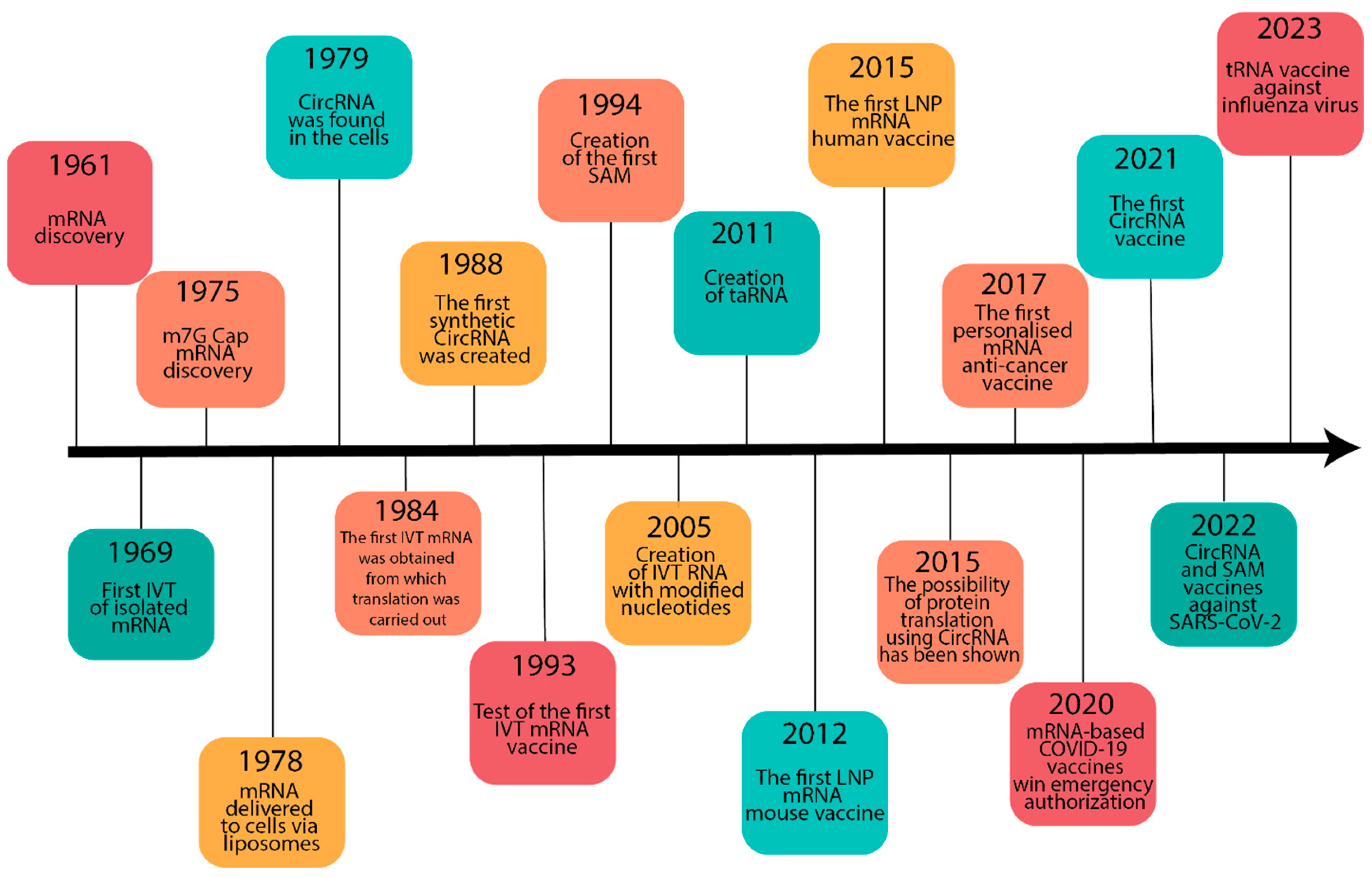
2. Historical Background

mRNA, as a ribonucleotide template for protein synthesis transcribed from DNA, was initially discovered in 1961 (Figure 1). However, it took several decades of scientific discoveries and experimental investigations before it was used as a vaccine [10].
In 1965, A.D. Bangham introduced lipid bilayer particles, later known as liposomes, which played a crucial role in the delivery of mRNA vaccines [11]. Liposomes were first employed for drug delivery in 1971, and only three years later, they were already being used for delivery of diphtheria toxoid in murine models. Concurrently, in 1969, the first laboratory-produced protein was obtained using isolated mRNA. These advances collectively enabled the delivery of mRNA to mouse cells and the subsequent production of the encoded protein using liposomes as early as 1978.
CircRNA, discovered in 1976, gained popularity as a vaccine several decades later [12]. Eight years after its discovery, a team led by Douglas Melton, under the leadership of Paul Krieg, successfully produced synthetic mRNA using RNA synthetase. In 1988, the first small-molecule synthetic circRNA was obtained by a chemical procedure [13].
By 1993, the considerable expertise and knowledge that had accumulated enabled the development of the first mRNA flu vaccine, which was tested in mice. Two years later, a cancer vaccine was also produced using mRNA technology [14]. However, it was not until 2015 that mRNA-based influenza vaccines progressed to clinical trials. This slow progress can be attributed to several limitations of mRNA, such as its instability, short half-life within cells, and relatively high production costs. Consequently, considerable investments were directed toward alternative vaccine technologies considered more promising at that time, which led to the advancement of DNA and protein vaccines [6]. Nonetheless, in 1994, the first SAM mRNA was successfully developed, although its widespread adoption occurred much later [15].
It is notable that mRNA vaccines held greater promise for the treatment of oncological conditions compared to viral diseases. Towards the end of the 20th century, Eli Gilboa and colleagues initiated human testing of mRNA cancer vaccines. However, the vaccine candidate for late-stage cancer did not yield the expected outcomes and was not approved, thereby impeding the progress of mRNA vaccine technology. Nonetheless, E. Gilboa’s research served as a catalyst for CureVac and BioNTech to embark on dedicated development of mRNA vaccines in the early 2000s. Studies demonstrated that direct administration of mRNA vaccines triggers a robust immune response against the mRNA, which presented a serious hurdle for their utilization. It was not until 2005 that Kariko and Weissman successfully addressed this challenge by substituting uridine in the vaccine mRNA with pseudouridine, substantially reducing the immunogenicity of the mRNA [14]. In 2011, a significant advancement in mRNA technology known as SAM was achieved with the creation of the first trans-amplifying mRNA (taRNA) [16].
The delivery of RNA vaccines posed another challenge in their development. Initially, cationic lipids with a positively charged head group were utilized; the head group interacts with the negatively charged RNA [17]. However, the sustained positive charge of these lipids was toxic within the body, preventing their use in vaccines [18]. In 2012, Pieter Cullis and his team started experimenting with various lipid compositions of liposomes for RNA delivery. The funding provided by the US Defense Advanced Research Projects Agency for the study and development of RNA vaccines in the same year added impetus to the progress of mRNA vaccine technology [14]. Ultimately, Cullis and his team successfully developed an optimal lipid formulation that incorporated ionizable lipids capable of transitioning from a positive to a neutral charge under physiological conditions, reducing the toxicity of liposomes significantly, while extending the storage duration of mRNA vaccines within this lipid-based delivery system [18].
These important discoveries culminated in the groundbreaking work of Ugur Sahin in 2017, who developed a personalized mRNA vaccine against melanoma. Sahin’s research yielded promising results in therapy and ignited considerable interest in the advancement of mRNA-based cancer vaccines [19]. While mRNA-based therapeutic vaccines for cancer demonstrated notable progress, prophylactic vaccines against viral infections using mRNA technology lagged behind. Notably, Moderna, one of the leading mRNA vaccine companies, had developed nine mRNA vaccines for various human infectious diseases by 2020, but none achieved significant success. Other companies involved in the development of mRNA-based vaccines had similar experiences. However, the emergence of the SARS-CoV-2 virus in 2019 dramatically altered the landscape, motivating large companies to leverage their accumulated experience and swiftly create mRNA-based vaccines in response to the global pandemic [14,20]. As early as one year after the onset of the pandemic, promising mRNA prototypes of vaccines against SARS-CoV-2 virus were generated [3]. As a result, mRNA vaccines are currently undergoing rapid development and closing the technological gap with DNA vaccines, which were developed much earlier.

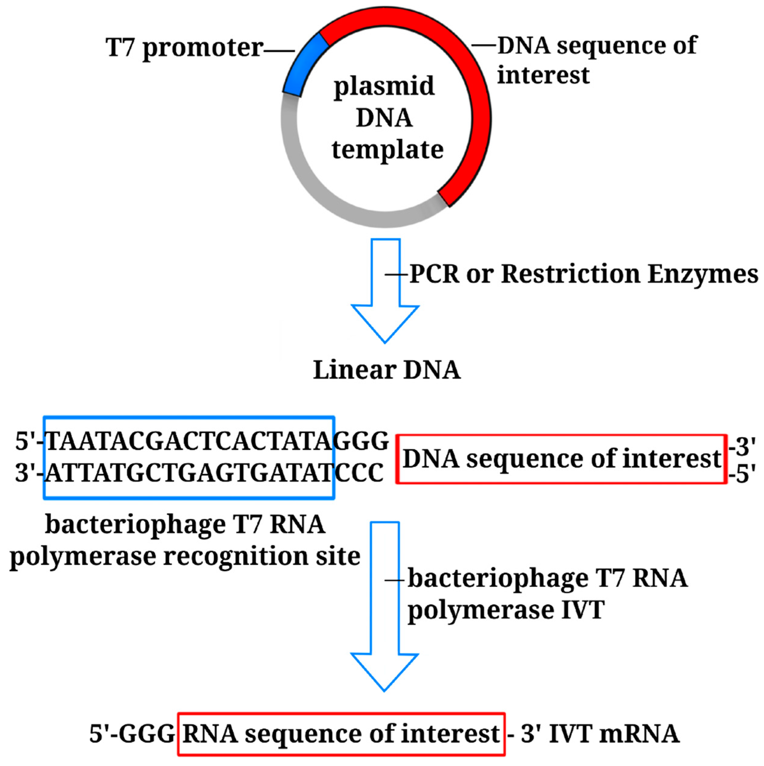
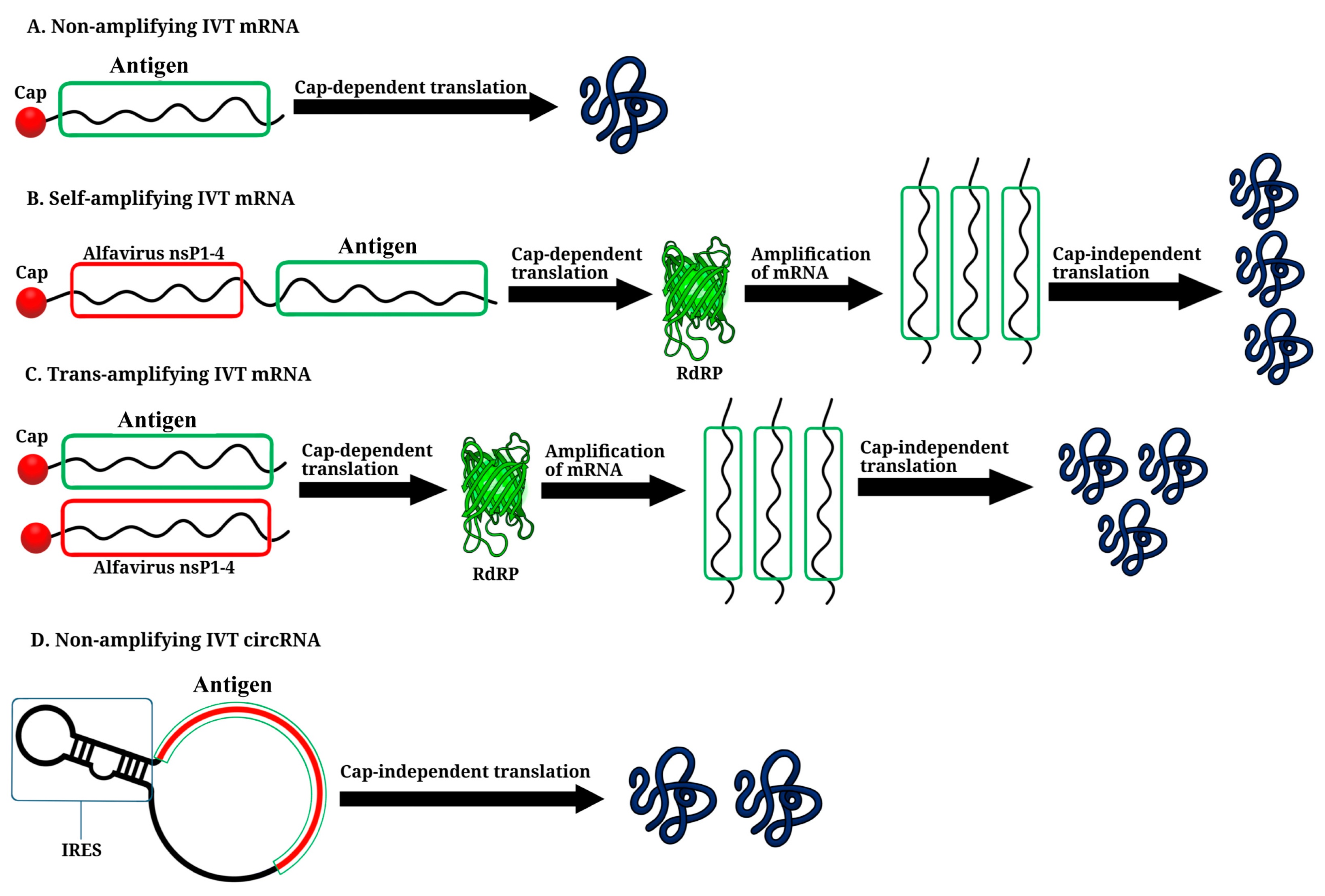
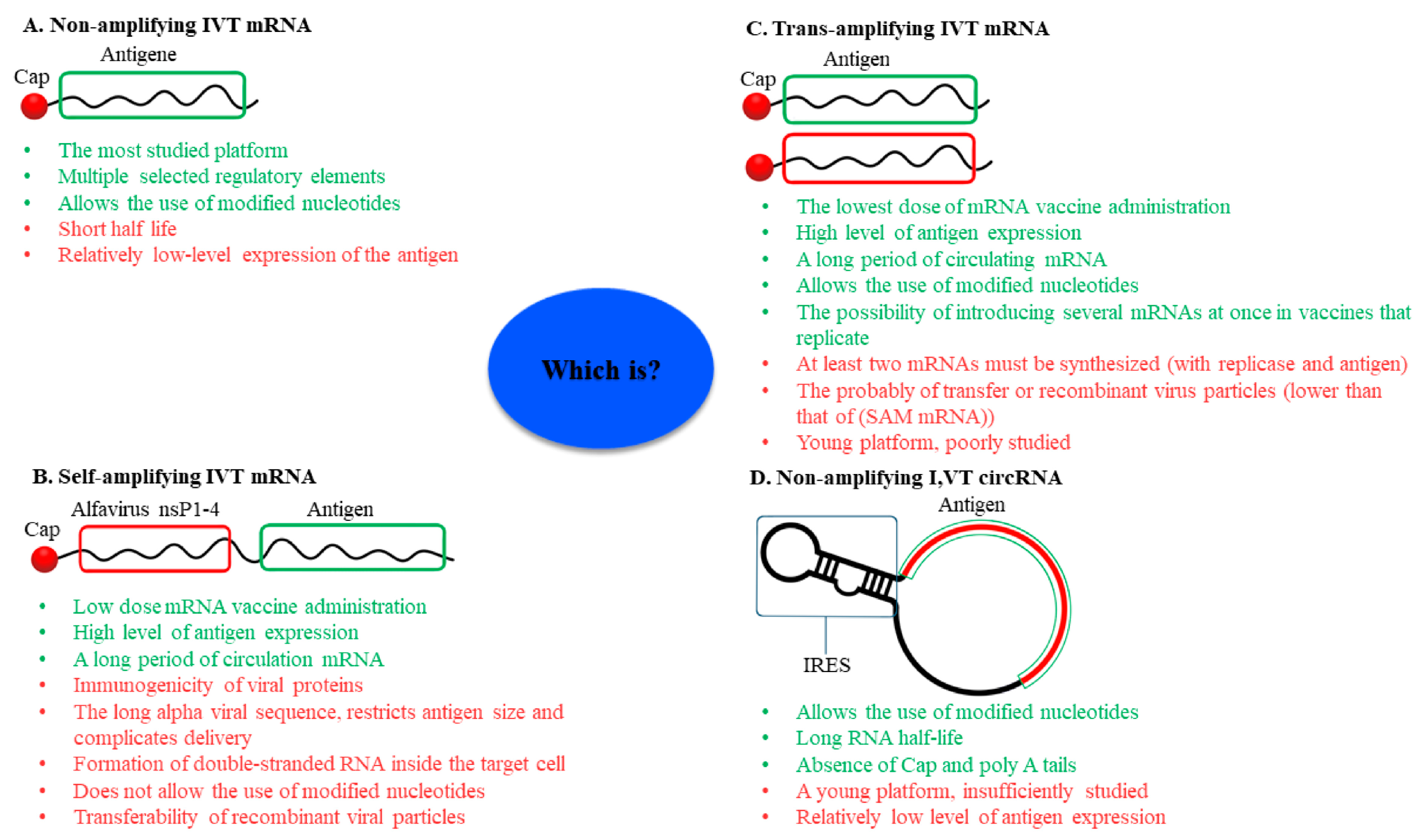
3. Non-Amplifying In Vitro-Transcribed mRNA Platform

Before mRNA synthesis in vitro, the DNA template needs to be linearized. This is typically done with restriction enzymes or by performing PCR to obtain the desired DNA fragment in a linear form suitable for transcription. In vitro-transcribed mRNA (IVT mRNA) is synthesized with bacteriophage T3, T7, or SP6 RNA polymerase [21]. A specific sequence recognized by the corresponding polymerase is incorporated at the 5’ end of the DNA template. For instance, if bacteriophage T7 is to be used, the sequence 5’-TAATACGACTCACTATA-3’ is commonly employed [22] (Figure 2).
A mRNA molecule comprises five essential elements: 5′ cap, 5′-untranslated region (5′-UTR), open reading frame (ORF), 3′-UTR, and 3′ poly(A) tail (Figure 3). Each of these elements plays a crucial role in its function as a template for protein synthesis [21,23].

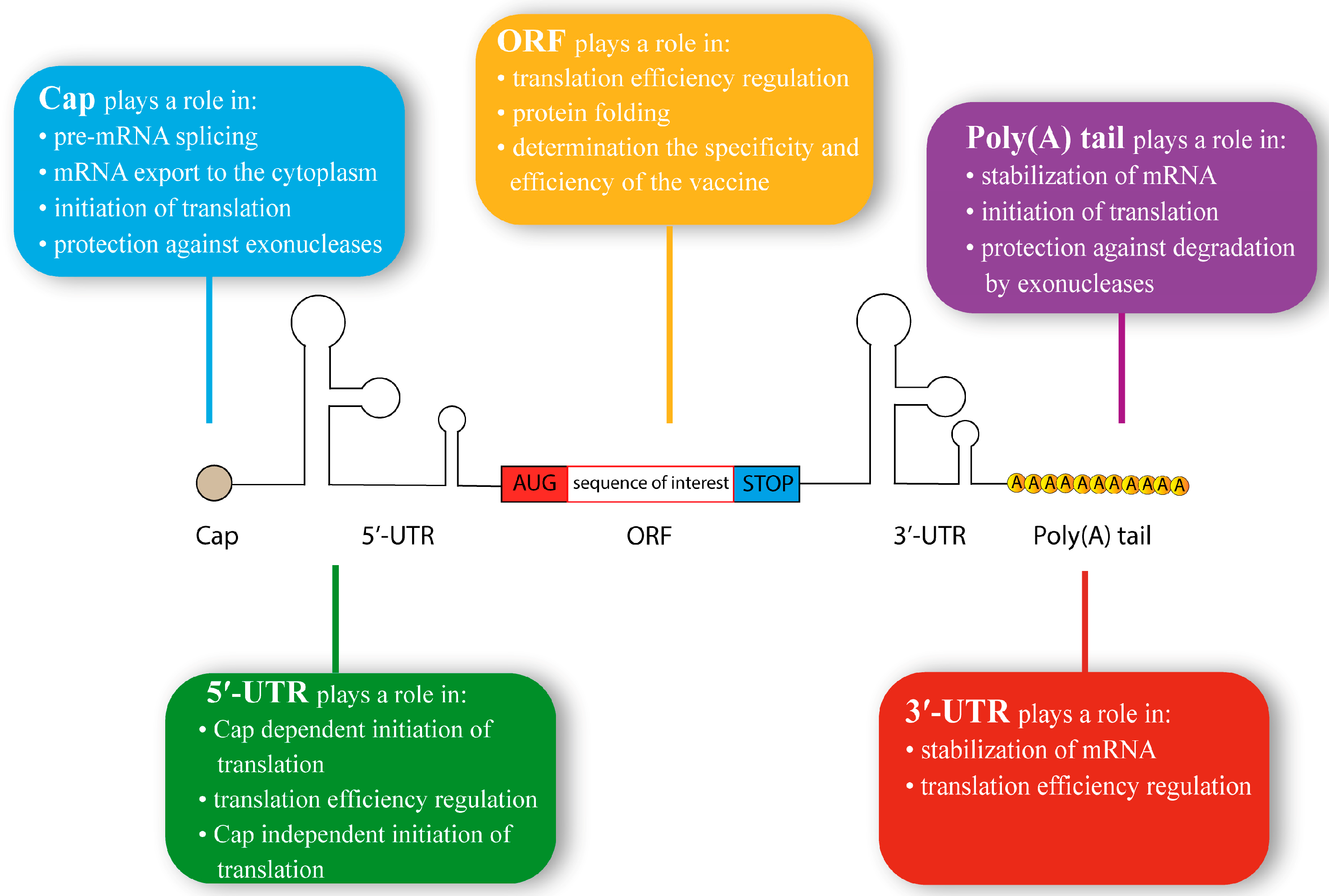
3.1. 5′ Cap Structure

The 5′ cap is a methylated guanoside structure that serves multiple important functions in mRNA processing and stability (Figure 3). It facilitates pre-mRNA splicing, export of mRNA from the nucleus to the cytoplasm, translation initiation, and protection against exonucleases. In eukaryotes, the simplest form of the cap structure is 7-methylguanosine (m7G), which is linked to the mRNA molecule by a 5’-5’-triphosphate bridge. This minimal structure at the 5’ end of mRNA is referred to as Cap 0 (m7GpppNp, where p represents a phosphate group and N denotes any mRNA nucleotide). Cap 0 formation occurs during mRNA transcription in the nucleus [24]. Cap 0 is recognized by the eukaryotic translation initiation factor 4E (eIF4E) and is commonly found in plants (including plant viruses) and fungi. In animals, however, Cap 0 is relatively rare and is found only in certain organisms, such as Drosophila melanogaster. Consequently, mammalian cells may perceive Cap 0 mRNA as foreign and trigger an inflammatory response, which might impede the translation of the vaccine RNA [25]. Cap 0 undergoes modification by Cap 2’-O-methyltransferase 1 (CMTR1) in the cell nucleus, resulting in the formation of m7GpppNm2’-Op, known as Cap 1. Cap 2 (m7GpppNm2’-OpNm2’-Op) differs from Cap 0 by the additional CMTR2-catalyzed methylation of the first and second mRNA nucleotides at the 2’-O-position of the ribose [26]. In animals, Cap 1 and Cap 2 are typical, and the ratio between them in a cell depends on the species. In mammals, certain cells may also contain N6-methyladenosine (m6A), and the first transcribed nucleotide can be m6Am instead of m7G. The level of protein production using mRNAs containing different types of caps largely depends on the specific cellular context into which the RNA is introduced. Studies have demonstrated that mRNA with Cap 1 generally yields higher protein levels compared to Cap 0, although no distinct advantages have been established in comparison to Cap 2. Therefore, the choice of cap modification depends on the particular cell line or cellular environment in which the mRNA is to be delivered [27,28]. Another important function of the cap is to protect mRNA from degradation by 5’-end RNases, such as the XRN1 family [29].
In the past, mRNA was capped by sequential enzymatic reactions catalyzed by RNA 5’-triphosphatase (RTPase), guanylyltransferase (GTase), and N7-methyltransferase (N7MTase). This set of enzymes results in the formation of IVT (in vitro-translated) mRNA containing Cap 0 [26]. However, more recently, Vaccinia Capping Enzyme (VCE) has become the preferred method because it combines the activities of all three enzymes (RTPase, GTase, N7MTase), resulting in the direct formation of a Cap 0 in a single step (Table 1). To produce Cap 1 mRNA, an additional enzymatic reaction with mRNA cap 2’-O-methyltransferase (MTase) is required. The efficiency of this capping process can reach 100% [30]. Consequently, the creation of Cap 1 mRNA entails the synthesis of the mRNA itself, as well as two supplementary enzymatic reactions, thereby prolonging the procedure and increasing its cost. To address this issue, synthetic analogs of the cap have been developed to enable co-transcriptional capping. The first variant of a cap analog was m7GpppG, which could successfully incorporate into the 5’ end of IVT mRNA [31]. However, a significant drawback of the cap m7GpppG analog is its ability to attach in the reverse orientation, leading to the formation of non-functional RNAs [32]. One of the early successful alternatives was the Anti-Reverse Cap Analog (ARCA), which is a 3’-O-Me-m7G(5’)ppp(5’)G and serves as an analogue of Cap 0 with the correct orientation, since the 3’-OH group is methylated and does not allow attachment to the 3’ end of the mRNA [33]. Studies have indicated that the protein production level in mRNA containing the conventional Cap 0 was slightly higher than that of mRNA containing ARCA. To obtain Cap 1, an additional reaction is performed by adding 2’-O-methyltransferase to ARCA mRNA. Subsequently, an analogue of the cap (CleanCap®) was introduced by TriLink BioTechnologies (San Diego, CA, USA). It incorporates co-transcriptionally and represents Cap 1 [34]. Numerous analogs of CleanCap® have been developed, such as CleanCap® Reagent AG 3’OMe (m7G3’OMepppA2’OMepG), CleanCap® Reagent AG (m7GpppA2’OMepG), and CleanCap® Reagent AU (m7GpppA2’OMepU) [35]. Studies have demonstrated that the use of CleanCap® cap analog and its modifications leads to a significant increase in the proportion of mRNAs containing cap structures (up to 99%) compared to ARCA (approximately 70%) [36]. The manufacturer of CleanCap® claims that these mRNAs are less immunogenic and produce more protein compared to Cap 0 [37]. However, recent investigations have indicated that the increased protein production observed in mammalian cells with high doses of the CleanCap® AG cap analog is accompanied by the heightened production of pro-inflammatory cytokines when compared to ARCA and also involves higher costs [38]. Ongoing research is currently focused on exploring and evaluating new analogs of the cap, with promising results [29].

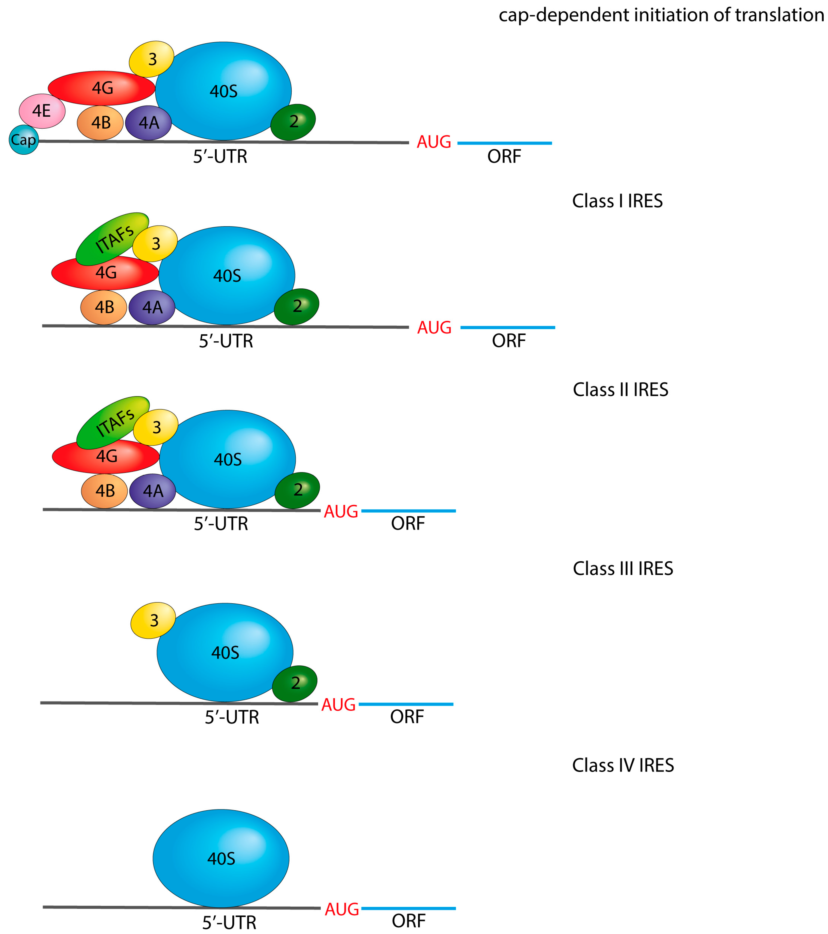
3.2. 5′-Untranslated Region Structure

The 5′-UTR of mRNA plays a crucial role in regulating protein translation (Figure 3). The significance of the 5’-UTR is highlighted by the rates of ribosome association with mRNA, which can differ by orders of magnitude, depending on the sequence [39]. In eukaryotes, the average length of the 5’-UTR ranges from 53 to 218 nucleotides, with a median of 210 nucleotides in humans [40]. The 5’-UTR serves as a platform for transcriptosome assembly, and its sequence can either enhance or hinder translation. Eukaryotic translation initiation begins with the assembly of the 43S pre-initiation complex (PIC), which includes methionyl initiation tRNA (Met-tRNAi) in complex with eukaryotic initiation factor 2 (eIF2). eIFs 1, 1A, 3, and 5 bind to the PIC complex, followed by mRNA interaction facilitated by the eIF4F complex recognizing the cap structure. The eIF4F complex consists of eIF4A, eIF4G, eIF4E RNA helicases, and poly(A)-binding protein (PABP). The PIC scans the mRNA in the 5’-3’ direction until it encounters an AUG start codon that is complementary to Met-tRNAi. The interaction between Met-tRNAi and AUG triggers the binding of the 60S ribosomal subunit, forming a complete 80S ribosome ready for protein synthesis. The context of the AUG codon’s location is a critical determinant of the interaction between Met-tRNAi and the start codon. If the first codon is situated in an “unfavorable” nucleotide environment, Met-tRNAi may “slip” past it. In mammals, the optimal context for the AUG codon is known as the Kozak consensus sequence, which consists of 5’-GCCRCCATGG-3’ [41,42].
Secondary structures of mRNA play a significant role in the regulation of translation. The presence of a hairpin structure following the AUG codon can impede the movement of the PIC complex, thereby reducing the likelihood of “slippage” even in unfavorable nucleotide contexts. Conversely, if a hairpin structure is located within the coding region of the mRNA sequence, it typically leads to a decrease in translation efficiency. Mammalian 5’-UTRs, in particular, contain numerous secondary structures, making them highly dependent on the activity of the eIF4A RNA helicase, which unwinds these structures [43].
In addition to the conventional translation initiation mechanism, there is an alternative mechanism by which the PIC interacts with internal ribosome entry sites (IRES). These IRES elements are found in many viral mRNAs, enabling them to recruit the 40S ribosomal subunit and PIC to internal sites within the 5’-UTR, bypassing the cap structure. Interestingly, IRES elements are also present in various mRNAs involved in the cellular stress response in mammals [44].
In developing mRNA vaccines, it is crucial to avoid secondary structures in the 5’UTR region, particularly those rich in GC content, and to exclude AUG codons to prevent premature translation initiation [45]. An elevated adenine content in the 5’UTR can trigger cap-independent translation initiation but may result in rapid mRNA degradation in the absence of translation [46].
Among the popular choices for 5’UTRs are those derived from the human or Xenopus laevis α- and β-globin genes, which produce good protein expression levels [47]. A further advancement in the design of 5’UTR involves the exploration of synthetic sequences. Through bioinformatics, 280,000 randomized 5’UTRs have been generated and analyzed, with 10 sequences showing promising potential [48]. Another approach is the utilization of minimal 5’UTRs consisting of 12–14 nucleotides, which has also shown promising outcomes [49].
Optimal 5’UTRs are being sought for incorporation into mRNA vaccines, including the exploration of highly successful viral IRES elements as potential candidates. One of the challenges encountered when using ARCA for creating 5’UTR in IVT mRNAs is the relatively low efficiency of capping. Moreover, the manufacture of Vaccinia Capping Enzyme (VCE) and m7GmAmG enzymes is expensive. Consequently, viral internal ribosome entry sites (IRES) present a viable alternative for initiating cap-independent translation [50]. Four classes of viral IRES have been identified, with each class recruiting different translation initiation factors (Figure 4) [51]. Class I IRES utilize all initiation factors except for eIF4E, which is typically involved in cap recognition. Notably, picornaviruses, including Coxsackievirus B3 (CVB3) and some rhinoviruses such as human rhinovirus (HRV), demonstrate particularly robust Class I IRES activity in IVT mRNA [50]. In contrast, Class II IRESs directly recruit translation factors to the AUG codon, bypassing the need for mRNA scanning by the pre-initiation complex, which distinguishes it from Class I IRES. Class II IRESs are exemplified by viruses such as encephalomyocarditis virus (EMCV) and foot-and-mouth disease virus (FMDV). On the other hand, Class III IRESs share similarities with Class II IRESs but recruit translation initiation factors specifically at the AUG codon by a mechanism that primarily involves the pre-initiation complex (PIC) and eIF3. Hepatitis C virus (HCV) and Japanese encephalitis virus (JEV) are representative examples of Class III IRESs. Class IV IRESs, on the other hand, exhibit a remarkable independence from eIFs and directly recruit the 40S subunit, thereby facilitating translation initiation. These IRESs structurally mimic Met-tRNAi. Cricket paralysis virus (CrPV) and Taura syndrome virus (TSV) are prominent examples of Class IV IRESs [50,51,52,53]. It is noteworthy that viral IRESs incorporated into IVT mRNAs have demonstrated higher protein production levels compared to cellular IRESs such as eukaryotic initiation factor 4G (eIF4G), fragile X Messenger Ribonucleoprotein 1 (FMR1), or connexin 43 (CN43) [50]. However, a limitation of viral IRESs is that they are longer than the typical cap-dependent 5’UTRs, which usually do not exceed 50 nucleotides.

3.3. Open Reading Frame Structure

The ORF in mRNA is the encoded sequence that determines the functional properties of a vaccine (Figure 3). In many cases, the first region of the ORF encodes a signal peptide, typically up to 30 amino acids long. The signal peptide plays a crucial role in determining the localization of the synthesized protein. For therapeutic vaccines, the desired localization may be the cell membrane or secretion as exosomes. One commonly employed approach in therapeutic vaccines is the use of MHC class I signal peptide fragments. For optimal results, an N-terminal signal peptide (SP) is combined with an MHC class I trafficking signal (MITD) fused to the C-terminus of the antigen. This combination significantly improves the antigen presentation of the epitopes of interest in MHC class I and class II in human and mouse dendritic cells [54].
In some cases, tissue plasminogen activator (tPA) is used along with MITD at the N-terminus of the target antigen. tPA may also be used without MITD [3]. The inclusion of additional signals aims to stimulate the CD4+ T-lymphocyte response. CD4+ T-lymphocytes are activated upon recognition of antigens presented by MHC class II, which is primarily responsible for presenting exogenous peptides.
The expression of IVT mRNA suggests an endogenous arrangement of proteins, which primarily stimulates the presentation of peptides in MHC class I, and, accordingly, CD8+ T-lymphocytes [55]. Early investigations have demonstrated that the incorporation of MHC class I signal peptides into vaccine proteins facilitates their intracellular trafficking to key compartments associated with MHC class II processing, including the endoplasmic reticulum, Golgi apparatus, endosomes, lysosomes, and the cell plasma membrane [54]. Consequently, these signal sequences contribute to the robust development of CD4+ T-lymphocyte immune responses elicited by the vaccine (Figure 5).
Another notable advantage of localizing vaccine proteins on the cell membrane surface is the concurrent stimulation of B-lymphocytes. B-lymphocytes typically scan surface-displayed native antigens on antigen-presenting cells via their receptors, thus enabling their activation and the subsequent initiation of a B-cell response [56].
Adjuvants play a crucial role in IVT RNA vaccines. One example is the use of the CD40 molecule to effectively stimulate antigen-presenting cells such as dendritic cells and macrophages, as well as CD8+ T lymphocytes and B lymphocytes. The use of CD40 as an adjuvant demonstrates its ability to stimulate all key components of the immune system necessary for the establishment of robust protection following vaccination [57]. Another strategy involves the incorporation of separate IVT mRNAs encoding adjuvants. A mix of three mRNA molecules (TriMix) vaccines have shown promising results in this regard. TriMix vaccines comprise mRNAs from three distinct adjuvants: CD40L, CD70, and TLR4, or IL-23, IL-36, and OX40L [58,59].
The selection of codons used in the coding region of a vaccine is an important consideration, particularly for allogeneic nucleotide sequences that might contain codons rarely used in human cells, such as those found in viral RNA. To enhance the expression of such proteins in human cells, synonymous mutations are introduced, artificially modifying certain nucleotides. These modified sequences, known as humanized sequences, are then used for protein expression [60,61]. In mammals, the third nucleotide in a triplet is usually cytosine or guanine. The presence of uracil or adenine in this position usually reduces the efficiency of protein expression, and studies have demonstrated that replacing uracil in the third position with cytosine or guanine can increase protein expression up to fivefold [56]. However, it is important to note that accelerating protein synthesis can sometimes disrupt proper folding. On the other hand, natural RNAs may contain rare codons, which may slow down the rate of protein synthesis but enable correct folding [62]. Therefore, when optimizing codons, close attention should be paid to RNA sequences encoding proteins that rely on complex folding processes, while IVT RNAs encoding proteins that do not require intricate folding can be optimized more freely.
Immunological bioinformatics is currently employed to determine the primary sequence of a vaccine, which plays a crucial role in its immunogenic properties [63]. This computational approach can predict most immunogenic epitopes, making it possible to select peptides as vaccine candidates rather than entire proteins.
The immunogenicity of epitopes is evaluated on three lymphocyte populations involved in long-term vaccine epitope memory: cytotoxic T lymphocytes (CTL), helper T lymphocytes (HTL), and linear B lymphocytes (LBL) [3,64]. Epitope selection is tailored for each cell population due to significant variations in their recognized fragment sizes. Consequently, the in silico approach significantly reduces vaccine development time and minimizes the size of the sequence encoded in IVT mRNA [65].
In addition, multi-epitope vaccines incorporate 10 or more immunogenic antigen fragments, each consisting of approximately 30 amino acids. This approach significantly reduces the cost of vaccine production without compromising its effectiveness [66]. Multi-epitope vaccines enable the inclusion of multiple distinct antigens within a single vaccine [67]. In a multi-epitope vaccine, the epitopes are separated by a linker or spacer sequence of low immunogenicity [66]. Linkers can be categorized into three groups: in vivo cleavable, rigid, and flexible [68]. Commonly used linkers include EAAAK, GPGPG, GGGGS, KK, and AAY. The EAAAK linker is often used to connect epitopes to adjuvants, AAY or GGGGS for separating CTL epitopes, GPGPG for separating Th epitopes, and KK or EAAAK for separating LBL epitopes [3,66,69,70,71,72].

3.4. 3′-Untranslated Region Structure

The 3′-UTR is an untranslated region of mRNA located at its 3’ end (Figure 3). The 3’-UTR plays a crucial role in mRNA localization, stability and translation efficiency, making it important in the design of IVT mRNA platforms [73]. The length of the 3’-UTR ranges from a few to hundreds of nucleotides in different organisms [74,75]. Notably, the length of the 3’-UTR can affect the level of protein expression, with longer fragments exhibiting shorter half-lives and shorter fragments generally having lower translation efficiency [76]. The 3’-UTR used in the creation of IVT mRNA is typically 130‒280 nucleotides long. Initially, 3’-UTRs derived from human hemoglobin subunits α (HBA) and β (HBB) mRNAs were commonly used.
However, more recently, it has been discovered that incorporating two repeats of the 3’-UTR of human β-globin (2hBg) in mRNA improves its stability and increases protein production [77]. 2hBg has become the gold standard for 3’-UTR constructs for in vitro mRNA synthesis. Subsequent research on 3’-UTR gene fragments has identified other effective options. For example, the mitochondrially encoded 12S rRNA (mtRNR1) has shown high protein levels when used alone or in combination with an amino-terminal enhancer of split (AES) gene fragment [76]. Various IVT mRNA manufacturers have successfully utilized 3’-UTR fragments from genes such as hydroxysteroid dehydrogenase (3β-HSD), tyrosine hydroxylase (TH), heat-shock protein 70 (Hsp70), dynein heavy chain 2 (DNAH2), and many others [78]. The future development of 3’-UTR involves the creation of synthetic fragments that may surpass the expression levels of existing natural analogs. However, the progress in creating artificial sequences is hindered by the limited research on the 3’-UTR compared to the 5’-UTR (5’ untranslated region) due to methodological limitations [73]. It has been observed that AU-rich regions in the 3’-UTR can significantly shorten mRNA lifetime and should be avoided when designing IVT mRNA [73,78]. However, AU-rich regions also contribute to the rapid induction of protein expression when immune cells are stimulated with lipopolysaccharide [79].

3.5. Poly(A) Tail Structure

The poly(A) tail, the 3’ end of the mRNA, is characterized by multiple adenine repeats (Figure 3) added to the mRNA molecule after transcription. In the cytoplasm, the poly(A) tail is recognized by the poly(A) binding protein (PABP), which requires at least 12 adenine residues on the poly(A) tail. PABP interacts with the cap structure of mRNA, facilitated by eIF4G, to form a translation complex [30,80]. Another function of the poly(A) tail is to protect the mRNA from degradation by exonucleases, thereby increasing its stability and prolonging its lifespan. This contributes to higher levels of protein production [80]. The length of the poly(A) tail typically ranges from 100 to 300 nucleotides and varies with the cell type. For instance, in dendritic cells, the poly(A) tail is 120–150 nucleotides long, while in T-lymphocytes, it is generally about 300 nucleotides [81,82]. Translation efficiency drops significantly when the poly(A) tail is shorter than 20 nucleotides [83,84].
The poly(A) tail can be generated in vitro using E. coli Poly(A) Polymerase I (E-PAP) or a DNA template. Enzymatic polyadenylation with E-PAP occurs as a separate step after transcription and can produce a poly(A) tail longer than 100 nucleotides. However, one significant drawback of using polyadenylation enzymes is the unpredictability of the poly(A) tail length, which prevents standardization and necessitates a separate adenylation step after transcription [85]. Another challenge is the increased frequency of hydrolysis of IVT mRNA during enzymatic polyadenylation, which occurs at pH > 7.5. IVT mRNAs longer than 3000 nucleotides are particularly sensitive to alkaline hydrolysis [86].
A poly(A) tail can also be added to mRNA in vitro by PCR or by using a plasmid DNA template. In the PCR-based method, a poly(A) tail is generated by incorporating a reverse poly(T) primer of a specific length during the PCR reaction. This method enables precise control of the length of the poly(A) tail, but it is not suitable for producing more than a few hundred milligrams of polyadenylated RNA. Additionally, there is a higher risk of mutagenesis during template amplification. Therefore, this method is commonly used in laboratory research but is not suitable for large-scale production. Using a plasmid DNA template is more scalable and cost-effective for in vitro transcription of up to grams of mRNA. This method also carries a lower risk of mutagenesis compared to PCR. However, one challenge associated with using plasmid DNA templates is the potential for recombination within the homopolymeric regions during plasmid amplification. Deletions in the plasmid sequence of IVT mRNA can occur, particularly when the poly(A) tail exceeds 100 nucleotides. At lengths greater than 150 nucleotides, it becomes difficult to obtain bacterial clones containing the complete, intended poly(A) tail sequence [52,82,87]. One approach to preserve a long poly(A) tail while ensuring high translation levels and avoiding deletions in bacterial cells is to introduce a linker within the tail sequence. This linker is typically one to ten nucleotides long and is not composed of adenine, effectively dividing the poly(A) tail into two approximately equal parts of about 60 nucleotides each [82,88,89]. An example of this approach can be seen in mRNA vaccines developed by Pfizer/BioNTech, which incorporate a spacer in the middle of the poly(A) tail [90,91]. The optimal length of a long poly(A) tail in IVT mRNA is 120–150 nucleotides, which is characteristic of vaccines targeting dendritic cells. This assumption is supported by experiments conducted on dendritic cells, where the elongation of the poly(A) tail in IVT mRNA is directly correlated with increased protein levels in cells persisting for up to 150 h after transfection [47]. On the other hand, mRNAs that are translated at a high rate typically have shorter poly(A) tails of 30–60 nucleotides and have longer half-lives [92]. Studies have demonstrated that mRNAs transcribed in vitro with synthetic 5’UTRs designed for high ribosome loading, despite having a poly(A) tail as short as 40 nucleotides expressed higher relative protein levels than mRNAs with the classic β-globin 5’UTR, which has a poly(A) tail of 148 nucleotides [47]. These findings challenge the conventional paradigm regarding the length of the poly(A) tail of IVT mRNA vaccines. Another paradigm that can also be reconsidered is the nucleotide composition of the poly(A) tail. The tail is known to contain only adenines, but studies show that the inclusion of other nucleotides increases the efficiency of mRNA translation [93].

4. Self-Amplifying IVT mRNA Platform

Two types of IVT mRNAs are utilized in the development of therapeutic and prophylactic vaccines: non-amplifying mRNA (NRM) and self-amplifying mRNA (SAM) (Figure 6) [94].
The concept of self-amplifying mRNA was first applied in 1994 using Semliki Forest virus (SFV) by Zhou et al. [15]. Other well-studied alphaviruses such as Sindbis virus and Venezuelan equine encephalitis virus are also employed for SAM production [95]. SAM, like NRM, consists of a 5’ cap, 5’-UTR, long open reading frame (ORF) region, 3’-UTR, and 3’ poly(A) tail. However, SAM differs significantly from other vaccine types because it can self-replicate by means of the replicase located downstream of the 5’ UTR. The replicase includes four non-structural proteins (nsP1, nsP2, nsP3, and nsP4) derived from alphaviruses [96]. nsP1 possesses GTase and N7MTase activities, while nsP2 exhibits RTPase activity, enabling the capping of IVT mRNA to form Cap 0 [97]. nsP2 also functions as a protease and helicase, facilitating the processing of the entire nsP complex. The role of nsP3 is not fully understood, but it interacts with various host cell proteins, contributing to the suppression of the antiviral response [97,98]. The most conserved protein among alphaviruses is nsP4, which functions as an RNA-dependent RNA polymerase (RdRp) responsible for amplifying the number of copies of the original IVT mRNA [99]. After the SAM enters the cytoplasm of the target cell, the protein translation process is initiated, primarily involving the nsPs, which are located upstream of the main protein coding region. The nsPs form an early replication complex that utilizes the SAM template to generate a complementary RNA strand. Subsequently, the nsP polypeptide is cleaved into individual proteins, and a late replication complex is formed. This late replication complex is responsible for synthesizing a copy of SAM on the RNA template, thereby increasing the initial number of IVT RNA copies. One notable distinction between SAM and NRM is the presence in SAM of an alphavirus subgenomic promoter (SGP) region located before the gene of interest (GOI) in the main protein coding sequence. The SGP facilitates the initiation of GOI transcription by bypassing the reading of the sequence encoding viral nsP proteins. This mechanism promotes the formation of mRNA copies containing only the GOI, which are then translated [78,98,99,100,101,102]. This replication mechanism allows for a 30 to 1000-fold reduction (0.1–10 μg versus 30–100 μg) in the required dose of SAM compared to NRM drugs [103]. Consequently, the lower dose requirement of SAM is less immunogenic, safer, and has a longer duration of action, which contributes to the development of a robust immune response [104], and also requires less raw material to synthesize. Indeed, SAMs do have some of the drawbacks associated with the inclusion of viral sequences. The length of the nsP fragment, which is approximately 7 kb, imposes limitations on the length of the gene of interest (GOI) and complicates the creation of a vector for bacterial propagation and vaccine packaging. Additionally, viral nsPs can have an impact on the host cell and trigger immune hyperactivation [78,100,104]. Another characteristic of SAM is the generation of intracellular double-stranded RNA (dsRNA) during replication, which activates innate immunity and can interfere with protein translation [105]. Efforts have been invested in optimizing the codons of the viral replicase to enhance its translation rate [106,107], thereby improving the properties of SAM.
Several vaccines based on SAM are in pre-clinical trials. These vaccines target pathogens such as SARS-CoV-2, influenza virus, rabies virus, Zika virus, Ebola virus, VEEV (Venezuelan equine encephalitis virus), HIV-1, and certain microbes causing bacterial infections or parasitic infestations, as well as cancer [108,109,110,111,112,113,114,115,116,117].

5. Trans-Amplifying IVT mRNA Platform

taRNA is a form of SAM wherein the viral sequences, nsPs, and GOI are contained in different mRNAs but are used together (Figure 6) [106]. The viral replicase can be either nrRNA or saRNA, and the mRNA encoding the GOI is called trans-replicon (TR)-RNA. To achieve TR-RNA amplification, conserved sequence elements (5’CSE and 3’CSE) derived from the alphavirus flank the GOI, with SGP of the alphavirus upstream of the GOI [107]. The concept of the trans-replication system was initially proposed by Pirjo Spuul et al. in 2011 [16]. This design encompasses the advantages of SAM while mitigating certain drawbacks. Notably, by encoding the replicase separately in an RNA platform, the limitation on the GOI length is circumvented and the use of modified nucleotides is not constrained. Additionally, this approach is safer because it reduces the likelihood of producing recombinant virus particles [118]. Intriguingly, taRNA is more immunogenic, so lower doses than saRNA (as low as 50 ng) are sufficient to elicit a comparable immune response [106]. Recently, a variation of taRNA has been reported. It includes two additional TR-RNAs alongside the replicase-encoding RNA [107]. Ongoing progress in taRNA technology has led to the proposal of a modified version that incorporates adenine-rich regions in the 5’UTR. Notably, this modified taRNA lacks the alphavirus SGP, resulting in a shorter RNA and a tenfold reduction in vaccine dose without compromising in vitro expression levels [119]. However, a significant drawback of taRNA is the requirement for at least two distinct RNAs, one for the replicase and the other for the GOI [120]. Overall, taRNA technology is still in its early stages but it has promising prospects for practical applications. Pre-clinical investigations are currently underway for a taRNA vaccine targeting the influenza virus [78,106]. Furthermore, a bivalent vaccine targeting Chikungunya and Ross River Virus has been developed using this approach [121].

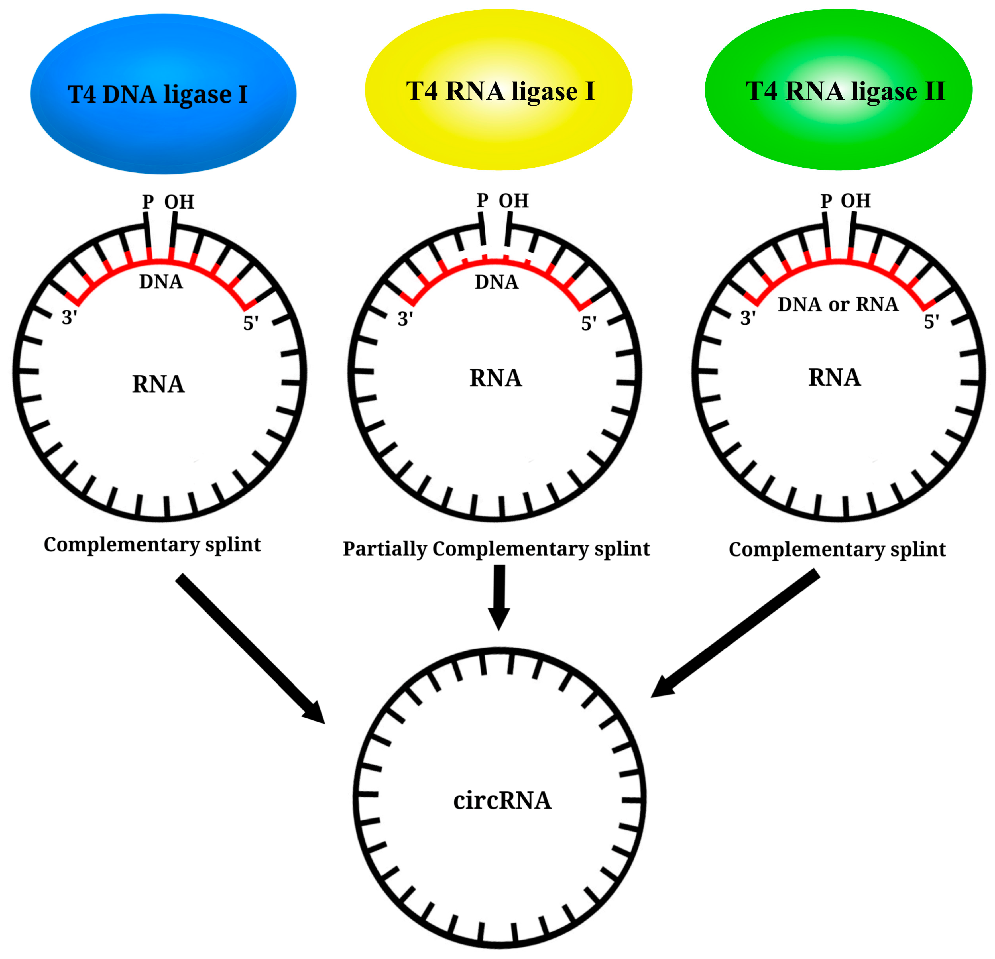
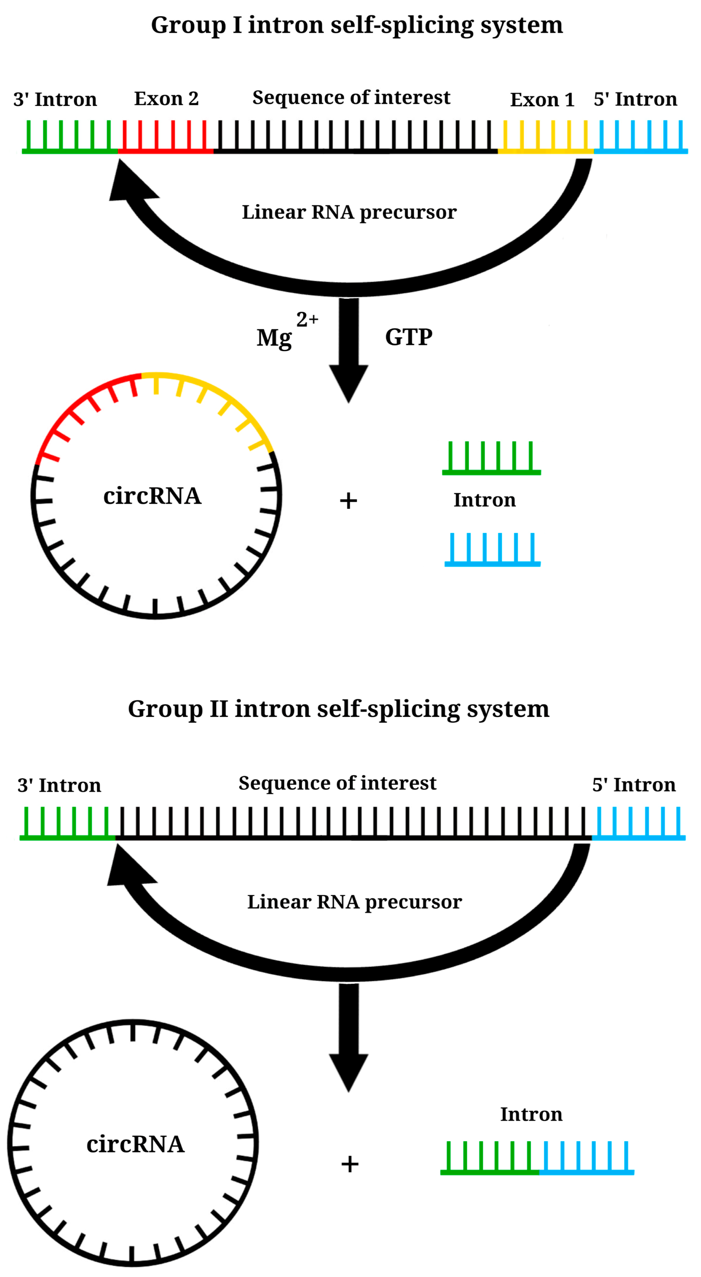
6. Circular RNA Platform

After circular RNA (circRNA) was discovered in cells in 1979 by electronic microscopy, it was initially regarded as a by-product of erroneous splicing [122,123]. But its vaccine potential was recognized in 2015 with the discovery of translated circRNAs in fruit flies [124], and it has gained recognition as a vaccine platform.
CircRNA is self-circularized and therefore lacks traditional 5’ and 3’ ends (Figure 6), a conventional cap structure, and a poly(A) tail. This structure makes it resistant to exonucleases, such as RNase R, and enhances its stability. It has a half-life of up to 84 h, in contrast to the average half-life of <20 h for linear mRNA [125,126,127]. Translation is initiated by a mechanism mediated by the IRES [51,78,128,129]. A commonly utilized IRES for translating synthetic circRNA is the encephalomyocarditis virus (EMCV) IRES, which is efficient in various cell lines [51,130,131], as well as those derived from coxsackievirus B3 (CVB3) and human rhinovirus B3 (HRV-B3), along with their modified versions, which have translation levels 3–4 times higher than that of EMCV IRES [131,132,133]. The crucial step in IVT circRNA synthesis is the circularization of linear RNA molecules by chemical or enzymatic methods [134]. The chemical approach for IVT circRNA generation, which originated in 1988, can be regarded as a pivotal milestone in the field. This method uses cyanogen bromide (BrCN) or 1-ethyl-3-(3-dimethylaminopropyl) carbodiimide (EDC) for the formation of bonds between the 5’-terminal phosphate and 3’-terminal hydroxyl groups of the linear RNA. However, this reaction may yield circRNA variants containing 2’-5’ phosphodiester bonds at the junction of the linear molecule, which are considered by-products [127]. The method is no longer commonly employed due to its high cost and low yield, and its applicability to circularization of RNA of only up to 70 nucleotides in length [135].
Enzymatic approaches for RNA circularization use bacteriophage T4 enzymes or ribozymes [134]. For the creation of IVT CircRNA, three bacteriophage T4 enzymes are used: T4 DNA ligase (T4 Dnl 1), T4 RNA ligase 1 (T4 Rnl 1), and T4 RNA ligase 2 (T4 Rnl 2) (Figure 7). To circularize the ends of a linear IVT RNA molecule using T4 bacteriophage enzymes, a nucleoside monophosphate must be present at the 5’ end bonded to the OH group at the 3’ end of the RNA [136]. Typically, IVT RNA contains guanosine triphosphate (GTP) at the 5’ end, as nucleoside triphosphates are added during the reaction. There are three strategies to obtain 5’-guanosine monophosphate (GMP) at the 5’ end of IVT RNA:
(1) Addition of an excess amount of guanosine monophosphate (GMP) compared to GTP in the IVT reaction, typically in a ratio of approximately 5/1, increases the likelihood of GMP being present at the 5’ end of the RNA [130,135,137].
(2) Treatment of IVT RNA with E. coli 5’-RNA pyrophosphohydrolase (RppH) or similar phosphatases cleaves phosphate groups from the 5’-terminal GTP, resulting in the formation of a 5’-terminal GMP [138,139].
(3) Treatment of IVT RNA with alkaline phosphatases to remove all phosphate groups from GTP, followed by treatment with T4 polynucleotide kinases to phosphorylate guanosine and generate GMP [134,135].
The subsequent step in the generation of IVT circRNA involves the addition of T4 bacteriophage enzymes to the linear RNA with a 5’-terminal GMP. T4 Dnl1, initially employed in 1992, is the first enzyme used for circRNA formation. T4 Dnl1 is used by bacteriophages to repair breaks in double-stranded DNA substrates [140]. A necessary condition for T4 Dnl1 is the presence of a double-stranded nucleic acid substrate. However, since IVT RNA is single-stranded, the reaction requires a short single-stranded DNA molecule complementary to the RNA site being joined. This single-stranded DNA (ssDNA) typically consists of at least 20 nucleotides, 10 of which are complementary to the 3’-end of the IVT RNA and the other 10 complementary to the 5’-end. This positions the ssDNA in the middle of the junction between the ends to form a double-stranded structure [137,141,142]. It should be noted that the efficiency of the RNA ligation reaction is relatively low because T4 Dnl1 is specific for double-stranded DNA (dsDNA) and exhibits weaker binding to RNA/DNA heteroduplexes, necessitating an increase in enzyme concentration in the reaction [135].
Due to the low yield and high consumption of T4 Dnl1, this method is now rarely used. Conversely, T4 Rnl 1, which was first used in 1999, is commonly employed as a ligase for circRNA synthesis [134]. T4 Rnl 1 exhibits an affinity for single-stranded RNA molecules (ssRNA) but possesses low specificity [143]. The efficiency of end-joining is primarily influenced by the nucleotide composition of the ligated RNA ends, because adenosine is optimal at the 3’ end and cytidine at the 5’ end [144]. However, IVT RNA typically contains guanosine at the 5’ end, which is the least favorable nucleotide for ligation in this position and thereby complicates the enzymatic reaction. Consequently, IVT circRNA of 6–8 nucleotides is typically produced, as increasing its length significantly reduces the reaction efficiency [145]. The close proximity of the 3’ and 5’ ends that are to be ligated is crucial for the activity of T4 Rnl 1 [146]. However, as the length of IVT circRNA increases, the likelihood of interaction between the RNA ends and the formation of double-stranded RNA (dsRNA) and secondary structures also increases, significantly reducing the efficiency of end ligation with this enzyme [137,147]. To remedy this issue, DNA complementary to IVT RNA is introduced into the reaction, which brings the 3’ and 5’ ends of the RNA in close proximity, with several non-complementary nucleotides at the ends, creating a short single-stranded fragment for ligation [148]. Recently developed is an optimal motif that forms an RNA hairpin structure, effectively bringing the 3’ and 5’ ends closer together. This approach enabled Carmona et al. (2019) to successfully obtain IVT circRNA of up to 4 kb in length [149].
The final enzyme used in the creation of IVT circRNA is T4 Rnl 2, which was first employed in 2002 [134]. Like T4 Rnl 1, this ligase can ligate ssRNA into a circular molecule, but it exhibits greater activity in the ligation of dsRNA molecules [145,150]. To generate dsRNA from a linear IVT RNA molecule, secondary structures are employed at the ligation site, and an RNA or DNA fragment complementary to the ends of the IVT molecule is added. However, the primary challenge remaining in the use of T4 Rnl 2 and T4 Rnl 1 is the low efficiency of the reaction and the increased production of by-products as the length of the coupled IVT RNA increases [134,135]. More recently, a strategy has been proposed to generate IVT circRNAs longer than 1.5 kb. Liang Qu et al. (2022) employed bioinformatic methods to identify a suitable site for RNA ligation within the CVB3 IRES (385 nucleotides). At this site, a favorable secondary structure is formed, enabling high-efficiency ligation with T4 Rnl 2. Using this approach, the authors successfully developed a highly effective vaccine against SARS-CoV-2, which was tested in mice and monkeys [132].
Significant progress has been made to resolve the primary challenge associated with the use of T4 bacteriophage enzymes, which is the limited length of IVT circRNA. However, a persistent problem is the formation of RNA concatemers, as most of the ligases used can cross-link ssRNAs into long molecules. To mitigate this problem, the volume of the reaction mixture was increased to reduce the likelihood of close proximity between linear IVT RNA molecules [139,151].
Ribozymes are RNAs that possess enzymatic activity, and this unique property is harnessed in the creation of IVT circRNA. Ribozyme sequences facilitate self-splicing, enabling the conversion of linear RNA molecules into circRNA without the need for additional enzymes (Figure 8). The self-splicing process involves two successive transesterification reactions at specific sites, ensuring the production of the desired circRNA product [134]. Three methods are commonly employed for generating circRNA using ribozymes: the group I intron self-splicing method, also known as permuted introns and exons (PIE), the group II intron self-splicing method, and the hairpin ribozyme method [134,135]. The group I intron method is the most widely used in circRNA design [131]. The PIE method was first employed in 1992 to create circRNA using group I introns derived from the Anabaena pre-tRNA gene [152]. Two years later, group I introns from the thymidylate synthase (td) gene of bacteriophage T4 were identified, enabling the generation of circRNA from td exons [153]. These two variants of group I introns are still used to generate circRNA [135,154].
It has been observed that the composition of group I introns influences the efficiency of circRNA production. The yield of circRNA is notably higher when using group I introns from the Anabaena pre-tRNA gene compared to those from the td gene [131]. In the PIE method, only GTP and Mg2+ need to be added as co-factors in the reaction [153]. The use of group I introns enables the generation of circRNA longer than 5 kb, which is a significant advantage [131,145]. A distinctive characteristic of group I introns is the requirement for an additional exon sequence derived from the same organism as the group I introns, flanking the GOI. The recognition of specific exon sequences by group I introns allows for precise self-splicing according to these markers. The exchange of exons typically ranges from 70 to 180 nucleotides, while the introns themselves are approximately 300‒400 nucleotides long. After self-splicing, the ends are joined within the exon sequence [134,135]. One significant drawback of using group I introns is that allogeneic exon sequences remain after RNA self-splicing, which separate the GOI and IRES. This separation could affect the expression of the IVT CircRNA. However, efforts are underway to improve the limitations associated with group I introns, and there have been promising results [154,155]. The unique characteristics of the PIE method make it possible to generate circRNA not only in vitro but also in vivo, making it a distinctive approach [134,156].
The self-splicing mechanism of group II introns shares a similar principle with the formation of circRNA, but its advantage is using introns without the need for exons [157]. Group II introns are transposable genetic elements present in organelle genomes and bacteria [158]. A distinguishing structural characteristic of group II introns is the presence of six domains, with the first domain playing a role in determining the specificity of self-splicing through its binding to a specific RNA sequence. This method enables the precise joining of linear RNA sequences without the inclusion of foreign exon fragments during circularization. Recently, there has been a growing interest in the development of vaccines using group II introns, which can be seen as a logical progression from the use of group I introns [159]. However, it is important to note that the self-splicing of group II introns forms 2’, 5’-phosphodiester bonds at the ligation site instead of the natural 3’, 5’-phosphodiester bonds, and the precise mechanism of this process is not fully understood [134,135,160]. Nevertheless, studies have demonstrated that IVT circRNAs containing group II introns are highly stable, weakly immunogenic, have low toxicity, and can produce high levels of protein [159].
Another approach for producing IVT circRNA from a linear RNA molecule is the utilization of hairpin ribozyme (HPR). HPR enables the production of circRNA using T7 RNA polymerase on a single-stranded circular DNA template by a rolling circle mechanism. The resulting linear RNA molecule can undergo self-cleavage and ligation to form circRNA. HPR elements have been identified in the genomes of certain viruses, such as the hepatitis delta virus, and have been employed as a platform for the synthesis of IVT circRNA [134,135,161,162,163]. This method is particularly suitable for producing small circRNA molecules (up to 150 nucleotides) with high yields [134,164]. However, the instability of ligation and self-cleavage mediated by HPR can lead to the generation of reaction by-products, and the resulting circRNA may contain HPR fragments that retain catalytic activity. This renders the current HPR method unsuitable for the development of RNA vaccines [134,144].
In conclusion, among the various ribozyme methods available for generating circRNA, the PIE method has been extensively studied and proven to be useful. However, group II introns hold promise for advancements. Ribozyme methods, in general, offer advantages such as cost-effectiveness, as they do not require additional enzymes, and the reactions can occur co-transcriptionally. These features make ribozyme methods more attractive for circRNA synthesis than enzymatic methods.

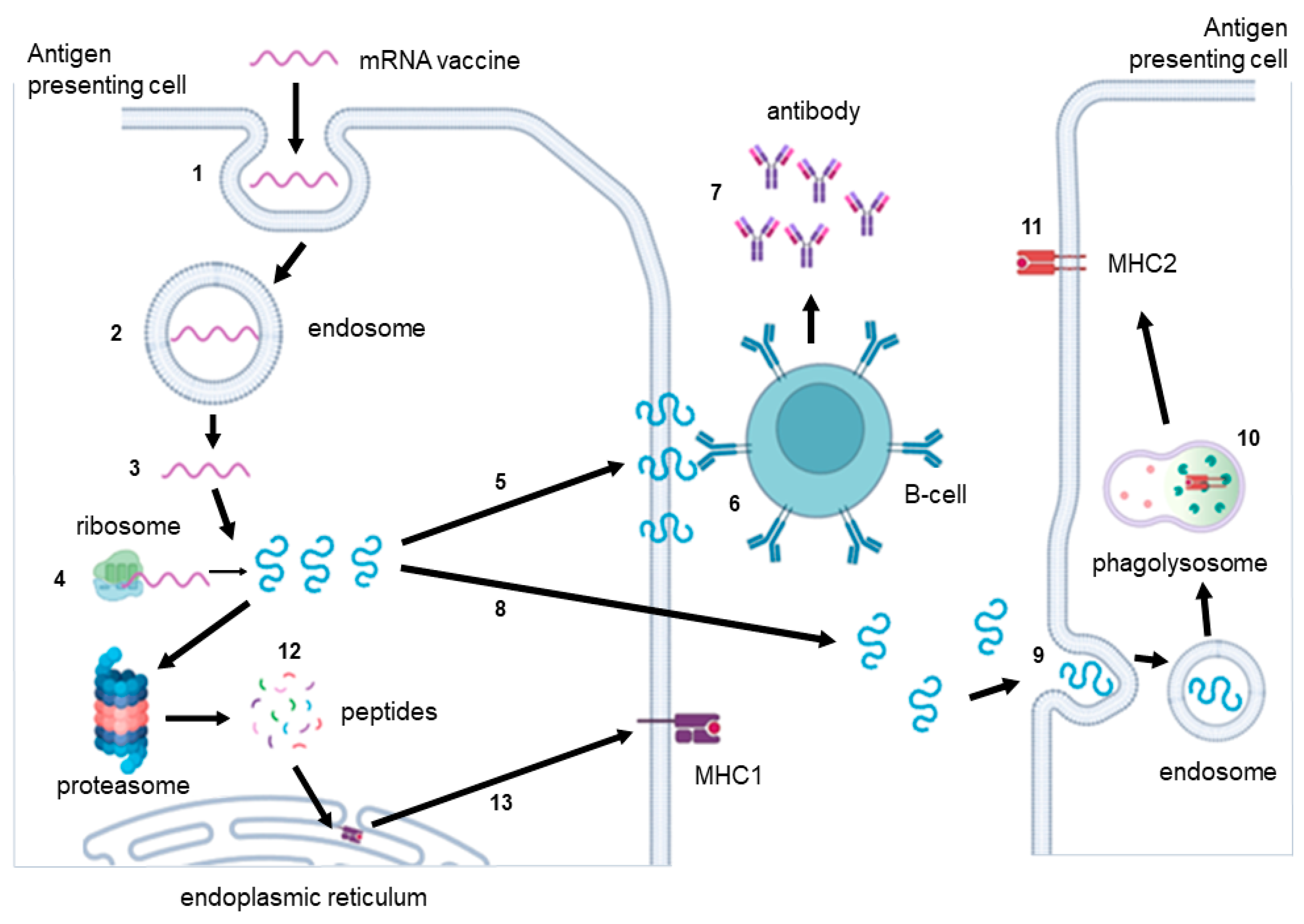
7. Immunogenicity of RNA Vaccines

The immunogenicity of IVT RNA vaccines is a significant challenge [165]. This immunogenicity arises from the absence of nucleotide modifications that are characteristic of natural mRNA, thereby preventing its effective discrimination by the immune system [85,165]. Hence, the triggered immunological reactions destroy the RNA, nullifying the desired effect. Therefore, the immunogenicity of vaccine RNA should be reduced, while the immunogenicity of the proteins translated from the vaccine RNA should be increased to develop persistent immunity. Among the receptors involved in recognizing IVT RNA are pattern recognition receptors (PRRs), which are widely expressed on various immune cells, particularly antigen-presenting cells [30]. PRRs involved in RNA recognition can be categorized into endosomal PRRs and cytoplasmic PRRs. Endosomal PRRs, primarily Toll-like receptors (TLRs), including TLR-3 for double-stranded RNA (dsRNA) and TLR-7/TLR-8 for single-stranded RNA (ssRNA), are predominantly localized within the endosomes (Table 2). Activation of these endosomal TLRs by RNA triggers intracellular signaling cascades leading to the production of type 1 interferon (IFN-I) and pro-inflammatory cytokines [78,166,167]. IFN-I, acting in an autocrine and a paracrine manner, initiates intracellular signaling pathways that induce the expression of IFN-stimulated genes (ISGs), which exert regulatory functions by inhibiting cellular RNA translation and promoting its accelerated degradation [78].
Among the main pattern recognition receptors (PRRs) in the cytosol are nucleotide oligomerization domain (NOD)-like receptors (NLRs), oligoadenylate synthetase (OAS) receptors, RNA-dependent protein kinase (PKR), and retinoic acid-inducible gene-I-like (RIG-I-like) receptors (RLRs). NOD2, a member of the NLR family, plays a crucial role in recognizing ssRNA. RLRs consist of RIG-I (DExD/H-box helicase 58), which interacts with both ssRNA and dsRNA, as well as melanoma differentiation-associated gene 5 (MDA-5) and LGP2 (DEXH-box helicase 58), which recognize dsRNA [168,169]. Like TLRs and NLRs, RLRs activate the synthesis of IFN-I and pro-inflammatory cytokines in response to RNA recognition [78]. Conversely, PRR interacts with dsRNA and phosphorylates eIF2, leading to the repression of translation. Oligoadenylate synthetase binds to dsRNA and facilitates the activation of RNase L, resulting in RNA degradation [170,171]. Thus, it is essential to create IVT RNA that evades recognition by the immune system [172].
To address this challenge, various modified nucleotides have been proposed and incorporated during IVT mRNA synthesis to reduce its immunogenicity [173]. Of the 172 proposed modified bases, only some demonstrated the ability to enhance mRNA expression and stability and reduce immunogenicity [174,175]. These modified nucleotides include N1-methyladenosine (m1A), N6-methyladenosine (m6A), 5-methoxyuridine (mo5U), 2-thiouridine (s2U), pseudouridine (ψ), N1-methylpseudouridine (m1ψ), 5-methylcytidine (m5C), 5-methoxycytidine (5moC), and 5-hydroxymethylcytidine (5hmC) [78,167,176,177,178]. The primary objective of incorporating these modified nucleotides, along with natural nucleotides, is to inhibit the activation of PRRs, such as TLR-3, TLR-7, TLR-8, OAS, RIG-I, and PKR, and thereby enhance the translation of IVT mRNA [78,173,179,180,181,182]. However, it is important to note that the same modified nucleotide can have different effects on the translation processes, depending on the cell type [183]. Therefore, when employing modified nucleotides, it is crucial to consider the specific cell type to which the IVT RNA will be targeted and select the nucleotides accordingly [177].
SAM is highly sensitive to the incorporation of modified bases, as they can disrupt the replicase activity, and therefore, they are usually not used. The lower immunogenicity of SAM vaccines compared to NRM is attributed primarily to the significantly lower doses of SAM employed, rather than to the use of modified nucleotides. Notably, modified nucleotides could be used with optimized taRNAs to reduce immunogenicity and decrease vaccine doses without diminishing the response. However, restrictions still exist in the use of modified nucleotides in TR-RNA synthesis, though the RNA encoding the replicase can be modified [104]. Notably, taRNAs offer an additional advantage over SAM because even lower vaccine doses could be used, further reducing immunogenicity [106,118].
Like mRNA, IVT circRNA incorporates modified nucleotides that reduce immunogenicity while minimally affecting translational efficiency. The incorporation of 5% m6A in circRNA significantly decreases its immunogenicity without impairing translation [133]. m6A is a prevalent RNA modification, occurring in approximately 0.5% of cellular RNA, including circRNA [184,185]. Notably, the YTHDF2 protein, which hinders innate immune activation, binds to m6A within endogenous circRNA in cells [186]. Furthermore, even the presence of a single m6A modification near the start codon can initiate cap-independent translation of circRNA by directly interacting with eIF3 [187]. Studies have demonstrated that the inclusion of multiple copies of the GGACU motif containing m6A enables translation initiation without an IRES, thus permitting the substitution of classical viral sequences with short motifs of approximately 20 nucleotides [188]. Overall, the RR(m6A)CH motif has been identified as particularly favorable for initiating cap-independent translation of circRNA [189]. In contrast, the use of m1ψ in IVT circRNA does not alter its immunogenicity but can negatively affect RNA circularization efficiency and translation levels [190].
It is important to note that the immune response to IVT circRNA has not been sufficiently investigated. However, it is postulated that the primary PRRs involved in recognizing circRNA are RIG-I and PKR. RIG-I activates intracellular signaling pathways, including MAVS, leading to the activation of the transcription factor IRF3 and subsequent synthesis of IFN-I, whereas PKR represses RNA translation [186]. It is hypothesized that the immunogenicity of IVT circRNA primarily arises from residual exons resulting from PIE circularization, which can form secondary structures and trigger PRR activation [191]. Indeed, IVT circRNAs generated using T4 RNA ligase do not activate the innate immune response. However, it is worth noting that viral IRESs also form secondary structures, potentially contributing to PRR activation. Further investigations are required to ascertain the factors underlying the immunogenicity of in vitro-transcribed circRNA. Such studies should focus on the influence of the primary RNA sequence, secondary structure, size, and the presence of modified nucleosides [192].
By-products of the synthesis of artificial mRNA or circRNA inevitably emerge, including dsDNA, ssDNA, and DNA/RNA heterodimers. Each of these by-products possesses specific receptors that recognize them and activate the innate immune system. For instance, TLR-9, located in the cell endosome, recognizes dsDNA and DNA/RNA hybrids, and cytosolic sensors such as cGAS (cyclic GMP-AMP synthase) and AIM2 (absent in melanoma 2) also detect these molecules, leading to the synthesis of IFN-I and other cytokines [193,194,195,196]. Therefore, it is crucial to purify the IVT mRNA from DNA impurities and DNA/RNA hybrids. DNase-I is effective in eliminating DNA contaminants [176]. However, purifying IVT mRNA from random RNA and dsRNA fragments remains a challenge. A reduction in dsRNA levels can be achieved by decreasing the concentration of Mg2+ ions during in vitro transcription or by incorporating modified nucleotides [173,197]. Moreover, after the synthesis of IVT circRNA, linear RNA molecules have to be removed as well because they are highly immunogenic by-products of the reaction. The treatment of IVT circRNA with RNase R or exonuclease T has been proposed [135,139,198].
Two main approaches are currently employed to purify IVT mRNA and circRNA from dsRNA. The first approach involves reversed-phase high-performance liquid chromatography (HPLC), while the second approach utilizes cellulose [78,199]. Reversed-phase HPLC, however, has several limitations, including poor scalability, toxicity of the eluant (acetonitrile), and considerable losses of IVT mRNA and circRNA [135,190,200]. On the other hand, the cellulose-based method for IVT mRNA purification does not suffer from these drawbacks. It removes up to 90% of dsRNA impurities, though it may not effectively eliminate short RNA fragments [81]. While the use of cellulose to purify IVT circRNA has not been described extensively, it holds potential for such applications. The significance of purifying IVT mRNA from dsRNA impurities is evident from studies demonstrating that purification alone can enhance translated protein levels by 10–1000 times compared to unpurified mRNA [78,166,201]. Similar investigations on IVT CircRNA highlight the critical importance of removing reaction by-products to reduce immunogenicity [131,141,144,188,190,191,192].

8. Future of mRNA-Based Platform Vaccine

Further development of mRNA vaccines is reduced to the search for a suitable alternative to replace the cap and to find a more suitable variant of the poly A tail. The use of a classical cap or a synthetic analog of the cap seems to be quite financially costly when creating mRNA vaccines. A significant reduction in cost is possible by using IRES, which does not require additional financial costs, instead of the cap. However, at the moment, the mRNA created using IRES is still inferior in terms of protein expression level. Therefore, it seems promising to search for new stronger synthetic analogs of IRES, which will allow us to completely abandon the cap [202].
Another branch of mRNA platform development is self-amplifying mRNA, an improved version of which is represented by trans-amplifying RNA. taRNA will have to be studied in more detail in the future and the main problem related to the safety of its use will have to be solved. The presence of alphavirus enzymes synthesized with taRNA will have to be replaced by enzymes that are safer for humans, and trans-replicon RNA and non-replicating mRNA encoding a replicase will have to be combined into a single mRNA. The last RNA-based vaccine variant in use is circRNA, which has a good chance to become a leader among other RNA platforms in the future. The future development of circRNA is connected with the creation of strong synthetic IRES, which allow us to significantly increase protein production using such RNA [133,203]. A separate direction in the development of this platform is the search for simpler and cheaper ways to synthesize a circular RNA molecule. One of the options is related to the search and use of non-immunogenic ribozymes that allow to create circRNA without additional costs.

9. Conclusions

Sixty-two years after its discovery, mRNA has evolved from an object of research into a tool for the prevention and therapy of a number of diseases. Vaccines based on mRNA have become a promising technological platform that is being actively developed. Currently, self-amplifying and trans-amplifying IVT RNA vaccines based on mRNA have been created. In 2015, the discovery of the possibility of translating proteins from circRNA in mammalian cells led to the development of viral vaccines based on them. Each of the four platforms used for vaccine development (mRNA, self-amplifying mRNA, trans-amplifying mRNA, and circRNA) has a number of advantages and disadvantages (Figure 9).
The non-amplifying mRNA platform is the most studied platform for the in vitro synthesis of RNA for its use as a vaccine. It allows for the selection of multiple regulatory elements to increase antigen translation in different cell types and tissues. Various modified bases can also be used to reduce the immunogenicity of the mRNA and prevent immune activation before translation takes place. However, mRNA-based vaccines have a number of disadvantages, mainly the relatively short half-life of the matrix and consequently the small amount of antigen produced. Self-amplifying mRNA is a logical extension of mRNA and is designed to solve its main problems because the mRNA is amplified inside the cell. The amplification of mRNA inside the cell leads to an increase in the initial number of mRNAs inside the cell, and therefore increases the amount of antigen that is translated. Thus, self-amplifying mRNA vaccines can be administered in low doses, have a long half-life inside cells, and result in the production of relatively high amounts of antigen. These unique properties of self-amplifying mRNA are due to the inclusion of a sequence encoding four non-structural proteins (nsP1, nsP2, nsP3, and nsP4) derived from alphaviruses. However, these virus sequences are the main drawbacks of this platform. Since the viral proteins are highly immunogenic and the sequence encoding them is more than 7000 nucleotide pairs, this limits the size of the antigen in such a vaccine. Furthermore, the double-stranded by-products of mRNA amplification are immunogenic. Yet another disadvantage of self-amplifying mRNA is the inability to use modified nitrogenous bases.
To solve the several problems of the self-amplifying mRNA platform, a trans-amplifying mRNA vaccine platform was created using two kinds of mRNA. The first encodes non-structural proteins of the alphavirus necessary for amplification of the mRNA of interest, which encodes the antigen and contains elements important for its recognition by RNA-dependent RNA polymerase. This platform has all the advantages of self-amplifying mRNA but lacks a number of the disadvantages. The trans-amplifying mRNA platform allows for the use of modified nitrogenous bases, reducing the immunogenicity of the mRNA. But this mRNA platform requires at least two types of mRNA, still contains viral sequences, and is still poorly understood.
The last type of RNA vaccines is circRNA, which has a unique structure that protects it from degradation by exonucleases and enables it to have a longer half-life, which leads to higher levels of the translated antigen. Furthermore, the circRNA platform does not contain a cap and poly A tail, which pose a number of problems in vaccine development, and allows for the use of modified nitrogenous bases. However, this platform (as well as trans-amplifying mRNA) is still insufficiently studied and the level of synthesized antigen is lower than that in self-replicating mRNA platforms. Nevertheless, at this time, it seems that the circRNA platform is the most promising because, on the one hand, it produces a higher level of antigen than the mRNA platform, and it is also safer than self-replicating mRNAs, which contain viral sequences. The application of any mRNA-based platform is feasible for both therapeutic cancer vaccines and prophylactic vaccines against various viral diseases.

Author Contributions

Conceptualization, A.D.P.; writing—original draft preparation, A.D.P.; writing—review and editing, A.D.P., M.V.V. and D.V.K.; visualization, A.D.S. and M.V.V.; project administration, A.D.S. and M.V.V.; funding acquisition, M.V.V. and. D.V.K. All authors have read and agreed to the published version of the manuscript.

Funding

This work was supported by a grant from Russian Science Foundation (RSF, project no. 22-15-00376, https://rscf.ru/en/project/22-15-00376/).

Conflicts of Interest

The authors declare no conflict of interest.

References

  1. Plotkin, S.A. Vaccines: The Fourth Century. Clin. Vaccine Immunol. 2009, 16, 1709–1719. [Google Scholar] [CrossRef] [PubMed]
  2. Probst, J.; Weide, B.; Scheel, B.; Pichler, B.J.; Hoerr, I.; Rammensee, H.G.; Pascolo, S. Spontaneous cellular uptake of exogenous messenger RNA in vivo is nucleic acid-specific, saturable and ion dependent. Gene Ther. 2007, 14, 1175–1180. [Google Scholar] [CrossRef] [PubMed]
  3. Ahammad, I.; Lira, S.S. Designing a novel mRNA vaccine against SARS-CoV-2: An immunoinformatics approach. Int. J. Biol. Macromol. 2020, 162, 820–837. [Google Scholar] [CrossRef]
  4. Li, W.; Joshi, M.D.; Singhania, S.; Ramsey, K.H.; Murthy, A.K. Peptide Vaccine: Progress and Challenges. Vaccines 2014, 2, 515–536. [Google Scholar] [CrossRef] [PubMed]
  5. Schmidt, S.T.; Foged, C.; Korsholm, K.S.; Rades, T.; Christensen, D. Liposome-Based Adjuvants for Subunit Vaccines: Formulation Strategies for Subunit Antigens and Immunostimulators. Pharmaceutics 2016, 8, 7. [Google Scholar] [CrossRef] [PubMed]
  6. Schlake, T.; Thess, A.; Fotin-Mleczek, M.; Kallen, K.J. Developing mRNA-vaccine technologies. RNA Biol. 2012, 9, 1319–1330. [Google Scholar] [CrossRef]
  7. Pardi, N.; Hogan, M.J.; Porter, F.W.; Weissman, D. mRNA vaccines—A new era in vaccinology. Nat. Rev. Drug Discov. 2018, 17, 261–279. [Google Scholar] [CrossRef]
  8. Chen, N.; Xia, P.; Li, S.; Zhang, T.; Wang, T.T.; Zhu, J. RNA sensors of the innate immune system and their detection of pathogens. IUBMB Life 2017, 69, 297–304. [Google Scholar] [CrossRef]
  9. Pardi, N.; Hogan, M.J.; Pelc, R.S.; Muramatsu, H.; Andersen, H.; DeMaso, C.R.; Dowd, K.A.; Sutherland, L.L.; Scearce, R.M.; Parks, R.; et al. Zika virus protection by a single low-dose nucleoside-modified mRNA vaccination. Nature 2017, 543, 248–251. [Google Scholar] [CrossRef]
  10. Cobb, M. Who discovered messenger RNA? Curr. Biol. 2015, 25, 526–532. [Google Scholar] [CrossRef]
  11. Zahednezhad, F.; Saadat, M.; Valizadeh, H.; Zakeri-Milani, P.; Baradaran, B. Liposome and immune system interplay: Challenges and potentials. J. Control. Release 2019, 10, 194–209. [Google Scholar] [CrossRef] [PubMed]
  12. Sanger, H.L.; Klotz, G.; Riesner, D.; Gross, H.J.; Kleinschmidt, A.K. Viroids are single-stranded covalently closed circular RNA molecules existing as highly base-paired rod-like structures. Proc. Natl. Acad. Sci. USA 1976, 73, 3852–3856. [Google Scholar] [CrossRef] [PubMed]
  13. Krieg, P.A.; Melton, D.A. In vitro RNA synthesis with SP6 RNA polymerase. Methods Enzymol. 1987, 155, 397–415. [Google Scholar] [PubMed]
  14. Dolgin, E. The tangled history of mRNA vaccines. Nature 2021, 597, 318–324. [Google Scholar] [CrossRef]
  15. Zhou, X.; Berglund, P.; Rhodes, G.; Parker, S.E.; Jondal, M.; Liljeström, P. Self-replicating Semliki Forest virus RNA as recombinant vaccine. Vaccine 1994, 12, 1510–1514. [Google Scholar] [CrossRef]
  16. Spuul, P.; Balistrer, G.; Hellstrom, K.; Golubtsov, A.V.; Jokitalo, E.; Ahola, T. Assembly of alphavirus replication complexes from RNA and protein components in a novel trans-replication system in mammalian cells. J. Virol. 2011, 85, 4739–4751. [Google Scholar] [CrossRef]
  17. Hou, X.; Zaks, T.; Langer, R.; Youn, H.; Im, H.J. Lipid nanoparticles for mRNA delivery. Nat. Rev. Mater. 2021, 6, 1078–1094. [Google Scholar] [CrossRef]
  18. Albertsen, C.H.; Kulkarni, J.A.; Witzigmann, D.; Lind, M.; Petersson, K.; Simonsen, J.B. The role of lipid components in lipid nanoparticles for vaccines and gene therapy. Adv. Drug Deliv. Rev. 2022, 188, 114416. [Google Scholar] [CrossRef]
  19. Sahin, U.; Derhovanessian, E.; Miller, M.; Kloke, B.P.; Simon, P.; Löwer, M.; Bukur, V.; Tadmor, A.D.; Luxemburger, U.; Schrörs, B.; et al. Personalized RNA mutanome vaccines mobilize poly-specific therapeutic immunity against cancer. Nature 2017, 547, 222–226. [Google Scholar] [CrossRef]
  20. Pascolo, S. Synthetic Messenger RNA-Based Vaccines: From Scorn to Hype. Viruses 2021, 13, 270. [Google Scholar] [CrossRef]
  21. Pardi, N.; Muramatsu, H.; Weissman, D.; Kariko, K. In vitro transcription of long RNA containing modified nucleosides. Methods Mol. Biol. 2013, 969, 29–42. [Google Scholar] [CrossRef]
  22. Rio, D.C. Expression and purification of active recombinant T7 RNA polymerase from E. coli. In RNA: A Laboratory Manual; CSHL Press: Cold Spring Harbor, NY, USA, 2013. [Google Scholar] [CrossRef]
  23. Deng, Z.; Tian, Y.; Song, J.; An, G.; Yang, P. mRNA Vaccines: The Dawn of a New Era of Cancer Immunotherapy. Front. Immunol. 2022, 13, 887125. [Google Scholar] [CrossRef] [PubMed]
  24. Klocker, N.; Weissenboeck, F.P.; Dulmen, M.; Spacek, P.; Huwel, S.; Rentmeister, A. Photocaged 5′ cap analogues for optical control of mRNA translation in cells. Nat. Chem. 2022, 14, 905–913. [Google Scholar] [CrossRef] [PubMed]
  25. To, K.K.; Cho, W.C. An overview of rational design of mRNA-based therapeutics and vaccines. Expert Opin. Drug Discov. 2021, 16, 1307–1317. [Google Scholar] [CrossRef]
  26. Ramanathan, A.; Robb, G.B.; Chan, S.-H. mRNA capping: Biological functions and applications. Nucleic Acids Res. 2016, 44, 7511–7526. [Google Scholar] [CrossRef]
  27. Mauer, J.; Luo, X.; Blanjoie, A.; Jiao, X.; Grozhik, A.V.; Patil, D.P.; Linder, B.; Pickering, B.F.; Vasseur, J.J.; Chen, Q.; et al. Reversible methylation of m6Am in the 5′ cap controls mRNA stability. Nature 2017, 541, 371–375. [Google Scholar] [CrossRef]
  28. Tat, T.T.; Kiss, D.L. From m6A to Cap-Adjacent m6Am and their Effects on mRNAs. Epitranscriptomics 2021, 12, 325–351. [Google Scholar] [CrossRef]
  29. Drazkowska, K.; Tomecki, R.; Warminski, M.; Baran, N.; Cysewski, D.; Depaix, A.; Kasprzyk, R.; Kowalska, J.; Jemielity, J.; Sikorski, P.J. 2′-O-Methylation of the second transcribed nucleotide within the mRNA 5′ cap impacts the protein production level in a cell-specific manner and contributes to RNA immune evasion. Nucleic Acids Res. 2022, 50, 9051–9071. [Google Scholar] [CrossRef] [PubMed]
  30. Linares-Fernandez, S.; Lacroix, C.; Exposito, J.-Y.; Verrier, B. Tailoring mRNA Vaccine to Balance Innate/Adaptive Immune Response. Trends Mol. Med. 2020, 26, 311–323. [Google Scholar] [CrossRef] [PubMed]
  31. Pardi, N.; Hogan, M.J.; Weissman, D. Recent advances in mRNA vaccine technology. Curr. Opin. Immunol. 2020, 65, 14–20. [Google Scholar] [CrossRef]
  32. Stepinski, J.; Waddell, C.; Stolarski, R.; Darzynkiewicz, E.; Rhoads, R.E. Synthesis and properties of mRNAs containing the novel “anti-reverse” cap analogs 7-methyl (3′-O-methyl) GpppG and 7-methyl (3′-deoxy) GpppG. RNA 2001, 7, 1486–1495. [Google Scholar] [PubMed]
  33. Jemielity, J.; Fowler, T.; Zuberek, J.; Stepinski, J.; Lewdorowicz, M.; Niedzwiecka, A.; Stolarski, R.; Darzynkiewicz, E.; Rhoads, R.E. Novel “anti-reverse” cap analogs with superior translational properties. RNA 2003, 9, 1108–1122. [Google Scholar] [CrossRef] [PubMed]
  34. Henderson, J.M.; Ujita, A.; Hill, E.; Yousif-Rosales, S.; Smith, C.; Ko, N.; McReynolds, T.; Cabral, C.R.; Escamilla-Powers, J.R.; Houston, M.E. Cap 1 messenger RNA synthesis with co-transcriptional CleanCap® analog by in vitro transcription. Curr. Protoc. 2021, 1, e39. [Google Scholar] [CrossRef] [PubMed]
  35. TriLink BioTechnologies. Cap Analogs. Available online: https://www.trilinkbiotech.com/products-services/nucleoside-triphosphates-nucleotides.html?cap_analogs=1878 (accessed on 10 September 2023).
  36. Hornblower, B.; Robb, G.B.; Tzertzinis, G. Minding your caps and tails—Considerations for functional mRNA synthesis. DNA 2018, 5, 3. [Google Scholar]
  37. TriLink BioTechnologies. CleanCap. Available online: https://www.trilinkbiotech.com/cleancap-old21-3 (accessed on 10 September 2023).
  38. Moradian, H.; Roch, T.; Anthofer, L.; Lendlein, A.; Gossen, M. Chemical modification of uridine modulates mRNA-mediated proinflammatory and antiviral response in primary human macrophages. Mol. Ther. Nucleic Acids 2022, 27, 854–869. [Google Scholar] [CrossRef]
  39. Niederer, R.O.; Rojas-Duran, M.F.; Zinshteyn, B.; Gilbert, W.V. Direct analysis of ribosome targeting illuminates thousand-fold regulation of translation initiation. Cell Syst. 2022, 13, 256–264. [Google Scholar] [CrossRef]
  40. Leppek, K.; Das, R.; Barna, M. Functional 5′ UTR mRNA structures in eukaryotic translation regulation and how to find them. Nat. Rev. Mol. Cell Biol. 2018, 19, 158–174. [Google Scholar] [CrossRef]
  41. Hinnebusch, A.G.; Ivanov, I.P.; Sonenberg, N. Translational control by 5′-untranslated regions of eukaryotic mRNAs. Science 2016, 352, 1413–1416. [Google Scholar] [CrossRef]
  42. Haizel, S.A.; Bhardwaj, U.; Gonzalez, R.L., Jr.; Mitra, S.; Goss, D.J. 5′-UTR recruitment of the translation initiation factor eIF4GI or DAP5 drives cap-independent translation of a subset of human mRNAs. J. Biol. Chem. 2020, 295, 11693–11706. [Google Scholar] [CrossRef]
  43. Hinnebusch, A.G. Molecular mechanism of scanning and start codon selection in eukaryotes. Microbiol. Mol. Biol. Rev. 2011, 75, 434–467. [Google Scholar] [CrossRef]
  44. Weingarten-Gabbay, S.; Elias-Kirma, S.; Nir, R.; Gritsenko, A.A.; Stern-Ginossar, N.; Yakhini, Z.; Weinberger, A.; Segal, E. Comparative genetics. Systematic discovery of cap-independent translation sequences in human and viral genomes. Science 2016, 351, aad4939. [Google Scholar] [CrossRef] [PubMed]
  45. Wang, Y.; Zhang, R.; Tang, L.; Yang, L. Nonviral Delivery Systems of mRNA Vaccines for Cancer Gene Therapy. Pharmaceutics 2022, 14, 512. [Google Scholar] [CrossRef] [PubMed]
  46. Jia, L.; Mao, Y.; Ji, Q.; Dersh, D.; Yewdell, J.W.; Qian, S.B. Decoding mRNA translatability and stability from the 5′ UTR. Nat. Struct. Mol. Biol. 2020, 27, 814–821. [Google Scholar] [CrossRef] [PubMed]
  47. Linares-Fernández, S.; Moreno, J.; Lambert, E.; Mercier-Gouy, P.; Vachez, L.; Verrier, B.; Exposito, J.Y. Combining an optimized mRNA template with a double purification process allows strong expression of in vitro transcribed mRNA. Mol. Ther. Nucleic Acids 2021, 26, 945–956. [Google Scholar] [CrossRef]
  48. Sample, P.J.; Wang, B.; Reid, D.W.; Presnyak, V.; McFadyen, I.J.; Morris, D.R.; Seelig, G. Human 5′ UTR design and variant effect prediction from a massively parallel translation assay. Nat. Biotechnol. 2019, 37, 803–809. [Google Scholar] [CrossRef]
  49. Trepotec, Z.; Aneja, M.K.; Geiger, J.; Hasenpusch, G.; Plank, C.; Rudolph, C. Maximizing the Translational Yield of mRNA Therapeutics by Minimizing 5′-UTRs. Tissue Eng. Part A 2019, 25, 69–79. [Google Scholar] [CrossRef]
  50. Ko, H.L.; Park, H.-J.; Kim, J.; Kim, H.; Youn, H.; Nam, J.H. Development of an RNA Expression Platform Controlled by Viral Internal Ribosome Entry Sites. J. Microbiol. Biotechnol. 2019, 29, 127–140. [Google Scholar] [CrossRef]
  51. Deviatkin, A.A.; Simonov, R.A.; Trutneva, K.A.; Maznina, A.A.; Soroka, A.B.; Kogan, A.A.; Feoktistova, S.G.; Khavina, E.M.; Mityaeva, O.N.; Volchkov, P.Y. Cap-Independent Circular mRNA Translation Efficiency. Vaccines 2023, 11, 238. [Google Scholar] [CrossRef]
  52. Svitkin, Y.V.; Siddiqui, N.; Sonenberg, N. Protein Synthesis Initiation in Eukaryotes: IRES-mediated Internal Initiation. eLS 2015, 1–11. [Google Scholar] [CrossRef]
  53. Trainor, B.M.; Shcherbik, N. Short and Sweet: Viral 5′-UTR as a Canonical and Non-Canonical Translation Initiation Switch. J. Cell. Immunol. 2021, 3, 296–304. [Google Scholar] [CrossRef]
  54. Kreiter, S.; Selmi, A.; Diken, M.; Sebastian, M.; Osterloh, P.; Schild, H.; Huber, C.; Tureci, O.; Sahin, U. Increased antigen presentation efficiency by coupling antigens to MHC class I trafficking signals. J. Immunol. 2008, 180, 309–318. [Google Scholar] [CrossRef] [PubMed]
  55. Mediani, L.; Guillen-Boixet, J.; Vinet, J.; Franzmann, T.M.; Bigi, I.; Mateju, D.; Carrà, A.D.; Morelli, F.F.; Tiago, T.; Poser, I.; et al. Defective ribosomal products challenge nuclear function by impairing nuclear condensate dynamics and immobilizing ubiquitin. EMBO J. 2019, 38, e101341. [Google Scholar] [CrossRef] [PubMed]
  56. Weissman, D. mRNA transcript therapy. Expert Rev. Vaccines 2015, 14, 265–281. [Google Scholar] [CrossRef]
  57. Bullock, T.N.J. CD40 stimulation as a molecular adjuvant for cancer vaccines and other immunotherapies. Cell. Mol. Immunol. 2022, 19, 14–22. [Google Scholar] [CrossRef] [PubMed]
  58. Hewitt, S.L.; Bai, A.; Bailey, D.; Ichikawa, K.; Zielinski, J.; Karp, R.; Apte, A.; Arnold, K.; Zacharek, S.J.; Iliou, M.S.; et al. Durable anticancer immunity from intratumoral administration of IL-23, IL-36γ, and OX40L mRNAs. Sci. Transl. Med. 2019, 11, eaat9143. [Google Scholar] [CrossRef]
  59. Fan, N.; Chen, K.; Zhu, R.; Zhang, Z.; Huang, H.; Qin, S.; Zheng, Q.; He, Z.; He, X.; Xiao, W.; et al. Manganese-coordinated mRNA vaccines with enhanced mRNA expression and immunogenicity induce robust immune responses against SARS-CoV-2 variants. Sci. Adv. 2022, 8, eabq3500. [Google Scholar] [CrossRef]
  60. Tanida-Miyake, E.; Koike, M.; Uchiyama, Y.; Tanida, I. Optimization of mNeonGreen for Homo sapiens increases its fluorescent intensity in mammalian cells. PLoS ONE 2018, 13, e0191108. [Google Scholar] [CrossRef]
  61. Zhong, F.; Cao, W.; Chan, E.; Tay, P.N.; Cahya, F.F.; Zhang, H.; Lu, J. Deviation from major codons in the Toll-like receptor genes is associated with low Toll-like receptor expression. Immunology 2005, 114, 83–93. [Google Scholar] [CrossRef]
  62. Kimchi-Sarfaty, C.; Oh, J.M.; Kim, I.-W.; Sauna, Z.E.; Calcagno, A.M.; Ambudkar, S.V.; Gottesman, M.M. A “silent” polymorphism in the MDR1 gene changes substrate specificity. Science 2007, 315, 525–528. [Google Scholar] [CrossRef]
  63. Kanampalliwar, A.M.; Soni, R.; Girdhar, A.; Tiwari, A. Reverse Vaccinology: Basics and Applications. Methods Mol. Biol. 2020, 2131, 1–16. [Google Scholar] [CrossRef]
  64. Yang, Z.; Bogdan, P.; Nazarian, S. An in silico deep learning approach to multi-epitope vaccine design: A SARS-CoV-2 case study. Sci. Rep. 2021, 11, 3238. [Google Scholar] [CrossRef]
  65. Serruto, D.; Rappuoli, R. Post-genomic vaccine development. FEBS Lett. 2006, 580, 2985–2992. [Google Scholar] [CrossRef] [PubMed]
  66. Fadilah, F.; Paramita, R.I.; Erlina, L.; Khaerunissa, A.I.; Puspita, E.W.; Aryo, T. Linker Optimization in Breast Cancer Multiepitope Peptide Vaccine Design Based on Molecular Study. ICOLIB 2023, 27, 528–538. [Google Scholar] [CrossRef]
  67. Suhrbier, A. Multi-epitope DNA vaccines. Immunol. Cell Biol. 1997, 75, 402–408. [Google Scholar] [CrossRef]
  68. Chen, X.; Zaro, J.L.; Shen, W.-C. Fusion protein linkers: Property, design and functionality. Adv. Drug Deliv. Rev. 2013, 65, 1357–1369. [Google Scholar] [CrossRef]
  69. Ali, M.; Pandey, R.K.; Khatoon, N.; Narula, A.; Mishra, A.K.; Prajapati, V.K. Exploring dengue genome to construct a multi-epitope based subunit vaccine by utilizing immunoinformatics approach to battle against dengue infection. Sci. Rep. 2017, 7, 9232. [Google Scholar] [CrossRef] [PubMed]
  70. Sanami, S.; Azadegan-Dehkordi, F.; Rafieian-Kopaei, M.; Salehi, M.; Ghasemi-Dehnoo, M.; Mahooti, M.; Alizadeh, M.; Bagheri, N. Design of a multi-epitope vaccine against cervical cancer using immunoinformatics approaches. Sci. Rep. 2021, 11, 12397. [Google Scholar] [CrossRef]
  71. Banerjee, S.; Majumder, K.; Gutierrez, G.J.; Gupta, D.; Mittal, B. Immuno-informatics approach for multi-epitope vaccine designing against SARS-CoV-2. BioRxiv 2020. BioRxiv:2020-07. [Google Scholar] [CrossRef]
  72. Mittal, A.; Sasidharan, S.; Raj, S.; Balaji, S.N.; Saudagar, P. Exploring the Zika Genome to Design a Potential Multiepitope Vaccine Using an Immunoinformatics Approach. Int. J. Pept. Res. Ther. 2020, 26, 2231–2240. [Google Scholar] [CrossRef]
  73. Mayr, C. What Are 3′ UTRs Doing? Cold Spring Harb. Perspect. Biol. 2019, 11, a034728. [Google Scholar] [CrossRef]
  74. Siepel, A.; Bejerano, G.; Pedersen, J.S.; Hinrichs, A.S.; Hou, M.; Rosenbloom, K.; Clawson, H.; Spieth, J.; Hillier, L.W.; Richards, S.; et al. Evolutionarily conserved elements in vertebrate, insect, worm, and yeast genomes. Genome Res. 2005, 15, 1034–1050. [Google Scholar] [CrossRef] [PubMed]
  75. Xie, X.; Lu, J.; Kulbokas, E.J.; Golub, T.R.; Mootha, V.; Lindblad-Toh, K.; Lander, E.S.; Kellis, M. Systematic discovery of regulatory motifs in human promoters and 3′ UTRs by comparison of several mammals. Nature 2005, 434, 338–345. [Google Scholar] [CrossRef] [PubMed]
  76. Orlandini von Niessen, A.G.; Poleganov, M.A.; Rechner, C.; Plaschke, A.; Kranz, L.M.; Fesser, S.; Diken, M.; Löwer, M.; Vallazza, B.; Beissert, T.; et al. Improving mRNA-Based Therapeutic Gene Delivery by Expression-Augmenting 3′ UTRs Identified by Cellular Library Screening. Mol. Ther. 2019, 27, 824–836. [Google Scholar] [CrossRef]
  77. Holtkamp, S.; Kreiter, S.; Selmi, A.; Simon, P.; Koslowski, M.; Huber, C.; Türeci, O.; Sahin, U. Modification of antigen-encoding RNA increases stability, translational efficacy, and T-cell stimulatory capacity of dendritic cells. Blood 2006, 108, 4009–4017. [Google Scholar] [CrossRef] [PubMed]
  78. Fang, E.; Liu, X.; Li, M.; Zhang, Z.; Song, L.; Zhu, B.; Wu, X.; Liu, J.; Zhao, D.; Li, Y. Advances in COVID-19 mRNA vaccine development. Signal Transduct. Target. Ther. 2022, 7, 94. [Google Scholar] [CrossRef]
  79. Kontoyiannis, D.; Pasparakis, M.; Pizarro, T.T.; Cominelli, F.; Kollias, G. Impaired on/off regulation of TNF biosynthesis in mice lacking TNF AU-rich elements: Implications for joint and gut-associated immunopathologies. Immunity 1999, 10, 387–398. [Google Scholar] [CrossRef]
  80. Trepotec, Z.; Geiger, J.; Plank, C.; Aneja, M.K.; Rudolph, C. Segmented poly(A) tails significantly reduce recombination of plasmid DNA without affecting mRNA translation efficiency or half-life. RNA 2019, 25, 507–518. [Google Scholar] [CrossRef]
  81. Pardi, N.; Parkhouse, K.; Kirkpatrick, E.; McMahon, M.; Zost, S.J.; Mui, B.L.; Tam, Y.K.; Karikó, K.; Barbosa, C.J.; Madden, T.D.; et al. Nucleoside-Modified mRNA Immunization Elicits Influenza Virus Hemagglutinin Stalk-Specific Antibodies. Nat. Commun. 2018, 9, 3361. [Google Scholar] [CrossRef]
  82. Grier, A.; Burleigh, S.; Sahni, J.; Clough, C.A.; Cardot, V.; Choe, D.C.; Krutein, M.C.; Rawlings, D.J.; Jensen, M.C.; Scharenberg, A.M.; et al. pEVL: A Linear Plasmid for Generating mRNA IVT Templates with Extended Encoded Poly(A) Sequences. Mol. Ther. Nucleic Acids 2016, 5, e306. [Google Scholar] [CrossRef]
  83. Park, J.-E.; Yi, H.; Kim, Y.; Chang, H.; Kim, V.N. Regulation of Poly(A) Tail and Translation during the Somatic Cell Cycle. Mol. Cell 2016, 62, 462–471. [Google Scholar] [CrossRef]
  84. Qin, S.; Tang, X.; Chen, Y.; Chen, K.; Fan, N.; Xiao, W.; Zheng, Q.; Li, G.; Teng, Y.; Wu, M.; et al. mRNA-based therapeutics: Powerful and versatile tools to combat diseases. Signal Transduct. Target. Ther. 2022, 7, 166. [Google Scholar] [CrossRef] [PubMed]
  85. Sahin, U.; Kariko, K.; Tureci, O. mRNA-based therapeutics-developing a new class of drugs. Nat. Rev. Drug Discov. 2014, 13, 759–780. [Google Scholar] [CrossRef]
  86. Voet, D.; Voet, J.G. Biochemistry, 4th ed.; John Wiley & Sons: New York, NY, USA, 2011; pp. 123–126. [Google Scholar]
  87. Preiss, T.; Muckenthaler, M.; Hentze, M.W. Poly(A)-tail-promoted translation in yeast: Implications for translational control. RNA 1998, 4, 1321–1331. [Google Scholar] [CrossRef]
  88. Vogel, A.B.; Kanevsky, I.; Che, Y.; Swanson, K.A.; Muik, A.; Vormehr, M.; Kranz, L.M.; Walzer, K.C.; Hein, S.; Güler, A.; et al. A prefusion SARS-CoV-2 spike RNA vaccine is highly immunogenic and prevents lung infection in non-human primates. Biorxiv 2020. BioRxiv:2020-09. [Google Scholar] [CrossRef]
  89. Xia, X. Detailed dissection and critical evaluation of the Pfizer/BioNTech and Moderna mRNA vaccines. Vaccines 2021, 9, 734. [Google Scholar] [CrossRef] [PubMed]
  90. Nance, K.D.; Meier, J.L. Modifications in an Emergency: The Role of N1-Methylpseudouridine in COVID-19 Vaccines. ACS Cent. Sci. 2021, 7, 748–756. [Google Scholar] [CrossRef]
  91. Jeeva, S.; Kim, K.-H.; Shin, C.H.; Wang, B.Z.; Kang, S.M. An Update on mRNA-Based Viral Vaccines. Vaccines 2021, 9, 965. [Google Scholar] [CrossRef]
  92. Lima, S.A.; Chipman, L.; Nicholson, A.; Chen, Y.H.; Yee, B.A.; Yeo, G.W.; Coller, J.; Pasquinelli, A.E. Short poly(A) tails are a conserved feature of highly expressed genes. Nat. Struct. Mol. Biol. 2017, 24, 1057–1063. [Google Scholar] [CrossRef]
  93. Liu, Y.; Nie, H.; Sun, R.; Wang, J.; Lu, F. Enhancement of synthetic mRNA translation efficiency through engineered poly(A) tails. BioRxiv 2021. [Google Scholar] [CrossRef]
  94. Bhattacharya, M.; Sharma, A.R.; Ghosh, P.; Patra, P.; Patra, B.C.; Lee, S.S.; Chakraborty, C. Bioengineering of Novel Non-Replicating mRNA (NRM) and Self-Amplifying mRNA (SAM) Vaccine Candidates Against SARS-CoV-2 Using Immunoinformatics Approach. Mol. Biotechnol. 2022, 64, 510–525. [Google Scholar] [CrossRef]
  95. Brito, L.A.; Kommareddy, S.; Maione, D.; Uematsu, Y.; Giovani, C.; Berlanda Scorza, F.; Otten, G.R.; Yu, D.; Mandl, C.W.; Mason, P.W.; et al. Self-amplifying mRNA vaccines. Adv. Genet. 2015, 89, 179–233. [Google Scholar] [CrossRef]
  96. Tan, Y.B.; Yien Law, M.C.; Luo, D. Targeting the alphavirus virus replication process for antiviral development. Antivir. Res. 2023, 210, 105494. [Google Scholar] [CrossRef] [PubMed]
  97. Bakar, F.A.; Ng, L.F.P. Nonstructural Proteins of Alphavirus-Potential Targets for Drug Development. Viruses 2018, 10, 71. [Google Scholar] [CrossRef] [PubMed]
  98. Gotte, B.; Liu, L.; McInerney, G.M. The enigmatic alphavirus non-structural protein 3 (nsP3) revealing its secrets at last. Viruses 2018, 10, 105. [Google Scholar] [CrossRef] [PubMed]
  99. Forrester, N.L.; Guerbois, M.; Seymour, R.L.; Spratt, H.; Weaver, S.C. Vector-borne transmission imposes a severe bottleneck on an RNA virus population. PLoS Pathog. 2012, 8, e1002897. [Google Scholar] [CrossRef]
  100. Kim, J.; Eygeris, Y.; Gupta, M.; Sahay, G. Self-assembled mRNA vaccines. Adv. Drug Deliv. Rev. 2021, 170, 83–112. [Google Scholar] [CrossRef]
  101. Blakney, A.K.; McKay, P.F.; Shattock, R.J. Structural Components for Amplification of Positive and Negative Strand VEEV Splitzicons. Front. Mol. Biosci. 2018, 5, 71. [Google Scholar] [CrossRef]
  102. Bloom, K.; Berg, F.; Arbuthnot, P. Self-amplifying RNA vaccines for infectious diseases. Gene Ther. 2021, 28, 117–129. [Google Scholar] [CrossRef]
  103. Pollock, K.M.; Cheeseman, H.M.; Szubert, A.J.; Libri, V.; Boffito, M.; Owen, D.; Bern, H.; O’Hara, J.; McFarlane, L.R.; Lemm, N.M.; et al. Safety and Immunogenicity of a Self-Amplifying RNA Vaccine Against COVID-19: COVAC1, a Phase I, Dose-Ranging Trial. EClinicalMedicine 2022, 44, 101262. [Google Scholar] [CrossRef]
  104. Maruggi, G.; Zhang, C.; Li, J.; Ulmer, J.B.; Yu, D. mRNA as a transformative technology for vaccine development to control infectious diseases. Mol. Ther. 2019, 27, 757–772. [Google Scholar] [CrossRef]
  105. Carpentier, K.S.; Morrison, T.E. Innate immune control of alphavirus infection. Curr. Opin. Virol. 2018, 28, 53–60. [Google Scholar] [CrossRef] [PubMed]
  106. Beissert, T.; Perkovic, M.; Vogel, A.; Erbar, S.; Walzer, K.C.; Hempel, T.; Brill, S.; Haefner, E.; Becker, R.; Türeci, Ö.; et al. A Trans-amplifying RNA Vaccine Strategy for Induction of potent protective immunity. Mol. Ther. 2020, 28, 119–128. [Google Scholar] [CrossRef]
  107. Schmidt, C.; Haefner, E.; Gerbeth, J.; Beissert, T.; Sahin, U.; Perkovic, M.; Schnierle, B.S. A taRNA vaccine candidate induces a specific immune response that protects mice against Chikungunya virus infections. Mol. Ther. Nucleic Acids 2022, 28, 743–754. [Google Scholar] [CrossRef] [PubMed]
  108. Palladino, G.; Chang, C.; Lee, C.; Music, N.; De Souza, I.; Nolasco, J.; Amoah, S.; Suphaphiphat, P.; Otten, G.R.; Settembre, E.C.; et al. Self-amplifying mRNA SARS-CoV-2 vaccines raise cross-reactive immune response to variants and prevent infection in animal models. Mol. Ther. Methods Clin. Dev. 2022, 25, 225–235. [Google Scholar] [CrossRef] [PubMed]
  109. McCafferty, S.; Haque, A.K.M.A.; Vandierendonck, A.; Weidensee, B.; Plovyt, M.; Stuchlíková, M.; François, N.; Valembois, S.; Heyndrickx, L.; Michiels, J.; et al. A dual-antigen self-amplifying RNA SARS-CoV-2 vaccine induces potent humoral and cellular immune responses and protects against SARS-CoV-2 variants through T cell-mediated immunity. Mol. Ther. 2022, 30, 2968–2983. [Google Scholar] [CrossRef]
  110. Chang, C.; Music, N.; Cheung, M.; Rossignol, E.; Bedi, S.; Patel, H.; Safari, M.; Lee, C.; Otten, G.R.; Settembre, E.C.; et al. Self-amplifying mRNA bicistronic influenza vaccines raise cross-reactive immune responses in mice and prevent infection in ferrets. Mol. Ther. Methods Clin. Dev. 2022, 27, 195–205. [Google Scholar] [CrossRef]
  111. Stokes, A.; Pion, J.; Binazon, O.; Laffont, B.; Bigras, M.; Dubois, G.; Blouin, K.; Young, J.K.; Ringenberg, M.A.; Ben Abdeljelil, N.; et al. Nonclinical safety assessment of repeated administration and biodistribution of a novel rabies self-amplifying mRNA vaccine in rats. Regul. Toxicol. Pharmacol. 2020, 113, 104648. [Google Scholar] [CrossRef]
  112. Erasmus, J.H.; Khandhar, A.P.; O’Connor, M.A.; Walls, A.C.; Hemann, E.A.; Murapa, P.; Archer, J.; Leventhal, S.; Fuller, J.T.; Lewis, T.; et al. An Alphavirus-derived replicon RNA vaccine induces SARS-CoV-2 neutralizing antibody and T cell responses in mice and nonhuman primates. Sci. Transl. Med. 2020, 12, eabc9396. [Google Scholar] [CrossRef]
  113. Luisi, K.; Morabito, K.M.; Burgomaster, K.E.; Sharma, M.; Kong, W.P.; Foreman, B.M.; Patel, S.; Fisher, B.; Aleshnick, M.A.; Laliberte, J.; et al. Development of a potent Zika virus vaccine using self-amplifying messenger RNA. Sci. Adv. 2020, 6, eaba5068. [Google Scholar] [CrossRef]
  114. Chahal, J.S.; Khan, O.F.; Cooper, C.L.; McPartlan, J.S.; Tsosie, J.K.; Tilley, L.D.; Sidik, S.M.; Lourido, S.; Langer, R.; Bavari, S.; et al. Dendrimer-RNA nanoparticles generate protective immunity against lethal Ebola, H1N1 influenza, and Toxoplasma gondii challenges with a single dose. Proc. Natl. Acad. Sci. USA 2016, 113, E4133–E4142. [Google Scholar] [CrossRef]
  115. Samsa, M.M.; Dupuy, L.C.; Beard, C.W.; Six, C.M.; Schmaljohn, C.S.; Mason, P.W.; Geall, A.J.; Ulmer, J.B.; Yu, D. Self-Amplifying RNA Vaccines for Venezuelan Equine Encephalitis Virus Induce Robust Protective Immunogenicity in Mice. Mol. Ther. 2019, 27, 850–865. [Google Scholar] [CrossRef] [PubMed]
  116. Moyo, N.; Vogel, A.B.; Buus, S.; Erbar, S.; Wee, E.G.; Sahin, U.; Hanke, T. Efficient Induction of T Cells against Conserved HIV-1 Regions by Mosaic Vaccines Delivered as Self-Amplifying mRNA. Mol. Ther. Methods Clin. Dev. 2019, 12, 32–46. [Google Scholar] [CrossRef] [PubMed]
  117. Li, Y.; Teague, B.; Zhang, Y.; Su, Z.; Porter, E.; Dobosh, B.; Wagner, T.; Irvine, D.J.; Weiss, R. In vitro evolution of enhanced RNA replicons for immunotherapy. Sci. Rep. 2019, 9, 6932. [Google Scholar] [CrossRef]
  118. Fuller, D.H.; Berglund, P. Amplifying RNA vaccine development. N. Engl. J. Med. 2020, 382, 2469–2471. [Google Scholar] [CrossRef] [PubMed]
  119. Perkovic, M.; Gawletta, S.; Hempel, T.; Brill, S.; Nett, E.; Sahin, U.; Beissert, T. A trans-amplifying RNA simplified to essential elements is highly replicative and robustly immunogenic in mice. Mol. Ther. 2023, 31, 1636–1646. [Google Scholar] [CrossRef] [PubMed]
  120. Schmidt, C.; Schnierle, B.S. Self-Amplifying RNA Vaccine Candidates: Alternative Platforms for mRNA Vaccine Development. Pathogens 2023, 12, 138. [Google Scholar] [CrossRef] [PubMed]
  121. Schmidt, C.; Hastert, F.D.; Gerbeth, J.; Beissert, T.; Sahin, U.; Perkovic, M.; Schnierle, B.S. A Bivalent Trans-Amplifying RNA Vaccine Candidate Induces Potent Chikungunya and Ross River Virus Specific Immune Responses. Vaccines 2022, 10, 1374. [Google Scholar] [CrossRef]
  122. Hsu, M.T.; Coca-Prados, M. Electron microscopic evidence for the circular form of RNA in the cytoplasm of eukaryotic cells. Nature 1979, 280, 339–340. [Google Scholar] [CrossRef]
  123. Nigro, J.M.; Cho, K.R.; Fearon, E.R.; Kern, S.E.; Ruppert, J.M.; Oliner, J.D.; Kinzler, K.W.; Vogelstein, B. Scrambled exons. Cell 1991, 64, 607–613. [Google Scholar] [CrossRef]
  124. Wang, Y.; Wang, Z. Efficient backsplicing produces translatable circular mRNAs. RNA 2015, 21, 172–179. [Google Scholar] [CrossRef]
  125. Memczak, S.; Jens, M.; Elefsinioti, A.; Torti, F.; Krueger, J.; Rybak, A.; Maier, L.; Mackowiak, S.D.; Gregersen, L.H.; Munschauer, M. Circular RNAs are a large class of animal RNAs with regulatory potency. Nature 2013, 495, 333–338. [Google Scholar] [CrossRef] [PubMed]
  126. Jeck, W.R.; Sorrentino, J.A.; Wang, K.; Slevin, M.K.; Burd, C.E.; Liu, J.; Marzluff, W.F.; Sharpless, N.E. Circular RNAs are abundant, conserved, and associated with ALU repeats. RNA 2013, 19, 141–157. [Google Scholar] [CrossRef] [PubMed]
  127. Bai, Y.; Liu, D.; He, Q.; Liu, J.; Mao, Q.; Liang, Z. Research progress on circular RNA vaccines. Front. Immunol. 2022, 13, 1091797. [Google Scholar] [CrossRef]
  128. Pamudurti, N.R.; Bartok, O.; Jens, M.; Ashwal-Fluss, R.; Stottmeister, C.; Ruhe, L.; Hanan, M.; Wyler, E.; Perez-Hernandez, D.; Ramberger, E.; et al. Translation of CircRNAs. Mol. Cell 2017, 66, 9–21. [Google Scholar] [CrossRef] [PubMed]
  129. Liang, W.-C.; Wong, C.-W.; Liang, P.-P.; Shi, M.; Cao, Y.; Rao, S.T.; Tsui, S.K.; Waye, M.M.; Zhang, Q.; Fu, W.M. Translation of the circular RNA circβ-catenin promotes liver cancer cell growth through activation of the Wnt pathway. Genome Biol. 2019, 20, 84. [Google Scholar] [CrossRef]
  130. Chen, C.; Sarnow, P. Initiation of protein synthesis by the eukaryotic translational apparatus on circular RNAs. Science 1995, 268, 415–417. [Google Scholar] [CrossRef]
  131. Wesselhoeft, R.A.; Kowalski, P.S.; Anderson, D.G. Engineering circular RNA for potent and stable translation in eukaryotic cells. Nat. Commun. 2018, 9, 2629. [Google Scholar] [CrossRef]
  132. Qu, L.; Yi, Z.; Shen, Y.; Lin, L.; Chen, F.; Xu, Y.; Wu, Z.; Tang, H.; Zhang, X.; Tian, F. Circular RNA vaccines against SARS-CoV-2 and emerging variants. Cell 2022, 185, 1728–1744.e16. [Google Scholar] [CrossRef]
  133. Chen, R.; Wang, S.K.; Belk, J.A.; Amaya, L.; Li, Z.; Cardenas, A.; Abe, B.T.; Chen, C.K.; Wender, P.A.; Chang, H.Y. Engineering circular RNA for enhanced protein production. Nat. Biotechnol. 2023, 41, 262–272. [Google Scholar] [CrossRef]
  134. Chen, X.; Lu, Y. Circular RNA: Biosynthesis in vitro. Front. Bioeng. Biotechnol. 2021, 9, 787881. [Google Scholar] [CrossRef]
  135. Obi, P.; Chen, Y.G. The Design and Synthesis of Circular RNAs. Methods 2021, 196, 85–103. [Google Scholar] [CrossRef]
  136. Moore, M.J. Joining RNA molecules with T4 DNA ligase. Methods Mol. Biol. 1999, 118, 11–19. [Google Scholar] [CrossRef] [PubMed]
  137. Abe, N.; Kodama, A.; Abe, H. Preparation of Circular RNA In Vitro. Methods Mol. Biol. 2018, 1724, 181–192. [Google Scholar] [CrossRef] [PubMed]
  138. Deana, A.; Celesnik, H.; Belasco, J.G. The bacterial enzyme RppH triggers messenger RNA degradation by 5′ pyrophosphate removal. Nature 2008, 451, 355–358. [Google Scholar] [CrossRef] [PubMed]
  139. Petkovic, S.; Müller, S. Synthesis and Engineering of Circular RNAs. Methods Mol. Biol. 2018, 1724, 167–180. [Google Scholar] [CrossRef]
  140. Lohman, G.J.S.; Tabor, S.; Nichols, N.M. Current Protocols in Molecular Biology—DNA ligases. Curr. Protoc. Mol. Biol. 2011, 94, 3.14.1–3.14.7. [Google Scholar] [CrossRef]
  141. Chen, Y.G.; Kim, M.V.; Chen, X.; Batista, P.J.; Aoyama, S.; Wilusz, J.E.; Iwasaki, A.; Chang, H.Y. Sensing Self and Foreign Circular RNAs by Intron Identity. Mol. Cell 2017, 67, 228–238. [Google Scholar] [CrossRef]
  142. Kershaw, C.J.; O’Keefe, R.T. Splint ligation of RNA with T4 DNA ligase—Recombinant and in vitro RNA synthesis, methods and protocols. Methods Mol. Biol. 2012, 941, 257–269. [Google Scholar] [CrossRef]
  143. Uhlenbeck, O.C.; Gumport, R.I. T4 RNA ligase. Enzyme 1982, 15, 31–58. [Google Scholar]
  144. Muller, S.; Appel, B. In Vitro circularization of RNA. RNA Biol. 2017, 14, 1018–1027. [Google Scholar] [CrossRef]
  145. Petkovic, S.; Muller, S. RNA Circularization Strategies In Vivo and In Vitro. Nucleic Acids Res. 2015, 43, 2454–2465. [Google Scholar] [CrossRef]
  146. Wang, L.; Ruffner, D.E. Oligoribonucleotide circularization by ‘template-mediated’ ligation with T4 RNA ligase: Synthesis of circular hammerhead ribozymes. Nucleic Acids Res. 1998, 26, 2502–2504. [Google Scholar] [CrossRef] [PubMed]
  147. Costello, A.; Lao, N.T.; Barron, N.; Clynes, M. Reinventing the Wheel: Synthetic Circular RNAs for Mammalian Cell Engineering. Trends Biotechnol. 2020, 38, 217–230. [Google Scholar] [CrossRef] [PubMed]
  148. Breuer, J.; Rossbach, O. Production and purification of artificial circular RNA sponges for application in molecular biology and medicine. Methods Protoc. 2020, 3, 42. [Google Scholar] [CrossRef]
  149. Carmona, E. Circular RNA: Design Criteria for Optimal Therapeutical Utility; Harvard University: Cambridge, UK, 2019. [Google Scholar]
  150. Bullard, D.R.; Bowater, R.P. Direct Comparison of Nick-Joining Activity of the Nucleic Acid Ligases from Bacteriophage T4. Biochem. J. 2006, 398, 135–144. [Google Scholar] [CrossRef]
  151. Chen, H.; Cheng, K.; Liu, X.; An, R.; Komiyama, M.; Liang, X. Preferential production of RNA rings by T4 RNA ligase 2 without any splint through rational design of precursor strand. Nucleic Acids Res. 2020, 48, e54. [Google Scholar] [CrossRef] [PubMed]
  152. Puttaraju, M.; Been, M.D. Group I permuted intron-exon (PIE) sequences self-splice to produce circular exons. Nucleic Acids Res. 1992, 20, 5357–5364. [Google Scholar] [CrossRef]
  153. Ford, E.; Ares, M., Jr. Synthesis of circular RNA in bacteria and yeast using RNA cyclase ribozymes derived from a group I intron of phage T4. Proc. Natl. Acad. Sci. USA 1994, 91, 3117–3121. [Google Scholar] [CrossRef]
  154. Rausch, J.W.; Hein, W.F.; Payea, M.J.; Sherpa, C.; Gorospe, M.; Le Grice, S.F.J. Characterizing and circumventing sequence restrictions for synthesis of circular RNA in vitro. Nucleic Acids Res. 2021, 49, e35. [Google Scholar] [CrossRef]
  155. Lee, K.H.; Kim, S.; Song, J.; Han, S.R.; Kim, J.H.; Lee, S.W. Efficient circular RNA engineering by end-to-end self-targeting and splicing reaction using Tetrahymena group I intron ribozyme. Nucleic Acids Res. 2023, 51, e78. [Google Scholar] [CrossRef]
  156. Rostain, W.; Shen, S.; Cordero, T.; Rodrigo, G.; Jaramillo, A. Engineering a circular riboregulator in Escherichia coli. Biodesign Res. 2020, 1–9. [Google Scholar] [CrossRef]
  157. Mikheeva, S.; Hakim-Zarga, M.; Carlson, D.; Jarrell, K. Use of an engineered ribozyme to produce a circular human exon. Nucleic Acids Res. 1997, 25, 5085–5094. [Google Scholar] [CrossRef] [PubMed]
  158. Lambowitz, A.M.; Zimmerly, S. Group II introns: Mobile ribozymes that invade DNA. Cold Spring Harb. Perspect. Biol. 2011, 3, a003616. [Google Scholar] [CrossRef] [PubMed]
  159. Chen, C.; Wei, H.; Zhang, K.; Li, Z.; Wei, T.; Tang, C.; Yang, Y.; Wang, Z. A flexible, efficient, and scalable platform to produce circular RNAs as new therapeutics. BioRxiv 2022. [Google Scholar] [CrossRef]
  160. Murray, H.L.; Mikheeva, S.; Coljee, V.W.; Turczyk, B.M.; Donahue, W.F.; Bar-Shalom, A.; Jarrell, K.A. Excision of group II introns as circles. Mol. Cell 2001, 8, 201–211. [Google Scholar] [CrossRef]
  161. Diegelman, A.M.; Kool, E.T. Generation of circular RNAs and trans—Cleaving catalytic RNAs by rolling transcription of circular DNA oligonucleotides encoding hairpin ribozymes. Nucleic Acids Res. 1998, 26, 3235–3241. [Google Scholar] [CrossRef]
  162. Petkovic, S.; Muller, S. RNA Self-Processing: Formation of Cyclic Species and Concatemers from a Small Engineered RNA. FEBS Lett. 2013, 587, 2435–2440. [Google Scholar] [CrossRef]
  163. Dallas, A.; Balatskaya, S.V.; Kuo, T.-C.; Ilves, H.; Vlassov, A.V.; Kaspar, R.L.; Kisich, K.O.; Kazakov, S.A.; Johnston, B.H. Hairpin Ribozyme-Antisense RNA Constructs Can Act as Molecular Lassos. Nucleic Acids Res. 2008, 36, 6752–6766. [Google Scholar] [CrossRef]
  164. Hieronymus, R.; Muller, S. Engineering of Hairpin Ribozyme Variants for RNA Recombination and Splicing. Ann. N. Y. Acad. Sci. 2019, 1447, 135–143. [Google Scholar] [CrossRef]
  165. Fisher, A.J.; Beal, P.A. Structural basis for eukaryotic mRNA modification. Curr. Opin. Struct. Biol. 2018, 53, 59–68. [Google Scholar] [CrossRef]
  166. Kariko, K.; Muramatsu, H.; Ludwig, J.; Weissman, D. Generating the optimal mRNA for therapy: HPLC purification eliminates immune activation and improves translation of nucleoside-modified, protein-encoding mRNA. Nucleic Acids Res. 2011, 39, e142. [Google Scholar] [CrossRef] [PubMed]
  167. Vaidyanathan, S.; Azizian, K.T.; Haque, A.A.; Henderson, J.M.; Hendel, A.; Shore, S.; Antony, J.S.; Hogrefe, R.I.; Kormann, M.S.D.; Porteus, M.H.; et al. Uridine depletion and chemical modification increase Cas9 mRNA activity and reduce immunogenicity without HPLC purification. Mol. Ther. Nucleic Acids 2018, 12, 530–542. [Google Scholar] [CrossRef]
  168. Hornung, V.; Ellegast, J.; Kim, S.; Brzózka, K.; Jung, A.; Kato, H.; Poeck, H.; Akira, S.; Conzelmann, K.K.; Schlee, M.; et al. Hartmann, G. 5′-Triphosphate RNA is the ligand for RIG-I. Science 2006, 314, 994–997. [Google Scholar] [CrossRef] [PubMed]
  169. Kato, H.; Takeuchi, O.; Mikamo-Satoh, E.; Hirai, R.; Kawai, T.; Matsushita, K.; Hiiragi, A.; Dermody, T.S.; Fujita, T.; Akira, S. Length-dependent recognition of double-stranded ribonucleic acids by retinoic acid-inducible gene-I and melanoma differentiation-associated gene 5. J. Exp. Med. 2008, 205, 1601–1610. [Google Scholar] [CrossRef] [PubMed]
  170. Pulit-Penaloza, J.A.; Scherbik, S.V.; Brinton, M.A. Activation of Oas1a gene expression by type I IFN requires both STAT1 and STAT2 while only STAT2 is required for Oas1b activation. Virology 2012, 425, 71–81. [Google Scholar] [CrossRef]
  171. Xu, S.; Yang, K.; Li, R.; Zhang, L. mRNA Vaccine Era-Mechanisms, Drug Platform and Clinical Prospection. Int. J. Mol. Sci. 2020, 21, 6582. [Google Scholar] [CrossRef] [PubMed]
  172. Bidram, M.; Zhao, Y.; Shebardina, N.G.; Baldin, A.V.; Bazhin, A.V.; Ganjalikhany, M.R.; Zamyatnin, A.A., Jr.; Ganjalikhani-Hakemi, M. mRNA-Based Cancer Vaccines: A Therapeutic Strategy for the Treatment of Melanoma Patients. Vaccines 2021, 9, 1060. [Google Scholar] [CrossRef]
  173. Freund, I.; Eigenbrod, T.; Helm, M.; Dalpke, A. RNA Modifications Modulate Activation of Innate Toll-Like Receptors. Genes 2019, 10, 92. [Google Scholar] [CrossRef]
  174. Rouf, N.Z.; Biswas, S.; Tarannum, N.; Oishee, L.M.; Muna, M.M. Demystifying mRNA vaccines: An emerging platform at the forefront of cryptic diseases. RNA Biol. 2022, 19, 386–410. [Google Scholar] [CrossRef]
  175. Boccaletto, P.; Machnicka, M.A.; Purta, E.; Feder, M.; Grosjean, H.; Bujnicki, J.M. MODOMICS: A database of RNA modification pathways. Nucleic Acids Res. 2018, 46, D303–D307. [Google Scholar] [CrossRef]
  176. Kariko, K.; Buckstein, M.; Ni, H.; Weissman, D. Suppression of RNA recognition by Toll-like receptors: The impact of nucleoside modification and the evolutionary origin of RNA. Immunity 2005, 23, 165–175. [Google Scholar] [CrossRef] [PubMed]
  177. Yuan, Y.; Gao, F.; Chang, Y.; Zhao, Q.; He, X. Advances of mRNA vaccine in tumor: A maze of opportunities and challenges. Biomark. Res. 2023, 11, 6. [Google Scholar] [CrossRef] [PubMed]
  178. Gao, M.; Zhang, Q.; Feng, X.H.; Liu, J. Synthetic modified messenger RNA for therapeutic applications. Acta Biomater. 2021, 131, 1–15. [Google Scholar] [CrossRef] [PubMed]
  179. Kwon, H.; Kim, M.; Seo, Y.; Moon, Y.S.; Lee, H.J.; Lee, K.; Lee, H. Emergence of synthetic mRNA: In vitro synthesis of mRNA and its applications in regenerative medicine. Biomaterials 2018, 156, 172–193. [Google Scholar] [CrossRef] [PubMed]
  180. Song, J.; Yi, C. Chemical Modifications to RNA: A New Layer of Gene Expression Regulation. ACS Chem. Biol. 2017, 12, 316–325. [Google Scholar] [CrossRef]
  181. Durbin, A.F.; Wang, C.; Marcotrigiano, J.; Gehrke, L. RNAs Containing Modified Nucleotides Fail To Trigger RIG-I Conformational Changes for Innate Immune Signaling. mBio 2016, 7, e00833-16. [Google Scholar] [CrossRef]
  182. Gote, V.; Bolla, P.K.; Kommineni, N.; Butreddy, A.; Nukala, P.K.; Palakurthi, S.S.; Khan, W. A Comprehensive Review of mRNA Vaccines. Int. J. Mol. Sci. 2023, 24, 2700. [Google Scholar] [CrossRef]
  183. Mu, X.; Hur, S. Immunogenicity of In Vitro-Transcribed RNA. Acc. Chem. Res. 2021, 54, 4012–4023. [Google Scholar] [CrossRef]
  184. Dominissini, D.; Moshitch-Moshkovitz, S.; Schwartz, S.; Salmon-Divon, M.; Ungar, L.; Osenberg, S.; Cesarkas, K.; Jacob-Hirsch, J.; Amariglio, N.; Kupiec, M.; et al. Topology of the human and mouse m6A RNA methylomes revealed by m6A-seq. Nature 2012, 485, 201–206. [Google Scholar] [CrossRef]
  185. Meyer, K.D.; Saletore, Y.; Zumbo, P.; Elemento, O.; Mason, C.E.; Jaffrey, S.R. Comprehensive Analysis of mRNA Methylation Reveals Enrichment in 3′ UTRs and Near Stop Codons. Cell 2012, 149, 1635–1646. [Google Scholar] [CrossRef]
  186. Chen, Y.G.; Chen, R.; Ahmad, S.; Verma, R.; Kasturi, S.P.; Amaya, L.; Broughton, J.P.; Kim, J.; Cadena, C.; Pulendran, B.; et al. N6-Methyladenosine Modification Controls Circular RNA Immunity. Mol. Cell 2019, 76, 96–109.e9. [Google Scholar] [CrossRef]
  187. Kostyusheva, A.; Brezgin, S.; Glebe, D.; Kostyushev, D.; Chulanov, V. Host-cell interactions in HBV infection and pathogenesis: The emerging role of m6A modification. Emerg. Microbes Infect. 2021, 10, 2264–2275. [Google Scholar] [CrossRef]
  188. Yang, Y.; Fan, X.; Mao, M.; Song, X.; Wu, P.; Zhang, Y.; Jin, Y.; Yang, Y.; Chen, L.L.; Wang, Y.; et al. Extensive translation of circular RNAs driven by N6-methyladenosine. Cell Res. 2017, 27, 626–641. [Google Scholar] [CrossRef] [PubMed]
  189. Yang, B.; Wang, J.Q.; Tan, Y.; Yuan, R.; Chen, Z.S.; Zou, C. RNA methylation and cancer treatment. Pharmacol. Res. 2021, 174, 105937. [Google Scholar] [CrossRef] [PubMed]
  190. Wesselhoeft, R.A.; Kowalski, P.S.; Parker-Hale, F.C.; Huang, Y.; Bisaria, N.; Anderson, D.G. RNA Circularization Diminishes Immunogenicity and Can Extend Translation Duration In Vivo. Mol. Cell 2019, 74, 508–520.e4. [Google Scholar] [CrossRef] [PubMed]
  191. Liu, C.X.; Guo, S.K.; Nan, F.; Xu, Y.F.; Yang, L.; Chen, L.L. RNA circles with minimized immunogenicity as potent PKR inhibitors. Mol. Cell 2022, 82, 420–434.e6. [Google Scholar] [CrossRef]
  192. Tai, J.; Chen, Y.G. Differences in the immunogenicity of engineered circular RNAs. J. Mol. Cell Biol. 2023, 15, mjad002. [Google Scholar] [CrossRef]
  193. Rigby, R.E.; Webb, L.M.; Mackenzie, K.J.; Li, Y.; Leitch, A.; Reijns, M.A.; Lundie, R.J.; Revuelta, A.; Davidson, D.J.; Diebold, S.; et al. RNA:DNA hybrids are a novel molecular pattern sensed by TLR9. EMBO J. 2014, 33, 542–558. [Google Scholar] [CrossRef]
  194. Roers, A.; Hiller, B.; Hornung, V. Recognition of Endogenous Nucleic Acids by the Innate Immune System. Immunity 2016, 44, 739–754. [Google Scholar] [CrossRef]
  195. Lee, J.H.; Chiang, C.; Gack, M.U. Endogenous Nucleic Acid Recognition by RIG-I-Like Receptors and cGAS. J. Interferon Cytokine Res. Off. J. Int. Soc. Interferon Cytokine Res. 2019, 39, 450–458. [Google Scholar] [CrossRef]
  196. Wang, L.; Sun, L.; Byrd, K.M.; Ko, C.C.; Zhao, Z.; Fang, J. AIM2 Inflammasome’s First Decade of Discovery: Focus on Oral Diseases. Front. Immunol. 2020, 11, 1487. [Google Scholar] [CrossRef] [PubMed]
  197. Mu, X.; Greenwald, E.; Ahmad, S.; Hur, S. An origin of the immunogenicity of in vitro transcribed RNA. Nucleic Acids Res. 2018, 46, 5239–5249. [Google Scholar] [CrossRef] [PubMed]
  198. Xiao, M.-S.; Wilusz, J.E. An improved method for circular RNA purification using RNase R that efficiently removes linear RNAs containing G-quadruplexes or structured 3′ ends. Nucleic Acids Res. 2019, 47, 8755–8769. [Google Scholar] [CrossRef]
  199. Weissman, D.; Pardi, N.; Muramatsu, H.; Karikó, K. Synthetic messenger RNA and cell metabolism modulation, methods and protocols. Methods Mol. Biol. 2012, 969, 43–54. [Google Scholar] [CrossRef]
  200. Baiersdorfer, M.; Boros, G.; Muramatsu, H.; Mahiny, A.; Vlatkovic, I.; Sahin, U.; Karikó, K. A Facile Method for the Removal of dsRNA Contaminant from In Vitro-Transcribed mRNA. Mol. Therapy. Nucleic Acids 2019, 15, 26–35. [Google Scholar] [CrossRef] [PubMed]
  201. Rosa, S.S.; Prazeres, D.M.F.; Azevedo, A.M.; Marques, M.P.C. mRNA vaccines manufacturing: Challenges and bottlenecks. Vaccine 2021, 39, 2190–2200. [Google Scholar] [CrossRef]
  202. Rzymski, P.; Szuster-Ciesielska, A.; Dzieciątkowski, T.; Gwenzi, W.; Fal, A. mRNA vaccines: The future of prevention of viral infections? J. Med. Virol. 2023, 95, e28572. [Google Scholar] [CrossRef]
  203. Al Fayez, N.; Nassar, M.S.; Alshehri, A.A.; Alnefaie, M.K.; Almughem, F.A.; Alshehri, B.Y.; Alawad, A.O.; Tawfik, E.A. Recent Advancement in mRNA Vaccine Development and Applications. Pharmaceutics 2023, 15, 1972. [Google Scholar] [CrossRef]
Figure 1. Timeline of some basic discoveries in the development of RNA vaccine technology. Abbreviations: mRNA, messenger RNA; IVT, in vitro-transcribed; circRNA, circular RNA; SAM, self-amplifying mRNA; taRNA, trans-amplifying mRNA; LNP, lipid nanoparticles; COVID-19, coronavirus disease 2019.
Vaccines 11 01600 g001
Figure 2. In vitro generation of RNA molecule by a phage T7 RNA polymerase. The blue box represents bacteriophage T7 RNA polymerase recognition site; the red box represents the sequence of interest.
Vaccines 11 01600 g002
Figure 3. The typical structure of synthetic mRNA. mRNA consists of cap, 5′-untranslated region (5′-UTR), which can include internal ribosome entry sites (IRES), open reading frame (ORF), 3′-untranslated region (3′-UTR), and 3′ poly(A) tail. The colored box characterizes each element in the mRNA structure.
Vaccines 11 01600 g003
Figure 4. Scheme of cap-dependent and cap-independent initiation of mRNA translation. Cap-dependent translation initiation is the classical way to start the assembly of the eukaryotic initiator complex. The complex includes methionyl initiator tRNA (Met-tRNAi), eukaryotic initiation factor 1 (eIF1), eIF1A, eIF2, eIF3, eIF4A, eIF4B, eIF4G, eIF4E, eIF5, and poly(A)-binding protein (PABP). Class I–IV IRES initiates mRNA translation in a cap-independent way and is typical for viruses. The initiator complex of mRNA translation containing Class I IRES includes all the factors characteristic of the cap-dependent pathway except eIF4E, which recognizes the cap, but adds IRES trans-activating factors (ITAFs). Class II IRES includes all the initiator factors characteristic of Class I IRES, but the initiation itself occurs directly at the AUG codon. IRES class III, similar to IRES class II, attracts translation initiation factors directly to the AUG codon, but only Met-tRNAi, eIF2, and eIF3 perform initiation. Class IV IRES engages the 40S subunit independently without the eIF and AUG codons, promoting translation initiation by structurally mimicking Met-tRNAi.
Vaccines 11 01600 g004
Figure 5. Schematic representation of the mRNA mechanism of action in promoting the adaptive immune response. mRNA is first internalized into the antigen-presenting cell (1) and an endosome containing the mRNA is formed (2). mRNA must exit the endosome into the cytoplasm to avoid degradation in the phagolysosome (3). Once in the cytoplasm, the mRNA is translated by ribosomes (4). Part of the synthesized antigen is transported to the surface of the antigen-presenting cell (5), where it can be recognized by B-lymphocytes (6), which end up forming clones of plasma cells synthesizing antibodies against the antigen (7). Another portion of the synthesized antigen can be secreted into the intercellular space (8), where it can be captured (9) and degraded as part of the phagolysosome (10) of another antigen-presenting cell. After degradation, the antigen is presented as part of the MHC class II (11). The third pathway of the synthesized antigen is proteasomal degradation into peptides within the synthesizing cell (12). The peptides are then presented as part of the MHC class I (13).
Vaccines 11 01600 g005
Figure 6. Vaccine design for non-amplifying, self-amplifying, trans-amplifying, and circular in vitro-transcribed RNA (IVT circRNA). Non-amplifying IVT mRNA is the classic RNA vaccine (A). A self-replicating RNA vaccine was created on the basis of non-amplifying IVT mRNA by encoding viral RNA-dependent RNA polymerase (RdRp) (B). The trans-amplifying IVT mRNA vaccine was the logical extension of self-amplifying IVT mRNA. It includes two types of mRNA: mRNA encoding antigens and mRNA encoding viral replicase (С). Circular IVT RNA has a unique closed structure that allows translation initiation only through internal ribosome entry sites (IRES) (D). Green box: RNA fragment encoding the antigen conferring the vaccine properties. Red box: RNA fragment encoding a viral RNA-dependent RNA polymerase that can amplify the antigen-encoding site of the RNA to enhance the vaccine action of a specific dose. IRES: internal ribosome entry sites.
Vaccines 11 01600 g006
Figure 7. Enzymatic ligation of RNA using T4 ligases. T4 Dnl1 requires the presence of a double-stranded nucleic acid substrate, so a short single-stranded DNA molecule complementary to the circular RNA being synthesized is added to the reaction. T4 Rnl 1 has an affinity for single-stranded RNA molecules. For T4 Rnl 1 to work, the convergence of the joined 3’- and 5’-ends in the RNA molecule is very important, so DNA not fully complementary to the linear RNA molecule is added to the reaction. This DNA splint brings the RNA ends closer together but a few non-complementary terminal nucleotides leave the ends single-stranded. T4 Rnl 2 requires a double-stranded structure to form a circular RNA molecule, so a short RNA or DNA molecule fully complementary to the ends to be joined is added to the reaction.
Vaccines 11 01600 g007
Figure 8. Ribozyme methods for circRNA synthesis. The group I intron self-splicing system comprises RNA fragments of exons and introns of the foreign organism flanking the antigen sequence. The group I intron self-splicing system requires guanosine triphosphate (GTP) and Mg2+ in the reaction as co-factors for the formation of circRNA from a linear RNA molecule. The principle of the group II intron self-splicing method resembles the principle of the group I intron self-splicing method, but it can use foreign introns without exons. However, the self-splicing of group II introns is known to form 2’, 5’-phosphodiester bonds at the ligation site.
Vaccines 11 01600 g008
Figure 9. Advantages and disadvantages of different RNA vaccine platforms. Non-amplifying IVT mRNA platform (A), self-amplifying IVT mRNA (B), trans-amplifying IVT mRNA (C), IVT circRNA (D). Green text represents advantages and red text indicates disadvantages of the RNA platform. IVT: in vitro-transcribed, IRES: internal ribosome entry sites.
Vaccines 11 01600 g009
Table 1. Basic cap structures used in IVT mRNA modification [24,26,30,34,35,36,37,38].
Type of CapStructureCapping EnzymesCapping Stage
Cap 0m7G-ppp-N-pVaccinia Capping Enzyme (VCE)Separate stage
Cap 1m7G-ppp-N2’-Me-OpVaccinia Capping Enzyme (VCE) and mRNA cap 2′O-methyltransferase (MTase)Two separate stages
Anti-Reverse Cap Analog (ARCA)3′-O-Me-m7G5′-ppp-5′GRNA polymerases of bacteriophages T3, T7 or SP6Сo-transcriptionally
CleanCap® Reagent AG 3′OMem7G3′-O-Me-ppp-A2′-O-Me-p-GRNA polymerases of bacteriophages T3, T7 or SP6Сo-transcriptionally
CleanCap® Reagent AGm7G-ppp-A2′-O-Me-p-GRNA polymerases of bacteriophages T3, T7 or SP6Сo-transcriptionally
CleanCap® Reagent AUm7G-ppp-A2′-O-Me-p-URNA polymerases of bacteriophages T3, T7 or SP6Сo-transcriptionally
Abbreviations: p is a phosphate, N is any nucleotide, m7G is 7 methyl guanosine, O is oxygen, and Me is a methyl group.
Table 2. Basic pattern recognition receptors (PRRs) that recognize in vitro transcription nuclear acids [78,166,167,168,169].
Nucleic AcidPRREffects
ssRNATLR7Inhibition of protein translation;
Synthesis of pro-inflammatory cytokines
TLR8Synthesis of pro-inflammatory cytokines
NOD2Synthesis of pro-inflammatory cytokines
dsRNATLR3Inhibition of protein translation;
Synthesis of pro-inflammatory cytokines
RIG-IInhibition of protein translation;
Synthesis of pro-inflammatory cytokines
MDA-5Inhibition of protein translation;
Synthesis of pro-inflammatory cytokines
NLRP3Synthesis of pro-inflammatory cytokines
PKRStop of translation
OASRNA degradation
CircRNARIG-IInhibition of protein translation;
Synthesis of pro-inflammatory cytokines
PKRStop of translation
dsDNA, RNA/DNATLR9Synthesis of pro-inflammatory cytokines
cGASSynthesis of pro-inflammatory cytokines
AIM2Synthesis of pro-inflammatory cytokines
Abbreviations: TLR, Toll-like receptor; NOD, nucleotide oligomerization domain; RIG, retinoic acid-inducible gene-I; MDA, melanoma differentiation-associated gene 5; NLR, nucleotide oligomerization domain (NOD)-like receptor; PKR, RNA-dependent protein kinase; OAS, oligoadenylate synthetase; cGAS, cyclic GMP-AMP synthase; AIM2, absent in melanoma 2.
Disclaimer/Publisher’s Note: The statements, opinions and data contained in all publications are solely those of the individual author(s) and contributor(s) and not of MDPI and/or the editor(s). MDPI and/or the editor(s) disclaim responsibility for any injury to people or property resulting from any ideas, methods, instructions or products referred to in the content.

