Article

Phytochemical Constituents, Antioxidant, Cytotoxic, and Antimicrobial Activities of the Ethanolic Extract of Mexican Brown Propolis

by
J. Fausto Rivero-Cruz
1,*,
Jessica Granados-Pineda
1,
José Pedraza-Chaverri
1,
Jazmin Marlen Pérez-Rojas
2,
Ajit Kumar-Passari
3,
Gloria Diaz-Ruiz
1 and
Blanca Estela Rivero-Cruz
1
1
Facultad de Química, Universidad Nacional Autónoma de México, Ciudad Universitaria, Ciudad de México 04510, Mexico
2
División de Investigación Básica, Instituto Nacional de Cancerología, Av. San Fernando 22, Apartado Postal 22026, Tlalpan 14000, Mexico
3
Instituto de Investigaciones Biomédicas, Universidad Nacional Autónoma de México, Ciudad Universitaria, Ciudad de México 04510, Mexico
*
Author to whom correspondence should be addressed.
Antioxidants 2020, 9(1), 70; https://doi.org/10.3390/antiox9010070
Submission received: 30 November 2019 / Revised: 29 December 2019 / Accepted: 4 January 2020 / Published: 13 January 2020
(This article belongs to the Special Issue Natural Phenolic Compounds for Health, Food and Cosmetic Applications)

Abstract

:
Propolis is a complex mixture of natural sticky and resinous components produced by honeybees from living plant exudates. Globally, research has been dedicated to studying the biological properties and chemical composition of propolis from various geographical and climatic regions. However, the chemical data and biological properties of Mexican brown propolis are scant. The antioxidant activity of the ethanolic extract of propolis (EEP) sample collected in México and the isolated compounds is described. Cytotoxic activity was evaluated in a central nervous system and cervical cancer cell lines. Cytotoxicity of EEP was evaluated in a C6 cell line and cervical cancer (HeLa, SiHa, and CasKi) measured by the 3-(3,5-dimethylthiazol-2-yl)2,5-diphenyltetrazolium (MTT) assay. The antibacterial activity was tested using the minimum inhibitory concentration (MIC) assay. Twelve known compounds were isolated and identified by nuclear magnetic resonance spectroscopy (NMR). Additionally, forty volatile compounds were identified by means of headspace-solid phase microextraction with gas chromatography and mass spectrometry time of flight analysis (HS-SPME/GC-MS-TOF). The main volatile compounds detected include nonanal (18.82%), α-pinene (12.45%), neryl alcohol (10.13%), and α-pinene (8.04%). EEP showed an anti-proliferative effect on glioma cells better than temozolomide, also decreased proliferation and viability in cervical cancer cells, but its effectiveness was lower compared to cisplatin.

Graphical Abstract

1. Introduction

Propolis also known as “bee glue” is a nontoxic hive product accumulated by bees from diverse plants containing compounds such as flavonoid aglycones, phenolic acids and their esters, phenolic aldehydes, alcohols, ketones, sesquiterpenes, coumarins, steroids, amino acids, and inorganic compounds. It functions in sealing holes, cracks, reconstruction, and smothering the inner surfaces of the beehive. Propolis and its extracts have application in treating diseases due its anti-inflammatory, antioxidant, antibacterial, antimycotic, antifungal, antiulcer, anticancer, and immunomodulatory properties [1,2]. Egyptians, Greeks, Romans, Chinese, Arabs, and Incas have traditionally used it as an antiseptic to treat wounds and as an anti-pyretic agent [1,2,3]. The term propolis was described in the 16th Century in France and was considered as an official drug by London Pharmacopoeia [4].
Propolis contains mainly hydrophobic terpenes and phenolics compounds and can be classified in five types. The chemogeographic patterns of propolis types reflects the geographical distribution of plants species [2]. According to this distribution, the propolis produced in North America belongs to the poplar propolis and contains mainly flavonoids without B-ring substituents, such as pinocembrin, pinobanksin, galangin, and chrysin [2,5].
In recent years, it has gained wide acceptance as traditional medicine in various parts of the world where it is claimed to improve human health and to prevent diseases such as diabetes and cancer [6,7]. Nowadays, in several countries, it is possible to find propolis as liquid extracts in bottles, vaporizers, syrups capsules, tablets, candies, creams, among others [8,9].
Data about the chemical composition and biological activity of propolis from México is limited [10,11,12]. Propolis in Atliplano region is prepared in several forms, including syrups, tinctures, and creams as an alternative to improve health and prevent diseases. In view of the importance of propolis in Mexican traditional medicine the chemical composition and biological activities (antioxidant, antibacterial, and cytotoxic) of the ethanolic extract of Altiplano propolis (EEP) were investigated.
The aims of this study were: (a) To isolate the major compounds of brown Mexican propolis, and (b) to evaluate the antibacterial, antioxidant, and cytotoxic activities of the EEP.

2. Materials and Methods

2.1. Chemicals and Reagents

All the chemicals to investigate the antioxidant, antibacterial, and cytotoxic activities were supplied by Sigma-Aldrich (St. Louis, MO, USA). All the solvents and chromatographic supports were purchased from Merck (Darmstadt, Germany). Deuterated solvents and TMS were provided by Cambridge Isotope Laboratories (Tewksbury, MA, USA).

2.2. Instrumentation

Nuclear magnetic resonance (NMR) spectra were taken on a Bruker AVANCE III 400 (400 MHz) spectrometer, using tetramethylsilane (TMS) as an internal standard.

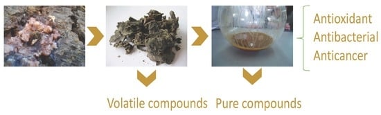
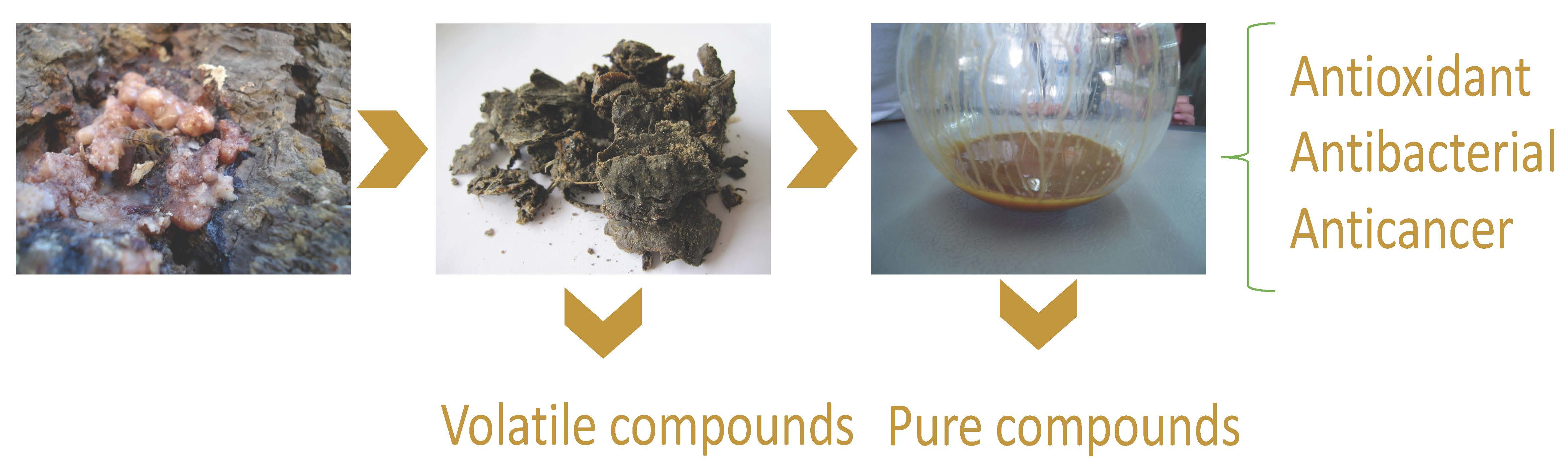
2.3. Propolis Samples

Beekeepers kindly provided four raw propolis samples from Silao and Irapuato, Guanajuato, México (Table 1). The samples were harvested using traps in 2018 and 2019 and were frozen and stored at −20 °C until analysis.

2.4. Antioxidant Activity

The EEP antioxidant activity was assessed using two different assays in vitro: DPPH and ABTS. Both methods were modified and translated into 96-well plates. Each test was done in three replicates.
Radical scavenging activity (RSA) for DPPH was evaluated according to the method described in [11]. Briefly, an ethanolic solution of 0.208 mM was added to 0.1 mL of different concentrations of extracts and pure compounds. The 96-well plate was maintained in a dark at room temperature for 20 min and the absorbance was recorded at 540 nm. The RSA was calculated as: RSA = 100 × (Acontrol − Asample)/Acontrol, where Acontrol and Asample are the absorbance. The IC50 values were calculated from the relationship curve of RSA versus concentrations of the respective sample curve.
The ABTS test was performed according to the methodology previously reported in [13,14] and slightly modified. The RSA of the ABTS radical was calculated using the following equation: % inhibition = 100 (Acontrol − Asample)/Acontrol. The IC50 was calculated from the scavenging activities (%) versus concentrations of the respective sample curve.

2.5. Total Phenol and Total Flavonoid Content

In this paper, we used spectrometric procedures for the quantification of the total phenolic and flavonoids content in propolis. The total phenol content in extracts was adapted from the method described by Singleton and Rossi [15]. The total flavonoid content was determined using the aluminum chloride reagent and the method described by Marquele et al. [16]. The total phenol content was expressed as mg equivalents of gallic acid/g of dry extract of propolis (EEP). The total flavonoid content was expressed as mg equivalents of quercetin/g of dry extract of propolis (EEP).

2.6. Extraction and Isolation of Compounds 1–12 from EEP GUA-4

The air-dried and powdered propolis samples GUA-4 were extracted with ethanol for up to one week and the resultant extract was concentrated in vacuo. A portion of ethanol-soluble extract (10.3 g) was subjected to a silica gel vacuum column chromatography (VLC) and eluted with a gradient mixture of dichloromethane–acetone (1:0 → 0:1) to give eight pooled fractions (F2–F8). Fractions F2, F4, and F7 showed the best antioxidant activity. Fraction F2 was chromatographed over a Sephadex LH-20 column and eluted with methanol to yield 1 (40 mg). Fraction F4, eluted with dichloromethane-acetone (9:1), was chromatographed over a Sephadex LH-20 column, using methanol as eluent, to give six fractions. Fraction F4-3 (100 mg) was separated by TLC with dichloromethane–acetone (99:1), followed by TLC with dichloromethane–acetone (98:2), to give 2 (20 mg) and 3 (65 mg). Fraction F5 was rechromatographed on a Sephadex LH-20 column using methanol as solvent to give six subfractions (F5-1: 10 mg; F5-2: 12.3 mg; F5-3: 17.5 mg; F5-4: 15.7 mg; F5-5: 25.3 mg; F5-6: 18.1 mg). Subfraction F5-1 yielded crystals of 4. F5-5 (25.3 mg) yielded crystals of 5 (9.0 mg). Mother liquor was subjected to a preparative thin-layer chromatography (PTLC) with acetone-toluene (5:95) to give 6. Subfraction F5-6 (18.1 mg) yielded crystals of 7 (11.0 mg). Fraction F7 was chromatographed on silica gel using a hexane–EtOAc gradient system to give five fractions (F7-1: 13.9 mg; F7-2: 517.7 mg; F7-3: 28.4 mg; F7-4: 16.7 mg; F7-5: 32.8 mg; F7-6: 56.8 mg; F7-7: 45.1 mg). Subfraction F7-1 was identified as 8. Subfraction F7-2 was chromatographed by RP-MPLC, using H2O–CH3CN (1:0 → 0:1) to afford five subfractions (F7-2.1: 125.3 mg; F7-2.2: 275.1 mg; F7-2.3: 67.8 mg; F7-2.4: 37.0 mg; F7-2.5: 12.3 mg). Subfraction F7-2.3 was identified as 9. Subfractions F7-2.4, F7-2.5, and F7-2.6 were dissolved in CH2Cl2–MeOH and left overnight to give crystals of 10, 11, and 12, respectively.

2.7. Head Space Solid Phase Microextraction (HS-SPME), GC-MS Analysis, and Identification of Volatile Components

The HS-SPME was performed using a Stableflex® fiber 50/30 μm DVB/CAR/PDMS (1 cm) as described in our previous work [17].
The analysis of volatile compounds was carried out on a GC Agilent 6890 N (Agilent Technology, Santa Clara, CA, USA) series gas chromatograph coupled to a LECO time of flight mass spectrometer (LECO Corporation, St. Joseph, MI, USA). The volatile compounds were separated on a 5% diphenyl-95% dimethyl polysiloxane (30 m × 0.18 mm i.d.; 0.18 μm film thickness) capillary column (Bellefonte, PA, USA). The carrier gas was helium with a flow rate of 1 mL/min and split ratio of 1:50. The column initial temperature was 40 °C. It was then raised to 300 °C with a rate of 20 °C/min and was held for 5 min. The ionization electron energy was 70 eV and the mass range scanned was 40–400 m/z. The injector and MS transfer were set at 300 and 250 °C, respectively. The volatile constituents of propolis were identified by co-injection of the sample with standard samples when available; based on their Kovats Index, calculated in relation to the retention times of a series of alkanes (C-8–C-20), in comparison with those of the chemical compounds gathered by Adams [18] and by comparing their MS fragmentation patterns with those of pure compounds in the spectral database of the National Institute of Standards and Technology (NIST) [19].

2.8. Cell Culture

Rat C6 glioma cell and human cervical cancer cell lines (HeLa, SiHa, and CaSki) were obtained from American Type Culture Collection (Manassas, VA, USA). Cells were routinely maintained in Dulbecco’s modified Eagle´s medium (DMEM) supplemented with fetal bovine serum (Gibco BRL) with 5% for C6 cells and 10% for cervical cancer cells, and incubated at 37 °C in an atmosphere comprising 5% CO2 and 95% air at high humidity. Cells were harvested with 0.025% trypsin and 0.01% EDTA (Gibco BRL).
The effect of EEP, cisplatin, and temozolomide on the proliferation of cells were evaluated using the MTT assay (3-(3,5-dimethylthiazol-2-yl)2,5-diphenyltetrazolium), which is based on the reduction of a tetrazolium salt in metabolically active cells. The procedure was as follows. Viable cells were seed into 96-well plates in 100 µL per well of DMEM culture medium at a density of 3 × 10³ for C6 cells, 2 × 10⁴ for CaSki cells, and 1 × 10⁴ cells for HeLa and SiHa cells. After treatment, the medium was removed and the MTT solution was added to each well, followed by 1 to 2 h in a humidified atmosphere containing 5% CO2 at 37 °C. The absorbance of the samples was measured spectrophotometrically at λ 570 nm using a microtiter plate ELISA reader. Results are expressed as the percentage of MMT reduction.
C6 cells were exposed for 72 h with 2 to 200 µg/mL of EEP, since it is the time used to make the exposure with temozolomide (first line treatment used for glioblastoma), whereas HeLa, SiHa, and CaSki cells were exposed for 24 h with 15 to 500 µg/mL of EEP; after that time, cell viability was quantified using the MTT assay. Temozolomide (250 µM) and cisplatin (5–320 µM) were used as a control. The concentration of drugs to reach 50% growth inhibition (IC50) was obtained from the survival curves. The experiments were conducted in triplicate in independent experiments. Values are expressed as the mean ± SEM of at least three independent experiments. SigmaPlot 12.3 software (Systat Software, Santa Clara, CA, USA) was used.

2.9. Antibacterial Activity

The in vitro antibacterial activity of EEP and compounds 14, 1013, and 1517 were determined using a broth microdilution test as recommended by Clinical and Laboratory Standards Institute (New York, NY, USA) M7-A11 for bacteria [20]. The minimum inhibitory concentration (MIC) was defined as the lowest concentration of the test agent that had restricted growth to a level <0.05 at 660 nm after incubation at 37 °C for 16–24 h.

3. Results and Discussion

3.1. Total Phenol and Flavonoid Content

The total polyphenol contents for the selected propolis samples were found to be 178.9 ± 5.7, 198.4 ± 4.1, 167.6 ± 7.8, and 246.3 ± 3.2 mg GAE/g of dry extract for GUA-1, GUA-2, GUA-3, and GUA-4, respectively. It should be noted that the Folin-Ciolcateau reagent was reported in the literature as not specific to only phenols and could react with other reducing compounds that could be oxidized by the Folin reagent [21]. The total flavonoid contents were 64.32 ± 5.75, 58.34 ± 2.8, 77.45 ± 6.9, and 87.5 ± 1.9 mg QE/g of dry extract for GUA-1, GUA-2, GUA-3, and GUA-4, respectively. Flavonoids can form complexes with aluminum chloride to yield a yellow solution. Valencia et al. [22] has been reported values of 629.6 ± 9.9 mg GAE/g of dry extract for TPC and 185.9 ± 3.2 mg QE/g of dry extract and TF in a propolis from Sonora, México. The most important flavonoids isolated in this propolis sample were pinocembrin, pinobanksin 3-acetate, and chrysin. The results also confirm the influence of the geographic region and the season of collection for the quality and properties of the propolis [2].

3.2. Antioxidant Activity Assays

The EEP was evaluated for its ability to quench the DPPH., which is one of the few stable organic nitrogen radicals and bears a purple colour. This assay is based on the measurement of the loss of DPPH after reaction with samples. It is considered as the prior mechanism involved in the electron transfer. The IC50 value is a parameter widely used to measure the antioxidant activity of test samples. It is calculated as the concentration of antioxidants needed to decrease the initial DPPH concentration by 50% [23]. Thus, the lower IC50 value the higher antioxidant activity. The EEP GUA-4 showed an antioxidant activity (IC50 = 67.9 μg/mL) comparable to the reference ascorbic acid (IC50 = 43.2 μg/mL) and tenfold lower than Trolox (IC50 = 6.3 μg/mL) and seven-fold lower than quercetin (IC50 = 9.9 μg/mL). While caffeic acid (12) showed the highest activity (IC50 = 5.9 μg/mL) along with ferulic acid (10) (IC50 = 9.9 μg/mL) and syringic acid (11) (IC50 = 9.8 μg/mL). The lowest antioxidant activity corresponds to 5-methylchrysin ether (9) (IC50 = 112.9 μg/mL) and 5-methyl-pinobanksin ether (5) (IC50 = 98.4 μg/mL).
There are reports of the DPPH scavenging capacity, in terms of IC50, for pinocembrin (1), chrysin (2), galangin (3), isorhamnetin (7), ferulic acid (10), syringic acid, (11) and caffeic acid (12) [23,24,25,26]. For alpinetin (4) only the percentage of scavenging activity of DPPH is reported [27]. Nevertheless, the information available for dillenetin (6), 5-methyl-pinobanksin ether (5), 5-methylchrysin ether (9), and 5-methylgalangin ether (8) is scarce. Thus, the present work stands for the first report of the IC50 as a measurement of their antioxidant activity (Table 2).
In the ABTS assay, the antioxidant activity is measured as the ability of test compounds to decrease the color reacting directly with the radical ABTS•+ [28]. Ferulic acid (10), syringic acid (11), caffeic acid (12), and chrysin (2) showed the lowest IC50 (Table 2). The EEP, 5-methylchrysin ether (9), and 5-methyl-pinobanksin ether (5) showed weak antioxidant activity (Table 2).
The analysis of their chemical structures can explain the weak antioxidant activity of the methylated flavonoids. According to Procházková et al. [29] the catechol structure in the B-ring, 2,3-double bond in conjugation with a 4-oxo function in the C-ring and the hydroxyl groups in meta position in ring-A. On the other hand, the activity of phenolic acids lies on the presence of two o-hydroxyl groups in the aromatic ring.

3.3. Cytotoxicity of EEP on Cancer Cells

Cytotoxicity was expressed as the percentage grow inhibition of C6, HeLa, SiHa, and CaSki cells treated with EEP. In all the cases, EEP shows a cytotoxicity concentration-depended manner. In Table 3, we show the IC50 value of EEP over four cancer cell lines. Those results show that EEP restricts glioblastoma cells (C6 cell cancer line) proliferation in vitro as efficiently as temozolomide (reference drug), whereas, for cervical cancer cell lines, it requires a higher concentration of the EEP compared to cisplatin.
There are a few studies of beneficial properties of Mexican propolis. Li et al. [11] reported that three of the 39 compounds isolated from the methanolic extract of Mexican propolis exhibited a potent cytotoxic effect in a colon, melanoma, lung, cervix, and fibrosarcoma cancer cell lines. Li et al. reported the isolation of flavonoids from methanolic extract of Mexican propolis, and one of them revealed significant cytotoxic effect against pancreatic human cancer cell line with IC50 values of 4 μM [10]. Other studies described the potent cytotoxic activity of galangin (3); ferulic acid (10); syringic acid (11); and caffeic acid (12) against different cancer cell lines [22]. Interestingly, a few researchers reported that these compounds could be useful for therapeutic treatments. For example, Benguedouar et al. [30] reported that ethanolic extract of Algerian propolis (EEP) and galangin (3) decreased the number of B16F1 melanoma cells in vitro compared to control. Celinska-Janowicz et al. [31] state that the ethanolic extract of propolis isolated abundant polyphenolic compounds such as ferulic acid (10) and caffeic acid (12) revealed pro-apoptotic activity on human tongue squamous carcinoma cells (CAL-27). From this information, we emphasized that EEP and compounds possesses anti-cancer effects against cancer cell lines.

3.4. Antibacterial Activity

As shown in Table 4, antimicrobial screening against four oral pathogens revealed that compounds 1, 3, 4, 12, and 16 were inhibitory to the growth of Streptococcus mutans, Streptococcus oralis, Streptococcus sanguinis, and Phorphyromonas gingivalis. Among these, compounds 1, 3, 4, and 12, were either equally or more potent than their respective crude extract of origin (Table 4). Earlier in vitro studies have shown that the Sonoran ethanolic extract of propolis exhibited antibacterial activity against E. coli (ATCC 25922) and S. aureus (ATCC 6538P). The propolis constituents CAPE, pinocembrin, pinobanksin 3-O-acetate, and naringenin exhibited significant inhibitory activity on the growth of S. aureus. CAPE exhibited the maximum inhibitory effect on the bacterial growth (CAPE (MIC 0.1 mmol/L), pinocembrin (MIC 0.4 mmol/L), pinobanksin 3-O-acetate (MIC 0.8 mmol/mL), and naringenin (0.8 mmol/L)). None of the propolis constituents influenced the growth of E. coli at any of the tested concentrations [32].

3.5. Chemical Composition of EEP GUA-4

The EtOH-soluble extract of sample GUA-4 was fractionated by chromatography on a VLC column and by Sephadex LH-20, giving 12 known compounds (112). The compounds were identified as pinocembrin (1, 405.4 mg) [33], chrysin (2, 126.3 mg) [34], galangin (3, 56.7 mg) [33], alpinetin (4, 15.6 mg) [35], 5-methyl-pinobanksin ether (5, 16.8 mg) [36], dillenetin (6, 12.3 mg) [37], isorhamnetin (7, 24.6 mg) [38], 5-methylgalangin ether (8, 9.8 mg) [39], 5-methylchrysin ether (9, 16.3 mg) [40], ferulic acid (10, 24.9 mg) [33], syringic acid (11, 6.7 mg), and caffeic acid (12, 8.9 mg) [33] (Figure S1) by means of 1D (Table S1) and 2D NMR spectral analysis. The presence of pinocembrin (1), chrysin (2), galangin (3), ferulic acid (10), syringic acid (11), and caffeic acid (12) suggest that its main botanical source are a species of Populus typical of the country, such as Populus mexicana Wesmael, Populus guzmanantlensis Vazques and Cuevas and Populus simaroa Rzedowski. Although flavonoids without B-ring substituents appear common in temperate propolis from both the northern and southern hemispheres, in this study we report for the first time the presence of dillenetin (6) and isorhamnetin (7) with B-ring substitution as constituents of Mexican poplar propolis. To the best of our knowledge, this is the first report of dillenetin (6) as a propolis constituent. 5-methylpinobanksin (5), alpinetin (4), isorhamnetin (7), 5-methylgalangin ether (8), and 5-methylchrysin ether (9) were previously identified by diode array detection and ESI mass spectrometry (LC-DAD-ESI-MS) as constituents of Italian, Portuguese, and Czech propolis [41,42].

3.6. Volatile Compounds

Forty volatile constituents were identified as shown in Table 5. The typical analytical ion current (AIC) chromatogram is shown in Figure S2. The chemical composition of volatiles was found in agreement with previous reports [43]. The main volatile compounds identified were nonanal (18.829%, 13), β-pinene (12.179%, 14), 1-octen-3-ol (12.129%, 15), neryl alcohol (10.135%, 16), α-pinene (8.046%, 17), 6-methyl-3,5-heptadiene-2-one (6.803%, 18), p-cymen-9-ol (3.108%, 19), and sylvestrene (3.022 %, 20) (Figure S3). It is important to note that the content of the compounds differed from a previous study carried out on the volatile compounds in propolis samples from Yucatan (México). In that sample, hexadecanoic acid (10.9%) and trans-verbenol (7.0%) were the predominant compounds [44].

4. Conclusions

In the present study, we have isolated twelve (112) components and identified 40 volatile compounds in Mexican propolis. The current study revealed the presence of antioxidant, antimicrobial, and cytotoxic phytochemicals [galangin (3); ferulic acid (10); syringic acid (11); and caffeic acid (12)] in EEP. It is concluded that EEP can be a potential addition in pharmaceutical products for the improvement of human health by contributing in the antioxidant defense system fighting against the production of free radicals. México, being a megadiverse country, has numerous numbers of propolis differing in chemical composition. However, unfortunately it is still unexplored.

Supplementary Materials

The following are available online at https://www.mdpi.com/2076-3921/9/1/70/s1, Figure S1: Flavonoids isolated from EEP GUA-4; Figure S2: Analytical ion current chromatogram (AIC) of propolis GUA-4; Figure S3: Major volatile compounds of EEP GUA-4; Table S1: 1H-RMN data of the flavonoids isolated from Mexican propolis.

Author Contributions

J.G.-P. performed the phytochemical study; J.F.R.-C. conceived and designed the phytochemical experiments and wrote the paper; J.M.P.-R. conceived and designed the in vitro experiments; A.K.-P.reviewed of the manuscript and contributed with in vitro experiments; G.D.-R. contributed with the antimicrobial assays; J.P.-C. reviewed the manuscript and contributed with in vitro experiments; B.E.R.-C. reviewed of the manuscript and contributed with in vitro experiments. All authors have read and agreed to the published version of the manuscript.

Funding

This research was funded by CONACyT CB-252006 and College of Chemistry, National Autonomous University of Mexico under the PAIP 5000-9138 Fellowship Scheme.

Acknowledgments

We are in debt to Médico Veterinario Zootecnista. Ángel López-Ramírez for technical assistance in propolis recolection. Jessica Granados Pineda is a doctoral student from the Programa de Doctorado en Ciencias Químicas, Universidad Nacional Autónoma de México (UNAM), Facultad de Química, and recieved a CONACyT fellowship # 273461. The authors wish to thank the technical assistance of Georgina Duarte-Lisci and Juan Rojas-Moreno.

Conflicts of Interest

The authors declare no conflict of interest.

References

  1. Bankova, V.; Bertelli, D.; Borba, R.; Conti, B.J.; da Silva Cunha, I.B.; Danert, C.; Eberlin, M.N.; I Falcão, S.; Isla, M.I.; Moreno, M.I.N.; et al. Standard methods for Apis mellifera propolis research. J. Apic. Res. 2016, 58, 1–49. [Google Scholar] [CrossRef] [Green Version]
  2. Salatino, A.; Fernandes-Silva, C.C.; Righi, A.A.; Salatino, M.L. Propolis research and the chemistry of plant products. Nat. Prod. Res. 2011, 28, 925–936. [Google Scholar] [CrossRef] [PubMed]
  3. Kuropatnicki, A.K.; Szliszka, E.; Krol, W. Historical aspects of propolis research in modern times. Evid. Based Cmnplement. Altern. Med. eCAM 2013, 2013, 964149. [Google Scholar] [CrossRef] [PubMed] [Green Version]
  4. Toreti, V.C.; Sato, H.H.; Pastore, G.M.; Park, Y.K. Recent progress of propolis for its biological and chemical compositions and its botanical origin. Evid. Based Cmnplement. Altern. Med. eCAM 2013, 2013, 697390. [Google Scholar] [CrossRef]
  5. Christov, R.; Trusheva, B.; Popova, M.; Bankova, V.; Bertrand, M. Chemical composition of propolis from canada, its antiradical activity and plant origin. Nat. Prod. Res. 2006, 20, 531–536. [Google Scholar] [CrossRef]
  6. Karapetsas, A.; Voulgaridou, G.-P.; Konialis, M.; Tsochantaridis, I.; Kynigopoulos, S.; Lambropoulou, M.; Stavropoulou, M.-I.; Stathopoulou, K.; Aligiannis, N.; Bozidis, P.; et al. Propolis extracts inhibit UV-induced photodamage in human experimental in vitro skin models. Antioxidants 2019, 8, 125. [Google Scholar] [CrossRef] [Green Version]
  7. Nna, V.U.; Abu Bakar, A.B.; Ahmad, A.; Eleazu, C.O.; Mohamed, M. Oxidative stress, NF-κb-mediated inflammation and apoptosis in the testes of streptozotocin–induced diabetic rats: Combined protective effects of malaysian propolis and metformin. Antioxidants 2019, 8, 465. [Google Scholar] [CrossRef] [Green Version]
  8. Papotti, G.; Bertelli, D.; Bortolotti, L.; Plessi, M. Chemical and functional characterization of italian propolis obtained by different harvesting methods. J. Agric. Food. Chem. 2012, 60, 2852–2862. [Google Scholar] [CrossRef]
  9. Navarro-Navarro, M.; Ruiz-Bustos, P.; Valencia, D.; Robles-Zepeda, R.; Ruiz-Bustos, E.; Virues, C.; Hernandez, J.; Dominguez, Z.; Velazquez, C. Antibacterial activity of sonoran propolis and some of its constituents against clinically significant vibrio species. Foodborne Pathog. Dis. 2013, 10, 150–158. [Google Scholar] [CrossRef]
  10. Li, F.; Awale, S.; Tezuka, Y.; Esumi, H.; Kadota, S. Study on the constituents of mexican propolis and their cytotoxic activity against panc-1 human pancreatic cancer cells. J. Nat. Prod. 2010, 73, 623–627. [Google Scholar] [CrossRef]
  11. Li, F.; Awale, S.; Tezuka, Y.; Kadota, S. Cytotoxicity of constituents from mexican propolis against a panel of six different cancer cell lines. Nat. Prod. Commun. 2010, 5, 1601–1606. [Google Scholar] [CrossRef] [PubMed] [Green Version]
  12. Lotti, C.; Campo Fernandez, M.; Piccinelli, A.L.; Cuesta-Rubio, O.; Marquez Hernandez, I.; Rastrelli, L. Chemical constituents of red mexican propolis. J. Agric. Food Chem. 2010, 58, 2209–2213. [Google Scholar] [CrossRef] [PubMed]
  13. Cheng, Z.; Moore, J.; Yu, L. High-throughput relative DPPH radical scavenging capacity assay. J. Agric. Food Chem. 2006, 54, 7429–7436. [Google Scholar] [CrossRef] [PubMed]
  14. Zhao, H.; Fan, W.; Dong, J.; Lu, J.; Chen, J.; Shan, L.; Lin, Y.; Kong, W. Evaluation of antioxidant activities and total phenolic contents of typical malting barley varieties. Food Chem. 2007, 107, 296–304. [Google Scholar] [CrossRef]
  15. Singleton, V.L.; Rossi, J.A. Colorimetry of total phenolics with phosphomolybdic-phosphotungstic acid reagents. Am. J. Enol. Vit. 1965, 16, 144–158. [Google Scholar]
  16. Marquele, F.D.; Di Mambro, V.M.; Georgetti, S.R.; Casagrande, R.; Valim, Y.M.L.; Fonseca, M.J.V. Assessment of the antioxidant activities of brazilian extracts of propolis alone and in topical pharmaceutical formulations. J. Pharm. Biomed. Anal. 2005, 39, 455–462. [Google Scholar] [CrossRef]
  17. Torres-González, A.; López-Rivera, P.; Duarte-Lisci, G.; López-Ramírez, Á.; Correa-Benítez, A.; Rivero-Cruz, J.F. Analysis of volatile components from Melipona beecheii geopropolis from Southeast Mexico by headspace solid-phase microextraction. Nat. Prod. Res. 2016, 30, 237–240. [Google Scholar] [CrossRef]
  18. Adams, R.P. Identification of Essential Oil Components by Gas Chromatography/Mass Spectrometry; Allured Publishing Corporation: Carol Stream, IL, USA, 2007. [Google Scholar]
  19. Linstrom, P.J. NIST Chemistry Webbook, NIST Standard Reference Database Number 69; National Institute of Standards and Technology: Gaithersburg, MD, USA, 2005. [Google Scholar]
  20. Clinical and Laboratory Standards Institute. Methods for Dilution Antimicrobial Susceptibility Tests for Bacteria that Grow Aerobically, Approved Standard, 10th ed.; M07-A11; Clinical and Laboratory Standards Institute: Wayne, PA, USA, 2018. [Google Scholar]
  21. Escarpa, A.; González, M. Approach to the content of total extractable phenolic compounds from different food samples by comparison of chromatographic and spectrophotometric methods. Anal. Chim. Acta 2001, 427, 119–127. [Google Scholar] [CrossRef]
  22. Valencia, D.; Alday, E.; Robles-Zepeda, R.; Garibay-Escobar, A.; Galvez-Ruiz, J.C.; Salas-Reyes, M.; Jimenez-Estrada, M.; Velazquez-Contreras, E.; Hernandez, J.; Velazquez, C. Seasonal effect on chemical composition and biological activities of sonoran propolis. Food Chem. 2012, 131, 645–651. [Google Scholar] [CrossRef]
  23. Sánchez-Moreno, C.; Larrauri, J.A.; Saura-Calixto, F. A procedure to measure the antiradical efficiency of polyphenols. J. Sci. Food Chem. 1998, 76, 270–276. [Google Scholar] [CrossRef]
  24. Lima, B.; Tapia, A.; Luna, L.; Fabani, M.P.; Schmeda-Hirschmann, G.; Podio, N.S.; Wunderlin, D.A.; Feresin, G.E. Main flavonoids, dpph activity, and metal content allow determination of the geographical origin of propolis from the province of San Juan (Argentina). J. Agric. Food Chem. 2009, 57, 2691–2698. [Google Scholar] [CrossRef] [PubMed]
  25. Tominaga, H.; Kobayashi, Y.; Goto, T.; Kasemura, K.; Nomura, M. DPPH radical-scavenging effect of several phenylpropanoid compounds and their glycoside derivatives. Yakugaku Zasshi J. Pharm. Soc. Jpn. 2005, 125, 371–375. [Google Scholar] [CrossRef] [PubMed] [Green Version]
  26. Pengfei, L.; Tiansheng, D.; Xianglin, H.; Jianguo, W. Antioxidant properties of isolated isorhamnetin from the sea buckthorn marc. Plant Foods Hum. Nutr. 2009, 64, 141–145. [Google Scholar] [CrossRef] [PubMed]
  27. Lu, H.T.; Zou, Y.L.; Deng, R.; Shan, H. Extraction, purification and antiradical activities of alpinetin and cardamomin from alpinia katsumadai hayata. Asian J. Chem. 2013, 25, 9503–9507. [Google Scholar] [CrossRef]
  28. Prior, R.L.; Wu, X.; Schaich, K. Standardized methods for the determination of antioxidant capacity and phenolics in foods and dietary supplements. J. Agric. Food Chem. 2005, 53, 4290–4302. [Google Scholar] [CrossRef]
  29. Procházková, D.; Boušová, I.; Wilhelmová, N. Antioxidant and prooxidant properties of flavonoids. Fitoterapia 2011, 82, 513–523. [Google Scholar] [CrossRef]
  30. Benguedouar, L.; Lahouel, M.; Gangloff, S.C.; Durlach, A.; Grange, F.; Bernard, P.; Antonicelli, F. Ethanolic extract of Algerian propolis and galangin decreased murine melanoma T. Anticancer Agents Med. Chem. 2016, 16, 1172–1183. [Google Scholar] [CrossRef]
  31. Celińska-Janowicz, K.; Zaręba, I.; Lazarek, U.; Teul, J.; Tomczyk, M.; Pałka, J.; Miltyk, W. Constituents of Propolis: Chrysin, Caffeic Acid, p-Coumaric Acid, and Ferulic Acid Induce PRODH/POX-Dependent Apoptosis in Human Tongue Squamous Cell Carcinoma Cell (CAL-27). Front. Pharmacol. 2018, 9, 336. [Google Scholar] [CrossRef] [Green Version]
  32. Velazquez, C.; Navarro, M.; Acosta, A.; Angulo, A.; Dominguez, Z.; Robles, R.; Robles-Zepeda, R.; Lugo, E.; Goycoolea, F.M.; Velazquez, E.F.; et al. Antibacterial and free-radical scavenging activities of Sonoran propolis. J. Appl. Microbiol. 2007, 103, 1747–1756. [Google Scholar] [CrossRef]
  33. Bertelli, D.; Papotti, G.; Bortolotti, L.; Marcazzan, G.L.; Plessi, M. 1H-NMR simultaneous identification of health-relevant compounds in propolis extracts. Phytochem. Anal. 2012, 23, 260–266. [Google Scholar] [CrossRef]
  34. Wawer, I.; Zielinska, A. 13c cp/mas nmr studies of flavonoids. Magn. Reson. Chem. 2001, 39, 374–380. [Google Scholar] [CrossRef]
  35. Dominguez, X.A.; Franco, R.; Zamudio, A.; Barradas, D.D.M.; Watson, W.H.; Zabel, V.; Merijanian, A. Mexican medicinal plants. Part 38. Flavonoids from Dalea scandens var. Paucifolia and Dalea thyrsiflora. Phytochemistry 1980, 19, 1262–1263. [Google Scholar] [CrossRef]
  36. Rossi, M.H.; Yoshida, M.; Soares Maia, J.G. Neolignans, styrylpyrones and flavonoids from an aniba species. Phytochemistry 1997, 45, 1263–1269. [Google Scholar] [CrossRef]
  37. Hosny, M.; Dhar, K.; Rosazza, J.P.N. Hydroxylations and methylations of quercetin, fisetin, and catechin by Streptomyces griseus. J. Nat. Prod. 2001, 64, 462–465. [Google Scholar] [CrossRef] [PubMed]
  38. Cao, X.; Wei, Y.; Ito, Y. Preparative isolation of isorhamnetin from Stigma maydis using high-speed countercurrent chromatography. J. Liq. Chromatogr. Relat. Technol. 2009, 32, 273–280. [Google Scholar] [CrossRef] [PubMed] [Green Version]
  39. Nagy, M.; Suchy, V.; Uhrin, D.; Ubik, K.; Budesinsky, M.; Grancai, D. Constituents of propolis of Czechoslovak origin. V. Chem. Pap. 1988, 42, 691–696. [Google Scholar]
  40. Falcão, S.; Vilas-Boas, M.; Estevinho, L.; Barros, C.; Domingues, M.M.; Cardoso, S. Phenolic characterization of northeast portuguese propolis: Usual and unusual compounds. Anal. Bioanal. Chem. 2010, 396, 887–897. [Google Scholar] [CrossRef] [Green Version]
  41. Gardana, C.; Scaglianti, M.; Pietta, P.; Simonetti, P. Analysis of the polyphenolic fraction of propolis from different sources by liquid chromatography–tandem mass spectrometry. J. Pharm. Biomed. Anal. 2007, 45, 390–399. [Google Scholar] [CrossRef]
  42. Falcao, S.I.; Vale, N.; Gomes, P.; Domingues, M.R.M.; Freire, C.; Cardoso, S.M.; Vilas-Boas, M. Phenolic profiling of portuguese propolis by LC-MS spectrometry: Uncommon propolis rich in flavonoid glycosides. Phytochem. Anal. 2013, 24, 309–318. [Google Scholar] [CrossRef] [Green Version]
  43. Pellati, F.; Prencipe, F.P.; Benvenuti, S. Headspace solid-phase microextraction-gas chromatography-mass spectrometry characterization of propolis volatile compounds. J. Pharm. Biomed. Anal. 2013, 84, 103–111. [Google Scholar] [CrossRef]
  44. Pino, J.A.; Marbot, R.; Delgado, A.; Zumarraga, C.; Sauri, E. Volatile constituents of propolis from honey bees and stingless bees from Yucatan. J. Essent. Oil Res. 2006, 18, 53–56. [Google Scholar] [CrossRef]
Table 1. Location of recollection, harvesting method, and weight recollected.
CodeLocation of RecollectionYear of RecollectionHarvesting Method Weight (g)
GUA-1Silao2018plastic nest6.0
GUA-2Irapuato2018plastic nest8.50
GUA-3Irapuato2019plastic nest6.95
GUA-4Silao2019plastic nest32.0
Table 2. Scavenging ability of ethanol extract of propolis (EEP) GUA-4 and compounds 112.
CompoundsIC50 (μg/mL)
DPPHABTS
EEP GUA-467.9 ± 0.198.7 ± 0.5
pinocembrin (1)23.5 ± 1.844.8 ± 2.1
chrysin (2)10.7 ± 0.216.3 ± 0.8
galangin (3)15.3 ± 0.526.8 ± 0.1
alpinetin (4)47.3 ± 1.969.5 ± 0.7
5-methylpinobanksin ether (5)98.4 ± 2.3126.9 ± 4.5
dillenetin (6)35.7 ± 3.548.9 ± 2.9
isorhamnetin (7)21.4 ± 3.136.7 ± 4.6
5-methylgalangin ether (8)63.2 ± 2.694.2 ± 6.2
5-methylchrysin ether (9)112.9 ± 1.9 158.4 ± 4.9
ferulic acid (10)9.9 ± 0.716.7 ± 0.2
syringic acid (11)9.8 ± 0.413.0 ± 0.9
caffeic acid (12)5.9 ± 0.49.7 ± 0.5
Quercetin9.9 ± 2.516.1 ± 2.1
Trolox6.3 ± 1.43.8 ± 1.2
ascorbic acid43.2 ± 10.336.8 ± 2.5
Table 3. Cytotoxic effect of EEP on several cancer cell lines.
Cell LineIC50Reference DrugReference Concentration
C692.2 µg/mLTemozolamideIC30 250 µM
(50 µg/mL)
HeLa>100 µg/mL
(357 µg/mL)
CisplatinIC50 46 µM
(14 µg/mL)
SiHa>100 µg/mL
(500 µg/mL)
IC50 121 µM
(36 µg/mL)
CaSki>100 µg/mL
(538 µg/mL)
IC50 163 µM
(50 µg/mL)
Table 4. Antimicrobial activity of ethanolic extract and compounds 14, 1013, 1517, and chlorhexidine digluconate (CHX) a.
CompoundsMIC b (μg/mL)
S. mutansS. oralisS. sanguinisP. gingivalis
EEP250125125500
EOP5005005001000
pinocembrin (1)256128128512
chrysin (2)5125125121024
galangin (3)2562562561024
alpinetin (4)128256128516
ferulic acid (10)500250250500
syringic acid (11)250250250500
caffeic acid (12)128128128256
nonanal (13)256256128512
neryl alcohol (15)10245125121024
α-pinene (16)250250250500
α-pinene (17)500500500500
a CHX0.020.020.020.12
a positive control; b minimum inhibitory concentration.
Table 5. Volatile components from Mexican propolis GUA-4.
NameRetention IndexArea %Method of Identification
1α-pinene 9398.046a, b, c
2β-pinene97912.179a, b, c
31-octen-3-ol98212.129a, b
4ethyl hexanoate10030.042a, b, c
5Octanal10061.273a, b
6Sylvestrene10303.022a, b
7acetophenone 10760.486a, b
81-octanol10780.107a, b
9Thujone11020.276a, b
10p-cymenene10820.291a, b
11Nonanal110018.829a, b, c
126-methyl-3,5-heptadiene-2-one11056.803a, b
13Eucalyptol10390.472a, b, c
14Camphor11460.583a, b, c
15trans-pinocamphone11622.386a, b
16trans-terpineol11630.102a, b
17p-menth-1,5-dien-8-ol11671.097a, b
18(2E)-nonenal11681.097a, b
19m-cymen-8-ol11801.024a, b, c
20Unknown 111850.064 -
21α-terpineol11891.571a, b
22Myrtenol11931.571a, b
23d-fenchone11871.828a, b
24p-cymen-9-ol12073.108a, b, c
25neryl alcohol121710.135a, b
26trans-carveol12192.953a, b
27Citronellol12281.916a, b
28Ocimenone12300.727a, b
29cis-chrysanthenyl acetate12530.243a, b
30Geraniol12780.314a, b
31neodehydro carveol acetate13071.843a, b
32ethyl nonanoate13191.283a, b
33trans-carvyl acetate13370.357a, b
34α-longipinene13520.181a, b
35β-cububene13880.248a, b
36β-bourbonene13980.217a, b
37n-decyl acetate14080.109a, b
38trans-geranylacetone14550.429a, b
39γ-gelinene14850.103a, b
40β-bisabolene15090.290a, b
a: Retention time; b: Retention index; c: Mass spectrum.

