Review

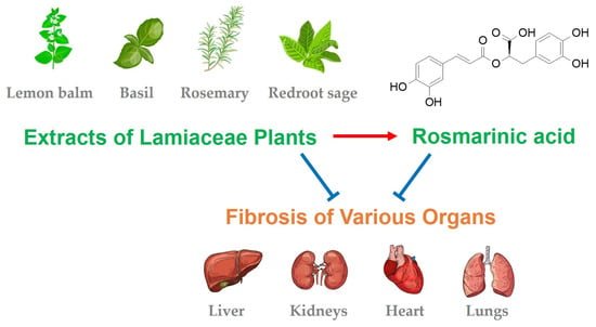
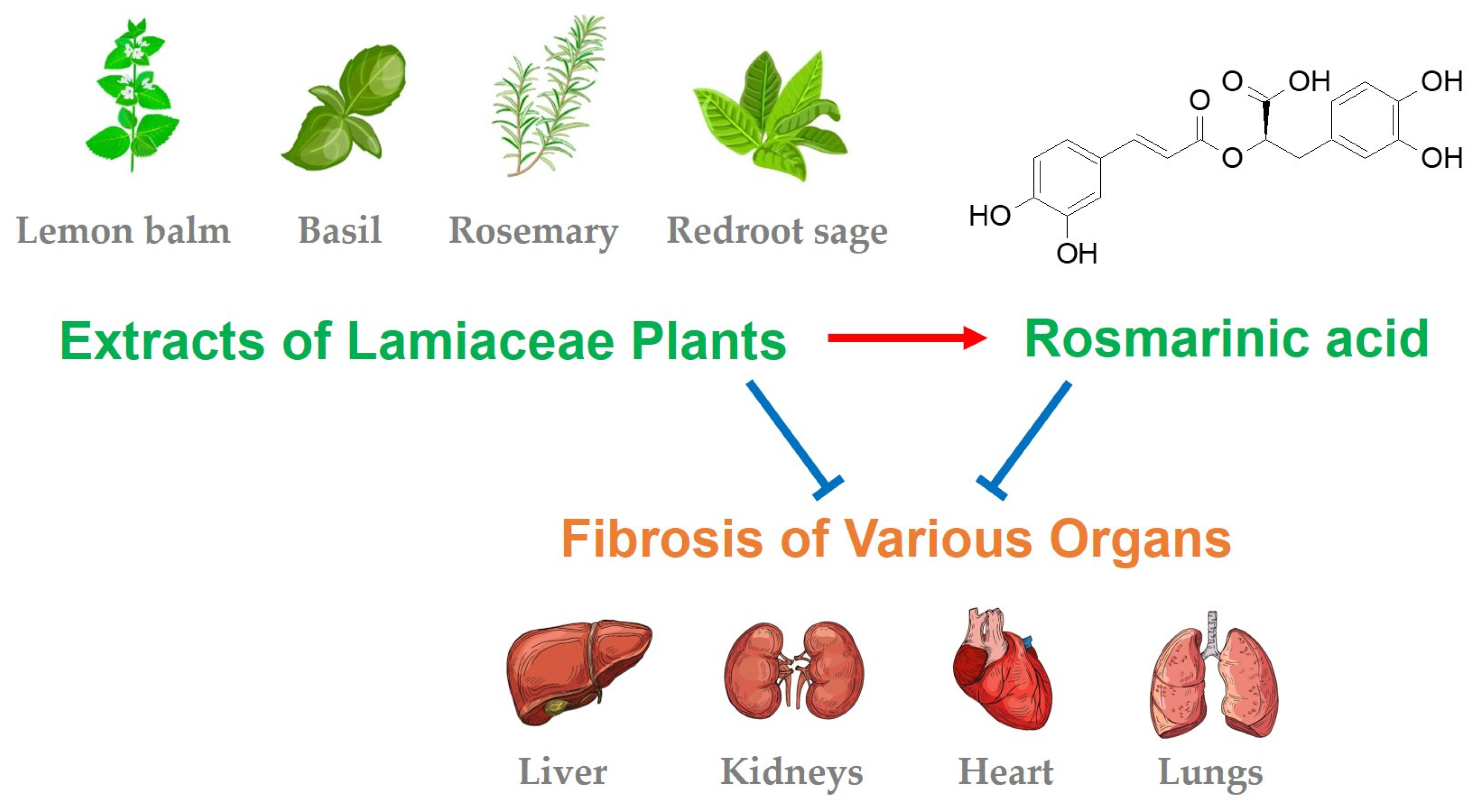
Therapeutic Potential and Mechanisms of Rosmarinic Acid and the Extracts of Lamiaceae Plants for the Treatment of Fibrosis of Various Organs

by
Yong Chool Boo
1,2,3
1
Department of Molecular Medicine, School of Medicine, Kyungpook National University, 680 Gukchaebosang-ro, Jung-gu, Daegu 41944, Republic of Korea
2
BK21 Plus KNU Biomedical Convergence Program, Department of Biomedical Science, The Graduate School, Kyungpook National University, 680 Gukchaebosang-ro, Jung-gu, Daegu 41944, Republic of Korea
3
Cell and Matrix Research Institute, Kyungpook National University, 680 Gukchaebosang-ro, Jung-gu, Daegu 41944, Republic of Korea
Antioxidants 2024, 13(2), 146; https://doi.org/10.3390/antiox13020146
Submission received: 11 December 2023 / Revised: 22 January 2024 / Accepted: 23 January 2024 / Published: 24 January 2024

Abstract

:
Fibrosis, which causes structural hardening and functional degeneration in various organs, is characterized by the excessive production and accumulation of connective tissue containing collagen, alpha-smooth muscle actin (α-SMA), etc. In traditional medicine, extracts of medicinal plants or herbal prescriptions have been used to treat various fibrotic diseases. The purpose of this narrative review is to discuss the antifibrotic effects of rosmarinic acid (RA) and plant extracts that contain RA, as observed in various experimental models. RA, as well as the extracts of Glechoma hederacea, Melissa officinalis, Elsholtzia ciliata, Lycopus lucidus, Ocimum basilicum, Prunella vulgaris, Salvia rosmarinus (Rosmarinus officinalis), Salvia miltiorrhiza, and Perilla frutescens, have been shown to attenuate fibrosis of the liver, kidneys, heart, lungs, and abdomen in experimental animal models. Their antifibrotic effects were associated with the attenuation of oxidative stress, inflammation, cell activation, epithelial–mesenchymal transition, and fibrogenic gene expression. RA treatment activated peroxisomal proliferator-activated receptor gamma (PPARγ), 5′ AMP-activated protein kinase (AMPK), and nuclear factor erythroid 2-related factor 2 (NRF2) while suppressing the transforming growth factor beta (TGF-β) and Wnt signaling pathways. Interestingly, most plants that are reported to contain RA and exhibit antifibrotic activity belong to the family Lamiaceae. This suggests that RA is an active ingredient for the antifibrotic effect of Lamiaceae plants and that these plants are a useful source of RA. In conclusion, accumulating scientific evidence supports the effectiveness of RA and Lamiaceae plant extracts in alleviating fibrosis and maintaining the structural architecture and normal functions of various organs under pathological conditions.

Graphical Abstract

1. Introduction

In 2007, the mechanism of action of Wen-pi-tang-Hab-Wu-ling-san (WHW), which is an oriental herbal prescription currently used in Korea for the treatment of chronic renal failure, was investigated in our laboratory [1]. WHW extract inhibited the expression of alpha-smooth muscle actin (α-SMA) and the phosphorylation of small mothers against decapentaplegic (SMAD) 2 that were induced by transforming growth factor (TGF) β1 in Madin–Darby canine kidney cells (MDCKs), thus supporting its renal protective effects via the inhibition of the TGF-β1/SMAD2 pathway. In the same study, rosmarinic acid (RA, Figure 1) was proposed as an active compound responsible for the antifibrotic activity of WHW extract. At that time, there was little research regarding the antifibrotic actions of RA; however, since then, related research has substantially increased.
Fibrosis, which causes structural hardening and functional degeneration of major organs of the body, is characterized by the excessive production and accumulation of connective tissues containing collagen and α-SMA [2,3]. Fibrosis is caused by chronic exposure to physical, chemical, and biological stresses, and its progression is mediated by various factors, such as TGF-β1, platelet-derived growth factor (PDGF), connective tissue growth factor (CTGF), Wnt, heat shock proteins (HSPs), and reactive oxygen species (ROS) [4,5]. Because the disease mechanism of fibrosis for each organ has both common and different elements, research has been conducted to identify the therapeutic targets specific to individual organs as well as treatment strategies to recover them to a normal state [6,7,8,9,10].
In traditional medicine in the East and West, herbal prescriptions that are composed of several medicinal plants have been used to alleviate fibrotic diseases of the major organs, including the liver, kidneys, and lungs [11,12,13]. Interestingly, plant extracts with these effects contain several polyphenolic compounds in common, and studies on the pharmacological effects of these compounds are attracting attention [14,15,16]. A literature search by Alberti showed that plant extracts that contain hydroxycinnamic acids inhibited experimentally induced liver fibrosis [17]. Contents of hydroxycinnamic acids, such as caffeic acid, chlorogenic acid, and RA, were particularly high in Lamiaceae plants, and RA was found to be the most common main compound in these plants. The antioxidant and anti-inflammatory properties of RA underlie its therapeutic potential against various liver diseases, such as hepatitis, fibrosis, cirrhosis, and carcinoma [18].
RA is a secondary metabolite that is found in various plants, including the Lamiaceae, Boraginaceae, and Apiaceae families, and it is structurally an ester compound of caffeic acid and 3,4-dihydroxyphenyllactic acid [19]. As a polyphenol compound, RA is considered to function as a defensive phytoalexin in plants and has multiple biological activities in addition to its antioxidant and antimicrobial properties [20]. Because oxidative stress, which is increased by various internal and external factors, leads to excessive inflammatory responses and various chronic diseases, the medical application of RA for its antioxidant and anti-inflammatory properties is currently underway [21,22]. The value of RA as a nutraceutical, which promotes the overall health of the body, is high, and the medical application of RA is gradually and continuously expanding, as discussed in recent review papers [23,24,25].
Compared to previous studies, this review was mainly focused on discussing the antifibrotic effects of RA and plant extracts that contain RA, as observed in various experimental models for fibrosis of the liver, kidneys, heart, lungs, and other organs, as well as evaluating their therapeutic potential for the treatment of human diseases.

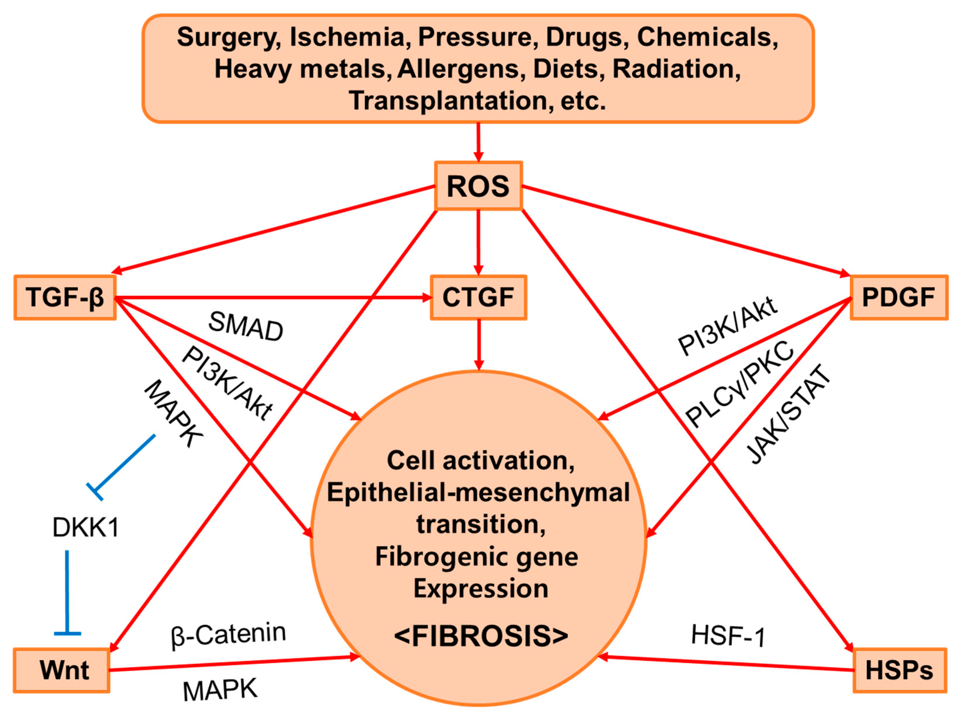
2. Common Mediators of Fibrosis

In this section, we briefly provide background information on the main mediators of fibrosis. The regulatory roles of TGF-β, PDGF, CTGF, Wnt, HSPs, and ROS in cell activation, epithelial–mesenchymal transition (EMT), or target gene expression related to fibrosis are discussed. Simplified illustrations are presented in Figure 2. There may be additional factors that play a vital role in fibrosis even if they are not covered here.

2.1. TGF-β

The mammalian genome encodes TGF-β1, -β2, and -β3, which bind to the TGF-β receptor (TGF-βR) type I and type II transmembrane protein kinases [26]. TGF-β synthesis is triggered by tissue injury and mediates the activation of various cells that are involved in immunological responses and tissue remodeling [5]. In the canonical pathway, the binding of TGF-β to the membrane receptors stimulates the phosphorylation of SMAD2 and SMAD3, which, in cooperation with SMAD4, induces the transcription of the target genes involved in cell proliferation and transdifferentiation, extracellular matrix (ECM) synthesis, and the expression of tissue inhibitor of metalloproteinase (TIMP) and SMAD7 (a feedback inhibitor of TGF-β signaling) [27]. In the noncanonical pathways, TGF-β stimulates the mitogen-activated protein kinase (MAPK) signaling pathway and the phosphoinositide 3-kinase (PI3K)/protein kinase B (PKB, Akt)/mammalian target of rapamycin (mTOR) signaling pathway, which results in the transcription of target genes including CTGF, α-SMA, collagen, fibronectin, and other ECM components [26].

2.2. PDGF

The PDGF family members, including PDGF-AA, -AB, -BB, -CC, and -DD, bind to the tyrosine kinase PDGF receptor (PDGFR) dimers αα, αβ, or ββ to stimulate auto-phosphorylation and the activation of the cytoplasmic signaling pathways [28]. The binding of the PDGF isoforms to the relevant PDGFRs initiates distinct receptor dimerization and phosphorylation and provides multiple binding sites for several adaptor molecules, thereby activating various downstream signaling pathways mediated by PI3K, Akt, extracellular signal-regulated kinase (ERK), phospholipase C (PLC) γ, diacylglycerol/protein kinase C (PKC), Janus kinase (JAK), signal transducers and activators of transcription (STAT), and/or Notch [29]. PDGF-BB has been demonstrated to enhance collagen gel contraction via the PI3K-PLCγ-PKC signaling pathway in immortalized human foreskin fibroblasts (BJ fibroblasts) [30] and collagen synthesis via the PI3K/Akt signaling pathway in oral mucosal fibroblasts [31]. The PDGF-mediated effects can induce fibroblast activation, transdifferentiation, migration, and accumulation at the damaged site of vessels [32,33,34]. The PDGF-mediated effects in hepatic stellate cells (HSCs) can increase cell motility through actin reorganization and cell proliferation during chronic liver injury [35].

2.3. CTGF

CTGF (also called CCN2), a protein of the CCN family, appears to be a central mediator of tissue remodeling and fibrosis induced by TGF-β [36,37]. Despite its name, CTGF is not a traditional growth factor or a cytokine because no specific receptors have been found to bind it with high affinity to induce signal transduction. Therefore, it is considered a type of matricellular protein that modulates cellular functions and the interaction of cells with the ECM by interacting with various regulatory proteins and ligands [36,37]. CTGF regulates the proliferation and differentiation of cells, as well as other biological processes, and participates in the development of inflammation, fibrosis, cancer, and other diseases [38]. Therefore, CTGF is a promising therapeutic target to treat various fibrotic diseases [39,40]. A specific monoclonal antibody (FG-3019) against CTGF recovered the impaired lung function in mice exposed to radiation, improved the reduced grip strength in rats induced by chronic overuse of the upper extremity skeletal muscle, and prevented fibrosis in both animal models [40,41].

2.4. Wnt

Wnt is a family of lipid-modified glycoprotein ligands that bind to a Frizzled family receptor that spans across the plasma membrane and interacts with the Dishevelled protein in the cytoplasm to transmit signals [42]. The canonical Wnt signaling pathway regulates the gene transcription of target genes, and the noncanonical Wnt signaling pathways regulate cell actin modification or calcium inside the cell. In the canonical Wnt signaling pathway, in the absence of Wnt, β-catenin is sequestered in the cytoplasm, phosphorylated by casein kinase 1 (CK1) and glycogen synthase kinase 3 beta (GSK3β), and degraded by the proteasome; however, in the presence of Wnt, β-catenin is stabilized and enters the nucleus, where it acts as a transcriptional coactivator that regulates the expression of various target genes. Under certain conditions, the Wnt/β-catenin signaling pathway induces EMT [43]. The Wnt/β-catenin signaling pathway also induces the gene expression of an antagonistic inhibitor, Dickkopf-1 (DKK1), and this negative feedback loop prevents the excessive activation of the Wnt signaling pathway [44]. Meanwhile, it has been reported that activation of the canonical Wnt signaling pathway is required for TGF-β-mediated fibrosis [45]. TGF-β1 suppresses DKK1 through an ERK-dependent mechanism and consequently activates the Wnt signaling pathway, which can induce cell activation and fibrosis. Therefore, the TGF-β and Wnt signaling pathways may cooperatively contribute to the development of fibrosis.

2.5. HSPs

HSPs appear to play a critical role in fibrotic diseases [46]. HSPs are a group of proteins that are rapidly and highly induced by heat shock factors (HSF) in response to heat shock and other stimuli and play a critical role in the folding and refolding of proteins via their chaperone functions, especially under stressful conditions [47]. HSF-1 is sequestered in the cytoplasm by HSPs under normal conditions; however, when HSPs bind to misfolded proteins under certain stressful conditions, they release HSF-1, which then undergoes phosphorylation, trimerization, and nuclear localization to bind to the heat shock elements (HSE) found in the promoters of the HSP genes and induce the gene transcription [48]. HSP47 is a collagen-specific chaperone, and its role is essential for the structural correction of fibrillar procollagens in fibrotic diseases whose hallmark is collagen deposition [49,50,51]. HSP27 also plays a pivotal role in cell transdifferentiation to myofibroblasts, thereby providing an additional therapeutic target for the treatment of fibrotic diseases [52,53].

2.6. ROS

ROS, including superoxide anion radicals (O2−•), hydrogen peroxide (H2O2), hydroxyl radicals (•OH), and nitric oxide (•NO), are key modulators of TGF-β-mediated signaling pathways as well as other cell signaling pathways associated with the development and persistence of fibrosis under pathophysiological conditions [54]. In cells, a certain level of ROS is generated as a result of mitochondrial electron transport and various enzymatic reactions, such as NADPH oxidase (NOX), thereby maintaining cellular homeostasis and the redox balance; however, exposure to ultraviolet rays, radiation, toxic chemicals, drugs, or physical damage can cause ROS production beyond the cell’s antioxidant defense capacity, which leads to an emergency state in the cells [55]. As a result, pathological signs such as inflammation, cell death, and tissue remodeling appear, and the chronic consequences can cause the dysfunction of various organs. Although oxidative stress has been implicated in a wide range of diseases, the degree to which oxidative stress contributes to the pathology of the disease varies, and antioxidant therapies that target oxidative stress do not always provide satisfactory results [56]. Research projects at various stages are trying to establish treatments for different fibrotic diseases through the application of antioxidants [57,58].

3. Methods: Study Search and Selection

We used the PubMed database (https://pubmed.ncbi.nlm.nih.gov/, accessed on 9 January 2024) to search for relevant studies with the keywords: fibrosis AND rosmarinic acid. The search results provided 43 articles in total, and 42 of these articles were cited and discussed in Section 1 (2 articles), Section 4 (4 articles), and Section 5 (36 articles). We did not discuss one article that reported on the cloning of RA glycosyltransferases from Arnebia euchroma (Boraginaceae) [59].

4. Ex Vivo Fibrosis Model Studies

Westra et al. developed an ex vivo model of liver fibrosis induced by the prolonged culture of precision-cut liver slices (PCLSs) [60]. In rat PCLSs cultured in vitro for 48 h, the expression of PDGF-B increased, while the expression of TGF-β1 did not change. The gene expression of the fibrotic markers, such as HSP47, α-SMA, and procollagen (PCOL) 1A1, increased as well, and their gene expression was further enhanced by external PDGF-BB and TGF-β1. The early gene expression of the fibrotic markers was significantly attenuated by PDGF inhibitors (e.g., imatinib, sorafenib, and sunitinib) but was not much affected by TGF-β inhibitors (e.g., perindopril, valproic acid, RA, tetrandrine, and pirfenidone), which indicates that the PDGF signaling pathway predominates during the early stages of fibrosis development in PCLSs.
They further developed a functional ex vivo model using precision-cut liver slices (fPCLSs) from rats with fibrotic livers after 3 weeks of bile duct ligation (BDL) [61]. In the in vitro culture of the fPCLSs for 48 h, the gene expression of the fibrotic markers (HSP47, α-SMA, and PCOL1A1) increased and the protein levels of collagen 1, mature fibrillar collagen, and total collagen increased, which indicates the continued fibrogenic process even after tissue excision. Both PDGF inhibitors (imatinib, sorafenib, and sunitinib) and TGF-β inhibitors (perindopril, valproic acid, RA, tetrandrine, and pirfenidone) significantly attenuated the gene expression of the fibrotic markers and the protein expression of collagen 1 in this model.
In their study using PCLSs from human donors, the early onset of fibrosis during the 48 h of organ culture was demonstrated by the increases in the gene expression of HSP47 and PCOL1A1 and the collagen 1 protein level [62]. The gene expression of HSP47 and PCOL1A1 was down-regulated with sunitinib and valproic acid, and the gene expression of PCOL1A1 was also reduced by RA, tetrandrine, and pirfenidone. The TGF-β1 signaling pathway inhibitors appeared to be more effective than the PDGF signaling pathway inhibitors in suppressing fibrogenesis in the human PCLS model, which was different from the observations in the rat PCLS models. These suggest that fibrogenesis in the PCLS model may develop in different ways depending on the species and that the effects of certain treatments may also present differently. Even within the same species, the biocompatibility, anti-inflammatory, and antifibrotic effects of each treatment were different between the liver and intestine slices from humans, mice, and rats [63]. Ex vivo fibrosis model studies suggest that because the fibrogenesis process and pathology are different depending on the organ, the treatment should also be different.

5. In Vivo Fibrosis Model Studies: Antifibrotic Effects of RA and the Plant Extracts Containing RA

In this section, the antifibrotic effects of RA and the plant extracts containing RA are described according to their target organs. The inducer of the experimental model and treatments in each test are briefly summarized in Table 1.

5.1. Liver Fibrosis

HSCs are considered one of the most important therapeutic targets for liver fibrosis because they are activated, undergo myofibroblastic transdifferentiation, and participate in the progression of the disease [95]. RA inhibited cell proliferation and the expression of TGF-β, CTGF, and α-SMA in cultured HSCs in vitro [64]. It also conferred antifibrotic effects by ameliorating the biochemical and histopathological changes in the liver and the expression of TGF-β and CTGF in mice with liver fibrosis induced by CCl4 [64].
Peroxisomal proliferator-activated receptor (PPAR) γ functions normally in quiescent HSCs, and its epigenetic repression causes cell transdifferentiation to myofibroblastic cells that mediate liver fibrosis progression [64,96]. Restored expression of PPARγ can recover the normal phenotypes of HSCs. The Wnt signaling pathway is implicated in the epigenetic repression of PPARγ, which leads to the progression of fibrosis in different organs [97]. Impeding the canonical Wnt signaling pathway using the co-receptor antagonist DKK1 abolished the epigenetic repression of PPARγ and restored the gene expression and normal phenotypes of HSCs [96].
The herbal prescription Yang-Gan-Wan, whose main active polyphenolic constituents were identified as RA and baicalin, was shown to restore the normal phenotypes of HSCs through an epigenetic mechanism [65]. Yang-Gan-Wan reduced the expression and recruitment of the DNA methyl-CpG-binding protein MeCP2 to the PPARγ promoter, the expression of histone methyltransferase, the enhancer of zeste homolog 2 (EZH2), and the methylation of histone H3 lysine 27 (H3K27), with the subsequent induction of PPARγ. RA and baicalin restored the normal phenotypes of HSCs by means of epigenetic PPARγ induction. These compounds further suppressed the expression of Wnt family members, such as Wnt10b and Wnt3a, and the canonical Wnt signaling pathway. In addition, RA treatment inhibited HSC activation and the progression of liver fibrosis in mice with cholestatic liver fibrosis induced by BDL [65]. These results suggest that RA could alleviate liver fibrosis via the suppression of the canonical Wnt signaling pathway and epigenetic de-repression of PPARγ in HSCs.
RA was shown to inhibit cell proliferation and induce apoptosis in the activated hepatic stellate cell line (HSC-T6), which correlated with the reduced phosphorylation in STAT3 and the downregulation of cyclin D1 and B cell lymphoma/leukemia (Bcl) 2 [98]. Oxidative stress plays a role in liver fibrogenesis by activating the HSCs [99]. RA inhibited the activation of matrix metalloproteinase (MMP) 2 in a nuclear factor kappa B (NF-κB)-dependent mechanism in HSC-T6 cells [100]. In addition, RA suppressed the generation of ROS and lipid peroxidation while increasing the cellular glutathione (GSH) levels and the expression of catalytic subunits of glutamate cysteine ligase (GCLc) in a nuclear factor erythroid 2-related factor 2 (NRF2)/antioxidant response element (ARE)-dependent manner.
In Sprague Dawley (SD) rats with cholestatic liver injury induced by BDL, oral administration of the extract of Glechoma hederacea lowered the expression levels of TGF-β1, CTGF, SMAD2/3, and collagen and reduced the number of α-SMA-positive matrix-producing cells [66]. In addition, Glechoma hederacea extract attenuated the BDL-induced inflammatory cell infiltration and accumulation, the NF-κB and activator protein-1 (AP-1) activation, and the inflammatory cytokine production, whereas it inhibited the axis of the high mobility group box (HMGB) 1/Toll-like receptor (TLR) 4 intracellular signaling pathways. RA, which was identified as one of the most abundant polyphenolic compounds in Glechoma hederacea, also demonstrated protective effects against cholestasis-related liver injury via the attenuation of oxidative stress and the downregulation of HMGB1/TLR4, NF-κB, AP-1, and TGF-β1/SMAD signaling [67].
The Fuzheng Huayu recipe (FZHY) is an herbal prescription that is used in China to treat liver fibrosis [101,102]. Yang et al. identified more than eleven compounds and two metabolites in the serum from rats that were orally administered an extract of Danshen (Salvia miltiorrhiza), the main component of FZHY [101]. Of these compounds, salvianolic acid B (6 and 48 μM), caffeic acid (6 and 48 μM), and RA (48 μM) inhibited the proliferation of the HSC cell line (LX-2) and down-regulated the in vitro expression of α-SMA, thus supporting their contribution to the antihepatic fibrosis effects of FZHY. Plasma pharmacokinetics and tissue distribution profiles were analyzed following the oral administration of FZHY to normal and fibrotic rats induced by intraperitoneal injections of dimethylnitrosamine [102]. The results revealed that the serum levels of danshensu, salvianolic acid B, and RA were 1.69-, 2.37-, and 1.49-fold higher, respectively, in rats with liver fibrosis compared with the normal/nonfibrotic rats. It is suggested that the absorption and metabolism of active ingredients, including RA, in a mixed herbal prescription may vary depending on the disease state in the body and that they can exert therapeutic effects by modulating the relevant cells when administered at effective concentrations.
The hepatoprotective and antifibrotic effects of RA were compared with silymarin in thioacetamide-intoxicated SD rats [68]. RA at a daily oral dose of 10 mg kg−1 had similar hepatoprotective effects as silymarin at a dose of 50 mg kg−1 in that it lowered the serum levels of alanine aminotransferase (ALT) and aspartate aminotransferase (AST), reduced the tissue levels of malondialdehyde (MDA), and increased the tissue levels of GSH in the thioacetamide-intoxicated rats. Thioacetamide increased the tissue levels of the fibrotic markers, such as TIMP-1 and hydroxyproline, in the liver, and these changes were attenuated by RA (10 mg kg−1) and silymarin (50 mg kg−1) treatment to a similar extent. Both RA and silymarin decreased the expression of collagen and α-SMA in the livers of thioacetamide-intoxicated rats. In vitro experiments using HSC-T6 cells showed that RA decreased cell viability and increased both caspase-3-positive cells and α-SMA-positive cells, thus supporting the antiproliferative and proapoptotic effects of RA in this cell type.
When lemon balm (Melissa officinalis) extract and its constituent, RA, were administered orally, they were shown to alleviate nonalcoholic steatohepatitis and hepatic fibrosis in db/db mice fed a methionine- and choline-deficient (MCD) diet [69]. Oral treatments with the extract (200 mg kg−1) or RA (10 or 30 mg kg−1) alleviated liver damage that was monitored by the serum levels of ALT and AST. These treatments inhibited the accumulation of hepatic triglycerides and hydroxyproline as well as the expression of α-SMA and collagen (COL) 1A1 in the mouse model fed a MCD diet. In the associated in vitro experiments using HepG2 cells, palmitic acid caused the accumulation of lipids and triglycerides and the expression of lipogenic genes, such as sterol regulatory element-binding protein (SREBP) 1c, fatty acid synthase (FAS), and stearoyl-CoA desaturase (SCD) 1, whereas it suppressed the expression of lipolytic genes, such as carnitine palmitoyl transferase (CPT) 1L [69]. Treatment with either the extract or RA reversed these changes and restored the 5′ AMP-activated protein kinase (AMPK) phosphorylation and NRF2 activation as well as its downstream targets, including superoxide dismutase (SOD) 1 and heme oxygenase (HO) 1.
Lyu et al. demonstrated that advanced glycation end-products (AGEs) mediate high levels of cross-linking in the ECM of cirrhotic liver tissue [70]. AGE cross-linking in the collagen matrix could be effectively inhibited by RA in vitro. Furthermore, RA inhibited the AGE-mediated cross-linking in the liver ECM and alleviated late-stage liver fibrosis, as shown by the reduced levels of collagen in the scar tissue, hepatic hydroxyproline, and the proportion of α-SMA-positive cells in male C57BL/6 mice that were treated with CCl4 or fed the high-fat choline-deficient L-amino acid-defined (HFCDAA) diet, thus suggesting its potential to alleviate liver cirrhosis.

5.2. Kidney Fibrosis

EMT is a process of cell transdifferentiation that occurs during the development of many tissues [103]. Epithelial and mesenchymal cells have different morphologies, functions, and other phenotypes; epithelial cells express high levels of E-cadherin, whereas mesenchymal cells express high levels of N-cadherin, fibronectin, vimentin, collagen, and α-SMA. EMT can occur during the early stages of wound healing, organ fibrosis, and cancer metastasis; therefore, it is important in the pathogenesis of various diseases and a useful therapeutic target [104].
WHW, a herbal prescription composed of 14 medicinal plants, has been used in oriental traditional medicine for the treatment of chronic renal failure as well as for other purposes [1,105,106]. Our previous study demonstrated that WHW extract (1 mg mL−1) attenuated the TGF-β1-induced morphological change of MDCKs undergoing EMT [1]. It inhibited the TGF-β1-stimulated expression of α-SMA and the phosphorylation of SMAD2. In the same study, activity-guided solvent fractionation and chromatographic separation of WHW extract identified RA as one of the main active compounds. It was further verified that RA (52–104 μM) suppressed the expression of α-SMA in MDCKs stimulated with TGF-β1 (unpublished data). In the study using cultured primary mouse macrophages and the RAW264.7 cell line, WHW extract (0.5 mg mL−1) suppressed the production of •NO as well as the mRNA expression and protein levels of nitric oxide synthase (NOS) 2, tumor necrosis factor (TNF) α, interleukin (IL) 1β, and IL-6 induced by lipopolysaccharide (LPS) [105]. WHW extract inhibited the phosphorylation of MAPK, such as ERK 1/2, p38, and c-Jun N-terminal kinase (JNK), and the nuclear translocation of NF-κB p65 protein in the LPS-stimulated RAW264.7 cells. These in vitro studies suggest anti-inflammatory and antifibrogenic properties of WHW extract that contains RA.
The effect of WHW on kidney ischemia/reperfusion injury was evaluated in a mouse model [71,72]. The mice were orally administered WHW extract (0.5–100 mg kg−1) daily for 14 days and then subjected to 30 min of bilateral renal ischemia. Then, oral administration of WHW continued for 2 days [71]. The ischemia/reperfusion resulted in the disruption of the kidney tubular epithelial cells and increased plasma creatinine levels in the model mice, but these changes were attenuated by the WHW treatment. WHW treatment also inhibited the post-ischemic increase of H2O2 and lipid peroxidation and the post-ischemic decrease in catalase, copper/zinc SOD, and manganese SOD activity in the kidney tissue. WHW treatment enhanced the post-ischemic increase in the expression levels of HSP27 and HSP72. In another study, WHW extract was administered orally to mice for 14 days, beginning 2 days after the ischemic procedure [72]. WHW treatment attenuated the progression of renal fibrosis and prevented the post-ischemic increase of H2O2 and lipid peroxidation, as well as the decrease of copper/zinc SOD and manganese SOD activity. In addition, WHW treatment reduced the phosphorylation of ERK1/2 and JNK1/2 and the activation of NF-κB in the kidneys induced by ischemia/reperfusion.
WHW treatment also inhibited the kidney fibrosis that was induced by unilateral ureteral obstruction (UUO) in mice [73]. Post-procedure WHW treatment (2, 10, or 50 mg kg−1) mitigated the UUO-induced kidney fibrosis, as determined by tubular atrophy and dilatation, collagen accumulation, interstitial space expansion, and leukocyte infiltration. WHW treatment prevented the increase in oxidative stress and decrease of catalase, copper/zinc SOD, and manganese SOD activity due to the UUO procedure. WHW reduced the expression of TGF-β1 and the phosphorylation of SMAD2/3 stimulated by the UUO procedure. Collectively, WHW extract can alleviate oxidative stress, inflammation, and fibrosis in the kidneys following an ischemia/reperfusion or UUO.
The effects of Elsholtzia ciliata extract, which contains luteolin and RA as its main phenolic compounds, on renal interstitial fibrosis were examined in male SD rats with UUO [74]. Orally administered extract (300 or 500 mg kg−1) reduced UUO-induced renal damage, histopathological alterations, and interstitial fibrosis. In addition, the renal expression levels of TNF-α, TGF-β1, and SMAD3 were reduced in the extract-treated rats compared to the untreated UUO model rats. The accompanying in vitro studies revealed that this extract (200 μg mL−l) ameliorated the LPS-induced overexpression of NF-κB, TNF-α, and IL-6, improved oxidative stress in RAW 264.7 cells, and suppressed the TGF-β-induced expression of α-SMA and MMP9 in human renal mesangial cells. Thus, Elsholtzia ciliata extract may impede the inflammatory and fibrogenic signaling pathways that cause renal fibrotic disease.
The inhibitory effects of RA on kidney fibrosis and EMT were examined using a UUO mouse model and an indoxyl sulfate-stimulated NRK-52E cell model [75]. C57BL/6 mice were orally administered RA (10 or 20 mg kg−1) before and after UUO surgery. UUO caused kidney damage, which was determined by the increase in serum creatine and blood urea nitrogen (BUN) levels, and the damage was reduced by RA treatment. The UUO-induced increase of α-SMA, collagen I, fibronectin, and vimentin and the decrease of E-cadherin were attenuated by RA treatment. NRK-52E cells were treated with 4 mM indoxyl sulfate with or without RA (20 or 40 μM). The indoxyl sulfate-induced proliferation and migration of the cells were inhibited by RA treatment. RA treatment further attenuated the increase of α-SMA, collagen I, fibronectin, vimentin, and TGF-β1 and the decrease of E-cadherin induced by indoxyl sulfate. RA treatment inhibited the phosphorylation of Akt in both the cell and animal models. Thus, RA was suggested to ameliorate renal interstitial fibrosis by inhibiting the Akt-mediated EMT.
RA was shown to attenuate the nephrotoxicity of cadmium both in vitro and in vivo [76]. RA prevented the apoptotic death of cultured mouse proximal tubular epithelial cells that were exposed to CdCl2. It attenuated the CdCl2-induced increased production of ROS/•NO, lipid peroxidation, protein carbonylation, the increased activity of NOX activity, and the decreased levels of coenzyme 9, coenzyme 10, and GSH, as well as the decreased activity of SOD, catalase, glucose-6-phosphate dehydrogenase (G6PDH), glutathione peroxidase (GPX), glutathione S-transferase (GST), and glutathione reductase (GR). CdCl2 stimulated apoptotic events that were mediated by Bcl-2, Bcl-2-associated death protein (Bad), apoptotic protease activating factor (Apaf) 1, cytochrome C, and caspases 3, 8, and 9, and the fibrogenic events that were accompanied by the upregulation of α-SMA, E-cadherin, and collagen IV in the cells, and all these events were attenuated by RA treatment. Swiss albino mice were treated with CdCl2 (4 mg kg−1) with or without RA treatment (50 mg kg−1), and the pathological changes were monitored by assessing the blood, urine, and tissues. The biochemical and histological data supported the pharmacological effects of RA attenuating oxidative stress, apoptosis, inflammation, and fibrosis in the kidneys caused by cadmium exposure.
The effects of Lycopus lucidus extract, which contains caffeic acid, luteolin-7-O-β-D-glucoside, and RA as the main phenolic compounds, on kidney fibrosis were examined in rhTGF-β1-stimulated SV40 MES13 cells and a streptozotocin-induced diabetic nephropathy rat model [77]. Treatment with the extract (50–400 μg mL−1) suppressed the activation of SMAD2 and ERK1/2 and downregulated TGF-βRI, TGF-βRII, SMAD4, and SMAD7 in TGF-β1-stimulated cells. This extract (3, 6, or 12 g kg−1) inhibited SMAD2 phosphorylation, reduced TGF-β1 expression, ameliorated the expansion of the mesangial area in glomerular tissue, reduced the levels of serum creatine and BUN, and reduced the total SOD activity in the streptozotocin-induced diabetic nephropathy rats. Thus, Lycopus lucidus extract is considered to be able to inhibit renal fibrosis by impeding the TGF-β signaling pathway and attenuating diabetic nephropathy.
Guanxinning injection, a herbal prescription composed of Radix et Rhizoma Salviae Miltiorrhizae (Salvia miltiorrhiza) and Chuanxiong Rhizoma (Ligusticum chuanxiong), was tested for its effects on renal fibrosis in mice with heart failure induced by transverse aortic constriction [78]. Guanxinning injection was administered via tail vein at doses of 3–12 mL kg−1, and it relieved the cardiac function indexes (ejection fraction, cardiac output, and left ventricle volume), kidney functional indexes (serum creatinine), and kidney fibrosis indexes (collagen volume fraction and CTGF) in the model mice. Guanxinning injection further increased catalase, GPX4, the x(c)(-)cysteine/glutamate antiporter SLC7A11, and ferritin heavy chain 1, while decreasing xanthine oxidase and NOS in the kidneys. Its major active ingredients include RA, caffeic acid, ferulic acid, etc. Thus, Guanxinning injection could help maintain cardiac function and alleviate the progression of fibrosis in the kidneys due to heart failure through the regulation of redox metabolism.
Patients with autosomal dominant polycystic kidney disease had higher expression levels of complement factor B and complement component 9 in the kidneys compared with control subjects or patients with other types of chronic kidney disease [107]. Oral administration of RA (300 mg kg−1, daily) for 4 weeks reduced serum creatine, BUN, kidney weight, cyst index, fibrosis, expression of complement factor B and C5b-9, and the number of Ki67-positive nuclei, as well as the inflammatory cells, without affecting the body weight in Pkd1−/− knockout mice. This supports the therapeutic potential of RA as an inhibitor of the complement pathway associated with autosomal dominant polycystic kidney disease progression [107].

5.3. Heart Fibrosis

The ethanolic extract of the aerial parts of basil (Ocimum basilicum), which contains RA as the principal phenolic compound, has been shown to improve cardiac function and inhibit the histopathological changes in response to isoproterenol-induced myocardial infarction (MI) in male Wistar albino rats [79]. Treatment with isoproterenol (100 mg kg−1) daily for 2 consecutive days caused an elevation in the ST segment in the electrocardiogram, indicative of a MI, a reduction in the left ventricular contractility, an increase in the left ventricular end-diastolic pressure, and an increase in myocardial necrosis and fibrosis, all of which were significantly attenuated by the subcutaneous injection of 10, 20, or 40 mg kg−1 of the extract twice per day. In addition, the isoproterenol-induced elevated MDA levels in the serum and myocardium were suppressed by the extract, thus suggesting that the cardioprotective effects of O. basilicum extract could be associated with its antioxidant activity.
The aqueous extract of Xia-Ku-Cao (Prunella vulgaris), as well as its caffeic acid, ursolic acid, and RA components, exhibited a cardioprotective effect in acute MI in male SD rats with left anterior descending coronary artery (LAD) ligation [80]. The extract (400 mg kg−1) administered by intragastric gavage after surgery improved cardiac function and reduced the infarct size, inflammation, fibrosis, oxidative damage, and apoptosis of cardiomyocytes. Phenolic compounds, such as caffeic acid (400 mg kg−1), ursolic acid (400 mg kg−1), and RA (400 mg kg−1), improved cardiac function and suppressed inflammatory aggregation and fibrosis in MI rat models. RNA-seq analysis and an additional in vitro study suggest that the therapeutic effect of P. vulgaris extract and its phenolic compounds might be related to the reduced expression of NOD-like receptor protein 3 (NLRP3), which is presumed to play a critical role in the inflammatory process during cardiac remodeling after a MI [108].
RA demonstrated antifibrotic effects in male SD rats with a MI induced by LAD ligation surgery [81]. The oral administration of RA (50, 100, or 200 mg kg−1) ameliorated the infarct size and MI-induced changes in the left ventricular systolic pressure, +dp/dtmax, and −dp/dtmax. RA attenuated the cardiac fibrosis, as determined by the collagen volume fraction and the expression of collagen I, collagen III, α-SMA, and hydroxyproline. RA treatment also decreased the expression of angiotensin-converting enzyme (ACE), angiotensin type 1 receptor (AT1R), and phospho-p38 MAPK while increasing the expression of ACE2, which may be associated with the cardioprotective effects of RA against cardiac dysfunction and fibrosis.
An injectable hydrogel encapsulating polydopamine-RA nanoparticles with dual responsiveness to pH and ROS was developed to achieve on-demand drug release in the MI microenvironment [82]. Oxidized xanthan gum grafted with 3-aminophenylboronic acid and dopamine-grafted gelatin were combined to prepare an “OGD” hydrogel. The polypyrrole-modified gelatin was combined with “OGD” to prepare an “OGDP” hydrogel. The polydopamine-RA nanoparticles were added to the “OGD” and “OGDP” hydrogels to prepare the “OGDR” and “OGDPR” hydrogels. Rats were subjected to LAD ligation to induce MI, and on day 2 after modeling, rats with a confirmed MI were subjected to multiple injections with the “OGD”, “OGDR”, “OGDP”, or “OGDPR” hydrogels in and around the infarct area. The OGDPR hydrogel showed the best results by exerting anti-inflammatory, antiapoptotic, and antifibrotic effects. The multifunctional hydrogel promoted the expression of heart-specific markers, thus restoring heart function after a MI.
RA also attenuated cardiac remodeling and fibrosis following a long-term pressure overload in male C57/B6 mice subjected to aortic banding to generate pressure overload, which led to cardiac dysfunction with reduced fractional shortening [83]. Orally administered RA (100 mg kg−1) attenuated the fibrotic response and cardiac dysfunction after pressure overload without affecting the hypertrophic response. Aortic banding increased the expression of collagen I and III, CTGF, fibronectin, TGF-β1, and α-SMA, and the phosphorylation of ERK, Akt, p38, AMPK, and acetyl-CoA carboxylase (ACC). RA attenuated the increased expression of collagen I and III, CTGF, fibronectin, TGF-β1, and α-SMA and enhanced the increased phosphorylation of AMPK and ACC without significantly affecting the phosphorylation of ERK, Akt, and p38. The cardioprotective effects of RA in reducing the expression of collagen I and III, the phosphorylation of SMAD, and the restored fractional shortening induced by aortic banding were abolished in AMPKα2 knockout mice.
RA has been demonstrated to alleviate the apoptosis of cardiomyocytes [109] and cardiac fibrosis induced by doxorubicin, an anticancer medicine, in male rats [84]. Doxorubicin (2 mg kg−1 per 48 h) increased the heart-to-body weight ratio, reduced the heart rate and blood pressure, and caused fibrosis and necrosis of the cardiac tissue [84]. These toxic effects of doxorubicin were attenuated by the administration of RA (10, 20, or 40 mg kg−1). RA administration also attenuated the doxorubicin-induced increase of MDA and decrease of GSH in the heart tissue, thereby implying that its antioxidant properties underlie the protective effects against doxorubicin-induced cardiotoxicity.

5.4. Lung Fibrosis

Rosemary (Rosmarinus officinalis) leaf extracts, which contain carnosic acid and RA, have been shown to inhibit bleomycin-induced pulmonary fibrosis [85]. Male Wistar rats were given a single dose of bleomycin (4 mg kg−1, intratracheal), and the extract (75 mg kg−1) was administered 3 days later and continued for 4 weeks (curative group), or administered 2 weeks before bleomycin administration and continued for 15 days thereafter (prophylactic group). The lung architecture was more retained in the curative and prophylactic groups compared to the bleomycin group, which was associated with lower fibrosis scores (2.33 and 1.8 vs. 3.7 of the bleomycin group, respectively), reduced MDA levels (141% and 108% vs. 258% of the normal value, respectively), high catalase levels (103% and 117% vs. 59% of the normal value, respectively), and enhanced GST activity (85% and 69% vs. 23% of the normal value, respectively). The antifibrotic and antioxidant effects of carnosic acid, RA, and their combination were verified in the bleomycin-induced fibrosis model in rats [86]. In addition, purified rosemary leaf extract with increased RA and carnosic acid contents and decreased essential oil content exhibited potent antifibrotic efficacy in an animal model [110].
RA attenuated the pulmonary fibrosis caused by irradiation. Treatments of male SD rats with RA (30, 60, or 120 mg kg−1) before 15 Gy of X-ray irradiation decreased the expression of inflammatory mediators, NF-κB phosphorylation, and the production of ROS [87]. RA inhibited Ras homolog family member A (RhoA)/Rho-associated protein kinase (Rock) signaling through the upregulation of the microRNA (miR) 19b-3p, which targets the myosin phosphatase target subunit 1 (MYPT1) and leads to the inhibition of TGF-β1 signaling and subsequent fibrosis [87].
Salvia miltiorrhiza extract, which contains salvianic acid A and B, caffeic acid, RA, and tanshinone IIA, attenuated ovalbumin-induced asthma in mice [88]. The ovalbumin challenge increased airway resistance, inflammatory cell infiltration, Th1/Th2 cytokine levels in the bronchoalveolar lavage fluid (BALF), cell hyperplasia, collagen deposition, and airway wall thickening. Daily treatment with the water extract of S. miltiorrhiza (156 mg kg−1) significantly alleviated these pathological changes. In the associated in vitro experiments, caffeic acid and RA potently reduced E-cadherin and vimentin in the TGF-β1-induced BEAS-2B cells and α-SMA and COL1A1 in the TGF-β1-induced MRC-5 cells. RA also attenuated the immunological and inflammatory responses and normalized the pathological features in the lungs of ovalbumin-induced asthmatic rats [89]. The asthmatic rats showed increased levels of IL-4, immunoglobulin (Ig) E, phospholipase A (PLA) 2, and total protein in the BALF and decreased levels of interferon (IFN) γ and the IFN-γ/IL-4 ratio compared to the control group. The RA treatment reduced the levels of IL-4, IgE, PLA2, and total protein and restored the IFN-γ/IL-4 ratio.

5.5. Post-Surgical Abdominal Adhesion

Peritoneal adhesion occurs when the peritoneum is damaged as a result of various injury events, including abdominal surgery [111]. Peritoneal bands connect the peritoneum to different visceral organs and cause abdominal discomfort, infertility in women, intestinal blockage, and other ailments [112]. RA (50 or 70 mg kg−1) inhibited the formation of postoperative peritoneal adhesion in male Wistar albino rats [90]. RA decreased the peritoneal adhesion score and was associated with reduced fibroblast proliferation, the expression of TGF-β1 (a fibrotic marker), TNF-α (an inflammation marker), and vascular endothelial growth factor (VEGF) (an angiogenesis marker), and reduced MDA levels (an oxidative stress marker). A polygalacturonic acid-conjugated cysteine hydrogel has been developed as a drug carrier for RA, and animal implant studies have shown that hydrogel films with and without RA reduce the incidence of post-surgical adhesion and early inflammatory reactions [91].

5.6. Fibrosis in the Salivary Glands

The three major types of salivary glands are the parotid, submaxillary, and sublingual glands [113]. The salivary glands have a high risk of exposure to oxidative damage after irradiation therapy for head and neck cancers [114]. The radioprotective effect of RA on the parotid gland was examined in SD rats irradiated with 15 Gy X-ray irradiation [92]. RA treatment (60 and 120 mg kg−1) attenuated the radiation-induced hyposalivation and oxidative stress in the parotid gland as effectively as amifostine (250 mg kg−1). The radiation increased NOX 4 and decreased PPARγ coactivator (PGC) 1α, which were normalized by RA treatment. RA reduced apoptosis by inhibiting p53/JNK activation and fibrosis by downregulating inflammatory factor levels. Therefore, RA has the therapeutic potential to treat radiation-induced parotid gland injuries.

5.7. Skin Wounds

The skin wound healing effects of RA were examined in Wistar albino rats with a 2 cm full-thickness skin wound [93]. The topical application of a 10% RA cream enhanced the wound size reduction more effectively compared to a 5% dexpanthenol cream application or nontreatment, which resulted in fewer scars. An oxidized dextran/amidated gelatin hydrogel, “ODex-AG”, and an RA-grafted hydrogel, “ODex-AG-RA”, were developed as dressings for skin injuries [94]. Both the “ODex-AG” and “ODex-AG-RA” hydrogels promoted skin wound healing in a rat model of a full-thickness skin wound. The “ODex-AG-RA” hydrogel effectively enhanced the collagen deposition and neovascularization (CD31) in the wounds and alleviated inflammation (TNF-α and CD163) and oxidative stress (MDA and H2O2) compared to the “ODex-AG” hydrogel. Overall, this study demonstrated the utility of RA-grafted hydrogels as a wound dressing with anti-inflammatory and antioxidant activities.

5.8. Pterygium in the Eyes

Pterygium occurs due to the uncontrolled excessive proliferation of epithelial tissue in the eyes, and the lesion can invade the cornea, potentially causing irregular corneal astigmatism and vision loss [115]. The antifibrotic effect of RA has been tested in pterygium epithelial cells [116]. RA treatment (100 µM) significantly decreased the cell viability and the protein expression of type I collagen, TGF-β1, TGF-βRII, phospho-SMAD1/5, phospho-SMAD2, phospho-SMAD, and SMAD4. Thus, RA is suggested to inhibit the TGF-β1/SMAD signaling pathway involved in pterygium epithelial cell-mediated fibrosis. Further studies are expected to evaluate the therapeutic potential of RA for the in vivo treatment of pterygium.

5.9. Fibrosis of Autologous Fat Grafts

After the autologous transplantation of inguinal fat pads to the parascapular area in SD rats, an ethanol solution of RA was intraperitoneally injected (20 mg kg−1) daily for a week and then weekly for 7 weeks [117]. The total oxidant status, MDA, and the expression levels of TNF-α and TGF-β1 were lower in the transplanted tissue of the RA-treated group compared to the control or vehicle (ethanol)-treated groups, which indicates that RA could reduce the necrosis, inflammation, and fibrosis of fat grafts [117].

6. Lamiaceae Plants with Antifibrotic Effects

Herbal remedies that have been used to treat fibrotic diseases in traditional medicine are mostly mixtures of multiple plants. Therefore, the question arises as to which compound of which plant is responsible for its efficacy. In previous research, the activity-guided solvent fractionation and chromatographic separation of WHW led to the identification of RA as the active compound [1]. Interestingly, all plants whose single extract was reported to contain RA and to show antifibrotic activity belong to the family Lamiaceae (Table 2). These include Glechoma hederacea, Melissa officinalis, Elsholtzia ciliata, Lycopus lucidus, Ocimum basilicum, Prunella vulgaris, Salvia rosmarinus (Rosmarinus officinalis), and Salvia miltiorrhiza.
Traditional herbal prescriptions, such as FZHY, WHW, and Guanxinning injections, commonly include Lamiaceae plants, i.e., Salvia miltiorrhiza and Perilla frutescens (Table 2). This suggests that RA is the active ingredient for the antifibrotic effect of Lamiaceae plants, and these plants are a valuable source of RA. Perilla frutescens also contains RA [119], and the therapeutic effects of the extracts of this plant and its polyphenolic components on chronic kidney disease have been investigated [120]. Meanwhile, Yang-Gan-Wan contains RA as one of its main compounds, even though this herbal prescription does not include Lamiaceae plants, and thus RA is presumed to have originated from Apiaceae plants.
Lamiaceae plants commonly contain RA, which means that they share a biosynthetic pathway for the generation of this compound [121]. The RA biosynthesis starts with the condensation of 4-coumaroyl-CoA and (R)-3-(4-hydroxyphenyl)lactate to produce 4-coumaroyl-(R)-3-(4-hydroxyphenyl)lactate. This compound is further oxidized via either 4-coumaroyl-(R)-3-(3,4-dihydroxyphenyl)lactate or caffeoyl-(R)-3-(4-hydroxyphenyl)lactate into RA, caffeoyl-(R)-3-(3,4-dihydroxyphenyl)lactate. Structurally similar compounds, such as metabolites of the RA synthesis pathway, may be contained in the plants of the Lamiaceae family and exhibit biological activity similar to RA in animal systems. Therefore, RA is not necessarily the only active ingredient. Furthermore, it should be noted that RA synthesis is not limited to Lamiaceae plants [121].

7. Key Mediators of the Antifibrotic Effects of RA

Many ex vivo and in vivo studies have supported that RA and several plant extracts have antifibrotic effects, attenuating cell activation, EMT, and the expression of fibrogenic genes. RA is considered to regulate multiple targets beyond the antioxidant effect of nonselectively relieving oxidative stress. RA could modulate common mediators of fibrosis, such as TGF-β, PDGF, CTGF, Wnt, HSPs, etc. In addition, PPARγ, AMPK, NF-κB, and NRF2 appear to be additional key mediators of the antifibrotic effects of RA, as discussed below. Based on the considerations and discussions in this review, the mechanism of antifibrotic action of RA is schematized in Figure 3.

7.1. PPARγ

The nuclear receptor superfamily PPARα, β/δ, and γ is known to regulate cellular metabolic energy homeostasis [122,123]. The ligand-activated transcription factor PPARγ forms a heterodimer with the retinoid X receptor (RXR) and binds to the PPAR response elements (PPREs), which are mainly identified in the promoters of genes involved in the metabolism of lipids and glucose [124]. PGC-1α binds PPARs and RXRs to co-activate the expression of target genes [125]. The activation of PPARγ appears to inhibit the canonical and noncanonical TGF-β-mediated signaling pathways, thereby supporting the cross-talk between the PPAR and TGF-β superfamilies [126]. The herbal prescription Yang-Gan-Wan and its constituents, RA and baicalin, induced the gene expression of PPARγ in HSCs [65]. RA treatment restored the gene expression of PPARα and PGC-1α that were suppressed by palmitic acid in HepG2 cells [69]. RA enhanced AMPKα phosphorylation and suppressed α-SMA expression in cardiac fibroblasts induced by TGF-β1, and the RA effects were abolished by the small interfering RNA (siRNA)-mediated depletion of PPARγ or its pharmacological inhibition with GW9662, a specific PPARγ antagonist [83]. Thus, it is suggested that PPARα, PPARγ, and PGC-1α are important mediators of the antifibrotic effects of RA.

7.2. AMPK

AMPK is a heterotrimeric protein complex that consists of α, β, and γ subunits and plays a role in maintaining cellular energy homeostasis [127]. It is allosterically activated by the binding of AMP to the γ subunit and deactivated upon the displacement of AMP by ATP [128]. It is also activated through the phosphorylation of the α subunit by liver kinase (LK) B1, calcium/calmodulin-dependent protein kinase (CAMK) 2, or TGFβ-activated kinase (TAK) 1 and deactivated through the dephosphorylation by several protein phosphatases [129]. The AMPK-mediated phosphorylation of ACC1 or SREBP1c suppressed the synthesis of fatty acids, cholesterol, and triglycerides while increasing the uptake and β-oxidation of fatty acids [129]. RA and RA-enriched lemon balm extract increased the phosphorylation of AMPK, its upstream protein kinases, such as LKB1 and CAMK2, and its downstream substrate, ACC1, in HepG2 cells [69]. RA also enhanced the phosphorylation of AMPK and ACC that were induced by pressure overload in mice, and the antifibrotic and cardioprotective effects of RA were abolished in AMPKα2 knockout mice [83]. RA treatment increased the phosphorylation of AMPKα and ACC in cardiac fibroblasts and suppressed the TGF-β1-induced α-SMA expression, which was abolished by the short hairpin RNA (shRNA)-mediated knockdown of AMPKα2 [83]. Thus, AMPK activation is a key mechanism for the antifibrotic effects of RA.

7.3. NRF2

NRF2 is a transcription factor that binds to AREs, which are commonly found in the promoter region of phase II metabolism/antioxidant enzymes [130,131]. In the base state, Kelch-like ECH-associated protein (KEAP) 1 and β-transducin repeat-containing protein (TrCP) sequester NRF2 in the cytosol and promote its proteasomal degradation. However, when their interaction is weakened by ROS, electrophilic substances, or protein kinases, NRF2 is released from KEAP1 or β-TrCP and enters the nucleus to induce the gene expression of target genes, such as GCLc, catalase, SOD, and HO-1 [132,133].
RA suppressed the generation of ROS while increasing the cellular GSH levels in HSC-T6 cells [100]. RA stimulated the nuclear translocation of NRF2 and the activation of AREs in the promoters, which led to the expression of the target genes, such as GCLc, which is involved in GSH synthesis. The shRNA-mediated knockdown of NRF2, but not NF-κB, abolished the RA-induced decrease of ROS levels and increase of GSH levels in the cells. RA restored the protein levels of NRF2 and its downstream targets, including SOD1 and HO-1, in palmitic acid-treated HepG2 cells [69]. RA increased GSH levels in the cultured mouse proximal tubular epithelial cells exposed to CdCl2 [76]. This effect of RA has also been observed in the livers of thioacetamide-intoxicated rats [68] and the hearts of doxorubicin-exposed rats [84]. Thus, RA can enhance cellular antioxidant capacity and resistance to oxidative stress via the NRF2/ARE-mediated gene expression of antioxidant enzymes.

7.4. NF-κB

NF-κB is a family of inducible transcription factors that mediate the transcription of target genes by binding to a specific DNA element and κB enhancer in a variety of immune and inflammatory processes [134]. The canonical NF-κB pathway responds to diverse signals transmitted through members of the TNF receptor (TNFR) superfamily as well as other relevant receptors. The NF-κB proteins, such as NF-κB1 (p50), are normally sequestered in the cytoplasm by inhibitory proteins, such as IκBα. However, under stimulated conditions, IκBα undergoes site-specific phosphorylation by an IκB kinase (IKK) complex and ubiquitin-dependent proteasomal degradation, resulting in the rapid and transient nuclear translocation of the NF-κB1 (p50)/RelA (p65) dimer, which transactivates the expression of the target gene [134]. The noncanonical NF-κB pathways are regulated by different mechanisms [135]. Accumulating evidence suggests that the dysregulation of NF-κB activity may be associated with various inflammatory diseases and cancers, and thus NF-κB has been one of the most studied targets for the therapy of diseases [136].
RA has been shown to induce the nuclear translocation of NF-κB and inhibit the expression of MMP2 in HSC-T6 cells [100]. The inhibitory effects of RA on the expression of MMP-2 were abolished by the shRNA-mediated depletion of NF-κB or NRF2. Thus, it is suggested that RA may impede the progression of HSC activation through NF-κB-dependent mechanisms. On the other hand, RA reduced the activation of the NF-κB pathway in mouse tubular epithelial cells exposed to CdCl2 [76]. RA and Glechoma hederacea extract reduced NF-κB activity in rat livers with cholestasis [66,67]. WHW attenuated the activation of NF-κB in RAW264.7 cells that were stimulated with LPS [105] and in the kidneys of mice exposed to ischemic surgery [72]. Elsholtzia ciliata extract also attenuated NF-κB activation in LPS-stimulated RAW264.7 cells [74]. Therefore, NF-κB is an important mediator in both the pathogenesis and treatment of diseases, and it is proposed that RA and plant extracts containing RA have a therapeutic effect by maintaining the NF-κB activity at an appropriate level.

8. Future Perspectives

RA and the extracts of the Lamiaceae plants that contain RA have various biological activities and a high potential to be used as nutraceuticals to improve human health or as medicines for the treatment of diseases. As explored in this review, they affect various signaling pathways and biological events in cells and animal models that are associated with fibrotic diseases of different organs. In addition, bioengineering technologies are being developed to increase the RA content in plants and RA production efficiency [137,138]. Furthermore, research on formulations, such as nanoparticles and hydrogels, that increase RA bioavailability or its pharmacological efficacy is also emerging [82,91,94,139,140]. Advances in these related fields will accelerate the industrial and medical utilization of RA. Information regarding the safety and pharmacokinetics of RA has been previously reported [141,142]. We expect that the development of advanced medicines specialized for fibrotic diseases in human organs based on the pharmacological properties of RA is a step further from the traditional use of plant extracts and herbal prescriptions.
We present several challenges for future studies, as follows:
  • To verify key mediators of RA effects as therapeutic targets for fibrotic diseases.
  • To compare RA with other competing substances in terms of efficacy and safety.
  • To discover new drug candidates using RA as a lead compound.
  • To develop formulations to enhance the bioavailability and targeted delivery of RA.
  • To evaluate the safety and therapeutic efficacy of RA through clinical trials.
  • To expand the industrial and medical applications of RA.

9. Conclusions

In conclusion, accumulating scientific evidence supports the effectiveness of RA and Lamiaceae plant extracts in alleviating fibrosis in various organs, including the liver, kidneys, heart, lungs, etc., as summarized in Figure 4.
RA modulates multiple signaling pathways mediated by PPARγ, AMPK, NRF2, and/or NF-κB in cells. It enhances cellular antioxidant capacity, alleviates ROS-mediated oxidative stress and inflammation, inhibits the TGF-β, CTGF, and Wnt-mediated signaling pathways, and suppresses cell activation, EMT, and fibrogenic gene expression in various organs, contributing to the recovery of structural and functional integrities in each organ.
Although RA-containing plant extracts are already used in traditional medicine, it is hoped that RA will be developed into more advanced nutraceuticals or pharmaceuticals based on updated information on its efficacy, mechanism of action, safety, and pharmacokinetics. For these industrial and medical applications, well-designed clinical trials on the efficacy and safety of RA-based preparations are needed.

Funding

This work was supported by a grant from the Korea Health Technology R&D Project through the Korea Health Industry Development Institute (KHIDI), funded by the Ministry of Health and Welfare, Republic of Korea (grant no. HP20C0004).

Institutional Review Board Statement

Not applicable.

Informed Consent Statement

Not applicable.

Data Availability Statement

Not applicable.

Conflicts of Interest

The author declares no conflicts of interest.

Abbreviations

ACCAcetyl-CoA carboxylase
ACEAngiotensin-converting enzyme
AGEsAdvanced glycation end-products
ALTAlanine aminotransferase
AMPK5′ AMP-activated protein kinase
AktProtein kinase B (PKB)
AP-1Activator protein 1
Apaf-1Apoptotic protease activating factor-1
AREAntioxidant response element
ASTAspartate aminotransferase
AT1RAngiotensin type 1 receptor
BadBcl-2-associated death protein
BALFBronchoalveolar lavage fluid
Bcl 2B cell lymphoma/leukemia 2
BDLBile duct ligation
BUNBlood urea nitrogen
CAMKCalcium/calmodulin-dependent protein kinase
COLCollagen
CTGFConnective tissue growth factor
DKK1Dickkopf-1
ECMExtracellular matrix
EMTEpithelial–mesenchymal transition
ERKExtracellular signal-regulated kinase
FASFatty acid synthase
FZHYFuzheng Huayu recipe
G6PDHGlucose-6-phosphate dehydrogenase
GCLcCatalytic subunits of glutamate cysteine ligase
GPXGlutathione peroxidase
GRGlutathione reductase
GSHGlutathione
GSTGlutathione S-transferase
HOHeme oxygenase
HSCsHepatic stellate cells
HSFHeat shock factor
HSPsHeat shock proteins
IFN-γInterferon gamma
IgImmunoglobulin
IKKIκB kinase
ILInterleukin
JNKc-Jun N-terminal kinase
KEAPKelch-like ECH-associated protein
LADLeft anterior descending coronary artery
LPSLipopolysaccharide
MAPKMitogen-activated protein kinase
MCDMethionine- and choline-deficient
MDAMalondialdehyde
MDCKsMadin–Darby canine kidney cells
MIMyocardial infarction
MMPMatrix metalloproteinase
NF-κBNuclear factor kappa B
NOSNitric oxide synthase
NOXNADPH oxidase
NRF2Nuclear factor erythroid 2-related factor 2
PCLSPrecision-cut liver slices
PCOLProcollagen
PDGFPlatelet-derived growth factor
PDGFRPDGF receptor
PGCPPARγ coactivator
PI3KPhosphoinositide 3-kinase
PKCProtein kinase C
PLAPhospholipase A
PLCPhospholipase C
PPARPeroxisomal proliferator-activated receptor
RARosmarinic acid
ROSReactive oxygen species
RXRRetinoid X receptor
SDSprague Dawley
shRNAShort hairpin RNA
SMASmooth muscle actin
SMADSmall mothers against decapentaplegic
SODSuperoxide dismutase
SREBPSterol regulatory element-binding protein
STATSignal transducers and activators of transcription
TGFTransforming growth factor
TGF-βRTGF-β receptor
TIMPTissue inhibitor of metalloproteinase
TLRToll-like receptor
TNFTumor necrosis factor
TNFRTNF receptor
TrCPTransducin repeat-containing protein
UUOUnilateral ureteral obstruction
WHWWen-pi-tang-Hab-Wu-ling-san

References

  1. Lee, S.I.; Kim, H.J.; Baek, M.C.; Park, K.M.; Park, Y.; Yoon, C.H.; Boo, Y.C. Wen-pi-tang-Hab-Wu-ling-san, an oriental herbal prescription, attenuates epithelial-mesenchymal transdifferentiation stimulated by TGF-beta1 in kidney cells. Phytother. Res. 2007, 21, 548–553. [Google Scholar] [CrossRef]
  2. Henderson, N.C.; Rieder, F.; Wynn, T.A. Fibrosis: From mechanisms to medicines. Nature 2020, 587, 555–566. [Google Scholar] [CrossRef] [PubMed]
  3. Antar, S.A.; Ashour, N.A.; Marawan, M.E.; Al-Karmalawy, A.A. Fibrosis: Types, Effects, Markers, Mechanisms for Disease Progression, and Its Relation with Oxidative Stress, Immunity, and Inflammation. Int. J. Mol. Sci. 2023, 24, 4004. [Google Scholar] [CrossRef] [PubMed]
  4. Lurje, I.; Gaisa, N.T.; Weiskirchen, R.; Tacke, F. Mechanisms of organ fibrosis: Emerging concepts and implications for novel treatment strategies. Mol. Asp. Med. 2023, 92, 101191. [Google Scholar] [CrossRef] [PubMed]
  5. Gyorfi, A.H.; Matei, A.E.; Distler, J.H.W. Targeting TGF-beta signaling for the treatment of fibrosis. Matrix Biol. 2018, 68–69, 8–27. [Google Scholar] [CrossRef] [PubMed]
  6. Distler, J.H.W.; Gyorfi, A.H.; Ramanujam, M.; Whitfield, M.L.; Konigshoff, M.; Lafyatis, R. Shared and distinct mechanisms of fibrosis. Nat. Rev. Rheumatol. 2019, 15, 705–730. [Google Scholar] [CrossRef] [PubMed]
  7. Tan, Z.; Sun, H.; Xue, T.; Gan, C.; Liu, H.; Xie, Y.; Yao, Y.; Ye, T. Liver Fibrosis: Therapeutic Targets and Advances in Drug Therapy. Front. Cell Dev. Biol. 2021, 9, 730176. [Google Scholar] [CrossRef] [PubMed]
  8. Sato, S.; Yanagihara, T.; Kolb, M.R.J. Therapeutic targets and early stage clinical trials for pulmonary fibrosis. Expert Opin. Investig. Drugs 2019, 28, 19–28. [Google Scholar] [CrossRef]
  9. Rayego-Mateos, S.; Valdivielso, J.M. New therapeutic targets in chronic kidney disease progression and renal fibrosis. Expert Opin. Ther. Targets 2020, 24, 655–670. [Google Scholar] [CrossRef]
  10. Park, S.; Nguyen, N.B.; Pezhouman, A.; Ardehali, R. Cardiac fibrosis: Potential therapeutic targets. Transl. Res. 2019, 209, 121–137. [Google Scholar] [CrossRef]
  11. Latief, U.; Ahmad, R. Herbal remedies for liver fibrosis: A review on the mode of action of fifty herbs. J. Tradit. Complement. Med. 2018, 8, 352–360. [Google Scholar] [CrossRef]
  12. Xu, Y.; Chen, J.; Wang, H.; Lu, Y. Research and application of herbal medicine in the treatment of chronic kidney disease since the 21st century: A visualized bibliometric analysis. Front. Pharmacol. 2022, 13, 971113. [Google Scholar] [CrossRef]
  13. Wu, X.; Huang, J.; Wang, J.; Xu, Y.; Yang, X.; Sun, M.; Shi, J. Multi-Pharmaceutical Activities of Chinese Herbal Polysaccharides in the Treatment of Pulmonary Fibrosis: Concept and Future Prospects. Front. Pharmacol. 2021, 12, 707491. [Google Scholar] [CrossRef]
  14. Wang, L.; Zhu, T.; Feng, D.; Li, R.; Zhang, C. Polyphenols from Chinese Herbal Medicine: Molecular Mechanisms and Therapeutic Targets in Pulmonary Fibrosis. Am. J. Chin. Med. 2022, 50, 1063–1094. [Google Scholar] [CrossRef] [PubMed]
  15. Li, S.; Tan, H.Y.; Wang, N.; Cheung, F.; Hong, M.; Feng, Y. The Potential and Action Mechanism of Polyphenols in the Treatment of Liver Diseases. Oxid. Med. Cell Longev. 2018, 2018, 8394818. [Google Scholar] [CrossRef] [PubMed]
  16. Zhou, Z.; Qiao, Y.; Zhao, Y.; Chen, X.; Li, J.; Zhang, H.; Lan, Q.; Yang, B. Natural products: Potential drugs for the treatment of renal fibrosis. Chin. Med. 2022, 17, 98. [Google Scholar] [CrossRef] [PubMed]
  17. Alberti, A. Importance of dietary hydroxycinnamic acids in the therapy of liver fibrosis. Orv. Hetil. 2012, 153, 948–953. [Google Scholar] [CrossRef] [PubMed]
  18. Elufioye, T.O.; Habtemariam, S. Hepatoprotective effects of rosmarinic acid: Insight into its mechanisms of action. Biomed. Pharmacother. 2019, 112, 108600. [Google Scholar] [CrossRef] [PubMed]
  19. Guan, H.; Luo, W.; Bao, B.; Cao, Y.; Cheng, F.; Yu, S.; Fan, Q.; Zhang, L.; Wu, Q.; Shan, M. A Comprehensive Review of Rosmarinic Acid: From Phytochemistry to Pharmacology and Its New Insight. Molecules 2022, 27, 3292. [Google Scholar] [CrossRef]
  20. Nieto, G.; Ros, G.; Castillo, J. Antioxidant and Antimicrobial Properties of Rosemary (Rosmarinus officinalis, L.): A Review. Medicines 2018, 5, 98. [Google Scholar] [CrossRef] [PubMed]
  21. Dahchour, A. Anxiolytic and antidepressive potentials of rosmarinic acid: A review with a focus on antioxidant and anti-inflammatory effects. Pharmacol. Res. 2022, 184, 106421. [Google Scholar] [CrossRef] [PubMed]
  22. Luo, C.; Zou, L.; Sun, H.; Peng, J.; Gao, C.; Bao, L.; Ji, R.; Jin, Y.; Sun, S. A Review of the Anti-Inflammatory Effects of Rosmarinic Acid on Inflammatory Diseases. Front. Pharmacol. 2020, 11, 153. [Google Scholar] [CrossRef] [PubMed]
  23. Noor, S.; Mohammad, T.; Rub, M.A.; Raza, A.; Azum, N.; Yadav, D.K.; Hassan, M.I.; Asiri, A.M. Biomedical features and therapeutic potential of rosmarinic acid. Arch. Pharm. Res. 2022, 45, 205–228. [Google Scholar] [CrossRef] [PubMed]
  24. Zhao, J.; Xu, L.; Jin, D.; Xin, Y.; Tian, L.; Wang, T.; Zhao, D.; Wang, Z.; Wang, J. Rosmarinic Acid and Related Dietary Supplements: Potential Applications in the Prevention and Treatment of Cancer. Biomolecules 2022, 12, 1410. [Google Scholar] [CrossRef] [PubMed]
  25. Azhar, M.K.; Anwar, S.; Hasan, G.M.; Shamsi, A.; Islam, A.; Parvez, S.; Hassan, M.I. Comprehensive Insights into Biological Roles of Rosmarinic Acid: Implications in Diabetes, Cancer and Neurodegenerative Diseases. Nutrients 2023, 15, 4297. [Google Scholar] [CrossRef] [PubMed]
  26. Budi, E.H.; Schaub, J.R.; Decaris, M.; Turner, S.; Derynck, R. TGF-β as a driver of fibrosis: Physiological roles and therapeutic opportunities. J. Pathol. 2021, 254, 358–373. [Google Scholar] [CrossRef] [PubMed]
  27. Tie, Y.; Tang, F.; Peng, D.; Zhang, Y.; Shi, H. TGF-beta signal transduction: Biology, function and therapy for diseases. Mol. Biomed. 2022, 3, 45. [Google Scholar] [CrossRef]
  28. Klinkhammer, B.M.; Floege, J.; Boor, P. PDGF in organ fibrosis. Mol. Asp. Med. 2018, 62, 44–62. [Google Scholar] [CrossRef]
  29. Zou, X.; Tang, X.Y.; Qu, Z.Y.; Sun, Z.W.; Ji, C.F.; Li, Y.J.; Guo, S.D. Targeting the PDGF/PDGFR signaling pathway for cancer therapy: A review. Int. J. Biol. Macromol. 2022, 202, 539–557. [Google Scholar] [CrossRef]
  30. Reyhani, V.; Tsioumpekou, M.; van Wieringen, T.; Rask, L.; Lennartsson, J.; Rubin, K. PDGF-BB enhances collagen gel contraction through a PI3K-PLCγ-PKC-cofilin pathway. Sci. Rep. 2017, 7, 8924. [Google Scholar] [CrossRef]
  31. Wang, J.; You, J.; Gong, D.; Xu, Y.; Yang, B.; Jiang, C. PDGF-BB induces conversion, proliferation, migration, and collagen synthesis of oral mucosal fibroblasts through PDGFR-beta/PI3K/ AKT signaling pathway. Cancer Biomark. 2021, 30, 407–415. [Google Scholar] [CrossRef] [PubMed]
  32. Takamura, N.; Renaud, L.; da Silveira, W.A.; Feghali-Bostwick, C. PDGF Promotes Dermal Fibroblast Activation via a Novel Mechanism Mediated by Signaling Through MCHR1. Front. Immunol. 2021, 12, 745308. [Google Scholar] [CrossRef] [PubMed]
  33. Yao, L.; Rathnakar, B.H.; Kwon, H.R.; Sakashita, H.; Kim, J.H.; Rackley, A.; Tomasek, J.J.; Berry, W.L.; Olson, L.E. Temporal control of PDGFRalpha regulates the fibroblast-to-myofibroblast transition in wound healing. Cell Rep. 2022, 40, 111192. [Google Scholar] [CrossRef] [PubMed]
  34. Zhang, F.; Kong, D.; Chen, L.; Zhang, X.; Lian, N.; Zhu, X.; Lu, Y.; Zheng, S. Peroxisome proliferator-activated receptor-gamma interrupts angiogenic signal transduction by transrepression of platelet-derived growth factor-beta receptor in hepatic stellate cells. J. Cell Sci. 2014, 127, 305–314. [Google Scholar] [PubMed]
  35. Kikuchi, A.; Pradhan-Sundd, T.; Singh, S.; Nagarajan, S.; Loizos, N.; Monga, S.P. Platelet-Derived Growth Factor Receptor alpha Contributes to Human Hepatic Stellate Cell Proliferation and Migration. Am. J. Pathol. 2017, 187, 2273–2287. [Google Scholar] [CrossRef] [PubMed]
  36. Lipson, K.E.; Wong, C.; Teng, Y.; Spong, S. CTGF is a central mediator of tissue remodeling and fibrosis and its inhibition can reverse the process of fibrosis. Fibrogenesis Tissue Repair. 2012, 5, S24. [Google Scholar] [CrossRef] [PubMed]
  37. Chen, Z.; Zhang, N.; Chu, H.Y.; Yu, Y.; Zhang, Z.K.; Zhang, G.; Zhang, B.T. Connective Tissue Growth Factor: From Molecular Understandings to Drug Discovery. Front. Cell Dev. Biol. 2020, 8, 593269. [Google Scholar] [CrossRef] [PubMed]
  38. Fu, M.; Peng, D.; Lan, T.; Wei, Y.; Wei, X. Multifunctional regulatory protein connective tissue growth factor (CTGF): A potential therapeutic target for diverse diseases. Acta Pharm. Sin. B 2022, 12, 1740–1760. [Google Scholar] [CrossRef]
  39. Liu, R.; Zhu, M.; Chen, J.; Gai, J.; Huang, J.; Zhou, Y.; Wan, Y.; Tu, C. Identification and Characterization of a Novel Nanobody Against Human CTGF to Reveal Its Antifibrotic Effect in an in vitro Model of Liver Fibrosis. Int. J. Nanomed. 2023, 18, 5407–5422. [Google Scholar] [CrossRef]
  40. Trampuz, S.R.; van Riet, S.; Nordling, A.; Ingelman-Sundberg, M. The Role of CTGF in Liver Fibrosis Induced in 3D Human Liver Spheroids. Cells 2023, 12, 302. [Google Scholar] [CrossRef]
  41. Barbe, M.F.; Hilliard, B.A.; Amin, M.; Harris, M.Y.; Hobson, L.J.; Cruz, G.E.; Popoff, S.N. Blocking CTGF/CCN2 reduces established skeletal muscle fibrosis in a rat model of overuse injury. FASEB J. 2020, 34, 6554–6569. [Google Scholar] [CrossRef] [PubMed]
  42. Liu, J.Q.; Xiao, Q.; Xiao, J.N.; Niu, C.X.; Li, Y.Y.; Zhang, X.J.; Zhou, Z.W.; Shu, G.; Yin, G. Wnt/β-catenin signalling: Function, biological mechanisms, and therapeutic opportunities. Signal Transduct. Target. Ther. 2022, 7, 3. [Google Scholar] [CrossRef] [PubMed]
  43. Jiang, Y.G.; Luo, Y.; He, D.L.; Li, X.; Zhang, L.L.; Peng, T.; Li, M.C.; Lin, Y.H. Role of Wnt/β-catenin signaling pathway in epithelial-mesenchymal transition of human prostate cancer induced by hypoxia-inducible factor-1α. Int. J. Urol. 2007, 14, 1034–1039. [Google Scholar] [CrossRef] [PubMed]
  44. Lyros, O.; Rafiee, P.; Nie, L.H.; Medda, R.; Jovanovic, N.; Schmidt, J.; Mackinnon, A.; Venu, N.; Shaker, R. Dickkopf-1, the Wnt antagonist, is induced by acidic pH and mediates epithelial cellular senescence in human reflux esophagitis. Am. J. Physiol. Gastrointest. Liver Physiol. 2014, 306, G557–G574. [Google Scholar] [CrossRef]
  45. Akhmetshina, A.; Palumbo, K.; Dees, C.; Bergmann, C.; Venalis, P.; Zerr, P.; Horn, A.; Kireva, T.; Beyer, C.; Zwerina, J.; et al. Activation of canonical Wnt signalling is required for TGF-β-mediated fibrosis. Nat. Commun. 2012, 3, 735. [Google Scholar] [CrossRef] [PubMed]
  46. Zhang, X.; Zhang, X.; Huang, W.; Ge, X. The role of heat shock proteins in the regulation of fibrotic diseases. Biomed. Pharmacother. 2021, 135, 111067. [Google Scholar] [CrossRef] [PubMed]
  47. Bunch, H.; Calderwood, S.K. Role of Heat Shock Factors in Stress-Induced Transcription: An Update. Methods Mol. Biol. 2023, 2693, 25–38. [Google Scholar] [PubMed]
  48. Kmiecik, S.W.; Mayer, M.P. Molecular mechanisms of heat shock factor 1 regulation. Trends Biochem. Sci. 2022, 47, 218–234. [Google Scholar] [CrossRef] [PubMed]
  49. Li, L.; Wu, T.; Huang, J.; Ma, K.; Xu, L.; Wang, H.; Fan, X.; Tao, R.; Ai, G.; Ning, Q. Expression of heat shock protein 47, transforming growth factor-beta 1, and connective tissue growth factor in liver tissue of patients with Schistosoma japonicum-induced hepatic fibrosis. Parasitology 2015, 142, 341–351. [Google Scholar] [CrossRef]
  50. Lonsmann, I.; Gudmann, N.S.; Manon-Jensen, T.; Thiele, M.; Moreno, Y.M.; Langholm, L.L.; Nielsen, M.J.; Detlefsen, S.; Karsdal, M.A.; Krag, A.A.; et al. Serologically assessed heat shock protein 47 is related to fibrosis stage in early compensated alcohol-related liver disease. Clin. Biochem. 2022, 104, 36–43. [Google Scholar] [CrossRef]
  51. Bellaye, P.S.; Burgy, O.; Bonniaud, P.; Kolb, M. HSP47: A potential target for fibrotic diseases and implications for therapy. Expert Opin. Ther. Targets 2021, 25, 49–62. [Google Scholar] [CrossRef] [PubMed]
  52. Vidyasagar, A.; Wilson, N.A.; Djamali, A. Heat shock protein 27 (HSP27): Biomarker of disease and therapeutic target. Fibrogenesis Tissue Repair. 2012, 5, 7. [Google Scholar] [CrossRef] [PubMed]
  53. Park, A.M.; Kanai, K.; Itoh, T.; Sato, T.; Tsukui, T.; Inagaki, Y.; Selman, M.; Matsushima, K.; Yoshie, O. Heat Shock Protein 27 Plays a Pivotal Role in Myofibroblast Differentiation and in the Development of Bleomycin-Induced Pulmonary Fibrosis. PLoS ONE 2016, 11, e0148998. [Google Scholar] [CrossRef] [PubMed]
  54. Richter, K.; Kietzmann, T. Reactive oxygen species and fibrosis: Further evidence of a significant liaison. Cell Tissue Res. 2016, 365, 591–605. [Google Scholar] [CrossRef] [PubMed]
  55. Sies, H.; Jones, D.P. Reactive oxygen species (ROS) as pleiotropic physiological signalling agents. Nat. Rev. Mol. Cell Biol. 2020, 21, 363–383. [Google Scholar] [CrossRef] [PubMed]
  56. Forman, H.J.; Zhang, H.Q. Targeting oxidative stress in disease: Promise and limitations of antioxidant therapy. Nat. Rev. Drug Discov. 2021, 20, 689–709. [Google Scholar] [CrossRef] [PubMed]
  57. Morry, J.; Ngamcherdtrakul, W.; Yantasee, W. Oxidative stress in cancer and fibrosis: Opportunity for therapeutic intervention with antioxidant compounds, enzymes, and nanoparticles. Redox Biol. 2017, 11, 240–253. [Google Scholar] [CrossRef]
  58. Saso, L.; Reza, A.; Ng, E.; Nguyen, K.; Lin, S.; Zhang, P.Z.; Fantozzi, P.J.; Armagan, G.; Romeo, U.; Cirillo, N. A Comprehensive Analysis of the Role of Oxidative Stress in the Pathogenesis and Chemoprevention of Oral Submucous Fibrosis. Antioxidants 2022, 11, 868. [Google Scholar] [CrossRef] [PubMed]
  59. Wang, R.S.; Wang, S.; Liang, J.W.; Li, T.; Zhou, L.; Zhan, Z.L.; Wan, X.F.; Kang, C.Z.; Guo, L.P. Cloning and functional analysis of caffeic acid and rosmarinic acid glycosyltransferases from Arnebia euchroma. Zhongguo Zhong Yao Za Zhi 2021, 46, 86–93. [Google Scholar]
  60. Westra, I.M.; Oosterhuis, D.; Groothuis, G.M.; Olinga, P. Precision-cut liver slices as a model for the early onset of liver fibrosis to test antifibrotic drugs. Toxicol. Appl. Pharmacol. 2014, 274, 328–338. [Google Scholar] [CrossRef]
  61. Westra, I.M.; Oosterhuis, D.; Groothuis, G.M.; Olinga, P. The effect of antifibrotic drugs in rat precision-cut fibrotic liver slices. PLoS ONE 2014, 9, e95462. [Google Scholar] [CrossRef]
  62. Westra, I.M.; Mutsaers, H.A.; Luangmonkong, T.; Hadi, M.; Oosterhuis, D.; de Jong, K.P.; Groothuis, G.M.; Olinga, P. Human precision-cut liver slices as a model to test antifibrotic drugs in the early onset of liver fibrosis. Toxicol. Vitr. 2016, 35, 77–85. [Google Scholar] [CrossRef]
  63. Iswandana, R.; Pham, B.T.; van Haaften, W.T.; Luangmonkong, T.; Oosterhuis, D.; Mutsaers, H.A.; Olinga, P. Organ- and species-specific biological activity of rosmarinic acid. Toxicol. Vitr. 2016, 32, 261–268. [Google Scholar] [CrossRef]
  64. Li, G.S.; Jiang, W.L.; Tian, J.W.; Qu, G.W.; Zhu, H.B.; Fu, F.H. In vitro and in vivo antifibrotic effects of rosmarinic acid on experimental liver fibrosis. Phytomedicine 2010, 17, 282–288. [Google Scholar] [CrossRef]
  65. Yang, M.D.; Chiang, Y.M.; Higashiyama, R.; Asahina, K.; Mann, D.A.; Mann, J.; Wang, C.C.; Tsukamoto, H. Rosmarinic acid and baicalin epigenetically derepress peroxisomal proliferator-activated receptor gamma in hepatic stellate cells for their antifibrotic effect. Hepatology 2012, 55, 1271–1281. [Google Scholar] [CrossRef] [PubMed]
  66. Wang, Y.Y.; Lin, S.Y.; Chen, W.Y.; Liao, S.L.; Wu, C.C.; Pan, P.H.; Chou, S.T.; Chen, C.J. Glechoma hederacea extracts attenuate cholestatic liver injury in a bile duct-ligated rat model. J. Ethnopharmacol. 2017, 204, 58–66. [Google Scholar] [CrossRef]
  67. Lin, S.Y.; Wang, Y.Y.; Chen, W.Y.; Liao, S.L.; Chou, S.T.; Yang, C.P.; Chen, C.J. Hepatoprotective activities of rosmarinic acid against extrahepatic cholestasis in rats. Food Chem. Toxicol. 2017, 108, 214–223. [Google Scholar] [CrossRef] [PubMed]
  68. El-Lakkany, N.M.; El-Maadawy, W.H.; Seif El-Din, S.H.; Hammam, O.A.; Mohamed, S.H.; Ezzat, S.M.; Safar, M.M.; Saleh, S. Rosmarinic acid attenuates hepatic fibrogenesis via suppression of hepatic stellate cell activation/proliferation and induction of apoptosis. Asian Pac. J. Trop. Med. 2017, 10, 444–453. [Google Scholar] [CrossRef] [PubMed]
  69. Kim, M.; Yoo, G.; Randy, A.; Son, Y.J.; Hong, C.R.; Kim, S.M.; Nho, C.W. Lemon Balm and Its Constituent, Rosmarinic Acid, Alleviate Liver Damage in an Animal Model of Nonalcoholic Steatohepatitis. Nutrients 2020, 12, 1166. [Google Scholar] [CrossRef] [PubMed]
  70. Lyu, C.; Kong, W.; Liu, Z.; Wang, S.; Zhao, P.; Liang, K.; Niu, Y.; Yang, W.; Xiang, C.; Hu, X.; et al. Advanced glycation end-products as mediators of the aberrant crosslinking of extracellular matrix in scarred liver tissue. Nat. Biomed. Eng. 2023, 7, 1437–1454. [Google Scholar] [CrossRef] [PubMed]
  71. Seok, Y.M.; Kim, J.; Choi, K.C.; Yoon, C.H.; Boo, Y.C.; Park, Y.; Park, K.M. Wen-pi-tang-Hab-Wu-ling-san attenuates kidney ischemia/reperfusion injury in mice A role for antioxidant enzymes and heat-shock proteins. J. Ethnopharmacol. 2007, 111, 333. [Google Scholar] [CrossRef]
  72. Seok, Y.M.; Kim, J.; Park, M.J.; Boo, Y.C.; Park, Y.K.; Park, K.M. Wen-pi-tang-Hab-Wu-ling-san attenuates kidney fibrosis induced by ischemia/reperfusion in mice. Phytother. Res. 2008, 22, 1057. [Google Scholar] [CrossRef] [PubMed]
  73. Jung, K.J.; Kim, J.; Park, Y.K.; Yoon, Y.R.; Park, K.M. Wen-pi-tang-Hab-Wu-ling-san reduces ureteral obstructive renal fibrosis by the reduction of oxidative stress, inflammation, and TGF-beta/Smad2/3 signaling. Food Chem. Toxicol. 2010, 48, 522–529. [Google Scholar] [CrossRef] [PubMed]
  74. Kim, T.W.; Kim, Y.J.; Seo, C.S.; Kim, H.T.; Park, S.R.; Lee, M.Y.; Jung, J.Y. Elsholtzia ciliata (Thunb.) Hylander attenuates renal inflammation and interstitial fibrosis via regulation of TGF-ss and Smad3 expression on unilateral ureteral obstruction rat model. Phytomedicine 2016, 23, 331–339. [Google Scholar] [CrossRef] [PubMed]
  75. Hsieh, Y.H.; Tsai, J.P.; Ting, Y.H.; Hung, T.W.; Chao, W.W. Rosmarinic acid ameliorates renal interstitial fibrosis by inhibiting the phosphorylated-AKT mediated epithelial-mesenchymal transition in vitro and in vivo. Food Funct. 2022, 13, 4641–4652. [Google Scholar] [CrossRef] [PubMed]
  76. Joardar, S.; Dewanjee, S.; Bhowmick, S.; Dua, T.K.; Das, S.; Saha, A.; De Feo, V. Rosmarinic Acid Attenuates Cadmium-Induced Nephrotoxicity via Inhibition of Oxidative Stress, Apoptosis, Inflammation and Fibrosis. Int. J. Mol. Sci. 2019, 20, 2027. [Google Scholar] [CrossRef] [PubMed]
  77. Yao, Y.; Yang, J.; Wang, D.; Zhou, F.; Cai, X.; Lu, W.; Hu, C.; Gu, Z.; Qian, S.; Guan, X.; et al. The aqueous extract of Lycopus lucidus Turcz ameliorates streptozotocin-induced diabetic renal damage via inhibiting TGF-beta1 signaling pathway. Phytomedicine 2013, 20, 1160–1167. [Google Scholar] [CrossRef] [PubMed]
  78. Wang, C.; Zhou, J.; Wang, S.; Liu, Y.; Long, K.; Sun, T.; Zhi, W.; Yang, Y.; Zhang, H.; Zhao, Y.; et al. Guanxining injection alleviates fibrosis in heart failure mice and regulates SLC7A11/GPX4 axis. J. Ethnopharmacol. 2023, 310, 116367. [Google Scholar] [CrossRef]
  79. Fathiazad, F.; Matlobi, A.; Khorrami, A.; Hamedeyazdan, S.; Soraya, H.; Hammami, M.; Maleki-Dizaji, N.; Garjani, A. Phytochemical screening and evaluation of cardioprotective activity of ethanolic extract of Ocimum basilicum L. (basil) against isoproterenol induced myocardial infarction in rats. Daru 2012, 20, 87. [Google Scholar] [CrossRef]
  80. Wei, J.; Leng, L.; Sui, Y.; Song, S.; Owusu, F.B.; Li, X.; Cao, Y.; Li, P.; Wang, H.; Li, R.; et al. Phenolic acids from Prunella vulgaris alleviate cardiac remodeling following myocardial infarction partially by suppressing NLRP3 activation. Phytother. Res. 2024, 38, 384–399. [Google Scholar] [CrossRef]
  81. Liu, Q.; Tian, J.; Xu, Y.; Li, C.; Meng, X.; Fu, F. Protective Effect of RA on Myocardial Infarction-Induced Cardiac Fibrosis via AT1R/p38 MAPK Pathway Signaling and Modulation of the ACE2/ACE Ratio. J. Agric. Food Chem. 2016, 64, 6716–6722. [Google Scholar] [CrossRef]
  82. Zhang, L.; Bei, Z.; Li, T.; Qian, Z. An injectable conductive hydrogel with dual responsive release of rosmarinic acid improves cardiac function and promotes repair after myocardial infarction. Bioact. Mater. 2023, 29, 132–150. [Google Scholar] [CrossRef] [PubMed]
  83. Zhang, X.; Ma, Z.G.; Yuan, Y.P.; Xu, S.C.; Wei, W.Y.; Song, P.; Kong, C.Y.; Deng, W.; Tang, Q.Z. Rosmarinic acid attenuates cardiac fibrosis following long-term pressure overload via AMPKalpha/Smad3 signaling. Cell Death Dis. 2018, 9, 102. [Google Scholar] [CrossRef] [PubMed]
  84. Rahbardar, M.G.; Eisvand, F.; Rameshrad, M.; Razavi, B.M.; Hosseinzadeh, H. In Vivo and In Vitro Protective Effects of Rosmarinic Acid against Doxorubicin-Induced Cardiotoxicity. Nutr. Cancer 2022, 74, 747–760. [Google Scholar] [CrossRef] [PubMed]
  85. Bahri, S.; Ben Ali, R.; Gasmi, K.; Mlika, M.; Fazaa, S.; Ksouri, R.; Serairi, R.; Jameleddine, S.; Shlyonsky, V. Prophylactic and curative effect of rosemary leaves extract in a bleomycin model of pulmonary fibrosis. Pharm. Biol. 2017, 55, 462–471. [Google Scholar] [CrossRef] [PubMed]
  86. Bahri, S.; Mies, F.; Ben Ali, R.; Mlika, M.; Jameleddine, S.; Mc Entee, K.; Shlyonsky, V. Rosmarinic acid potentiates carnosic acid induced apoptosis in lung fibroblasts. PLoS ONE 2017, 12, e0184368. [Google Scholar] [CrossRef]
  87. Zhang, T.; Ma, S.; Liu, C.; Hu, K.; Xu, M.; Wang, R. Rosmarinic Acid Prevents Radiation-Induced Pulmonary Fibrosis through Attenuation of ROS/MYPT1/TGFbeta1 Signaling Via miR-19b-3p. Dose Response 2020, 18, 1559325820968413. [Google Scholar] [CrossRef] [PubMed]
  88. Luo, J.; Zhang, L.; Zhang, X.; Long, Y.; Zou, F.; Yan, C.; Zou, W. Protective effects and active ingredients of Salvia miltiorrhiza Bunge extracts on airway responsiveness, inflammation and remodeling in mice with ovalbumin-induced allergic asthma. Phytomedicine 2019, 52, 168–177. [Google Scholar] [CrossRef]
  89. Shakeri, F.; Eftekhar, N.; Roshan, N.M.; Rezaee, R.; Moghimi, A.; Boskabady, M.H. Rosmarinic acid affects immunological and inflammatory mediator levels and restores lung pathological features in asthmatic rats. Allergol. Immunopathol. 2019, 47, 16–23. [Google Scholar] [CrossRef]
  90. Kakanezhadi, A.; Rezaei, M.; Raisi, A.; Dezfoulian, O.; Davoodi, F.; Ahmadvand, H. Rosmarinic acid prevents post-operative abdominal adhesions in a rat model. Sci. Rep. 2022, 12, 18593. [Google Scholar] [CrossRef]
  91. Peng, H.H.; Chen, Y.M.; Lee, C.I.; Lee, M.W. Synthesis of a disulfide cross-linked polygalacturonic acid hydrogel for biomedical applications. J. Mater. Sci. Mater. Med. 2013, 24, 1375–1382. [Google Scholar] [CrossRef] [PubMed]
  92. Zhang, T.; Liu, C.; Ma, S.; Gao, Y.; Wang, R. Protective Effect and Mechanism of Action of Rosmarinic Acid on Radiation-Induced Parotid Gland Injury in Rats. Dose Response 2020, 18, 1559325820907782. [Google Scholar] [CrossRef] [PubMed]
  93. Kuba, M.C.; Turkoglu, A.; Oguz, A.; Tuncer, M.C.; Kaya, S.; Basol, O.; Bilge, H.; Tatli, F. Comparison of local rosmarinic acid and topical dexpanthenol applications on wound healing in a rat experimental wound model. Folia Morphol. 2021, 80, 618–624. [Google Scholar] [CrossRef] [PubMed]
  94. Yin, Y.; Xu, Q.; Wei, X.; Ma, Q.; Li, D.; Zhao, J. Rosmarinic Acid-Grafted Dextran/Gelatin Hydrogel as a Wound Dressing with Improved Properties: Strong Tissue Adhesion, Antibacterial, Antioxidant and Anti-Inflammatory. Molecules 2023, 28, 4034. [Google Scholar] [CrossRef] [PubMed]
  95. Bansal, R.; Poelstra, K. Hepatic Stellate Cell Targeting Using Peptide-Modified Biologicals. Methods Mol. Biol. 2023, 2669, 269–284. [Google Scholar] [PubMed]
  96. Miao, C.G.; Yang, Y.Y.; He, X.; Huang, C.; Huang, Y.; Zhang, L.; Lv, X.W.; Jin, Y.; Li, J. Wnt signaling in liver fibrosis: Progress, challenges and potential directions. Biochimie 2013, 95, 2326–2335. [Google Scholar] [CrossRef] [PubMed]
  97. Xu, C.Y.; Wang, J.; Zhu, T.J.; Shen, Y.; Tang, X.S.; Fang, L.; Xu, Y.Z. Cross-Talking Between PPAR and WNT Signaling and its Regulation in Mesenchymal Stem Cell Differentiation. Curr. Stem Cell Res. Ther. 2016, 11, 247–254. [Google Scholar] [CrossRef] [PubMed]
  98. Zhang, J.J.; Wang, Y.L.; Feng, X.B.; Song, X.D.; Liu, W.B. Rosmarinic acid inhibits proliferation and induces apoptosis of hepatic stellate cells. Biol. Pharm. Bull. 2011, 34, 343–348. [Google Scholar] [CrossRef]
  99. De Smet, V.; Eysackers, N.; Merens, V.; Kazemzadeh Dastjerd, M.; Halder, G.; Verhulst, S.; Mannaerts, I.; van Grunsven, L.A. Initiation of hepatic stellate cell activation extends into chronic liver disease. Cell Death Dis. 2021, 12, 1110. [Google Scholar] [CrossRef]
  100. Lu, C.; Zou, Y.; Liu, Y.; Niu, Y. Rosmarinic acid counteracts activation of hepatic stellate cells via inhibiting the ROS-dependent MMP-2 activity: Involvement of Nrf2 antioxidant system. Toxicol. Appl. Pharmacol. 2017, 318, 69–78. [Google Scholar] [CrossRef] [PubMed]
  101. Yang, T.; Shen, D.P.; Wang, Q.L.; Tao, Y.Y.; Liu, C.H. Investigation of the absorbed and metabolized components of Danshen from Fuzheng Huayu recipe and study on the anti-hepatic fibrosis effects of these components. J. Ethnopharmacol. 2013, 148, 691–700. [Google Scholar] [CrossRef]
  102. Yang, T.; Liu, S.; Wang, C.H.; Tao, Y.Y.; Zhou, H.; Liu, C.H. Comparative pharmacokinetic and tissue distribution profiles of four major bioactive components in normal and hepatic fibrosis rats after oral administration of Fuzheng Huayu recipe. J. Pharm. Biomed. Anal. 2015, 114, 152–158. [Google Scholar] [CrossRef] [PubMed]
  103. Yang, J.; Antin, P.; Berx, G.; Blanpain, C.; Brabletz, T.; Bronner, M.; Campbell, K.; Cano, A.; Casanova, J.; Christofori, G.; et al. Guidelines and definitions for research on epithelial-mesenchymal transition. Nat. Rev. Mol. Cell Biol. 2020, 21, 341–352. [Google Scholar] [CrossRef] [PubMed]
  104. Dongre, A.; Weinberg, R.A. New insights into the mechanisms of epithelial-mesenchymal transition and implications for cancer. Nat. Rev. Mol. Cell Biol. 2019, 20, 69–84. [Google Scholar] [CrossRef] [PubMed]
  105. Jung, H.W.; Yoon, C.H.; Kim, Y.H.; Boo, Y.C.; Park, K.M.; Park, Y.K. Wen-Pi-Tang-Hab-Wu-Ling-San extract inhibits the release of inflammatory mediators from LPS-stimulated mouse macrophages. J. Ethnopharmacol. 2007, 114, 439–445. [Google Scholar] [CrossRef] [PubMed]
  106. Jung, H.W.; Jung, J.K.; Ramalingam, M.; Yoon, C.H.; Bae, H.S.; Park, Y.K. Anti-diabetic effect of Wen-pi-tang-Hab-Wu-ling-san extract in streptozotocin-induced diabetic rats. Indian J. Pharmacol. 2012, 44, 97–102. [Google Scholar] [PubMed]
  107. Su, Z.; Wang, X.; Gao, X.; Liu, Y.; Pan, C.; Hu, H.; Beyer, R.P.; Shi, M.; Zhou, J.; Zhang, J.; et al. Excessive activation of the alternative complement pathway in autosomal dominant polycystic kidney disease. J. Intern. Med. 2014, 276, 470–485. [Google Scholar] [CrossRef] [PubMed]
  108. Fei, Q.; Ma, H.; Zou, J.; Wang, W.; Zhu, L.; Deng, H.; Meng, M.; Tan, S.; Zhang, H.; Xiao, X.; et al. Metformin protects against ischaemic myocardial injury by alleviating autophagy-ROS-NLRP3-mediated inflammatory response in macrophages. J. Mol. Cell Cardiol. 2020, 145, 1–13. [Google Scholar] [CrossRef]
  109. Zhang, X.; Zhu, J.X.; Ma, Z.G.; Wu, H.M.; Xu, S.C.; Song, P.; Kong, C.Y.; Yuan, Y.P.; Deng, W.; Tang, Q.Z. Rosmarinic acid alleviates cardiomyocyte apoptosis via cardiac fibroblast in doxorubicin-induced cardiotoxicity. Int. J. Biol. Sci. 2019, 15, 556–567. [Google Scholar] [CrossRef]
  110. Bahri, S.; Ali, R.B.; Abdennabi, R.; Nahdi, A.; Mlika, M.; Jameleddine, S. Industrial Elimination of Essential Oils from Rosmarinus officinalis: In Support of the Synergic Antifibrotic Effect of Rosmarinic and Carnosic Acids in Bleomycin Model of Lung Fibrosis. Nutr. Cancer 2021, 73, 2376–2387. [Google Scholar] [CrossRef]
  111. Tang, J.; Xiang, Z.; Bernards, M.T.; Chen, S. Peritoneal adhesions: Occurrence, prevention and experimental models. Acta Biomater. 2020, 116, 84–104. [Google Scholar] [CrossRef]
  112. Oelhafen, K.; Shayota, B.J.; Muhleman, M.; Klaassen, Z.; Shoja, M.M.; Tubbs, R.S.; Loukas, M. Peritoneal bands: A review of anatomical distribution and clinical implications. Am. Surg. 2012, 78, 377–384. [Google Scholar] [CrossRef] [PubMed]
  113. de Paula, F.; Teshima, T.H.N.; Hsieh, R.; Souza, M.M.; Nico, M.M.S.; Lourenco, S.V. Overview of Human Salivary Glands: Highlights of Morphology and Developing Processes. Anat. Rec. 2017, 300, 1180–1188. [Google Scholar] [CrossRef] [PubMed]
  114. Castelli, J.; Simon, A.; Louvel, G.; Henry, O.; Chajon, E.; Nassef, M.; Haigron, P.; Cazoulat, G.; Ospina, J.D.; Jegoux, F.; et al. Impact of head and neck cancer adaptive radiotherapy to spare the parotid glands and decrease the risk of xerostomia. Radiat. Oncol. 2015, 10, 6. [Google Scholar] [CrossRef] [PubMed]
  115. Shahraki, T.; Arabi, A.; Feizi, S. Pterygium: An update on pathophysiology, clinical features, and management. Ther. Adv. Ophthalmol. 2021, 13, 25158414211020152. [Google Scholar] [CrossRef] [PubMed]
  116. Chen, Y.Y.; Tsai, C.F.; Tsai, M.C.; Chen, W.K.; Hsu, Y.W.; Lu, F.J. Anti-fibrotic effect of rosmarinic acid on inhibition of pterygium epithelial cells. Int. J. Ophthalmol. 2018, 11, 189–195. [Google Scholar] [PubMed]
  117. Cin, B.; Ciloglu, N.S.; Omar, S.; Kaya Terzi, N. Effect of Rosmarinic Acid and Alcohol on Fat Graft Survival in Rat Model. Aesthetic Plast. Surg. 2020, 44, 177–185. [Google Scholar] [CrossRef] [PubMed]
  118. Yeh, C.W.; Wu, W.J.; Lu, C.W.; Wang, S.E.; Chuang, W.C.; Lee, M.C.; Wu, C.H. Traditional Chinese Medicine Yang-Gan-Wan Alleviated Experimental Hepatic Damage by Inhibiting Oxidation, Inflammation, and Apoptosis in Cell and Mouse Models. Evid. Based Complement. Altern. Med. 2021, 2021, 556352. [Google Scholar] [CrossRef] [PubMed]
  119. Deguchi, Y.; Ito, M. Rosmarinic acid in Perilla frutescens and perilla herb analyzed by HPLC. J. Nat. Med. 2020, 74, 341–352. [Google Scholar] [CrossRef]
  120. Yong, C.; Zhang, Z.; Huang, G.; Yang, Y.; Zhu, Y.; Qian, L.; Tian, F.; Liu, L.; Wu, Q.; Xu, Z.; et al. Exploring the Critical Components and Therapeutic Mechanisms of Perilla frutescens L. in the Treatment of Chronic Kidney Disease via Network Pharmacology. Front. Pharmacol. 2021, 12, 717744. [Google Scholar] [CrossRef]
  121. Levsh, O.; Pluskal, T.; Carballo, V.; Mitchell, A.J.; Weng, J.K. Independent evolution of rosmarinic acid biosynthesis in two sister families under the Lamiids clade of flowering plants. J. Biol. Chem. 2019, 294, 15193–15205. [Google Scholar] [CrossRef]
  122. Lamichane, S.; Dahal Lamichane, B.; Kwon, S.M. Pivotal Roles of Peroxisome Proliferator-Activated Receptors (PPARs) and Their Signal Cascade for Cellular and Whole-Body Energy Homeostasis. Int. J. Mol. Sci. 2018, 19, 949. [Google Scholar] [CrossRef] [PubMed]
  123. Montaigne, D.; Butruille, L.; Staels, B. PPAR control of metabolism and cardiovascular functions. Nat. Rev. Cardiol. 2021, 18, 809–823. [Google Scholar] [CrossRef] [PubMed]
  124. Li, Z.; Luo, L.; Yu, W.; Li, P.; Ou, D.; Liu, J.; Ma, H.; Sun, Q.; Liang, A.; Huang, C.; et al. PPARgamma phase separates with RXRalpha at PPREs to regulate target gene expression. Cell Discov. 2022, 8, 37. [Google Scholar] [CrossRef] [PubMed]
  125. Mihaylov, S.R.; Castelli, L.M.; Lin, Y.H.; Gul, A.; Soni, N.; Hastings, C.; Flynn, H.R.; Paun, O.; Dickman, M.J.; Snijders, A.P.; et al. The master energy homeostasis regulator PGC-1alpha exhibits an mRNA nuclear export function. Nat. Commun. 2023, 14, 5496. [Google Scholar] [CrossRef] [PubMed]
  126. Kokeny, G.; Calvier, L.; Hansmann, G. PPARgamma and TGFbeta-Major Regulators of Metabolism, Inflammation, and Fibrosis in the Lungs and Kidneys. Int. J. Mol. Sci. 2021, 22, 10431. [Google Scholar] [CrossRef] [PubMed]
  127. Gowans, G.J.; Hardie, D.G. AMPK: A cellular energy sensor primarily regulated by AMP. Biochem. Soc. Trans. 2014, 42, 71–75. [Google Scholar] [CrossRef] [PubMed]
  128. Herzig, S.; Shaw, R.J. AMPK: Guardian of metabolism and mitochondrial homeostasis. Nat. Rev. Mol. Cell Biol. 2018, 19, 121–135. [Google Scholar] [CrossRef]
  129. Steinberg, G.R.; Hardie, D.G. New insights into activation and function of the AMPK. Nat. Rev. Mol. Cell Biol. 2022, 24, 255–272. [Google Scholar] [CrossRef]
  130. Tonelli, C.; Chio, I.I.C.; Tuveson, D.A. Transcriptional Regulation by Nrf2. Antioxid. Redox Signal 2018, 29, 1727–1745. [Google Scholar] [CrossRef]
  131. He, F.; Ru, X.; Wen, T. NRF2, a Transcription Factor for Stress Response and Beyond. Int. J. Mol. Sci. 2020, 21, 4777. [Google Scholar] [CrossRef]
  132. Boo, Y.C. Natural Nrf2 Modulators for Skin Protection. Antioxidants 2020, 9, 812. [Google Scholar] [CrossRef] [PubMed]
  133. Kuga, A.; Tsuchida, K.; Panda, H.; Horiuchi, M.; Otsuki, A.; Taguchi, K.; Katsuoka, F.; Suzuki, M.; Yamamoto, M. The beta-TrCP-Mediated Pathway Cooperates with the Keap1-Mediated Pathway in Nrf2 Degradation In Vivo. Mol. Cell Biol. 2022, 42, e0056321. [Google Scholar] [CrossRef] [PubMed]
  134. Liu, T.; Zhang, L.Y.; Joo, D.; Sun, S.C. NF-κB signaling in inflammation. Signal Transduct. Target. Ther. 2017, 2, 17023. [Google Scholar] [CrossRef] [PubMed]
  135. Sun, S.C. The non-canonical NF-κB pathway in immunity and inflammation. Nat. Rev. Immunol. 2017, 17, 545–558. [Google Scholar] [CrossRef] [PubMed]
  136. Yu, H.; Lin, L.B.; Zhang, Z.Q.; Zhang, H.Y.; Hu, H.B. Targeting NF-κB pathway for the therapy of diseases: Mechanism and clinical study. Signal Transduct. Target. Ther. 2020, 5, 209. [Google Scholar] [CrossRef] [PubMed]
  137. Khojasteh, A.; Mirjalili, M.H.; Alcalde, M.A.; Cusido, R.M.; Eibl, R.; Palazon, J. Powerful Plant Antioxidants: A New Biosustainable Approach to the Production of Rosmarinic Acid. Antioxidants 2020, 9, 1273. [Google Scholar] [CrossRef]
  138. Swamy, M.K.; Sinniah, U.R.; Ghasemzadeh, A. Anticancer potential of rosmarinic acid and its improved production through biotechnological interventions and functional genomics. Appl. Microbiol. Biotechnol. 2018, 102, 7775–7793. [Google Scholar] [CrossRef]
  139. García-Melero, J.; López-Mitjavila, J.J.; García-Celma, M.J.; Rodriguez-Abreu, C.; Grijalvo, S. Rosmarinic Acid-Loaded Polymeric Nanoparticles Prepared by Low-Energy Nano-Emulsion Templating: Formulation, Biophysical Characterization, and In Vitro Studies. Materials 2022, 15, 4572. [Google Scholar] [CrossRef]
  140. Chung, C.H.; Jung, W.; Keum, H.; Kim, T.W.; Jon, S. Nanoparticles Derived from the Natural Antioxidant Rosmarinic Acid Ameliorate Acute Inflammatory Bowel Disease. Acs Nano 2020, 14, 6887–6896. [Google Scholar] [CrossRef]
  141. Hitl, M.; Kladar, N.; Gavaric, N.; Bozin, B. Rosmarinic Acid-Human Pharmacokinetics and Health Benefits. Planta Med. 2021, 87, 273–282. [Google Scholar] [CrossRef]
  142. Min, J.B.; Chen, H.; Gong, Z.P.; Liu, X.; Wu, T.; Li, W.R.; Fang, J.S.; Huang, T.L.; Zhang, Y.F.; Zhao, W.; et al. Pharmacokinetic and Pharmacodynamic Properties of Rosmarinic Acid in Rat Cholestatic Liver Injury. Molecules 2018, 23, 2287. [Google Scholar] [CrossRef]
Figure 1. The chemical structure of rosmarinic acid (RA).
Antioxidants 13 00146 g001
Figure 2. Common mediators of fibrosis. Surgery, ischemia, pressure, drugs, chemicals, heavy metals, allergens, diets, radiation, transplantation, etc. stimulate the production of intracellular reactive oxygen species (ROS) and cause oxidative damage, structural deformation, and functional abnormalities in various organs. These stresses stimulate the expression of transforming growth factor beta (TGF-β), platelet-derived growth factor (PDGF), Wnt, heat shock proteins (HSPs), etc. The canonical TGF-β signaling pathway is mediated by the small mothers against decapentaplegic (SMAD) and induces the expression of the target genes. As a result, the expression of connective tissue growth factor (CTGF) is induced and the signaling pathway that it mediates is activated. Additionally, the noncanonical TGF-β signaling pathway activates the Wnt signaling pathway by suppressing Dickkopf-1 (DKK1) through an extracellular signal-regulated kinase (ERK)-dependent mechanism. The activation of the canonical Wnt signaling pathway activates β-catenin and induces the expression of the target genes. The TGF-β, CTGF, and Wnt signaling pathways cooperatively induce cell activation, epithelial–mesenchymal transition (EMT), and the expression of fibrogenic genes, such as alpha-smooth muscle actin (α-SMA) and collagens to promote fibrogenesis. Sharp red arrows (→) indicate stimulation, and blunt blue arrows (⊥) indicate suppression.
Antioxidants 13 00146 g002
Figure 3. Hypothetical mechanisms of the antifibrotic action of rosmarinic acid (RA). RA can remove ROS and suppress cell activation, EMT, and fibrogenic gene expression by inhibiting the stress-induced expression of TGF-β, CTGF, and Wnt and their signaling pathways. Peroxisomal proliferator-activated receptor (PPAR) γ is repressed in myofibroblastic cells, and RA restores the gene expression of PPAR γ through an epigenetic mechanism that reduces the methylation of histone H3 lysine 27 (H3K27). PPARγ activates 5′ AMP-activated protein kinase (AMPK), which inhibits cell activation, EMT, and fibrogenic gene expression. RA also activates the nuclear factor erythroid 2-related factor (NRF) 2 via multiple mechanisms to induce the transcriptional expression of phase II/antioxidant enzymes, and the increased expression of catalytic subunits of glutamate cysteine ligase (GCLc) promotes the synthesis of glutathione (GSH), thereby reducing ROS. Collectively, RA alleviates oxidative stress and maintains the structural and functional integrity of the organs through multiple mechanisms. Profibrotic and antifibrotic factors are shown in brown and green circles, respectively. Sharp red arrows (→) indicate stimulation, and blunt blue arrows (⊥) indicate suppression.
Antioxidants 13 00146 g003
Figure 4. Inhibitory effects of rosmarinic acid (RA) and Lamiaceae plant extracts on fibrosis of various organs in animal models exposed to various fibrosis-inducing factors. Profibrotic and antifibrotic factors are shown in brown and green boxes, respectively. Sharp red arrows (→) indicate stimulation or progression, and blunt blue arrows (⊥) indicate inhibition or suppression.
Antioxidants 13 00146 g004
Table 1. Animal studies on the protective effects of rosmarinic acid (RA) and plant extracts containing RA against fibrosis in different disease models.
Disease Model AnimalsInducerTest Materials and Doses Literature
Liver fibrosisSprague Dawley ratsCCl4RA, 2.5, 5, or 10 mg kg−1, i.g.[64]
Cholestatic liver fibrosisC57BL/6 miceBile duct ligationRA, 4 mg kg−1, i.p[65]
Cholestatic liver injurySprague Dawley ratsBile duct ligationGlechoma hederacea extract, 500 or 2000 mg kg−1, p.o.[66]
RA, 5 or 20 mg kg−1, p.o.[67]
Liver fibrosisSprague Dawley ratsThioacetamideRA, 10 mg kg−1, p.o.; silymarin, 50 mg kg−1, p.o.[68]
Nonalcoholic
steatohepatitis
db/db miceMethionine- and choline-deficient dietLemon balm (Melissa officinalis) extract, 200 mg kg−1, p.o.; RA, 10 or 30 mg kg−1, p.o.[69]
Liver cirrhosisC57BL/6 miceCCl4 or high-fat choline-deficient L-amino acid-defined diet RA, 30 mg kg−1, i.g.[70]
Kidney ischemia/reperfusion injuryBALB/c miceBilateral renal ischemiaWen-pi-tang-Hab-Wu-ling-san, 0.5, 2, 10, 50, or 100 mg/kg−1, p.o., pre- and post-ischemic treatments.[71]
Wen-pi-tang-Hab-Wu-ling-san, 10 or 100 mg kg−1, p.o. [72]
Kidney fibrosisC57BL/6 miceUnilateral ureteral obstructionWen-pi-tang-Hab-Wu-ling-san, 2, 10, or 50 mg kg−1, p.o.[73]
Kidney fibrosisSprague Dawley ratsUnilateral ureteral obstructionElsholtzia ciliata extract, 300 or 500 mg kg−1, p.o.;
captopril, 200 mg kg−1, p.o.
[74]
Kidney fibrosisC57BL/6 miceUnilateral ureteral obstructionRA, 10 or 20 mg kg−1, o.g.[75]
Nephrotoxicity Swiss miceCdCl2RA, 50 mg kg−1, p.o.[76]
Diabetic nephropathy Sprague Dawley ratsStreptozotocinLycopus lucidus extract, 3, 6, or 12 g kg−1[77]
Kidney fibrosis in heart failureC57BL/6 miceTransverse aortic constrictionGuanxinning injection, 3, 6, or 12 mL kg−1, i.v.;
telmisartan, 6.1 mg/kg, o.g.
[78]
Myocardial
infarction
Wistar ratsIsoproterenolBasil (Ocimum basilicum) extract, 10, 20, or 40 mg kg−1, p.o.[79]
Myocardial
infarction
Sprague Dawley ratsLeft anterior descending coronary artery ligationXia-Ku-Cao (Prunella vulgaris) extract, 200 or 400 mg kg−1, p.o.[80]
Myocardial
infarction
Sprague Dawley ratsLeft anterior descending coronary artery ligationRA, 50, 100, or 200 mg kg−1, p.o.[81]
Myocardial
infarction
Sprague Dawley ratsLeft anterior descending coronary artery ligationHydrogel encapsulating RA nanoparticles, 100 μL per animal heart, s.c.[82]
Long-term pressure overloadC57BL/6 miceAortic bandingRA, 100 mg kg−1 day−1, i.g.[83]
CardiotoxicityWistar ratsDoxorubicinRA, 10, 20, or 40 mg kg−1, i.p.;
vitamin E, 200 mg kg−1, i.p.
[84]
Lung fibrosisWistar ratsBleomycinRosemary (Rosmarinus officinalis) leaf extract, 75 mg kg−1, i.p.[85]
Lung fibrosisWistar ratsBleomycinRA, 5 mg kg−1, i.p.; carnosic acid, 5 mg kg−1, i.p.; vitamin E, 300 mg kg−1, i.p.[86]
Lung fibrosisSprague Dawley ratsX-ray irradiationRA, 30, 60, or 120 mg kg−1, i.g. [87]
AsthmaBALB/c miceOvalbuminSalvia miltiorrhiza water extract, 31.5 or 156 mg kg−1, p.o.; Salvia miltiorrhiza ethanol extract, 49.2 or 246 mg kg−1, p.o.[88]
AsthmaWistar ratsOvalbuminRA, 0.125, 0.250, or 0.500 mg mL−1 in drinking water;
dexamethasone, 1.25 g mL−1 in drinking water.
[89]
Peritoneal
adhesion
Wistar ratsAbdominal surgeryRA, 50 or 70 mg kg−1, 3 mL poured over the lesion site.[90]
Post-surgical
adhesion
Sprague Dawley ratsMidline laparotomy incisionPGAcys hydrogel; PGAcys/RA hydrogel; hyaluronate/carboxymethylcellulose. [91]
Parotid gland injurySprague Dawley ratsX-ray irradiationRA, 30, 60, or 120 mg kg−1, i.g.; amifostine, 250 mg kg−1, i.p.[92]
Skin woundWistar ratsFull-thickness skin wound10% RA cream; 5% dexpanthenol cream, applied on the wound.[93]
Skin woundSprague Dawley ratsFull-thickness skin woundRA-grafted hydrogel, 50 μL, placed on the wound. [94]
Table 2. Composition of herbal prescriptions and familial information of the plant species with antifibrotic effects.
Herbal Prescription or Plant NamePlants of LamiaceaePlants of Other FamiliesLiterature
Yang-Gan-Wan Angelica sinensis (Oliv.) Diels (Apiaceae);
Plantago asiatica L. (Plantago depressa Willd.) (Plantaginaceae); Paeonia lactiflora Pall. (Paeoniaceae); Saposhnikovia divaricate (Turcz.) Schischk. (Apiaceae); Prinsepia utilis Royle (Rosaceae); Rehmanniae preparata (Orobanchaceae); Ligusticum scoticum L. (Apiaceae); Citrus aurantium L. (Rutaceae)
[65,118]
Fuzheng Huayu
recipe
Salvia miltiorrhiza BungeCordyceps militaris (L.) Fr. (Cordycipitaceae);
Prunus persica (L.) Batsch (Rosaceae); Gynostemma pentaphyllum (Thunb.) Makino (Cucurbitaceae); Pinus massoniana Lamb. (Pinaceae); Schisandrae Chinensis (Turcz.) Baill. (Magnoliaceae)
[101,102]
Wen-pi-tang-Hab-Wu-ling-sanSalvia miltiorrhiza Bunge;
Perilla frutescens (L.) Britton
Codonopsis pilosula Franch. (Campanulaceae);
Pinellia ternata (Thunb.) Makino (Araceae);
Coptis chinensis Franch. (Ranunculaceae);
Epimedium koreanum Nakai (Berberidaceae);
Rheum palmatum L. (Polygonaceae); Glycyrrhiza uralensis Fisch. ex DC. (Fabaceae); Artemisia capillaris Thunb. (Asteraceae); Alisma plantago-aquatica var. orientale Samuels (Alismataceae);
Wolfiporia extensa (Peck) Wolfiporia extensa (Peck) Ginns (Poria cocos (Schw.) Wolf (Polyporaceae); Atractylodes macrocephala Koidz. (Asteraceae); Polyporus umbellatus (Pers.) Fr. (Polyporaceae); Cinnamomum cassia (L.) J.Presl (Lauraceae)
[71,72,73]
Guanxinning injectionSalvia miltiorrhiza BungeLigusticum chuanxiong Hort. (Apiaceae)[78]
Ground ivy (gill-over-the-ground)Glechoma hederacea L. [66]
Lemon balmMelissa officinalis L. [69]
Xiang-Ru (Vietnamese balm) Elsholtzia ciliata (Thunb.) Hyl. [74]
Ze-Lan (hirsute shiny bugleweed)Lycopus lucidus Turcz. ex Benth. [77]
BasilOcimum basilicum L. [79]
Xia-Ku-Cao (self-heal) Prunella vulgaris L. [80]
RosemarySalvia rosmarinus Spenn
(Rosmarinus officinalis L.)
[85]
Danshen (redroot sage) Salvia miltiorrhiza Bunge [88,101]
Disclaimer/Publisher’s Note: The statements, opinions and data contained in all publications are solely those of the individual author(s) and contributor(s) and not of MDPI and/or the editor(s). MDPI and/or the editor(s) disclaim responsibility for any injury to people or property resulting from any ideas, methods, instructions or products referred to in the content.

