Review

Unfolding the Interactions between Endoplasmic Reticulum Stress and Oxidative Stress

1
Department of Human Anatomy and Cell Science, Rady Faculty of Health Sciences, University of Manitoba, Winnipeg, MB R3E 0J9, Canada
2
CancerCare Manitoba Research Institute, Winnipeg, MB R3E 0V9, Canada
3
The Children’s Hospital Research Institute of Manitoba (CHRIM), Winnipeg, MB R3E 3P4, Canada
*
Author to whom correspondence should be addressed.
Antioxidants 2023, 12(5), 981; https://doi.org/10.3390/antiox12050981
Submission received: 26 March 2023 / Revised: 16 April 2023 / Accepted: 19 April 2023 / Published: 22 April 2023
(This article belongs to the Section ROS, RNS and RSS)

Abstract

:
Oxidative stress is caused by an imbalance in cellular redox state due to the accumulation of reactive oxygen species (ROS). While homeostatic levels of ROS are important for cell physiology and signaling, excess ROS can induce a variety of negative effects ranging from damage to biological macromolecules to cell death. Additionally, oxidative stress can disrupt the function of redox-sensitive organelles including the mitochondria and endoplasmic reticulum (ER). In the case of the ER, the accumulation of misfolded proteins can arise due to oxidative stress, leading to the onset of ER stress. To combat ER stress, cells initiate a highly conserved stress response called the unfolded protein response (UPR). While UPR signaling, within the context of resolving ER stress, is well characterised, how UPR mediators respond to and influence oxidative stress is less defined. In this review, we evaluate the interplay between oxidative stress, ER stress and UPR signaling networks. Specifically, we assess how UPR signaling mediators can influence antioxidant responses.

1. Introduction

Reactive oxygen species (ROS) are a set of highly unstable oxygen-containing molecules or free radicals. The main forms of ROS generated under physiologic conditions include hydrogen peroxide (H2O2), superoxide anion (O2•−) and hydroxyl radicals (OH). In cellular physiology, low levels of ROS are vital for cell signaling, but high levels constitute a threat by potentially damaging DNA, proteins and lipids. Therefore, cells must have strategies to carefully control ROS levels. Counterbalancing ROS is achieved via antioxidants; these compounds or redox systems (including glutathione and thioredoxin systems) convert ROS into stable molecules, thereby blocking their damaging effects. The maintenance of optimal cellular ROS involves a careful balance of ROS production versus antioxidant-controlled neutralisation. The disruption of this balance triggers ROS accumulation, a state known as oxidative stress, which if not resolved jeopardizes cellular health.
The mitochondria are the most recognised cellular source of ROS. During oxidative phosphorylation, a small percentage of electrons leak from complex I, II and III of the electron transport chain to form superoxide anions [1]. These ROS, while potentially harmful, also act as important signaling molecules. Mitochondrial ROS have been reported to induce PI3K signaling, activate hypoxia inducible factors (HIFs) and modify metabolism [2].
While the mitochondria are the most recognised cellular source of ROS, the endoplasmic reticulum (ER) is also a significant contributor, where it is estimated to account for around 25% of total ROS generation [3]. Similar to the mitochondria, within the ER, ROS generation is a by-product of an important biochemical process, namely protein folding. Given the large volume of ROS produced within the ER, effective countermeasures are essential to maintain redox balance. In this review article, we focus upon the ER, outlining the relationship between ER function, oxidative stress and the unfolded protein response (UPR), an ER-specific stress response pathway.

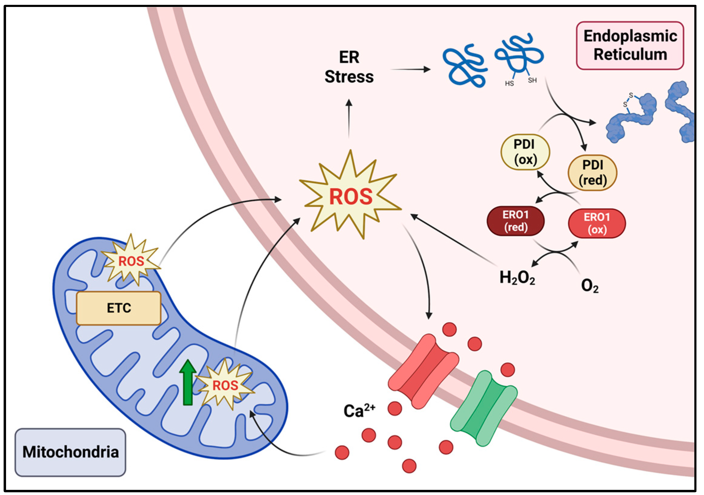
2. The Endoplasmic Reticulum and ROS Production

The ER is comprised of a series of tubules and sheet-like structures spanning throughout the cytoplasm and serves critical cellular functions including lipid and steroid production, calcium storage and protein synthesis, folding and transport [4,5]. Disulfide bond formation is one of the most common post-translational modifications occurring within the ER and is essential for the generation of stable, correctly folded proteins. To facilitate protein folding, the ER requires high levels of calcium (to support ER chaperone protein function) and must maintain a highly oxidizing environment to enable disulfide bond formation [6,7].
The protein disulfide isomerase (PDI) family of proteins play a central role in forming disulfide bridges. These calcium-binding chaperones possess oxidoreductase and isomerase activity, mediated by four thioredoxin-like domains a, b, a’, b’, with a and a’ containing a Cys-X-X-Cys sequence enabling disulfide oxidoreductase activity. While the b and b’ domains lack catalytic activity, they contribute to the binding of protein substrates [8]. Oxidized PDI is the working unit responsible for extracting electrons from the cysteine residues in newly synthesized proteins, thus creating an intramolecular disulfide bridge, a process that results in PDI reduction [9]. For further rounds of disulfide bond formation, reduced PDI requires re-oxidization, which is facilitated by several redox pathways. Endoplasmic reticulum oxidoreductase 1 (ERO1) replenishes oxidized PDI, but in doing so generates ROS by using O2 as an electron acceptor, which forms the reactive oxygen species H2O2 [9,10]. Alternatively, oxidized peroxiredoxin 4 (PRDX4), and glutathione peroxidase 7 and 8 (GPx7 and GPx8) can accept electrons to regenerate re-oxidized PDIs for subsequent rounds of protein disulfide bond formation. PRDX4, GPx7 and GPx8 also act as important ER-resident antioxidants as they utilize ERO1-generated H2O2 as an electron acceptor during cyclic redox reactions to form H2O [11,12,13]. Collectively, these enzymes are critical for ensuring the complete reduction of O2 to H2O and the maintenance of redox balance within the ER during the process of oxidative protein folding.
Protein disulfide bond formation can also be catalyzed through enzymatic reactions that do not depend on ERO1. This includes the vitamin K epoxide reductase (VKOR) pathway. VKOR can undergo a cyclic redox reaction by accepting electrons from thioredoxin-like proteins (which catalyze protein disulfide bond formation) and by reducing vitamin K epoxides [14]. The VKOR pathway has been described to be important in managing ER redox levels during oxidative protein folding [15]. Quiescin sulfhydryl oxidase (QSOX) is another enzyme that can directly catalyze protein disulfide bond formation through the reduction of O2 into H2O2 [16]. However, these pathways are not as well characterised and further investigation is required to assess their overall impact on ER ROS.
ER-derived ROS can also originate from enzymatic reactions beyond protein folding. The cytochrome P450 (CYP) family of enzymes are known for their role in lipid and drug metabolism, and many are localized to the endoplasmic reticulum of hepatocytes [17]. CYP catalyzes the hydroxylation of a hydrocarbon substrate with the assistance of electron donors in the presence of oxygen. However, ROS may leak during the intermediary steps of the reaction (O2•− and H2O2), thus contributing to endoplasmic reticulum ROS levels [18]. CYP2A1 and CYP2E1 have been characterised as ‘leaky’ enzymes which contribute to ROS production [19,20]. Meanwhile, the stimulation of hepatocytes with free fatty acids elevated ROS levels through CYP4A11 [21]. CYP2C9 was also demonstrated to generate H2O2, which was reduced upon interaction with cytochrome b5 [22].
While the ER has defense strategies, if ROS levels exceed the buffering capacity of ROS scavengers, ER function becomes impeded. Increased ROS can result in excess oxidation or hyperoxidation of ER-resident proteins, altering their conformation and function. For example, PRDX4 can be irreversibly inactivated through overoxidization into its sulfonic acid form (-SO3H) under high H2O2 levels [23,24]. Furthermore, perturbances within ER redox homeostasis disrupt the conditions required to support disulfide bond formation, thus negatively affecting protein folding [25]. This can result in the buildup of misfolded or unfolded proteins within the ER lumen, a state commonly referred to as ER stress. To combat ER stress, cells trigger the activation of the UPR. The UPR aims to restore ER homeostasis by promoting the folding of proteins that can be refolded, while initiating the degradation of those beyond repair via a process referred to as ER-associated degradation (ERAD). The UPR and its constituent signaling pathways have been well characterised over the past few decades.

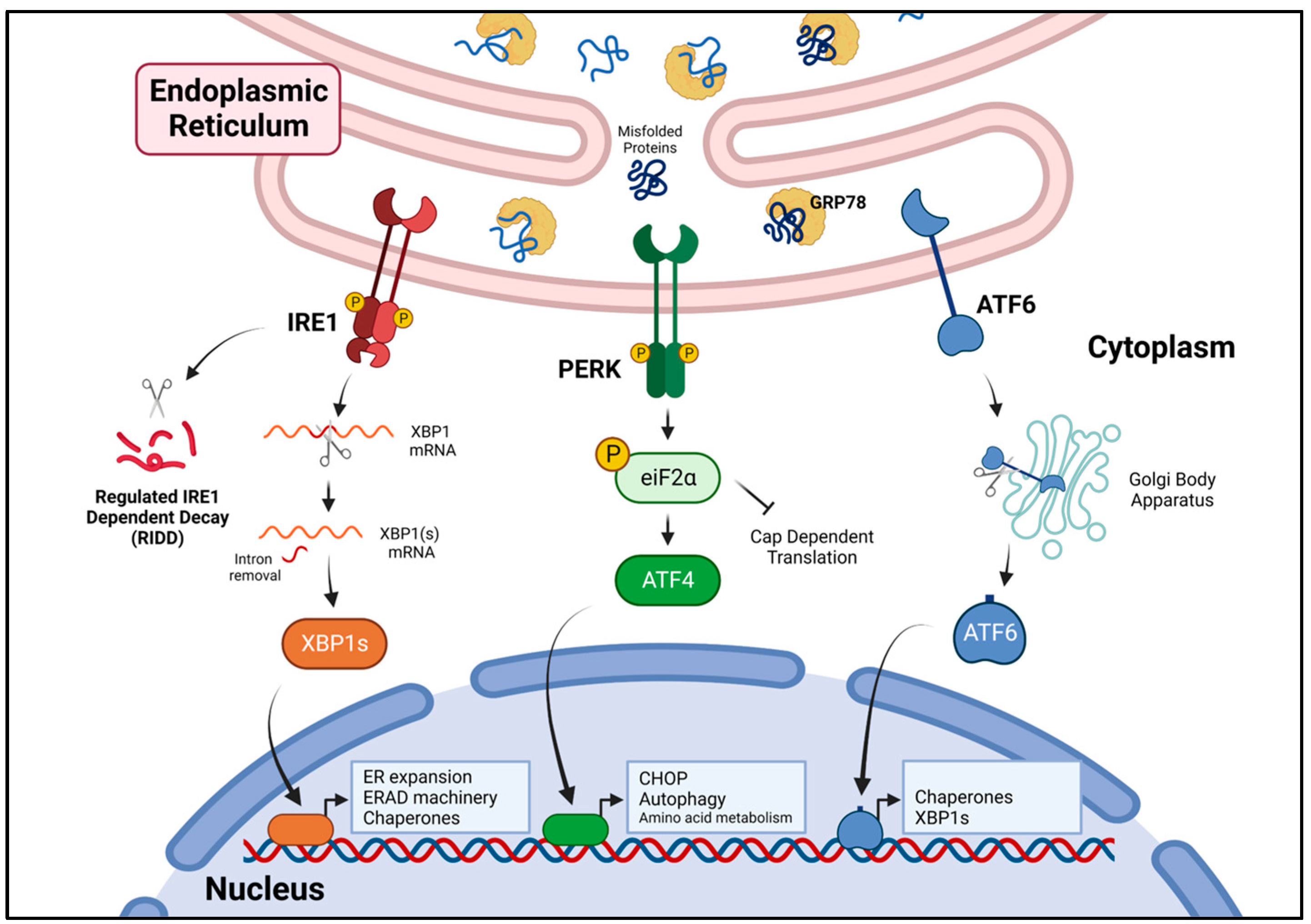
3. The Unfolded Protein Response

The UPR is a collective term for a series of complex, dynamic signaling processes controlled by three receptors, inositol requiring enzyme-1 (IRE1), protein kinase R-like endoplasmic reticulum kinase (PERK) and activating transcription factor 6 (ATF6). These receptors, anchored on the ER membrane, are single-pass transmembrane proteins with either cytosolic carboxyl termini (IRE1, PERK) or a cytosolic amino terminus (ATF6). Under non-stress conditions, IRE1, PERK and ATF6 remain in an “off” position via the binding of their luminal terminus to the ER chaperone glucose regulated protein 78 (GRP78) [26]. The accumulation of unfolded proteins within the ER lumen breaks the association with GRP78 (as GRP78 preferentially binds unfolded proteins), permitting IRE1, PERK and ATF6 to undergo changes facilitating their activation (Figure 1).

3.1. IRE1 Signaling

IRE1 consists of two isoforms, IRE1α (hereafter referred to as IRE1) which is ubiquitously expressed, and IRE1β whose expression is restricted to epithelial cells lining mucosal surfaces such as the intestine [27,28]. IRE1 is a type I transmembrane ER-anchored receptor protein consisting of cytosolic Serine/Threonine kinase and the ribonuclease domain [29]. Following the dissociation of GRP78, IRE1 homodimerizes and transautophosphorylates, facilitating the activation of its RNase activity [30,31]. Through its RNase activity, IRE1 cleaves X-box binding protein 1 (XBP1) mRNA, facilitating the removal of a 26-nucleotide intron [32]. The RNA 2’,3’-cyclic phosphate and 5’-OH ligase (RTCB) re-ligates cleaved XBP1 mRNA generating spliced XBP1 (XBP1s), which, when translated, produces the transcription factor XBP1s [33]. XBP1s binds to ER stress response element (ERSE), ER stress response element II (ERSE II) and unfolded protein response element (UPRE) sequences [34,35]. This upregulates the expression of genes encoding proteins that (A) promote protein refolding (ER chaperone proteins), (B) aid in the destruction of those proteins beyond repair (components of the ER associated degradation (ERAD) machinery) and (C) encode proteins that facilitate the expansion of the ER, thus increasing protein folding capacity [36,37,38]. In addition to splicing XBP1, IRE1 RNase activity has the capacity to cleave other RNA targets in a process known as regulated IRE1 dependent decay (RIDD) [39,40]. Initial studies demonstrated that RIDD targets contained a consensus sequence (CTGCAG) and a secondary structure similar to that found in XBP1 mRNA [41]. However, recent studies suggest that RIDD may be a more complex process, with the oligomerization/phosphorylation status of IRE1 influencing the selection of RIDD targets. Under relatively modest activation, IRE1 selectively targets substrates exhibiting the preferred consensus sequence. However, as IRE1 activation increases, RIDD activity becomes more promiscuous resulting in widespread mRNA degradation, a process that has been coined RIDD Lacking Endomotif, or RIDDLE [42]. Initial studies linked RIDD to the destruction of mRNAs encoding ER bound proteins. This suggested that RIDD aids a stressed ER by preventing the production of proteins destined for the ER [39]. As our knowledge of RIDD has increased, so too has our understanding of its functional outcomes, both within the specific setting of ER stress and its broader physiological impact. IRE1-RIDD has been demonstrated to maintain cell viability during ER stress by degrading death receptor 5 (DR5), thereby neutralising CHOP-mediated increases in DR5 [43]. The attenuation of RIDD activity, as observed during excessive ER stress, halts DR5 degradation resulting in DR5 mediated cell death [44]. In terms of its wider biological implications, RIDD activity contributes to a wide range of physiological processes ranging from lipid metabolism and insulin production to innate and adaptive immunity [45,46,47].
While IRE1 RNase activity has been the focus of most research, the kinase activity of IRE1 can also trigger downstream signaling. Although not extensively characterised, IRE1 kinase activity has been demonstrated to recruit TNF receptor-associated factor 2 (TRAF2) leading to c-Jun N terminal Kinase (JNK) signaling [48]. As JNK activation can phosphorylate BCL-2 family members, IRE1-JNK signaling may promote a shift towards pro-death signaling by increasing the activity of pro-apoptotic BCL-2 family members while reducing that of anti-apoptotic BCL-2 family members [49].

3.2. PERK Signaling

PERK, similar to IRE1, is a type I transmembrane ER-anchored receptor protein which undergoes dimerization and transautophosphorylation following the dissociation of GRP78, resulting in the activation of its cytosolic kinase domain [50]. Active PERK phosphorylates Ser-51 of eukaryotic translation initiation factor 2A (eIF2α), preventing interaction of eIF2α with the guanidine exchange factor eukaryotic translation initiation factor 2B (eIF2B), thereby blocking the assembly of the translational pre-initiation complex and ultimately preventing cap-dependent translation [51,52]. This pause in translation reduces the protein load placed upon the ER and by doing so offers the cell an opportunity to deal with the backlog of unfolded/misfolded proteins. While this translational block is widespread, it is not absolute. Due to the presence of upstream open reading frames and internal ribosomal entry sites, specific genes, such as activating transcription factor 4 (ATF4), are selectively translated under these conditions [53]. Increased ATF4 expression elevates downstream target genes implicated in a wide range of processes including amino acid metabolism and autophagy [54,55]. The transcription factor CCAAT/enhancer binding proteins-homologous protein (CHOP) is also upregulated by ATF4 [56]. Increased CHOP expression has been linked to a shift towards pro-apoptotic outcomes, both by direct regulation of downstream pro-apoptotic target genes and indirectly via the re-establishment of cap-dependent translation through the induction of growth arrest and DNA damage-inducible protein 34 (GADD34) which de-phosphorylates eIF2α [57,58,59].

3.3. ATF6 Signaling

Activating transcription factor 6 is a member of the ATF/CREB family of transcription factors. In an unstressed ER, owing to the presence of intra- and inter-disulphide bonds between conserved cysteine residues within the luminal domain, ATF6 exists in monomeric, dimeric and oligomeric forms [60]. During ER stress, ATF6 translocates to the Golgi apparatus where Site-1 and Site-2 proteases cleave it, generating the active transcription factor ATF6N [61]. For many years, precisely how the dissociation of Grp78 instigates a shift in ATF6 localisation from the ER to the Golgi apparatus was unclear. Recent studies suggest that the redox state of ATF6 affects its ability to undergo translocation and processing. Oka and colleagues demonstrated that during the induction of ER stress, ATF6 dimers linked by a C467-467 disulfide bond are preferentially translocated to the Golgi apparatus. Overexpression of the ER resident oxidoreductase ERp18 diminished the formation of ATF6 dimers (via reduction) and processing of ATF6 to ATF6N, whereas the loss of ERp18 improved the kinetics of ATF6 dimerization [62]. This observation suggests that the mechanisms governing ATF6 translocation and cleavage within the Golgi apparatus are complex, inferring a role of ERp18 in regulating this process. Upon cleavage, the N-terminal portion of ATF6 (ATF6N) is released and translocates to the nucleus where it binds to cis-regulatory elements, including ERSE I, ERSE II and UPRE promoter sequences [34,63,64]. ATF6 upregulates ER-associated genes including XBP1, thereby supporting IRE1 signaling and ER quality control proteins [37,65].

3.4. The Intersection of Oxidative Stress, ER Stress, and the Unfolded Protein Response

Given that protein folding is influenced by the redox state of the ER, it should be no surprise that ER stress and oxidative stress are two closely associated events. Indeed, UPR activation and increased ROS levels can coincide during the onset of stress. This phenomenon can be observed by certain thiol-containing compounds such as dithiothreitol (DTT), which disrupts the process of oxidative protein folding. Accordingly, DTT treatment initiates the UPR; however, increased ROS levels were also observed [66]. Similarly, α-lipoic acid has been reported to induce ROS production and UPR activation in hepatoma cells [67]. Tunicamycin and thapsigargin, which are classical well-known inducers of ER stress/UPR initiation, were also demonstrated to increase ROS levels [68,69]. These findings suggest that a relationship exists between oxidative stress and ER stress. However, the mechanisms that link these two stressors are rather complex.
Increasing the levels of ROS can exacerbate ER stress and trigger downstream signaling events within the ER. ROS-sensitive Ca2+ channels inositol-1,4,5-trisphosphate receptor (IP3R) and ryanodine receptors (RyR) on the ER membrane can be activated during oxidative stress to trigger the release of Ca2+ stores [70]. Regions of the ER that interact with the mitochondria, known as mitochondria-associated ER membranes or MAMs, facilitate the uptake of ER-Ca2+ within the mitochondria, resulting in increased metabolism which stimulates further ROS production [71]. Increased mitochondrial ROS in turn can leak to the ER, further aggravating ER stress. Additionally, components of the UPR also directly contribute to oxidative stress. For example, the transcription factor CHOP can upregulate ERO1 which further potentiates the reduction of oxygen into hydrogen peroxide during oxidative folding [72]. Subsequently, increased ERO1 expression and accumulation of H2O2 enhance the activation of IP3R, triggering additional Ca2+ movement from the ER to the mitochondria thereby further exacerbating oxidative stress [73]. High H2O2 levels can also result in superoxidation of PRDX4, thus resulting in its inactivation [24]. Ultimately, these events support a vicious cycle where increased ROS triggers the release of Ca2+; this in turn stimulates mitochondrial ROS production which feeds back into the ER triggering Ca2+ release and further aggravating ER stress (Figure 2). If not controlled, the concurrent stressors will result in cell death. Perhaps not surprisingly, UPR mediators have been linked to signaling pathways aimed at alleviating oxidative stress and ultimately the restoration of ER homeostasis.

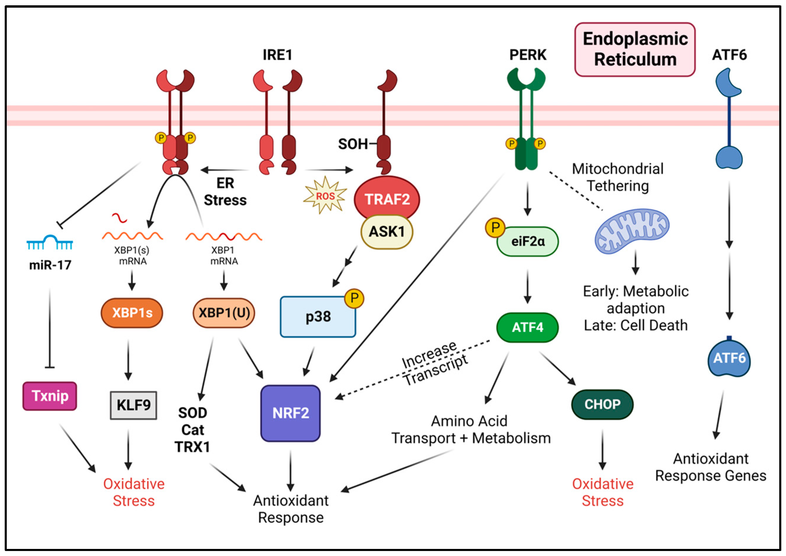
4. Regulation of Oxidative Stress by the Unfolded Protein Response

4.1. PERK in Response to Oxidative Stress

In 2003, a critical link between PERK and the master regulator of the oxidative stress response, nuclear factor-erythroid 2 related factor 2 (NRF2), was identified [74]. NRF2 is a Cap N’ Collar (CNC) and b-Zip transcription factor which binds to and promotes the activation of genes containing an antioxidant response element, including components of the thioredoxin and glutathione systems [75,76]. Under homeostatic conditions, the Kelch-like ECH-associated protein 1 (KEAP1) binds to the NRF2-ECH homology (Neh2) domain of NRF2. This interaction brings NRF2 into the KEAP1–Cul3–E3 ubiquitin ligase complex where it is ubiquitinated, leading to its degradation via the 26S proteasome [77]. During oxidative stress, NRF2 binding to KEAP1 is disrupted resulting in NRF2 stabilisation and the upregulation of NRF2 target genes, thereby boosting antioxidant responses. The disruption of KEAP1–NRF2 binding during oxidative stress is achieved via several mechanisms. Cysteine residues (Cys226/613/622/624) present on KEAP1 act as sensors for H2O2 becoming oxidized by H2O2 to form intramolecular disulfide bonds, thus preventing NRF2 binding [78]. Additionally, PERK-dependent signaling can also contribute to NRF2 stabilisation. PERK-mediated phosphorylation of NRF2 on Thr-80 prevents NRF2:KEAP1 complex formation, thereby stabilising NRF2 and, through this, enabling cellular adaption to oxidative stress [74,79]. The PERK–NRF2 axis can interact with downstream targets including hypoxia-inducible factor 1α (HIF1α), and can upregulate the antioxidants hemeoxygenase 1 (HO-1) and NAD(P)H:quinone oxidoreductase 1 (NQO1) [80,81].
PERK can also indirectly support NRF2 activation via upregulation of ATF4. Sarcinelli et al. demonstrated that ATF4 upregulates the Nrf2 transcript by binding to the C/ebp-Atf response element (CARE) cis regulatory element within the Nrf2 promoter [82]. This contributed to an increase in antioxidant response genes including heme-oxygenase 1 (HO1), an enzyme involved in producing the antioxidant bilirubin, and glutamate-cysteine ligase-catalytic subunit (GCLC), the subunit of the rate-limiting enzyme for glutathione (antioxidant) synthesis. ATF4 can also bind directly with NRF2 to form a heterodimer and upregulate antioxidant genes containing the cis regulatory antioxidant response element, stress response element, and amino acid response element to aid in suppressing oxidative stress [83,84].
PERK signaling through the PERK–p-eIF2α–ATF4 axis also supports oxidative stress resistance by upregulating genes involved in acid transport and glutathione biosynthesis. Harding et al. demonstrated that mouse embryonic fibroblasts (MEF) lacking PERK or ATF4 expression had increased sensitivity to oxidative stress and reduced expression of Glyt1 (glycine transporter 1) and SLC3A2 (heavy chain of xc cystine/glutamate exchanger) [54]. These genes express transporters responsible for the import of glycine and cysteine, which are important precursors for glutathione. PERK–p-eIF2α–ATF4 was also shown to upregulate GCLC, CTH (cystathionine gamma-lyase, an enzyme involved in cysteine synthesis) and SLC7A11 (solute carrier family 7 member 11, light chain of xc cysteine/glutamate antiporter) in MDA-MB-231 cells which resulted in increased amounts of reduced glutathione and decreased ROS levels [85]. Thus, PERK–p-eIF2α–ATF4-mediated increases in amino acid transport and metabolism are important for mitigating oxidative stress.
Conversely, the ATF4 target CHOP has been reported to contribute to increased oxidative stress by upregulating ERO1, thus leading to a heightened production of H2O2 and increased apoptosis [72]. Meanwhile, the deletion of CHOP lowered ERO1 expression and oxidative stress in diabetic mouse models [86].
While PERK’s function is mostly associated with UPR signaling via its kinase activity, it also has important structural roles, acting as a tethering protein within MAMs which also has implications in regulating oxidative stress. PERK-deficient MEFs exhibit decreased MAM formation, thereby reducing Ca2+ and ROS signaling from the ER to the mitochondria, ultimately suppressing ROS-induced apoptosis [87]. Similarly, in rat models of diabetic cardiomyopathy, PERK knockdown decreased ROS-induced apoptosis [88]. More recently, Bassot et al. have examined the PERK interactome and identified ERO1 as a PERK binding partner [89]. During the early stages of ER stress, PERK and ERO1 rapidly complex at mitochondrial ER contact sites, an interaction that supports Ca2+ flux between the two organelles, thus helping to maintain bioenergetics and suppress oxidative stress. These findings underscore the importance of PERK enrichment at MAMs to facilitate effective communication between the ER and the mitochondria for the regulation of oxidative stress.

4.2. IRE1 in Response to Oxidative Stress

IRE1-mediated signaling has been implicated in oxidative stress responses in various ways, both dependent and independent of its RNase activity. Guerra-Moreno et al. demonstrated a non-canonical activation of IRE1 in yeast (Saccharomyces cerevisiae) by sulfenylation of conserved cysteine residues present within the activation loop (C832 for yeast), activated antioxidant responses and reduced oxidative stress [90]. Similarly, Hourihan et al. described a unique mechanism where increased ROS production through the ER or mitochondria results in downstream activation of SKN-1 (C. elegans) and NRF2 (human) through IRE1. They demonstrated that the increased presence of ER ROS drives the sulfenylation of cysteine residues present within the activation loop of IRE1 [91]. In C. elegans, sulfenylated IRE1 recruits TRF1/NSY-1 (TRAF2/ASK1 in humans) which is required for subsequent phosphorylation of p38 MAPK signaling and downstream activation of NRF2 in a KEAP1 independent manner. Similarly, in human (HepG2) cells, IRE1 sulfenylation induced p38/NRF2 activation, highlighting the conservation of IRE1 antioxidant responses across species. IRE1 sulfenylation was mutually exclusive of IRE1 phosphorylation, as the induction of oxidative stress blocked XBP1s expression, while triggering ER stress repressed P-p38 expression [91].
Classical downstream signaling through IRE1-XBP1s can also influence oxidative stress. Fink et al. demonstrated that XBP1s upregulates Krüppel-like factor 9 (KLF9) during high levels of ER stress. Increased KLF9 amplifies both ER stress and oxidative stress through increased ER Ca2+ release, and suppression of thioredoxin reductase 2 (Txnrd2), resulting in cell death [92,93]. The weak interaction between XBP1s and UPRE sequences in the promoter region of KLF9 likely suggests that KLF9 upregulation requires a stronger signal (high XBP1s levels) due to unmitigated stress, thereby serving as a forward feedback mechanism to induce cell death [92,93].
While the impact of spliced XBP1(s) has been extensively studied, much less is known about its unspliced counterpart, XBP1u. Our lack of knowledge regarding XBP1u is, in part, a consequence of the unstable nature of XBP1u (half-life of 11 min) [94]. Despite this, evidence suggesting a selective requirement for XBP1u in oxidative stress responses is available. Liu et al. demonstrated that MEFs lacking total XBP1 displayed elevated cell death in response to H2O2-induced oxidative stress compared to wild-type counterparts. Further analysis demonstrated the reduced induction of antioxidant genes including superoxide dismutase 1 (SOD1), thioredoxin 1 (TRX1) and catalase (Cat) in H2O2-treated XBP1 knockout MEFs. The selective reestablishment of XBP1u, but not XBP1s, restored the induction of antioxidant genes, underscoring a specific role for unspliced XBP1 in antioxidant responses [95]. A similar role for XBP1u has also been reported in endothelial cells, where selective overexpression of XBP1u increased survival following the induction of oxidative stress. Within this setting, XBP1u promotion of cell survival increased NRF2 stabilisation and HO-1 expression [96]. Collectively, these studies suggest that XBP1u contributes to responses by antioxidant genes important for decomposing O2•− and H2O2.
IRE1 signaling has also been linked to the control of ROS production via the upregulation of thioredoxin inhibiting protein (TXNIP). TXNIP acts as an inhibitor of the key antioxidant enzyme thioredoxin (TRX), with TXNIP inhibiting thioredoxin reductase activity by binding to its redox active domain [97]. A loss of TXNIP expression has been demonstrated to inhibit ROS production, whereas an overexpression of TXNIP has been shown to increase ROS [98]. Based on these observations, pathways controlling TXNIP expression may have central roles in regulating oxidative stress. Lerner and colleagues demonstrated that IRE1 signaling increases TXNIP levels during ER stress. Mechanistically, IRE1-mediated TXNIP regulation was linked to RIDD-facilitated degradation of miR-17, a micro RNA that normally suppresses TXNIP expression. The induction of ER stress in INS-1 cells rapidly increased TXNIP expression in an IRE1-dependent manner [99]. Elevated ROS, in addition to damaging proteins and lipids, can also promote inflammation by triggering the formation of the NLRP3 inflammasome [99]. Indeed, TXNIP has been demonstrated to interact with NLRP3 and, through this, promote inflammasome formation [100]. Therefore, IRE1 activity, in addition to controlling ROS levels via TXNIP regulation, may also have the ability to influence inflammasome formation [99].

4.3. ATF6 in Response to Oxidative Stress

Of the three UPR mediators, within the context of oxidative stress, the contribution of ATF6 is the least characterised. Nonetheless, Jin et al. identified a potential role for ATF6 in suppressing necrosis induced by increased ROS. The overexpression of ATF6 in neonatal rat ventricular myocytes (NRVM) treated with hydrogen peroxide, or induced mitochondrial ROS simulated via ischemia/reperfusion, increased cell viability relative to controls [101]. Further qPCR gene array analysis revealed 11 antioxidant genes regulated by ATF6, including catalase, peroxiredoxin 5 (PRDX5) and thioredoxin reductase 1 (TXNRD1), glutathione peroxidase 3 and 4 (GPx3, GPx4) and NAPDH quinone dehydrogenase 1 (NQO1). The selective activation of ATF6 with AA147 increased the expression of catalase and suppressed oxidative stress and apoptosis in mouse neurons following cardiac arrest. Interestingly, NRF2/HO-1 activation was also demonstrated as a feature of AA147 treatment [102]. Overall, these findings indicate an antioxidant role for ATF6.

5. Conclusions and Perspectives

Classically, the UPR is considered a stress response system primarily aimed at resolving ER stress. However, the role of the ER in oxidative protein folding and its close association with mitochondria leaves the ER vulnerable to oxidative stress. Indeed, ER stress aggravates oxidative stress through increased protein folding and stimulated mitochondrial activity. As such, the UPR is also naturally equipped to deal with oxidative stress through the upregulation of antioxidant response genes to allow for initial adaptation (Figure 3). However, prolonged ER stress will initiate apoptosis in which some mechanisms are dependent on excessive ROS (oxidative stress) and mitochondrial crosstalk. Although the link between ER stress in ROS production has been established, the specific conditions for the ROS induction of ER stress, and, more specifically, UPR activation are not as well defined. The extent of UPR signaling appears to vary as some studies demonstrate activation by ROS [103], while others describe a limited, or lack of, activation [104]. Thus, further insights are required to elucidate the mechanisms by which ROS triggers ER stress and UPR activation. While the UPR is integral in maintaining ER homeostasis, it is amongst the many tools that the cell can utilize to adapt to stress. Indeed, other stress adaptation mechanisms, some of which are related to the UPR, can also aid in managing oxidative stress. Autophagy, a process that recycles damaged organelles including the ER and mitochondria, can supress oxidative stress and enable cell survival [105]. The maintenance of ER Ca2+ homeostasis and Ca2+ uptake through the sacro/endoplasmic reticulum calcium ATPase (SERCA) is also critical in preventing ER stress and elevated ROS levels [106]. Finally, heat shock proteins can be induced during oxidative stress to assist in protein folding and the degradation of damaged proteins [107,108]. Overall, understanding the interconnection amongst stress response pathways will provide a broader perspective on the progression of events that fine-tune the responses towards cellular adaptation and fate.

Author Contributions

Writing—original draft preparation, G.O.; writing—review and editing, G.O. and S.E.L. All authors have read and agreed to the published version of the manuscript.

Funding

This research was supported by the Canada Research Chairs Program, NSERC Discovery Grant Program and Research Manitoba New Investigator Grant awarded to S.E.L. G.O. is supported by a CancerCare Manitoba Foundation/Research Manitoba graduate studentship.

Acknowledgments

We thank Eftekhar Eftekharpour for helpful discussions and feedback on the manuscript.

Conflicts of Interest

The authors declare no conflict of interest.

References

  1. Zhao, R.-Z.; Jiang, S.; Zhang, L.; Yu, Z.-B. Mitochondrial Electron Transport Chain, ROS Generation and Uncoupling (Review). Int. J. Mol. Med. 2019, 44, 3–15. [Google Scholar] [CrossRef] [PubMed]
  2. Sullivan, L.B.; Chandel, N.S. Mitochondrial Reactive Oxygen Species and Cancer. Cancer Metab. 2014, 2, 17. [Google Scholar] [CrossRef] [PubMed]
  3. Tu, B.P.; Weissman, J.S. Oxidative Protein Folding in Eukaryotes: Mechanisms and Consequences. J. Cell Biol. 2004, 164, 341–346. [Google Scholar] [CrossRef] [PubMed]
  4. Shibata, Y.; Voeltz, G.K.; Rapoport, T.A. Rough Sheets and Smooth Tubules. Cell 2006, 126, 435–439. [Google Scholar] [CrossRef] [PubMed]
  5. Schwarz, D.S.; Blower, M.D. The Endoplasmic Reticulum: Structure, Function and Response to Cellular Signaling. Cell Mol. Life Sci. 2016, 73, 79–94. [Google Scholar] [CrossRef]
  6. Michalak, M.; Robert Parker, J.M.; Opas, M. Ca2+ Signaling and Calcium Binding Chaperones of the Endoplasmic Reticulum. Cell Calcium 2002, 32, 269–278. [Google Scholar] [CrossRef] [PubMed]
  7. Hwang, C.; Sinskey, A.J.; Lodish, H.F. Oxidized Redox State of Glutathione in the Endoplasmic Reticulum. Science 1992, 257, 1496–1502. [Google Scholar] [CrossRef]
  8. Kozlov, G.; Määttänen, P.; Thomas, D.Y.; Gehring, K. A Structural Overview of the PDI Family of Proteins. FEBS J. 2010, 277, 3924–3936. [Google Scholar] [CrossRef] [PubMed]
  9. Frand, A.R.; Kaiser, C.A. Ero1p Oxidizes Protein Disulfide Isomerase in a Pathway for Disulfide Bond Formation in the Endoplasmic Reticulum. Mol. Cell 1999, 4, 469–477. [Google Scholar] [CrossRef]
  10. Gross, E.; Sevier, C.S.; Heldman, N.; Vitu, E.; Bentzur, M.; Kaiser, C.A.; Thorpe, C.; Fass, D. Generating Disulfides Enzymatically: Reaction Products and Electron Acceptors of the Endoplasmic Reticulum Thiol Oxidase Ero1p. Proc. Natl. Acad. Sci. USA 2006, 103, 299–304. [Google Scholar] [CrossRef]
  11. Konno, T.; Pinho Melo, E.; Lopes, C.; Mehmeti, I.; Lenzen, S.; Ron, D.; Avezov, E. ERO1-Independent Production of H2O2 within the Endoplasmic Reticulum Fuels Prdx4-Mediated Oxidative Protein Folding. J. Cell Biol. 2015, 211, 253–259. [Google Scholar] [CrossRef]
  12. Wang, L.; Zhang, L.; Niu, Y.; Sitia, R.; Wang, C.-C. Glutathione Peroxidase 7 Utilizes Hydrogen Peroxide Generated by Ero1α to Promote Oxidative Protein Folding. Antioxid. Redox Signal. 2014, 20, 545–556. [Google Scholar] [CrossRef]
  13. Ramming, T.; Hansen, H.G.; Nagata, K.; Ellgaard, L.; Appenzeller-Herzog, C. GPx8 Peroxidase Prevents Leakage of H2O2 from the Endoplasmic Reticulum. Free Radic. Biol. Med. 2014, 70, 106–116. [Google Scholar] [CrossRef] [PubMed]
  14. Schulman, S.; Wang, B.; Li, W.; Rapoport, T.A. Vitamin K Epoxide Reductase Prefers ER Membrane-Anchored Thioredoxin-like Redox Partners. Proc. Natl. Acad. Sci. USA 2010, 107, 15027–15032. [Google Scholar] [CrossRef] [PubMed]
  15. Rutkevich, L.A.; Williams, D.B. Vitamin K Epoxide Reductase Contributes to Protein Disulfide Formation and Redox Homeostasis within the Endoplasmic Reticulum. Mol. Biol. Cell 2012, 23, 2017–2027. [Google Scholar] [CrossRef] [PubMed]
  16. Kodali, V.K.; Thorpe, C. Oxidative Protein Folding and the Quiescin–Sulfhydryl Oxidase Family of Flavoproteins. Antioxid. Redox Signal. 2010, 13, 1217–1230. [Google Scholar] [CrossRef] [PubMed]
  17. Kwon, D.; Kim, S.-M.; Correia, M.A. Cytochrome P450 Endoplasmic Reticulum-Associated Degradation (ERAD): Therapeutic and Pathophysiological Implications. Acta Pharm. Sin. B 2020, 10, 42–60. [Google Scholar] [CrossRef]
  18. Veith, A.; Moorthy, B. Role of Cytochrome P450S in the Generation and Metabolism of Reactive Oxygen Species. Curr. Opin. Toxicol. 2018, 7, 44–51. [Google Scholar] [CrossRef]
  19. Yue, Z.; Zhang, X.; Yu, Q.; Liu, L.; Zhou, X. Cytochrome P450-Dependent Reactive Oxygen Species (ROS) Production Contributes to Mn3O4 Nanoparticle-Caused Liver Injury. RSC Adv. 2018, 8, 37307–37314. [Google Scholar] [CrossRef]
  20. Harjumäki, R.; Pridgeon, C.S.; Ingelman-Sundberg, M. CYP2E1 in Alcoholic and Non-Alcoholic Liver Injury. Roles of ROS, Reactive Intermediates and Lipid Overload. Int. J. Mol. Sci. 2021, 22, 8221. [Google Scholar] [CrossRef]
  21. Gao, H.; Cao, Y.; Xia, H.; Zhu, X.; Jin, Y. CYP4A11 Is Involved in the Development of Nonalcoholic Fatty Liver Disease via ROS-induced Lipid Peroxidation and Inflammation. Int. J. Mol. Med. 2020, 45, 1121–1129. [Google Scholar] [CrossRef] [PubMed]
  22. Gómez-Tabales, J.; García-Martín, E.; Agúndez, J.A.G.; Gutierrez-Merino, C. Modulation of CYP2C9 Activity and Hydrogen Peroxide Production by Cytochrome B5. Sci. Rep. 2020, 10, 15571. [Google Scholar] [CrossRef] [PubMed]
  23. Ikeda, Y.; Nakano, M.; Ihara, H.; Ito, R.; Taniguchi, N.; Fujii, J. Different Consequences of Reactions with Hydrogen Peroxide and T-Butyl Hydroperoxide in the Hyperoxidative Inactivation of Rat Peroxiredoxin-4. J. Biochem. 2011, 149, 443–453. [Google Scholar] [CrossRef]
  24. Elko, E.A.; Manuel, A.M.; White, S.; Zito, E.; van der Vliet, A.; Anathy, V.; Janssen-Heininger, Y.M.W. Oxidation of Peroxiredoxin-4 Induces Oligomerization and Promotes Interaction with Proteins Governing Protein Folding and Endoplasmic Reticulum Stress. J. Biol. Chem. 2021, 296, 100665. [Google Scholar] [CrossRef] [PubMed]
  25. Malhotra, J.D.; Kaufman, R.J. Endoplasmic Reticulum Stress and Oxidative Stress: A Vicious Cycle or a Double-Edged Sword? Antioxid. Redox Signal. 2007, 9, 2277–2293. [Google Scholar] [CrossRef] [PubMed]
  26. Bertolotti, A.; Zhang, Y.; Hendershot, L.M.; Harding, H.P.; Ron, D. Dynamic Interaction of BiP and ER Stress Transducers in the Unfolded-Protein Response. Nat. Cell Biol. 2000, 2, 326–332. [Google Scholar] [CrossRef]
  27. Bertolotti, A.; Wang, X.; Novoa, I.; Jungreis, R.; Schlessinger, K.; Cho, J.H.; West, A.B.; Ron, D. Increased Sensitivity to Dextran Sodium Sulfate Colitis in IRE1β-Deficient Mice. J. Clin. Invest. 2001, 107, 585–593. [Google Scholar] [CrossRef]
  28. Martino, M.B.; Jones, L.; Brighton, B.; Ehre, C.; Abdulah, L.; Davis, C.W.; Ron, D.; O’Neal, W.K.; Ribeiro, C.M.P. The ER Stress Transducer IRE1β Is Required for Airway Epithelial Mucin Production. Mucosal Immunol. 2013, 6, 639–654. [Google Scholar] [CrossRef]
  29. Mori, K.; Ma, W.; Gething, M.J.; Sambrook, J. A Transmembrane Protein with a Cdc2+/CDC28-Related Kinase Activity Is Required for Signaling from the ER to the Nucleus. Cell 1993, 74, 743–756. [Google Scholar] [CrossRef]
  30. Shamu, C.E.; Walter, P. Oligomerization and Phosphorylation of the Ire1p Kinase during Intracellular Signaling from the Endoplasmic Reticulum to the Nucleus. EMBO J. 1996, 15, 3028–3039. [Google Scholar] [CrossRef]
  31. Sidrauski, C.; Walter, P. The Transmembrane Kinase Ire1p Is a Site-Specific Endonuclease That Initiates MRNA Splicing in the Unfolded Protein Response. Cell 1997, 90, 1031–1039. [Google Scholar] [CrossRef] [PubMed]
  32. Lee, K.; Tirasophon, W.; Shen, X.; Michalak, M.; Prywes, R.; Okada, T.; Yoshida, H.; Mori, K.; Kaufman, R.J. IRE1-Mediated Unconventional MRNA Splicing and S2P-Mediated ATF6 Cleavage Merge to Regulate XBP1 in Signaling the Unfolded Protein Response. Genes Dev. 2002, 16, 452–466. [Google Scholar] [CrossRef] [PubMed]
  33. Lu, Y.; Liang, F.-X.; Wang, X. A Synthetic Biology Approach Identifies the Mammalian UPR RNA Ligase RtcB. Mol. Cell 2014, 55, 758–770. [Google Scholar] [CrossRef] [PubMed]
  34. Yoshida, H.; Haze, K.; Yanagi, H.; Yura, T.; Mori, K. Identification of the Cis-Acting Endoplasmic Reticulum Stress Response Element Responsible for Transcriptional Induction of Mammalian Glucose-Regulated Proteins. Involvement of Basic Leucine Zipper Transcription Factors. J. Biol. Chem. 1998, 273, 33741–33749. [Google Scholar] [CrossRef]
  35. Yamamoto, K.; Sato, T.; Matsui, T.; Sato, M.; Okada, T.; Yoshida, H.; Harada, A.; Mori, K. Transcriptional Induction of Mammalian ER Quality Control Proteins Is Mediated by Single or Combined Action of ATF6alpha and XBP1. Dev. Cell 2007, 13, 365–376. [Google Scholar] [CrossRef]
  36. Yoshida, H.; Matsui, T.; Hosokawa, N.; Kaufman, R.J.; Nagata, K.; Mori, K. A Time-Dependent Phase Shift in the Mammalian Unfolded Protein Response. Dev. Cell 2003, 4, 265–271. [Google Scholar] [CrossRef]
  37. Yoshida, H.; Matsui, T.; Yamamoto, A.; Okada, T.; Mori, K. XBP1 MRNA Is Induced by ATF6 and Spliced by IRE1 in Response to ER Stress to Produce a Highly Active Transcription Factor. Cell 2001, 107, 881–891. [Google Scholar] [CrossRef]
  38. Shaffer, A.L.; Shapiro-Shelef, M.; Iwakoshi, N.N.; Lee, A.-H.; Qian, S.-B.; Zhao, H.; Yu, X.; Yang, L.; Tan, B.K.; Rosenwald, A.; et al. XBP1, Downstream of Blimp-1, Expands the Secretory Apparatus and Other Organelles, and Increases Protein Synthesis in Plasma Cell Differentiation. Immunity 2004, 21, 81–93. [Google Scholar] [CrossRef]
  39. Hollien, J.; Weissman, J.S. Decay of Endoplasmic Reticulum-Localized MRNAs during the Unfolded Protein Response. Science 2006, 313, 104–107. [Google Scholar] [CrossRef]
  40. Hollien, J.; Lin, J.H.; Li, H.; Stevens, N.; Walter, P.; Weissman, J.S. Regulated Ire1-Dependent Decay of Messenger RNAs in Mammalian Cells. J. Cell Biol. 2009, 186, 323–331. [Google Scholar] [CrossRef]
  41. Oikawa, D.; Tokuda, M.; Hosoda, A.; Iwawaki, T. Identification of a Consensus Element Recognized and Cleaved by IRE1α. Nucleic Acids Res. 2010, 38, 6265–6273. [Google Scholar] [CrossRef] [PubMed]
  42. Thomas, A.L.; Ferri, E.; Marsters, S.; Harnoss, J.M.; Modrusan, Z.; Li, W.; Rudolph, J.; Wang, W.; Wu, T.D.; Walter, P.; et al. Noncanonical MRNA Decay by the Endoplasmic-Reticulum Stress Sensor IRE1α Promotes Cancer-Cell Survival. bioRxiv 2021, 16. [Google Scholar] [CrossRef]
  43. Lu, M.; Lawrence, D.A.; Marsters, S.; Acosta-Alvear, D.; Kimmig, P.; Mendez, A.S.; Paton, A.W.; Paton, J.C.; Walter, P.; Ashkenazi, A. Opposing Unfolded-Protein-Response Signals Converge on Death Receptor 5 to Control Apoptosis. Science 2014, 345, 98–101. [Google Scholar] [CrossRef] [PubMed]
  44. Chang, T.-K.; Lawrence, D.A.; Lu, M.; Tan, J.; Harnoss, J.M.; Marsters, S.A.; Liu, P.; Sandoval, W.; Martin, S.E.; Ashkenazi, A. Coordination between Two Branches of the Unfolded Protein Response Determines Apoptotic Cell Fate. Mol. Cell 2018, 71, 629–636.e5. [Google Scholar] [CrossRef]
  45. Coelho, D.S.; Domingos, P.M. Physiological Roles of Regulated Ire1 Dependent Decay. Front. Genet. 2014, 5, 76. [Google Scholar] [CrossRef]
  46. Eletto, D.; Eletto, D.; Boyle, S.; Argon, Y. PDIA6 Regulates Insulin Secretion by Selectively Inhibiting the RIDD Activity of IRE1. FASEB J. 2016, 30, 653–665. [Google Scholar] [CrossRef]
  47. Almanza, A.; Mnich, K.; Blomme, A.; Robinson, C.M.; Rodriguez-Blanco, G.; Kierszniowska, S.; McGrath, E.P.; Le Gallo, M.; Pilalis, E.; Swinnen, J.V.; et al. Regulated IRE1α-Dependent Decay (RIDD)-Mediated Reprograming of Lipid Metabolism in Cancer. Nat. Commun. 2022, 13, 2493. [Google Scholar] [CrossRef]
  48. Urano, F.; Wang, X.; Bertolotti, A.; Zhang, Y.; Chung, P.; Harding, H.P.; Ron, D. Coupling of Stress in the ER to Activation of JNK Protein Kinases by Transmembrane Protein Kinase IRE1. Science 2000, 287, 664–666. [Google Scholar] [CrossRef]
  49. Szegezdi, E.; Logue, S.E.; Gorman, A.M.; Samali, A. Mediators of Endoplasmic Reticulum Stress-Induced Apoptosis. EMBO Rep. 2006, 7, 880–885. [Google Scholar] [CrossRef]
  50. Liu, C.Y.; Schröder, M.; Kaufman, R.J. Ligand-Independent Dimerization Activates the Stress Response Kinases IRE1 and PERK in the Lumen of the Endoplasmic Reticulum. J. Biol. Chem. 2000, 275, 24881–24885. [Google Scholar] [CrossRef]
  51. Harding, H.P.; Zhang, Y.; Ron, D. Protein Translation and Folding Are Coupled by an Endoplasmic-Reticulum-Resident Kinase. Nature 1999, 397, 271–274. [Google Scholar] [CrossRef] [PubMed]
  52. Harding, H.P.; Zhang, Y.; Bertolotti, A.; Zeng, H.; Ron, D. Perk Is Essential for Translational Regulation and Cell Survival during the Unfolded Protein Response. Mol. Cell 2000, 5, 897–904. [Google Scholar] [CrossRef] [PubMed]
  53. Vattem, K.M.; Wek, R.C. Reinitiation Involving Upstream ORFs Regulates ATF4 MRNA Translation in Mammalian Cells. Proc. Natl. Acad. Sci. USA 2004, 101, 11269–11274. [Google Scholar] [CrossRef]
  54. Harding, H.P.; Zhang, Y.; Zeng, H.; Novoa, I.; Lu, P.D.; Calfon, M.; Sadri, N.; Yun, C.; Popko, B.; Paules, R.; et al. An Integrated Stress Response Regulates Amino Acid Metabolism and Resistance to Oxidative Stress. Mol. Cell 2003, 11, 619–633. [Google Scholar] [CrossRef]
  55. B’chir, W.; Maurin, A.-C.; Carraro, V.; Averous, J.; Jousse, C.; Muranishi, Y.; Parry, L.; Stepien, G.; Fafournoux, P.; Bruhat, A. The EIF2α/ATF4 Pathway Is Essential for Stress-Induced Autophagy Gene Expression. Nucleic Acids Res. 2013, 41, 7683–7699. [Google Scholar] [CrossRef]
  56. Fawcett, T.W.; Martindale, J.L.; Guyton, K.Z.; Hai, T.; Holbrook, N.J. Complexes Containing Activating Transcription Factor (ATF)/CAMP-Responsive-Element-Binding Protein (CREB) Interact with the CCAAT/Enhancer-Binding Protein (C/EBP)-ATF Composite Site to Regulate Gadd153 Expression during the Stress Response. Biochem. J. 1999, 339 Pt 1, 135–141. [Google Scholar] [CrossRef]
  57. McCullough, K.D.; Martindale, J.L.; Klotz, L.O.; Aw, T.Y.; Holbrook, N.J. Gadd153 Sensitizes Cells to Endoplasmic Reticulum Stress by Down-Regulating Bcl2 and Perturbing the Cellular Redox State. Mol. Cell Biol. 2001, 21, 1249–1259. [Google Scholar] [CrossRef] [PubMed]
  58. Novoa, I.; Zeng, H.; Harding, H.P.; Ron, D. Feedback Inhibition of the Unfolded Protein Response by GADD34-Mediated Dephosphorylation of EIF2α. J. Cell Biol. 2001, 153, 1011–1022. [Google Scholar] [CrossRef]
  59. Han, J.; Back, S.H.; Hur, J.; Lin, Y.-H.; Gildersleeve, R.; Shan, J.; Yuan, C.L.; Krokowski, D.; Wang, S.; Hatzoglou, M.; et al. ER-Stress-Induced Transcriptional Regulation Increases Protein Synthesis Leading to Cell Death. Nat. Cell Biol. 2013, 15, 481–490. [Google Scholar] [CrossRef]
  60. Nadanaka, S.; Okada, T.; Yoshida, H.; Mori, K. Role of Disulfide Bridges Formed in the Luminal Domain of ATF6 in Sensing Endoplasmic Reticulum Stress. Mol. Cell Biol. 2007, 27, 1027–1043. [Google Scholar] [CrossRef]
  61. Ye, J.; Rawson, R.B.; Komuro, R.; Chen, X.; Davé, U.P.; Prywes, R.; Brown, M.S.; Goldstein, J.L. ER Stress Induces Cleavage of Membrane-Bound ATF6 by the Same Proteases That Process SREBPs. Mol. Cell 2000, 6, 1355–1364. [Google Scholar] [CrossRef] [PubMed]
  62. Oka, O.B.V.; Pierre, A.S.; Pringle, M.A.; Tungkum, W.; Cao, Z.; Fleming, B.; Bulleid, N.J. Activation of the UPR Sensor ATF6α Is Regulated by Its Redox-Dependent Dimerization and ER Retention by ERp18. Proc. Natl. Acad. Sci. USA 2022, 119, e2122657119. [Google Scholar] [CrossRef] [PubMed]
  63. Wang, Y.; Shen, J.; Arenzana, N.; Tirasophon, W.; Kaufman, R.J.; Prywes, R. Activation of ATF6 and an ATF6 DNA Binding Site by the Endoplasmic Reticulum Stress Response. J. Biol. Chem. 2000, 275, 27013–27020. [Google Scholar] [CrossRef]
  64. Kokame, K.; Kato, H.; Miyata, T. Identification of ERSE-II, a New Cis-Acting Element Responsible for the ATF6-Dependent Mammalian Unfolded Protein Response. J. Biol. Chem. 2001, 276, 9199–9205. [Google Scholar] [CrossRef]
  65. Adachi, Y.; Yamamoto, K.; Okada, T.; Yoshida, H.; Harada, A.; Mori, K. ATF6 Is a Transcription Factor Specializing in the Regulation of Quality Control Proteins in the Endoplasmic Reticulum. Cell Struct. Funct. 2008, 33, 75–89. [Google Scholar] [CrossRef] [PubMed]
  66. Wufuer, R.; Fan, Z.; Yuan, J.; Zheng, Z.; Hu, S.; Sun, G.; Zhang, Y. Distinct Roles of Nrf1 and Nrf2 in Monitoring the Reductive Stress Response to Dithiothreitol (DTT). Antioxidants 2022, 11, 1535. [Google Scholar] [CrossRef] [PubMed]
  67. Pibiri, M.; Sulas, P.; Camboni, T.; Leoni, V.P.; Simbula, G. α-Lipoic Acid Induces Endoplasmic Reticulum Stress-Mediated Apoptosis in Hepatoma Cells. Sci. Rep. 2020, 10, 7139. [Google Scholar] [CrossRef] [PubMed]
  68. Wang, C.; Li, T.; Tang, S.; Zhao, D.; Zhang, C.; Zhang, S.; Deng, S.; Zhou, Y.; Xiao, X. Thapsigargin Induces Apoptosis When Autophagy Is Inhibited in HepG2 Cells and Both Processes Are Regulated by ROS-Dependent Pathway. Environ. Toxicol. Pharmacol. 2016, 41, 167–179. [Google Scholar] [CrossRef] [PubMed]
  69. Yen, J.-H.; Wu, P.-S.; Chen, S.-F.; Wu, M.-J. Fisetin Protects PC12 Cells from Tunicamycin-Mediated Cell Death via Reactive Oxygen Species Scavenging and Modulation of Nrf2-Driven Gene Expression, SIRT1 and MAPK Signaling in PC12 Cells. Int. J. Mol. Sci. 2017, 18, 852. [Google Scholar] [CrossRef]
  70. Cao, S.S.; Kaufman, R.J. Endoplasmic Reticulum Stress and Oxidative Stress in Cell Fate Decision and Human Disease. Antioxid. Redox Signal. 2014, 21, 396–413. [Google Scholar] [CrossRef]
  71. Decuypere, J.-P.; Monaco, G.; Missiaen, L.; De Smedt, H.; Parys, J.B.; Bultynck, G. IP3 Receptors, Mitochondria, and Ca2+ Signaling: Implications for Aging. J. Aging Res. 2011, 2011, 920178. [Google Scholar] [CrossRef] [PubMed]
  72. Marciniak, S.J.; Yun, C.Y.; Oyadomari, S.; Novoa, I.; Zhang, Y.; Jungreis, R.; Nagata, K.; Harding, H.P.; Ron, D. CHOP Induces Death by Promoting Protein Synthesis and Oxidation in the Stressed Endoplasmic Reticulum. Genes Dev. 2004, 18, 3066–3077. [Google Scholar] [CrossRef] [PubMed]
  73. Bhattarai, K.R.; Riaz, T.A.; Kim, H.-R.; Chae, H.-J. The Aftermath of the Interplay between the Endoplasmic Reticulum Stress Response and Redox Signaling. Exp. Mol. Med. 2021, 53, 151–167. [Google Scholar] [CrossRef] [PubMed]
  74. Cullinan, S.B.; Zhang, D.; Hannink, M.; Arvisais, E.; Kaufman, R.J.; Diehl, J.A. Nrf2 Is a Direct PERK Substrate and Effector of PERK-Dependent Cell Survival. Mol. Cell Biol. 2003, 23, 7198–7209. [Google Scholar] [CrossRef] [PubMed]
  75. Alam, J.; Stewart, D.; Touchard, C.; Boinapally, S.; Choi, A.M.; Cook, J.L. Nrf2, a Cap’n’Collar Transcription Factor, Regulates Induction of the Heme Oxygenase-1 Gene. J. Biol. Chem. 1999, 274, 26071–26078. [Google Scholar] [CrossRef] [PubMed]
  76. Tonelli, C.; Chio, I.I.C.; Tuveson, D.A. Transcriptional Regulation by Nrf2. Antioxid. Redox Signal. 2018, 29, 1727–1745. [Google Scholar] [CrossRef]
  77. Villeneuve, N.F.; Lau, A.; Zhang, D.D. Regulation of the Nrf2-Keap1 Antioxidant Response by the Ubiquitin Proteasome System: An Insight into Cullin-Ring Ubiquitin Ligases. Antioxid. Redox Signal. 2010, 13, 1699–1712. [Google Scholar] [CrossRef]
  78. Suzuki, T.; Muramatsu, A.; Saito, R.; Iso, T.; Shibata, T.; Kuwata, K.; Kawaguchi, S.-I.; Iwawaki, T.; Adachi, S.; Suda, H.; et al. Molecular Mechanism of Cellular Oxidative Stress Sensing by Keap1. Cell Rep. 2019, 28, 746–758.e4. [Google Scholar] [CrossRef]
  79. Bobrovnikova-Marjon, E.; Grigoriadou, C.; Pytel, D.; Zhang, F.; Ye, J.; Koumenis, C.; Cavener, D.; Diehl, J.A. PERK Promotes Cancer Cell Proliferation and Tumor Growth by Limiting Oxidative DNA Damage. Oncogene 2010, 29, 3881–3895. [Google Scholar] [CrossRef]
  80. Küper, A.; Baumann, J.; Göpelt, K.; Baumann, M.; Sänger, C.; Metzen, E.; Kranz, P.; Brockmeier, U. Overcoming Hypoxia-Induced Resistance of Pancreatic and Lung Tumor Cells by Disrupting the PERK-NRF2-HIF-Axis. Cell Death Dis. 2021, 12, 82. [Google Scholar] [CrossRef]
  81. Wang, J.; Lu, L.; Chen, S.; Xie, J.; Lu, S.; Zhou, Y.; Jiang, H. Up-Regulation of PERK/Nrf2/HO-1 Axis Protects Myocardial Tissues of Mice from Damage Triggered by Ischemia-Reperfusion through Ameliorating Endoplasmic Reticulum Stress. Cardiovasc. Diagn. Ther. 2020, 10, 500–511. [Google Scholar] [CrossRef] [PubMed]
  82. Sarcinelli, C.; Dragic, H.; Piecyk, M.; Barbet, V.; Duret, C.; Barthelaix, A.; Ferraro-Peyret, C.; Fauvre, J.; Renno, T.; Chaveroux, C.; et al. ATF4-Dependent NRF2 Transcriptional Regulation Promotes Antioxidant Protection during Endoplasmic Reticulum Stress. Cancers 2020, 12, 569. [Google Scholar] [CrossRef] [PubMed]
  83. He, C.H.; Gong, P.; Hu, B.; Stewart, D.; Choi, M.E.; Choi, A.M.; Alam, J. Identification of Activating Transcription Factor 4 (ATF4) as an Nrf2-Interacting Protein. Implication for Heme Oxygenase-1 Gene Regulation. J. Biol. Chem. 2001, 276, 20858–20865. [Google Scholar] [CrossRef] [PubMed]
  84. Ye, P.; Mimura, J.; Okada, T.; Sato, H.; Liu, T.; Maruyama, A.; Ohyama, C.; Itoh, K. Nrf2- and ATF4-Dependent Upregulation of XCT Modulates the Sensitivity of T24 Bladder Carcinoma Cells to Proteasome Inhibition. Mol. Cell Biol. 2014, 34, 3421–3434. [Google Scholar] [CrossRef]
  85. Bai, X.; Ni, J.; Beretov, J.; Wasinger, V.C.; Wang, S.; Zhu, Y.; Graham, P.; Li, Y. Activation of the EIF2α/ATF4 Axis Drives Triple-Negative Breast Cancer Radioresistance by Promoting Glutathione Biosynthesis. Redox Biol. 2021, 43, 101993. [Google Scholar] [CrossRef]
  86. Song, B.; Scheuner, D.; Ron, D.; Pennathur, S.; Kaufman, R.J. Chop Deletion Reduces Oxidative Stress, Improves Beta Cell Function, and Promotes Cell Survival in Multiple Mouse Models of Diabetes. J. Clin. Investig. 2008, 118, 3378–3389. [Google Scholar] [CrossRef] [PubMed]
  87. Verfaillie, T.; Rubio, N.; Garg, A.D.; Bultynck, G.; Rizzuto, R.; Decuypere, J.-P.; Piette, J.; Linehan, C.; Gupta, S.; Samali, A.; et al. PERK Is Required at the ER-Mitochondrial Contact Sites to Convey Apoptosis after ROS-Based ER Stress. Cell Death Differ. 2012, 19, 1880–1891. [Google Scholar] [CrossRef] [PubMed]
  88. Liu, Z.-W.; Zhu, H.-T.; Chen, K.-L.; Dong, X.; Wei, J.; Qiu, C.; Xue, J.-H. Protein Kinase RNA-like Endoplasmic Reticulum Kinase (PERK) Signaling Pathway Plays a Major Role in Reactive Oxygen Species (ROS)-Mediated Endoplasmic Reticulum Stress-Induced Apoptosis in Diabetic Cardiomyopathy. Cardiovasc. Diabetol. 2013, 12, 158. [Google Scholar] [CrossRef] [PubMed]
  89. Bassot, A.; Chen, J.; Takahashi-Yamashiro, K.; Yap, M.C.; Gibhardt, C.S.; Le, G.N.T.; Hario, S.; Nasu, Y.; Moore, J.; Gutiérrez, T.; et al. The Endoplasmic Reticulum Kinase PERK Interacts with the Oxidoreductase ERO1 to Metabolically Adapt Mitochondria. Cell Rep. 2023, 42, 111899. [Google Scholar] [CrossRef]
  90. Guerra-Moreno, A.; Ang, J.; Welsch, H.; Jochem, M.; Hanna, J. Regulation of the Unfolded Protein Response in Yeast by Oxidative Stress. FEBS Lett. 2019, 593, 1080–1088. [Google Scholar] [CrossRef]
  91. Hourihan, J.M.; Moronetti Mazzeo, L.E.; Fernández-Cárdenas, L.P.; Blackwell, T.K. Cysteine Sulfenylation Directs IRE-1 to Activate the SKN-1/Nrf2 Antioxidant Response. Mol. Cell 2016, 63, 553–566. [Google Scholar] [CrossRef] [PubMed]
  92. Fink, E.E.; Moparthy, S.; Bagati, A.; Bianchi-Smiraglia, A.; Lipchick, B.C.; Wolff, D.W.; Roll, M.V.; Wang, J.; Liu, S.; Bakin, A.V.; et al. XBP1-KLF9 Axis Acts as a Molecular Rheostat to Control the Transition from Adaptive to Cytotoxic Unfolded Protein Response. Cell Rep. 2018, 25, 212–223.e4. [Google Scholar] [CrossRef] [PubMed]
  93. Zucker, S.N.; Fink, E.E.; Bagati, A.; Mannava, S.; Bianchi-Smiraglia, A.; Bogner, P.N.; Wawrzyniak, J.A.; Foley, C.; Leonova, K.I.; Grimm, M.J.; et al. Nrf2 Amplifies Oxidative Stress via Induction of Klf9. Mol. Cell 2014, 53, 916–928. [Google Scholar] [CrossRef] [PubMed]
  94. He, Y.; Sun, S.; Sha, H.; Liu, Z.; Yang, L.; Xue, Z.; Chen, H.; Qi, L. Emerging Roles for XBP1, a SUPeR Transcription Factor. Gene Expr. 2010, 15, 13–25. [Google Scholar] [CrossRef]
  95. Liu, Y.; Adachi, M.; Zhao, S.; Hareyama, M.; Koong, A.C.; Luo, D.; Rando, T.A.; Imai, K.; Shinomura, Y. Preventing Oxidative Stress: A New Role for XBP1. Cell Death Differ. 2009, 16, 847–857. [Google Scholar] [CrossRef]
  96. Martin, D.; Li, Y.; Yang, J.; Wang, G.; Margariti, A.; Jiang, Z.; Yu, H.; Zampetaki, A.; Hu, Y.; Xu, Q.; et al. Unspliced X-Box-Binding Protein 1 (XBP1) Protects Endothelial Cells from Oxidative Stress through Interaction with Histone Deacetylase 3. J. Biol. Chem. 2014, 289, 30625–30634. [Google Scholar] [CrossRef]
  97. Nishiyama, A.; Matsui, M.; Iwata, S.; Hirota, K.; Masutani, H.; Nakamura, H.; Takagi, Y.; Sono, H.; Gon, Y.; Yodoi, J. Identification of Thioredoxin-Binding Protein-2/Vitamin D(3) up-Regulated Protein 1 as a Negative Regulator of Thioredoxin Function and Expression. J. Biol. Chem. 1999, 274, 21645–21650. [Google Scholar] [CrossRef] [PubMed]
  98. Lerner, A.G.; Upton, J.-P.; Praveen, P.V.K.; Ghosh, R.; Nakagawa, Y.; Igbaria, A.; Shen, S.; Nguyen, V.; Backes, B.J.; Heiman, M.; et al. IRE1α Induces Thioredoxin-Interacting Protein to Activate the NLRP3 Inflammasome and Promote Programmed Cell Death under Irremediable ER Stress. Cell Metab. 2012, 16, 250–264. [Google Scholar] [CrossRef]
  99. Dostert, C.; Pétrilli, V.; Van Bruggen, R.; Steele, C.; Mossman, B.T.; Tschopp, J. Innate Immune Activation through Nalp3 Inflammasome Sensing of Asbestos and Silica. Science 2008, 320, 674–677. [Google Scholar] [CrossRef]
  100. Zhou, R.; Tardivel, A.; Thorens, B.; Choi, I.; Tschopp, J. Thioredoxin-Interacting Protein Links Oxidative Stress to Inflammasome Activation. Nat. Immunol. 2010, 11, 136–140. [Google Scholar] [CrossRef]
  101. Jin, J.-K.; Blackwood, E.A.; Azizi, K.; Thuerauf, D.J.; Fahem, A.G.; Hofmann, C.; Kaufman, R.J.; Doroudgar, S.; Glembotski, C.C. ATF6 Decreases Myocardial Ischemia/Reperfusion Damage and Links ER Stress and Oxidative Stress Signaling Pathways in the Heart. Circ. Res. 2017, 120, 862–875. [Google Scholar] [CrossRef] [PubMed]
  102. Yuan, M.; Gong, M.; He, J.; Xie, B.; Zhang, Z.; Meng, L.; Tse, G.; Zhao, Y.; Bao, Q.; Zhang, Y.; et al. IP3R1/GRP75/VDAC1 Complex Mediates Endoplasmic Reticulum Stress-Mitochondrial Oxidative Stress in Diabetic Atrial Remodeling. Redox Biol. 2022, 52, 102289. [Google Scholar] [CrossRef] [PubMed]
  103. Pallepati, P.; Averill-Bates, D.A. Activation of ER Stress and Apoptosis by Hydrogen Peroxide in HeLa Cells: Protective Role of Mild Heat Preconditioning at 40 °C. Biochim. Biophys. Acta 2011, 1813, 1987–1999. [Google Scholar] [CrossRef] [PubMed]
  104. Santos, C.X.C.; Tanaka, L.Y.; Wosniak, J.; Laurindo, F.R.M. Mechanisms and Implications of Reactive Oxygen Species Generation during the Unfolded Protein Response: Roles of Endoplasmic Reticulum Oxidoreductases, Mitochondrial Electron Transport, and NADPH Oxidase. Antioxid. Redox Signal. 2009, 11, 2409–2427. [Google Scholar] [CrossRef]
  105. Yun, H.R.; Jo, Y.H.; Kim, J.; Shin, Y.; Kim, S.S.; Choi, T.G. Roles of Autophagy in Oxidative Stress. Int. J. Mol. Sci. 2020, 21, 3289. [Google Scholar] [CrossRef] [PubMed]
  106. Guerrero-Hernández, A.; Leon-Aparicio, D.; Chavez-Reyes, J.; Olivares-Reyes, J.A.; DeJesus, S. Endoplasmic Reticulum Stress in Insulin Resistance and Diabetes. Cell Calcium 2014, 56, 311–322. [Google Scholar] [CrossRef]
  107. Reeg, S.; Jung, T.; Castro, J.P.; Davies, K.J.A.; Henze, A.; Grune, T. The Molecular Chaperone Hsp70 Promotes the Proteolytic Removal of Oxidatively Damaged Proteins by the Proteasome. Free Radic. Biol. Med. 2016, 99, 153–166. [Google Scholar] [CrossRef]
  108. Szyller, J.; Bil-Lula, I. Heat Shock Proteins in Oxidative Stress and Ischemia/Reperfusion Injury and Benefits from Physical Exercises: A Review to the Current Knowledge. Oxid. Med. Cell Longev. 2021, 2021, 6678457. [Google Scholar] [CrossRef]
Figure 1. The unfolded protein response and its role in ER stress. As misfolded proteins accumulate within the endoplasmic reticulum lumen, GRP78 dissociates from IRE1, PERK and ATF6 allowing for their activation. IRE1 RNase cleaves its downstream target XBP1 mRNA which becomes religated to form XBP1s mRNA encoding for the transcription factor XBP1s. PERK phosphorylates its downstream target eiF2α which suppresses CAP-dependent translation while allowing for selective translation of ATF4. Upon ER stress, ATF6 translocates to the Golgi where it becomes cleaved into its active form. The initiation of the UPR signaling pathways enhances expression of genes that enable adaptation and resolution of ER stress. Created with BioRender.com.
Antioxidants 12 00981 g001
Figure 2. Both the endoplasmic reticulum and mitochondria are major sources of cellular ROS. If ROS levels increase, Ca2+ release by the ER occurs and can stimulate further ROS production in the mitochondria. This in turn contributes to further ER stress and oxidative stress in the ER. A vicious feedback loop occurs where ER stress aggravates oxidative stress and vice versa. Created with BioRender.com.
Antioxidants 12 00981 g002
Figure 3. The three UPR signaling pathways (IRE1, PERK and ATF6) and their contribution to both antioxidant signaling and oxidative stress. Created with BioRender.com.
Antioxidants 12 00981 g003
Disclaimer/Publisher’s Note: The statements, opinions and data contained in all publications are solely those of the individual author(s) and contributor(s) and not of MDPI and/or the editor(s). MDPI and/or the editor(s) disclaim responsibility for any injury to people or property resulting from any ideas, methods, instructions or products referred to in the content.

