Article

Comparative Study of Different H2S Donors as Vasodilators and Attenuators of Superoxide-Induced Endothelial Damage

1
Department of Drug Science and Technology, University of Turin, 10125 Turin, Italy
2
Department of Pharmacy, “Federico II” University of Naples, 80131 Naples, Italy
3
Rita Levi-Montalcini Department of Neuroscience, University of Turin, 10125 Turin, Italy
*
Author to whom correspondence should be addressed.
Antioxidants 2023, 12(2), 344; https://doi.org/10.3390/antiox12020344
Submission received: 18 January 2023 / Revised: 25 January 2023 / Accepted: 30 January 2023 / Published: 1 February 2023

Abstract

:
In the last years, research proofs have confirmed that hydrogen sulfide (H2S) plays an important role in various physio-pathological processes, such as oxidation, inflammation, neurophysiology, and cardiovascular protection; in particular, the protective effects of H2S in cardiovascular diseases were demonstrated. The interest in H2S-donating molecules as tools for biological and pharmacological studies has grown, together with the understanding of H2S importance. Here we performed a comparative study of a series of H2S donor molecules with different chemical scaffolds and H2S release mechanisms. The compounds were tested in human serum for their stability and ability to generate H2S. Their vasorelaxant properties were studied on rat aorta strips, and the capacity of the selected compounds to protect NO-dependent endothelium reactivity in an acute oxidative stress model was tested. H2S donors showed different H2S-releasing kinetic and produced amounts and vasodilating profiles; in particular, compound 6 was able to attenuate the dysfunction of relaxation induced by pyrogallol exposure, showing endothelial protective effects. These results may represent a useful basis for the rational development of promising H2S-releasing agents also conjugated with other pharmacophores.

1. Introduction

Hydrogen sulfide (H2S), together with carbon monoxide (CO) and nitric oxide (NO), belong to the group of gaseous signaling molecules or “gasotransmitters”. These three species play a pivotal role in many pathophysiological processes of the cardiovascular system [1,2,3,4].
Although NO is still considered the major gaseous vasodilator, the identification of the biological importance of H2S has aroused increasing interest in its role [3,5,6]. The ongoing research clearly shows that H2S is an important independent mediator [7,8,9], as well as an enhancer of NO-mediated effects on the cardiovascular system [10,11]. Over the past decade, it has been progressively demonstrated that H2S, either endogenously produced or intentionally administered using H2S-donating compounds, is able to influence a wide range of physiological and pathophysiological processes [12]. Indeed, it takes part in the homeostatic regulation of cardiovascular, respiratory, gastroenteric, nervous, immune, and endocrine systems [1,13].
Nowadays, increasing research evidence has corroborated the protective effects of H2S in cardiovascular diseases (CVD) [14,15,16,17,18,19,20,21,22] such as cardiac hypertrophy, heart failure, myocardial ischemia/reperfusion (I/R) injury [23], hypertension [16,24] and atherosclerosis [25]. H2S reveals its protective potential by acting as an activator of angiogenesis [26], as a basal vasorelaxant agent [27], and as a blood pressure and heart rate regulator [28,29]. It has also been demonstrated that the mechanisms of these cardioprotective effects implicate antioxidative, anti-inflammatory, and pro-angiogenic behavior, in addition to the inhibition of cell apoptosis, and ion channel regulation [30,31]. In particular, it has been reported that H2S-mediated endothelial cells’ protection from oxidative stress could be due to direct antioxidant action, as well as maintenance of mitochondrial structure and function [32].
The first pharmacological tools used for the studies of H2S effects were inorganic salts, and in particular, sodium sulfide (Na2S) and hydrosulfide (NaHS), which were very helpful in clarifying their physio-pathological roles in mammalian organisms [33].
Although these compounds are commercially available and easy to manipulate, they have some important limitations. In particular, these salts hydrolyze upon reaction with water giving rise to an instantaneous supraphysiological level of H2S followed by a quick decrease in its concentration [34]. To overcome this problem, several organic structures able to release H2S under physiological conditions have been proposed. H2S donors differ in the mechanism of activation and H2S release kinetic [33].
Here we report a comparative study of a series of H2S donor molecules with extensively varied chemical scaffolds and mechanisms of H2S release (Figure 1). Their stability in human serum and ability to generate H2S, as well as their vasodilator effects, were evaluated. The capacity of the selected compounds to preserve NO-dependent endothelium reactivity in an in vitro model of acute oxidative stress was also reported.

2. Materials and Methods
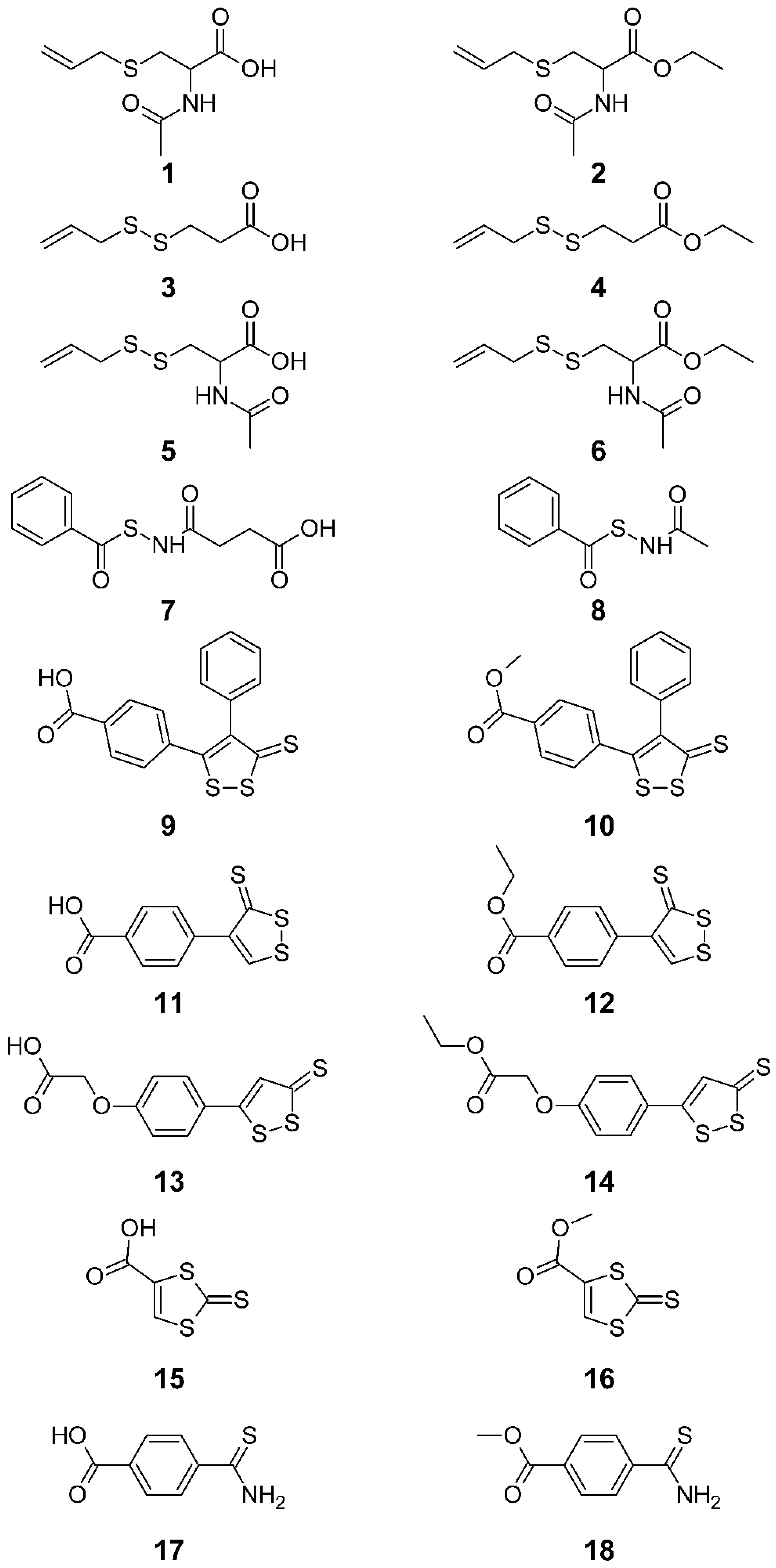
2.1. Synthesis

The synthetic procedures and physicochemical characterization of all studied compounds are reported in Supplementary Material.

2.2. Stability and H2S Release

2.2.1. Stability of the Compounds at pH 7.4 in Phosphate Buffer (PBS) in the Absence and Presence of L-Cysteine

The compounds were solubilized at a concentration of 100 µM (starting from a 10 mM stock solution in DMSO) in 0.1 M PBS, pH = 7.4, in the absence or presence of 5 mM L-cysteine (50×). The solutions were incubated at 37.0 ± 0.5 °C, and at appropriate time intervals, aliquots were analyzed by RP-HPLC as described below. All experiments were performed at least in triplicate.

2.2.2. Stability of the Compounds in Human Serum

A solution of each compound (10 mM) in DMSO was added to human serum (obtained by filtration and sterilization from AB, Sigma-Aldrich (Merck KGaA, 64,271 Darmstadt, Germany) male human blood) and was preheated to 37 °C to obtain a final concentration of 200 µM. The resulting solution was incubated at 37.0 ± 0.5 °C, and aliquots of 300 µL were taken at appropriate time intervals and added to 300 µL of CH3CN acidified with 0.1% formic acid in order to deproteinize the serum. The mixture was centrifuged for 10 min at 2150× g, and the clear supernatant was filtered (PTFE filters, 0.45 µm, VWR (VWR International S.r.l., Milano, Italy)) and analyzed by RP-HPLC as described below. All experiments were performed at least in triplicate.

2.2.3. RP-HPLC Analysis of Stability Assays

RP-HPLC analysis was performed with an HP 1100 chromatograph system (Agilent Technologies, Palo Alto, CA, USA) equipped with an injector (Rheodyne, Cotati, CA, USA), a quaternary pump (model G1311A), a membrane degasser (model G1379A), and a diode-array detector (DAD, model G1315B) integrated into the HP1100 system. The data were processed using an HP ChemStation system (Agilent Technologies, Palo Alto, CA, USA). A stationary phase Tracer Excel 120 ODSB (25 × 0.46, 5 μm; Tecnokroma (Teknokroma Analytical S.A., Barcelona, Spain)) was used. The mobile phase consisted of acetonitrile 0.1% HCOOH/water 0.1% HCOOH at a flow rate of 1.0 mL/min, with gradient elution: 35% acetonitrile up to 5 min, 35 to 80% acetonitrile between 5 and 10 min, 80% acetonitrile between 10 and 20 min. The injection volume was 20 μL. The column effluent was monitored for each compound at the wavelength of maximum absorption (referenced against a 700 nm wavelength). Using this RP-HPLC procedure, we separated the compounds from any degradation products and we quantified the compounds during incubation time. The quantification was performed using a calibration curve obtained with standard solutions of compounds chromatographed under the same experimental conditions, in a concentration range of 1–100 μM (r2 > 0.99).

2.2.4. Determination of H2S Release in Human Serum (Dansilazide Method)

60 μL of dansylazide solution (10 mM in ethanol) and 40 μL of H2S donor compound stock solution (10 mM in DMSO) were added to 1900 μL of human serum prewarmed at 37 °C to obtain an initial compound concentration of 200 μM. The solution was incubated at 37 ± 0.5 °C, and at fixed time (1, 4, and 24 h) 200 μL of the reaction mixture was diluted with 200 μL of CH3CN to have a final concentration of compound of 100 μM. The mixture was vortexed, centrifuged (10 min at 2150× g) and the clear supernatant was filtered (0.45 μm PTFE filters) and analyzed by RP-HPLC. All experiments were performed at least in triplicate. HPLC analyses were performed with a HP 1200 chromatograph system (Agilent Technologies, Palo Alto, CA, USA) equipped with a quaternary pump (model G1311A), a membrane degasser (G1322A), a UV detector, MWD (model G1365D) and a fluorescence detector (model G1321A) integrated into the HP1200 system. Data analysis was performed using an HP ChemStation system (Agilent Technologies). The sample was eluted on a Tracer Excel 120 ODSB C18 (250 × 4.6 mm, 5 μm; Teknokroma); the injection volume was 20 μL. The mobile phase consisted of 0.1% aqueous HCOOH and CH3CN 20/80 v/v; elution was in isocratic mode at a flow rate of 1.0 mL/min. The signals were obtained in fluorescence using an excitation and emission wavelength of 340 and 535 nm, respectively, and a gain factor = 10. The values obtained from integrating the dansyl amide peak were interpolated into a calibration curve, prepared using NaHS as a standard so that the concentration of dansyl amide in each sample is an index of the amount of H2S.

2.3. Functional Experiments

Animals were handled humanely in accordance with recognized guidelines on experimentation; the “3 Rs” policy (99/167/EC: Council Decision of 25/1/99) of replacement by alternative methods, reduction of the number of animals, and the refinement of experiments has been fully applied. The protocol has been approved by Ministero della Salute, “Studio preliminare del profilo farmacocinetico e farmacodinamico di composti di nuova sintesi ad attività multifattoriale”. Responsible: Elisabetta Marini. Cod. n. 56105.N.ZMT, approved on 23 June 2018.

2.3.1. Vasodilating Activity

Vasodilating activity was studied on the thoracic aortas from male Wistar rats (180–200 g), following a protocol published elsewhere [35]. All synthesized compounds were dissolved in DMSO. The addition of the vehicle had no perceptible consequence on contraction level. Results were expressed as EC50 ± SE (μM), n = 3–8.

2.3.2. Vasoprotection in Rat Aorta with Endothelium Impairment Induced by Pyrogallol

The experiments were performed on rat thoracic aortic rings from male Wistar rats (180–200 g), prepared as previously described [36]. The effect of H2S donor or catechin, taken as a reference, on acetylcholine-induced relaxation in aortic rings pre-incubated with pyrogallol was studied as published elsewhere [37], with some modifications. Aortic rings were incubated with diethyldithiocarbamate (DETCA) 10 mM for 1 h to irreversibly inactivate endogenous superoxide dismutase (SOD). Then, the aortic rings were washed and randomly divided into four groups: (1) control: endothelium-intact rings incubated in Krebs’ solution with vehicle only (DMSO); (2) H2S donor: endothelium-intact rings incubated with H2S donor (100 μM) or reference catechin (10 mM) for 30 min; (3) pyrogallol: endothelium-intact rings incubated with 500 μM pyrogallol for 15 min; (4) H2S donor plus pyrogallol: endothelium-intact rings incubated with H2S donor (100 μM) or reference catechin (10 mM) for 30 min and 500 μM pyrogallol for the last 15 min. After the incubation time, the rings were washed, 1 μM phenylephrine was added to the organ baths, then acetylcholine (10−9–l0−5 M) was added cumulatively. Tension was measured and calculated as the percentage of contraction in response to phenylephrine (1 μM). Data were expressed as mean ± SEM, n = 3–9.

2.3.3. Data Analysis

All data were expressed as mean ± SEM. Statistical comparisons were evaluated by Student’s t-test for unpaired data. p values < 0.05 were considered significantly different. The Gaussian distribution of data was verified by the D’Agostino–Pearson normality test. Statistical analyses were performed by GraphPad Prism 7.0 (GraphPad Software Inc., San Diego, CA, USA).

3. Results

3.1. Stability and H2S Release

H2S donor acids were first evaluated for their chemical stability under physiological-like conditions at pH 7.4 in a phosphate buffer solution. The results are reported in the Supplementary Materials (Table S1) as % residual concentration of compounds after 24 h of incubation. In these experiments, most of the compounds showed good stability, suggesting the absence of spontaneous H2S release at physiological pH. Only compound 7 displayed a reduced residual concentration, attributed to the hydrolytic instability of the N-bezoylsulfanil group. The experiment was repeated in the presence of cysteine to evaluate the stability of the compounds toward reactivity with the sulfhydryl group. The stability profiles of the H2S donor in PBS, at three different incubation times, in the presence of an excess of cysteine (50×) are reported in Supplementary Materials (Table S1).
To understand the potential of these H2S-donor substructures, it seemed important to evaluate their behavior in human serum, which is a complex sample, rich in metabolic enzymes, proteins and various endogenous thiols (cysteine, glutathione, homocysteine and –SH groups of proteins), which can profoundly influence the reactivity of these molecules. In these experiments, both stability and H2S release of all compounds were evaluated (Table 1). Stability data in human serum show that under these conditions, the carboxylic acids are characterized by slow degradation over time, which becomes significant after 24 h. At the same time, the poor stability of some esters (e.g., 2, 4, 6, 14, and 16) is mainly due to hydrolysis of the ester group under the action of esterases present in human serum. Indeed, the capacity of these compounds to release H2S was almost equal to that of the corresponding acids. On the other hand, the aromatic esters (10, 12, and 18) were more stable, and consequently, their kinetics of hydrolysis was different from that of corresponding acids (9, 11, and 17). It seems that the absence of an ionizable carboxyl group facilitates the release of H2S from H2S donors. Generally speaking, the H2S released from all the studied molecules turned out to be gradual and prolonged in time. From these experiments, it is possible to confirm that the series of synthesized compounds cover a wide range in terms of the quantity of H2S released.

3.2. Functional Studies

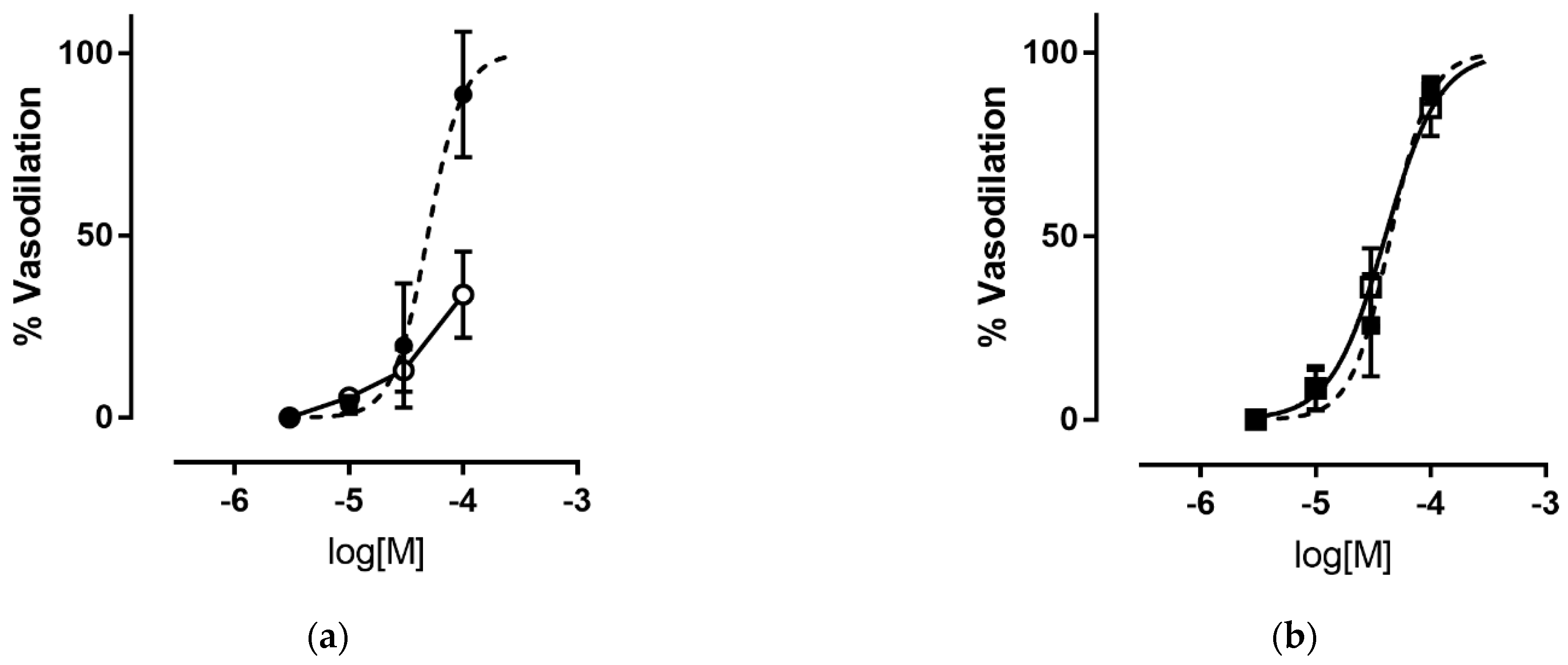
3.2.1. Vasodilating Activity

The vasodilator action of H2S-releasing compounds was evaluated on endothelium-denuded rat aorta strips precontracted with KCl. The results reported in Table 1 show the heterogeneous behavior of the tested compounds. For the most active compounds, vasodilation was found to be concentration dependent, with EC50 values ranging from 20 to 78 μM. In experiments performed in the presence of ATP-modulated K+-channels (K+ATP channels) blocker—glibenclamide the concentration–response curves obtained for compounds 4, 6, and 9 were significantly rightward shifted. These results confirmed that the relaxation induced by these compounds was mediated by the activation of the K+ATP channels. On the contrary, for compounds 8 and 12, the EC50 values obtained in the presence and absence of glibenclamide were almost the same, suggesting that these compounds exerted vasorelaxation with a different mechanism(s). An example of the vasodilation profile of the H2S donors tested is reported in Figure 2. For the less potent compounds, the percent of relaxation at the maximal testable concentration is reported in Table 1; due to solubility, the maximal concentration tested varied from 10 to 300 μM.

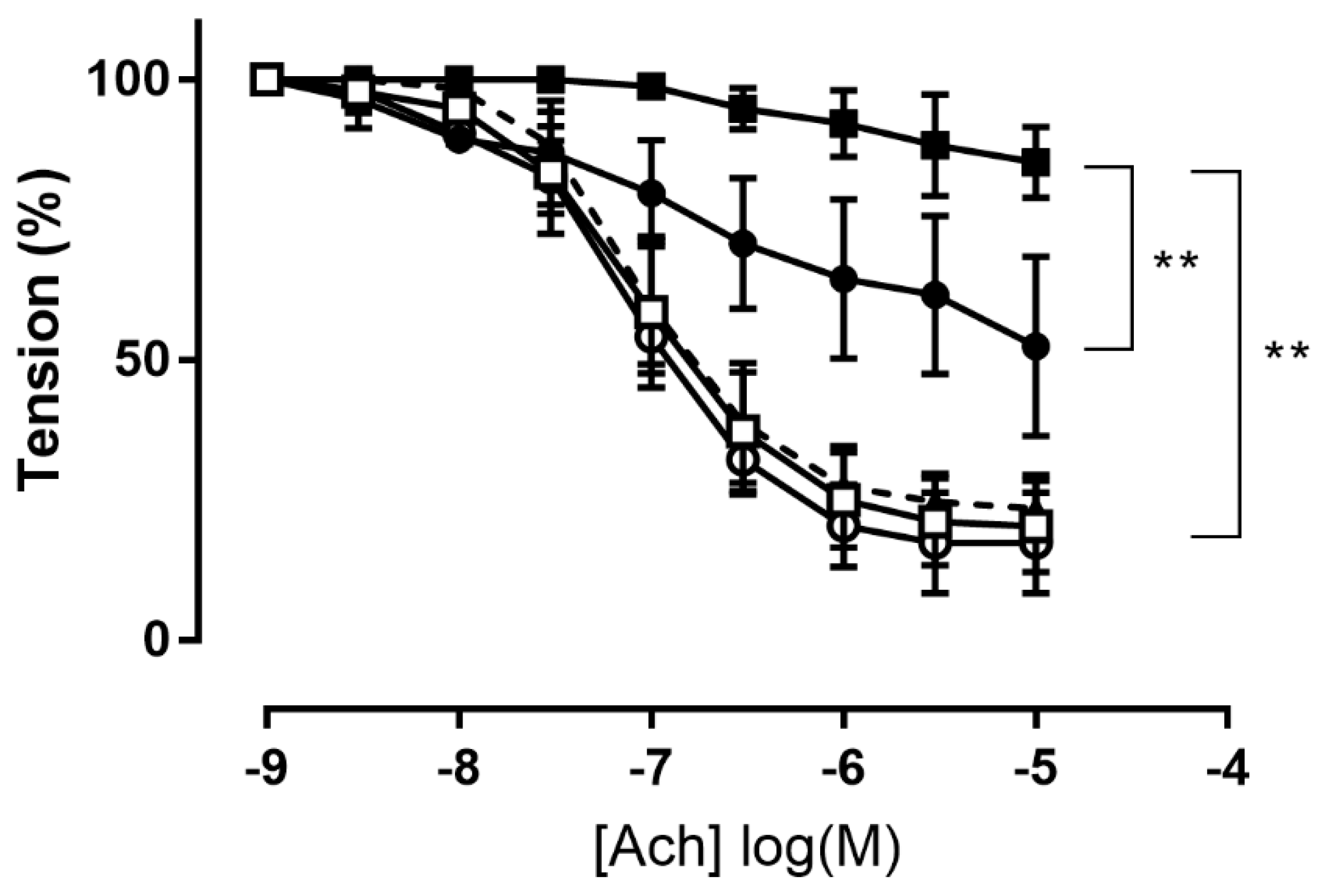
3.2.2. Effect of H2S Donor on Acetylcholine-Induced Vasodilation in Aorta Incubated with Pyrogallol

Superoxide anion (O2) generated by pyrogallol significantly reduced maximum Ach-induced relaxation in aortic rings from 80 ± 3% to 15 ± 3% (p = 0.0077, t-test). Treatment with catechin (10 mM), a known O2 scavenger [38], restored Ach-induced relaxation to control levels (77 ± 3%; Figure 3).
Treatment with compound 6 (100 μM) without exposure to pyrogallol had no effect on maximum relaxation (83 ± 6%; Figure 3), confirming that the amount of H2S released did not impair endothelium function. Pre-incubation with compound 6 attenuated the relaxation dysfunction induced by pyrogallol exposure, with a significant increase in Ach-induced maximal relaxation to 48 ± 11% compared to 15 ± 3% in the pyrogallol group (p = 0.0085, t-test; Figure 3). On the contrary, compound 8 (100 μM) was not able to reduce pyrogallol-induced endothelium impairment (Figure S1, Supplementary Materials).

4. Discussion

Experimental evidence has increasingly shown that alteration in H2S production is connected to cardiac pathologies. H2S has been hypothesized to have a protective role against the onset and development of atherosclerosis. While failures in H2S signaling, including the enzymes that synthesize it, can lead to cardiovascular diseases (CVD) and associated complications, H2S-based interventions have proved to be helpful in avoiding adult-onset CVD in animal studies by reversing disease-programming processes [6]. Indeed, the cardiovascular system can be programmed by a series of early-life offenses, driving to CVD in adulthood. Various models of developmental programming have been studied, including the genetic and maternal hypertension model, the suramin-induced preeclampsia model, maternal nicotine exposure, and high-fat diet: in these models, the reprogramming effects of H2S-based interventions have been reported in rats ranging from 12 weeks to 8 months of age, which approximately corresponds to human ages from young to middle adulthood [39].
However, the fine balance of H2S amount coming from endogenous production or exogenous H2S-releasing agents is very important. Similar to other gaseous transmitters, H2S has a double-face behavior with beneficial effects at low concentrations but potentially damaging effects at higher doses. The balance between endogenous H2S synthesis and exogenous H2S-releasing compounds that can have an effect on the fine H2S balance is important and requires a careful examination of the complex relationship between H2S and CVD.
Although many compounds have been synthetized and studied through in vitro and in vivo experiments, displaying H2S-release and positive effects in the treatment of cardiovascular diseases, to date, it is still not possible to identify an optimal compound, due to some drawbacks for each of them. In fact, the major limits of endogenous H2S-donor molecules are their incapacity to imitate endogenous H2S generation together with reactive byproducts formation, the nature and biological activity of which are frequently unknown [33]. A great number of H2S releasing molecules have been developed and evaluated, including N-(benzoylthio)benzamides [40], acyl perthiols [41,42], arylthioamides [43], 1,2,4-thiadiazolidin-3,5-diones [44], iminothioethers [45], mercaptopyruvate [46], dithioates [47], isothiocyanate [48], and thiocarbamates [49].
In this work, we decided to investigate various classes of H2S donors with different activation mechanisms as well as different kinetics and amounts of released H2S. All of the designed molecules were furnished with a carboxyl group that allows the desired H2S donors to be conjugated to other pharmacophores in the future. The corresponding alkyl esters were also studied to exclude the possibility of ionization and increase the lipophilicity of the studied molecules. In particular, S-allyl sulfide, S-allyl disulfide, benzylthioamide, N-(aryloylsulfanyl)acetamide, 3H-1,2-dithiole-3-thione and 1,3-dithiole-2-thione substructures were taken in account (Figure 1).
The H2S donors showed a very varied and complex vasodilating profile. Among the series, compounds 4, 6, 8, and 12 were found to be the most potent vasodilators (Table 1). There is no direct correlation between the quantity and velocity of H2S release and vasodilatation. For example, a good H2S releaser bearing the thioamide substructure 18 was a less potent vasodilator than 3H-1,2-dithiole-3-thione derivative 9 with rather poor H2S production. In the acid/ester pair, the better H2S production of the ester derivatives results in a better vasodilatation profile, with the only exception of 9/10. It seems that for effective vasorelaxation, H2S release should occur within endothelial cells and that optimal lipophilicity is essential for intracellular penetration of the compounds. Ester derivatives have higher lipophilicity and better H2S-releasing properties, which result in better vasorelaxation. On the other hand, the highly lipophilic acid 9 gives rise to ester 10 with very high lipophilicity, which probably hinders its intracellular accumulation resulting in a reduced vasodilating activity.
In the series of 3H-1,2-dithiole-3-thiones (914), the substituents present on the heteroring appear to be crucial for the potency and mechanism of vasodilation. Indeed, compound 9, bearing two aryl substituents on 1,2-dithiole-3-thione ring, showed modest vasodilating activity, with a significant rightward shift in the concentration-response curve obtained in the presence of the K+ATP channel blocker glibenclamide. The same behavior was observed for disulfuric compounds 4, and 6: the higher EC50 values obtained in the presence of glibenclamide (Table 1, Figure 2a) confirmed that the vasorelaxation induced by these compounds was due to the opening by H2S of K+ATP channels by sulfhydration of the Kir6.1 subunit, which is thought to be the main mechanism underlying the vasorelaxant effects of H2S [50].
Removing the aryl from the 5-position of the 3H-1,2-dithiole-3-thione ring yields the best vasodilator in the series, compound 12. This compound probably combines an optimal lipophilic profile with good H2S-releasing capacity, but its vasodilation has a different mechanism. Indeed, the EC50 values obtained in the presence and absence of glibenclamide were almost the same, suggesting that the observed vasorelaxation was not caused by the activation of K+ATP channels. The same behavior was observed for compound 8, bearing an N-mercapto substructure (Table 1; Figure 2b). Both of these derivatives released more H2S than the other compounds in the library, but the vasodilating effects were not induced by the main vasorelaxant mechanism of H2S. It is known that the K+ATP channels’ activation is not the only mechanism by which H2S exerts vasodilation; Bucci et al. [51] demonstrated that H2S could act as an endogenous nonselective phosphodiesterase inhibitor that raises cyclic nucleotide levels in tissues, causing vasodilation. Additional ion channel-independent mechanisms include, among others, decreased ATP levels through metabolic inhibition [52] and intracellular pH decrease exerted by activation of the chloride/bicarbonate exchanger [53], albeit H2S-induced stimulation of the type 2 anion exchanger of vascular smooth muscle cells, resulting in bicarbonate influx and superoxide anion efflux, has been connected to nitric oxide inactivation and vasoconstriction [54]. Moreover, it was demonstrated that not only K+ATP channels but also Kv7 channels (particularly the Kv7.4 subtype) are relevant pharmacological targets for H2S, and that direct activation of this family of ion channels mediates an important part of the vasodilatory effects of this gaseous mediator [55]. The relative contribution of K+ channels versus alternative pathways in the vasorelaxation due to H2S is expected to vary depending on the vascular bed, the species studied, and the amount of H2S present in the microenvironment [56], thus justifying the K+ATP-insensitive dilatory response of compounds 8 and 12.
Shifting the aryl substituent from the 4 to 5 position of the heteroring leads to molecules that are less active (13, 14) as both H2S releasers and vasodilators.
Endothelial dysfunction is associated with the onset of different CVDs, such as hypertension, myocardial infarction, cardiovascular complications of diabetes, and atherosclerosis. H2S could have a beneficial effect by triggering the angiogenesis of endothelial cells and relaxing vascular smooth muscle cells, thereby decreasing blood pressure [57]. Indeed, a low level of H2S endogenous production is implicated in the pathogenesis of endothelial dysfunction, while the administration of exogenous H2S, using H2S donors, can help to recover endothelial function and slow down the progression of CVD [27]. Oxidative stress is one of the most important mechanisms involved in endothelial dysfunction and associated cardiovascular pathogenesis. Therefore, reducing endothelial cells’ injury caused by oxidative stress could be a key to the treatment and/or prevention of CVD. Wen et al. reported the capacity of H2S to protect endothelial cells against oxidative stress due to its direct antioxidant activity, as well as its role in maintaining mitochondrial integrity [32]. There are still some uncertainties about the exact mechanisms of the antioxidant action of H2S, although different hypotheses have been proposed. The family of silent information regulator 2 (SIR2) is functionally significant in endothelial cells under oxidative stress, and some works point out that H2S is able to tune the activity of the sirtuin family, as the upregulation of sirtuin1 (SIRT1) in human PC12 cells and human umbilical vein endothelial cells (HUVECs) and the rise of SIRT3 and sirtuin 6 (SIRT6), to carry out either physiological or pathophysiological effects [58]. Moreover, H2S (H2S/HS/S2−) was helpful in biological states in which free radicals play an unfavorable role. H2S protects vascular smooth muscle, neurons, and different cells from oxidative stress exerted by biological or physicochemical conditions [59,60,61,62]. Indeed, the direct interaction of H2S with both free radicals and reactive oxygen species (ROS) have been hypothesized as a part of its antioxidant action [63,64,65].
To better understand the antioxidant activity of H2S in vascular tissue, we studied the ability of selected H2S donors to preserve NO-dependent endothelial reactivity in an in vitro model of acute oxidative stress. Probably the best-characterized mechanism by which oxidative stress can injure vascular function is the reaction of superoxide with NO [66,67], resulting in reduced bioavailability of this vasoprotective molecule. Given the ability of H2S to scavenge O2, we tested whether H2S donors could, in turn, preserve the endogenous bioavailability of NO in isolated rat aortae treated with the O2 generator pyrogallol to generate acute excessive levels of superoxide in vitro. Pyrogallol quickly auto-oxidizes in an oxygen-containing aqueous medium to generate O2. It has been previously reported that this model significantly inhibits ACh-induced relaxation through a reduction in the bioavailability of endogenous NO [68]. Catechin, a well-known O2 scavenger [38], was able to completely restore Ach-induced relaxation, albeit at high doses (10 mM) (Figure 3). For testing the ability of new H2S donors to protect against superoxide-mediated impairment of NO bioavailability, we selected two compounds with similar potencies but different vasorelaxant mechanisms. Compound 6 was selected as the most potent vasodilator agent via the opening of K+ATP channels, while compound 8 was selected as one of the compounds releasing higher amounts of H2S but exhibiting a K+ATP-independent vasorelaxation mechanism. Both selected compounds were first tested on rat aortic rings without acute oxidative stress to verify whether the compounds could induce an impairment of NO availability. The results showed that compounds 6 and 8 (100 μM) did not reduce Ach-induced maximum relaxation (Figure 3; Figure S1, Supplementary Materials), so the amount of H2S released by both compounds had no toxic or pro-oxidant effects.
Pretreatment with compound 6 significantly attenuated the pyrogallol-induced impairment of vasorelaxation and partially restored Ach-induced vasorelaxation (Figure 3). We suppose that the antioxidant effect of H2S release from 6 exerts vascular protection. The inhibitory effect of H2S on superoxide anion has been studied using different biochemical protocols. Polysulfides formation after several minutes incubation in buffered stock solutions of Na2S or NaHS at pH 7.4 have been demonstrated; Misak et al. showed that the formation of polysulfides and other H2S oxidation products is time-dependent when freshly prepared Na2S in H2O is diluted in Tris-HCl buffer, pH 7.4 [68]. Thus, we may assume that under our experimental conditions, polysulfides and other sulfide derivatives might be formed during incubation of the H2S donor solution in organ baths, and the observed protective effects of H2S could be affected by the formation of polysulfides or other H2S derivatives.
Although 8 is a more effective H2S donor, this compound tested under the same experimental conditions did not protect the endothelium from O2-induced damage. The lack of protection cannot be due to a too high amount of H2S being released into the organ bath since incubation of 8 alone had no negative effects on Ach-induced vasorelaxation (Figure S1). This behavior may be related to the H2S release mechanism for compound 8. Indeed, the release of 1 mole of H2S from 8 requires an excess (3 moles) of sulfhydryl groups, mainly glutathione (GSH), with GSSG formation [40,69]. GSH is the most abundant natural cellular antioxidant and plays a key role in maintaining the cellular redox balance. The antioxidant effect of GSH is due to the formation of its oxidized form: glutathione disulfide (GSSG). The latter is reduced to GSH by glutathione reductase to maintain a proper cellular GSH/GSSG ratio [70]. The H2S release process from compound 8 can significantly impair the GSH/GSSG ratio in endothelial cells. At the same time, in the experimental pyrogallol model, the incubation time of the H2S donor before the induction of endothelial damage was too short to allow for the recovery of glutathione lost, and the amount of H2S released was not able to compensate for the reduction in GSH/GSSG ratio and the consequent alteration of the cellular redox state. On the other side, the S-allyl disulfide substructure of compound 6 has been demonstrated to up-regulate the glutathione level by enhancing the expression of the regulatory subunit of glutamate-cysteine ligase (GCLM) and to increase the expression of phase II detoxifying enzymes, improving protection from oxidative stress [71,72].
This work provided a small library of H2S donor molecules with an extensively varied chemical scaffold showing different H2S-releasing rates and amounts, and vasodilating profiles. In particular, compound 6 was shown to vasodilate rat aorta strips through K+ATP channels activation and, more interestingly, was also able to protect vascular endothelium from oxidative damage. Therefore, this work could allow rationalizing of the development of H2S-donor molecules that are potentially useful for the treatment of cardiovascular diseases.

Supplementary Materials

The following supporting information can be downloaded at: https://www.mdpi.com/article/10.3390/antiox12020344/s1, Figure S1: Effect of 8 on acetylcholine-induced vasorelaxation in endothelium-intact rat aortic rings; Table S1: Stability of compounds in pH = 7.4 buffered solution (PBS) in the absence and in the presence of an excess of L-cysteine (50×). References [70,72,73,74,75,76] are cited in the Supplementary Materials.

Author Contributions

Conceptualization, K.C. and E.M.; validation, B.R. and E.M.; formal analysis, E.M.; investigation, E.M., B.R., F.S., F.B. and S.G.; resources, G.C.; writing—original draft preparation, E.M., K.C. and B.R.; writing—review and editing, L.L.; visualization, E.M.; supervision, K.C.; project administration, K.C.; funding acquisition, S.G. All authors have read and agreed to the published version of the manuscript.

Funding

This research was funded by MIUR PRIN 2017XP72RF_003 to S.G. and by the Università di Torino, Ricerca Locale 2017, and 2019 to E.M., and 2021 to K.C.

Institutional Review Board Statement

The animal study protocol was approved by the Ethics Committee of MINISTERO DELLA SALUTE (protocol code 56105.N.ZMT, approved on 23 June 2018).

Informed Consent Statement

Not applicable.

Data Availability Statement

Data supporting the findings of this study are available from the corresponding authors upon reasonable request.

Acknowledgments

In this section, you can acknowledge any support given which is not covered by the author contribution or funding sections. This may include administrative and technical support or donations in kind (e.g., materials used for experiments).

Conflicts of Interest

The authors declare no conflict of interest.

References

  1. Wang, R. Hydrogen sulfide: The third gasotransmitter in biology and medicine. Antioxid. Redox Signal. 2010, 12, 1061–1064. [Google Scholar] [CrossRef]
  2. Wang, R. Two’s company, three’s a crowd: Can H2S be the third endogenous gaseous transmitter. FASEB J. 2002, 16, 1792–1798. [Google Scholar] [CrossRef]
  3. Abe, K.; Kimura, H. The possible role of hydrogen sulfide as an endogenous neuromodulator. J. Neurosci. 1996, 16, 1066–1071. [Google Scholar] [CrossRef]
  4. Szabo, C. Gasotransmitters in cancer: From pathophysiology to experimental therapy. Nat. Rev. Drug Discov. 2016, 15, 185–203. [Google Scholar] [CrossRef]
  5. Szabo, C. A timeline of hydrogen sulfide (H2S) research: From environmental toxin to biological mediator. Biochem. Pharmacol. 2018, 149, 5–19. [Google Scholar] [CrossRef]
  6. Kolluru, G.K.; Shen, X.; Bir, S.C.; Kevil, C.G. Hydrogen sulfide chemical biology: Pathophysiological roles and detection. Nitric Oxide 2013, 35, 5–20. [Google Scholar] [CrossRef]
  7. Kolluru, G.K.; Shackelford, R.E.; Shen, X.; Dominic, P.; Kevil, C.G. Sulfide regulation of cardiovascular function in health and disease. Nat. Rev. Cardiol. 2023, 20, 109–125. [Google Scholar] [CrossRef]
  8. Liu, Y.H.; Lu, M.; Hu, L.F.; Wong, P.T.; Webb, G.D.; Bian, J.S. Hydrogen sulfide in the mammalian cardiovascular system. Antioxid. Redox Signal. 2012, 17, 141–185. [Google Scholar] [CrossRef]
  9. Pan, L.-L.; Qin, M.; Liu, X.-H.; Zhu, Y.-Z. The role of hydrogen sulfide on cardiovascular homeostasis: An overview with update on immunomodulation. Front. Pharmacol. 2017, 8, 686. [Google Scholar] [CrossRef]
  10. Szabo, C. Hydrogen sulfide, an enhancer of vascular nitric oxide signaling: Mechanisms and implications. Am. J. Physiol. Cell Physiol. 2017, 312, C3–C15. [Google Scholar] [CrossRef]
  11. Bir, S.C.; Kolluru, G.K.; McCarthy, P.; Shen, X.; Pardue, S.; Pattillo, C.B.; Kevil, C.G. Hydrogen sulfide stimulates ischemic vascular remodeling through nitric oxide synthase and nitrite reduction activity regulating hypoxiainducible factor-1α and vascular endothelial growth factor-dependent angiogenesis. J. Am. Heart Assoc. 2012, 1, e004093. [Google Scholar] [CrossRef] [PubMed]
  12. Li, Z.; Polhemus, D.J.; Lefer, D.J. Evolution of hydrogen sulfide therapeutics to treat cardiovascular disease. Circ. Res. 2018, 123, 590–600. [Google Scholar] [CrossRef] [PubMed]
  13. Li, L.; Rose, P.; Moore, P.K. Hydrogen sulfide and cell signaling. Annu. Rev. Pharmacol. Toxicol. 2011, 51, 169–187. [Google Scholar] [CrossRef]
  14. Polhemus, D.J.; Lefer, D.J. Emergence of hydrogen sulfide as an endogenous gaseous signaling molecule in cardiovascular disease. Circ. Res. 2014, 114, 730–737. [Google Scholar] [CrossRef]
  15. Huang, S.; Li, H.; Ge, J. A Cardioprotective insight of the cystathionine-lyase/hydrogen sulfide pathway. IJC Heart Vasc. 2015, 7, 51–57. [Google Scholar] [CrossRef]
  16. Meng, G.; Ma, Y.; Xie, L.; Ferro, A.; Ji, Y. Emerging role of hydrogen sulfide in hypertension and related cardiovascular diseases. Br. J. Pharmacol. 2015, 172, 5501–5511. [Google Scholar] [CrossRef]
  17. Wang, X.-H.; Wang, F.; You, S.-J.; Cao, Y.-J.; Cao, L.-D.; Han, Q.; Liu, C.-F.; Hu, L.-F. Dysregulation of cystathionine-lyase (CSE)/hydrogen sulfide pathway contributes to ox-LDL-induced inflammation in macrophage. Cell Signal. 2013, 25, 2255–2262. [Google Scholar] [CrossRef]
  18. Whiteman, M.; Moore, P.K. Hydrogen sulfide and the vasculature: A novel vasculoprotective entity and regulator of nitric oxide bioavailability? J. Cell. Mol. Med. 2009, 13, 488–507. [Google Scholar] [CrossRef]
  19. Xu, Y.; Du, H.-P.; Li, J.; Xu, R.; Wang, Y.-L.; You, S.-J.; Liu, H.; Wang, F.; Cao, Y.-J.; Liu, C.-F.; et al. Statins upregulate cystathionine-lyase transcription and H2S generation via activating Akt signaling in macrophage. Pharmacol. Res. 2014, 87, 18–25. [Google Scholar] [CrossRef]
  20. Yu, X.-H.; Cui, L.-B.; Wu, K.; Zheng, X.-L.; Cayabyab, F.S.; Chen, Z.-W.; Tang, C.-K. Hydrogen sulfide as a potent cardiovascular protective agent. Clin. Chim. Acta 2014, 437, 78–87. [Google Scholar] [CrossRef]
  21. Zheng, Y.; Ji, X.; Ji, K.; Wang, B. Hydrogen sulfide prodrugs-A review. Acta Pharm. Sin. B 2015, 5, 367–377. [Google Scholar] [CrossRef]
  22. Zhang, L.; Wang, Y.; Li, Y.; Li, L.; Xu, S.; Feng, X.; Liu, S. Hydrogen sulfide (H2S)-releasing cCompounds: Therapeutic potential in cardiovascular diseases. Front. Pharmacol. 2018, 9, 1066. [Google Scholar] [CrossRef] [PubMed]
  23. Calvert, J.W.; Elston, M.; Nicholson, C.K.; Gundewar, S.; Jha, S.; Elrod, J.W.; Ramachandran, A.; Lefer, D.J. Genetic and pharmacologic hydrogen sulfide therapy attenuates ischemia-induced heart failure in mice. Circulation 2010, 122, 11–19. [Google Scholar] [CrossRef] [PubMed]
  24. Yang, G.; Wu, L.; Jiang, B.; Yang, W.; Qi, J.; Cao, K.; Meng, Q.; Mustafa, A.K.; Mu, W.; Zhang, S.; et al. H2S as a Physiologic vasorelaxant: Hypertension in mice with deletion of cystathionine gamma-lyase. Science 2008, 322, 587–590. [Google Scholar] [CrossRef]
  25. Wang, Y.; Zhao, X.; Jin, H.; Wei, H.; Li, W.; Bu, D.; Tang, X.; Ren, Y.; Tang, C.; Du, J. Role of hydrogen sulfide in the development of atherosclerotic lesions in apolipoprotein E knockout mice. Arterioscler. Thromb. Vasc. Biol. 2009, 29, 173–179. [Google Scholar] [CrossRef]
  26. Van Den Born, J.C.; Mencke, R.; Conroy, S.; Zeebregts, C.J.; Van Goor, H.; Hillebrands, J.L. Cystathionine gamma-lyase is expressed in human atherosclerotic plaque microvessels and is involved in micro-angiogenesis. Sci. Rep. 2016, 6, 34608. [Google Scholar] [PubMed]
  27. Zhao, W.; Zhang, J.; Lu, Y.; Wang, R. The vasorelaxant effect of H(2)S as a novel endogenous gaseous K(ATP) channel opener. EMBO J. 2001, 20, 6008–6016. [Google Scholar] [CrossRef]
  28. Dawe, G.S.; Han, S.P.; Bian, J.S.; Moore, P.K. Hydrogen sulphide in the hypothalamus causes an ATP-sensitive K+ channel-dependent decrease in blood pressure in freely moving rats. Neuroscience 2008, 152, 169–177. [Google Scholar] [CrossRef]
  29. Liu, W.Q.; Chai, C.; Li, X.Y.; Yuan, W.J.; Wang, W.Z.; Lu, Y. The cardiovascular effects of central hydrogen sulfide are related to K(ATP) channels activation. Physiol. Res. 2011, 60, 729–738. [Google Scholar] [CrossRef]
  30. Wang, R. Physiological implications of hydrogen sulfide: A whiff exploration that blossomed. Physiol. Rev. 2012, 92, 791–896. [Google Scholar] [CrossRef] [Green Version]
  31. Xu, S.; Liu, Z.; Liu, P. Targeting hydrogen sulfide as a promising tTherapeutic strategy for atherosclerosis. Int. J. Cardiol. 2014, 172, 313–317. [Google Scholar] [CrossRef]
  32. Wen, Y.D.; Wang, H.; Kho, S.H.; Rinkiko, S.; Sheng, X.; Shen, H.M.; Zhu, Y.Z. Hydrogen sulfide protects HUVECs against hydrogen peroxide induced mitochondrial dysfunction and oxidative stress. PLoS ONE 2013, 8, e53147. [Google Scholar] [CrossRef]
  33. Corvino, A.; Frecentese, F.; Magli, E.; Perissutti, E.; Santagada, V.; Scognamiglio, A.; Caliendo, G.; Fiorino, F.; Severino, B. Trends in H2S-donors chemistry and their effects in cardiovascular diseases. Antioxidants 2021, 10, 429. [Google Scholar] [CrossRef]
  34. Hughes, M.N.; Centelles, M.N.; Moore, K.P. Making and working with hydrogen sulfide. Free Radic. Biol. Med. 2009, 47, 1346–1353. [Google Scholar] [CrossRef]
  35. Lazzarato, L.; Chegaev, K.; Marini, E.; Rolando, B.; Borretto, E.; Guglielmo, S.; Joseph, S.; Di Stilo, A.; Fruttero, R.; Gasco, A. New nitric oxide or hydrogen sulfide releasing aspirins. J. Med. Chem. 2011, 54, 5478–5484. [Google Scholar] [CrossRef]
  36. Brusotti, G.; Papetti, A.; Serra, M.; Temporini, C.; Marini, E.; Orlandini, S.; Sanda, A.K.; Watcho, P.; Kamtchouing, P. Allanblackia floribunda Oliv.: An aphrodisiac plant with vasorelaxant properties. J. Ethnopharmacol. 2016, 192, 480–485. [Google Scholar] [CrossRef]
  37. Jin, B.H.; Qian, L.B.; Chen, S.; Li, J.; Wang, H.P.; Bruce, I.C.; Lin, J.; Xia, Q. Apigenin protects endothelium-dependent relaxation of rat aorta against oxidative stress. Eur. J. Pharmacol. 2009, 616, 200–205. [Google Scholar] [CrossRef]
  38. Liu, S.; Xing, J.; Zheng, Z.; Song, F.; Liu, Z.; Liu, S. Ultrahigh performance liquid chromatography-triple quadrupole mass spectrometry inhibitors fishing assay: A novel method for simultaneously screening of xanthine oxidase inhibitor and superoxide anion scavenger in a single analysis. Anal. Chim. Acta 2012, 715, 64–70. [Google Scholar] [CrossRef]
  39. Hsu, C.N.; Tain, Y.L. Preventing developmental origins of cardiovascular disease: Hydrogen sulfide as a potential target? Antioxidants 2021, 10, 247. [Google Scholar] [CrossRef]
  40. Zhao, Y.; Wang, H.; Xian, M. Cysteine-activated hydrogen sulfide (H2S) donors. J. Am. Chem. Soc. 2011, 133, 15–17. [Google Scholar] [CrossRef] [Green Version]
  41. Zhao, Y.; Bhushan, S.; Yang, C.; Otsuka, H.; Stein, J.D.; Pacheco, A.; Peng, B.; Devarie-Baez, N.O.; Aguilar, H.C.; Lefer, D.J.; et al. Controllable hydrogen sulfide donors and their activity against myocardial ischemia-reperfusion injury. ACS Chem. Biol. 2013, 8, 1283–1290. [Google Scholar] [CrossRef]
  42. Roger, T.; Raynaud, F.; Bouillaud, F.; Ransy, C.; Simonet, S.; Crespo, C.; Bourguignon, M.-P.; Villeneuve, N.; Vilaine, J.-P.; Artaud, I.; et al. New biologically active hydrogen sulfide donors. ChemBioChem 2013, 14, 2268–2271. [Google Scholar] [CrossRef]
  43. Martelli, A.; Testai, L.; Citi, V.; Marino, A.; Pugliesi, I.; Barresi, E.; Nesi, G.; Rapposelli, S.; Taliani, S.; Da Settimo, F.; et al. Arylthioamides as H2S donors: L-cysteine-activated releasing properties and vascular effects in vitro and in vivo. ACS Med. Chem. Lett. 2013, 4, 904–908. [Google Scholar] [CrossRef]
  44. Severino, B.; Corvino, A.; Fiorino, F.; Luciano, P.; Frecentese, F.; Magli, E.; Saccone, I.; Di Vaio, P.; Citi, V.; Calderone, V.; et al. 1,2,4-Thiadiazolidin-3,5-diones as novel hydrogen sulfide donors. Eur. J. Med. Chem. 2018, 143, 1677–1686. [Google Scholar] [CrossRef]
  45. Barresi, E.; Nesi, G.; Citi, V.; Piragine, E.; Piano, I.; Taliani, S.; Da Settimo, F.; Rapposelli, S.; Testai, L.; Breschi, M.C.; et al. Iminothioethers as hydrogen sulfide donors: From the gasotransmitter release to the vascular effects. J. Med. Chem. 2017, 60, 7512–7523. [Google Scholar] [CrossRef]
  46. Mitidieri, E.; Tramontano, T.; Gurgone, D.; Citi, V.; Calderone, V.; Brancaleone, V.; Katsouda, A.; Nagahara, N.; Papapetropoulos, A.; Cirino, G.; et al. Mercaptopyruvate acts as endogenous vasodilator independently of 3-mercaptopyruvate sulfurtransferase activity. Nitric Oxide 2018, 75, 53–59. [Google Scholar] [CrossRef]
  47. Ercolano, G.; De Cicco, P.; Frecentese, F.; Saccone, I.; Corvino, A.; Giordano, F.; Magli, E.; Fiorino, F.; Severino, B.; Calderone, V.; et al. Anti-metastatic properties of naproxen-HBTA in a murine model of cutaneous melanoma. Front. Pharmacol. 2019, 10, 66. [Google Scholar] [CrossRef]
  48. Martelli, A.; Testai, L.; Citi, V.; Marino, A.; Bellagambi, F.G.; Ghimenti, S.; Breschi, M.C.; Calderone, V. Pharmacological characterization of the vascular effects of aryl isothiocyanates: Is hydrogen sulfide the real player? Vasc. Pharmacol. 2014, 60, 32–41. [Google Scholar] [CrossRef]
  49. Citi, V.; Corvino, A.; Fiorino, F.; Frecentese, F.; Magli, E.; Perissutti, E.; Santagada, V.; Brogi, S.; Flori, L.; Gorica, E.; et al. Structure-activity relationships study of isothiocyanates for H2S releasing properties: 3-pyridyl-isothiocyanate as a new promising cardioprotective agent. J. Adv. Res. 2021, 27, 41–53. [Google Scholar] [CrossRef]
  50. Zhao, Y.; Steiger, A.K.; Pluth, M.D. Cyclic sulfenyl thiocarbamates release carbonyl culfide and hydrogen sulfide independently in thiol-promoted pathways. J. Am. Chem. Soc. 2019, 141, 13610–13618. [Google Scholar] [CrossRef]
  51. Kang, M.; Hashimoto, A.; Gade, A.; Akbarali, H.I. Interaction between hydrogen sulfide-induced sulfhydration and tyrosine nitration in the KATP channel complex. Am. J. Physiol. Gastrointest. Liver Physiol. 2015, 308, G532–G539. [Google Scholar] [CrossRef]
  52. Bucci, M.; Papapetropoulos, A.; Vellecco, V.; Zhou, Z.; Pyriochou, A.; Roussos, C.; Roviezzo, F.; Brancaleone, V.; Cirino, G. Hydrogen sulfide is an endogenous inhibitor of phosphodiesterase activity. Arterioscler. Thromb. Vasc. Biol. 2010, 30, 1998–2004. [Google Scholar] [CrossRef]
  53. Kiss, L.; Deitch, E.A.; Szabó, C. Hydrogen sulfide decreases adenosine triphosphate levels in aortic rings and leads to vasorelaxation via metabolic inhibition. Life Sci. 2008, 83, 589–594. [Google Scholar] [CrossRef]
  54. Lee, S.W.; Cheng, Y.; Moore, P.K.; Bian, J.S. Hydrogen sulphide regulates intracellular pH in vascular smooth muscle cells. Biochem. Biophys. Res. Commun. 2007, 358, 1142–1147. [Google Scholar] [CrossRef]
  55. Liu, Y.H.; Bian, J.S. Bicarbonate-dependent effect of hydrogen sulfide on vascular contractility in rat aortic rings. Am. J. Physiol. Cell Physiol. 2010, 299, C866–C872. [Google Scholar] [CrossRef]
  56. Martelli, A.; Testai, L.; Breschi, M.C.; Lawson, K.; McKay, N.G.; Miceli, F.; Taglialatela, M.; Calderone, V. Vasorelaxation by hydrogen sulphide involves activation of Kv7 potassium channels. Pharmacol. Res. 2013, 70, 27–34. [Google Scholar] [CrossRef]
  57. Bucci, M.; Papapetropoulos, A.; Vellecco, V.; Zhou, Z.; Zaid, A.; Giannogonas, P.; Cantalupo, A.; Dhayade, S.; Karalis, K.P.; Wang, R.; et al. cGMP-dependent protein kinase contributes to hydrogen sulfide-stimulated vasorelaxation. PLoS ONE 2012, 7, e53319. [Google Scholar] [CrossRef]
  58. Sun, H.J.; Wu, Z.Y.; Nie, X.W.; Bian, J.S. Role of endothelial dysfunction in cardiovascular diseases: The link between inflammation and hydrogen sulfide. Front. Pharmacol. 2020, 10, 1568. [Google Scholar] [CrossRef]
  59. Xie, L.; Feng, H.; Li, S.; Meng, G.; Liu, S.; Tang, X.; Ma, Y.; Han, Y.; Xiao, Y.; Gu, Y.; et al. SIRT3 mediates the antioxidant effect of hydrogen sulfide in endothelial cells. Antioxid. Redox Signal. 2016, 24, 329–343. [Google Scholar] [CrossRef]
  60. Kimura, Y.; Kimura, H. Hydrogen sulfide protects neurons from oxidative stress. FASEB J. 2004, 18, 1165–1167. [Google Scholar] [CrossRef]
  61. Tyagi, N.; Moshal, K.S.; Sen, U.; Vacek, T.P.; Kumar, M.; Hughes, W.M., Jr.; Kundu, S.; Tyagi, S.C. H2S protects against methionine-induced oxidative stress in brain endothelial cells. Antioxid. Redox Signal. 2009, 11, 25–33. [Google Scholar] [CrossRef]
  62. Zhang, L.M.; Jiang, C.X.; Liu, D.W. Hydrogen sulfide attenuates neuronal injury induced by vascular dementia via inhibiting apoptosis in rats. Neurochem. Res. 2009, 34, 1984–1992. [Google Scholar] [CrossRef]
  63. Szabó, G.; Veres, G.; Radovits, T.; Ger, D.; Módis, K.; Miesel-Gröschel, C.; Horkay, F.; Karck, M.; Szabó, C. Cardioprotective effects of hydrogen sulfide. Nitric Oxide 2011, 25, 201–210. [Google Scholar] [CrossRef]
  64. Mistry, R.K.; Brewer, A.C. Redox regulation of gasotransmission in the vascular system: A focus on angiogenesis. Free Radic. Biol. Med. 2017, 108, 500–516. [Google Scholar] [CrossRef]
  65. Fukuto, J.M.; Carrington, S.J.; Tantillo, D.J.; Harrison, J.G.; Ignarro, L.J.; Freeman, B.A.; Chen, A.; Wink, D.A. Small molecule signaling agents: The integrated chemistry and biochemistry of nitrogen oxides, oxides of carbon, dioxygen, hydrogen sulfide, and their derived species. Chem. Res. Toxicol. 2012, 25, 769–793. [Google Scholar] [CrossRef]
  66. Ono, K.; Akaike, T.; Sawa, T.; Kumagai, Y.; Wink, D.A.; Tantillo, D.J.; Hobbs, A.J.; Nagy, P.; Xian, M.; Lin, J.; et al. Redox chemistry and chemical biology of H2S, hydropersulfides, and derived species: Implications of their possible biological activity and utility. Free Radic. Biol. Med. 2014, 77, 82–94. [Google Scholar] [CrossRef]
  67. Gryglewski, R.J.; Palmer, R.M.; Moncada, S. Superoxide anion is involved in the breakdown of endothelium-derived vascular relaxing factor. Nature 1986, 320, 454–456. [Google Scholar] [CrossRef]
  68. MacKenzie, A.; Martin, W. Loss of endothelium-derived nitric oxide in rabbit aorta by oxidant stress: Restoration by superoxide dismutase mimetics. Br. J. Pharmacol. 1998, 124, 719–728. [Google Scholar] [CrossRef]
  69. Misak, A.; Grman, M.; Bacova, Z.; Rezuchova, I.; Hudecova, S.; Ondriasova, E.; Krizanova, O.; Brezova, V.; Chovanec, M.; Ondrias, K. Polysulfides and products of H2S/S-nitrosoglutathione in comparison to H2S, glutathione and antioxidant Trolox are potent scavengers of superoxide anion radical and produce hydroxyl radical by decomposition of H2O2. Nitric Oxide 2018, 76, 136–151. [Google Scholar] [CrossRef]
  70. Zhao, Y.; Yang, C.; Organ, C.; Li, Z.; Bhushan, S.; Otsuka, H.; Pacheco, A.; Kang, J.; Aguilar, H.C.; Lefer, D.J.; et al. Design, Synthesis, and cardioprotective effects of N-mercapto-based hydrogen sulfide donors. J. Med. Chem. 2015, 58, 7501–7511. [Google Scholar] [CrossRef]
  71. Dickinson, D.A.; Forman, H.J. Glutathione in defense and signaling: Lessons from a small thiol. Ann. N. Y. Acad. Sci. 2002, 973, 488–504. [Google Scholar] [CrossRef] [PubMed]
  72. Izigov, N.; Farzam, N.; Savion, N. S-allylmercapto-N-acetylcysteine up-regulates cellular glutathione and protects vascular endothelial cells from oxidative stress. Free Radic. Biol. Med. 2011, 50, 1131–1139. [Google Scholar] [CrossRef] [PubMed]
  73. Chegaev, K.; Rolando, B.; Cortese, D.; Gazzano, E.; Buondonno, I.; Lazzarato, L.; Fanelli, M.; Hattinger, C.M.; Serra, M.; Riganti, C.; et al. H2S-Donating doxorubicins may overcome cardiotoxicity and multidrug resistance. J. Med. Chem. 2016, 59, 4881–4889. [Google Scholar] [CrossRef]
  74. Lee, M.; Tazzari, V.; Giustarini, D.; Rossi, R.; Sparatore, A.; Del Soldato, P.; McGeer, E.; McGeer, P.L. Effects of hydrogen sulfide-releasing L-DOPA derivatives on glial activation. J. Biol. Chem. 2010, 23, 17318–17328. [Google Scholar] [CrossRef]
  75. Dartigues, B.; Cambar, J.; Trebaul, C.; Brelivet, J.; Guglielmetti, R. Proprietes diuretiques de derives des dithiole-thiones. Recherche d’une relation structure-activite. Eur. J. Med. Chem. 1980, 15, 405–412. [Google Scholar]
  76. Terauchi, T.; Kobayashi, Y.; Misaki, Y. Synthesis of bis-fused tetrathiafulvalene with mono- and dicarboxylic acids. Tetrahedron Lett. 2012, 53, 3277–3280. [Google Scholar] [CrossRef]
Figure 1. Structures of H2S donor molecules.
Antioxidants 12 00344 g001
Figure 2. Vasodilating effects on rat aorta strips precontracted with 25 mM KCl: (a) 6 (dotted line, ●), 6 and 10 μM glibenclamide (straight line, ○); (b) 8 (dotted line, ■), 8 and 10 μM glibenclamide (straight line, □).
Antioxidants 12 00344 g002
Figure 3. Effect of 6 (100 μM) on acetylcholine-induced vasodilation in endothelium-intact rat aortic rings. Control: rings incubated with vehicle only (DMSO) (straight line, □); rings incubated with 6 (straight line, ○); rings incubated with 500 μM pyrogallol (straight line, ■); rings incubated with 6 and 500 μM pyrogallol (straight line, ●); rings incubated with catechin (10 mM) and 500 μM pyrogallol (dotted line, ∆). Statistical analyses were made using Student’s t-test for unpaired data. ** p < 0.01 vs. the pyrogallol group.
Antioxidants 12 00344 g003
Table 1. Stability, H2S release, and vasodilating activity of H2S donors.
Compd 1Stability and H2S Release In
Human Serum
Vasodilating Activity
% Compd
(at 1, 4, 24 h) ± SE
or Half-Life
% H2S mol/mol
(at 1, 4, 24 h) Hours ± SE
EC50 (μM) ± SEM
[% Vasodilation]
+ Glib. 10 μM
EC50 (μM) ± SEM
[% Vasodilation]
188 ± 11 h0.4 ± 0.11 hNANA
35 ± 14 h1.5 ± 0.14 h
8.0 ± 0.524 h3.9 ± 0.924 h
2t1/2 < 30 min0.6 ± 0.31 hNANA
1.8 ± 0.34 h
5 ± 124 h
380.0 ± 1.01 h1.5 ± 0.21 hNANA
62 ± 24 h8.2 ± 0.34 h
44 ± 124 h23 ± 224 h
4t1/2 < 30 min5.8 ± 0.21 h64 ± 6[44 ± 3] 2
10 ± 14 h
24.2 ± 0.924 h
564 ± 11 h2.7 ± 0.21 h[16 ± 4] 3
39 ± 14 h11 ± 24 h
16 ± 124 h21 ± 124 h
6t1/2 < 30 min5.8 ± 0.21 h47 ± 8[34 ± 7] 2
10 ± 14 h
24.2 ± 0.824 h
719.8 ± 0.71 h0.3 ± 0.21 h[12 ± 1] 3
0 4 h1.4 ± 0.24 h
024 h5 ± 124 h
8t1/2 < 30 min16 ± 11 h40 ± 340 ± 4
25.8 ± 0.44 h
47 ± 124 h
994.2 ± 0.21 h0.4 ± 0.11 h78 ± 7262 ± 62
90.0 ± 0.84 h1.7 ± 0.64 h
83 ± 124 h4.4 ± 0.924 h
10t1/2 = 2.4 h1.8 ± 0.31 h[29 ± 3] 4
7.5 ± 0.64 h
12.0 ± 0.924 h
1196.3 ± 0.21 h0.4 ± 0.41 h[42 ± 3] 2
90.0 ± 0.94 h4 ± 14 h
53 ± 224 h33 ± 424 h
12t1/2 = 2.7 h17 ± 81 h20 ± 220 ± 3
63 ± 304 h
146 ± 3424 h
1399.2 ± 0.11 h01 h[39 ± 8] 2
96.0 ± 0.84 h0.7 ± 0.24 h
94.3 ± 224 h2.4 ± 0.524 h
14t1/2 = 30 min01 h[37 ± 3] 4
0.5 ± 0.34 h
1.8 ± 0.624 h
1599.2 ± 0.11 h0.2 ± 0.41 h[17 ± 3] 3
95 ± 14 h1.3 ± 0.24 h
94.8 ± 224 h2.8 ± 0.524 h
16t1/2 = 1.5 h0.4 ± 0.21 h[25 ± 5] 2
1.7 ± 0.54 h
2.5 ± 0.924 h
1791.6 ± 0.31 h0.2 ± 0.21 h[27 ± 6] 3
85.0 ± 0.14 h1.0 ± 0.34 h
64.7 ± 0.224 h6 ± 124 h
18t1/2 = 3.7 h1.4 ± 0.41 h[33 ± 6] 4
11.0 ± 0.34 h
44 ± 224 h
1 Compound. 2 Percent vasodilation at the maximum concentration testable (100 μM). 3 Percent vasodilation at the maximum concentration testable (300 μM). 4 Percent vasodilation at the maximum concentration testable (10 μM).
Disclaimer/Publisher’s Note: The statements, opinions and data contained in all publications are solely those of the individual author(s) and contributor(s) and not of MDPI and/or the editor(s). MDPI and/or the editor(s) disclaim responsibility for any injury to people or property resulting from any ideas, methods, instructions or products referred to in the content.

