Article

The Pleiotropic Regulator AdpA Regulates the Removal of Excessive Sulfane Sulfur in Streptomyces coelicolor

1
State Key Laboratory of Microbial Technology, Shandong University, 72 Binhai Road, Qingdao 266237, China
2
School of Health and Life Sciences, University of Health and Rehabilitation Sciences, Qingdao 266071, China
3
School of Molecular Biosciences, Washington State University, Pullman, WA 991647520, USA
*
Authors to whom correspondence should be addressed.
Antioxidants 2023, 12(2), 312; https://doi.org/10.3390/antiox12020312
Submission received: 4 January 2023 / Revised: 17 January 2023 / Accepted: 26 January 2023 / Published: 29 January 2023
(This article belongs to the Special Issue Reactive Sulfur Species in Microorganisms)

Abstract

:
Reactive sulfane sulfur (RSS), including persulfide, polysulfide, and elemental sulfur (S8), has important physiological functions, such as resisting antibiotics in Pseudomonas aeruginosa and Escherichia coli and regulating secondary metabolites production in Streptomyces spp. However, at excessive levels it is toxic. Streptomyces cells may use known enzymes to remove extra sulfane sulfur, and an unknown regulator is involved in the regulation of these enzymes. AdpA is a multi-functional transcriptional regulator universally present in Streptomyces spp. Herein, we report that AdpA was essential for Streptomyces coelicolor survival when facing external RSS stress. AdpA deletion also resulted in intracellular RSS accumulation. Thioredoxins and thioredoxin reductases were responsible for anti-RSS stress via reducing RSS to gaseous hydrogen sulfide (H2S). AdpA directly activated the expression of these enzymes at the presence of excess RSS. Since AdpA and thioredoxin systems are widely present in Streptomyces, this finding unveiled a new mechanism of anti-RSS stress by these bacteria.

1. Introduction

The physiological functions of hydrogen sulfide (H2S) have been studied for about two decades and was initiated by the discovery of endogenously generated H2S in mammalian tissue [1]. Currently, the conception that H2S is the third “gasotransmitter” has been widely accepted because it is involved in numerous physiology and biological processes [2,3,4,5,6]. H2S acts as a signaling molecule mainly via two mechanisms. One involves the interaction and modification of the cysteine residues (persulfidation) of some target hemoproteins or proteins by H2S [7,8]. Concerning the other mechanism, numerous studies indicate that the direct effector is zero-valent sulfur (S0), the oxidation product of H2S. Inside cells, S0 exists in many forms, including persulfide (HSSH and RSSH) and polysulfide (HSnH and RSnH, n > 2) [9,10,11], which are collectively named as reactive sulfane sulfur (RSS).
The endogenously produced RSS has many beneficial functions, such as quenching the strong destructive effect of hydroxyl radical on lipid peroxidation [12,13], contributing to the de novo biosynthesis of cysteine [14], and antagonizing electrophilic stress caused by heavy metal ions [15,16], but it has harmful effects at high intracellular concentrations. The octasulfur (S8) has been used as a common bactericide and fungicide for many years [17,18]. A recent study indicated that S8 depletes glutathione (GSH) and causes disulfide stress in the yeast Saccharomyces cerevisiae [19]. Consequently, cells need to maintain intracellular RSS homeostasis. Persulfide dioxygenase (PDO) plays an important role to remove excess RSS in most microorganisms [20,21]. It oxidizes RSS to sulfite (SO32−) using oxygen (O2) as a co-substrate. It is unclear whether there is other mechanism(s) involved in antagonizing RSS stress.
Streptomyces spp. are filamentous Gram-positive bacteria colonizing a wide range of terrestrial and aquatic niches [22,23,24,25]. AdpA is a transcriptional regulator that is universally present in Streptomyces spp. [26,27,28]. It is a key regulator of both secondary metabolism and morphological differentiation [29,30,31,32]. We previously demonstrated that AdpA senses intracellular RSS via the persulfidation of its Cys residues and affects actinorhodin production and morphological differentiation in Streptomyces coelicolor [33]. Herein, we report that AdpA was indispensable for maintaining intracellular RSS homeostasis. It activated expression of thioredoxins, thereby reducing excess RSS to H2S that may evaporate into the gas phase or be consumed by other microorganisms living in the same habitat. This finding expands the function spectrum of the versatile regulator AdpA.

2. Materials and Methods

2.1. Strains, Plasmids, and Growth Conditions

All strains and plasmids used in this study are summarized in Supplementary File (Table S1). The wild type S. coelicolor strain and its derivations were cultivated on mannitol soya flour (MS) solid medium [34] for spore production and intergeneric conjugation. Liquid YBP (yeast–beef–peptone) medium [35] was used for cultivation when RNA extraction, growth analysis, H2S measurement, intracellular RSS quantitation were performed. Minimal medium (MM) [36] and N-Evans media (2 mM Na2SO4, 1 µM Na2MoO4, 1.25 mM MgCl2, 10 mM KCl, 0.25 mM CaCl2, 2 mM citric acid, 25 mM TES, 0.5% Evans trace elements, 2.5% glucose, 100 mM NaNO3, 2 mM NaH2PO4, and pH 7.2) were used for detecting the sensitivity of strains to sulfur stress [37]. Escherichia coli DH5α was used for plasmid construction and Escherichia coli BL21 was used for AdpA expression. Escherichia coli ET12567 (pUZ8002) was used to transfer nonmethylated DNA into S. coelicolor M145. S. coelicolor strains grew at 30 °C. E. coli strains grew at 37 °C.

2.2. S. coelicolor ΔadpA, ΔadpA::adpA, and Trx-Overexpression Strains

All primers used in this study are listed in Supplementary File (Table S2). To overexpress trx genes, the coding sequence of these genes were amplified by PCR using the corresponding primers. The amplicon was purified and inserted into pre-cut integrating plasmid pMS82 (containing the kasOp* promoter [38]) to generate pCom-trxA, pCom-trxA3/C, pCom-trxA4, and pCom-trxB. These plasmids were introduced into S. coelicolor M145 using a conjugation method [39]. The obtained strains were named as wt::trxA, wt::trxA3/C, wt::trxA4, and wt::trxB.

2.3. Quantitative Determination of H2S and RSS Content

The released H2S from cultures was measured with a lead–acetate paper strip method [40]. Specifically, S. coelicolor and its derivations (2 × 10⁶ spores) were cultured in liquid YBP medium. The lead–-acetate paper strip was placed in the gas phase and replaced every 24 h. For cellular RSS determination, a previously reported method was used [41]. Briefly, 1-OD450 cells were collected by centrifugation (12,000× g, 5 min, 4 °C) and resuspended in 220 µL of Tris-HCl buffer (50 mM, pH 9.5, 1% Triton, 50 µM DTPA). 200 µM DTT or 1 mM sulfite was added as required. The cells were broken up at 95 °C for 10 min to release RSS, which then reacts with sulfite to form thiosulfate. After centrifugation at 12,000× g for 3 min, 50 µL supernatant was mixed with 5 µL mBBr (50 mM) for derivatization in the dark for 30 min, and then 110 µL acetic acid and acetonitrile mixture (v/v, 1:9) was added to stop the reaction. Finally, the samples were analyzed using HPLC with a fluorescence detector.

2.4. RNA Preparation and RT-qPCR

To extract RNA, S. coelicolor M145 and its derivations were grown at 30 °C in liquid YBP medium, and the mycelia were collected. Total RNA was isolated with a SteadyPure universal RNA extraction kit (Accurate Biology, Changsha, China) and treated twice with Turbo DNA-free DNase reagents (Ambion, Austin, TX, USA). The quality of RNA was analyzed by electrophoresis. RT-PCR experiments were conducted with a reverse transcriptase kit (Accurate Biology, Changsha, China), and RT-qPCR assay was performed on a Roche LightCycler480 thermal cycler system as previously described [42], using the primers listed in Supplementary File (Table S2). The data were normalized to the expression level of the hrdB gene that encodes the major sigma factor of Streptomyces [43].

2.5. AdpA Expression and Purification

The adpA coding sequence was amplified from genomic DNA using primers adpA-exp-f (with an NdeI adaptor) and adpA-exp-r (with an XhoI adaptor) (Table S2), and the PCR product was purified and inserted into NdeI/XhoI-cut pET15b to generate pEX-adpA. After sequence verification, the plasmid was transformed into E. coli BL21(DE3) cells. The cells were grown in LB medium at 37 °C to an optical density at 0.6 (OD600 nm), induced by adding 0.4 mM isopropyl β-d-1-thiogalactopyranoside (IPTG), and further incubated at 16 °C overnight. The cells were collected by centrifugation and washed twice with ice-cold lysis buffer (50 mM NaH2PO4, 300 mM NaCl, and 20 mM imidazole, pH 8.0). Next, these cultures were resuspended in the lysis buffer and disrupted though a SPCH-18 pressure cell homogenizer (Stansted Fluid Power LTD, Harlow, United Kingdom). The lysate was centrifuged at 5000× g for 10 min, and the supernatant was loaded onto Ni-NTA-Sefinose column (Sangon, Shanghai, China) with the binding buffer (50 mM NaH2PO4, 250 mM NaCl, 20 mM imidazole, pH 8.0), the washing buffer (50 mM NaH2PO4, 250 mM NaCl, 40 mM imidazole, pH 8.0), and the elution buffer (50 mM NaH2PO4, 250 mM NaCl, 250 mM imidazole, pH 8.0). The eluted protein was centrifuged in ultrafiltration tubes (Millipore), and then loaded onto a PD-10 desalting column (GE Healthcare) for buffer exchange to a sodium phosphate buffer (50 mM NaH2PO4, 50 mM NaCl and 10% glycerin, pH 8.0). The isolation process was performed at 4 °C. The purity of the protein was assessed by SDS-PAGE, and its concentration was determined by using the bicinchoninic acid (BCA) protein assay reagent (Thermo Fisher Scientific, Waltham, MA, USA).

2.6. Electrophoretic Mobility Shift Assays (EMSA)

EMSA experiments were performed as previously described [33]. Briefly, DNA probes containing the upstream region of selected genes were amplified by PCR with primers listed in Supplementary File (Table S2). The PCR products were purified using a gel extraction kit (Omega Bio Tek, Guangzhou, China), and the probe concentrations were determined by using a Thermo NanoDrop. The 20 nM probes were mixed with different amounts of purified AdpA in the EMSA binding buffer (20 mM Tris-HCl, 2 mM EDTA, 20 mM KCl, 0.5 mM DTT, 4% Ficoll-400, pH 8.0) containing 2 µg poly(dI-dC) at room temperature for 25 min. After incubation, samples were loaded and separated on 8% (w/v) non-denaturing polyacrylamide gels in ice-cold 0.5% Tris-Boric acid-EDTA at 120 V, and the images were captured with a Flour ChemQ system (Alpha Innotech, San Leandro, CA, USA).

3. Results

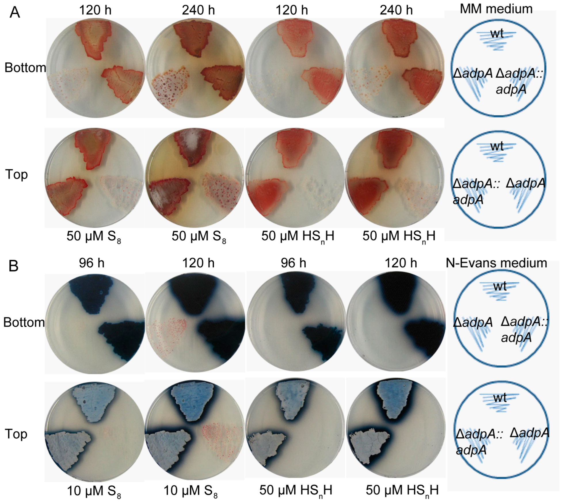
3.1. Deleting AdpA Increased the Sensitivity of S. coelicolor M145 to RSS

On MM plates with 50 µM HSnH or 50 µM S8, the adpA (sco2792, Gene ID: 1098226) deletion strain (ΔadpA) displayed severely impaired growth in comparison with the wild type strain (wt), and the growth was restored by adpA complementation (ΔadpA::adpA) (Figure 1A). For confirmation, we conducted the same experiments with another medium, the chemically limited medium (N-Evans medium), in which 50 µM HSnH or 10 µM S8 was added. Again, the growth of ΔadpA was obviously impaired and the growth of ΔadpA::adpA was fully recovered (Figure 1B). The growth of the strains was also reflected through the production of two secondary metabolites: blue-pigmented actinorhodin and red-pigmented undecylprodigiosin. S. coelicolor M145 produces blue-pigmented actinorhodin in MM medium [44], but red-pigmented undecylprodigiosin in N-Evans medium [45].

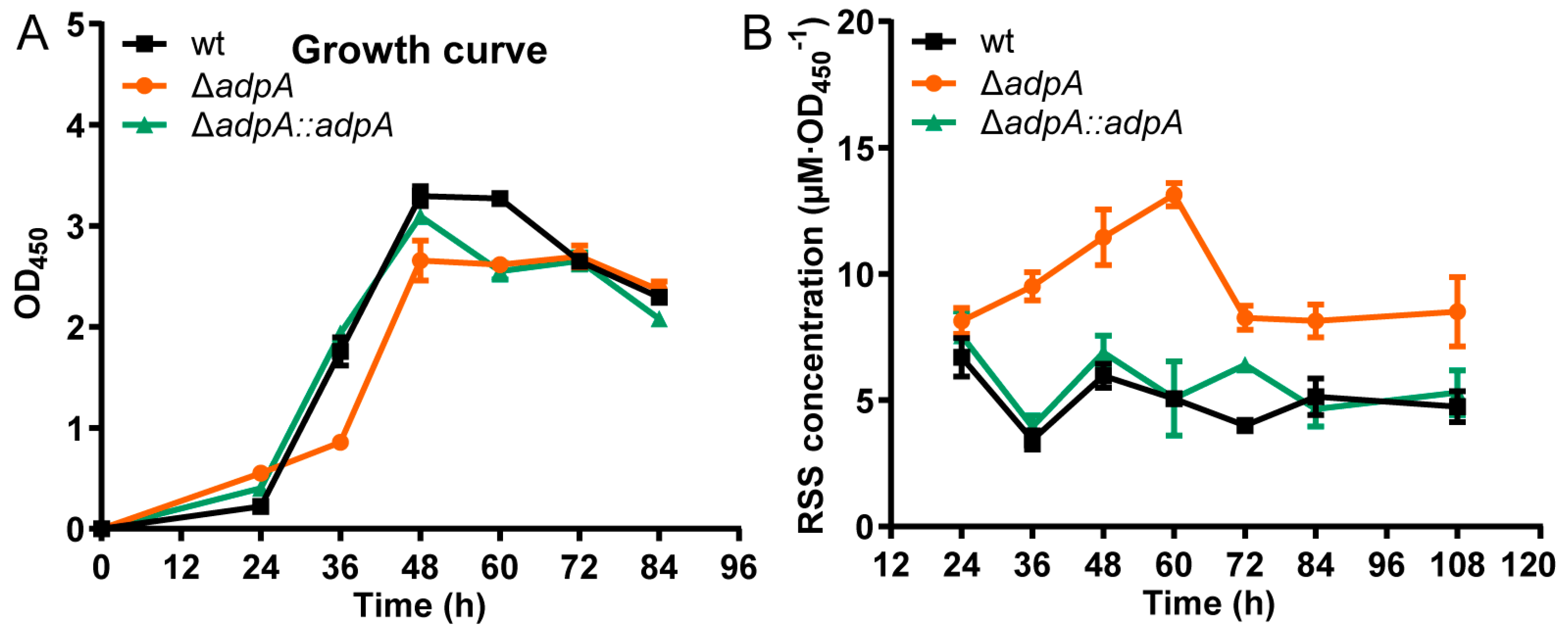
3.2. Deleting AdpA Led to RSS Accumulation in S. coelicolor M145 Cells

To investigate whether AdpA affects RSS metabolism in S. coelicolor M145, we cultivated wt, ΔadpA, and ΔadpA::adpA in YBP medium. Compared with wt and ΔadpA::adpA, ΔadpA showed a slightly impaired growth (Figure 2A). We then analyzed the intracellular RSS contents of S. coelicolor M145. ΔadpA accumulated higher content of intracellular RSS than wt and ΔadpA::adpA. For ΔadpA, the maximum content reached 13.13 µM·OD450−1 (60 h), but in comparison, wt and ΔadpA::adpA had the maximum content lower than 6.70 µM·OD450−1 (Figure 2B).

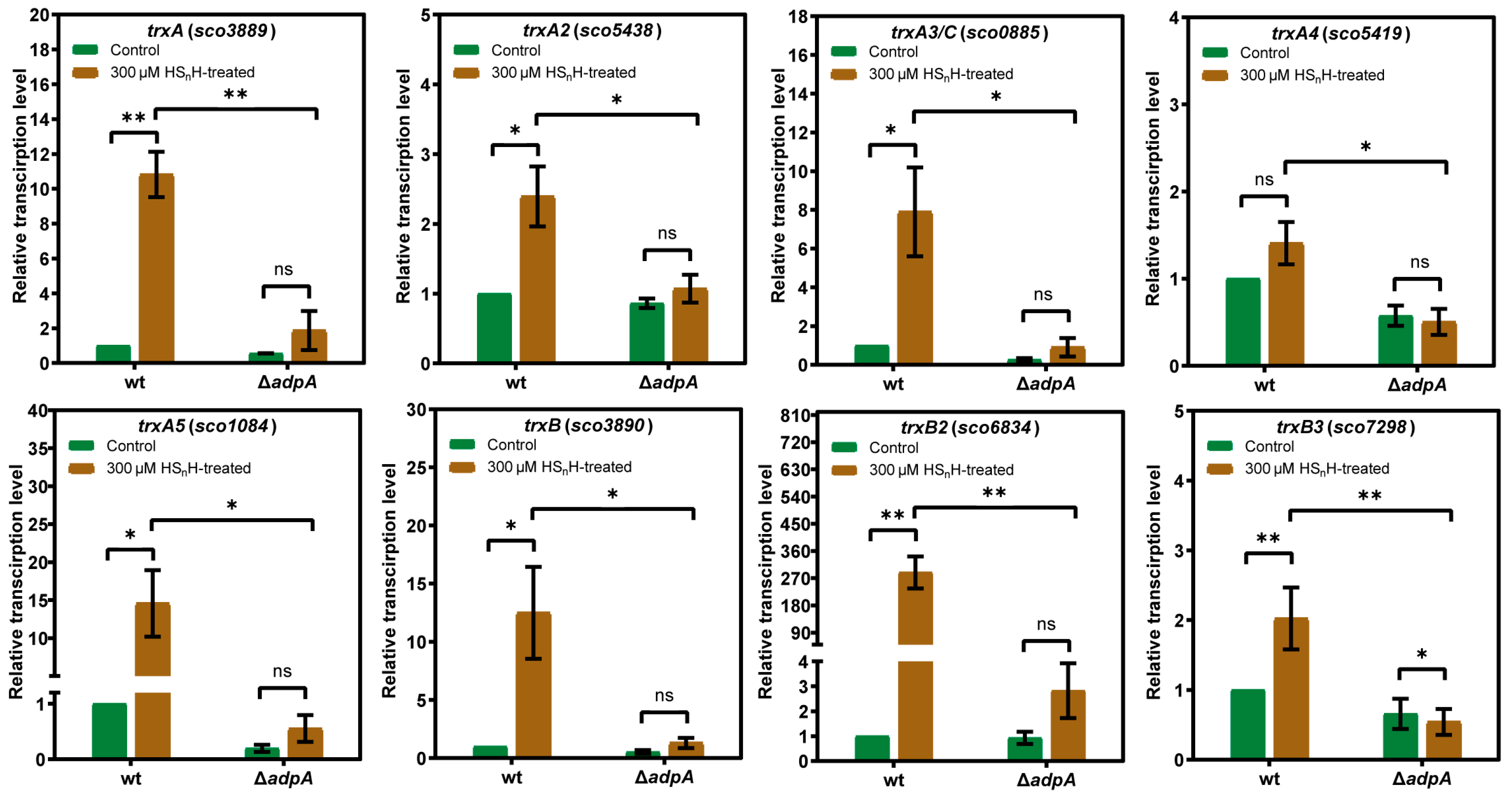
3.3. AdpA-Dependent Expression of Thioredoxins Was Induced by RSS

To examine whether the expression of thioredoxins was induced by RSS, we performed HSnH induction experiments. The wt and ΔadpA strains were cultivated in liquid YBP medium for 36 h (exponential phase). 300 µM HSnH was added, and the cultivation was continued for 1 h. Untreated cultures were used as the controls. Cells were harvested for RNA extraction and RT-qPCR analysis. In wt, the expression levels of trxA (sco3889, Gene ID: 1099325), trxB2 (sco6834, Gene ID: 1102273), and trxB3 (sco7298, Gene ID: 1102736) were significantly increased after HSnH treatment; the expression levels of trxA2 (sco5438, Gene ID: 1100878), trxA3/C (sco0885, Gene ID: 1096308), trxA5 (sco1084, Gene ID: 1096507), and trxB (sco3890, Gene ID: 1099326) were less significantly increased, while the expression level of trxA4 (sco5419, Gene ID: 1100859) was not significantly increased (Figure 3). In ΔadpA, for except trxB3 (sco7298), other thioredoxins did not show significantly increased expression after HSnH treatment. For each thioredoxin, the expression level in wt was significantly higher than that in ΔadpA after HSnH treatment (Figure 3). These results indicate that RSS-containing compounds induce the expression of thioredoxins, which requires the presence of AdpA.

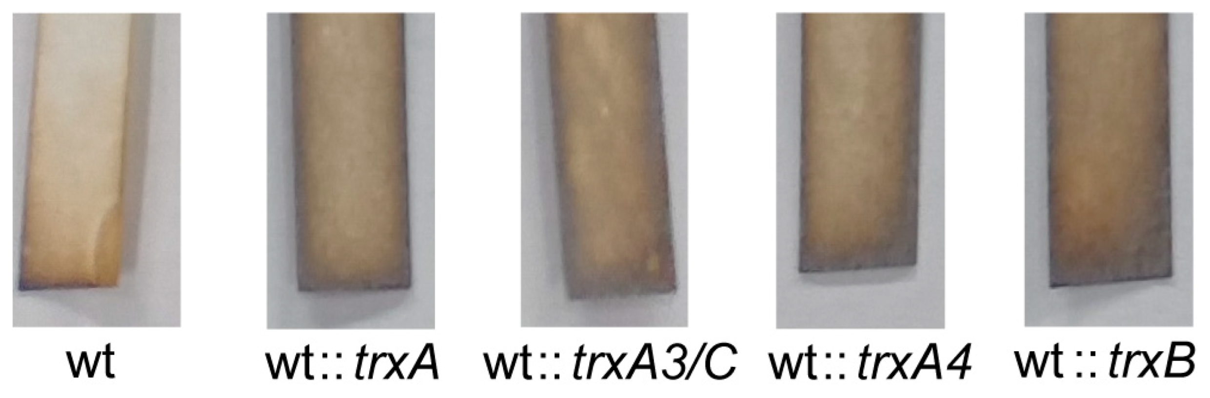
3.4. Overexpressing Thioredoxins Promoted H2S Release

To test whether thioredoxins were involved in the process of S0 reduction to H2S, we overexpressed trxA, trxA3, trxA4, or trxB genes in S. coelicolor M145. Lead–acetate test strips [Pb(Ac)2] were used to detected the production of H2S. The test strip was attached to the inside of the shaker containing 100 mL YBP medium and replaced with a new test strip after 24 h. The amount of H2S produced was compared by the color of the strip (more H2S leads to darker color). The overexpression thioredoxins led to increased H2S release (Figure 4), suggesting that these thioredoxins catalyze the reduction of RSS to H2S.

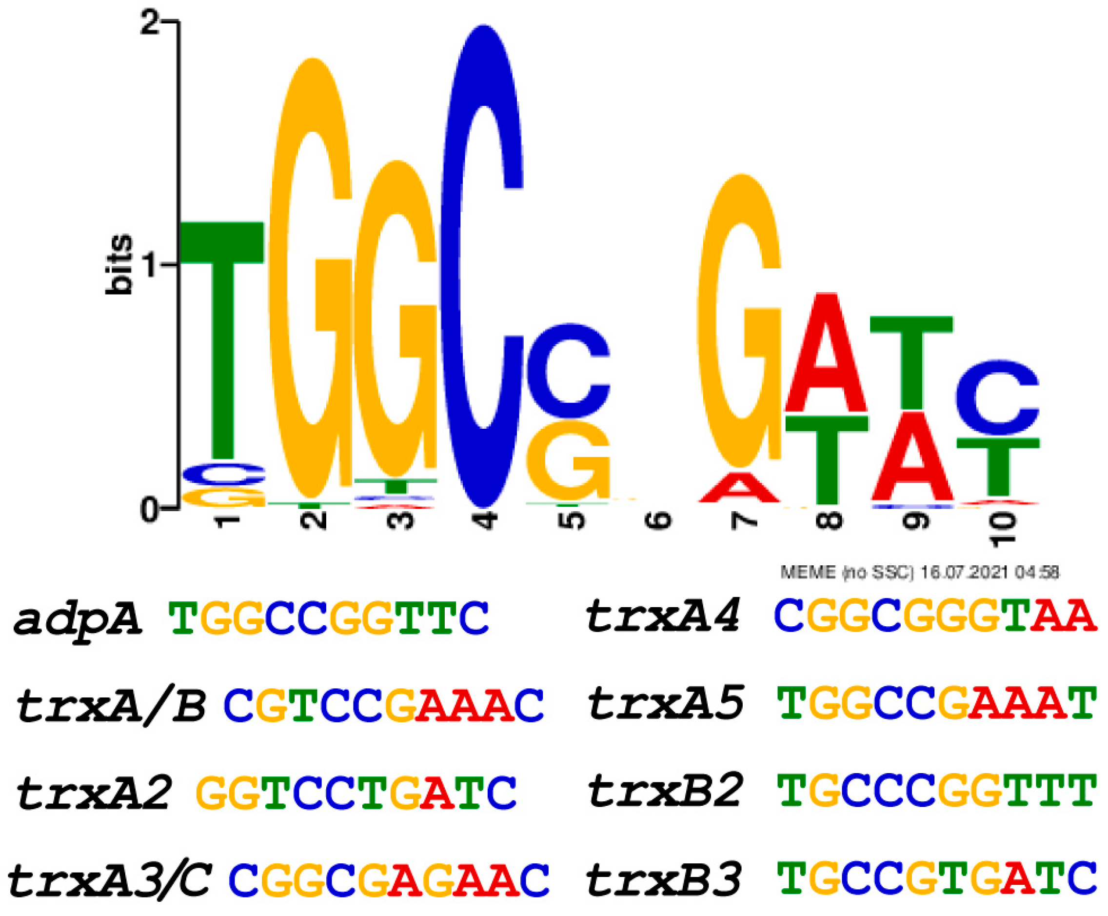
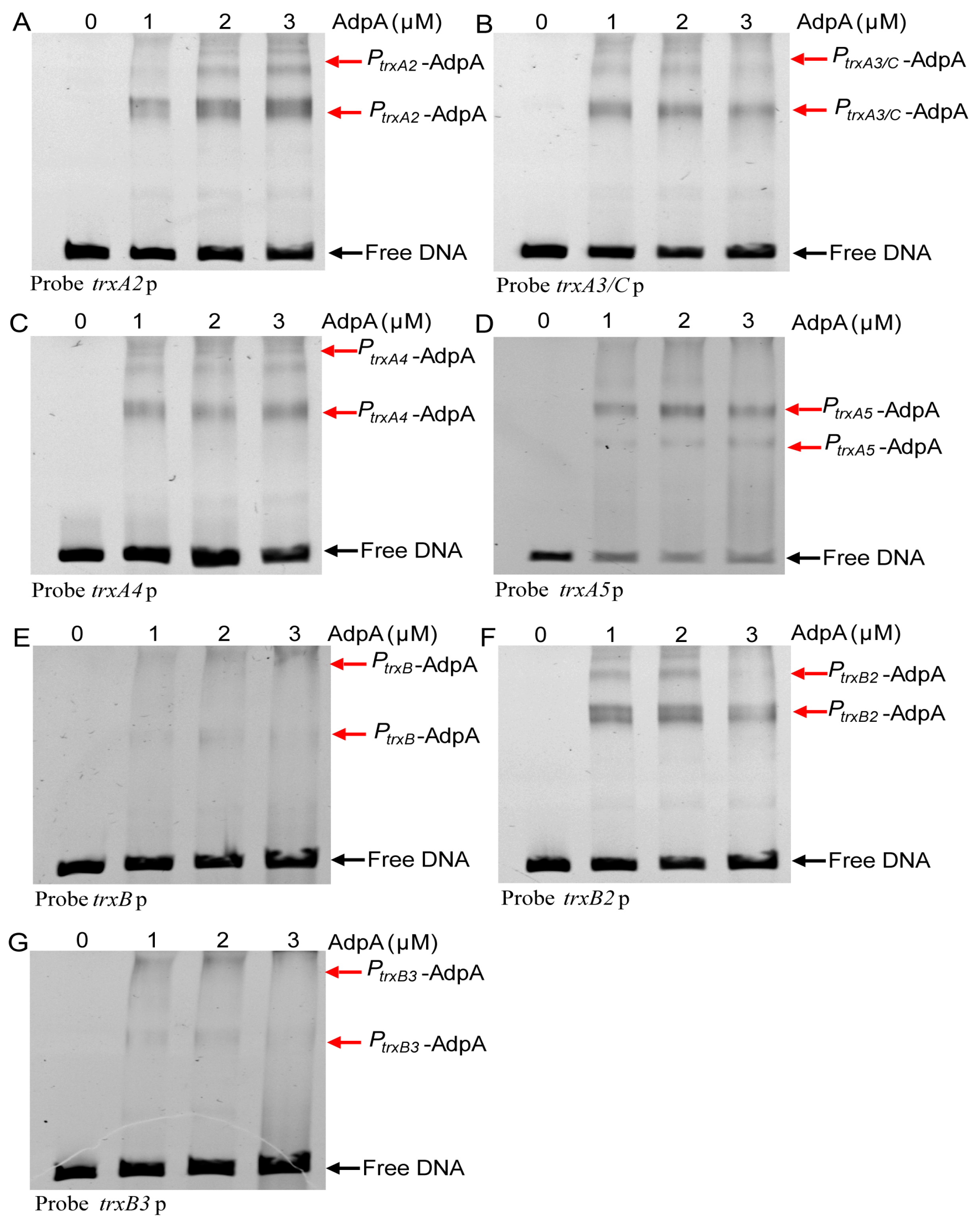
3.5. AdpA Directly Regulated the Transcription of Trx Genes
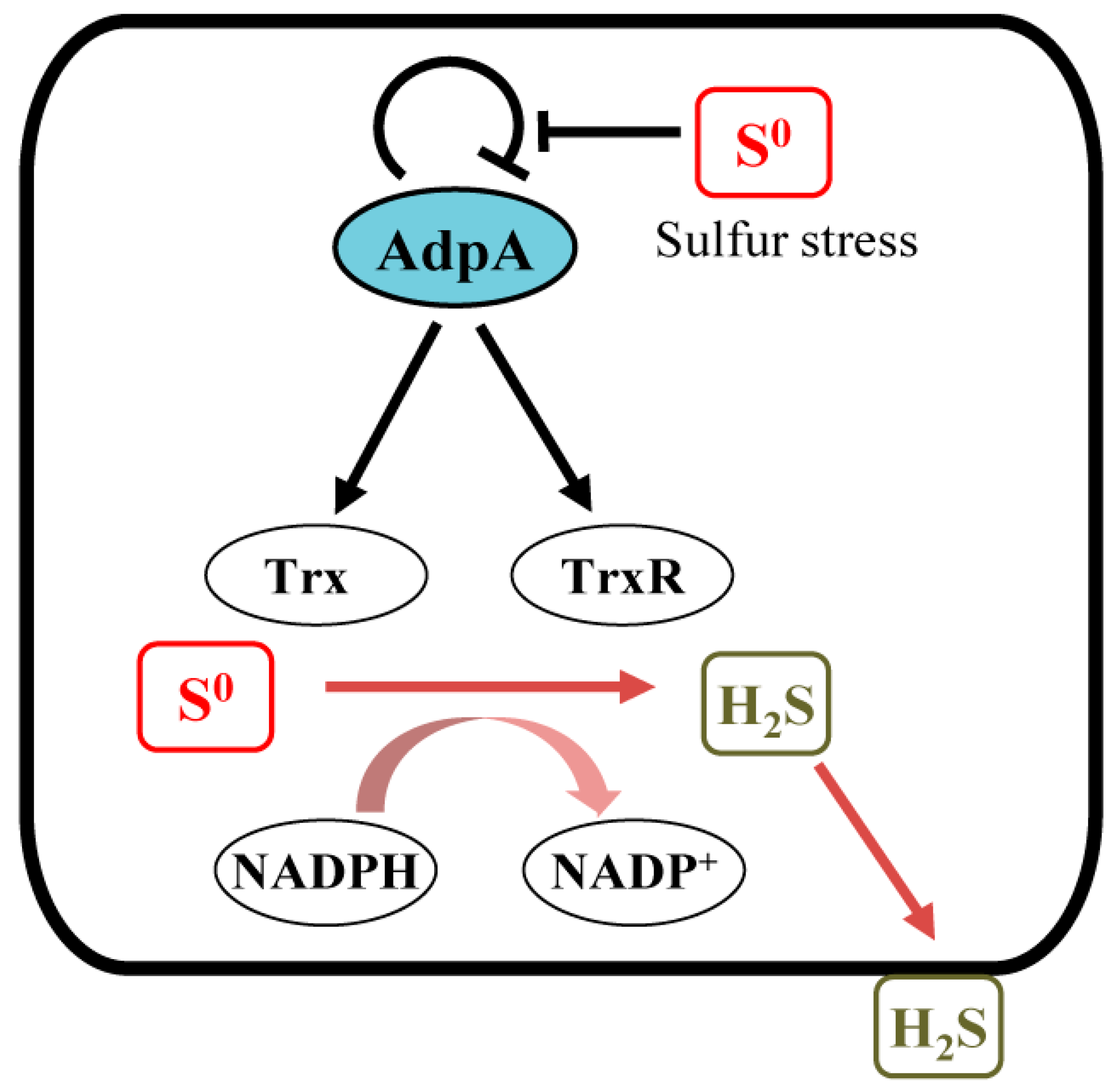
We previously reported that RSS releases the transcriptional self-inhibition on AdpA expression, thereby increasing AdpA levels [33]. We suspected that over-expressed AdpA can activate expression of thioredoxins. We analyzed the promoters of thioredoxin genes using the MEME software (http://meme-suite.org/tools/meme, accessed on 25 January 2023). The binding consensus sequence of AdpA is 5′-TGGCSNGWWY-3′ (S: G or C; W: A or T; Y: T or C; N: any nucleotide) [46,47]. This consensus sequence was found in all seven trx promoters (trxA and trxB are co-transcribed) (Figure 5). For experimental confirmation, we used synthesized promoter probes and purified AdpA to perform EMSA and observed that AdpA bound to all seven promoter probes (Figure 6A–G). These findings suggest that AdpA directly regulates the transcription of these trx genes.
We also mixed AdpA and HSnH at a molar ratio 1:50 (AdpA/HSnH); the binding of AdpA to seven trx promoters showed no obvious change (data not shown). These results suggested that increasing AdpA expression is the main mechanism for RSS to activate the expression of thioredoxins.

4. Discussion

Living organisms are always exposed to different environmental stresses and have developed specialized strategy to cope with them. Several transcription factors dealing with RSS stress have been identified. They sense intra- and extracellular RSS and maintain the intracellular RSS homeostasis. For instance, CstR negatively regulates the transcription of H2S oxidation genes in Staphylococcus aureus [48]. SqrR is responsible for the regulation of 45% sulfide-responsive genes in Rhodobacter capsulatus [49]. FisR transcriptionally activates the expression of H2S oxidation genes in Cupriavidus pinatubonensis [50] and Acinetobacter baumannii [51]. In addition to these RSS specific sensors, a few global or multifunctional regulators also have RSS sensing functions, such as MgrA in Staphylococcus aureus [52], MexR in Pseudomonas aeruginosa [53], OxyR in Escherichia coli [54], and AdpA in S. coelicolor [33]. In a previous study, we demonstrated that RSS can release the self-inhibition of AdpA expression via modifying its Cys62 residue, thereby affecting secondary metabolites production and morphological differentiation [33]. In this study, we demonstrated that AdpA is also responsible for maintaining intracellular RSS homeostasis. Deleting adpA makes S. coelicolor more sensitive to exogenous RSS stresses, suggesting that AdpA can activate expression of trx genes, whose products (thioredoxins) can reduce RSS to H2S, which evaporates into the gas phaseto relieve the RSS stress (Figure 7). AdpA is global transcriptional regulator widely present in Streptomyces spp. AdpA binds to more than hundreds of operator regions, and these sequences are less conserved. The crystal structure of the AdpA-DBD-DNA complex and mutation analysis of AdpA-DBD revealed its unique DNA recognition mode, with only two arginine residues directly recognizing the conserved site, explaining its strict recognition of G and C at position 2 and 4, respectively, as well as its tolerance recognition to other positions in the consensus sequence [55]. Although AdpA has been reported to regulate many important physiological processes [56,57,58,59], this study is the first report of its role in the maintenance of intracellular RSS homeostasis. Our finding revealed a new role that AdpA may commonly play in Streptomyces spp.
We previously demonstrated that the H2O2-response regulator OxyR senses intracellular RSS and activates the expression of catalase (KatG), glutaredoxin A (GrxA), and thioredoxin C (TrxC) in E. coli. These enzymes remove excess RSS via either oxidation or reduction [54]. AdpA also senses RSS and activates thioredoxins expression. These studies suggest that thioredoxins, conserved in many bacteria, constitute a general mechanism used by bacteria to maintain intracellular RSS homeostasis.
However, it is noteworthy that during the late period of cultivation, the intracellular RSS content of ΔadpA sharply decreased (Figure 2). This phenomenon suggests that AdpA might not be the only regulator controlling thioredoxins expression or that the removel of RSS is due to other factors other than thioredoxins. Further studies are thus required to elucidate the cause of this abrupt decrease of the RSS content in the ΔadpA mutant.
Notably, actinorhodin (ACT) is highly correlated with oxidative response. ACT is a pH-responsive benzoisochromanequinone produced by a type II polyketide synthase-based pathway. Its quinone groups participate in the redox-cycling reactions. The reduced form may react with oxygen to produce superoxide anions, which in turn generate reactive oxygen species (ROS) [60]. This may be one of the reasons why ACT has bactericidal properties. The oxidized ACT can act as an oxidative biocatalyst to react with L-ascorbic acid and L-cysteine to produce hydrogen peroxide (H2O2) [61]. This observation is consistent with the view that ACT as a redox antibiotic to perform its sterilizing function. ACT biosynthesis can be triggered by common oxidative stress such as nitric oxide (NO), RSS, and ROS has been recently reported [35,62,63,64,65]. These results suggest that ACT is both an oxidant and an antioxidant. ACT can be used as an endogenous signaling molecule to induce the expression of the redox-sensitive regulator SoxR and its regulon in S. coelicolor [66,67]. SoxR directly regulates the thioredoxins to promote resistance of thiol-oxidative stress in Streptomyces avermitilis [68]. We speculate that SoxR is not the only regulatory factor involved in antioxidant defense and ACT biosynthesis. In this study, AdpA not only directly regulates antibiotic production, but also regulates the oxidative stress resistance of thioredoxins. Deletion of adpA resulted in low expression of thioredoxins and accumulation of oxidant RSS, and AdpA is essential for the production of antioxidant ACT, aggravating the stress sensitivity of the cells. It was further confirmed that antibiotic production was a survival strategy to cope with the hostile environment in Streptomyces, however, the mechanism needs to be further explored.

5. Conclusions

Sulfane–sulfur compounds containing active sulfur atoms with a valence state of 0 or −1 are common components of cells. Sulfane sulfur is the inducer of numerous regulatory factors and participates in many physiological functions and metabolic pathways. Given the oxidizing and reducing properties of sulfane sulfur, it can be both oxidant and antioxidant. A high concentration of sulfane sulfur causes oxidative stress response and damages cells. Organisms develop different strategies to remove excess RSS in cells and maintain redox balance, but more studies are required to comprehensively understand the mechanisms. In this study, we demonstrated that the global regulator AdpA was critical for eliminating excessive RSS in S. coelicolor. The sensitivity of adpA deletion mutant to sulfur stress was significantly increased and the endogenous RSS accumulated in the mutant. Streptomyces has multiple pathways and enzymes to protect itself from oxidative stress, of which the thioredoxin systems are commonly involved. Overexpression of these thioredoxin systems resulted in more H2S release in S. coelicolor. The results showed that thioredoxin systems affected sulfur metabolism and reduced intracellular RSS. In addition, AdpA significantly activated the expression of thioredoxins and thioredoxin reductases to deal with sulfur stress. Overall, we found that AdpA has the regulatory function to promote sulfane sulfur reduction and maintain redox balance. This discovery further expands the pathway of antioxidant regulation in Streptomyces.

Supplementary Materials

The following supporting information can be downloaded at: https://www.mdpi.com/article/10.3390/antiox12020312/s1, Table S1. Strains and plasmids used in this study. Table S2. Primers sequence (5′→3′) used in this study.

Author Contributions

Data acquisition and analysis, T.L., Q.W. and Q.C.; supervision, Y.X. and H.L.; design and writing, T.L., L.X. and H.L. All authors have read and agreed to the published version of the manuscript.

Funding

This research was funded by the National Key R&D Program of China (2022YFC3401301) and the National Natural Science Foundation of China (91951202).

Institutional Review Board Statement

Not applicable.

Informed Consent Statement

Not applicable.

Data Availability Statement

The data are contained within the article and supplementary materials.

Conflicts of Interest

The authors declare no conflict of interest.

References

  1. Abe, K.; Kimura, H. The possible role of hydrogen sulfide as an endogenous neuromodulator. J. Neurosci. 1996, 16, 1066–1071. [Google Scholar] [CrossRef] [Green Version]
  2. Hine, C.; Harputlugil, E.; Zhang, Y.; Ruckenstuhl, C.; Lee, B.C.; Brace, L.; Longchamp, A.; Trevino-Villarreal, J.H.; Mejia, P.; Ozaki, C.K.; et al. Endogenous hydrogen sulfide production is essential for dietary restriction benefits. Cell 2015, 160, 132–144. [Google Scholar] [CrossRef] [Green Version]
  3. Gao, X.H.; Krokowski, D.; Guan, B.J.; Bederman, I.; Majumder, M.; Parisien, M.; Diatchenko, L.; Kabil, O.; Willard, B.; Banerjee, R.; et al. Quantitative H2S-mediated protein sulfhydration reveals metabolic reprogramming during the integrated stress response. eLife 2015, 4, e10067. [Google Scholar] [CrossRef]
  4. Wang, R. Physiological implications of hydrogen sulfide: A whiff exploration that blossomed. Physiol. Rev. 2012, 92, 791–896. [Google Scholar] [CrossRef] [PubMed] [Green Version]
  5. Chen, Y.W.; Camacho, M.I.; Chen, Y.; Bhat, A.H.; Chang, C.; Peluso, E.A.; Wu, C.; Das, A.; Ton-That, H. Genetic determinants of hydrogen sulfide biosynthesis in fusobacterium nucleatum are required for bacterial fitness, antibiotic sensitivity, and virulence. mBio 2022, 13, e0193622. [Google Scholar] [CrossRef] [PubMed]
  6. Lim, D.R.X.; Chen, Y.; Ng, L.F.; Gruber, J.; Gan, Y.H. Glutathione catabolism by Enterobacteriaceae species to hydrogen sulphide adversely affects the viability of host systems in the presence of 5’fluorodeoxyuridine. Mol. Microbiol. 2022, 117, 1089–1103. [Google Scholar] [CrossRef]
  7. Pietri, R.; Roman-Morales, E.; Lopez-Garriga, J. Hydrogen sulfide and hemeproteins: Knowledge and mysteries. Antioxid. Redox Signal. 2011, 15, 393–404. [Google Scholar] [CrossRef] [Green Version]
  8. Paul, B.D.; Snyder, S.H. H2S signalling through protein sulfhydration and beyond. Nat. Rev. Mol. Cell Biol. 2012, 13, 499–507. [Google Scholar] [CrossRef]
  9. Toohey, J.I. Sulfur signaling: Is the agent sulfide or sulfane? Anal. Biochem. 2011, 413, 1–7. [Google Scholar] [CrossRef]
  10. Walsh, B.J.C.; Giedroc, D.P. H2S and reactive sulfur signaling at the host-bacterial pathogen interface. J. Biol. Chem. 2020, 295, 13150–13168. [Google Scholar] [CrossRef]
  11. Kimura, H. Hydrogen sulfide and polysulfide signaling. Antioxid. Redox Signal. 2017, 27, 619–621. [Google Scholar] [CrossRef]
  12. Wang, Q.; Chen, Z.; Zhang, X.; Xin, Y.; Xia, Y.; Xun, L.; Liu, H. Rhodanese Rdl2 produces reactive sulfur species to protect mitochondria from reactive oxygen species. Free Radic. Biol. Med. 2021, 177, 287–298. [Google Scholar] [CrossRef]
  13. Barayeu, U.; Schilling, D.; Eid, M.; Xavier da Silva, T.N.; Schlicker, L.; Mitreska, N.; Zapp, C.; Grater, F.; Miller, A.K.; Kappl, R.; et al. Hydropersulfides inhibit lipid peroxidation and ferroptosis by scavenging radicals. Nat. Chem. Biol. 2023, 19, 28–37. [Google Scholar] [CrossRef]
  14. Zhang, X.; Xin, Y.; Chen, Z.; Xia, Y.; Xun, L.; Liu, H. Sulfide-quinone oxidoreductase is required for cysteine synthesis and indispensable to mitochondrial health. Redox Biol. 2021, 47, 102169. [Google Scholar] [CrossRef]
  15. Alvarez, L.; Bianco, C.L.; Toscano, J.P.; Lin, J.; Akaike, T.; Fukuto, J.M. Chemical biology of hydropersulfides and related species: Possible roles in cellular protection and redox signaling. Antioxid. Redox Signal. 2017, 27, 622–633. [Google Scholar] [CrossRef]
  16. Kumagai, Y.; Akiyama, M.; Unoki, T. Adaptive responses to electrophilic stress and reactive sulfur species as their regulator molecules. Toxicol. Res. 2019, 35, 303–310. [Google Scholar] [CrossRef] [Green Version]
  17. Williams, J.S.; Cooper, R.M. Elemental sulphur is produced by diverse plant families as a component of defence against fungal and bacterial pathogens. Physiol. Mol. Plant Pathol. 2003, 63, 3–16. [Google Scholar] [CrossRef]
  18. Leslie, K.S.; Millington, G.W.; Levell, N.J. Sulphur and skin: From Satan to Saddam! J. Cosmet. Dermatol. 2004, 3, 94–98. [Google Scholar] [CrossRef]
  19. Wang, T.; Yang, Y.; Liu, M.; Liu, H.; Liu, H.; Xia, Y.; Xun, L. Elemental sulfur inhibits yeast growth via producing toxic sulfide and causing disulfide stress. Antioxidants 2022, 11, 576. [Google Scholar] [CrossRef]
  20. Shen, J.C.; Keithly, M.E.; Armstrong, R.N.; Higgins, K.A.; Edmonds, K.A.; Giedroc, D.P. Staphylococcus aureus CstB is a novel multidomain persulfide dioxygenase-sulfurtransferase involved in hydrogen sulfide detoxification. Biochemistry 2015, 54, 4542–4554. [Google Scholar] [CrossRef]
  21. Giedroc, D.P. A new player in bacterial sulfide-inducible transcriptional regulation. Mol. Microbiol. 2017, 105, 347–352. [Google Scholar] [CrossRef] [Green Version]
  22. Barka, E.A.; Vatsa, P.; Sanchez, L.; Gaveau-Vaillant, N.; Jacquard, C.; Meier-Kolthoff, J.P.; Klenk, H.P.; Clement, C.; Ouhdouch, Y.; van Wezel, G.P. Taxonomy, physiology, and natural products of Actinobacteria. Microbiol. Mol. Biol. Rev. 2016, 80, 1–43. [Google Scholar] [CrossRef] [PubMed] [Green Version]
  23. Sekurova, O.N.; Zehl, M.; Predl, M.; Hunyadi, P.; Rattei, T.; Zotchev, S.B. Targeted metabolomics and high-throughput RNA sequencing-based transcriptomics reveal massive changes in the Streptomyces venezuelae NRRL B-65442 metabolism caused by ethanol shock. Microbiol. Spectr. 2022, 10, e0367222. [Google Scholar] [CrossRef]
  24. van der Meij, A.; Worsley, S.F.; Hutchings, M.I.; van Wezel, G.P. Chemical ecology of antibiotic production by actinomycetes. FEMS Microbiol. Rev. 2017, 41, 392–416. [Google Scholar] [CrossRef] [PubMed] [Green Version]
  25. Shepherdson, E.M.F.; Elliot, M.A. Cryptic specialized metabolites drive Streptomyces exploration and provide a competitive advantage during growth with other microbes. Proc. Natl. Acad. Sci. USA 2022, 119, e2211052119. [Google Scholar] [CrossRef] [PubMed]
  26. Rabyk, M.; Yushchuk, O.; Rokytskyy, I.; Anisimova, M.; Ostash, B. Genomic insights into evolution of AdpA family master regulators of morphological differentiation and secondary metabolism in Streptomyces. J. Mol. Evol. 2018, 86, 204–215. [Google Scholar] [CrossRef] [PubMed]
  27. Tistechok, S.; Stierhof, M.; Kachor, A.; Myronovskyi, M.; Gromyko, O.; Luzhetskyy, A. Flavacol and its novel derivative 3-beta-hydroxy flavacol from Streptomyces sp. Pv 4-95 after the expression of heterologous AdpA. Microorganisms 2022, 10, 2335. [Google Scholar] [CrossRef]
  28. Huang, R.; Liu, H.; Zhao, W.; Wang, S.; Wang, S.; Cai, J.; Yang, C. AdpA, a developmental regulator, promotes epsilon-poly-L-lysine biosynthesis in Streptomyces albulus. Microb. Cell Fact. 2022, 21, 60. [Google Scholar] [CrossRef] [PubMed]
  29. Plachetka, M.; Krawiec, M.; Zakrzewska-Czerwinska, J.; Wolanski, M. AdpA positively regulates morphological differentiation and chloramphenicol biosynthesis in Streptomyces venezuelae. Microbiol. Spectr. 2021, 9, e0198121. [Google Scholar] [CrossRef]
  30. Yushchuk, O.; Ostash, I.; Vlasiuk, I.; Gren, T.; Luzhetskyy, A.; Kalinowski, J.; Fedorenko, V.; Ostash, B. Heterologous AdpA transcription factors enhance landomycin production in Streptomyces cyanogenus S136 under a broad range of growth conditions. Appl. Microbiol. Biotechnol. 2018, 102, 8419–8428. [Google Scholar] [CrossRef]
  31. Bu, X.L.; Weng, J.Y.; He, B.B.; Xu, M.J.; Xu, J. A novel AdpA homologue negatively regulates morphological differentiation in Streptomyces xiamenensis 318. Appl. Environ. Microbiol. 2019, 85, e03107-18. [Google Scholar] [CrossRef] [PubMed] [Green Version]
  32. Kang, Y.; Wang, Y.; Hou, B.; Wang, R.; Ye, J.; Zhu, X.; Wu, H.; Zhang, H. AdpAlin, a pleiotropic transcriptional regulator, is involved in the cascade regulation of lincomycin biosynthesis in Streptomyces lincolnensis. Front. Microbiol. 2019, 10, 2428. [Google Scholar] [CrossRef] [PubMed]
  33. Lu, T.; Wu, X.; Cao, Q.; Xia, Y.; Xun, L.; Liu, H. Sulfane sulfur posttranslationally modifies the global regulator AdpA to influence actinorhodin production and morphological differentiation of Streptomyces coelicolor. mBio 2022, 13, e0386221. [Google Scholar] [CrossRef] [PubMed]
  34. Westhoff, S.; Kloosterman, A.M.; van Hoesel, S.F.A.; van Wezel, G.P.; Rozen, D.E. Competition sensing changes antibiotic production in Streptomyces. mBio 2021, 12, e02729-20. [Google Scholar] [CrossRef]
  35. Lu, T.; Cao, Q.; Pang, X.; Xia, Y.; Xun, L.; Liu, H. Sulfane sulfur-activated actinorhodin production and sporulation is maintained by a natural gene circuit in Streptomyces coelicolor. Microb. Biotechnol. 2020, 13, 1917–1932. [Google Scholar] [CrossRef]
  36. Kieser, T.; Bibb, M.J.; Buttner, M.J.; Chater, K.F.; Hopwood, D.A. Practical Streptomyces Genetics, 2nd ed.; John Innes Foundation: Norwich, UK, 2000. [Google Scholar]
  37. Fink, D.; Weissschuh, N.; Reuther, J.; Wohlleben, W.; Engels, A. Two transcriptional regulators GlnR and GlnRII are involved in regulation of nitrogen metabolism in Streptomyces coelicolor A3(2). Mol. Microbiol. 2002, 46, 331–347. [Google Scholar] [CrossRef]
  38. Bai, C.; Zhang, Y.; Zhao, X.; Hu, Y.; Xiang, S.; Miao, J.; Lou, C.; Zhang, L. Exploiting a precise design of universal synthetic modular regulatory elements to unlock the microbial natural products in Streptomyces. Proc. Natl. Acad. Sci. USA 2015, 112, 12181–12186. [Google Scholar] [CrossRef] [Green Version]
  39. Zhu, Y.; Zhang, P.; Lu, T.; Wang, X.; Li, A.; Lu, Y.; Tao, M.; Pang, X. Impact of MtrA on phosphate metabolism genes and the response to altered phosphate conditions in Streptomyces. Environ. Microbiol. 2021, 23, 6907–6923. [Google Scholar] [CrossRef]
  40. Chen, Z.; Zhang, X.; Li, H.; Liu, H.; Xia, Y.; Xun, L. The complete pathway for thiosulfate utilization in Saccharomyces cerevisiae. Appl. Environ. Microbiol. 2018, 84, e01241-18. [Google Scholar] [CrossRef] [Green Version]
  41. Ran, M.; Wang, T.; Shao, M.; Chen, Z.; Liu, H.; Xia, Y.; Xun, L. Sensitive method for reliable quantification of sulfane sulfur in biological samples. Anal. Chem. 2019, 91, 11981–11986. [Google Scholar] [CrossRef]
  42. Lu, T.; Zhu, Y.; Zhang, P.; Sheng, D.; Cao, G.; Pang, X. SCO5351 is a pleiotropic factor that impacts secondary metabolism and morphological development in Streptomyces coelicolor. FEMS Microbiol. Lett. 2018, 365, fny150. [Google Scholar] [CrossRef] [Green Version]
  43. Zhu, Y.; Zhang, P.; Zhang, J.; Xu, W.; Wang, X.; Wu, L.; Sheng, D.; Ma, W.; Cao, G.; Chen, X.L.; et al. The developmental regulator MtrA binds GlnR boxes and represses nitrogen metabolism genes in Streptomyces coelicolor. Mol. Microbiol. 2019, 112, 29–46. [Google Scholar] [CrossRef] [PubMed]
  44. Bystrykh, L.V.; Fernandez-Moreno, M.A.; Herrema, J.K.; Malpartida, F.; Hopwood, D.A.; Dijkhuizen, L. Production of actinorhodin-related “blue pigments” by Streptomyces coelicolor A3(2). J. Bacteriol. 1996, 178, 2238–2244. [Google Scholar] [CrossRef] [PubMed] [Green Version]
  45. Liu, G.; Chater, K.F.; Chandra, G.; Niu, G.; Tan, H. Molecular regulation of antibiotic biosynthesis in streptomyces. Microbiol. Mol. Biol. Rev. 2013, 77, 112–143. [Google Scholar] [CrossRef] [PubMed] [Green Version]
  46. Yamazaki, H.; Tomono, A.; Ohnishi, Y.; Horinouchi, S. DNA-binding specificity of AdpA, a transcriptional activator in the A-factor regulatory cascade in Streptomyces griseus. Mol. Microbiol. 2004, 53, 555–572. [Google Scholar] [CrossRef] [PubMed]
  47. Chen, Q.; Zhu, J.; Li, X.; Wen, Y. Transcriptional regulator DasR represses daptomycin production through both direct and cascade mechanisms in Streptomyces roseosporus. Antibiotics 2022, 11, 1065. [Google Scholar] [CrossRef] [PubMed]
  48. Grossoehme, N.; Kehl-Fie, T.E.; Ma, Z.; Adams, K.W.; Cowart, D.M.; Scott, R.A.; Skaar, E.P.; Giedroc, D.P. Control of copper resistance and inorganic sulfur metabolism by paralogous regulators in Staphylococcus aureus. J. Biol. Chem. 2011, 286, 13522–13531. [Google Scholar] [CrossRef] [PubMed] [Green Version]
  49. Shimizu, T.; Shen, J.; Fang, M.; Zhang, Y.; Hori, K.; Trinidad, J.C.; Bauer, C.E.; Giedroc, D.P.; Masuda, S. Sulfide-responsive transcriptional repressor SqrR functions as a master regulator of sulfide-dependent photosynthesis. Proc. Natl. Acad. Sci. USA 2017, 114, 2355–2360. [Google Scholar] [CrossRef] [Green Version]
  50. Li, H.; Li, J.; Lu, C.; Xia, Y.; Xin, Y.; Liu, H.; Xun, L.; Liu, H. FisR activates σ54-dependent transcription of sulfide-oxidizing genes in Cupriavidus pinatubonensis JMP134. Mol. Microbiol. 2017, 105, 373–384. [Google Scholar] [CrossRef] [Green Version]
  51. Walsh, B.J.C.; Wang, J.; Edmonds, K.A.; Palmer, L.D.; Zhang, Y.; Trinidad, J.C.; Skaar, E.P.; Giedroc, D.P. The response of Acinetobacter baumannii to hydrogen sulfide reveals two independent persulfide-sensing systems and a connection to biofilm regulation. mBio 2020, 11, e01254-20. [Google Scholar] [CrossRef]
  52. Peng, H.; Zhang, Y.; Palmer, L.D.; Kehl-Fie, T.E.; Skaar, E.P.; Trinidad, J.C.; Giedroc, D.P. Hydrogen sulfide and reactive sulfur species impact proteome S-sulfhydration and global virulence regulation in Staphylococcus aureus. ACS Infect. Dis. 2017, 3, 744–755. [Google Scholar] [CrossRef] [PubMed] [Green Version]
  53. Xuan, G.; Lu, C.; Xu, H.; Chen, Z.; Li, K.; Liu, H.; Liu, H.; Xia, Y.; Xun, L. Sulfane Sulfur is an intrinsic signal activating MexR-regulated antibiotic resistance in Pseudomonas aeruginosa. Mol. Microbiol. 2020, 114, 1038–1048. [Google Scholar] [CrossRef]
  54. Hou, N.; Yan, Z.; Fan, K.; Li, H.; Zhao, R.; Xia, Y.; Xun, L.; Liu, H. OxyR senses sulfane sulfur and activates the genes for its removal in Escherichia coli. Redox Biol. 2019, 26, 101293. [Google Scholar] [CrossRef] [PubMed]
  55. Yao, M.D.; Ohtsuka, J.; Nagata, K.; Miyazono, K.; Zhi, Y.; Ohnishi, Y.; Tanokura, M. Complex structure of the DNA-binding domain of AdpA, the global transcription factor in Streptomyces griseus, and a target duplex DNA reveals the structural basis of its tolerant DNA sequence specificity. J. Biol. Chem. 2013, 288, 31019–31029. [Google Scholar] [CrossRef] [Green Version]
  56. Zacharia, V.M.; Ra, Y.; Sue, C.; Alcala, E.; Reaso, J.N.; Ruzin, S.E.; Traxler, M.F. Genetic network architecture and environmental cues drive spatial organization of phenotypic division of labor in Streptomyces coelicolor. mBio 2021, 12, e00794-21. [Google Scholar] [CrossRef] [PubMed]
  57. Yan, H.; Lu, X.; Sun, D.; Zhuang, S.; Chen, Q.; Chen, Z.; Li, J.; Wen, Y. BldD, a master developmental repressor, activates antibiotic production in two Streptomyces species. Mol. Microbiol. 2020, 113, 123–142. [Google Scholar] [CrossRef] [PubMed]
  58. Wang, W.; Zhang, J.; Liu, X.; Li, D.; Li, Y.; Tian, Y.; Tan, H. Identification of a butenolide signaling system that regulates nikkomycin biosynthesis in Streptomyces. J. Biol. Chem. 2018, 293, 20029–20040. [Google Scholar] [CrossRef] [Green Version]
  59. Xu, J.; Zhang, J.; Zhuo, J.; Li, Y.; Tian, Y.; Tan, H. Activation and mechanism of a cryptic oviedomycin gene cluster via the disruption of a global regulatory gene, adpA, in Streptomyces ansochromogenes. J. Biol. Chem. 2017, 292, 19708–19720. [Google Scholar] [CrossRef] [Green Version]
  60. Mak, S.; Nodwell, J.R. Actinorhodin is a redox-active antibiotic with a complex mode of action against Gram-positive cells. Mol. Microbiol. 2017, 106, 597–613. [Google Scholar] [CrossRef]
  61. Nishiyama, T.; Hashimoto, Y.; Kusakabe, H.; Kumano, T.; Kobayashi, M. Natural low-molecular mass organic compounds with oxidase activity as organocatalysts. Proc. Natl. Acad. Sci. USA 2014, 111, 17152–17157. [Google Scholar] [CrossRef]
  62. Honma, S.; Ito, S.; Yajima, S.; Sasaki, Y. Nitric oxide signaling for actinorhodin production in Streptomyces coelicolor A3(2) via the DevS/R two-component system. Appl. Environ. Microbiol. 2021, 87, e0048021. [Google Scholar] [CrossRef] [PubMed]
  63. Lejeune, C.; Sago, L.; Cornu, D.; Redeker, V.; Virolle, M.J. A proteomic analysis indicates that oxidative stress is the common feature triggering antibiotic production in Streptomyces coelicolor and in the pptA mutant of Streptomyces lividans. Front. Microbiol. 2021, 12, 813993. [Google Scholar] [CrossRef] [PubMed]
  64. Esnault, C.; Dulermo, T.; Smirnov, A.; Askora, A.; David, M.; Deniset-Besseau, A.; Holland, I.B.; Virolle, M.J. Strong antibiotic production is correlated with highly active oxidative metabolism in Streptomyces coelicolor M145. Sci. Rep. 2017, 7, 200. [Google Scholar] [CrossRef] [PubMed] [Green Version]
  65. Shikura, N.; Darbon, E.; Esnault, C.; Deniset-Besseau, A.; Xu, D.; Lejeune, C.; Jacquet, E.; Nhiri, N.; Sago, L.; Cornu, D.; et al. The phosin PptA plays a negative role in the regulation of antibiotic production in Streptomyces lividans. Antibiotics 2021, 10, 325. [Google Scholar] [CrossRef] [PubMed]
  66. Shin, J.H.; Singh, A.K.; Cheon, D.J.; Roe, J.H. Activation of the SoxR regulon in Streptomyces coelicolor by the extracellular form of the pigmented antibiotic actinorhodin. J. Bacteriol. 2011, 193, 75–81. [Google Scholar] [CrossRef] [Green Version]
  67. Dela Cruz, R.; Gao, Y.; Penumetcha, S.; Sheplock, R.; Weng, K.; Chander, M. Expression of the Streptomyces coelicolor SoxR regulon is intimately linked with actinorhodin production. J. Bacteriol. 2010, 192, 6428–6438. [Google Scholar] [CrossRef] [Green Version]
  68. Wang, Q.; Lu, X.; Yang, H.; Yan, H.; Wen, Y. Redox-sensitive transcriptional regulator SoxR directly controls antibiotic production, development and thiol-oxidative stress response in Streptomyces avermitilis. Microb. Biotechnol. 2022, 15, 561–576. [Google Scholar] [CrossRef]
Figure 1. The ΔadpA strain was more sensitive to RSS than wt. (A) Phenotypes of S. coelicolor M145 (wt), ΔadpA, and ΔadpA::adpA grown at 30 °C on MM agar containing 50 µM S8 or 50 µM HSnH. The red pigment was undecyl prodigiosin in the colonies. (B) Phenotypes of wt, ΔadpA, and ΔadpA::adpA grown on N-Evans agar plates containing 10 µM S8 or 50 µM HSnH. The blue pigment is actinorhodin in the colonies. Images were taken at 120 h and 240 h (panel (A)) or 96 h and 120 h (panel (B)), and the views from top and bottom sides of the plates.
Antioxidants 12 00312 g001
Figure 2. The accumulation of RSS content in ΔadpA. (A) The growth curve of S. coelicolor M145 wt (black bar), ΔadpA (red bar), and ΔadpA::adpA (green bar) cultured in YBP medium. OD450 was checked to represent the growth. (B) Deletion of adpA increased intracellular RSS. RSS concentrations of wt (black bar), ΔadpA (red bar), and ΔadpA::adpA (green bar) were detected at the indicated times.
Antioxidants 12 00312 g002
Figure 3. The trx genes in S. coelicolor M145 were induced in response to HSnH treatment in an adpA-dependent manner. RT-qPCR analysis was performed to compare the transcriptional level of trxA, trxA2, trxA3/C, trxA4, trxA5, trxB, trxB2, and trxB3 in wt and ΔadpA when induced by 300 µM HSnH. The hrdB transcription was used as the internal control for normalization. The expression level of each gene in wt without induction was arbitrarily set to 1, and the fold changes in expression levels in wt and ΔadpA without induction (green bars) and with HSnH induction (brown bars) were shown. All data were averages from three samples with standard deviation (error bar). T-tests were performed to calculate the p-values, and asterisks indicate statistically significant difference (* p < 0.05, ** p < 0.01).
Antioxidants 12 00312 g003
Figure 4. The effect of overexpression trx genes in S. coelicolor M145 on H2S production. H2S production by S. coelicolor M145 wt, wt::trxA, wt::trxA3/C, wt::trxA4, and wt::trxB in YBP medium between 24 h and 48 h culture was detected in the gas phase with lead–acetate paper. The experiment was repeated at least three times.
Antioxidants 12 00312 g004
Figure 5. The consensus sequence for AdpA binding predicted by MEME analysis. The sequence contains nine relative conserved and one non-conserved (the sixth) nucleotides.
Antioxidants 12 00312 g005
Figure 6. EMSA analysis His-tagged AdpA with the upstream promoter sequence probes of trx genes. 20 nM DNA probe was incubated with different amounts of His-tagged AdpA (0, 1, 2, 3 µM). Black arrow indicates the free DNA probe and red arrow indicates the AdpA-DNA complex. (A) A 299-bp DNA fragment containing the promoter and partial ORF sequence of trxA2 (sco5438) was synthesized as the probe. (B) A 311-bp DNA fragment containing the promoter and partial ORF sequence of trxA3/C (sco0885) was synthesized and used as the probe. (C) A 335-bp DNA fragment containing the promoter and partial ORF sequence of trxA4 (sco5419) was synthesized and used as the probe. (D) A 155-bp DNA fragment containing the promoter and partial ORF sequence of trxA5 (sco1084) was synthesized and used as the probe. (E) A 334-bp DNA fragment containing the promoter and partial ORF sequence of trxB (sco3890) was synthesized and used as the probe. The trxA and trxB were co-transcribed, trxB the first gene of the operon. (F) A 285-bp DNA fragment containing the promoter and partial ORF sequence of trxB2 (sco6834) was synthesized and used as the probe. The probe is promoter, (G) A 277-bp DNA fragment containing the promoter and partial ORF sequence of trxB3 (sco7298) was synthesized and used as the probe.
Antioxidants 12 00312 g006
Figure 7. The proposed model of AdpA regulates the removal of excess intracellular RSS via thioredoxin systems in Streptomyces coelicolor.
Antioxidants 12 00312 g007
Disclaimer/Publisher’s Note: The statements, opinions and data contained in all publications are solely those of the individual author(s) and contributor(s) and not of MDPI and/or the editor(s). MDPI and/or the editor(s) disclaim responsibility for any injury to people or property resulting from any ideas, methods, instructions or products referred to in the content.

