Review

Non-Excitatory Amino Acids, Melatonin, and Free Radicals: Examining the Role in Stroke and Aging

by
Victoria Jiménez Carretero
1,
Eva Ramos
2,
Pedro Segura-Chama
3,
Adan Hernández
4,
Andrés M Baraibar
5,
Iris Álvarez-Merz
1,
Francisco López Muñoz
6,7,
Javier Egea
8,
José M. Solís
9,
Alejandro Romero
2 and
Jesús M. Hernández-Guijo
1,10,*
1
Department of Pharmacology and Therapeutic, Teófilo Hernando Institute, Faculty of Medicine, Universidad Autónoma de Madrid, Av. Arzobispo Morcillo 4, 28029 Madrid, Spain
2
Department of Pharmacology and Toxicology, Faculty of Veterinary Medicine, Complutense University of Madrid, 28040 Madrid, Spain
3
Investigador por México-CONAHCYT, Instituto Nacional de Psiquiatría “Ramón de la Fuente Muñiz”, Calzada México-Xochimilco 101, Huipulco, Tlalpan, Mexico City 14370, Mexico
4
Institute of Neurobiology, Universidad Nacional Autónoma of México, Juriquilla, Santiago de Querétaro 76230, Querétaro, Mexico
5
Department of Neurosciences, Universidad del País Vasco UPV/EHU, Achucarro Basque Center for Neuroscience, Barrio Sarriena, s/n, 48940 Leioa, Spain
6
Faculty of Health Sciences, University Camilo José Cela, C/Castillo de Alarcón 49, Villanueva de la Cañada, 28692 Madrid, Spain
7
Neuropsychopharmacology Unit, Hospital 12 de Octubre Research Institute (i + 12), Avda. Córdoba, s/n, 28041 Madrid, Spain
8
Molecular Neuroinflammation and Neuronal Plasticity Research Laboratory, Hospital Universitario Santa Cristina, Health Research Institute, Hospital Universitario de la Princesa, 28006 Madrid, Spain
9
Neurobiology-Research Service, Hospital Ramón y Cajal, Carretera de Colmenar Viejo, Km. 9, 28029 Madrid, Spain
10
Ramón y Cajal Institute for Health Research (IRYCIS), Hospital Ramón y Cajal, Carretera de Colmenar Viejo, Km. 9, 28029 Madrid, Spain
*
Author to whom correspondence should be addressed.
Antioxidants 2023, 12(10), 1844; https://doi.org/10.3390/antiox12101844
Submission received: 5 September 2023 / Revised: 23 September 2023 / Accepted: 26 September 2023 / Published: 10 October 2023
(This article belongs to the Special Issue Melatonin Metabolism and Aging)

Abstract

:
The aim of this review is to explore the relationship between melatonin, free radicals, and non-excitatory amino acids, and their role in stroke and aging. Melatonin has garnered significant attention in recent years due to its diverse physiological functions and potential therapeutic benefits by reducing oxidative stress, inflammation, and apoptosis. Melatonin has been found to mitigate ischemic brain damage caused by stroke. By scavenging free radicals and reducing oxidative damage, melatonin may help slow down the aging process and protect against age-related cognitive decline. Additionally, non-excitatory amino acids have been shown to possess neuroprotective properties, including antioxidant and anti-inflammatory in stroke and aging-related conditions. They can attenuate oxidative stress, modulate calcium homeostasis, and inhibit apoptosis, thereby safeguarding neurons against damage induced by stroke and aging processes. The intracellular accumulation of certain non-excitatory amino acids could promote harmful effects during hypoxia-ischemia episodes and thus, the blockade of the amino acid transporters involved in the process could be an alternative therapeutic strategy to reduce ischemic damage. On the other hand, the accumulation of free radicals, specifically mitochondrial reactive oxygen and nitrogen species, accelerates cellular senescence and contributes to age-related decline. Recent research suggests a complex interplay between melatonin, free radicals, and non-excitatory amino acids in stroke and aging. The neuroprotective actions of melatonin and non-excitatory amino acids converge on multiple pathways, including the regulation of calcium homeostasis, modulation of apoptosis, and reduction of inflammation. These mechanisms collectively contribute to the preservation of neuronal integrity and functions, making them promising targets for therapeutic interventions in stroke and age-related disorders.

1. Introduction

Melatonin (N-acetyl-5-methoxytryptamine), an indoleamine secreted from the pineal gland that plays a crucial role in regulating circadian rhythms and sleep–wake cycles [1,2], has garnered significant attention in recent years due to its diverse physiological functions and potential therapeutic benefits. Emerging research suggests that melatonin may have broader implications beyond its role in sleep [3,4,5,6,7,8,9,10,11,12,13,14,15]. In this context, the aim of this review is to explore the intricate relationship between melatonin, free radicals, and non-excitatory amino acids, and their role in stroke and aging. By understanding these mechanisms, we can gain insights into potential interventions for age-related diseases and stroke prevention. The aim of this review is to explore the potential role of melatonin in stroke and aging, shedding light on its neuroprotective effects and potential therapeutic applications.
Non-excitatory amino acids, such as glycine and taurine, have been shown to possess neuroprotective properties in stroke and aging-related conditions [14,16,17]. Glycine acts as a co-agonist at the N-methyl-D-aspartate (NMDA) receptor, thereby counteracting the excitotoxicity caused by excessive glutamate release during stroke. Excitotoxicity leads to neuronal cell death and contributes to the progression of neurodegenerative diseases. Taurine, on the other hand, exhibits multiple neuroprotective effects, including antioxidant and anti-inflammatory properties [18]. It can attenuate oxidative stress, modulate calcium homeostasis, and inhibit apoptosis, thereby safeguarding neurons against damage induced by stroke and aging processes [19]. Furthermore, the intracellular accumulation of certain non-excitatory amino acids, which were previously not considered excitotoxic, could promote harmful effects during hypoxia-ischemia episodes through the activation of NMDAR [20,21]. Therefore, given that NMDAR antagonists have been shown to be ineffective in clinical trials of ischemic stroke [22], blocking the amino acid transporters involved in the process could be an alternative therapeutic strategy to reduce ischemic damage.
Studies have shown that melatonin exhibits neuroprotective properties in the context of stroke [23]. By reducing oxidative stress, inflammation, and apoptosis, melatonin has been found to mitigate ischemic brain damage caused by stroke [24]. Additionally, melatonin’s ability to regulate cerebral blood flow and attenuate excitotoxicity further contributes to its neuroprotective effects [25]. These findings suggest that melatonin may hold promise as an adjunct therapy for stroke patients.
Over 75% of strokes are caused by a thromboembolic mechanism. Of the remaining cases, after extended diagnoses, it turns out that half are also caused by this mechanism. The current treatment for the prevention of the development of thrombi is the use of Direct Acting Oral Anticoagulants (DOACs) which have a specific mechanism of action that allows them to interfere with the blood coagulation process directly and more selectively than anticoagulants, i.e., traditional drugs such as warfarin [26,27]. DOACs act on key proteins involved in the coagulation cascade, specifically coagulation factors, to prevent clot formation: (i) they include drugs such as rivaroxaban, apixaban, and edoxaban whose mechanism of action is based on the inhibition of factor Xa, a central component in the coagulation cascade necessary to convert prothrombin to thrombin, an enzyme essential in clot formation; and (ii) drugs such as dabigatran, a direct thrombin inhibitor. Thrombin is a key enzyme in the coagulation cascade that converts fibrinogen to fibrin, a protein that forms the main structure of a clot. By inhibiting thrombin, dabigatran reduces the blood’s ability to form clots. However, it is important to note that these types of drugs can have interactions with melatonin. Current evidence suggests that melatonin inhibits platelet aggregation and might affect the coagulation cascade, altering fibrin clot structure and/or resistance to fibrinolysis [28,29,30,31]. The mechanisms behind melatonin-associated reduction in procoagulant response are not fully known.
Aging is associated, among other factors, with a decline in melatonin production, which may contribute to age-related disorders [32]. Melatonin’s antioxidant properties are particularly relevant in the context of aging, as oxidative stress plays a significant role in age-related neurodegenerative diseases such as Alzheimer’s and Parkinson’s [33]. By scavenging free radicals and reducing oxidative damage, melatonin may help slow down the aging process and protect against age-related cognitive decline. The impact of free radicals on aging has been a subject of investigation for many researchers. Several studies [34,35,36,37,38] revealed that the accumulation of free radicals, specifically mitochondrial reactive oxygen and nitrogen species (RONS), highly reactive chemicals, accelerates cellular senescence and contributes to age-related decline.
Furthermore, [39] explored the interplay between free radicals, inflammation, and aging, highlighting the role of oxidative stress in the aging process.
Oxidative stress arises from an imbalance between the production of RONS and the body’s ability to neutralize them. Free radicals, such as RONS, can cause cellular damage, leading to various diseases, including stroke and age-related disorders. Several studies have demonstrated the involvement of free radicals in the pathophysiology of stroke. Many reports have focused on how ROS contribute to oxidative stress and neuronal damage following an ischemic stroke [40,41,42]. Several comprehensive reviews of the role of free radicals in stroke have been published [43,44]. In this regard, melatonin has strong antioxidant activity, and it acts as a free radical scavenger by directly neutralizing RONS, thereby reducing oxidative damage [45]. Additionally, it enhances the activity of antioxidant enzymes and upregulates the expression of endogenous antioxidant molecules [45]. These actions collectively help mitigate the detrimental effects of free radicals and protect cells from oxidative, stress-induced injury.
Recent research suggests a complex interplay between melatonin, free radicals, and non-excitatory amino acids in stroke and aging. Melatonin’s antioxidant properties contribute to the reduction of free radicals, including RONS, thereby indirectly influencing the modulation of non-excitatory amino acids. Furthermore, melatonin has been shown to enhance the endogenous production of glycine and taurine, potentially amplifying their neuroprotective effects [46]. The neuroprotective actions of melatonin, glycine, and taurine converge on multiple pathways, including the regulation of calcium homeostasis, modulation of apoptosis, and reduction of inflammation. These mechanisms collectively contribute to the preservation of neuronal integrity and function, making them promising targets for therapeutic interventions in stroke and age-related disorders.
In summary, melatonin, free radicals, and non-excitatory amino acids play integral roles in stroke and aging processes. Melatonin’s potent antioxidant properties help mitigate oxidative stress, while non-excitatory amino acids provide neuroprotection by modulating excitotoxicity, calcium homeostasis, and inflammation.

Mechanisms Underlying Local Blood Flow

The relationship between local blood flow and aging is a complex and multifaceted topic [47,48,49,50,51,52]. As we age, a number of changes occur in the cardiovascular system and blood vessels that can affect local blood flow in several ways: (i) with aging, arteries tend to become stiffer due to plaque buildup, atherosclerotic disease, and increased stiffness of arterial walls. This stiffness can decrease the arteries’ ability to dilate and contract properly, negatively affecting local blood flow; (ii) over time, capillary density may decrease, which may reduce the ability to deliver blood and nutrients to tissues efficiently; (iii) the endothelium is the inner layer of the arteries and plays a crucial role in regulating blood flow by releasing nitric oxide and other substances, but with age, endothelial function can deteriorate, which can affect the ability of the blood vessels to dilate and contract appropriately; (iv) aging is associated with an increase in the production of free radicals and oxidative stress, which can damage cells and blood vessels, contributing to vascular dysfunction and impairment of local blood flow; and (v) in some cases, aging can lead to reduced blood flow in specific tissues, which can contribute to age-related diseases, such as cardiovascular disease or Alzheimer’s disease [53,54,55,56].
Local blood flow is a fundamental process for the delivery of oxygen and nutrients to tissues, and the removal of waste products. Local blood flow is regulated by several mechanisms, including the action of substances such as nitric oxide (NO) and carbon monoxide (CO) [57,58,59,60]. Nitric oxide is a gas molecule produced by the endothelial cells that line the inside of blood vessels, especially arteries, and acts as a powerful vasodilator, relaxing the smooth muscles surrounding the arteries, allowing them to widen and increase local blood flow. This dilation of the vessels is essential to regulate blood flow and blood pressure in different parts of the body [61]. Although carbon monoxide is primarily known as a toxic gas when inhaled in large quantities, it is also produced naturally in the body as a result of the breakdown of hemoglobin [62]. In the context of local blood flow, it has been found that CO can act as a vasodilator similar to NO under certain circumstances [63,64,65,66,67,68]. CO can relax the smooth muscles of the arteries and increase local blood flow, although its role in this regulation is less known and less studied than NO. Both NO and CO play crucial roles in regulating local blood flow [69]. Its function is complex and is influenced by numerous factors, including tissue metabolic activity, blood pressure, the presence of other chemicals, and the health of the vascular endothelium. Dysfunction in the production or action of these molecules can contribute to vascular disorders [70], neurodegenerative pathologies [71], and others [72,73,74].
The guanylate cyclase pathway is related to the production of NO, which, among other physiological functions, plays an important role in the regulation of blood flow [75]. Melatonin is a hormone that regulates the circadian rhythm and plays a role in sleep and wakefulness. These two molecules are indirectly related through their effects on the vascular system. Melatonin can influence the guanylate cyclase pathway and NO production in the following way: (i) by an indirect route, since melatonin can have indirect effects on the vascular system through its influence on the autonomic nervous system and the regulation of vascular tone influencing the production of NO; and (ii) by a direct route, since on the one hand it has been described that melatonin causes an enhancement of the activity of the guanylate cyclase-cyclic GMP system [76] and NO interacts with melatonin as a long-range signaling molecule, and helps regulate oxidative homeostasis [77]; however, on the other hand it has been reported, and seems to be the most accepted by the scientific community, that when endogenous melatonin levels are elevated, it results in a significant decrease in NOS activity [78] via complex formation with calmodulin [79,80], and even melatonin synthetic analogs such as nitric oxide synthase inhibitors have been developed [81]. In summary, melatonin is known to modulate cGMP concentration via the MT2 receptor. It has been found to affect vascular function and blood pressure regulation, which involves cGMP signaling pathways in blood vessels. In this sense, melatonin regulates coronary vasomotor tone through the MT2 receptor and stimulation of PDE5, which in turn, increases degradation of cGMP. However, the relationship between melatonin and guanylate cyclase is complex and may vary depending on the specific tissues and conditions being studied [82,83,84].
Vascular smooth muscle contraction plays a relevant role in aging and disease. The processes that regulate vascular smooth muscle function, and thus, influence the vascular diameter, involve complex-interacting systems such as the renin–angiotensin–aldosterone system, sympathetic nervous system, immune activation, and oxidative stress [85,86,87,88]. Vascular smooth muscle contraction is triggered by an increase in intracellular free calcium concentration ([Ca2+]i), promoting actin–myosin cross-bridge formation. This contraction is also regulated by calcium-independent mechanisms involving RhoA-Rho kinase [89,90,91], protein Kinase C [88,92], and mitogen-activated protein kinase signaling [93,94], reactive oxygen species [95,96,97], and reorganization of the actin cytoskeleton [98,99,100]. Perturbations in vascular smooth muscle cell signaling and altered function influence vascular reactivity and tone, which are important determinants of vascular resistance and blood flow [101]. The regulation of vascular diameter and consequently vascular resistance depends on the activation status of the contractile machinery involving actin: myosin interaction in vascular smooth muscle cells [102]. Changes in [Ca2+]i, ion fluxes, and membrane potential lead to calcium–calmodulin-mediated phosphorylation of the regulatory myosin light chains and actin–myosin cross-bridge cycling with consequent rapid vasoconstriction [103]. Calcium-independent mechanisms associated with altered calcium sensitization and actin filament remodeling and increased bioavailability of reactive oxygen species (ROS) (oxidative stress), also modulate vascular contraction [104].
In any case, melatonin is capable of interfering with all of these agents. Melatonin MT1 and MT2 receptors are G-protein-coupled receptors that are expressed in various parts of the CNS and peripheral organs (blood vessels included). Melatonin receptors mediate a plethora of intracellular effects depending on the cellular milieu. These effects comprise changes in intracellular cyclic nucleotides (cAMP and cGMP) and calcium levels and activation of protein kinase C [105,106,107]. Melatonin attenuated choroidal neovascularization is an important characteristic of advanced wet age-related macular degeneration and leads to severe visual impairment among elderly patients, reduced vascular leakage, and inhibited vascular proliferation via inhibition of the RhoA signaling pathway [108]. Melatonin treatment restored impaired contractility via the normalization of Ca2+ handling and Ca2+ sensitization pathways [109]. Melatonin exerts a modulation of the mitogen-activated protein kinases mediating intracellular processes [110,111], and so, leads to a protective effect during hepatic ischemia-reperfusion injury [112] and attenuates cerebral ischemic injury [113].
Mastoparan-7, an analog of the peptide mastoparan, which is derived from wasp venom, is a direct activator of Pertussis toxin-sensitive G-proteins that produce several biological effects in different cell types [114,115]. Mastoparan-7 activates guanine nucleotide-binding proteins (G-proteins), increases cytoplasmic calcium concentration, and induces smooth muscle contraction [116,117,118]. Considering that the receptors for mastoparan and melatonin activate Gi, they may share the same pool of inhibitory G-proteins in a similar way to what happens with opioidergic and purinergic receptors [119,120], or the activation of both receptors may even have a synergistic effect. In any case, it is necessary to carry out studies to understand the interaction at the vascular level between these two molecules. On the other hand, it has been reported that mastoparan induces the production of ROS [121] via arachidonic cascade [122], an important effect considering that precise morphogenetic and cellular mechanisms act in endothelial cells to drive angiogenesis during growth and throughout adulthood, and that ROS and their metabolism are proving to be crucial participants in the shaping and stabilizing of blood vessels [123]. In fact, ROS derived from NADPH oxidase as well as mitochondria play an important role in promoting the angiogenic switch from quiescent endothelial cells [124]; often, the same mechanisms are responsible for the insurgence of vascular-associated pathologies, the excessive ROS generation results in the initiation and progression of cardiovascular diseases [125].
The reactivity of small resistance vessels can be influenced by various factors, including the nature of tissue response, and it is indeed possible for vasoconstrictors to have a direct effect on these vessels, as well as influence their behavior through the production of free radicals [126,127]. Small resistance vessels, arterioles, play a crucial role in regulating blood flow and blood pressure. Their reactivity refers to their ability to constrict or dilate in response to various physiological and pharmacological stimuli. Vasoconstrictors cause blood vessels to constrict, leading to an increase in vascular resistance and blood pressure. Some vasoconstrictors, such as mastoparan-7, can have a direct effect on small resistance vessels by binding to receptors on the vessel walls and causing them to contract. On the other hand, free radicals can lead to damage to cells and tissues, including endothelial cells. Some vasoconstrictors, particularly when present in high concentrations or under certain conditions, can stimulate the production of free radicals. These free radicals can then impair the function of endothelial cells and contribute to vasoconstriction. In summary, the reactivity of small resistance vessels can be influenced by vasoconstrictors both through their direct action on vessel smooth muscle and through their ability to promote the production of free radicals, which can have detrimental effects on the vessels’ function. This interplay between vasoconstrictors and free radicals can have implications for blood pressure regulation and overall cardiovascular health.
Thus, the general objective of this review focuses on evaluating the role of melatonin, free radicals, and non-excitatory amino acids in stroke and aging, focusing on the multiple properties of melatonin, such as antioxidants, and neuroprotection. provided by non-excitatory amino acids in the modulation of excitotoxicity and calcium homeostasis, to understand its therapeutic potential in the prevention and treatment of stroke. From this general objective, we can define several specific objectives to guide the review: (i) to analyse the current scientific evidence on the role of melatonin in mitigating oxidative stress in the context of stroke and aging; (ii) to examine the molecular mechanisms involved in the antioxidant properties of melatonin and how these may influence the reduction in brain damage associated with stroke; (iii) to examine the properties of melatonin linked to inflammatory processes and evaluate its involvement in the damage associated with stroke; (iv) to investigate the effects of free radicals and oxidative stress on the development and progression of strokes and how melatonin can counteract these processes; (v) to review the scientific literature related to non-excitatory amino acids and their ability to provide neuroprotection by modulating excitotoxicity and calcium homeostasis in the context of stroke; (vi) to identify possible interaction pathways between melatonin, non-excitatory amino acids, and other biological systems relevant to the pathogenesis of stroke; (vii) to evaluate the effectiveness and potential therapeutic benefits of the administration of melatonin and non-excitatory amino acids in preclinical models and clinical trials related to stroke; and (viii) to synthesize the findings of the review to provide an overview of how melatonin and non-excitatory amino acids can be considered as potential therapeutic strategies in the prevention and treatment of stroke.

2. Stroke and Aging

Stroke is the most common cerebrovascular disease, being the second largest cause of death and the leading cause of disability worldwide [128,129]. From 1990 to 2019, the number of strokes worldwide has increased by 70%, with an incidence of around 15 million new strokes globally [130].
Although there is a small genetic predisposition to suffer from a stroke, the majority of them are related to other factors [131]. Eighty-seven percent of strokes occur in individuals aged above 49 years, indicating a relationship between the increase in life expectancy and the prevalence of strokes. The risk of stroke increases with each passing year of age [132,133]. Additionally, the prognosis for outcomes after a stroke is worse for elderly individuals, with women experiencing a higher mortality rate than men [134,135].
Among the modifiable risk factors for stroke are hypertension, hyperlipidemia, diabetes mellitus, smoking, physical inactivity, poor diet, and obesity [136]. However, many risk factors are non-modifiable, with advanced age being the most significant risk factor. Like other diseases associated with aging, there are different mechanisms that play a role in developing a stroke, acting additively or synergistically. Some of these mechanisms include genomic instability, telomere attrition, epigenetic alterations, loss of proteostasis, deregulated nutrient sensing, stem cell exhaustion, altered intercellular communication, cellular senescence, mitochondrial dysfunction, disabled macroautophagy, chronic inflammation, and dysbiosis [137,138].
Strokes can be either hemorrhagic or ischemic. The former occurs because of a rupture of a blood vessel in the brain, causing bleeding. On the other hand, the latter, which is the most prevalent form, is caused when the blood supply to a certain region of the brain is reduced due to an obstruction in a blood vessel [139]. This blood supply impairment leads to a reduction in the delivery of oxygen and glucose, resulting in diminished energetics required to maintain ionic gradients. Consequently, it causes a loss of membrane potential, depolarization of neurons and glial cells, and an increased release of excitatory neurotransmitters (such as glutamate) that ultimately lead to neuronal death [140]. The excessive release of glutamate activates post-synaptic NMDAR and metabotropic glutamate-receptors, as well as L-type voltage-gated calcium channels. Their activation contributes to a Ca2+ overload in the cells, activating intracellular signaling cascades that ultimately result in an increased generation of RONS [43,129,140].
The aging process also causes structural and functional impairments in the neurovascular unit (NVU), which is composed of neurons, astrocytes, endothelial cells of the blood-brain barrier (BBB), myocytes, pericytes, and extracellular matrix components [134,141]. The vulnerability of this NVU exacerbates the risk and severity of ischemic stroke by aggravating its initial phases and reducing the subsequent tissue repair and regeneration processes [134].
Epigenetic modifications correlate with chronological age, but since they are influenced by environmental factors, they can accelerate biological aging [142]. This biological age is the most determinant of the two when it comes to determining the outcome of an ischemic stroke [143]. Impairment of macroautophagy is directly connected with oxidative stress and inflammation [144], and dysbiosis is closely related to neuroinflammation and stroke outcomes [145].
Chronic inflammation associated with aging is a result of cell damage that accumulates with age, senescent cells, dysbiosis, immunosenescence, and the increasing activation of the coagulation system [146]. In the brain, chronic inflammation leads to an exacerbated microglial inflammatory response to stroke, as well as overall inflammation that persists longer than usual after the disease due to elevated expression of pro-inflammatory molecules [134,147].

3. Non-Excitatory Amino Acids as an Alternative Therapeutic Strategy to Reduce Ischemic Damage

The majority of excitatory and inhibitory synapses in the brain utilizes, as neurotransmitters, the amino acids glutamate [148] and GABA [149], respectively. Thus, no wonder that both amino acids are involved directly or indirectly in the plethora of brain disorders, which, in the case of glutamate, give rise to the hypothesis of “excitotoxicity” [150], ascribing this neurotransmitter as the main culprit causing cell damage. Experimental evidences obtained on this topic created an early enthusiasm that was cooled down when clinical trial evidence came to show the inefficiency of NMDAR antagonists to protect cell damage developed during ischemia [22]. Nowadays, the neuroprotective strategies against ischemic excitotoxicity have several targets including extracellular glutamate levels and its receptors and the downstream activation of cell death pathways [151].
Various brain pathologies are associated with swelling of both neurons and astrocytes, which activates volume-regulatory mechanisms that involve the efflux of osmolytes including AA such as glutamate, aspartate, alanine, glutamine, glycine, and taurine, whose efflux through volume-regulated anion channels (VRAC) [152,153,154] try to recover cellular volume even if the cause of the swelling is still present [152,155]. Furthermore, the loss of plasma membrane integrity produced during ischemic cell death not only releases glutamate but many cytoplasmic substances, among them other non-excitatory amino acids not directly involved in neurotransmission. In this sense, it is important to highlight the observation showing that the ischemic damage volume was proportional to the amount of amino acids released during ischemia, and that it was not exclusively neurotoxic [156]. In relation with this issue, we have identified a group of non-excitatory amino acids, L- and D-alanine, L- and D-serine, L- and D-threonine, L-glutamine, glycine, L-histidine, and taurine, whose individual application to rat hippocampal slices at high unphysiological concentrations (10 mM) produces extracellular space shrinkage, which is probably due to the intracellular accumulation of the applied amino acids [157]. This was not produced by any amino acid applied at high concentrations, because a 10 mM concentration of either L-arginine, L-leucine, L-methionine, L- or D-proline, or L-valine did not induce changes in evoked field potentials compatible with cell swelling [157] (Figure 1).
In a later study, we observed that the application of a mixture of L-alanine, L-glutamine, glycine, L-histidine, L-serine, taurine, and L-threonine at their plasmatic concentrations also induced the intracellular accumulation of these amino acids, accompanied by an increase in the slice electrical resistance, which indicates the occurrence of cellular swelling [20] (Figure 2). This phenomenon was not associated with changes in the basic electrical properties of recording neurons, and it was resistant to the presence of an NMDA receptor antagonist, suggesting that this AA mixture does not activate ionotropic glutamate receptors. Extracellular increments of these non-excitatory amino acids at such concentration ranges have been detected in experimental models of ischemia [158,159,160] and in patients with head injury [161], subarachnoid hemorrhage [161,162,163], and acute focal ischemia [164]. The impact of these amino acids on the evolution of ischemic-induced damage is unknown. Recent experiments carried out by our group have shed light on this question.
In rat hippocampal slices, we observed that the induction of 40 min of hypoxia caused the full loss of synaptic responses, evidenced by the disappearance of field EPSPs, which recovered completely after reoxygenation, as previously demonstrated by many groups [166,167]. In contrast, the presence of the seven-AA mixture during the hypoxia period made both the synaptic silencing [20] and the loss of membrane potential irreversible [165] (Figure 3), indicating that neurons have suffered irreversible damage. Lately, we showed that all the seven amino acids were not required to induce this detrimental effect, because with a mixture of just four AA (alanine, glutamine, glycine, and serine) the hypoxia effect was irreversible [21,165]. We also found that the presence of glutamine in this mixture was necessary but not sufficient (i.e., neither the application of alanine, glycine plus serine without glutamine, nor the individual application of glutamine during hypoxia induced permanent synaptic silencing). These results are remarkable given that L-glutamine is the most concentrated AA in the mixture and it is a substrate for the synthesis of glutamate and GABA [21]. Nevertheless, equimolar substitution of glutamine by histidine or threonine (a mixture of alanine, glycine and serine, plus histidine or threonine at 733 µM concentration) also produced detrimental effects of synaptic transmission [165]. Moreover, the individual application of each of the seven AA at a final concentration such as that of the whole AA mixture (2.1 mM) showed that serine, glycine, and histidine are the most potent AA, causing the irreversible loss of synaptic transmission during hypoxia, while alanine and glutamine, or taurine and threonine, allowed partial or total recovery of synaptic potentials, respectively [165]. This suggests that both the identity and quantity of each AA in a mixture determine the magnitude of hypoxic-induced synaptic transmission deterioration.
Recent studies, in which images of neurons and astrocytes in hippocampal slices were obtained with a multiphoton system, have clearly shown that the permanent synaptic loss induced by hypoxia in the presence of alanine, glutamine, glycine, and serine, was accompanied by the swelling of both neurons and astrocytes [21]. In the case of astrocytes, it was observed that they also swell in normoxic conditions with the mere presence of the AA mixture. Additionally, this work revealed the existence of irreversible dendritic beading and mitochondrial swelling, which demonstrates the acute damage caused under these experimental conditions.
Some of the detrimental effects caused by these non-excitatory amino acids during hypoxia, such as permanent synaptic silence, neuronal swelling and dendritic beading are attributable to the activation of NMDA receptors, because they were prevented by an NMDA antagonist [20,21]. However, this NMDA antagonist does not inhibit the astroglial swelling induced by the AA mixture and hypoxia [21], indicating that neuronal swelling and damage is secondary to the swelling occurring in astrocytes, which direct- or indirectly could favour the neurotoxic processes mediated by glutamate underlying ischemic damage. Glial swelling may “passively” increase extracellular glutamate concentrations, and those of other interstitial substances, just by the reduction of extracellular volume as a consequence cellular edema. Furthermore, glial swelling activates volume-regulated anion channels (VRAC) which operate as a glutamate efflux pathway [168,169]. The use of a VRAC antagonist has revealed that these channels seem to participate in most of the detrimental effects induced by the AA mixture during hypoxia, including synaptic silencing, neuronal and astroglial swelling, and dendritic beading [21].
The astrocytic swelling, that was observed when the AA mixture was applied either in normoxic or hypoxic conditions, as pointed out above, was resistant to the presence of an NMDA antagonist [21]. We initially hypothesized that the intracellular accumulation of the applied amino acids, driven by specific amino acid transporters, creates the osmotic conditions promoting astroglial swelling. Several systems of neutral amino acid transporters with a substantial brain expression, have the amino acids composing our AA mixtures as substrates. Among these transporters, we examine the participation of the sodium-dependent systems, A, L, N, and ASCT2 [170,171], in the deleterious effects provoked by the non-excitatory amino acid that we are describing. Based on substrate specificity of these transporters and the use of specific transporter inhibitors (MeAIB against system A [172], and BCH against system L [173,174]) we considered the participation of system A [20] and system L [165] in the deleterious effect of non-excitatory amino acid on synaptic recovery unlikely. In contrast, we have compelling evidence that system ASC was involved in this phenomenon. ASC transporters are sodium-dependent AA exchangers, primarily located in astrocytes and neurons [175,176]. The following evidence supports the contribution of the variant ASCT2 of this transport system in the harmful effects of AAs during hypoxia: (i) L-threonine is one of the preferred substrates of ASCT2, along with L-alanine, L-serine, and L-glutamine [171]; (ii) when L- or D-threonine replaces L-glutamine in the four-AA mixture, synaptic silencing occurs after hypoxia; and (iii) GPNA, an inhibitor of ASCT2 [177,178], affects the irreversible silencing of synaptic potentials [21,165] and neuronal and astroglial swelling [21] caused by alanine, glycine, glutamine, and serine in hypoxia. Another feature of ASCT2 relevant to our study is that it can release D-serine in exchange for L-alanine and L-glutamine [179]. This opens an alternative possibility where, during hypoxia, the AA mixture stimulates the release of D-serine from neuronal ASCT2 which would participate in the coactivation NMDAR involved in neuronal soma swelling and dendritic beading.
Because glutamine, alanine, serine, and histidine also activate system N [180,181], the question remains as to whether this transport system is involved in the deleterious process described here. When histidine replaces glutamine in the AA mixture also containing alanine, glycine, and serine, there was a permanent loss of synaptic potentials upon reoxygenation, and histidine was greatly accumulated in the slice [165], suggesting the involvement of system N in the detrimental effect of these amino acids.
Another aspect to consider is that the reduction of ATP levels induced by hypoxia [182] could decrease the activity of Na+/K+-ATPase, thereby reducing the driving force necessary for the active transport of AA. However, there is evidence showing that concentrative AA transporters continue to function during short or mild periods of ischemia [183,184], indicating that even under hypoxic conditions, some transporters can still accumulate AA.
In summary, the intracellular accumulation of certain AA, which are not considered excitotoxic, could promote harmful effects during hypoxia-ischemia episodes through the activation of NMDAR. These non-excitatory amino acids at plasma concentrations provoke cellular swelling during hypoxia [20,21], which can increase extracellular glutamate concentration in two ways: (i) releasing glutamate through volume-regulated anion channels (VRAC) activated by astroglial swelling, which was induced by carrier-mediated amino acid accumulation, and (ii) the reduction of interstitial volume consequent to cellular swelling would increase the concentration of extracellular molecules, including some neurotoxins such as glutamate and D-serine. Therefore, given that NMDAR antagonists have been shown to be ineffective in clinical trials of ischemic stroke [22], blocking the AA transporters involved in the process described here could be an alternative or adjuvant therapeutic strategy to reduce ischemic damage (see Figure 4).

4. Neuroprotective Effects of Melatonin in Ischemic Stroke

4.1. Melatonin’s Effects on Cerebral Edema

One of the features of ischemic stroke is the disruption of the BBB, which is caused by the degradation of tight junctions between cells and enhanced transport of endothelial vesicles. As a result, vasogenic edema appears and worsens the patient’s prognosis [185], contributing to the high mortality rate associated with ischemic stroke [186]. Unfortunately, there is currently no effective treatment available [187].
Due to its high lipophilicity and ability to cross the BBB and enter the brain parenchyma easily, melatonin has been the focus of numerous studies aiming to better understand its mechanism of action and test its potential use as a treatment for stroke-induced brain edema [188,189]. In a study conducted by Kondoh et al., rats were subjected to one hour of middle cerebral artery occlusion (MCAO) followed by reperfusion. The effects of oral administration of melatonin (6.0 mg/kg) prior to MCAO and one day after were tested, and it was observed that the total volume of cerebral edema was reduced by over 40% in the melatonin-treated animals compared to the control group [190]. This could be explained by the ability of melatonin to reduce the increase in BBB permeability when administered at the time of reperfusion [191].
Furthermore, melatonin has been shown to reduce the upregulated expression of aquaporin-4 in astrocytes, which occurs during ischemic stroke-induced hypoxia. This reduction helps decrease water uptake by astrocytes and the formation of brain edema [192]. In melatonin-treated rats after cerebral ischemia, the BBB was better preserved compared to control rats, likely due to the restoration of downregulated Na+/K+/ATPase activity, which is necessary to maintain cell membrane integrity [193].
Moreover, melatonin can reduce the increase in vascular permeability by acting on endothelial cells [194] and lowering the concentrations of vascular endothelial growth factor (VEGF) and nitric oxide (NO), two factors that are elevated during edema [195]. In addition to these factors, melatonin can also decrease the levels of matrix metalloproteinase 9 (MMP-9), a protein secreted by pericytes and induced by interleukin-1β (IL-1β), which contributes to BBB damage. Therefore, melatonin plays a protective role in maintaining BBB integrity [196].

4.2. Melatonin’s Effects on the Post-Stroke Inflammatory Response

In response to the necrotic cells that appear within the infarct area after an ischemic stroke, and because of an increase in oxidative stress, an inflammatory cascade is activated, in which danger-associated molecular pattern molecules (DAMPs) released from the necrotic cells and other immune molecules interact with TLR4 and other toll-like receptors, activating the microglia. This activation leads to increased cytokine production, damaging the BBB and promoting the migration of leukocytes to the ischemic injury. It also deregulates the expression of many adhesion molecules, such as integrins and selectins [197,198]. In this regard, melatonin has been shown to ameliorate inflammation in different tissues by downregulating the expression of NF-κB, a transcription factor that encodes various proinflammatory cytokines, through its action on different pathways (Figure 5) [199,200,201]. Additionally, it can modulate the activation of astrocytes and microglial cells, reducing both their apoptotic and inflammatory actions through multiple pathways [202,203,204,205]. Melatonin can also preserve adhesion molecules involved in tight junctions, which are necessary to maintain BBB integrity, by reducing the levels and activity of MMP-9 [196,206,207]. This action prevents the infiltration of leukocytes into the brain [208]. Lastly, by interacting with TLR4 and TLR2 and disrupting their binding to DAMPs, melatonin interferes with the activation of the inflammatory cascade (Figure 5), thereby downregulating all inflammatory signals in the post-ischemic brain tissue [4].

4.3. Melatonin’s Effects on Oxidative Stress

The production of RONS, which are incredibly damaging to cells due to their ability to activate inflammatory mechanisms and damage proteins and DNA, is increased during both the ischemic and reperfusion phases of an ischemic stroke. This increase is a result of mitochondrial damage caused by the aforementioned overload of Ca2+ inside the cells, the expression of cyclooxygenase-2 (COX-2), and the activation of nitric oxide synthases (NOS) [44,129,209,210,211,212].
Melatonin has been proven to act as a direct scavenger of free radicals, superoxide anion radicals (O2), hydrogen peroxide (H2O2), hydroxyl radicals (OH), nitric oxide (NO), and peroxynitrite anions (ONOO), in cells and tissues. It acts through the modulation of several signaling cascades [205,213,214,215] (Figure 5). Additionally, it acts indirectly by suppressing pro-oxidative enzymes such as COX-2 [216] and activating antioxidative enzymes [45]. Melatonin also inhibits the release of cytochrome C (an apoptotic protein) from damaged mitochondria into the cytosol [217].

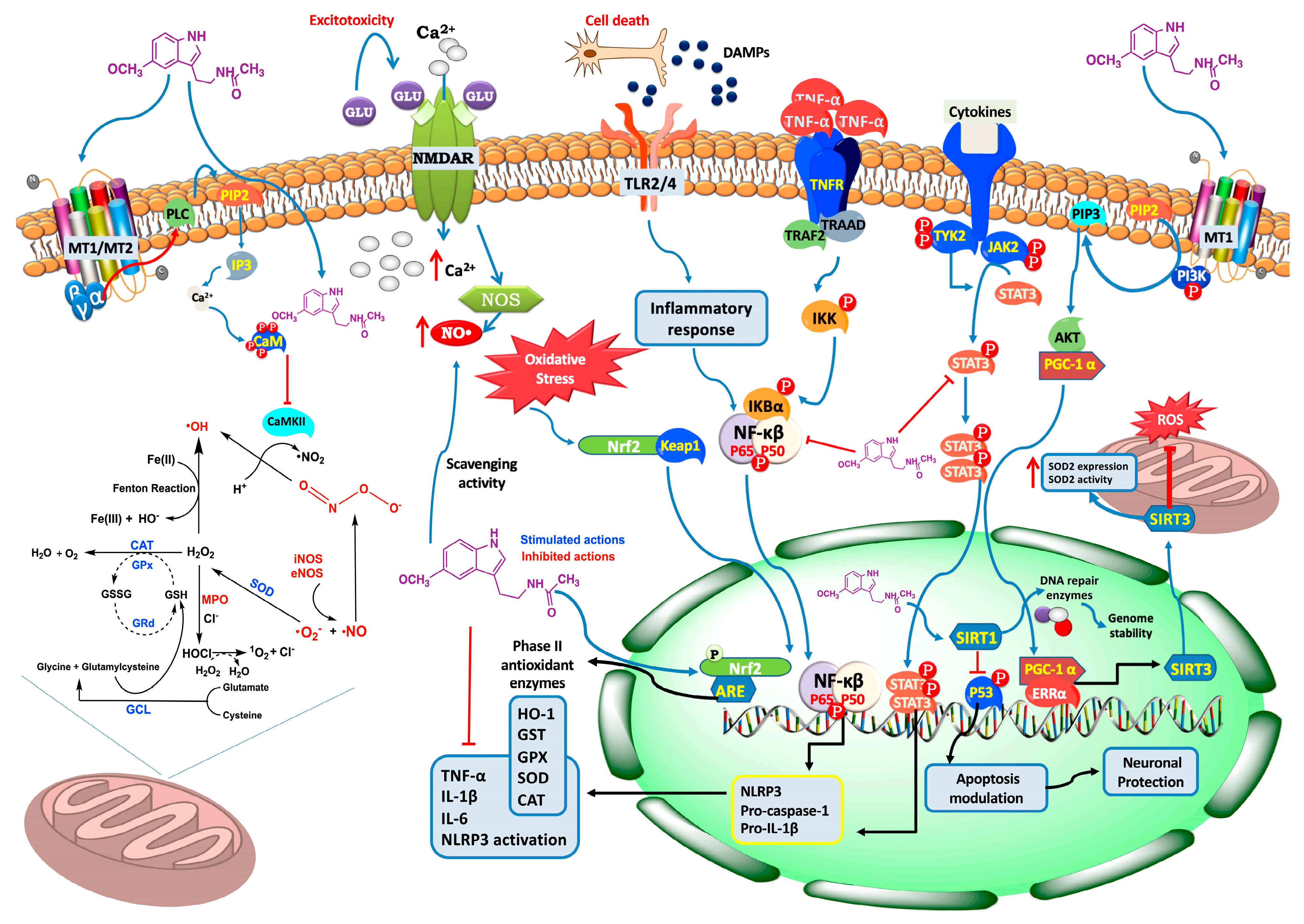
4.4. Exploring the Neuroprotective Potential of Melatonin in Stroke: Insights into Signaling Pathways

In stroke, the excessive release of glutamate leads to the activation of NMDA, AMPA, and kainate receptors, resulting in a detrimental influx of Ca2+ into the cells (Figure 5). This influx ultimately triggers apoptotic cell death [218]. Melatonin acts as an inhibitor of NMDA receptors in the brain, and when combined with memantine (an NMDA receptor inhibitor), it can significantly reduce the infarct volume in an MCAO model [219].
Additionally, melatonin exerts its neuroprotective effect by modulating different signaling pathways. The heme oxygenase-1 (HO-1)/CREB signaling pathway is involved not only in the development of depression-like behaviors that can be developed after an ischemic episode, but also in the transformation of astrocytes into a neurotoxic and inflammatory type cell. Melatonin interacts with HO-1/CREB, inducing an anti-depression effect [220].
The protein kinase B (Akt) is a molecule resistant to oxidative stress in the brain, whereas sirtuin 3 (SIRT3) activates superoxide dismutase 2 (SOD2, which converts H2O2 radicals into non-toxic molecules) and modulates mitochondrial oxidative stress, thus maintaining ROS homeostasis [221]. In fact, SIRT3 expression is downregulated after an ischemic episode, which contributes to the oxidative stress that appears after it [204]. The activation of the Akt-SIRT3-SOD2 pathway by melatonin as well as the increase in SIRT3 expression alone by it are mechanisms by which melatonin can decrease infarct volume and apoptotic rate after an ischemic stroke [204,222].
One way to alleviate mitochondrial damage after an ischemic episode is by targeting mitochondrial fission. Excessive mitochondrial fission causes an increased RONS production and the liberation of pro-apoptotic factors. By stimulating the Yap–Hippo pathway (decreased in ischemia-reperfusion injuries), melatonin can promote mitochondrial fusion by enhancing the activity of optic atrophy 1 (OPA-1). This makes fragmented mitochondria interact with each other, allowing mitochondrial recovery and, in the end, decreasing the brain reperfusion stress [223].
STAT3 is one of the many transcription factors that modulate the microglia/macrophage response to tissue damage. This damage in the tissue promotes the activation of STAT3, which promotes the pro-inflammatory activation of microglial cells. Melatonin induces an anti-inflammatory effect by promoting the STAT3 phosphorylation (Figure 5), thus shifting the pro-inflammatory phenotype of microglia to an anti-inflammatory one and reducing the ischemic stroke-induced brain damage [204].
A predominant NMDA receptor 2 (NR2a) stimulation at the synaptic level promotes pro-survival signaling via activation of pro-survival proteins such as Akt, ERK, and CREB, while NR2b at extra-synaptic areas favors excitotoxic death. During ischemic conditions, NR2a levels decrease, but melatonin may exert its protective effects by attenuating the NR2a cleavage and, additionally, by increasing the γ-enolase/PI3K/AKT/CRMP2 survival pathways [224].
The antioxidant activity of melatonin involves the modulation of several signaling pathways described in detail in the next section.

5. Melatonin as an Antioxidant and Free Radical Scavenger in Stroke: Mechanisms and Therapeutic Implications

The generation of free radicals in stroke has significant implications for neuronal damage and the progression of ischemic injury. In this regard, the ischemic cascade triggered by stroke results in a sequence of molecular and cellular events, including the generation of RONS [225]. Thus, several mechanisms contribute to the generation of free radicals in stroke, among them (i) oxidative stress, characterized by an imbalance between the production of ROS, including O2, H2O2, and OH, and the ability of endogenous antioxidants to neutralize them, leads to lipid peroxidation, protein oxidation, and DNA damage, which disrupt cellular function and contribute to neuronal injury and cell death [225]; (ii) mitochondrial dysfunction, leads to electron leakage from the electron transport chain, resulting in the generation of O2 (Figure 5). Additionally, the release of mitochondrial pro-apoptotic factors during stroke contributes to the generation of ROS [226] and; (iii) the inflammatory response that occurs in stroke, is a double-edged sword, despite the fact that it plays a role in clearing debris and initiating tissue repair processes, excessive inflammation can exacerbate brain damage by promoting the release of pro-inflammatory cytokines and attracting more immune cells, such as microglia and infiltrating neutrophils, which produce RONS through the activation of NADPH oxidase and inducible nitric oxide synthase (iNOS), respectively [227]. In this context, ROS-induced oxidative stress compromises the integrity of the BBB, promoting the infiltration of immune cells and inflammatory mediators into the brain, and therefore, exacerbating neuronal damage [228].
Given all the above information, stroke is a complex condition involving multiple pathophysiological processes and molecular targets. Therefore, the exploration of novel therapeutic approaches focussing on multitarget modulation, would be a good starting point for identifying and targeting multiple critical mechanisms involved in stroke, such as inflammation and oxidative stress, among others [229]. In this regard, melatonin holds great promise in stroke management [23,230]. It is able to cross the BBB efficiently [231], ensuring effective delivery to the site of injury, and this, combined with its ability to modulate various neuroprotective pathways [232] (Figure 5), makes it an attractive therapeutic strategy. Additionally, melatonin exhibits indirect antioxidant effects by enhancing the activity of endogenous antioxidant defense systems [33]. It upregulates the expression and activity of various antioxidant enzymes, including superoxide dismutase (SOD), catalase (CAT), glutathione peroxidase (GPx), and heme oxygenase-1 (HO-1) [45,233]. These enzymes play critical roles in neutralizing RONS and detoxifying oxidative products (Figure 5). Therefore, melatonin’s ability to enhance antioxidant enzyme activity helps to restore redox balance and mitigate oxidative stress in stroke.
The antioxidant activity of melatonin against stroke involves the modulation of several signaling pathways [234]. In this context, melatonin activates the master regulator of cellular antioxidant defense mechanisms, the Nuclear Factor Erythroid 2-Related Factor 2 (Nrf2) Pathway [233], where Nrf2 translocates to the nucleus upon activation and binds to antioxidant response elements (ARE) in the DNA, leading to the upregulation of genes encoding antioxidant enzymes, such as SOD, CAT, and GPx. Activation of the Nrf2 pathway by melatonin enhances the cellular antioxidant capacity, reducing oxidative stress and protecting against stroke-induced damage [233,235] (Figure 5). Likewise, melatonin can also modulate the Kelch-like ECH-Associated Protein 1 (Keap1) pathway [236], which regulates the stability and activity of Nrf2; melatonin interacts with Keap1, preventing its inhibitory effect on Nrf2. This interaction leads to the stabilization and nuclear translocation of Nrf2, thereby increasing the expression of antioxidant enzymes and enhancing the antioxidant defenses against stroke-induced oxidative stress. Another important pathway that contributes to the neuroprotective effects of melatonin against stroke concerns protein kinase C (PKC) [237,238], which is involved in cell signaling and oxidative stress regulation. PKC activation by melatonin triggers downstream signaling events that promote antioxidant effects, such as the upregulation of antioxidant enzymes and the inhibition of pro-oxidant enzymes. PKC-mediated melatonin signaling in the Mitogen-Activated Protein Kinase (MAPK) pathway, including extracellular signal-regulated kinase (ERK), c-Jun N-terminal kinase (JNK), and p38, is involved in cellular responses to oxidative stress [239,240,241,242]. Activation of ERK and other MAPKs by melatonin [242] leads to the phosphorylation and activation of transcription factors that regulate antioxidant enzyme expression, such as activator protein 1 (AP-1) and cAMP response element-binding protein (CREB) [243]. Therefore, by modulating MAPK signaling, melatonin enhances antioxidant defenses and protects against oxidative damage in stroke. The phosphoinositide 3-kinase (PI3K)/Akt pathway, known for its role in cell survival and protection against ischemic injury, also contributes to melatonin’s antioxidant effects. Activation of the PI3K/Akt pathway by melatonin [244] leads to the phosphorylation and activation of Akt, which in turn phosphorylates and inhibits pro-apoptotic proteins and transcription factors involved in oxidative stress-induced cell death (Figure 5). By promoting cell survival and inhibiting oxidative stress-induced apoptosis, melatonin’s activation of the PI3K/Akt pathway contributes to its antioxidant effects in stroke [244,245]. Moreover, the silent information regulator 1 (SIRT1) pathway, is a NAD+-dependent protein deacetylase involved in cellular stress responses and longevity. Melatonin can activate SIRT1, which regulates various pathways involved in neuroprotection, including antioxidant defense, mitochondrial function, and anti-apoptotic processes [222] (Figure 5).
As a whole, these signaling pathways interact and crosstalk with each other, forming a complex network of antioxidant mechanisms that mediate the protective effects of melatonin against oxidative stress in stroke. Further research is needed to fully understand the intricate interplay and downstream effects of these pathways to optimize the use of melatonin as an antioxidant therapeutic strategy in stroke management.
Melatonin’s capability to scavenge free radicals is attributed to its unique chemical structure, containing electron-donating functional groups, such as indole and methoxy groups, which facilitate electron transfer and RONS quenching, which reduces oxidative (H2O2, OH, and O2) and nitrosative (inhibiting the formation of ONOO, a highly reactive and cytotoxic molecule formed from the reaction between nitric oxide and superoxide) stress [33,246]. Furthermore, the scavenger action of melatonin also involves the preservation of mitochondrial function in stroke; enhancing mitochondrial respiration, ATP production, electron transport chain efficiency, preventing mitochondrial permeability transition pore opening, and reducing ROS generation [24,247,248]. On the basis of this complex background, inflammation plays a key role in stroke pathogenesis and exacerbates oxidative stress [249,250]. Relative to this, melatonin indirectly reduces RONS production and scavenges free radicals in stroke by modulation of the inflammatory response (Figure 5), regulating the production of pro-inflammatory cytokines, such as tumor necrosis factor-alpha (TNF-α), IL-1β, and IL-6 as well as inhibiting the activation of NF-κB [251,252].
Overall, while melatonin’s direct antioxidant effects have been extensively studied [253,254,255,256], emerging research has shed light on the importance of its metabolites in mediating its antioxidative actions [256,257,258,259]. In this sense, after melatonin is metabolized in the body, it undergoes various enzymatic reactions, leading to the formation of several metabolites that act as free radical scavengers. The free radical scavenging properties exhibited by melatonin metabolites are attributed to their ability to directly interact with and neutralize RONS. Melatonin metabolites act as potent antioxidants by donating electrons or hydrogen atoms to RONS, thereby stabilizing, and neutralizing them. One of the primary metabolites of melatonin, 6-Hydroxymelatonin (6-OHM), is formed through the action of cytochrome P450 enzymes and exhibits potent antioxidative activity. It acts as a direct free radical scavenger, neutralizing a variety of RONS, including H2O2, OH, and ONOO and reducing oxidative stress [256]. Furthermore, 6-OHM can also stimulate the activity of various antioxidant enzymes, and inhibits lipid peroxidation, thereby reducing oxidative damage to brain cells. N1-Acetyl-N2-formyl-5-methoxykynuramine (AFMK) is another important melatonin metabolite generated through the action of cytochrome P450 enzymes, myelo- or heme peroxidases, or formed from the reaction between melatonin and free radicals, particularly RONS. AFMK possesses strong antioxidant properties and can efficiently scavenge a wide range of free radicals, including OH, ONOO, and peroxyl radicals (ROO) [260]. AFMK also exhibits anti-inflammatory effects, further contributing to its neuroprotective potential in stroke. N1-Acetyl-5-methoxykynuramine (AMK) is another metabolite formed through the enzymatic conversion of melatonin by indoleamine 2,3-dioxygenase (IDO) and is an active metabolite with antioxidant properties. AMK acts as a free radical scavenger, specifically targeting ROO [261].
These melatonin metabolites possess inherent free radical scavenging properties and contribute to the overall antioxidant effects of melatonin. They can directly neutralize a wide range of RONS, thereby reducing oxidative stress and protecting against oxidative damage in stroke and other oxidative stress-related conditions. However, further research is needed to fully elucidate the specific roles and mechanisms of these melatonin metabolites in neuroprotection and stroke therapy.
Several preclinical studies have investigated the potential neuroprotective effects of melatonin in stroke models [222,244,262,263,264]. These studies have consistently demonstrated the ability of melatonin to reduce infarct size, improve neurological outcomes, and attenuate oxidative stress-related markers. Furthermore, melatonin has shown the potential to enhance cerebral blood flow [265] and protect against reperfusion injury [266,267], a critical component of stroke pathogenesis. However, although the preclinical data are promising, clinical trials exploring the efficacy of melatonin in stroke are relatively limited. Some early-phase clinical studies have reported positive outcomes [268], including improved functional recovery and reduced markers of oxidative stress in patients treated with melatonin following ischemia and reperfusion injury [269]. However, larger randomized controlled trials are needed to further validate these findings and determine the optimal dosing, timing, and long-term benefits of melatonin treatment in stroke patients. If proven successful, melatonin could revolutionize stroke treatment by providing a novel and cost-effective approach to reduce neuronal damage and improve functional outcomes.

6. Melatonin Improved Cognitive and Behavioral Performance

The circadian rhythm of endogenous melatonin is thought to be important in maintaining the normal sleep–wake rhythm [270,271]. The sleep–wake rhythm plays an important role in the development and maintenance of cognitive functions such as memory consolidation and learning. Similarly, sleep disturbances in mild to moderate Alzheimer’s disease (AD) are associated with increased memory and cognitive impairments [272]. Melatonin secretion by the pineal gland gradually decreases with age, in circadian disorders, but especially in stress-related pathologies that trigger degenerative diseases. Degenerative diseases are the result of a continuous process of dysfunction of cells affecting tissues or organs that progressively deteriorate over time. These diseases include neurodegenerative diseases such as Parkinson’s and Alzheimer’s disease, cardiovascular diseases, metabolic syndromes such as type 2 diabetes, and neoplastic pathologies such as cancer [273]. In addition, clinical trials of melatonin have been reported for all the disorders previously mentioned [274].
The role of melatonin on cognitive function has been evaluated in different situations; for example, increased sleep efficiency and total sleep time was successfully improved, without any effect on cognitive function in breast cancer patients [275], and cognitive deficits induced by benzodiazepine treatment does not seem affected by melatonin in patients with severe mental illness [276]. Prolonged release of melatonin has positive effects on cognitive functioning and sleep maintenance in AD patients, particularly in those with insomnia comorbidity, suggesting a causal link between poor sleep and cognitive decline [277].
Melatonin exerts biological processes via activation of two G-protein-coupled receptors, and non-receptors mediated pathways [278]. Two receptors named MT1 and MT2 are preferentially coupled to Gi proteins. MT1, and most likely MT2, is also coupled to Gq/11 proteins [279,280]. The third type of receptor MT3 is an enzyme quinone reductase 2 [281] and one nuclear receptor [282]. Some of the non-receptor activities highlight the property as a free radical scavenger [283,284], with special activity in mitochondria [248] and preserving the membrane fluidity, protecting against free radicals [285], and also attenuating the laser radiation-induced mutation and deletion of mitochondrial DNA [286]. Other activity includes a direct protein interaction inhibiting the calcium-calmodulin-dependent kinase II (CaMKII) activity [287].
The rhythmic release of melatonin from the pineal gland is coordinated with the local release of melatonin in the retina and suprachiasmatic nucleus via the activation of two G protein-coupled receptors, MT1 and MT2 [288]. Melatonin concentrations in cerebrospinal fluid (CSF) is higher compared to peripheral blood, and melatonin in CSF exhibits a concentration gradient in sheep [289,290] and humans [291]. The highest concentration is measured in the third ventricle near the gland pineal recess. Thereafter, melatonin concentrations gradually decrease in CSF sampled from the middle of the third ventricle, the aqueduct, the fourth ventricle, and the lumbar subarachnoid space [292].
Different animal models with different pathologies show cognitive impairment, and several behavior protocols are used to evaluate the role of melatonin in improving this function. The Morris water maze (MWM) is a robust and reliable spatial learning and memory test and is correlated with hippocampal synaptic plasticity and NMDA receptor function [293]. The novel object recognition test (NORT) is another behavioral paradigm to evaluate recognition memory [294], and the Y-maze protocol is used to evaluate spatial working and reference memory [295]. The MWM was widely used to evaluate cognitive function, and melatonin shows an improvement in learning and memory in this behavioral test in a cognitive impairment induced by liver fibrosis in rats [296], obese mice model [297], type 2 diabetic mice model [298], cortical compact impact model [299], and repeated mild traumatic brain injury if melatonin is administered during early pathological stages but not in late (chronic) stages [300]. Induced cognitive impairment shows learning and memory improvement by melatonin in MWM, NORT, and the Y-maze behavioral tests. In contrast, intact rats show learning and memory impairment induced by melatonin treatment in the MWM test [301].
The evidence of melatonin receptors in the hippocampus [302,303] suggests the role of melatonin in the modulation of learning and memory processes. Electrophysiological studies have demonstrated the role of melatonin in modulating synaptic transmission [302,303,304] and regulation of the short-term plasticity evaluated by the paired-pulse stimulation paradigm [305]. Results exploring the excitability indicate a regulation of the firing in hippocampal neurons [302,306] through the activation of melatonin receptors. Nevertheless, melatonin reduces the long-term potentiation (LTP) in the hippocampus CA1 [307], and this LTP inhibition correlates with an impairment in cognitive behavior [301]. In agreement with behavioral data, inhibition of the LTP induction by melatonin and cognitive impairment was observed in intact subjects; whereas, the early administration of melatonin in the traumatic brain injury (TBI) model shows an increase in the frequency of spontaneous excitatory and inhibitory postsynaptic currents without affecting the frequency of the miniature postsynaptic currents in the prefrontal cortex and hippocampus. These changes in synaptic transmission were associated with an improvement of the learning and memory in the MWM behavioral test. In summary, melatonin shows beneficial effects, improving cognitive functions associated with pathological processes such as oxidative stress, neuroinflammation, excitotoxicity, or scavenging free radicals as part of the mechanisms involved in neurodegeneration, traumatic brain injury, and vascular or metabolic diseases.

7. Suppressing Inflammatory Response by Melatonin: Effects on the Inflammasome

As previously mentioned, the pathophysiology of ischemic brain damage involves the activation of many deleterious signaling cascades, including ionic imbalances, excessive release of glutamate [308], inflammation, and free radical-induced oxidative and nitrosative stress [309]. Oxidative stress induces the release of DAMPs that trigger an inflammatory response, resulting in microglial activation, and increased BBB permeability that leads to peripheral immune cell infiltration [310,311]. Accumulating evidence suggests that post-ischemic inflammation is responsible for the secondary progression of brain injury and that stroke severity depends on the magnitude of this inflammatory response [312,313]. One of the most potent pro-inflammatory cytokines is IL-1β, which plays an important role in ischemic stroke through the following mechanisms: (i) increased microvascular permeability, BBB dysfunction and vasogenic cerebral edema [314]; (ii) exacerbation of the inflammatory response leading to secondary brain damage by secreting and releasing various neurotoxic substances, such as IL-1β, IL-6, TNF-α, and promoting the activation of iNOS [315]; and (iii) promoting apoptosis of injured cells by activating apoptotic molecular machinery, gradually transforming the original ischemic perimeter into the cerebral infarct area, ultimately exacerbating brain injury [316].
NOD-like receptor family pyrin domain containing 3 (NLRP3) inflammasome is the most widely characterized inflammasome that activates caspase-1 which contributes to the maturation and secretion of the cytokines IL-18 and IL-1β and induces pyroptosis cell death [317]. NLRP3 inflammasome is a key step in the innate immune response. Recently, the NLRP3 inflammasome was shown to play an important role in renal, myocardial, hepatic, and cerebral ischemic stroke [318]. Inhibition of NLRP3 inflammasome activation has been proposed as a promising new therapeutic target for inflammation-related diseases. Canonical activation of the NLRP3 inflammasome requires two sequential signals, priming and activation. The priming signal is initiated through the TLR4 receptors by DAMPs or pathogen-associated molecular patterns (PAMPs) such as LPS, leading to transcription of NLRP3 inflammatory components via NF-κB activation and nuclear translocation. Activation signals are stimulations of NLRP3 by various recognized factors, leading to procaspase-1 cleavage [319]. Active caspase-1 cleaves the pro-inflammatory cytokines IL-1β, IL-18, and the protein gasdermin D into their mature forms. Finally, mature gasdermin D forms pores in the cell membrane, causing pyroptotic cell death and cytokine release [320]. IL-1β and IL-18 enhance inflammation and promote immune cell infiltration.
Melatonin has been shown to inhibit NLRP3 inflammasome activation by modulating multiple proteins and signaling pathways. First, as mentioned above, RONS are key triggers for the activation of the NLRP3 inflammasome. Thioredoxin-interacting protein (TXNIP) is one of the most important redox regulators. By inhibiting thioredoxins, TXNIP regulates the intracellular redox balance. In a model of LPS-induced endometritis, melatonin downregulates TXNIP-mediated activation of NLRP3 inflammasome [321]. Furthermore, melatonin reduces TXNIP levels in cadmium-treated primary hepatocytes and suppresses RONS production and NLRP3 activity [322]. Another signaling pathway that melatonin can regulate is Nrf2. Under oxidative stress conditions, Nrf2 translocates to the nucleus and plays a role in protecting cells from oxidative damage, leading to increased expression of antioxidant and detoxifying enzymes, thus reducing oxidative stress [323]. It has been shown that melatonin provides protection against NLRP3 inflammasome activity through the removal and clearance of Nrf2-mediated RONS [203,233].
Autophagy is known to be a negative regulator of NLRP3 activation. Mitophagy is a subtype of autophagy that helps clear dysfunctional mitochondria, and in this sense, it has been shown that melatonin increased the expression of the autophagy markers Atg 5 and LC3-II/LC3-I and the mitophagy markers PINK-1 and Parkin and suppresses NLRP3 inflammasome activation in a model of subarachnoid hemorrhage [324]. Moreover, LPS-induced inflammation leads to an increase in NLRP3 inflammasome protein due to an autophagic flux impairment, and melatonin has a potent anti-inflammatory effect by restoring the LPS-induced autophagic flux blockage, hence reducing NLRP3 inflammasome components expression and decrease in ROS production [325]. Furthermore, melatonin may inhibit multiple transcription factors associated with the priming step of NLRP3 inflammasome activation. Thus, melatonin inhibits NF-κB signaling through silent information regulator 1 (SIRT1)-dependent deacetylation of NF-κB and retinoid orphan receptor α (RORα), thereby blocking septic responses induced by cecal ligation and puncture [326]. SIRT1 is an NAD+-dependent deacetylase and a potent regulator of intracellular inflammatory, metabolic, and oxidative stressors. Therefore, it has been shown that SIRT1 reduces NLRP3 inflammasome activation by deacetylating NLRP3 protein, and melatonin may increase SIRT1 activity and thus inhibit NLRP3 inflammasome activation in different inflammatory models [327,328]. Finally, the regulatory function of melatonin on NLRP3 inflammasome also occurs through post-transcriptional modifications since melatonin is able to inhibit the formation of NLRP3 inflammasome complex by altering the expression of various miRNAs [327]. These are, therefore, the main mechanisms by which melatonin reduces NLRP3 inflammasome activation.

8. Melatonin, iNOS, and Calcium/Calmodulin

As previously mentioned, melatonin is considered a multitasking molecule, but some physiological roles have yet to be established. Melatonin is a highly lipophilic molecule that crosses easily through cellular membranes and produces its cellular functions through diverse mechanisms: first, by its interaction with specific G-protein coupled receptors identified as the MT1 and MT2 leading down-stream second messengers, activation of signaling pathways, and gene transcription [246,329,330,331]; second, by acting as a free radical scavenger detoxifying RONS [246,254]; or third, through its binding to nuclear proteins [332,333,334] and intracellular proteins such as calmodulin (CaM), regulating intracellular Ca2+ signaling pathways because CaM is an essential mediator of Ca2+ signaling within cells [335,336,337,338] (Figure 5).
CaM has been defined as one melatonin-binding protein with considerable physiological significance depending on its affinity. In 1993, Benitez-King et al. described that melatonin binds to a single site on the CaM molecule with high affinity, in a saturable, reversible, Ca2+-dependent, and ligand-selective manner (Kd of 188 pM and a total binding capacity Bmax of 35 pM/µ of CaM) [336]. However, other investigations indicated a much lower affinity between melatonin and CaM [339]. The controversy remains open whether melatonin effects related to CaM are direct or because of indirect intracellular signaling mechanisms. Melatonin cascades may be sometimes crosslinked, therefore protein kinase or a transcription factor may be activated by several mechanisms. The precise interconnections between melatonin and CaM widely remain to be clarified [340]. It has been reported that melatonin increased the total cellular level and synthesis of CaM [335,337]. Additionally, melatonin directly binds to antagonize CaM [335,336,341]. Activated CaM acts both directly by interaction with key target enzymes, and structural proteins, and indirectly via specific protein kinases such as CaMKII [337].
CaMKII is a family of protein kinases encoded by four genes (α, β, γ, and δ) that mediates different physiological responses triggered by increased [Ca2+]i, by its autophosphorylation, and by the binding of Ca2+/CaM complex on its target proteins producing phosphorylation of serine and threonine residues [246,342]. CaMKII has a dumbbell shape connected by seven-turn alpha helixes and two hydrophobic clefts with four Ca2+-binding domains with typical EF-hand conformation [343]. Each isoform of CaMKII is composed of three domains: (i) the association domain located at the C-terminal region, (ii) the catalytic domain located at the N-terminal region, and (iii) the regulatory domain located between association and catalytic domains [342]. The Ca2+/CaM complex binds to the 293–310 AA sequence in the regulatory domain to disrupt the interaction between the regulatory and the catalytic domain [344]. After its activation, the enzyme undergoes autophosphorylation at the threonine-286 residue, changing its activity to a Ca2+/CaM-independent manner [345,346]. Therefore, the activated Ca2+/CaM complex can interact with other targets such as NOS [345].
In rat hippocampal neurons, it has been shown that CaMKII activates and translocates from the cytoplasm to the presynaptic active zone [345,347] where it regulates neurotransmitter synthesis and release through phosphorylation of different targets such as K+ channels [348] or synaptotagmin and synapsin I [347]; additionally, CaMKII translocates to the post-synaptic density [345,349] during neuronal activation where nNOS is mainly located, therefore an interaction between both molecules occurs [345].
It has been stated that the regulation of CaMKII activity depends on its relative concentration and the concentration of its activator Ca2+/CaM complex [345,350]. Importantly, melatonin upregulates CaM synthesis; therefore, an increase in the activation of CaMKII is associated with melatonin. A direct effect of melatonin on PKC or its activation by diacylglycerol phosphorylates is the augmented CaM, activating CaMKII which autophosphorylates to maintain its activity-inducing dendrite growth and arborization, essential for synaptogenesis to reestablish the synaptic connectivity lost in aging neuropsychiatric disorders [338], indicating that melatonin can be used to prevent neuronal dysfunction.
As previously indicated, melatonin synthesis is upregulated under circumstances where free-radical generation is exaggerated [246,340], because it has protective actions against free-radical species acting through anti-oxidative and pro-oxidative enzymes [246,351,352] including eosinophil peroxidase, myeloperoxidase (MPO), and NOS [351,353,354,355].
It is well known that in aging the progressive accumulation of oxidative debris promotes the functional inefficiency of cellular processes inducing free-radical generation, the oxidative damage provokes apoptosis, and this loss of cells contributes to age-related deterioration. Additionally, the increased age leads to a gradually diminished melatonin production such that, in the elderly, the nocturnal melatonin rise in the circulation is greatly attenuated; this situation can have health consequences [246]. Moreover, the maintenance and progression of several diseases (neurodegenerative, cardiovascular, skin deterioration, cancer, metabolic syndrome, among others) where oxidative stress has a great role, can be accelerated by reduced production of melatonin during aging [246], supporting that melatonin is an important molecule to prevent age-related deterioration. In this sense, as reported in the previous sections, the antioxidant effect of melatonin occurs because of its ability to scavenge ROS such as hypochlorous acid (HOCl), H2O2, OH, ONOO, and O2 [353,356,357]. This occurs due to the effect on NOS activity. The link between heme destruction and disturbance of the zinc-tetrathiolate center of inducible NOS (iNOS) was described recently by Camp, leading to iNOS monomerization, protein unfolding, and accumulation of toxic free iron that occurs in many inflammatory diseases. Importantly, these events can be prevented in the presence of melatonin [353]. In this regard, iNOS expressed by immunoactive cytokines generated in tissues at sites of inflammation or in response to viral infection produces NO that is connected with essential functions for the pathogenesis of several diseases including diabetes, multiple sclerosis, and cancer, among others [358,359]. In animals, iNOS from cytokine-stimulated macrophage cell lines is a zinc homodimeric enzyme consisting of two identical subunits, containing a reductase and an oxygenase domain [353,359,360,361]. The reductase domain, C-terminus, binds NADPH, flavin mononucleotide (FMN), and flavin adenine dinucleotide (FAD), at various sites and importantly, contains a CaM binding linker peptide that tightly binds CaM [359]. The oxygenase domain (N-terminal portion) binds the iron protoporphyrin IX (heme) prosthetic group, the substrate L-arginine (L-Arg), the cofactor tetrahydrobiopterin (H4B), and a zinc atom [359]. The zinc atom is coordinated symmetrically by two cysteine residues from each subunit in a tetrahedral arrangement at the bottom of the dimer complex [361]. This complex plays an important role in maintaining the dimeric iNOS stability and preventing its monomerization [353].
The Ca2+/CaM binding events allow cross-subunit electron transfer from the FMN to the heme and is the essential electron transfer step because it enables O2 to bind to the NOS and thus start the process of NO biosynthesis [362,363]. In the brain, NO participates in numerous functions, including neurotransmission, development, and neuroprotection [364]. However, NO reacts with O2 to form ONOO, a more powerful oxidant that acts as a toxicant molecule [365] that causes lipid peroxidation, protein nitration and direct DNA damage leading to cell death [345,366]. NO is a small molecule synthesized by NOS named neuronal NOS (nNOS) and endothelial (eNOS), which are constitutive isoforms, and the iNOS that is negligible in resting cells but is induced by inflammatory cytokines and LPS [358,359,362,367,368]. LPS binds to the TLR4 on the surface of macrophage membranes, leading to the activation of MAPK or NF-κB signaling pathways and further iNOS gene expression [369] to produce NO. Additionally, microglia can exhibit a pro-inflammatory phenotype by LPS contributing to the activation of iNOS. Thus, during infection, which is more related to aging, microbial LPS can induce a prolonged low-grade inflammatory state where macrophage and microglial iNOS is upregulated [370,371].
Increased vulnerability to infection and the development of inflammatory diseases is more frequent during aging, and the onset and progression of some diseases such as neurodegeneration, cancer, metabolic syndrome, multiple sclerosis, septic shock, and adjuvant arthritis [360,372] are strongly associated with changes in the immune system function, and the loss of immune responses has been associated with resistance to bacterial infection [370]. Within the immune system the release of cytokines and chemokines is regulated by circadian phases [371,373] to maintain homeostasis through a well-organized sequence of immune defensive responses against pathogens. The decline of diurnally rhythmic immune responses related to aging amplifies disease-causing inflammation [370]. This situation could be associated with the reduced circadian production of melatonin during aging [246]. The principal biological modulators of the mammalian immune response are the macrophages population, which is heterogeneous but predominantly composed of mononuclear leukocytes [367]. Macrophages are classified as classically-activated M1 or alternatively-activated M2 [374]. M1 macrophages are pro-inflammatory cells responsible for the initiation of the immune response, while M2 macrophages are anti-inflammatory cells. M1 macrophages are activated by the microbial LPS to induce the release of pro-inflammatory interleukins IL1β, Il-6, IL-12, IL-23, and TNF-α, chemokines, hydrolyzed proteases interferons, and ROS that are released downstream in the innate immunity response [375]; this activation promotes the increase in NO-mediated by iNOS activation.
On the other hand, microglia are cells involved in regulatory processes critical for development, maintenance of the neural environment, response to injury and subsequent repair, and sense pathological events in the CNS to orchestrate innate immune responses. They are regulated by the CNS microenvironment and are the first line of defense against invading microbes and via interactions with neurons can be the first to detect critical changes in neuronal activity and health [376]. With aging, a decrease in synaptic plasticity is accompanied by the ability of microglia to express pro-inflammatory cytokines to contribute to mild chronic inflammatory conditions, also accompanied by a decrease in anti-inflammatory cytokines [376] and an increase in NO synthesis. In this sense, melatonin increased the amount of CaM and phosphorylation of CaMKII prompting dendritogenesis in rat hippocampal slices suggesting that melatonin could repair the loss of synaptic connectivity in aging neuropsychiatric disorders [338]. Additionally, melatonin has been associated with neurogenesis stimulation and neuronal survival [377,378], this adds evidence for its potential use as an adjuvant in neurodegenerative diseases
The effects of melatonin in immunoregulation have been well-described [367,379,380]. Melatonin shows an inhibitory effect against the LPS effects only if melatonin is administered before LPS in J774.2 macrophages. This effect occurs because melatonin reduces iNOS steady-state mRNA levels and iNOS protein expression, and it was associated with inhibition of the NFκ-B. Inhibition of iNOS-derived NO production contributes to the anti-inflammatory effect produced by melatonin [356]. Additionally, the inhibitory effect of melatonin on iNOS is due to an interaction with CaM [79], and iNOS is associated with CaM in a tightly bound form [358]. In the immunostimulated macrophages, prolonged exposure to melatonin during the induction of iNOS and its association with CaM increases the inhibitory effect on iNOS activity [356]. Melatonin exerts a potent anti-inflammatory effect and reduces NO production in murine models of carrageenan-induced inflammation [381]. Protection by melatonin in shock and inflammation is due to the inhibition of iNOS expression [246,356]. However as described previously, the reduced melatonin production by aging could be a factor that contributes to low-grade inflammatory conditions.
On another hand, visfatin an adipokine highly expressed in adipocytes and macrophages [382] exerts an inflammatory activity through the expression of pro-inflammatory cytokines (TNF-α, IL-6, and IL-1β) in different cell types [382,383,384]. In RAW 264.7 macrophages, melatonin reduces the visfatin-induced iNOS expression through the suppression of NF-κB in a similar way as LPS-induced iNOS expression. Visfatin may contribute to the enhancement of obesity-associated inflammation via the release from macrophages. Interestingly visfatin has been found to increase in human dental pulps with age [385], contributing to senescence, and is highly expressed in inflammatory diseases and some cancers [384]; however, the functional role of visfatin in aging has not yet been well established, but the protective melatonin effect against visfatin-induced inflammation through iNOS activation, strongly supports its use to treat the low-grade inflammatory condition.

9. Melatonin in Microglial and Astrocyte Cell Activation

The beneficial effects of melatonin have been shown in pre-clinical and/or clinical studies for several neurodegenerative conditions caused by insults such as infections, hypoxia/ischemia, trauma, or toxins as well as for neurodegenerative diseases, such as Alzheimer’s disease (AD), Parkinson’s disease (PD), multiple sclerosis (MS), and amyotrophic lateral sclerosis (ALS) [12,221,258,386,387,388,389,390,391,392,393]. A shared feature among these pathologies is neuroinflammation, a complex process mediated primarily by microglia and astrocytes. In this regard, recent studies have revealed the potential use of melatonin as a neuroprotective agent, especially in the context of glial cell activation.

9.1. Melatonin in Astrocyte Activation

Astrocytes, the most abundant non-neuronal cells in the brain, were classically considered to serve passive structural and support roles in the brain. They are now considered full-fledged participants in brain circuitry and processing, with a wide range of functions at the cellular level. These functions include the formation, maturation, and elimination of synapses, maintaining the integrity of the BBB and ionic homeostasis, clearing neurotransmitters, regulating the volume of the extracellular space, and modulating synaptic activity and plasticity [394,395,396]. Moreover, astrocytes play an essential role in the initiation, execution, and regulation of immune responses in the CNS [397,398,399].
Astrocytes undergo morphological, molecular, and functional remodeling, known as astrocyte activation, in response to various pathological stimuli, including inflammation, tumors, trauma, ischemia, epilepsy, and neurodegeneration [400,401,402]. Astrocyte activation leads to the loss of their normal homeostatic functions and the acquisition of protective or detrimental roles, including proliferation, scar-border formation, immune cell recruitment, and neurotoxicity [400,403,404,405]. Just as the M1/M2 activation axis has been described for microglia, there has been a proposal for astrocyte A1/A2 phenotypic polarization. According to this model, A1 denotes reactive cells that have lost their neurosupportive functions and have become toxic toward neurons, while A2 refers to inflammation-resolving astrocytes [406,407]. However, this simplistic binary classification statement to characterize the reactive states of astrocytes is under debate [400].
Following an ischemic stroke, the peri-infarct region is commonly divided into two parts: an inner region adjacent to the lesion with a high density of monocytes/macrophages and an outer region rich in astrocytes [408]. Reactive astrocytes in the outer region secrete various pro-inflammatory cytokines, chemokines, and matrix metalloproteinases, which disrupt the BBB and attract peripheral leukocytes, exerting deleterious effects on the brain after ischemia [408,409]. Additionally, astrocytes in this region display elongated and polarized processes with an increased GFAP expression and expression of factors involved in extracellular matrix remodeling and clustering of reactive astrocytes [410]. After ischemia and reperfusion injury, melatonin has been shown to attenuate reactive astrogliosis and glial scar formation, reduce the infarct volume, and, consequently, enhance axonal regeneration and promote neurobehavioral recovery in adult rats [411,412]. These effects of melatonin are related to a reduction in the activity of glycogen synthase kinase-3 beta (GSK-3β) and receptor-interacting serine/threonine-protein 1 kinase (RIP1K), proteins involved in astrocyte responses, after the treatment [412]. Furthermore, melatonin pretreatment has been shown to reduce the increased expression of Nox2 and Nox4 in astrocyte neurons and endothelial cells, reduce RONS levels, and inhibit cell apoptosis [413].
Traumatic brain injury (TBI) also induces reactive astrogliosis accompanied by an increased expression in intermediate filaments such as glial fibrillary acidic protein (GFAP) and vimentin, along with astrocyte hypertrophy and functional alterations as glutamate and potassium clearance changes [414]. Melatonin has been shown to partially reverse TBI-induced anxiety-like behavior in rats and decrease the number of activated astrocytes and neuronal apoptosis in the amygdala induced by TBI [415]. Melatonin also has been demonstrated to decrease the number of A1-type astrocytes in the NAc after TBI and mitigate TBI-induced depression by activating of HO-1/CREB signaling [220]. Similar results have been obtained in the hippocampus, dentate gyrus [300], and cortex [300,416] where melatonin reduces activated astrocytes and cytokine production and promotes cell survival and cognitive function after TBI induction. Furthermore, melatonin treatment suppresses the accumulation and the proliferation of microglia and astrocytes, down-regulates caspase-3, Bax and GFAP expressions, and the pro-inflammatory markers iNOS, IL-1β, and TNF-α expressions in a spinal cord injury model [417].
Astrocytes play a crucial role in the development of neurological and neurodegenerative diseases, primarily due to the disruption of normal homeostatic function and the acquisition of toxic functions [418]. Additionally, the accumulation of certain protein aggregates such as alpha-synuclein (SNCA), Aβ, and Tau in astrocytic cytoplasm can contribute to the pathology of these diseases [419], representing a significant characteristic in various neurodegenerative conditions. Furthermore, astrocytes can undergo various detrimental transformations, resulting in either atrophy and loss of function or reactive astrogliosis with hypertrophy [420]. Although melatonin has been shown to have a beneficial role in different neurodegenerative diseases such as AD [421,422], PD [389,423], MS [424], or ALS [425], less is known of its effects on astrocyte activation. In the rat hippocampus, melatonin has been shown to attenuate synaptic dysfunction and reduce astrogliosis improving the Aβ1–42-induced impairment in spatial learning and memory [426]. On the other hand, in results obtained by Andrade et al., melatonin reduces Aβ levels but does not reduce GFAP levels in rats receiving ICV-STZ (intracerebroventricular streptozotocin STZ) [427]. In an MS model induced by cuprizone, melatonin also reduces astrocyte activation in young and aging mice due to the antiapoptotic, antioxidant, anti-inflammatory, and neurotrophic effects [428]. Melatonin has also been demonstrated to prevent the development of neuropathic pain in the cuneate nucleus in a lysophosphatidylcholine (LPC)-induced median nerve demyelination neuropathy model. Here, melatonin reduces astrogliosis and through MT2 receptors inhibits the activation of astrocytic MAPKs, production of pro-inflammatory cytokines, and development of demyelination-induced neuropathic pain behavior.
Some neurotoxins and drugs also produce neuroinflammation and neuronal cell death. Melatonin also has been demonstrated to be effective in decreasing astrogliosis and the production of pro-inflammatory cytokines in these situations. In trimethyltin chloride- treated mice, melatonin reduces the expression of C3, Gbp2, and Serping1, indicating the suppression of A1 reactive astrocytes in the brain [429]. Furthermore, melatonin has inhibitory effects on caspase-3 and GFAP up-regulation induced by the plasticizer Diisononyl phthalate [430]. Additionally, melatonin reduced reactive astrocytes associated with the activation of the TLR4/MyD88/NFκB signaling pathway by methamphetamine [384,431].

9.2. Melatonin in Microglia Activation

The diverse functions of microglia play a crucial role in the onset, progression, and resolution of inflammation within the CNS. These processes also involve communication and interaction between microglia and various other cell types, particularly astrocytes and neurons, although it is not limited to them. Classically, phenotyping macrophages and microglia (cells that are functionally and developmentally related) into M1 (resting) and M2 (activated) based on the expression of markers related to these categories, was used to indirectly assume a detrimental M1 or beneficial M2 microglial role. In this classic description, M1 microglia releases inflammatory mediators and induce inflammation and neurotoxicity, while M2 microglia release anti-inflammatory mediators and induce anti-inflammatory and neuroprotection. However, this dualistic classification of good or bad microglia is very controversial and is inconsistent with the wide repertoire of microglial states and functions in development, plasticity, aging, and diseases that were elucidated in recent years (see [432] for more information).
As previously mentioned, melatonin can suppress proinflammatory signals and has been extensively documented. Notably, research has focused on its impact on iNOS and COX-2, as well as its ability to inhibit inflammasome activation, particularly NLRP3. Moreover, it also activates processes in an anti-inflammatory network, in which SIRT1 activation, upregulation of Nrf2, downregulation of NF-κB, and release of the anti-inflammatory cytokines IL-4 and IL-10 are involved [258,433,434]. However, these findings often did not specifically focus on microglia.
Microglia and infiltrated macrophages initially polarize toward a neuroprotective anti-inflammatory phenotype after stroke, but gradually transform into a detrimental pro-inflammatory phenotype [435,436]. Melatonin treatment ameliorates brain damage after ischemic stroke at least partially through shifting microglia phenotype from pro-inflammatory to anti-inflammatory polarity [204,411,437,438,439]. This M2 phenotype promoting effect by melatonin is via signal transducer and activator of transcription 1/6 and 3 (STAT1/6 and 3) and neuronal melatonin type 1 receptor activation [204,411,439]. After TBI, melatonin also suppresses microglial activation and the production of proinflammatory cytokines reducing mTOR pathway phosphorylation [440,441]. Similar effects of melatonin have been shown in the spinal cord injury (SCI) model. Melatonin decreases the expression levels of M1 microglia phenotypic markers (CD16, iNOS, and TNF-α) and increases M2 markers (Arg1, CD206, and TGF-β) also reducing the levels of pro-inflammatory cytokines (TNF-α, IL-6, and IL-1β) in the SCI mice and rats, facilitating functional recovery [417,442].
Both M1 and M2 microglial phenotypes are involved in the pathogenesis of neurodegenerative diseases [443], and promoting microglia polarization shift from M1 to M2 phenotype may be a prospective strategy in the therapy of neurodegenerative diseases such as AD, PD, ALS, and MS [444]. Melatonin has been shown to ameliorate neuroinflammation by inhibiting STAT-related pro-inflammatory (M1-like) polarization of microglia in a cellular PD model [445]. Moreover, melatonin attenuates microglial activation by negatively regulating NLRP3 inflammasome activation via a sirtuin 1 (SIRT1)-dependent pathway in 1-methyl-4-phenyl-1,2,3,6-tetrahydropyridine (MPTP)-induced murine PD models [446]. Regarding AD, melatonin has been shown to promote anti-inflammatory microglial activation rescuing SIRT1 and BDNF expression/release following β-amyloid (Aβ42)-induced microglial activation [447] and remediates the cytokine profile of Tau-exposed microglia [448]. In other neurological diseases, such as epilepsy, melatonin can also promote the polarization status of microglia from an M1 to M2 phenotype. In this case, Li et al. [445] have demonstrated that melatonin plays an antiepileptic role in KA-induced temporal lobe epilepsy, reducing the frequency and severity of seizures and changing microglia polarization status by regulating the RhoA/ROCK signaling pathway.

10. Post-Stroke Melatonin Treatment

Currently, the only treatment for ischemic stroke, which is limited and ineffective in a large number of patients, is thrombolysis when administered within 4 h of its onset [449]. However, it is very common for stroke patients to experience multiple complications such as depression, movement disorders, epilepsy, fatigue, dysphagia, or vascular dementia [450,451,452,453,454,455]. Due to its beneficial properties and lack of toxicity even at high concentrations, melatonin could be proposed as a potential treatment for alleviating post-stroke complications, although research on this topic is limited and more research is required to determine the correct doses and timing at which melatonin may be most effective.
Central post-stroke pain (CPSP) is another issue that can arise because of a stroke. It is a neuropathic pain syndrome characterized by pain and sensory abnormalities in the body parts corresponding to the injured brain territory [456]. A study found that CPSP-model rats showed improved performance in several pain tests when treated with intraperitoneal melatonin, in a dose-dependent manner. This suggests a neuromodulatory effect of melatonin in the treatment of CPSP [263,457].
Considering melatonin’s anti-inflammatory and antioxidant properties, it could be administered as a post-stroke treatment to alleviate the sequelae caused by increased inflammation and RONS. In an aged MCAO rat model, Rancan et al. [458] demonstrated that oral melatonin administration after surgery can inhibit the upregulation of pro-apoptotic markers associated with ischemic stroke, suggesting a reduction in neuronal damage. Melatonin administration after ischemic injury can also significantly decrease the activity of mitochondrial enzymes responsible for oxidative stress, such as NOS and COX-2, thereby reducing the infarct area [459]. Additionally, in a rat model of ischemia/reperfusion injury, another study found that intranasal administration of melatonin loaded in lipidic nanocapsules post-stroke can increase the number of surviving neurons in the hippocampal CA1 region and improve oxidative stress and inflammatory marker levels in the hippocampus. These effects were even more pronounced than those observed with oral melatonin administration [460].
Additionally, although melatonin does not possess direct antiviral capacity, supplemental melatonin has shown favorable effects, eliminating the pathogenicity of highly deadly viruses [461,462,463,464,465]. This has been extensively documented in various case reports, with the prevention of hemorrhagic shock syndrome from Ebola virus infection being particularly promising [466]. Given melatonin’s pharmacological profile, which highlights its potent abilities to moderate inflammation, mitigate oxidative stress, and regulate immune response, this indoleamine should be considered a preferred candidate for testing in the context of attenuating hyper-inflammation in COVID-19 patients and enhancing the success of their clinical management.

11. Conclusions

The conclusions that can be drawn from this review are the following:
Melatonin as a Promising Neuroprotective Agent: Melatonin shows considerable potential beyond its role in regulating sleep–wake cycles. Its neuroprotective properties position it as a promising candidate for stroke therapy, offering protection against ischemic brain damage.
Powerful Antioxidant and Possible Anti-Aging Agent: Melatonin exhibits antioxidant effects, making it a potential anti-aging agent by mitigating oxidative stress, which is implicated in age-related neurodegenerative diseases.
Implications of Free Radicals in Stroke and Aging: Free radicals, highly reactive molecules, are of great interest in medical research due to their involvement in multiple pathologies. Free radicals play a crucial role in oxidative stress and cellular damage, which has significant implications for understanding complex biological processes such as stroke and aging.
Interaction of Melatonin, Free Radicals, and Non-Exciting Amino Acids: Both melatonin and non-excitatory amino acids play integral roles in the stroke and aging processes, with melatonin acting as an antioxidant and non-excitatory amino acids providing neuroprotection through modulation of several key factors.
Potential of Therapeutic Strategies: Understanding the interaction between melatonin, free radicals, and non-excitatory amino acids opens opportunities for therapeutic strategies in age-related diseases and stroke prevention/treatment. We highlight the need for additional research to fully understand these mechanisms and explore their therapeutic applications in future clinical trials.
Future Perspectives and Improvement of Quality of Life: Deepening our understanding of these mechanisms brings us closer to developing specific interventions that can improve neuronal resilience and quality of life in people affected by stroke and age-related neurodegenerative disorders.

Author Contributions

Writing, review and editing, and data curation, V.J.C.; editing and data curation, I.Á.-M.; editing and data curation, E.R.; writing and review and editing, P.S.-C.; editing and review, A.H.; review and editing, F.L.M.; review and editing, A.M.B.; review and editing, J.M.S.; review and editing, J.E.; writing, review and editing, and supervision, A.R.; conceptualization, methodology, writing—original draft preparation, review and editing, supervision, and funding acquisition, J.M.H.-G. All authors have read and agreed to the published version of the manuscript.

Funding

This work was supported by MICIU (grant number PID2021-128133NB-I00/AEI/FEDER10.13039/501100011033) to J.M.H.-G. and V.J.C. enjoys a contract from the CAM “Investigo” program (PIP/2022-09971). A.R. thanks UCJC (INFLAMAMEL 2022-07 project) for its continued support.

Institutional Review Board Statement

Not applicable.

Informed Consent Statement

Not applicable.

Data Availability Statement

No new data were created or analyzed in this study. Data sharing is not applicable to this article.

Acknowledgments

The authors want to acknowledge Translational Stroke Research and Amino Acids for granting permission to adapt the Figure 1, Figure 2 and Figure 3.

Conflicts of Interest

The authors declare no conflict of interest.

References

  1. Arendt, J.; Ravault, J.P. Suppression of melatonin secretion in Ile-de-France rams by different light intensities. J. Pineal Res. 1988, 5, 245–250. [Google Scholar] [CrossRef] [PubMed]
  2. Comai, S.; Gobbi, G. Unveiling the role of melatonin MT2 receptors in sleep, anxiety and other neuropsychiatric diseases: A novel target in psychopharmacology. J. Psychiatry Neurosci. JPN 2014, 39, 6–21. [Google Scholar] [CrossRef]
  3. Carrascal, L.; Nunez-Abades, P.; Ayala, A.; Cano, M. Role of Melatonin in the Inflammatory Process and its Therapeutic Potential. Curr. Pharm. Des. 2018, 24, 1563–1588. [Google Scholar] [CrossRef] [PubMed]
  4. Chen, B.; You, W.; Shan, T. The regulatory role of melatonin in skeletal muscle. J. Muscle Res. Cell Motil. 2020, 41, 191–198. [Google Scholar] [CrossRef] [PubMed]
  5. Chitimus, D.M.; Popescu, M.R.; Voiculescu, S.E.; Panaitescu, A.M.; Pavel, B.; Zagrean, L.; Zagrean, A.M. Melatonin’s Impact on Antioxidative and Anti-Inflammatory Reprogramming in Homeostasis and Disease. Biomolecules 2020, 10, 1211. [Google Scholar] [CrossRef]
  6. Gunata, M.; Parlakpinar, H.; Acet, H.A. Melatonin: A review of its potential functions and effects on neurological diseases. Rev. Neurol. 2020, 176, 148–165. [Google Scholar] [CrossRef]
  7. Lalanne, S.; Fougerou-Leurent, C.; Anderson, G.M.; Schroder, C.M.; Nir, T.; Chokron, S.; Delorme, R.; Claustrat, B.; Bellissant, E.; Kermarrec, S.; et al. Melatonin: From Pharmacokinetics to Clinical Use in Autism Spectrum Disorder. Int. J. Mol. Sci. 2021, 22, 1490. [Google Scholar] [CrossRef]
  8. Malakoti, F.; Zare, F.; Zarezadeh, R.; Raei Sadigh, A.; Sadeghpour, A.; Majidinia, M.; Yousefi, B.; Alemi, F. The role of melatonin in bone regeneration: A review of involved signaling pathways. Biochimie 2022, 202, 56–70. [Google Scholar] [CrossRef]
  9. Patel, R.; Parmar, N.; Pramanik Palit, S.; Rathwa, N.; Ramachandran, A.V.; Begum, R. Diabetes mellitus and melatonin: Where are we? Biochimie 2022, 202, 2–14. [Google Scholar] [CrossRef]
  10. Pivonello, C.; Negri, M.; Patalano, R.; Amatrudo, F.; Montò, T.; Liccardi, A.; Graziadio, C.; Muscogiuri, G.; Pivonello, R.; Colao, A. The role of melatonin in the molecular mechanisms underlying metaflammation and infections in obesity: A narrative review. Obes. Rev. Off. J. Int. Assoc. Study Obes. 2022, 23, e13390. [Google Scholar] [CrossRef]
  11. Roy, J.; Wong, K.Y.; Aquili, L.; Uddin, M.S.; Heng, B.C.; Tipoe, G.L.; Wong, K.H.; Fung, M.L.; Lim, L.W. Role of melatonin in Alzheimer’s disease: From preclinical studies to novel melatonin-based therapies. Front. Neuroendocrinol. 2022, 65, 100986. [Google Scholar] [CrossRef]
  12. Skarlis, C.; Anagnostouli, M. The role of melatonin in Multiple Sclerosis. Neurol. Sci. Off. J. Ital. Neurol. Soc. Ital. Soc. Clin. Neurophysiol. 2020, 41, 769–781. [Google Scholar] [CrossRef] [PubMed]
  13. Talib, W.H. Melatonin and Cancer Hallmarks. Molecules 2018, 23, 518. [Google Scholar] [CrossRef] [PubMed]
  14. Wang, G.H.; Jiang, Z.L.; Fan, X.J.; Zhang, L.; Li, X.; Ke, K.F. Neuroprotective effect of taurine against focal cerebral ischemia in rats possibly mediated by activation of both GABAA and glycine receptors. Neuropharmacology 2007, 52, 1199–1209. [Google Scholar] [CrossRef] [PubMed]
  15. Zhang, R.; Ni, L.; Di, X.; Ma, B.; Niu, S.; Rong, Z.; Liu, C. Potential Role of Melatonin as an Adjuvant for Atherosclerotic Carotid Arterial Stenosis. Molecules 2021, 26, 811. [Google Scholar] [CrossRef]
  16. Chen, J.; Zhuang, Y.; Zhang, Y.; Liao, H.; Liu, R.; Cheng, J.; Zhang, Z.; Sun, J.; Gao, J.; Wang, X.; et al. A synthetic BBB-permeable tripeptide GCF confers neuroprotection by increasing glycine in the ischemic brain. Front. Pharmacol. 2022, 13, 950376. [Google Scholar] [CrossRef]
  17. Piniella, D.; Zafra, F. Functional crosstalk of the glycine transporter GlyT1 and NMDA receptors. Neuropharmacology 2023, 232, 109514. [Google Scholar] [CrossRef]
  18. Surai, P.F.; Earle-Payne, K.; Kidd, M.T. Taurine as a Natural Antioxidant: From Direct Antioxidant Effects to Protective Action in Various Toxicological Models. Antioxidants 2021, 10, 1876. [Google Scholar] [CrossRef]
  19. Jakaria, M.; Azam, S.; Haque, M.E.; Jo, S.H.; Uddin, M.S.; Kim, I.S.; Choi, D.K. Taurine and its analogs in neurological disorders: Focus on therapeutic potential and molecular mechanisms. Redox Biol. 2019, 24, 101223. [Google Scholar] [CrossRef]
  20. Álvarez-Merz, I.; Luengo, J.G.; Muñoz, M.D.; Hernández-Guijo, J.M.; Solís, J.M. Hypoxia-induced depression of synaptic transmission becomes irreversible by intracellular accumulation of non-excitatory amino acids. Neuropharmacology 2021, 190, 108557. [Google Scholar] [CrossRef]
  21. Álvarez-Merz, I.; Fomitcheva, I.V.; Sword, J.; Hernández-Guijo, J.M.; Solís, J.M.; Kirov, S.A. Novel mechanism of hypoxic neuronal injury mediated by non-excitatory amino acids and astroglial swelling. Glia 2022, 70, 2108–2130. [Google Scholar] [CrossRef]
  22. Kemp, J.A.; McKernan, R.M. NMDA receptor pathways as drug targets. Nat. Neurosci. 2002, 5 (Suppl. S11), 1039–1042. [Google Scholar] [CrossRef] [PubMed]
  23. Ramos, E.; Patiño, P.; Reiter, R.J.; Gil-Martín, E.; Marco-Contelles, J.; Parada, E.; de Los Rios, C.; Romero, A.; Egea, J. Ischemic brain injury: New insights on the protective role of melatonin. Free Radic. Biol. Med. 2017, 104, 32–53. [Google Scholar] [CrossRef] [PubMed]
  24. Wang, Z.; Zhou, F.; Dou, Y.; Tian, X.; Liu, C.; Li, H.; Shen, H.; Chen, G. Melatonin Alleviates Intracerebral Hemorrhage-Induced Secondary Brain Injury in Rats via Suppressing Apoptosis, Inflammation, Oxidative Stress, DNA Damage, and Mitochondria Injury. Transl. Stroke Res. 2018, 9, 74–91. [Google Scholar] [CrossRef]
  25. Yang, Y.; Sun, B.; Huang, J.; Xu, L.; Pan, J.; Fang, C.; Li, M.; Li, G.; Tao, Y.; Yang, X.; et al. Up-regulation of miR-325-3p suppresses pineal aralkylamine N-acetyltransferase (Aanat) after neonatal hypoxia-ischemia brain injury in rats. Brain Res. 2017, 1668, 28–35. [Google Scholar] [CrossRef] [PubMed]
  26. Rodríguez-Padial, L.; Arias, M.Á.; Martín Rodríguez, F. Real-world data on direct-acting oral anticoagulants. Med. Clin. 2018, 150, 161. [Google Scholar] [CrossRef]
  27. Roldán Rabadán, I.; Alonso de Leciñana, M.; Barba Martín, R.; Páramo Fernández, J.A.; por el Foro Multidisciplinar en Trombosis Security profile of direct anticoagulants. Preferred use in atrial fibrillation. Clin. E Investig. En Arterioscler. Publ. Of. Soc. Esp. Arterioscler. 2019, 31, 263–270. [Google Scholar] [CrossRef]
  28. Cardinali, D.P.; Del Zar, M.M.; Vacas, M.I. The effects of melatonin in human platelets. Acta Physiol. Pharmacol. Ther. Latinoam. Organo Asoc. Latinoam. Cienc. Fisiol. Asoc. Latinoam. Farmacol. 1993, 43, 1–13. [Google Scholar]
  29. Otamas, A.; Grant, P.J.; Ajjan, R.A. Diabetes and atherothrombosis: The circadian rhythm and role of melatonin in vascular protection. Diab. Vasc. Dis. Res. 2020, 17, 1479164120920582. [Google Scholar] [CrossRef]
  30. Hosseinzadeh, A.; Bagherifard, A.; Koosha, F.; Amiri, S.; Karimi-Behnagh, A.; Reiter, R.J.; Mehrzadi, S. Melatonin effect on platelets and coagulation: Implications for a prophylactic indication in COVID-19. Life Sci. 2022, 307, 120866. [Google Scholar] [CrossRef]
  31. Amirzargar, M.R.; Shahriyary, F.; Shahidi, M.; Kooshari, A.; Vafajoo, M.; Nekouian, R.; Faranoush, M. Angiogenesis, coagulation, and fibrinolytic markers in acute promyelocytic leukemia (NB4): An evaluation of melatonin effects. J. Pineal Res. 2023, 75, e12901. [Google Scholar] [CrossRef] [PubMed]
  32. Karasek, M. Melatonin, human aging, and age-related diseases. Exp. Gerontol. 2004, 39, 1723–1729. [Google Scholar] [CrossRef] [PubMed]
  33. Reiter, R.J.; Mayo, J.C.; Tan, D.X.; Sainz, R.M.; Alatorre-Jimenez, M.; Qin, L. Melatonin as an antioxidant: Under promises but over delivers. J. Pineal Res. 2016, 61, 253–278. [Google Scholar] [CrossRef] [PubMed]
  34. Kowalska, M.; Piekut, T.; Prendecki, M.; Sodel, A.; Kozubski, W.; Dorszewska, J. Mitochondrial and Nuclear DNA Oxidative Damage in Physiological and Pathological Aging. DNA Cell Biol. 2020, 39, 1410–1420. [Google Scholar] [CrossRef]
  35. Leyane, T.S.; Jere, S.W.; Houreld, N.N. Oxidative Stress in Ageing and Chronic Degenerative Pathologies: Molecular Mechanisms Involved in Counteracting Oxidative Stress and Chronic Inflammation. Int. J. Mol. Sci. 2022, 23, 7273. [Google Scholar] [CrossRef]
  36. Liguori, I.; Russo, G.; Curcio, F.; Bulli, G.; Aran, L.; Della-Morte, D.; Gargiulo, G.; Testa, G.; Cacciatore, F.; Bonaduce, D.; et al. Oxidative stress, aging, and diseases. Clin. Interv. Aging 2018, 13, 757–772. [Google Scholar] [CrossRef]
  37. Sastre, J.; Pallardó, F.V.; García de la Asunción, J.; Viña, J. Mitochondria, oxidative stress and aging. Free Radic. Res. 2000, 32, 189–198. [Google Scholar] [CrossRef]
  38. Warraich, U.E.A.; Hussain, F.; Kayani, H.U.R. Aging—Oxidative stress, antioxidants and computational modeling. Heliyon 2020, 6, e04107. [Google Scholar] [CrossRef]
  39. Krnjević, K. Electrophysiology of cerebral ischemia. Neuropharmacology 2008, 55, 319–333. [Google Scholar] [CrossRef]
  40. Jelinek, M.; Jurajda, M.; Duris, K. Oxidative Stress in the Brain: Basic Concepts and Treatment Strategies in Stroke. Antioxidants 2021, 10, 1886. [Google Scholar] [CrossRef]
  41. Jurcau, A.; Ardelean, A.I. Oxidative Stress in Ischemia/Reperfusion Injuries following Acute Ischemic Stroke. Biomedicines 2022, 10, 574. [Google Scholar] [CrossRef] [PubMed]
  42. Orellana-Urzúa, S.; Rojas, I.; Líbano, L.; Rodrigo, R. Pathophysiology of Ischemic Stroke: Role of Oxidative Stress. Curr. Pharm. Des. 2020, 26, 4246–4260. [Google Scholar] [CrossRef] [PubMed]
  43. Sun, M.S.; Jin, H.; Sun, X.; Huang, S.; Zhang, F.L.; Guo, Z.N.; Yang, Y. Free Radical Damage in Ischemia-Reperfusion Injury: An Obstacle in Acute Ischemic Stroke after Revascularization Therapy. Oxid. Med. Cell. Longev. 2018, 2018, 3804979. [Google Scholar] [CrossRef]
  44. Yang, J.L.; Mukda, S.; Chen, S.D. Diverse roles of mitochondria in ischemic stroke. Redox Biol. 2018, 16, 263–275. [Google Scholar] [CrossRef]
  45. Rodriguez, C.; Mayo, J.C.; Sainz, R.M.; Antolín, I.; Herrera, F.; Martín, V.; Reiter, R.J. Regulation of antioxidant enzymes: A significant role for melatonin. J. Pineal Res. 2004, 36, 1–9. [Google Scholar] [CrossRef]
  46. Bikjdaouene, L.; Escames, G.; León, J.; Ferrer, J.M.R.; Khaldy, H.; Vives, F.; Acuña-Castroviejo, D. Changes in brain amino acids and nitric oxide after melatonin administration in rats with pentylenetetrazole-induced seizures. J. Pineal Res. 2003, 35, 54–60. [Google Scholar] [CrossRef] [PubMed]
  47. Guzik, T.J.; Touyz, R.M. Oxidative Stress, Inflammation, and Vascular Aging in Hypertension. Hypertension 2017, 70, 660–667. [Google Scholar] [CrossRef]
  48. Liu, Y.; Bloom, S.I.; Donato, A.J. The role of senescence, telomere dysfunction and shelterin in vascular aging. Microcirculation 2019, 26, e12487. [Google Scholar] [CrossRef]
  49. Ungvari, Z.; Tarantini, S.; Donato, A.J.; Galvan, V.; Csiszar, A. Mechanisms of Vascular Aging. Circ. Res. 2018, 123, 849–867. [Google Scholar] [CrossRef]
  50. Ungvari, Z.; Tarantini, S.; Sorond, F.; Merkely, B.; Csiszar, A. Mechanisms of Vascular Aging, A Geroscience Perspective: JACC Focus Seminar. J. Am. Coll. Cardiol. 2020, 75, 931–941. [Google Scholar] [CrossRef]
  51. Du, S.; Ling, H.; Guo, Z.; Cao, Q.; Song, C. Roles of exosomal miRNA in vascular aging. Pharmacol. Res. 2021, 165, 105278. [Google Scholar] [CrossRef] [PubMed]
  52. Barbu, E.; Popescu, M.R.; Popescu, A.C.; Balanescu, S.M. Inflammation as A Precursor of Atherothrombosis, Diabetes and Early Vascular Aging. Int. J. Mol. Sci. 2022, 23, 963. [Google Scholar] [CrossRef] [PubMed]
  53. Tarantini, S.; Tran, C.H.T.; Gordon, G.R.; Ungvari, Z.; Csiszar, A. Impaired neurovascular coupling in aging and Alzheimer’s disease: Contribution of astrocyte dysfunction and endothelial impairment to cognitive decline. Exp. Gerontol. 2017, 94, 52–58. [Google Scholar] [CrossRef]
  54. Jakovljevic, D.G. Physical activity and cardiovascular aging: Physiological and molecular insights. Exp. Gerontol. 2018, 109, 67–74. [Google Scholar] [CrossRef]
  55. Cortes-Canteli, M.; Iadecola, C. Alzheimer’s Disease and Vascular Aging: JACC Focus Seminar. J. Am. Coll. Cardiol. 2020, 75, 942–951. [Google Scholar] [CrossRef]
  56. Harraz, O.F.; Jensen, L.J. Aging, calcium channel signaling and vascular tone. Mech. Ageing Dev. 2020, 191, 111336. [Google Scholar] [CrossRef]
  57. Durante, W. Carbon monoxide and bile pigments: Surprising mediators of vascular function. Vasc. Med. 2002, 7, 195–202. [Google Scholar] [CrossRef]
  58. Perry, S.; Kumai, Y.; Porteus, C.S.; Tzaneva, V.; Kwong, R.W.M. An emerging role for gasotransmitters in the control of breathing and ionic regulation in fish. J. Comp. Physiol. B 2016, 186, 145–159. [Google Scholar] [CrossRef]
  59. Wareham, L.K.; Southam, H.M.; Poole, R.K. Do nitric oxide, carbon monoxide and hydrogen sulfide really qualify as “gasotransmitters” in bacteria? Biochem. Soc. Trans. 2018, 46, 1107–1118. [Google Scholar] [CrossRef]
  60. Tain, Y.L.; Hsu, C.N. The NOS/NO System in Renal Programming and Reprogramming. Antioxidants 2023, 12, 1629. [Google Scholar] [CrossRef]
  61. Wang, Y.; Hong, F.; Yang, S. Roles of Nitric Oxide in Brain Ischemia and Reperfusion. Int. J. Mol. Sci. 2022, 23, 4243. [Google Scholar] [CrossRef]
  62. Barbagallo, I.; Marrazzo, G.; Frigiola, A.; Zappala, A.; Li Volti, G. Role of carbon monoxide in vascular diseases. Curr. Pharm. Biotechnol. 2012, 13, 787–796. [Google Scholar] [CrossRef]
  63. Durante, W.; Schafer, A.I. Carbon monoxide and vascular cell function (review). Int. J. Mol. Med. 1998, 2, 255–262. [Google Scholar] [CrossRef]
  64. Wang, R. Resurgence of carbon monoxide: An endogenous gaseous vasorelaxing factor. Can. J. Physiol. Pharmacol. 1998, 76, 1. [Google Scholar] [CrossRef]
  65. Leffler, C.W.; Parfenova, H.; Jaggar, J.H. Carbon monoxide as an endogenous vascular modulator. Am. J. Physiol. Heart Circ. Physiol. 2011, 301, H1–H11. [Google Scholar] [CrossRef]
  66. Marazioti, A.; Bucci, M.; Coletta, C.; Vellecco, V.; Baskaran, P.; Szabó, C.; Cirino, G.; Marques, A.R.; Guerreiro, B.; Gonçalves, A.M.L.; et al. Inhibition of nitric oxide-stimulated vasorelaxation by carbon monoxide-releasing molecules. Arterioscler. Thromb. Vasc. Biol. 2011, 31, 2570–2576. [Google Scholar] [CrossRef]
  67. Failli, P.; Vannacci, A.; Di Cesare Mannelli, L.; Motterlini, R.; Masini, E. Relaxant effect of a water soluble carbon monoxide-releasing molecule (CORM-3) on spontaneously hypertensive rat aortas. Cardiovasc. Drugs Ther. 2012, 26, 285–292. [Google Scholar] [CrossRef]
  68. Li, B.; Xiong, J.; Liu, H.X.; Li, D.; Chen, G. Devil or angel: Two roles of carbon monoxide in stroke. Med. Gas Res. 2022, 12, 125–130. [Google Scholar] [CrossRef]
  69. Cirino, G.; Szabo, C.; Papapetropoulos, A. Physiological roles of hydrogen sulfide in mammalian cells, tissues, and organs. Physiol. Rev. 2023, 103, 31–276. [Google Scholar] [CrossRef]
  70. Hsu, C.N.; Tain, Y.L. Gasotransmitters for the Therapeutic Prevention of Hypertension and Kidney Disease. Int. J. Mol. Sci. 2021, 22, 7808. [Google Scholar] [CrossRef]
  71. Iova, O.M.; Marin, G.E.; Lazar, I.; Stanescu, I.; Dogaru, G.; Nicula, C.A.; Bulboacă, A.E. Nitric Oxide/Nitric Oxide Synthase System in the Pathogenesis of Neurodegenerative Disorders-An Overview. Antioxidants 2023, 12, 753. [Google Scholar] [CrossRef]
  72. Rengarajan, A.; Mauro, A.K.; Boeldt, D.S. Maternal disease and gasotransmitters. Nitric Oxide Biol. Chem. 2020, 96, 1–12. [Google Scholar] [CrossRef]
  73. Ali, A.; Wang, Y.; Wu, L.; Yang, G. Gasotransmitter signaling in energy homeostasis and metabolic disorders. Free Radic. Res. 2021, 55, 83–105. [Google Scholar] [CrossRef]
  74. Juin, S.K.; Ouseph, R.; Gondim, D.D.; Jala, V.R.; Sen, U. Diabetic Nephropathy and Gaseous Modulators. Antioxidants 2023, 12, 1088. [Google Scholar] [CrossRef]
  75. Murad, F.; Waldman, S.; Molina, C.; Bennett, B.; Leitman, D. Regulation and role of guanylate cyclase-cyclic GMP in vascular relaxation. Prog. Clin. Biol. Res. 1987, 249, 65–76. [Google Scholar] [PubMed]
  76. Vesely, D.L. Melatonin enhances guanylate cyclase activity in a variety of tissues. Mol. Cell. Biochem. 1981, 35, 55–58. [Google Scholar] [CrossRef]
  77. Zhu, Y.; Gao, H.; Lu, M.; Hao, C.; Pu, Z.; Guo, M.; Hou, D.; Chen, L.Y.; Huang, X. Melatonin-Nitric Oxide Crosstalk and Their Roles in the Redox Network in Plants. Int. J. Mol. Sci. 2019, 20, 6200. [Google Scholar] [CrossRef] [PubMed]
  78. Bettahi, I.; Pozo, D.; Osuna, C.; Reiter, R.J.; Acuña-Castroviejo, D.; Guerrero, J.M. Melatonin reduces nitric oxide synthase activity in rat hypothalamus. J. Pineal Res. 1996, 20, 205–210. [Google Scholar] [CrossRef] [PubMed]
  79. Pozo, D.; Reiter, R.J.; Calvo, J.R.; Guerrero, J.M. Inhibition of cerebellar nitric oxide synthase and cyclic GMP production by melatonin via complex formation with calmodulin. J. Cell. Biochem. 1997, 65, 430–442. [Google Scholar] [CrossRef]
  80. León, J.; Macías, M.; Escames, G.; Camacho, E.; Khaldy, H.; Martín, M.; Espinosa, A.; Gallo, M.A.; Acuña-Castroviejo, D. Structure-related inhibition of calmodulin-dependent neuronal nitric-oxide synthase activity by melatonin and synthetic kynurenines. Mol. Pharmacol. 2000, 58, 967–975. [Google Scholar] [CrossRef] [PubMed]
  81. Camacho, M.E.; Carrion, M.D.; Lopez-Cara, L.C.; Entrena, A.; Gallo, M.A.; Espinosa, A.; Escames, G.; Acuna-Castroviejo, D. Melatonin synthetic analogs as nitric oxide synthase inhibitors. Mini Rev. Med. Chem. 2012, 12, 600–617. [Google Scholar] [CrossRef]
  82. Genade, S.; Genis, A.; Ytrehus, K.; Huisamen, B.; Lochner, A. Melatonin receptor-mediated protection against myocardial ischaemia/reperfusion injury: Role of its anti-adrenergic actions. J. Pineal Res. 2008, 45, 449–458. [Google Scholar] [CrossRef]
  83. Paulis, L.; Simko, F.; Laudon, M. Cardiovascular effects of melatonin receptor agonists. Expert Opin. Investig. Drugs 2012, 21, 1661–1678. [Google Scholar] [CrossRef]
  84. Tobeiha, M.; Jafari, A.; Fadaei, S.; Mirazimi, S.M.A.; Dashti, F.; Amiri, A.; Khan, H.; Asemi, Z.; Reiter, R.J.; Hamblin, M.R.; et al. Evidence for the Benefits of Melatonin in Cardiovascular Disease. Front. Cardiovasc. Med. 2022, 9, 888319. [Google Scholar] [CrossRef] [PubMed]
  85. Bastin, G.; Heximer, S.P. Intracellular regulation of heterotrimeric G-protein signaling modulates vascular smooth muscle cell contraction. Arch. Biochem. Biophys. 2011, 510, 182–189. [Google Scholar] [CrossRef] [PubMed]
  86. Brozovich, F.V.; Nicholson, C.J.; Degen, C.V.; Gao, Y.Z.; Aggarwal, M.; Morgan, K.G. Mechanisms of Vascular Smooth Muscle Contraction and the Basis for Pharmacologic Treatment of Smooth Muscle Disorders. Pharmacol. Rev. 2016, 68, 476–532. [Google Scholar] [CrossRef]
  87. Lacolley, P.; Regnault, V.; Segers, P.; Laurent, S. Vascular Smooth Muscle Cells and Arterial Stiffening: Relevance in Development, Aging, and Disease. Physiol. Rev. 2017, 97, 1555–1617. [Google Scholar] [CrossRef]
  88. Liu, Z.; Khalil, R.A. Evolving mechanisms of vascular smooth muscle contraction highlight key targets in vascular disease. Biochem. Pharmacol. 2018, 153, 91–122. [Google Scholar] [CrossRef]
  89. Shimokawa, H.; Sunamura, S.; Satoh, K. RhoA/Rho-Kinase in the Cardiovascular System. Circ. Res. 2016, 118, 352–366. [Google Scholar] [CrossRef] [PubMed]
  90. Lin, L.; Xu, C.; Carraway, M.S.; Piantadosi, C.A.; Whorton, A.R.; Li, S. RhoA inactivation by S-nitrosylation regulates vascular smooth muscle contractive signaling. Nitric Oxide Biol. Chem. 2018, 74, 56–64. [Google Scholar] [CrossRef]
  91. Nunes, K.P.; Webb, R.C. New insights into RhoA/Rho-kinase signaling: A key regulator of vascular contraction. Small GTPases 2021, 12, 458–469. [Google Scholar] [CrossRef] [PubMed]
  92. Ringvold, H.C.; Khalil, R.A. Protein Kinase C as Regulator of Vascular Smooth Muscle Function and Potential Target in Vascular Disorders. Adv. Pharmacol. 2017, 78, 203–301. [Google Scholar] [CrossRef] [PubMed]
  93. Epstein, A.M.; Throckmorton, D.; Brophy, C.M. Mitogen-activated protein kinase activation: An alternate signaling pathway for sustained vascular smooth muscle contraction. J. Vasc. Surg. 1997, 26, 327–332. [Google Scholar] [CrossRef]
  94. Touyz, R.M.; El Mabrouk, M.; He, G.; Wu, X.H.; Schiffrin, E.L. Mitogen-activated protein/extracellular signal-regulated kinase inhibition attenuates angiotensin II-mediated signaling and contraction in spontaneously hypertensive rat vascular smooth muscle cells. Circ. Res. 1999, 84, 505–515. [Google Scholar] [CrossRef] [PubMed]
  95. Trebak, M.; Ginnan, R.; Singer, H.A.; Jourd’heuil, D. Interplay between calcium and reactive oxygen/nitrogen species: An essential paradigm for vascular smooth muscle signaling. Antioxid. Redox Signal. 2010, 12, 657–674. [Google Scholar] [CrossRef] [PubMed]
  96. Tsai, M.H.; Jiang, M.J. Reactive oxygen species are involved in regulating alpha1-adrenoceptor-activated vascular smooth muscle contraction. J. Biomed. Sci. 2010, 17, 67. [Google Scholar] [CrossRef]
  97. MacKay, C.E.; Knock, G.A. Control of vascular smooth muscle function by Src-family kinases and reactive oxygen species in health and disease. J. Physiol. 2015, 593, 3815–3828. [Google Scholar] [CrossRef]
  98. Tang, D.D.; Anfinogenova, Y. Physiologic properties and regulation of the actin cytoskeleton in vascular smooth muscle. J. Cardiovasc. Pharmacol. Ther. 2008, 13, 130–140. [Google Scholar] [CrossRef] [PubMed]
  99. Ohanian, J.; Pieri, M.; Ohanian, V. Non-receptor tyrosine kinases and the actin cytoskeleton in contractile vascular smooth muscle. J. Physiol. 2015, 593, 3807–3814. [Google Scholar] [CrossRef]
  100. Tang, D.D. The Dynamic Actin Cytoskeleton in Smooth Muscle. Adv. Pharmacol. 2018, 81, 1–38. [Google Scholar] [CrossRef]
  101. Touyz, R.M.; Alves-Lopes, R.; Rios, F.J.; Camargo, L.L.; Anagnostopoulou, A.; Arner, A.; Montezano, A.C. Vascular smooth muscle contraction in hypertension. Cardiovasc. Res. 2018, 114, 529–539. [Google Scholar] [CrossRef]
  102. Sacharidou, A.; Stratman, A.N.; Davis, G.E. Molecular mechanisms controlling vascular lumen formation in three-dimensional extracellular matrices. Cells Tissues Organs 2012, 195, 122–143. [Google Scholar] [CrossRef] [PubMed]
  103. Kim, H.R.; Appel, S.; Vetterkind, S.; Gangopadhyay, S.S.; Morgan, K.G. Smooth muscle signalling pathways in health and disease. J. Cell. Mol. Med. 2008, 12, 2165–2180. [Google Scholar] [CrossRef]
  104. Matchkov, V.V.; Kudryavtseva, O.; Aalkjaer, C. Intracellular Ca2+ signalling and phenotype of vascular smooth muscle cells. Basic Clin. Pharmacol. Toxicol. 2012, 110, 42–48. [Google Scholar] [CrossRef]
  105. Antón-Tay, F.; Ramírez, G.; Martínez, I.; Benítez-King, G. In vitro stimulation of protein kinase C by melatonin. Neurochem. Res. 1998, 23, 601–606. [Google Scholar] [CrossRef] [PubMed]
  106. Pandi-Perumal, S.R.; Trakht, I.; Srinivasan, V.; Spence, D.W.; Maestroni, G.J.M.; Zisapel, N.; Cardinali, D.P. Physiological effects of melatonin: Role of melatonin receptors and signal transduction pathways. Prog. Neurobiol. 2008, 85, 335–353. [Google Scholar] [CrossRef] [PubMed]
  107. Ramírez-Rodríguez, G.; Ortiz-López, L.; Benítez-King, G. Melatonin increases stress fibers and focal adhesions in MDCK cells: Participation of Rho-associated kinase and protein kinase C. J. Pineal Res. 2007, 42, 180–190. [Google Scholar] [CrossRef]
  108. Xu, Y.; Cui, K.; Li, J.; Tang, X.; Lin, J.; Lu, X.; Huang, R.; Yang, B.; Shi, Y.; Ye, D.; et al. Melatonin attenuates choroidal neovascularization by regulating macrophage/microglia polarization via inhibition of RhoA/ROCK signaling pathway. J. Pineal Res. 2020, 69, e12660. [Google Scholar] [CrossRef]
  109. Gomez-Pinilla, P.J.; Gomez, M.F.; Swärd, K.; Hedlund, P.; Hellstrand, P.; Camello, P.J.; Andersson, K.E.; Pozo, M.J. Melatonin restores impaired contractility in aged guinea pig urinary bladder. J. Pineal Res. 2008, 44, 416–425. [Google Scholar] [CrossRef]
  110. Li, W.; Wang, Z.; Chen, Y.; Wang, K.; Lu, T.; Ying, F.; Fan, M.; Li, Z.; Wu, J. Melatonin treatment induces apoptosis through regulating the nuclear factor-κB and mitogen-activated protein kinase signaling pathways in human gastric cancer SGC7901 cells. Oncol. Lett. 2017, 13, 2737–2744. [Google Scholar] [CrossRef]
  111. Lai, C.P.; Chen, Y.S.; Ying, T.H.; Kao, C.Y.; Chiou, H.L.; Kao, S.H.; Hsieh, Y.H. Melatonin acts synergistically with pazopanib against renal cell carcinoma cells through p38 mitogen-activated protein kinase-mediated mitochondrial and autophagic apoptosis. Kidney Res. Clin. Pract. 2023, 42, 487–500. [Google Scholar] [CrossRef] [PubMed]
  112. Gim, S.A.; Koh, P.O. Melatonin attenuates hepatic ischemia through mitogen-activated protein kinase signaling. J. Surg. Res. 2015, 198, 228–236. [Google Scholar] [CrossRef] [PubMed]
  113. Koh, P.O. Melatonin attenuates the cerebral ischemic injury via the MEK/ERK/p90RSK/bad signaling cascade. J. Vet. Med. Sci. 2008, 70, 1219–1223. [Google Scholar] [CrossRef]
  114. Leal Denis, M.F.; Incicco, J.J.; Espelt, M.V.; Verstraeten, S.V.; Pignataro, O.P.; Lazarowski, E.R.; Schwarzbaum, P.J. Kinetics of extracellular ATP in mastoparan 7-activated human erythrocytes. Biochim. Biophys. Acta 2013, 1830, 4692–4707. [Google Scholar] [CrossRef]
  115. Ramírez, V.T.; Ramos-Fernández, E.; Inestrosa, N.C. The Gαo Activator Mastoparan-7 Promotes Dendritic Spine Formation in Hippocampal Neurons. Neural Plast. 2016, 2016, 4258171. [Google Scholar] [CrossRef] [PubMed]
  116. Grześk, G.; Malinowski, B.; Grześk, E.; Wiciński, M.; Szadujkis-Szadurska, K. Direct regulation of vascular smooth muscle contraction by mastoparan-7. Biomed. Rep. 2014, 2, 34–38. [Google Scholar] [CrossRef]
  117. Grześk, E.; Darwish, N.; Stolarek, W.; Wiciński, M.; Malinowski, B.; Burdziński, I.; Grześk, G. Effect of reperfusion on vascular smooth muscle reactivity in three contraction models. Microvasc. Res. 2019, 121, 24–29. [Google Scholar] [CrossRef]
  118. Grześk, E.; Mackiewicz-Milewska, M.; Mackiewicz-Nartowicz, H.; Wiciński, M.; Burdziński, I.; Korsak, M.; Kopczyńska, A.; Hagner, W.; Grześk, G. Modulatory effect of laser irradiation on mastoparan-7-induced contraction. Biomed. Rep. 2020, 12, 23–29. [Google Scholar] [CrossRef]
  119. Hernández-Guijo, J.M.; Gandía, L.; Lara, B.; García, A.G. Autocrine/paracrine modulation of calcium channels in bovine chromaffin cells. Pflug. Arch. 1998, 437, 104–113. [Google Scholar] [CrossRef]
  120. Hernández-Guijo, J.M.; Carabelli, V.; Gandía, L.; García, A.G.; Carbone, E. Voltage-independent autocrine modulation of L-type channels mediated by ATP, opioids and catecholamines in rat chromaffin cells. Eur. J. Neurosci. 1999, 11, 3574–3584. [Google Scholar] [CrossRef]
  121. Alhakamy, N.A.; Ahmed, O.A.A.; Md, S.; Fahmy, U.A. Mastoparan, a Peptide Toxin from Wasp Venom Conjugated Fluvastatin Nanocomplex for Suppression of Lung Cancer Cell Growth. Polymers 2021, 13, 4225. [Google Scholar] [CrossRef] [PubMed]
  122. Nishikawa, H.; Kitani, S. Inhibitory effect of ganglioside on mastoparan-induced cytotoxicity and degranulation in lipid raft of connective tissue type mast cell. J. Biochem. Mol. Toxicol. 2011, 25, 158–168. [Google Scholar] [CrossRef] [PubMed]
  123. Santoro, M.M. Fashioning blood vessels by ROS signalling and metabolism. Semin. Cell Dev. Biol. 2018, 80, 35–42. [Google Scholar] [CrossRef]
  124. Fukai, T.; Ushio-Fukai, M. Cross-Talk between NADPH Oxidase and Mitochondria: Role in ROS Signaling and Angiogenesis. Cells 2020, 9, 1849. [Google Scholar] [CrossRef] [PubMed]
  125. Wang, Q.; Zou, M.H. Measurement of Reactive Oxygen Species (ROS) and Mitochondrial ROS in AMPK Knockout Mice Blood Vessels. Methods Mol. Biol. 2018, 1732, 507–517. [Google Scholar] [CrossRef] [PubMed]
  126. Pugsley, M.K.; Tabrizchi, R. The vascular system. An overview of structure and function. J. Pharmacol. Toxicol. Methods 2000, 44, 333–340. [Google Scholar] [CrossRef]
  127. Shimokawa, H.; Satoh, K. Vascular function. Arterioscler. Thromb. Vasc. Biol. 2014, 34, 2359–2362. [Google Scholar] [CrossRef]
  128. Feske, S.K. Ischemic Stroke. Am. J. Med. 2021, 134, 1457–1464. [Google Scholar] [CrossRef]
  129. Paul, S.; Candelario-Jalil, E. Emerging neuroprotective strategies for the treatment of ischemic stroke: An overview of clinical and preclinical studies. Exp. Neurol. 2021, 335, 113518. [Google Scholar] [CrossRef]
  130. Saini, V.; Guada, L.; Yavagal, D.R. Global Epidemiology of Stroke and Access to Acute Ischemic Stroke Interventions. Neurology 2021, 97 (Suppl. S2), S6–S16. [Google Scholar] [CrossRef]
  131. Rutten-Jacobs, L.C.; Larsson, S.C.; Malik, R.; Rannikmäe, K.; MEGASTROKE consortium; International Stroke Genetics Consortium; Sudlow, C.L.; Dichgans, M.; Markus, H.S.; Traylor, M. Genetic risk, incident stroke, and the benefits of adhering to a healthy lifestyle: Cohort study of 306 473 UK Biobank participants. BMJ 2018, 363, k4168. [Google Scholar] [CrossRef] [PubMed]
  132. GBD 2019 Stroke Collaborators Global, regional, and national burden of stroke and its risk factors, 1990-2019: A systematic analysis for the Global Burden of Disease Study 2019. Lancet Neurol. 2021, 20, 795–820. [CrossRef] [PubMed]
  133. Tsao, C.W.; Aday, A.W.; Almarzooq, Z.I.; Anderson, C.A.M.; Arora, P.; Avery, C.L.; Baker-Smith, C.M.; Beaton, A.Z.; Boehme, A.K.; Buxton, A.E.; et al. Heart Disease and Stroke Statistics-2023 Update: A Report From the American Heart Association. Circulation 2023, 147, e93–e621. [Google Scholar] [CrossRef]
  134. Cai, W.; Zhang, K.; Li, P.; Zhu, L.; Xu, J.; Yang, B.; Hu, X.; Lu, Z.; Chen, J. Dysfunction of the neurovascular unit in ischemic stroke and neurodegenerative diseases: An aging effect. Ageing Res. Rev. 2017, 34, 77–87. [Google Scholar] [CrossRef]
  135. Drozdowska, B.A.; Singh, S.; Quinn, T.J. Thinking About the Future: A Review of Prognostic Scales Used in Acute Stroke. Front. Neurol. 2019, 10, 274. [Google Scholar] [CrossRef]
  136. Bangad, A.; Abbasi, M.; de Havenon, A. Secondary Ischemic Stroke Prevention. Neurother. J. Am. Soc. Exp. Neurother. 2023, 20, 721–731. [Google Scholar] [CrossRef] [PubMed]
  137. López-Otín, C.; Blasco, M.A.; Partridge, L.; Serrano, M.; Kroemer, G. The hallmarks of aging. Cell 2013, 153, 1194–1217. [Google Scholar] [CrossRef]
  138. López-Otín, C.; Blasco, M.A.; Partridge, L.; Serrano, M.; Kroemer, G. Hallmarks of aging: An expanding universe. Cell 2023, 186, 243–278. [Google Scholar] [CrossRef]
  139. Donnan, G.A.; Fisher, M.; Macleod, M.; Davis, S.M. Stroke. Lancet 2008, 371, 1612–1623. [Google Scholar] [CrossRef]
  140. Dirnagl, U.; Iadecola, C.; Moskowitz, M.A. Pathobiology of ischaemic stroke: An integrated view. Trends Neurosci. 1999, 22, 391–397. [Google Scholar] [CrossRef]
  141. Muoio, V.; Persson, P.B.; Sendeski, M.M. The neurovascular unit–concept review. Acta Physiol. 2014, 210, 790–798. [Google Scholar] [CrossRef]
  142. Knobel, P.; Litke, R.; Mobbs, C.V. Biological age and environmental risk factors for dementia and stroke: Molecular mechanisms. Front. Aging Neurosci. 2022, 14, 1042488. [Google Scholar] [CrossRef]
  143. Soriano-Tárraga, C.; Mola-Caminal, M.; Giralt-Steinhauer, E.; Ois, A.; Rodríguez-Campello, A.; Cuadrado-Godia, E.; Gómez-González, A.; Vivanco-Hidalgo, R.M.; Fernández-Cadenas, I.; Cullell, N.; et al. Biological age is better than chronological as predictor of 3-month outcome in ischemic stroke. Neurology 2017, 89, 830–836. [Google Scholar] [CrossRef] [PubMed]
  144. Kitada, M.; Koya, D. Autophagy in metabolic disease and ageing. Nat. Rev. Endocrinol. 2021, 17, 647–661. [Google Scholar] [CrossRef]
  145. Pluta, R.; Januszewski, S.; Czuczwar, S.J. The Role of Gut Microbiota in an Ischemic Stroke. Int. J. Mol. Sci. 2021, 22, 915. [Google Scholar] [CrossRef] [PubMed]
  146. Franceschi, C.; Campisi, J. Chronic inflammation (inflammaging) and its potential contribution to age-associated diseases. J. Gerontol. A. Biol. Sci. Med. Sci. 2014, 69 (Suppl. S1), S4–S9. [Google Scholar] [CrossRef] [PubMed]
  147. Gallizioli, M.; Arbaizar-Rovirosa, M.; Brea, D.; Planas, A.M. Differences in the post-stroke innate immune response between young and old. Semin. Immunopathol. 2023, 45, 367–376. [Google Scholar] [CrossRef]
  148. Broman, J.; Rinvik, E.; Sassoe-Pognetto, M.; Shandiz, H.K.; Ottersen, O.P. CHAPTER 36—Glutamate. In The Rat Nervous System, 3rd ed.; Paxinos, G., Ed.; Academic Press: Burlington, NJ, USA, 2004; pp. 1269–1292. Available online: https://www.sciencedirect.com/science/article/pii/B9780125476386500377 (accessed on 22 September 2023).
  149. Charych, E.I.; Liu, F.; Moss, S.J.; Brandon, N.J. GABA(A) receptors and their associated proteins: Implications in the etiology and treatment of schizophrenia and related disorders. Neuropharmacology 2009, 57, 481–495. [Google Scholar] [CrossRef] [PubMed]
  150. Olney, J.W. Excitatory transmitter neurotoxicity. Neurobiol. Aging 1994, 15, 259–260. [Google Scholar] [CrossRef]
  151. Choi, D.W. Excitotoxicity: Still Hammering the Ischemic Brain in 2020. Front. Neurosci. 2020, 14, 579953. [Google Scholar] [CrossRef]
  152. Szydlowska, K.; Tymianski, M. Calcium, ischemia and excitotoxicity. Cell Calcium 2010, 47, 122–129. [Google Scholar] [CrossRef]
  153. Bode, B.P.; Kilberg, M.S. Amino acid-dependent increase in hepatic system N activity is linked to cell swelling. J. Biol. Chem. 1991, 266, 7376–7381. [Google Scholar] [CrossRef] [PubMed]
  154. Häussinger, D.; Schliess, F. Glutamine metabolism and signaling in the liver. Front. Biosci. J. Virtual Libr. 2007, 12, 371–391. [Google Scholar] [CrossRef] [PubMed]
  155. Kristensen, L.O. Associations between transports of alanine and cations across cell membrane in rat hepatocytes. Am. J. Physiol. 1986, 251 Pt 1, G575–G584. [Google Scholar] [CrossRef]
  156. Matsumoto, K.; Lo, E.H.; Pierce, A.R.; Halpern, E.F.; Newcomb, R. Secondary elevation of extracellular neurotransmitter amino acids in the reperfusion phase following focal cerebral ischemia. J. Cereb. Blood Flow Metab. Off. J. Int. Soc. Cereb. Blood Flow Metab. 1996, 16, 114–124. [Google Scholar] [CrossRef]
  157. Luengo, J.G.; Muñoz, M.D.; Álvarez-Merz, I.; Herranz, A.S.; González, J.C.; Martín Del Río, R.; Hernández-Guijo, J.M.; Solís, J.M. Intracellular accumulation of amino acids increases synaptic potentials in rat hippocampal slices. Amino Acids 2019, 51, 1337–1351. [Google Scholar] [CrossRef] [PubMed]
  158. Shimada, N.; Graf, R.; Rosner, G.; Heiss, W.D. Ischemia-induced accumulation of extracellular amino acids in cerebral cortex, white matter, and cerebrospinal fluid. J. Neurochem. 1993, 60, 66–71. [Google Scholar] [CrossRef]
  159. Uchiyama-Tsuyuki, Y.; Araki, H.; Yae, T.; Otomo, S. Changes in the extracellular concentrations of amino acids in the rat striatum during transient focal cerebral ischemia. J. Neurochem. 1994, 62, 1074–1078. [Google Scholar] [CrossRef] [PubMed]
  160. Li, H.; Li, C.; Yan, Z.Y.; Yang, J.; Chen, H. Simultaneous monitoring multiple neurotransmitters and neuromodulators during cerebral ischemia/reperfusion in rats by microdialysis and capillary electrophoresis. J. Neurosci. Methods 2010, 189, 162–168. [Google Scholar] [CrossRef]
  161. Hutchinson, P.J.; O’Connell, M.T.; Al-Rawi, P.G.; Kett-White, C.R.; Gupta, A.K.; Maskell, L.B.; Pickard, J.D.; Kirkpatrick, P.J. Increases in GABA concentrations during cerebral ischaemia: A microdialysis study of extracellular amino acids. J. Neurol. Neurosurg. Psychiatry 2002, 72, 99–105. [Google Scholar] [CrossRef]
  162. Zetterling, M.; Hillered, L.; Samuelsson, C.; Karlsson, T.; Enblad, P.; Ronne-Engström, E. Temporal patterns of interstitial pyruvate and amino acids after subarachnoid haemorrhage are related to the level of consciousness--a clinical microdialysis study. Acta Neurochir. 2009, 151, 771–780, discussion 780. [Google Scholar] [CrossRef]
  163. Jung, C.S.; Lange, B.; Zimmermann, M.; Seifert, V. CSF and Serum Biomarkers Focusing on Cerebral Vasospasm and Ischemia after Subarachnoid Hemorrhage. Stroke Res. Treat. 2013, 2013, 560305. [Google Scholar] [CrossRef]
  164. Kanthan, R.; Shuaib, A.; Griebel, R.; Miyashita, H. Intracerebral human microdialysis. In vivo study of an acute focal ischemic model of the human brain. Stroke 1995, 26, 870–873. [Google Scholar] [CrossRef] [PubMed]
  165. Álvarez-Merz, I.; Muñoz, M.; Hernández-Guijo, J.M.; Solís, J.M. Identification of non-excitatory amino acids and transporters mediating the irreversible synaptic silencing after hypoxia. Transl. Stroke Res. 2023, in press. [Google Scholar] [CrossRef]
  166. Dale, N.; Pearson, T.; Frenguelli, B.G. Direct measurement of adenosine release during hypoxia in the CA1 region of the rat hippocampal slice. J. Physiol. 2000, 526 Pt 1, 143–155. [Google Scholar] [CrossRef]
  167. Duarte, J.M.N.; Cunha, R.A.; Carvalho, R.A. Adenosine A1 receptors control the metabolic recovery after hypoxia in rat hippocampal slices. J. Neurochem. 2016, 136, 947–957. [Google Scholar] [CrossRef]
  168. Chebabo, S.R.; Hester, M.A.; Jing, J.; Aitken, P.G.; Somjen, G.G. Interstitial space, electrical resistance and ion concentrations during hypotonia of rat hippocampal slices. J. Physiol. 1995, 487 Pt 3, 685–697. [Google Scholar] [CrossRef] [PubMed]
  169. Traynelis, S.F.; Dingledine, R. Role of extracellular space in hyperosmotic suppression of potassium-induced electrographic seizures. J. Neurophysiol. 1989, 61, 927–938. [Google Scholar] [CrossRef]
  170. Wilson, C.S.; Bach, M.D.; Ashkavand, Z.; Norman, K.R.; Martino, N.; Adam, A.P.; Mongin, A.A. Metabolic constraints of swelling-activated glutamate release in astrocytes and their implication for ischemic tissue damage. J. Neurochem. 2019, 151, 255–272. [Google Scholar] [CrossRef] [PubMed]
  171. Jayakumar, A.R.; Rao, K.V.R.; Murthy, C.R.K.; Norenberg, M.D. Glutamine in the mechanism of ammonia-induced astrocyte swelling. Neurochem. Int. 2006, 48, 623–628. [Google Scholar] [CrossRef]
  172. Utsunomiya-Tate, N.; Endou, H.; Kanai, Y. Cloning and functional characterization of a system ASC-like Na+-dependent neutral amino acid transporter. J. Biol. Chem. 1996, 271, 14883–14890. [Google Scholar] [CrossRef] [PubMed]
  173. Liu, Q.R.; López-Corcuera, B.; Mandiyan, S.; Nelson, H.; Nelson, N. Molecular characterization of four pharmacologically distinct gamma-aminobutyric acid transporters in mouse brain [corrected]. J. Biol. Chem. 1993, 268, 2106–2112. [Google Scholar] [CrossRef] [PubMed]
  174. Bröer, A.; Wagner, C.; Lang, F.; Bröer, S. Neutral amino acid transporter ASCT2 displays substrate-induced Na+ exchange and a substrate-gated anion conductance. Biochem. J. 2000, 346 Pt 3, 705–710. [Google Scholar] [CrossRef] [PubMed]
  175. Bak, L.K.; Schousboe, A.; Waagepetersen, H.S. The glutamate/GABA-glutamine cycle: Aspects of transport, neurotransmitter homeostasis and ammonia transfer. J. Neurochem. 2006, 98, 641–653. [Google Scholar] [CrossRef] [PubMed]
  176. Pineda, M.; Fernández, E.; Torrents, D.; Estévez, R.; López, C.; Camps, M.; Lloberas, J.; Zorzano, A.; Palacín, M. Identification of a membrane protein, LAT-2, that Co-expresses with 4F2 heavy chain, an L-type amino acid transport activity with broad specificity for small and large zwitterionic amino acids. J. Biol. Chem. 1999, 274, 19738–19744. [Google Scholar] [CrossRef]
  177. Yamakami, J.; Sakurai, E.; Sakurada, T.; Maeda, K.; Hikichi, N. Stereoselective blood-brain barrier transport of histidine in rats. Brain Res. 1998, 812, 105–112. [Google Scholar] [CrossRef]
  178. Ennis, S.R.; Kawai, N.; Ren, X.D.; Abdelkarim, G.E.; Keep, R.F. Glutamine uptake at the blood-brain barrier is mediated by N-system transport. J. Neurochem. 1998, 71, 2565–2573. [Google Scholar] [CrossRef]
  179. Keep, R.F.; Xiang, J. N-system amino acid transport at the blood--CSF barrier. J. Neurochem. 1995, 65, 2571–2576. [Google Scholar] [CrossRef]
  180. Meier, C.; Ristic, Z.; Klauser, S.; Verrey, F. Activation of system L heterodimeric amino acid exchangers by intracellular substrates. EMBO J. 2002, 21, 580–589. [Google Scholar] [CrossRef] [PubMed]
  181. Kittl, M.; Dobias, H.; Beyreis, M.; Kiesslich, T.; Mayr, C.; Gaisberger, M.; Ritter, M.; Kerschbaum, H.H.; Jakab, M. Glycine Induces Migration of Microglial BV-2 Cells via SNAT-Mediated Cell Swelling. Cell. Physiol. Biochem. Int. J. Exp. Cell. Physiol. Biochem. Pharmacol. 2018, 50, 1460–1473. [Google Scholar] [CrossRef]
  182. Baird, F.E.; Beattie, K.J.; Hyde, A.R.; Ganapathy, V.; Rennie, M.J.; Taylor, P.M. Bidirectional substrate fluxes through the system N (SNAT5) glutamine transporter may determine net glutamine flux in rat liver. J. Physiol. 2004, 559 Pt 2, 367–381. [Google Scholar] [CrossRef] [PubMed]
  183. Chaudhry, F.A.; Krizaj, D.; Larsson, P.; Reimer, R.J.; Wreden, C.; Storm-Mathisen, J.; Copenhagen, D.; Kavanaugh, M.; Edwards, R.H. Coupled and uncoupled proton movement by amino acid transport system N. EMBO J. 2001, 20, 7041–7051. [Google Scholar] [CrossRef]
  184. Kaplan, E.; Zubedat, S.; Radzishevsky, I.; Valenta, A.C.; Rechnitz, O.; Sason, H.; Sajrawi, C.; Bodner, O.; Konno, K.; Esaki, K.; et al. ASCT1 (Slc1a4) transporter is a physiologic regulator of brain d-serine and neurodevelopment. Proc. Natl. Acad. Sci. USA 2018, 115, 9628–9633. [Google Scholar] [CrossRef] [PubMed]
  185. Jiang, X.; Andjelkovic, A.V.; Zhu, L.; Yang, T.; Bennett, M.V.L.; Chen, J.; Keep, R.F.; Shi, Y. Blood-brain barrier dysfunction and recovery after ischemic stroke. Prog. Neurobiol. 2018, 163–164, 144–171. [Google Scholar] [CrossRef]
  186. Jia, S.W.; Liu, X.Y.; Wang, S.C.; Wang, Y.F. Vasopressin Hypersecretion-Associated Brain Edema Formation in Ischemic Stroke: Underlying Mechanisms. J. Stroke Cerebrovasc. Dis. Off. J. Natl. Stroke Assoc. 2016, 25, 1289–1300. [Google Scholar] [CrossRef]
  187. Wu, S.; Yuan, R.; Wang, Y.; Wei, C.; Zhang, S.; Yang, X.; Wu, B.; Liu, M. Early Prediction of Malignant Brain Edema After Ischemic Stroke. Stroke 2018, 49, 2918–2927. [Google Scholar] [CrossRef]
  188. Rathnasamy, G.; Ling, E.A.; Kaur, C. Therapeutic implications of melatonin in cerebral edema. Histol. Histopathol. 2014, 29, 1525–1538. [Google Scholar] [CrossRef]
  189. Tordjman, S.; Chokron, S.; Delorme, R.; Charrier, A.; Bellissant, E.; Jaafari, N.; Fougerou, C. Melatonin: Pharmacology, Functions and Therapeutic Benefits. Curr. Neuropharmacol. 2017, 15, 434–443. [Google Scholar] [CrossRef]
  190. Kondoh, T.; Uneyama, H.; Nishino, H.; Torii, K. Melatonin reduces cerebral edema formation caused by transient forebrain ischemia in rats. Life Sci. 2002, 72, 583–590. [Google Scholar] [CrossRef] [PubMed]
  191. Chen, T.Y.; Lee, M.Y.; Chen, H.Y.; Kuo, Y.L.; Lin, S.C.; Wu, T.S.; Lee, E.J. Melatonin attenuates the postischemic increase in blood-brain barrier permeability and decreases hemorrhagic transformation of tissue-plasminogen activator therapy following ischemic stroke in mice. J. Pineal Res. 2006, 40, 242–250. [Google Scholar] [CrossRef]
  192. Kaur, C.; Sivakumar, V.; Zhang, Y.; Ling, E.A. Hypoxia-induced astrocytic reaction and increased vascular permeability in the rat cerebellum. Glia 2006, 54, 826–839. [Google Scholar] [CrossRef]
  193. Toklu, H.; Deniz, M.; Yüksel, M.; Keyer-Uysal, M.; Sener, G. The protective effect of melatonin and amlodipine against cerebral ischemia/reperfusion-induced oxidative brain injury in rats. Marmara Med. J. 2009, 22, 34–44. [Google Scholar]
  194. Lotufo, C.M.C.; Yamashita, C.E.; Farsky, S.H.P.; Markus, R.P. Melatonin effect on endothelial cells reduces vascular permeability increase induced by leukotriene B4. Eur. J. Pharmacol. 2006, 534, 258–263. [Google Scholar] [CrossRef]
  195. Sivakumar, V.; Lu, J.; Ling, E.A.; Kaur, C. Vascular endothelial growth factor and nitric oxide production in response to hypoxia in the choroid plexus in neonatal brain. Brain Pathol. 2008, 18, 71–85. [Google Scholar] [CrossRef]
  196. Qin, W.; Li, J.; Zhu, R.; Gao, S.; Fan, J.; Xia, M.; Zhao, R.C.; Zhang, J. Melatonin protects blood-brain barrier integrity and permeability by inhibiting matrix metalloproteinase-9 via the NOTCH3/NF-κB pathway. Aging 2019, 11, 11391–11415. [Google Scholar] [CrossRef]
  197. Kawabori, M.; Yenari, M.A. Inflammatory responses in brain ischemia. Curr. Med. Chem. 2015, 22, 1258–1277. [Google Scholar] [CrossRef]
  198. Yawoot, N.; Govitrapong, P.; Tocharus, C.; Tocharus, J. Ischemic stroke, obesity, and the anti-inflammatory role of melatonin. Biofactors 2021, 47, 41–58. [Google Scholar] [CrossRef]
  199. Kumar, J.; Haldar, C.; Verma, R. Melatonin Ameliorates LPS-Induced Testicular Nitro-oxidative Stress (iNOS/TNFα) and Inflammation (NF-kB/COX-2) via Modulation of SIRT-1. Reprod. Sci. 2021, 28, 3417–3430. [Google Scholar] [CrossRef]
  200. Tang, L.; Zhang, C.; Lu, L.; Tian, H.; Liu, K.; Luo, D.; Qiu, Q.; Xu, G.T.; Zhang, J. Melatonin Maintains Inner Blood-Retinal Barrier by Regulating Microglia via Inhibition of PI3K/Akt/Stat3/NF-κB Signaling Pathways in Experimental Diabetic Retinopathy. Front. Immunol. 2022, 13, 831660. [Google Scholar] [CrossRef]
  201. Zhi, S.M.; Fang, G.X.; Xie, X.M.; Liu, L.H.; Yan, J.; Liu, D.B.; Yu, H.Y. Melatonin reduces OGD/R-induced neuron injury by regulating redox/inflammation/apoptosis signaling. Eur. Rev. Med. Pharmacol. Sci. 2020, 24, 1524–1536. [Google Scholar] [CrossRef]
  202. Ali, T.; Rahman, S.U.; Hao, Q.; Li, W.; Liu, Z.; Ali Shah, F.; Murtaza, I.; Zhang, Z.; Yang, X.; Liu, G.; et al. Melatonin prevents neuroinflammation and relieves depression by attenuating autophagy impairment through FOXO3a regulation. J. Pineal Res. 2020, 69, e12667. [Google Scholar] [CrossRef] [PubMed]
  203. Arioz, B.I.; Tastan, B.; Tarakcioglu, E.; Tufekci, K.U.; Olcum, M.; Ersoy, N.; Bagriyanik, A.; Genc, K.; Genc, S. Melatonin Attenuates LPS-Induced Acute Depressive-Like Behaviors and Microglial NLRP3 Inflammasome Activation Through the SIRT1/Nrf2 Pathway. Front. Immunol. 2019, 10, 1511. [Google Scholar] [CrossRef] [PubMed]
  204. Liu, Z.J.; Ran, Y.Y.; Qie, S.Y.; Gong, W.J.; Gao, F.H.; Ding, Z.T.; Xi, J.N. Melatonin protects against ischemic stroke by modulating microglia/macrophage polarization toward anti-inflammatory phenotype through STAT3 pathway. CNS Neurosci. Ther. 2019, 25, 1353–1362. [Google Scholar] [CrossRef]
  205. Xu, D.; Liu, L.; Zhao, Y.; Yang, L.; Cheng, J.; Hua, R.; Zhang, Z.; Li, Q. Melatonin protects mouse testes from palmitic acid-induced lipotoxicity by attenuating oxidative stress and DNA damage in a SIRT1-dependent manner. J. Pineal Res. 2020, 69, e12690. [Google Scholar] [CrossRef]
  206. Hung, Y.C.; Chen, T.Y.; Lee, E.J.; Chen, W.L.; Huang, S.Y.; Lee, W.T.; Lee, M.Y.; Chen, H.Y.; Wu, T.S. Melatonin decreases matrix metalloproteinase-9 activation and expression and attenuates reperfusion-induced hemorrhage following transient focal cerebral ischemia in rats. J. Pineal Res. 2008, 45, 459–467. [Google Scholar] [CrossRef]
  207. Song, J.; Kang, S.M.; Lee, W.T.; Park, K.A.; Lee, K.M.; Lee, J.E. The beneficial effect of melatonin in brain endothelial cells against oxygen-glucose deprivation followed by reperfusion-induced injury. Oxid. Med. Cell. Longev. 2014, 2014, 639531. [Google Scholar] [CrossRef]
  208. Lee, M.Y.; Kuan, Y.H.; Chen, H.Y.; Chen, T.Y.; Chen, S.T.; Huang, C.C.; Yang, I.P.; Hsu, Y.S.; Wu, T.S.; Lee, E.J. Intravenous administration of melatonin reduces the intracerebral cellular inflammatory response following transient focal cerebral ischemia in rats. J. Pineal Res. 2007, 42, 297–309. [Google Scholar] [CrossRef]
  209. Feng, J.; Chen, X.; Shen, J. Reactive nitrogen species as therapeutic targets for autophagy: Implication for ischemic stroke. Expert Opin. Ther. Targets 2017, 21, 305–317. [Google Scholar] [CrossRef] [PubMed]
  210. He, Z.; Ning, N.; Zhou, Q.; Khoshnam, S.E.; Farzaneh, M. Mitochondria as a therapeutic target for ischemic stroke. Free Radic. Biol. Med. 2020, 146, 45–58. [Google Scholar] [CrossRef]
  211. Hoye, A.T.; Davoren, J.E.; Wipf, P.; Fink, M.P.; Kagan, V.E. Targeting mitochondria. Acc. Chem. Res. 2008, 41, 87–97. [Google Scholar] [CrossRef]
  212. Pawluk, H.; Kołodziejska, R.; Grześk, G.; Woźniak, A.; Kozakiewicz, M.; Kosinska, A.; Pawluk, M.; Grzechowiak, E.; Wojtasik, J.; Kozera, G. Increased Oxidative Stress Markers in Acute Ischemic Stroke Patients Treated with Thrombolytics. Int. J. Mol. Sci. 2022, 23, 15625. [Google Scholar] [CrossRef]
  213. He, R.; Cui, M.; Lin, H.; Zhao, L.; Wang, J.; Chen, S.; Shao, Z. Melatonin resists oxidative stress-induced apoptosis in nucleus pulposus cells. Life Sci. 2018, 199, 122–130. [Google Scholar] [CrossRef]
  214. Moniruzzaman, M.; Ghosal, I.; Das, D.; Chakraborty, S.B. Melatonin ameliorates H2O2-induced oxidative stress through modulation of Erk/Akt/NFkB pathway. Biol. Res. 2018, 51, 17. [Google Scholar] [CrossRef]
  215. Zhao, F.; Whiting, S.; Lambourne, S.; Aitken, R.J.; Sun, Y.P. Melatonin alleviates heat stress-induced oxidative stress and apoptosis in human spermatozoa. Free Radic. Biol. Med. 2021, 164, 410–416. [Google Scholar] [CrossRef]
  216. Farhood, B.; Goradel, N.H.; Mortezaee, K.; Khanlarkhani, N.; Najafi, M.; Sahebkar, A. Melatonin and cancer: From the promotion of genomic stability to use in cancer treatment. J. Cell. Physiol. 2019, 234, 5613–5627. [Google Scholar] [CrossRef]
  217. Rao, V.K.; Carlson, E.A.; Yan, S.S. Mitochondrial permeability transition pore is a potential drug target for neurodegeneration. Biochim. Biophys. Acta 2014, 1842, 1267–1272. [Google Scholar] [CrossRef] [PubMed]
  218. Orrenius, S.; Zhivotovsky, B.; Nicotera, P. Regulation of cell death: The calcium-apoptosis link. Nat. Rev. Mol. Cell Biol. 2003, 4, 552–565. [Google Scholar] [CrossRef] [PubMed]
  219. Kılıç, E.; Çağlayan, B.; Caglar Beker, M. Physiological and pharmacological roles of melatonin in the pathophysiological components of cellular injury after ischemic stroke. Turk. J. Med. Sci. 2020, 50 (Suppl. S2), 1655–1664. [Google Scholar] [CrossRef]
  220. Xie, L.L.; Rui, C.; Li, Z.Z.; Li, S.S.; Fan, Y.J.; Qi, M.M. Melatonin mitigates traumatic brain injury-induced depression-like behaviors through HO-1/CREB signal in rats. Neurosci. Lett. 2022, 784, 136754. [Google Scholar] [CrossRef] [PubMed]
  221. Li, M.; Wu, C.; Muhammad, J.S.; Yan, D.; Tsuneyama, K.; Hatta, H.; Cui, Z.G.; Inadera, H. Melatonin sensitises shikonin-induced cancer cell death mediated by oxidative stress via inhibition of the SIRT3/SOD2-AKT pathway. Redox Biol. 2020, 36, 101632. [Google Scholar] [CrossRef]
  222. Liu, L.; Cao, Q.; Gao, W.; Li, B.Y.; Zeng, C.; Xia, Z.; Zhao, B. Melatonin ameliorates cerebral ischemia-reperfusion injury in diabetic mice by enhancing autophagy via the SIRT1-BMAL1 pathway. FASEB J. Off. Publ. Fed. Am. Soc. Exp. Biol. 2021, 35, e22040. [Google Scholar] [CrossRef] [PubMed]
  223. Wei, N.; Pu, Y.; Yang, Z.; Pan, Y.; Liu, L. Therapeutic effects of melatonin on cerebral ischemia reperfusion injury: Role of Yap-OPA1 signaling pathway and mitochondrial fusion. Biomed. Pharmacother. 2019, 110, 203–212. [Google Scholar] [CrossRef] [PubMed]
  224. Shah, F.A.; Liu, G.; Al Kury, L.T.; Zeb, A.; Abbas, M.; Li, T.; Yang, X.; Liu, F.; Jiang, Y.; Li, S.; et al. Melatonin Protects MCAO-Induced Neuronal Loss via NR2A Mediated Prosurvival Pathways. Front. Pharmacol. 2019, 10, 297. [Google Scholar] [CrossRef]
  225. Briyal, S.; Ranjan, A.K.; Gulati, A. Oxidative stress: A target to treat Alzheimer’s disease and stroke. Neurochem. Int. 2023, 165, 105509. [Google Scholar] [CrossRef]
  226. Andrabi, S.S.; Parvez, S.; Tabassum, H. Ischemic stroke and mitochondria: Mechanisms and targets. Protoplasma 2020, 257, 335–343. [Google Scholar] [CrossRef]
  227. Shen, X.; Li, M.; Shao, K.; Li, Y.; Ge, Z. Post-ischemic inflammatory response in the brain: Targeting immune cell in ischemic stroke therapy. Front. Mol. Neurosci. 2023, 16, 1076016. [Google Scholar] [CrossRef]
  228. Wang, P.; Ren, Q.; Shi, M.; Liu, Y.; Bai, H.; Chang, Y.Z. Overexpression of Mitochondrial Ferritin Enhances Blood-Brain Barrier Integrity Following Ischemic Stroke in Mice by Maintaining Iron Homeostasis in Endothelial Cells. Antioxidants 2022, 11, 1257. [Google Scholar] [CrossRef] [PubMed]
  229. Qin, C.; Yang, S.; Chu, Y.H.; Zhang, H.; Pang, X.W.; Chen, L.; Zhou, L.Q.; Chen, M.; Tian, D.S.; Wang, W. Signaling pathways involved in ischemic stroke: Molecular mechanisms and therapeutic interventions. Signal Transduct. Target. Ther. 2022, 7, 215. [Google Scholar] [CrossRef] [PubMed]
  230. Cheung, R.T.F. The utility of melatonin in reducing cerebral damage resulting from ischemia and reperfusion. J. Pineal Res. 2003, 34, 153–160. [Google Scholar] [CrossRef]
  231. Vitte, P.A.; Harthe, C.; Lestage, P.; Claustrat, B.; Bobillier, P. Plasma, cerebrospinal fluid, and brain distribution of 14C-melatonin in rat: A biochemical and autoradiographic study. J. Pineal Res. 1988, 5, 437–453. [Google Scholar] [CrossRef]
  232. Kilic, U.; Kilic, E.; Reiter, R.J.; Bassetti, C.L.; Hermann, D.M. Signal transduction pathways involved in melatonin-induced neuroprotection after focal cerebral ischemia in mice. J. Pineal Res. 2005, 38, 67–71. [Google Scholar] [CrossRef] [PubMed]
  233. Parada, E.; Buendia, I.; León, R.; Negredo, P.; Romero, A.; Cuadrado, A.; López, M.G.; Egea, J. Neuroprotective effect of melatonin against ischemia is partially mediated by alpha-7 nicotinic receptor modulation and HO-1 overexpression. J. Pineal Res. 2014, 56, 204–212. [Google Scholar] [CrossRef]
  234. Chen, K.H.; Lin, K.C.; Ko, S.F.; Chiang, J.Y.; Guo, J.; Yip, H.K. Melatonin against acute ischaemic stroke dependently via suppressing both inflammatory and oxidative stress downstream signallings. J. Cell. Mol. Med. 2020, 24, 10402–10419. [Google Scholar] [CrossRef] [PubMed]
  235. Wang, L.; Zhang, X.; Xiong, X.; Zhu, H.; Chen, R.; Zhang, S.; Chen, G.; Jian, Z. Nrf2 Regulates Oxidative Stress and Its Role in Cerebral Ischemic Stroke. Antioxidants 2022, 11, 2377. [Google Scholar] [CrossRef] [PubMed]
  236. Vriend, J.; Reiter, R.J. The Keap1-Nrf2-antioxidant response element pathway: A review of its regulation by melatonin and the proteasome. Mol. Cell. Endocrinol. 2015, 401, 213–220. [Google Scholar] [CrossRef]
  237. Bright, R.; Mochly-Rosen, D. The role of protein kinase C in cerebral ischemic and reperfusion injury. Stroke 2005, 36, 2781–2790. [Google Scholar] [CrossRef]
  238. Weiss, M.D.; Carloni, S.; Vanzolini, T.; Coppari, S.; Balduini, W.; Buonocore, G.; Longini, M.; Perrone, S.; Sura, L.; Mohammadi, A.; et al. Human-rat integrated microRNAs profiling identified a new neonatal cerebral hypoxic-ischemic pathway melatonin-sensitive. J. Pineal Res. 2022, 73, e12818. [Google Scholar] [CrossRef]
  239. Fan, X.; Kavelaars, A.; Heijnen, C.J.; Groenendaal, F.; van Bel, F. Pharmacological neuroprotection after perinatal hypoxic-ischemic brain injury. Curr. Neuropharmacol. 2010, 8, 324–334. [Google Scholar] [CrossRef]
  240. Kilic, U.; Yilmaz, B.; Ugur, M.; Yüksel, A.; Reiter, R.J.; Hermann, D.M.; Kilic, E. Evidence that membrane-bound G protein-coupled melatonin receptors MT1 and MT2 are not involved in the neuroprotective effects of melatonin in focal cerebral ischemia. J. Pineal Res. 2012, 52, 228–235. [Google Scholar] [CrossRef]
  241. Kilic, U.; Yilmaz, B.; Reiter, R.J.; Yüksel, A.; Kilic, E. Effects of memantine and melatonin on signal transduction pathways vascular leakage and brain injury after focal cerebral ischemia in mice. Neuroscience 2013, 237, 268–276. [Google Scholar] [CrossRef]
  242. Kilic, U.; Elibol, B.; Caglayan, A.B.; Beker, M.C.; Beker, M.; Altug-Tasa, B.; Uysal, O.; Yilmaz, B.; Kilic, E. Delayed Therapeutic Administration of Melatonin Enhances Neuronal Survival Through AKT and MAPK Signaling Pathways Following Focal Brain Ischemia in Mice. J. Mol. Neurosci. 2022, 72, 994–1007. [Google Scholar] [CrossRef]
  243. Kryl’skii, E.D.; Popova, T.N.; Safonova, O.A.; Stolyarova, A.O.; Razuvaev, G.A.; de Carvalho, M.A.P. Transcriptional Regulation of Antioxidant Enzymes Activity and Modulation of Oxidative Stress by Melatonin in Rats Under Cerebral Ischemia/Reperfusion Conditions. Neuroscience 2019, 406, 653–666. [Google Scholar] [CrossRef]
  244. Ran, Y.; Ye, L.; Ding, Z.; Gao, F.; Yang, S.; Fang, B.; Liu, Z.; Xi, J. Melatonin Protects Against Ischemic Brain Injury by Modulating PI3K/AKT Signaling Pathway via Suppression of PTEN Activity. ASN Neuro 2021, 13, 17590914211022888. [Google Scholar] [CrossRef] [PubMed]
  245. Kilic, U.; Caglayan, A.B.; Beker, M.C.; Gunal, M.Y.; Caglayan, B.; Yalcin, E.; Kelestemur, T.; Gundogdu, R.Z.; Yulug, B.; Yılmaz, B.; et al. Particular phosphorylation of PI3K/Akt on Thr308 via PDK-1 and PTEN mediates melatonin’s neuroprotective activity after focal cerebral ischemia in mice. Redox Biol. 2017, 12, 657–665. [Google Scholar] [CrossRef] [PubMed]
  246. Reiter, R.J.; Tan, D.X.; Galano, A. Melatonin: Exceeding expectations. Physiology 2014, 29, 325–333. [Google Scholar] [CrossRef]
  247. Mayo, J.C.; Sainz, R.M.; González-Menéndez, P.; Hevia, D.; Cernuda-Cernuda, R. Melatonin transport into mitochondria. Cell. Mol. Life Sci. CMLS 2017, 74, 3927–3940. [Google Scholar] [CrossRef]
  248. Reiter, R.J.; Rosales-Corral, S.; Tan, D.X.; Jou, M.J.; Galano, A.; Xu, B. Melatonin as a mitochondria-targeted antioxidant: One of evolution’s best ideas. Cell. Mol. Life Sci. CMLS 2017, 74, 3863–3881. [Google Scholar] [CrossRef]
  249. Iadecola, C.; Alexander, M. Cerebral ischemia and inflammation. Curr. Opin. Neurol. 2001, 14, 89–94. [Google Scholar] [CrossRef]
  250. Przykaza, Ł. Understanding the Connection Between Common Stroke Comorbidities, Their Associated Inflammation, and the Course of the Cerebral Ischemia/Reperfusion Cascade. Front. Immunol. 2021, 12, 782569. [Google Scholar] [CrossRef]
  251. Hardeland, R. Melatonin and inflammation-Story of a double-edged blade. J. Pineal Res. 2018, 65, e12525. [Google Scholar] [CrossRef] [PubMed]
  252. Wang, K.; Ru, J.; Zhang, H.; Chen, J.; Lin, X.; Lin, Z.; Wen, M.; Huang, L.; Ni, H.; Zhuge, Q.; et al. Melatonin Enhances the Therapeutic Effect of Plasma Exosomes Against Cerebral Ischemia-Induced Pyroptosis through the TLR4/NF-κB Pathway. Front. Neurosci. 2020, 14, 848. [Google Scholar] [CrossRef]
  253. Manchester, L.C.; Coto-Montes, A.; Boga, J.A.; Andersen, L.P.H.; Zhou, Z.; Galano, A.; Vriend, J.; Tan, D.X.; Reiter, R.J. Melatonin: An ancient molecule that makes oxygen metabolically tolerable. J. Pineal Res. 2015, 59, 403–419. [Google Scholar] [CrossRef]
  254. Reiter, R.J.; Tan, D.X.; Poeggeler, B.; Menendez-Pelaez, A.; Chen, L.D.; Saarela, S. Melatonin as a free radical scavenger: Implications for aging and age-related diseases. Ann. N. Y. Acad. Sci. 1994, 719, 1–12. [Google Scholar] [CrossRef] [PubMed]
  255. Tamura, H.; Takasaki, A.; Taketani, T.; Tanabe, M.; Kizuka, F.; Lee, L.; Tamura, I.; Maekawa, R.; Asada, H.; Yamagata, Y.; et al. Melatonin as a free radical scavenger in the ovarian follicle. Endocr. J. 2013, 60, 1–13. [Google Scholar] [CrossRef]
  256. Tan, D.X.; Manchester, L.C.; Esteban-Zubero, E.; Zhou, Z.; Reiter, R.J. Melatonin as a Potent and Inducible Endogenous Antioxidant: Synthesis and Metabolism. Molecules 2015, 20, 18886–18906. [Google Scholar] [CrossRef]
  257. Galano, A.; Reiter, R.J. Melatonin and its metabolites vs oxidative stress: From individual actions to collective protection. J. Pineal Res. 2018, 65, e12514. [Google Scholar] [CrossRef] [PubMed]
  258. Hardeland, R. Melatonin, Its Metabolites and Their Interference with Reactive Nitrogen Compounds. Molecules 2021, 26, 4105. [Google Scholar] [CrossRef] [PubMed]
  259. Reiter, R.J.; Tan, D.; Terron, M.P.; Flores, L.J.; Czarnocki, Z. Melatonin and its metabolites: New findings regarding their production and their radical scavenging actions. Acta Biochim. Pol. 2007, 54, 1–9. [Google Scholar] [CrossRef]
  260. Tan, D.X.; Manchester, L.C.; Burkhardt, S.; Sainz, R.M.; Mayo, J.C.; Kohen, R.; Shohami, E.; Huo, Y.S.; Hardeland, R.; Reiter, R.J. N1-acetyl-N2-formyl-5-methoxykynuramine, a biogenic amine and melatonin metabolite, functions as a potent antioxidant. FASEB J. Off. Publ. Fed. Am. Soc. Exp. Biol. 2001, 15, 2294–2296. [Google Scholar] [CrossRef]
  261. Ressmeyer, A.R.; Mayo, J.C.; Zelosko, V.; Sáinz, R.M.; Tan, D.X.; Poeggeler, B.; Antolín, I.; Zsizsik, B.K.; Reiter, R.J.; Hardeland, R. Antioxidant properties of the melatonin metabolite N1-acetyl-5-methoxykynuramine (AMK): Scavenging of free radicals and prevention of protein destruction. Redox Rep. Commun. Free Radic. Res. 2003, 8, 205–213. [Google Scholar] [CrossRef]
  262. Cambiaghi, M.; Cherchi, L.; Comai, S. Photothrombotic Mouse Models for the Study of Melatonin as a Therapeutic Tool After Ischemic Stroke. Methods Mol. Biol. 2022, 2550, 433–441. [Google Scholar] [CrossRef]
  263. Kaur, T.; Huang, A.C.W.; Shyu, B.C. Modulation of Melatonin in Pain Behaviors Associated with Oxidative Stress and Neuroinflammation Responses in an Animal Model of Central Post-Stroke Pain. Int. J. Mol. Sci. 2023, 24, 5413. [Google Scholar] [CrossRef] [PubMed]
  264. Ling, L.; Alattar, A.; Tan, Z.; Shah, F.A.; Ali, T.; Alshaman, R.; Koh, P.O.; Li, S. A Potent Antioxidant Endogenous Neurohormone Melatonin, Rescued MCAO by Attenuating Oxidative Stress-Associated Neuroinflammation. Front. Pharmacol. 2020, 11, 1220. [Google Scholar] [CrossRef]
  265. Candia, A.A.; Arias, P.V.; González-Candia, C.; Navarrete, A.; Ebensperger, G.; Reyes, R.V.; Llanos, A.J.; González-Candia, A.; Herrera, E.A. Melatonin treatment during chronic hypoxic gestation improves neonatal cerebrovascular function. Vascul. Pharmacol. 2022, 144, 106971. [Google Scholar] [CrossRef] [PubMed]
  266. Saleh, D.O.; Jaleel, G.A.A.; Al-Awdan, S.W.; Hassan, A.; Asaad, G.F. Melatonin suppresses the brain injury after cerebral ischemia/reperfusion in hyperglycaemic rats. Res. Pharm. Sci. 2020, 15, 418–428. [Google Scholar] [CrossRef]
  267. Zheng, Y.; Gao, N.; Zhang, W.; Ma, R.; Chi, F.; Gao, Z.; Cong, N. Melatonin Alleviates the Oxygen-Glucose Deprivation/Reperfusion-Induced Pyroptosis of HEI-OC1 Cells and Cochlear Hair Cells via MT-1,2/Nrf2 (NFE2L2)/ROS/NLRP3 Pathway. Mol. Neurobiol. 2023, 60, 629–642. [Google Scholar] [CrossRef] [PubMed]
  268. Mehrpooya, M.; Mazdeh, M.; Rahmani, E.; Khazaie, M.; Ahmadimoghaddam, D. Melatonin supplementation may benefit patients with acute ischemic stroke not eligible for reperfusion therapies: Results of a pilot study. J. Clin. Neurosci. Off. J. Neurosurg. Soc. Australas. 2022, 106, 66–75. [Google Scholar] [CrossRef]
  269. Zhao, Z.; Lu, C.; Li, T.; Wang, W.; Ye, W.; Zeng, R.; Ni, L.; Lai, Z.; Wang, X.; Liu, C. The protective effect of melatonin on brain ischemia and reperfusion in rats and humans: In vivo assessment and a randomized controlled trial. J. Pineal Res. 2018, 65, e12521. [Google Scholar] [CrossRef]
  270. McArthur, A.J.; Lewy, A.J.; Sack, R.L. Non-24-h sleep-wake syndrome in a sighted man: Circadian rhythm studies and efficacy of melatonin treatment. Sleep 1996, 19, 544–553. [Google Scholar] [CrossRef] [PubMed]
  271. Uchiyama, M.; Okawa, M.; Shibui, K.; Kim, K.; Tagaya, H.; Kudo, Y.; Kamei, Y.; Hayakawa, T.; Urata, J.; Takahashi, K. Altered phase relation between sleep timing and core body temperature rhythm in delayed sleep phase syndrome and non-24-h sleep-wake syndrome in humans. Neurosci. Lett. 2000, 294, 101–104. [Google Scholar] [CrossRef]
  272. Moran, M.; Lynch, C.A.; Walsh, C.; Coen, R.; Coakley, D.; Lawlor, B.A. Sleep disturbance in mild to moderate Alzheimer’s disease. Sleep Med. 2005, 6, 347–352. [Google Scholar] [CrossRef] [PubMed]
  273. Socaciu, A.I.; Ionuţ, R.; Socaciu, M.A.; Ungur, A.P.; Bârsan, M.; Chiorean, A.; Socaciu, C.; Râjnoveanu, A.G. Melatonin, an ubiquitous metabolic regulator: Functions, mechanisms and effects on circadian disruption and degenerative diseases. Rev. Endocr. Metab. Disord. 2020, 21, 465–478. [Google Scholar] [CrossRef] [PubMed]
  274. Samanta, S. Physiological and pharmacological perspectives of melatonin. Arch. Physiol. Biochem. 2022, 128, 1346–1367. [Google Scholar] [CrossRef] [PubMed]
  275. Hansen, M.V.; Madsen, M.T.; Andersen, L.T.; Hageman, I.; Rasmussen, L.S.; Bokmand, S.; Rosenberg, J.; Gögenur, I. Effect of Melatonin on Cognitive Function and Sleep in relation to Breast Cancer Surgery: A Randomized, Double-Blind, Placebo-Controlled Trial. Int. J. Breast Cancer 2014, 2014, 416531. [Google Scholar] [CrossRef]
  276. Baandrup, L.; Fagerlund, B.; Glenthoj, B. Neurocognitive performance, subjective well-being, and psychosocial functioning after benzodiazepine withdrawal in patients with schizophrenia or bipolar disorder: A randomized clinical trial of add-on melatonin versus placebo. Eur. Arch. Psychiatry Clin. Neurosci. 2017, 267, 163–171. [Google Scholar] [CrossRef]
  277. Wade, A.G.; Farmer, M.; Harari, G.; Fund, N.; Laudon, M.; Nir, T.; Frydman-Marom, A.; Zisapel, N. Add-on prolonged-release melatonin for cognitive function and sleep in mild to moderate Alzheimer’s disease: A 6-month, randomized, placebo-controlled, multicenter trial. Clin. Interv. Aging 2014, 9, 947–961. [Google Scholar] [CrossRef]
  278. Ahmad, S.B.; Ali, A.; Bilal, M.; Rashid, S.M.; Wani, A.B.; Bhat, R.R.; Rehman, M.U. Melatonin and Health: Insights of Melatonin Action, Biological Functions, and Associated Disorders. Cell. Mol. Neurobiol. 2023, 43, 2437–2458. [Google Scholar] [CrossRef]
  279. Brydon, L.; Petit, L.; de Coppet, P.; Barrett, P.; Morgan, P.J.; Strosberg, A.D.; Jockers, R. Polymorphism and signalling of melatonin receptors. Reprod. Nutr. Dev. 1999, 39, 315–324. [Google Scholar] [CrossRef]
  280. Jarzynka, M.J.; Passey, D.K.; Ignatius, P.F.; Melan, M.A.; Radio, N.M.; Jockers, R.; Rasenick, M.M.; Brydon, L.; Witt-Enderby, P.A. Modulation of melatonin receptors and G-protein function by microtubules. J. Pineal Res. 2006, 41, 324–336. [Google Scholar] [CrossRef]
  281. Nosjean, O.; Ferro, M.; Coge, F.; Beauverger, P.; Henlin, J.M.; Lefoulon, F.; Fauchere, J.L.; Delagrange, P.; Canet, E.; Boutin, J.A. Identification of the melatonin-binding site MT3 as the quinone reductase 2. J. Biol. Chem. 2000, 275, 31311–31317. [Google Scholar] [CrossRef]
  282. Carrillo-Vico, A.; García-Pergañeda, A.; Naji, L.; Calvo, J.R.; Romero, M.P.; Guerrero, J.M. Expression of membrane and nuclear melatonin receptor mRNA and protein in the mouse immune system. Cell. Mol. Life Sci. CMLS 2003, 60, 2272–2278. [Google Scholar] [CrossRef]
  283. Galano, A.; Medina, M.E.; Tan, D.X.; Reiter, R.J. Melatonin and its metabolites as copper chelating agents and their role in inhibiting oxidative stress: A physicochemical analysis. J. Pineal Res. 2015, 58, 107–116. [Google Scholar] [CrossRef]
  284. Tan, D.X.; Manchester, L.C.; Terron, M.P.; Flores, L.J.; Reiter, R.J. One molecule, many derivatives: A never-ending interaction of melatonin with reactive oxygen and nitrogen species? J. Pineal Res. 2007, 42, 28–42. [Google Scholar] [CrossRef] [PubMed]
  285. García, J.J.; López-Pingarrón, L.; Almeida-Souza, P.; Tres, A.; Escudero, P.; García-Gil, F.A.; Tan, D.X.; Reiter, R.J.; Ramírez, J.M.; Bernal-Pérez, M. Protective effects of melatonin in reducing oxidative stress and in preserving the fluidity of biological membranes: A review. J. Pineal Res. 2014, 56, 225–237. [Google Scholar] [CrossRef]
  286. Jou, M.J.; Jou, S.B.; Chen, H.M.; Lin, C.H.; Peng, T.I. Critical role of mitochondrial reactive oxygen species formation in visible laser irradiation-induced apoptosis in rat brain astrocytes (RBA-1). J. Biomed. Sci. 2002, 9 Pt 1, 507–516. [Google Scholar] [CrossRef]
  287. Benítez-King, G.; Ríos, A.; Martínez, A.; Antón-Tay, F. In vitro inhibition of Ca2+/calmodulin-dependent kinase II activity by melatonin. Biochim. Biophys. Acta 1996, 1290, 191–196. [Google Scholar] [CrossRef]
  288. Liu, J.; Clough, S.J.; Hutchinson, A.J.; Adamah-Biassi, E.B.; Popovska-Gorevski, M.; Dubocovich, M.L. MT1 and MT2 Melatonin Receptors: A Therapeutic Perspective. Annu. Rev. Pharmacol. Toxicol. 2016, 56, 361–383. [Google Scholar] [CrossRef]
  289. Tricoire, H.; Locatelli, A.; Chemineau, P.; Malpaux, B. Melatonin enters the cerebrospinal fluid through the pineal recess. Endocrinology 2002, 143, 84–90. [Google Scholar] [CrossRef] [PubMed]
  290. Tricoire, H.; Malpaux, B.; Møller, M. Cellular lining of the sheep pineal recess studied by light-, transmission-, and scanning electron microscopy: Morphologic indications for a direct secretion of melatonin from the pineal gland to the cerebrospinal fluid. J. Comp. Neurol. 2003, 456, 39–47. [Google Scholar] [CrossRef] [PubMed]
  291. Longatti, P.; Perin, A.; Rizzo, V.; Comai, S.; Giusti, P.; Costa, C.V.L. Ventricular cerebrospinal fluid melatonin concentrations investigated with an endoscopic technique. J. Pineal Res. 2007, 42, 113–118. [Google Scholar] [CrossRef]
  292. Tan, D.X.; Manchester, L.C.; Sanchez-Barcelo, E.; Mediavilla, M.D.; Reiter, R.J. Significance of High Levels of Endogenous Melatonin in Mammalian Cerebrospinal Fluid and in the Central Nervous System. Curr. Neuropharmacol. 2010, 8, 162–167. [Google Scholar] [CrossRef]
  293. Vorhees, C.V.; Williams, M.T. Morris water maze: Procedures for assessing spatial and related forms of learning and memory. Nat. Protoc. 2006, 1, 848–858. [Google Scholar] [CrossRef]
  294. Leger, M.; Quiedeville, A.; Bouet, V.; Haelewyn, B.; Boulouard, M.; Schumann-Bard, P.; Freret, T. Object recognition test in mice. Nat. Protoc. 2013, 8, 2531–2537. [Google Scholar] [CrossRef]
  295. Kraeuter, A.K.; Guest, P.C.; Sarnyai, Z. The Y-Maze for Assessment of Spatial Working and Reference Memory in Mice. Methods Mol. Biol. 2019, 1916, 105–111. [Google Scholar] [CrossRef]
  296. Haeger, P.; Bouchet, A.; Ossandon, C.; Bresky, G. Treatment with Melatonin Improves Cognitive Behavior and Motor Skills in a Rat Model of Liver Fibrosis. Ann. Hepatol. 2019, 18, 101–108. [Google Scholar] [CrossRef]
  297. Mansouri, S.; Salari, A.A.; Abedi, A.; Mohammadi, P.; Amani, M. Melatonin treatment improves cognitive deficits by altering inflammatory and neurotrophic factors in the hippocampus of obese mice. Physiol. Behav. 2022, 254, 113919. [Google Scholar] [CrossRef] [PubMed]
  298. Cui, Y.; Yang, M.; Wang, Y.; Ren, J.; Lin, P.; Cui, C.; Song, J.; He, Q.; Hu, H.; Wang, K.; et al. Melatonin prevents diabetes-associated cognitive dysfunction from microglia-mediated neuroinflammation by activating autophagy via TLR4/Akt/mTOR pathway. FASEB J. Off. Publ. Fed. Am. Soc. Exp. Biol. 2021, 35, e21485. [Google Scholar] [CrossRef]
  299. Lin, X.J.; Liu, R.; Li, C.; Yi, X.; Fu, B.; Walker, M.J.; Xu, X.M.; Sun, G.; Lin, C.H. Melatonin ameliorates spatial memory and motor deficits via preserving the integrity of cortical and hippocampal dendritic spine morphology in mice with neurotrauma. Inflammopharmacology 2020, 28, 1553–1566. [Google Scholar] [CrossRef] [PubMed]
  300. Cao, R.; Li, L.; Zhang, W.; Lu, J.; Wang, Y.; Chen, Q.; Zhang, W.; Chen, M.; Sheng, L.; Cai, K.; et al. Melatonin attenuates repeated mild traumatic brain injury-induced cognitive deficits by inhibiting astrocyte reactivation. Biochem. Biophys. Res. Commun. 2021, 580, 20–27. [Google Scholar] [CrossRef] [PubMed]
  301. Cao, X.J.; Wang, M.; Chen, W.H.; Zhu, D.M.; She, J.Q.; Ruan, D.Y. Effects of chronic administration of melatonin on spatial learning ability and long-term potentiation in lead-exposed and control rats. Biomed. Environ. Sci. BES 2009, 22, 70–75. [Google Scholar] [CrossRef]
  302. Musshoff, U.; Riewenherm, D.; Berger, E.; Fauteck, J.D.; Speckmann, E.J. Melatonin receptors in rat hippocampus: Molecular and functional investigations. Hippocampus 2002, 12, 165–173. [Google Scholar] [CrossRef] [PubMed]
  303. Wan, Q.; Man, H.Y.; Liu, F.; Braunton, J.; Niznik, H.B.; Pang, S.F.; Brown, G.M.; Wang, Y.T. Differential modulation of GABAA receptor function by Mel1a and Mel1b receptors. Nat. Neurosci. 1999, 2, 401–403. [Google Scholar] [CrossRef] [PubMed]
  304. Hogan, M.V.; El-Sherif, Y.; Wieraszko, A. The modulation of neuronal activity by melatonin: In vitro studies on mouse hippocampal slices. J. Pineal Res. 2001, 30, 87–96. [Google Scholar] [CrossRef] [PubMed]
  305. El-Sherif, Y.; Tesoriero, J.; Hogan, M.V.; Wieraszko, A. Melatonin regulates neuronal plasticity in the hippocampus. J. Neurosci. Res. 2003, 72, 454–460. [Google Scholar] [CrossRef] [PubMed]
  306. Zeise, M.L.; Semm, P. Melatonin lowers excitability of guinea pig hippocampal neurons in vitro. J. Comp. Physiol. A 1985, 157, 23–29. [Google Scholar] [CrossRef]
  307. Wang, L.M.; Suthana, N.A.; Chaudhury, D.; Weaver, D.R.; Colwell, C.S. Melatonin inhibits hippocampal long-term potentiation. Eur. J. Neurosci. 2005, 22, 2231–2237. [Google Scholar] [CrossRef]
  308. Pulsinelli, W. Pathophysiology of acute ischaemic stroke. Lancet 1992, 339, 533–536. [Google Scholar] [CrossRef]
  309. Nelson, C.W.; Wei, E.P.; Povlishock, J.T.; Kontos, H.A.; Moskowitz, M.A. Oxygen radicals in cerebral ischemia. Am. J. Physiol. 1992, 263, H1356–H1362. [Google Scholar] [CrossRef]
  310. Banjara, M.; Ghosh, C. Sterile Neuroinflammation and Strategies for Therapeutic Intervention. Int. J. Inflamm. 2017, 2017, 8385961. [Google Scholar] [CrossRef]
  311. Gülke, E.; Gelderblom, M.; Magnus, T. Danger signals in stroke and their role on microglia activation after ischemia. Ther. Adv. Neurol. Disord. 2018, 11, 1756286418774254. [Google Scholar] [CrossRef]
  312. Chamorro, Á.; Meisel, A.; Planas, A.M.; Urra, X.; van de Beek, D.; Veltkamp, R. The immunology of acute stroke. Nat. Rev. Neurol. 2012, 8, 401–410. [Google Scholar] [CrossRef]
  313. Wang, Q.; Tang, X.N.; Yenari, M.A. The inflammatory response in stroke. J. Neuroimmunol. 2007, 184, 53–68. [Google Scholar] [CrossRef]
  314. Dinarello, C.A. Immunological and inflammatory functions of the interleukin-1 family. Annu. Rev. Immunol. 2009, 27, 519–550. [Google Scholar] [CrossRef]
  315. Liu, G.; Tsuruta, Y.; Gao, Z.; Park, Y.J.; Abraham, E. Variant IL-1 receptor-associated kinase-1 mediates increased NF-kappa B activity. J. Immunol. 2007, 179, 4125–4134. [Google Scholar] [CrossRef]
  316. Chaparro-Huerta, V.; Rivera-Cervantes, M.C.; Flores-Soto, M.E.; Gómez-Pinedo, U.; Beas-Zárate, C. Proinflammatory cytokines and apoptosis following glutamate-induced excitotoxicity mediated by p38 MAPK in the hippocampus of neonatal rats. J. Neuroimmunol. 2005, 165, 53–62. [Google Scholar] [CrossRef]
  317. Schroder, K.; Tschopp, J. The inflammasomes. Cell 2010, 140, 821–832. [Google Scholar] [CrossRef] [PubMed]
  318. Alishahi, M.; Farzaneh, M.; Ghaedrahmati, F.; Nejabatdoust, A.; Sarkaki, A.; Khoshnam, S.E. NLRP3 inflammasome in ischemic stroke: As possible therapeutic target. Int. J. Stroke Off. J. Int. Stroke Soc. 2019, 14, 574–591. [Google Scholar] [CrossRef] [PubMed]
  319. Jo, E.K.; Kim, J.K.; Shin, D.M.; Sasakawa, C. Molecular mechanisms regulating NLRP3 inflammasome activation. Cell. Mol. Immunol. 2016, 13, 148–159. [Google Scholar] [CrossRef]
  320. De Vasconcelos, N.M.; Van Opdenbosch, N.; Van Gorp, H.; Parthoens, E.; Lamkanfi, M. Single-cell analysis of pyroptosis dynamics reveals conserved GSDMD-mediated subcellular events that precede plasma membrane rupture. Cell Death Differ. 2019, 26, 146–161. [Google Scholar] [CrossRef] [PubMed]
  321. Hu, X.; Li, D.; Wang, J.; Guo, J.; Li, Y.; Cao, Y.; Zhang, N.; Fu, Y. Melatonin inhibits endoplasmic reticulum stress-associated TXNIP/NLRP3 inflammasome activation in lipopolysaccharide-induced endometritis in mice. Int. Immunopharmacol. 2018, 64, 101–109. [Google Scholar] [CrossRef] [PubMed]
  322. Stone, W.M.; van Heerden, J.A.; Zimmerman, D. An 8-year-old patient with complicated primary hyperparathyroidism. J. Pediatr. Surg. 1989, 24, 1113–1114. [Google Scholar] [CrossRef] [PubMed]
  323. Ma, Q. Role of nrf2 in oxidative stress and toxicity. Annu. Rev. Pharmacol. Toxicol. 2013, 53, 401–426. [Google Scholar] [CrossRef] [PubMed]
  324. Cao, S.; Shrestha, S.; Li, J.; Yu, X.; Chen, J.; Yan, F.; Ying, G.; Gu, C.; Wang, L.; Chen, G. Melatonin-mediated mitophagy protects against early brain injury after subarachnoid hemorrhage through inhibition of NLRP3 inflammasome activation. Sci. Rep. 2017, 7, 2417. [Google Scholar] [CrossRef] [PubMed]
  325. Farré-Alins, V.; Narros-Fernández, P.; Palomino-Antolín, A.; Decouty-Pérez, C.; Lopez-Rodriguez, A.B.; Parada, E.; Muñoz-Montero, A.; Gómez-Rangel, V.; López-Muñoz, F.; Ramos, E.; et al. Melatonin Reduces NLRP3 Inflammasome Activation by Increasing α7 nAChR-Mediated Autophagic Flux. Antioxidants 2020, 9, 1299. [Google Scholar] [CrossRef]
  326. García, J.A.; Volt, H.; Venegas, C.; Doerrier, C.; Escames, G.; López, L.C.; Acuña-Castroviejo, D. Disruption of the NF-κB/NLRP3 connection by melatonin requires retinoid-related orphan receptor-α and blocks the septic response in mice. FASEB J. Off. Publ. Fed. Am. Soc. Exp. Biol. 2015, 29, 3863–3875. [Google Scholar] [CrossRef]
  327. Che, H.; Wang, Y.; Li, H.; Li, Y.; Sahil, A.; Lv, J.; Liu, Y.; Yang, Z.; Dong, R.; Xue, H.; et al. Melatonin alleviates cardiac fibrosis via inhibiting lncRNA MALAT1/miR-141-mediated NLRP3 inflammasome and TGF-β1/Smads signaling in diabetic cardiomyopathy. FASEB J. Off. Publ. Fed. Am. Soc. Exp. Biol. 2020, 34, 5282–5298. [Google Scholar] [CrossRef]
  328. Peng, Z.; Zhang, W.; Qiao, J.; He, B. Melatonin attenuates airway inflammation via SIRT1 dependent inhibition of NLRP3 inflammasome and IL-1β in rats with COPD. Int. Immunopharmacol. 2018, 62, 23–28. [Google Scholar] [CrossRef]
  329. Dubocovich, M.L. Pharmacology and function of melatonin receptors. FASEB J. Off. Publ. Fed. Am. Soc. Exp. Biol. 1988, 2, 2765–2773. [Google Scholar] [CrossRef]
  330. Ebisawa, T.; Karne, S.; Lerner, M.R.; Reppert, S.M. Expression cloning of a high-affinity melatonin receptor from Xenopus dermal melanophores. Proc. Natl. Acad. Sci. USA 1994, 91, 6133–6137. [Google Scholar] [CrossRef]
  331. Molis, T.M.; Spriggs, L.L.; Hill, S.M. Modulation of estrogen receptor mRNA expression by melatonin in MCF-7 human breast cancer cells. Mol. Endocrinol. 1994, 8, 1681–1690. [Google Scholar] [CrossRef]
  332. Acuña-Castroviejo, D.; Reiter, R.J.; Menéndez-Peláez, A.; Pablos, M.I.; Burgos, A. Characterization of high-affinity melatonin binding sites in purified cell nuclei of rat liver. J. Pineal Res. 1994, 16, 100–112. [Google Scholar] [CrossRef] [PubMed]
  333. Dubocovich, M.L.; Delagrange, P.; Krause, D.N.; Sugden, D.; Cardinali, D.P.; Olcese, J. International Union of Basic and Clinical Pharmacology. LXXV. Nomenclature, classification, and pharmacology of G protein-coupled melatonin receptors. Pharmacol. Rev. 2010, 62, 343–380. [Google Scholar] [CrossRef] [PubMed]
  334. Steinhilber, D.; Brungs, M.; Werz, O.; Wiesenberg, I.; Danielsson, C.; Kahlen, J.P.; Nayeri, S.; Schräder, M.; Carlberg, C. The nuclear receptor for melatonin represses 5-lipoxygenase gene expression in human B lymphocytes. J. Biol. Chem. 1995, 270, 7037–7040. [Google Scholar] [CrossRef] [PubMed]
  335. Benítez-King, G.; Huerto-Delgadillo, L.; Antón-Tay, F. Melatonin modifies calmodulin cell levels in MDCK and N1E-115 cell lines and inhibits phosphodiesterase activity in vitro. Brain Res. 1991, 557, 289–292. [Google Scholar] [CrossRef]
  336. Benítez-King, G.; Huerto-Delgadillo, L.; Antón-Tay, F. Binding of 3H-melatonin to calmodulin. Life Sci. 1993, 53, 201–207. [Google Scholar] [CrossRef]
  337. Benítez-King, G.; Antón-Tay, F. Calmodulin mediates melatonin cytoskeletal effects. Experientia 1993, 49, 635–641. [Google Scholar] [CrossRef]
  338. Domínguez-Alonso, A.; Valdés-Tovar, M.; Solís-Chagoyán, H.; Benítez-King, G. Melatonin stimulates dendrite formation and complexity in the hilar zone of the rat hippocampus: Participation of the Ca++/Calmodulin complex. Int. J. Mol. Sci. 2015, 16, 1907–1927. [Google Scholar] [CrossRef]
  339. Turjanski, A.G.; Vaqué, J.P.; Gutkind, J.S. MAP kinases and the control of nuclear events. Oncogene 2007, 26, 3240–3253. [Google Scholar] [CrossRef]
  340. Hardeland, R. Melatonin: Signaling mechanisms of a pleiotropic agent. Biofactors 2009, 35, 183–192. [Google Scholar] [CrossRef]
  341. Dai, J.; Inscho, E.W.; Yuan, L.; Hill, S.M. Modulation of intracellular calcium and calmodulin by melatonin in MCF-7 human breast cancer cells. J. Pineal Res. 2002, 32, 112–119. [Google Scholar] [CrossRef]
  342. Skelding, K.A.; Rostas, J.A.P. Regulation of CaMKII in vivo: The importance of targeting and the intracellular microenvironment. Neurochem. Res. 2009, 34, 1792–1804. [Google Scholar] [CrossRef]
  343. Babu, Y.S.; Bugg, C.E.; Cook, W.J. Structure of calmodulin refined at 2.2 A resolution. J. Mol. Biol. 1988, 204, 191–204. [Google Scholar] [CrossRef]
  344. Colbran, R.J.; Brown, A.M. Calcium/calmodulin-dependent protein kinase II and synaptic plasticity. Curr. Opin. Neurobiol. 2004, 14, 318–327. [Google Scholar] [CrossRef] [PubMed]
  345. Araki, S.; Osuka, K.; Takata, T.; Tsuchiya, Y.; Watanabe, Y. Coordination between Calcium/Calmodulin-Dependent Protein Kinase II and Neuronal Nitric Oxide Synthase in Neurons. Int. J. Mol. Sci. 2020, 21, 7997. [Google Scholar] [CrossRef] [PubMed]
  346. Fink, C.C.; Meyer, T. Molecular mechanisms of CaMKII activation in neuronal plasticity. Curr. Opin. Neurobiol. 2002, 12, 293–299. [Google Scholar] [CrossRef]
  347. Tao-Cheng, J.H.; Dosemeci, A.; Winters, C.A.; Reese, T.S. Changes in the distribution of calcium calmodulin-dependent protein kinase II at the presynaptic bouton after depolarization. Brain Cell Biol. 2006, 35, 117–124. [Google Scholar] [CrossRef]
  348. Liu, Q.; Chen, B.; Ge, Q.; Wang, Z.W. Presynaptic Ca2+/calmodulin-dependent protein kinase II modulates neurotransmitter release by activating BK channels at Caenorhabditis elegans neuromuscular junction. J. Neurosci. Off. J. Soc. Neurosci. 2007, 27, 10404–10413. [Google Scholar] [CrossRef] [PubMed]
  349. Strack, S.; Choi, S.; Lovinger, D.M.; Colbran, R.J. Translocation of autophosphorylated calcium/calmodulin-dependent protein kinase II to the postsynaptic density. J. Biol. Chem. 1997, 272, 13467–13470. [Google Scholar] [CrossRef]
  350. Fong, D.K.; Rao, A.; Crump, F.T.; Craig, A.M. Rapid synaptic remodeling by protein kinase C: Reciprocal translocation of NMDA receptors and calcium/calmodulin-dependent kinase II. J. Neurosci. Off. J. Soc. Neurosci. 2002, 22, 2153–2164. [Google Scholar] [CrossRef] [PubMed]
  351. Hardeland, R. Antioxidative protection by melatonin: Multiplicity of mechanisms from radical detoxification to radical avoidance. Endocrine 2005, 27, 119–130. [Google Scholar] [CrossRef]
  352. Reiter, R.J.; Paredes, S.D.; Manchester, L.C.; Tan, D.X. Reducing oxidative/nitrosative stress: A newly-discovered genre for melatonin. Crit. Rev. Biochem. Mol. Biol. 2009, 44, 175–200. [Google Scholar] [CrossRef]
  353. Camp, O.G.; Bai, D.; Awonuga, A.; Goud, P.T.; Abu-Soud, H.M. Hypochlorous acid facilitates inducible nitric oxide synthase subunit dissociation: The link between heme destruction, disturbance of the zinc-tetrathiolate center, and the prevention by melatonin. Nitric Oxide Biol. Chem. 2022, 124, 32–38. [Google Scholar] [CrossRef]
  354. Galijasevic, S.; Abdulhamid, I.; Abu-Soud, H.M. Melatonin is a potent inhibitor for myeloperoxidase. Biochemistry 2008, 47, 2668–2677. [Google Scholar] [CrossRef] [PubMed]
  355. Lu, T.; Galijasevic, S.; Abdulhamid, I.; Abu-Soud, H.M. Analysis of the mechanism by which melatonin inhibits human eosinophil peroxidase. Br. J. Pharmacol. 2008, 154, 1308–1317. [Google Scholar] [CrossRef] [PubMed]
  356. Gilad, E.; Cuzzocrea, S.; Zingarelli, B.; Salzman, A.L.; Szabó, C. Melatonin is a scavenger of peroxynitrite. Life Sci. 1997, 60, PL169–PL174. [Google Scholar] [CrossRef] [PubMed]
  357. Gulcin, I.; Buyukokuroglu, M.E.; Kufrevioglu, O.I. Metal chelating and hydrogen peroxide scavenging effects of melatonin. J. Pineal Res. 2003, 34, 278–281. [Google Scholar] [CrossRef] [PubMed]
  358. Nathan, C.; Xie, Q.W. Regulation of biosynthesis of nitric oxide. J. Biol. Chem. 1994, 269, 13725–13728. [Google Scholar] [CrossRef]
  359. Stuehr, D.J.; Haque, M.M. Nitric oxide synthase enzymology in the 20 years after the Nobel Prize. Br. J. Pharmacol. 2019, 176, 177–188. [Google Scholar] [CrossRef]
  360. Abu-Soud, H.M.; Loftus, M.; Stuehr, D.J. Subunit dissociation and unfolding of macrophage NO synthase: Relationship between enzyme structure, prosthetic group binding, and catalytic function. Biochemistry 1995, 34, 11167–11175. [Google Scholar] [CrossRef]
  361. Li, H.; Raman, C.S.; Glaser, C.B.; Blasko, E.; Young, T.A.; Parkinson, J.F.; Whitlow, M.; Poulos, T.L. Crystal structures of zinc-free and -bound heme domain of human inducible nitric-oxide synthase. Implications for dimer stability and comparison with endothelial nitric-oxide synthase. J. Biol. Chem. 1999, 274, 21276–21284. [Google Scholar] [CrossRef]
  362. Knowles, R.G.; Moncada, S. Nitric oxide synthases in mammals. Biochem. J. 1994, 298 Pt 2, 249–258. [Google Scholar] [CrossRef] [PubMed]
  363. Stuehr, D.J. Structure-function aspects in the nitric oxide synthases. Annu. Rev. Pharmacol. Toxicol. 1997, 37, 339–359. [Google Scholar] [CrossRef] [PubMed]
  364. Garthwaite, J. NO as a multimodal transmitter in the brain: Discovery and current status. Br. J. Pharmacol. 2019, 176, 197–211. [Google Scholar] [CrossRef]
  365. Alderton, W.K.; Cooper, C.E.; Knowles, R.G. Nitric oxide synthases: Structure, function and inhibition. Biochem. J. 2001, 357 Pt 3, 593–615. [Google Scholar] [CrossRef] [PubMed]
  366. Lau, A.; Tymianski, M. Glutamate receptors, neurotoxicity and neurodegeneration. Pflug. Arch. 2010, 460, 525–542. [Google Scholar] [CrossRef] [PubMed]
  367. Kashfi, K.; Kannikal, J.; Nath, N. Macrophage Reprogramming and Cancer Therapeutics: Role of iNOS-Derived NO. Cells 2021, 10, 3194. [Google Scholar] [CrossRef]
  368. Murphy, M.P. Nitric oxide and cell death. Biochim. Biophys. Acta 1999, 1411, 401–414. [Google Scholar] [CrossRef]
  369. Shi, Y.; Liu, H.; Liu, H.; Yu, Y.; Zhang, J.; Li, Y.; Luo, G.; Zhang, X.; Xu, N. Increased expression levels of inflammatory cytokines and adhesion molecules in lipopolysaccharide-induced acute inflammatory apoM-/- mice. Mol. Med. Rep. 2020, 22, 3117–3126. [Google Scholar] [CrossRef]
  370. Blacher, E.; Tsai, C.; Litichevskiy, L.; Shipony, Z.; Iweka, C.A.; Schneider, K.M.; Chuluun, B.; Heller, H.C.; Menon, V.; Thaiss, C.A.; et al. Aging disrupts circadian gene regulation and function in macrophages. Nat. Immunol. 2022, 23, 229–236. [Google Scholar] [CrossRef]
  371. Rahman, S.A.; Castanon-Cervantes, O.; Scheer, F.A.J.L.; Shea, S.A.; Czeisler, C.A.; Davidson, A.J.; Lockley, S.W. Endogenous circadian regulation of pro-inflammatory cytokines and chemokines in the presence of bacterial lipopolysaccharide in humans. Brain. Behav. Immun. 2015, 47, 4–13. [Google Scholar] [CrossRef]
  372. Schmidt, H.H.; Walter, U. NO at work. Cell 1994, 78, 919–925. [Google Scholar] [CrossRef]
  373. Geiger, S.S.; Curtis, A.M.; O’Neill, L.A.J.; Siegel, R.M. Daily variation in macrophage phagocytosis is clock-independent and dispensable for cytokine production. Immunology 2019, 157, 122–136. [Google Scholar] [CrossRef]
  374. Italiani, P.; Boraschi, D. From Monocytes to M1/M2 Macrophages: Phenotypical vs. Functional Differentiation. Front. Immunol. 2014, 5, 514. [Google Scholar] [CrossRef]
  375. Benoit, M.; Desnues, B.; Mege, J.L. Macrophage polarization in bacterial infections. J. Immunol. 2008, 181, 3733–3739. [Google Scholar] [CrossRef]
  376. Harry, G.J. Microglia during development and aging. Pharmacol. Ther. 2013, 139, 313–326. [Google Scholar] [CrossRef] [PubMed]
  377. Ramírez-Rodríguez, G.; Ortíz-López, L.; Domínguez-Alonso, A.; Benítez-King, G.A.; Kempermann, G. Chronic treatment with melatonin stimulates dendrite maturation and complexity in adult hippocampal neurogenesis of mice. J. Pineal Res. 2011, 50, 29–37. [Google Scholar] [CrossRef] [PubMed]
  378. Ramírez-Rodríguez, G.; Klempin, F.; Babu, H.; Benítez-King, G.; Kempermann, G. Melatonin modulates cell survival of new neurons in the hippocampus of adult mice. Neuropsychopharmacol. Off. Publ. Am. Coll. Neuropsychopharmacol. 2009, 34, 2180–2191. [Google Scholar] [CrossRef] [PubMed]
  379. Gonzalez-Haba, M.G.; Garcia-Mauriño, S.; Calvo, J.R.; Goberna, R.; Guerrero, J.M. High-affinity binding of melatonin by human circulating T lymphocytes (CD4+). FASEB J. Off. Publ. Fed. Am. Soc. Exp. Biol. 1995, 9, 1331–1335. [Google Scholar] [CrossRef]
  380. Maestroni, G.J. T-helper-2 lymphocytes as a peripheral target of melatonin. J. Pineal Res. 1995, 18, 84–89. [Google Scholar] [CrossRef]
  381. Cuzzocrea, S.; Zingarelli, B.; Gilad, E.; Hake, P.; Salzman, A.L.; Szabó, C. Protective effect of melatonin in carrageenan-induced models of local inflammation: Relationship to its inhibitory effect on nitric oxide production and its peroxynitrite scavenging activity. J. Pineal Res. 1997, 23, 106–116. [Google Scholar] [CrossRef]
  382. Sonoli, S.S.; Shivprasad, S.; Prasad, C.V.B.; Patil, A.B.; Desai, P.B.; Somannavar, M.S. Visfatin—A review. Eur. Rev. Med. Pharmacol. Sci. 2011, 15, 9–14. [Google Scholar] [PubMed]
  383. Luk, T.; Malam, Z.; Marshall, J.C. Pre-B cell colony-enhancing factor (PBEF)/visfatin: A novel mediator of innate immunity. J. Leukoc. Biol. 2008, 83, 804–816. [Google Scholar] [CrossRef] [PubMed]
  384. Moschen, A.R.; Gerner, R.R.; Tilg, H. Pre-B cell colony enhancing factor/NAMPT/visfatin in inflammation and obesity-related disorders. Curr. Pharm. Des. 2010, 16, 1913–1920. [Google Scholar] [CrossRef] [PubMed]
  385. Ok, C.Y.; Park, S.; Jang, H.O.; Takata, T.; Bae, M.K.; Kim, Y.D.; Ryu, M.H.; Bae, S.K. Visfatin Induces Senescence of Human Dental Pulp Cells. Cells 2020, 9, 193. [Google Scholar] [CrossRef]
  386. Anderson, G.; Reiter, R.J. Melatonin: Roles in influenza, COVID-19, and other viral infections. Rev. Med. Virol. 2020, 30, e2109. [Google Scholar] [CrossRef]
  387. Bald, E.M.; Nance, C.S.; Schultz, J.L. Melatonin may slow disease progression in amyotrophic lateral sclerosis: Findings from the Pooled Resource Open-Access ALS Clinic Trials database. Muscle Nerve 2021, 63, 572–576. [Google Scholar] [CrossRef] [PubMed]
  388. Gonzalez, A. Antioxidants and Neuron-Astrocyte Interplay in Brain Physiology: Melatonin, a Neighbor to Rely on. Neurochem. Res. 2021, 46, 34–50. [Google Scholar] [CrossRef]
  389. Guo, Y.L.; Wei, X.J.; Zhang, T.; Sun, T. Molecular mechanisms of melatonin-induced alleviation of synaptic dysfunction and neuroinflammation in Parkinson’s disease: A review. Eur. Rev. Med. Pharmacol. Sci. 2023, 27, 5070–5082. [Google Scholar] [CrossRef]
  390. Hardeland, R.; Cardinali, D.P.; Brown, G.M.; Pandi-Perumal, S.R. Melatonin and brain inflammaging. Prog. Neurobiol. 2015, 127–128, 46–63. [Google Scholar] [CrossRef]
  391. Mauriz, J.L.; Collado, P.S.; Veneroso, C.; Reiter, R.J.; González-Gallego, J. A review of the molecular aspects of melatonin’s anti-inflammatory actions: Recent insights and new perspectives. J. Pineal Res. 2013, 54, 1–14. [Google Scholar] [CrossRef] [PubMed]
  392. Naseem, M.; Parvez, S. Role of melatonin in traumatic brain injury and spinal cord injury. ScientificWorldJournal 2014, 2014, 586270. [Google Scholar] [CrossRef]
  393. Paprocka, J.; Kijonka, M.; Rzepka, B.; Sokół, M. Melatonin in Hypoxic-Ischemic Brain Injury in Term and Preterm Babies. Int. J. Endocrinol. 2019, 2019, 9626715. [Google Scholar] [CrossRef]
  394. Araque, A.; Carmignoto, G.; Haydon, P.G.; Oliet, S.H.R.; Robitaille, R.; Volterra, A. Gliotransmitters travel in time and space. Neuron 2014, 81, 728–739. [Google Scholar] [CrossRef] [PubMed]
  395. Dallérac, G.; Rouach, N. Astrocytes as new targets to improve cognitive functions. Prog. Neurobiol. 2016, 144, 48–67. [Google Scholar] [CrossRef] [PubMed]
  396. Simard, M.; Nedergaard, M. The neurobiology of glia in the context of water and ion homeostasis. Neuroscience 2004, 129, 877–896. [Google Scholar] [CrossRef]
  397. Colombo, E.; Farina, C. Astrocytes: Key Regulators of Neuroinflammation. Trends Immunol. 2016, 37, 608–620. [Google Scholar] [CrossRef]
  398. Geyer, S.; Jacobs, M.; Hsu, N.J. Immunity Against Bacterial Infection of the Central Nervous System: An Astrocyte Perspective. Front. Mol. Neurosci. 2019, 12, 57. [Google Scholar] [CrossRef]
  399. Klegeris, A. Targeting neuroprotective functions of astrocytes in neuroimmune diseases. Expert Opin. Ther. Targets 2021, 25, 237–241. [Google Scholar] [CrossRef]
  400. Escartin, C.; Galea, E.; Lakatos, A.; O’Callaghan, J.P.; Petzold, G.C.; Serrano-Pozo, A.; Steinhäuser, C.; Volterra, A.; Carmignoto, G.; Agarwal, A.; et al. Reactive astrocyte nomenclature, definitions, and future directions. Nat. Neurosci. 2021, 24, 312–325. [Google Scholar] [CrossRef]
  401. Linnerbauer, M.; Wheeler, M.A.; Quintana, F.J. Astrocyte Crosstalk in CNS Inflammation. Neuron 2020, 108, 608–622. [Google Scholar] [CrossRef] [PubMed]
  402. Patabendige, A.; Singh, A.; Jenkins, S.; Sen, J.; Chen, R. Astrocyte Activation in Neurovascular Damage and Repair Following Ischaemic Stroke. Int. J. Mol. Sci. 2021, 22, 4280. [Google Scholar] [CrossRef]
  403. Guttenplan, K.A.; Weigel, M.K.; Prakash, P.; Wijewardhane, P.R.; Hasel, P.; Rufen-Blanchette, U.; Münch, A.E.; Blum, J.A.; Fine, J.; Neal, M.C.; et al. Neurotoxic reactive astrocytes induce cell death via saturated lipids. Nature 2021, 599, 102–107. [Google Scholar] [CrossRef]
  404. Huang, J.; Li, C.; Shang, H. Astrocytes in Neurodegeneration: Inspiration From Genetics. Front. Neurosci. 2022, 16, 882316. [Google Scholar] [CrossRef] [PubMed]
  405. Qian, K.; Jiang, X.; Liu, Z.Q.; Zhang, J.; Fu, P.; Su, Y.; Brazhe, N.A.; Liu, D.; Zhu, L.Q. Revisiting the critical roles of reactive astrocytes in neurodegeneration. Mol. Psychiatry 2023. [Google Scholar] [CrossRef] [PubMed]
  406. Fan, Y.Y.; Huo, J. A1/A2 astrocytes in central nervous system injuries and diseases: Angels or devils? Neurochem. Int. 2021, 148, 105080. [Google Scholar] [CrossRef] [PubMed]
  407. Liddelow, S.A.; Guttenplan, K.A.; Clarke, L.E.; Bennett, F.C.; Bohlen, C.J.; Schirmer, L.; Bennett, M.L.; Münch, A.E.; Chung, W.S.; Peterson, T.C.; et al. Neurotoxic reactive astrocytes are induced by activated microglia. Nature 2017, 541, 481–487. [Google Scholar] [CrossRef]
  408. Gliem, M.; Krammes, K.; Liaw, L.; van Rooijen, N.; Hartung, H.P.; Jander, S. Macrophage-derived osteopontin induces reactive astrocyte polarization and promotes re-establishment of the blood brain barrier after ischemic stroke. Glia 2015, 63, 2198–2207. [Google Scholar] [CrossRef] [PubMed]
  409. Choudhury, G.R.; Ding, S. Reactive astrocytes and therapeutic potential in focal ischemic stroke. Neurobiol. Dis. 2016, 85, 234–244. [Google Scholar] [CrossRef]
  410. Li, H.; Zhang, N.; Lin, H.Y.; Yu, Y.; Cai, Q.Y.; Ma, L.; Ding, S. Histological, cellular and behavioral assessments of stroke outcomes after photothrombosis-induced ischemia in adult mice. BMC Neurosci. 2014, 15, 58. [Google Scholar] [CrossRef]
  411. Li, D.; He, T.; Zhang, Y.; Liu, J.; Zhao, H.; Wang, D.; Wang, Q.; Yuan, Y.; Zhang, S. Melatonin regulates microglial polarization and protects against ischemic stroke-induced brain injury in mice. Exp. Neurol. 2023, 367, 114464. [Google Scholar] [CrossRef] [PubMed]
  412. Yawoot, N.; Sengking, J.; Wicha, P.; Govitrapong, P.; Tocharus, C.; Tocharus, J. Melatonin attenuates reactive astrogliosis and glial scar formation following cerebral ischemia and reperfusion injury mediated by GSK-3β and RIP1K. J. Cell. Physiol. 2022, 237, 1818–1832. [Google Scholar] [CrossRef]
  413. Li, H.; Wang, Y.; Feng, D.; Liu, Y.; Xu, M.; Gao, A.; Tian, F.; Zhang, L.; Cui, Y.; Wang, Z.; et al. Alterations in the time course of expression of the Nox family in the brain in a rat experimental cerebral ischemia and reperfusion model: Effects of melatonin. J. Pineal Res. 2014, 57, 110–119. [Google Scholar] [CrossRef] [PubMed]
  414. Muñoz-Ballester, C.; Robel, S. Astrocyte-mediated mechanisms contribute to traumatic brain injury pathology. WIREs Mech. Dis. 2023, 15, e1622. [Google Scholar] [CrossRef] [PubMed]
  415. Xie, L.L.; Li, S.S.; Fan, Y.J.; Qi, M.M.; Li, Z.Z. Melatonin alleviates traumatic brain injury-induced anxiety-like behaviors in rats: Roles of the protein kinase A/cAMP-response element binding signaling pathway. Exp. Ther. Med. 2022, 23, 248. [Google Scholar] [CrossRef]
  416. Babaee, A.; Eftekhar-Vaghefi, S.H.; Asadi-Shekaari, M.; Shahrokhi, N.; Soltani, S.D.; Malekpour-Afshar, R.; Basiri, M. Melatonin treatment reduces astrogliosis and apoptosis in rats with traumatic brain injury. Iran. J. Basic Med. Sci. 2015, 18, 867–872. [Google Scholar]
  417. Yang, Z.; Bao, Y.; Chen, W.; He, Y. Melatonin exerts neuroprotective effects by attenuating astro- and microgliosis and suppressing inflammatory response following spinal cord injury. Neuropeptides 2020, 79, 102002. [Google Scholar] [CrossRef]
  418. Phatnani, H.; Maniatis, T. Astrocytes in neurodegenerative disease. Cold Spring Harb. Perspect. Biol. 2015, 7, a020628. [Google Scholar] [CrossRef]
  419. Li, Q.; Haney, M.S. The role of glia in protein aggregation. Neurobiol. Dis. 2020, 143, 105015. [Google Scholar] [CrossRef]
  420. Pathak, D.; Sriram, K. Neuron-astrocyte omnidirectional signaling in neurological health and disease. Front. Mol. Neurosci. 2023, 16, 1169320. [Google Scholar] [CrossRef] [PubMed]
  421. Knight, E.; Geetha, T.; Broderick, T.L.; Babu, J.R. The Role of Dietary Antioxidants and Their Potential Mechanisms in Alzheimer’s Disease Treatment. Metabolites 2023, 13, 438. [Google Scholar] [CrossRef]
  422. Li, Y.; Zhang, J.; Wan, J.; Liu, A.; Sun, J. Melatonin regulates Aβ production/clearance balance and Aβ neurotoxicity: A potential therapeutic molecule for Alzheimer’s disease. Biomed. Pharmacother. 2020, 132, 110887. [Google Scholar] [CrossRef]
  423. Mack, J.M.; Schamne, M.G.; Sampaio, T.B.; Pértile, R.A.N.; Fernandes, P.A.C.M.; Markus, R.P.; Prediger, R.D. Melatoninergic System in Parkinson’s Disease: From Neuroprotection to the Management of Motor and Nonmotor Symptoms. Oxid. Med. Cell. Longev. 2016, 2016, 3472032. [Google Scholar] [CrossRef] [PubMed]
  424. Muñoz-Jurado, A.; Escribano, B.M.; Caballero-Villarraso, J.; Galván, A.; Agüera, E.; Santamaría, A.; Túnez, I. Melatonin and multiple sclerosis: Antioxidant, anti-inflammatory and immunomodulator mechanism of action. Inflammopharmacology 2022, 30, 1569–1596. [Google Scholar] [CrossRef] [PubMed]
  425. Anderson, G. Amyotrophic Lateral Sclerosis Pathoetiology and Pathophysiology: Roles of Astrocytes, Gut Microbiome, and Muscle Interactions via the Mitochondrial Melatonergic Pathway, with Disruption by Glyphosate-Based Herbicides. Int. J. Mol. Sci. 2022, 24, 587. [Google Scholar] [CrossRef] [PubMed]
  426. Zhang, S.; Wang, P.; Ren, L.; Hu, C.; Bi, J. Protective effect of melatonin on soluble Aβ1-42-induced memory impairment, astrogliosis, and synaptic dysfunction via the Musashi1/Notch1/Hes1 signaling pathway in the rat hippocampus. Alzheimers Res. Ther. 2016, 8, 40. [Google Scholar] [CrossRef] [PubMed]
  427. Andrade, M.K.; Souza, L.C.; Azevedo, E.M.; Bail, E.L.; Zanata, S.M.; Andreatini, R.; Vital, M.A.B.F. Melatonin reduces β-amyloid accumulation and improves short-term memory in streptozotocin-induced sporadic Alzheimer’s disease model. IBRO Neurosci. Rep. 2023, 14, 264–272. [Google Scholar] [CrossRef] [PubMed]
  428. Labunets, I.; Rodnichenko, A.; Savosko, S.; Pivneva, T. Reaction of different cell types of the brain on neurotoxin cuprizone and hormone melatonin treatment in young and aging mice. Front. Cell. Neurosci. 2023, 17, 1131130. [Google Scholar] [CrossRef]
  429. Xi, Y.; Liu, M.; Xu, S.; Hong, H.; Chen, M.; Tian, L.; Xie, J.; Deng, P.; Zhou, C.; Zhang, L.; et al. Inhibition of SERPINA3N-dependent neuroinflammation is essential for melatonin to ameliorate trimethyltin chloride-induced neurotoxicity. J. Pineal Res. 2019, 67, e12596. [Google Scholar] [CrossRef] [PubMed]
  430. Ma, P.; Liu, X.; Wu, J.; Yan, B.; Zhang, Y.; Lu, Y.; Wu, Y.; Liu, C.; Guo, J.; Nanberg, E.; et al. Cognitive deficits and anxiety induced by diisononyl phthalate in mice and the neuroprotective effects of melatonin. Sci. Rep. 2015, 5, 14676. [Google Scholar] [CrossRef]
  431. Lwin, T.; Yang, J.L.; Ngampramuan, S.; Viwatpinyo, K.; Chancharoen, P.; Veschsanit, N.; Pinyomahakul, J.; Govitrapong, P.; Mukda, S. Melatonin ameliorates methamphetamine-induced cognitive impairments by inhibiting neuroinflammation via suppression of the TLR4/MyD88/NFκB signaling pathway in the mouse hippocampus. Prog. Neuropsychopharmacol. Biol. Psychiatry 2021, 111, 110109. [Google Scholar] [CrossRef]
  432. Paolicelli, R.C.; Sierra, A.; Stevens, B.; Tremblay, M.E.; Aguzzi, A.; Ajami, B.; Amit, I.; Audinat, E.; Bechmann, I.; Bennett, M.; et al. Microglia states and nomenclature: A field at its crossroads. Neuron 2022, 110, 3458–3483. [Google Scholar] [CrossRef] [PubMed]
  433. Favero, G.; Franceschetti, L.; Bonomini, F.; Rodella, L.F.; Rezzani, R. Melatonin as an Anti-Inflammatory Agent Modulating Inflammasome Activation. Int. J. Endocrinol. 2017, 2017, 1835195. [Google Scholar] [CrossRef] [PubMed]
  434. Hardeland, R. Aging, Melatonin, and the Pro- and Anti-Inflammatory Networks. Int. J. Mol. Sci. 2019, 20, 1223. [Google Scholar] [CrossRef] [PubMed]
  435. Hu, X.; Li, P.; Guo, Y.; Wang, H.; Leak, R.K.; Chen, S.; Gao, Y.; Chen, J. Microglia/macrophage polarization dynamics reveal novel mechanism of injury expansion after focal cerebral ischemia. Stroke 2012, 43, 3063–3070. [Google Scholar] [CrossRef]
  436. Williams, L.; Bradley, L.; Smith, A.; Foxwell, B. Signal transducer and activator of transcription 3 is the dominant mediator of the anti-inflammatory effects of IL-10 in human macrophages. J. Immunol. 2004, 172, 567–576. [Google Scholar] [CrossRef]
  437. Azedi, F.; Mehrpour, M.; Talebi, S.; Zendedel, A.; Kazemnejad, S.; Mousavizadeh, K.; Beyer, C.; Zarnani, A.H.; Joghataei, M.T. Melatonin regulates neuroinflammation ischemic stroke damage through interactions with microglia in reperfusion phase. Brain Res. 2019, 1723, 146401. [Google Scholar] [CrossRef]
  438. Chumboatong, W.; Khamchai, S.; Tocharus, C.; Govitrapong, P.; Tocharus, J. Agomelatine Exerts an Anti-inflammatory Effect by Inhibiting Microglial Activation Through TLR4/NLRP3 Pathway in pMCAO Rats. Neurotox. Res. 2022, 40, 259–266. [Google Scholar] [CrossRef] [PubMed]
  439. Suofu, Y.; Jauhari, A.; Nirmala, E.S.; Mullins, W.A.; Wang, X.; Li, F.; Carlisle, D.L.; Friedlander, R.M. Neuronal melatonin type 1 receptor overexpression promotes M2 microglia polarization in cerebral ischemia/reperfusion-induced injury. Neurosci. Lett. 2023, 795, 137043. [Google Scholar] [CrossRef]
  440. Ding, K.; Wang, H.; Xu, J.; Lu, X.; Zhang, L.; Zhu, L. Melatonin reduced microglial activation and alleviated neuroinflammation induced neuron degeneration in experimental traumatic brain injury: Possible involvement of mTOR pathway. Neurochem. Int. 2014, 76, 23–31. [Google Scholar] [CrossRef]
  441. Wang, J.; Jiang, C.; Zhang, K.; Lan, X.; Chen, X.; Zang, W.; Wang, Z.; Guan, F.; Zhu, C.; Yang, X.; et al. Melatonin receptor activation provides cerebral protection after traumatic brain injury by mitigating oxidative stress and inflammation via the Nrf2 signaling pathway. Free Radic. Biol. Med. 2019, 131, 345–355. [Google Scholar] [CrossRef]
  442. Zhang, Y.; Liu, Z.; Zhang, W.; Wu, Q.; Zhang, Y.; Liu, Y.; Guan, Y.; Chen, X. Melatonin improves functional recovery in female rats after acute spinal cord injury by modulating polarization of spinal microglial/macrophages. J. Neurosci. Res. 2019, 97, 733–743. [Google Scholar] [CrossRef] [PubMed]
  443. Tang, Y.; Le, W. Differential Roles of M1 and M2 Microglia in Neurodegenerative Diseases. Mol. Neurobiol. 2016, 53, 1181–1194. [Google Scholar] [CrossRef]
  444. Song, G.J.; Suk, K. Pharmacological Modulation of Functional Phenotypes of Microglia in Neurodegenerative Diseases. Front. Aging Neurosci. 2017, 9, 139. [Google Scholar] [CrossRef] [PubMed]
  445. Li, J.; Liu, H.; Wang, X.; Xia, Y.; Huang, J.; Wang, T.; Lin, Z.; Xiong, N. Melatonin ameliorates Parkinson’s disease via regulating microglia polarization in a RORα-dependent pathway. NPJ Park. Dis. 2022, 8, 90. [Google Scholar] [CrossRef] [PubMed]
  446. Zheng, B.; Meng, J.; Zhu, Y.; Ding, M.; Zhang, Y.; Zhou, J. Melatonin enhances SIRT1 to ameliorate mitochondrial membrane damage by activating PDK1/Akt in granulosa cells of PCOS. J. Ovarian Res. 2021, 14, 152. [Google Scholar] [CrossRef]
  447. Caruso, G.I.; Spampinato, S.F.; Costantino, G.; Merlo, S.; Sortino, M.A. SIRT1-Dependent Upregulation of BDNF in Human Microglia Challenged with Aβ: An Early but Transient Response Rescued by Melatonin. Biomedicines 2021, 9, 466. [Google Scholar] [CrossRef]
  448. Das, R.; Balmik, A.A.; Chinnathambi, S. Melatonin Reduces GSK3β-Mediated Tau Phosphorylation, Enhances Nrf2 Nuclear Translocation and Anti-Inflammation. ASN Neuro 2020, 12, 1759091420981204. [Google Scholar] [CrossRef]
  449. Campbell, B.C.V.; De Silva, D.A.; Macleod, M.R.; Coutts, S.B.; Schwamm, L.H.; Davis, S.M.; Donnan, G.A. Ischaemic stroke. Nat. Rev. Dis. Primer 2019, 5, 70. [Google Scholar] [CrossRef]
  450. Acciarresi, M.; Bogousslavsky, J.; Paciaroni, M. Post-stroke fatigue: Epidemiology, clinical characteristics and treatment. Eur. Neurol. 2014, 72, 255–261. [Google Scholar] [CrossRef]
  451. Cohen, D.L.; Roffe, C.; Beavan, J.; Blackett, B.; Fairfield, C.A.; Hamdy, S.; Havard, D.; McFarlane, M.; McLauglin, C.; Randall, M.; et al. Post-stroke dysphagia: A review and design considerations for future trials. Int. J. Stroke Off. J. Int. Stroke Soc. 2016, 11, 399–411. [Google Scholar] [CrossRef]
  452. Guo, J.; Wang, J.; Sun, W.; Liu, X. The advances of post-stroke depression: 2021 update. J. Neurol. 2022, 269, 1236–1249. [Google Scholar] [CrossRef]
  453. Kalaria, R.N.; Akinyemi, R.; Ihara, M. Stroke injury, cognitive impairment and vascular dementia. Biochim. Biophys. Acta 2016, 1862, 915–925. [Google Scholar] [CrossRef] [PubMed]
  454. Tanaka, T.; Ihara, M. Post-stroke epilepsy. Neurochem. Int. 2017, 107, 219–228. [Google Scholar] [CrossRef] [PubMed]
  455. Tater, P.; Pandey, S. Post-stroke Movement Disorders: Clinical Spectrum, Pathogenesis, and Management. Neurol. India 2021, 69, 272–283. [Google Scholar] [CrossRef]
  456. Klit, H.; Finnerup, N.B.; Jensen, T.S. Central post-stroke pain: Clinical characteristics, pathophysiology, and management. Lancet Neurol. 2009, 8, 857–868. [Google Scholar] [CrossRef] [PubMed]
  457. Kaur, T.; Shih, H.C.; Huang, A.C.W.; Shyu, B.C. Modulation of melatonin to the thalamic lesion-induced pain and comorbid sleep disturbance in the animal model of the central post-stroke hemorrhage. Mol. Pain 2022, 18, 17448069221127180. [Google Scholar] [CrossRef]
  458. Rancan, L.; Paredes, S.D.; García, C.; González, P.; Rodríguez-Bobada, C.; Calvo-Soto, M.; Hyacinthe, B.; Vara, E.; Tresguerres, J.A.F. Comparison of the Effect of Melatonin Treatment before and after Brain Ischemic Injury in the Inflammatory and Apoptotic Response in Aged Rats. Int. J. Mol. Sci. 2018, 19, 2097. [Google Scholar] [CrossRef]
  459. Nair, S.M.; Rahman, R.M.A.; Clarkson, A.N.; Sutherland, B.A.; Taurin, S.; Sammut, I.A.; Appleton, I. Melatonin treatment following stroke induction modulates L-arginine metabolism. J. Pineal Res. 2011, 51, 313–323. [Google Scholar] [CrossRef]
  460. Bseiso, E.A.; AbdEl-Aal, S.A.; Nasr, M.; Sammour, O.A.; El Gawad, N.A.A. Nose to brain delivery of melatonin lipidic nanocapsules as a promising post-ischemic neuroprotective therapeutic modality. Drug Deliv. 2022, 29, 2469–2480. [Google Scholar] [CrossRef]
  461. Molina-Carballo, A.; Palacios-López, R.; Jerez-Calero, A.; Augustín-Morales, M.C.; Agil, A.; Muñoz-Hoyos, A.; Muñoz-Gallego, A. Protective Effect of Melatonin Administration against SARS-CoV-2 Infection: A Systematic Review. Curr. Issues Mol. Biol. 2021, 44, 31–45. [Google Scholar] [CrossRef]
  462. Ramos, E.; López-Muñoz, F.; Gil-Martín, E.; Egea, J.; Álvarez-Merz, I.; Painuli, S.; Semwal, P.; Martins, N.; Hernández-Guijo, J.M.; Romero, A. The Coronavirus Disease 2019 (COVID-19): Key Emphasis on Melatonin Safety and Therapeutic Efficacy. Antioxidants 2021, 10, 1152. [Google Scholar] [CrossRef] [PubMed]
  463. Renn, T.Y.; Yang, C.P.; Wu, U.I.; Chen, L.Y.; Mai, F.D.; Tikhonova, M.A.; Amstislavskaya, T.G.; Liao, W.C.; Lin, C.T.; Liu, Y.C.; et al. Water composed of reduced hydrogen bonds activated by localized surface plasmon resonance effectively enhances anti-viral and anti-oxidative activities of melatonin. Chem. Eng. J. 2022, 427, 131626. [Google Scholar] [CrossRef]
  464. Tan, D.X.; Hardeland, R. Potential utility of melatonin in deadly infectious diseases related to the overreaction of innate immune response and destructive inflammation: Focus on COVID-19. Melatonin Res. 2020, 3, 120–143. [Google Scholar] [CrossRef]
  465. Wongchitrat, P.; Yasawong, M.; Jumpathong, W.; Chanmanee, T.; Samutpong, A.; Dangsakul, W.; Govitrapong, P.; Reiter, R.J.; Puthavathana, P. Melatonin inhibits Zika virus replication in Vero epithelial cells and SK-N-SH human neuroblastoma cells. Melatonin Res. 2022, 5, 171–185. [Google Scholar] [CrossRef]
  466. Tan, D.X.; Korkmaz, A.; Reiter, R.J.; Manchester, L.C. Ebola virus disease: Potential use of melatonin as a treatment. J. Pineal Res. 2014, 57, 381–384. [Google Scholar] [CrossRef] [PubMed]
Figure 1. Positive correlation between fEPSP potentiation and amino acid accumulation represented by the solid straight line of linear regression (R2 = 0.45; p < 0.001). The dashed line indicates the total amino acid content of control slices. Adapted with permission from ref. [157].
Antioxidants 12 01844 g001
Figure 2. Amino acid content of control slices (white boxes) and after the application of AGHQSTU (alanine, glutamine, glycine, histidine, serine, taurine, and threonine; grey boxes). The total amino acid content has been determined as the sum of all amino acids quantified and reflected in this figure, including tyrosine. * p < 0.05; ** p < 0.01; and *** p < 0.001. One-way ANOVA, followed by Tukey’s test for multiple comparisons of parametric data and Kruskal–Wallis test followed by Dunn’s test for multiple comparisons of nonparametric data. The numbers in parentheses indicate the number of slices. Adapted with permission from ref. [165].
Antioxidants 12 01844 g002
Figure 3. The hypoxia-induced depression of synaptic potentials becomes irreversible in the presence of AGHQSTU (alanine, glutamine, glycine, histidine, serine, taurine, and threonine; black symbols). Time course of changes in fEPSP (left panel) and FV (right panel) elicited by a 40 min period of hypoxia (indicated by the horizontal bar). LCRA: artificial cerebrospinal fluid. Adapted with permission from ref. [165].
Antioxidants 12 01844 g003
Figure 4. Graphical representation of the proposed model. (1) During hypoxia, adenosine release inhibits glutamatergic transmission via its A1 receptors. (2) Uptake of non-excitatory amino acids by glial cells (via transporters such as those of the N-system and ASCT2) leads to an increase in cell volume. The activation of volume-regulated anion channels (VRAC) releases excitotoxins (such as glutamate, aspartate, and D-serine) into the already reduced extracellular space. This glutamate is in addition to glutamate released by other pathways during ischemia (synaptic glutamate, reversal of its transporters, etc.). (3) Glutamate and D-serine released into the reduced interstitial space leads to a progressive increase in their extracellular concentration and the activation of NMDA receptors, triggering excitotoxicity phenomena that result in increased cell volume and dendritic beading. Glu: glutamate, Gln: glutamine, D-Ser: D-serine, AA: other amino acids. Image designed with BioRender.com.
Antioxidants 12 01844 g004
Figure 5. Schematic view showing melatonin-mediated actions in stroke. In neuronal cells, glutamate (GLU) interacts with the NMDA receptor (NMDAR), boosting a high intracellular Ca2+ and NO levels by activating nitric oxide synthase (NOS), which causes a reactive oxygen and nitrogen species (RONS) flux overflow within the cell. Subsequently, melatonin’s ability to easily diffuse allows it to enter neuronal cells, where it binds to calmodulin with high affinity inhibiting CaMKII (Ca2+/calmodulin dependent protein kinase-II), a major mediator of neuronal cell death involved in pathological glutamate signaling, thereby mitigating excitotoxicity damage. Furthermore, radicals are generated as a result of electron leakage from the inner mitochondrial membrane’s electron transport chain; the rogue electrons chemically convert adjacent oxygen molecules to produce the superoxide anion radical (O2). This reactant either combines with nitric oxide to form the highly oxidizing peroxynitrite anion (ONOO) or is immediately dismutated by superoxide dismutase 2 (SOD2) to hydrogen peroxide (H2O2). Both the Fenton reaction and the kinetically sluggish Haber–Weiss reaction require a transition metal, such as ferrous iron (Fe2+), to convert H2O2 to the hydroxyl radical (OH). The OH damages molecules along with other oxidants, which promotes neuronal cell death. In this regard, melatonin acts as a direct radical scavenger (directly neutralizes OH and the ONOO), regulating RONS overload, as well as an indirect agent, boosting antioxidant enzymes such as glutathione peroxidase (GPx) and SOD2, preserving mitochondrial homeostasis and, as a result, enhancing cellular energy efficiency. In addition, melatonin attenuates oxidative stress by stimulating Nuclear erythroid-related factor 2 (Nrf2), the principal transcription factor that regulates antioxidant response element (ARE)-mediated expression of phase II detoxifying antioxidant enzymes. Under normal conditions, Nrf2 is sequestered in the cytoplasm by an actin-binding (Kelch-like) protein (Keap1); on exposure of cells to oxidative stress, Nrf2 dissociates from Keap1, translocates into the nucleus, binds to ARE, and transactivates phase II detoxifying and antioxidant genes. Among the spectrum of antioxidant genes controlled by Nrf2 are catalase, SOD, hemoxigenase-1 (HO-1), and GPx. Melatonin increases Nrf2 gene expression. Melatonin also functions as an anti-inflammatory. Thus, DAMPs, released from damaged or dying cells, promote pathological inflammatory response through toll-like receptors (TLR2/4). In this context, TNF-α acts by binding to its receptor TNFR (TNF receptor), which recruits TRADD (TNF Receptor-Associated Death Domain). This protein binds to TRAF2 (TNF Receptor-Associated Factor-2) to phosphorylate and activate the IKK (I-KappaB-Alpha kinase complex). Then, the IKK complex phosphorylates IKBα, resulting in the translocation of NF-κβ (Nuclear Factor-κβ) to the nucleus where it targets many coding genes for mediators of inflammatory responses. Subsequently, in response to cytokine receptor stimulation STAT3 (Signal Transducers and Activators of Transcription) following tyrosine and JAK2 (Janus-family tyrosine kinases) phosphorylation, dimerize and translocate to the nucleus. In addition, melatonin reverts these pro-inflammatory effects by inhibiting the JAK2/STAT3 signaling pathway and NF-κβ translocation. Additionally, melatonin-mediated signaling through MT1 receptors promotes PI3K/Akt phosphorylation. Akt coactivator-1-α (PGC-1α) complex dimerize and translocate to the nucleus where estrogen-related receptor-α (ERRα) codes to sirtuin 3 (SIRT3) gene, upregulating the expression and activity of SOD2. In this regard, melatonin is able to activate the multifaceted regulator SIRT1, which enhances the stress response by repressing p53 and promoting DNA repair. Stimulation (blue colored) or inhibition (red colored) by melatonin are also shown.
Antioxidants 12 01844 g005
Disclaimer/Publisher’s Note: The statements, opinions and data contained in all publications are solely those of the individual author(s) and contributor(s) and not of MDPI and/or the editor(s). MDPI and/or the editor(s) disclaim responsibility for any injury to people or property resulting from any ideas, methods, instructions or products referred to in the content.

