
Green Synthesis and Characterization of ZnO Nanoparticles Using Pelargonium odoratissimum (L.) Aqueous Leaf Extract and Their Antioxidant, Antibacterial and Anti-inflammatory Activities

 , , ,
, , ,  and
and
Abstract
:1. Introduction
2. Materials and Methods
2.1. Chemicals
2.2. Plant Collection and Processing
2.3. Preparation of P. odoratissimum Leaf Extract
2.4. Qualitative Phytochemical Screening
2.5. HPLC-Analysis
2.6. Estimation of Total Phenolic and Flavonoid Contents (TPC and TFC)
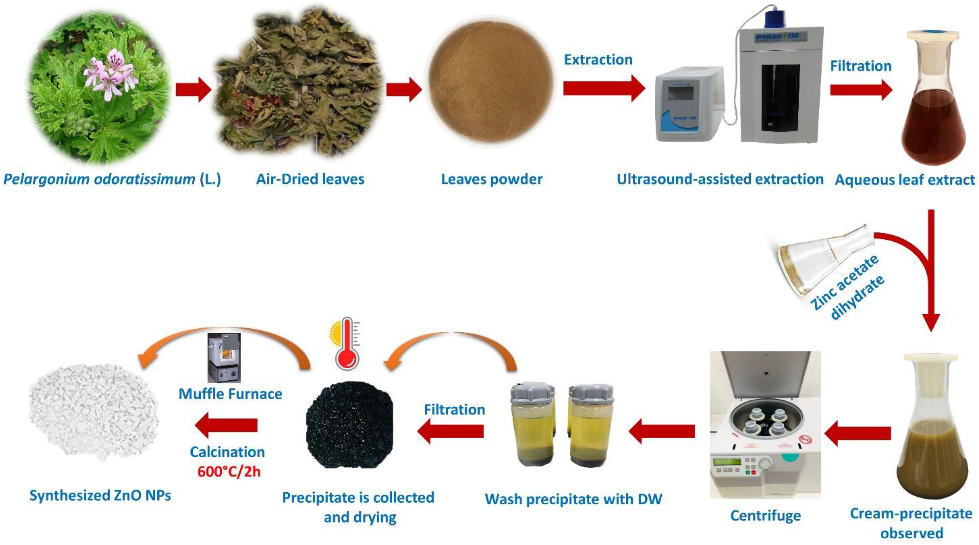
2.7. Green Synthesis of ZnO Nanoparticles
2.8. Characterization Methods of ZnO NPs
2.8.1. UV-Vis Spectroscopy
2.8.2. Dynamic Light Scattering (DLS)
2.8.3. Fourier Transform Infra-Red Spectroscopy (FTIR)
2.8.4. X-ray Diffraction (XRD)
2.8.5. Field Emission-Scanning Electron Microscopy (FE-SEM)
2.8.6. High-Resolution Transmission Electron Microscopy (HRTEM)
2.9. Estimation of Antioxidant Activity—DPPH Radical Scavenging Activity
2.10. Estimation of Antibacterial Activity
2.10.1. Bacteria Strains
2.10.2. Antibacterial Assay
2.11. Estimation of Anti-inflammatory Activity
2.12. Statistical Analysis
3. Results and Discussion
3.1. Qualitative Phytochemical Screening (QPS)
3.2. HPLC-Analysis
3.3. Characterization of ZnO NPs
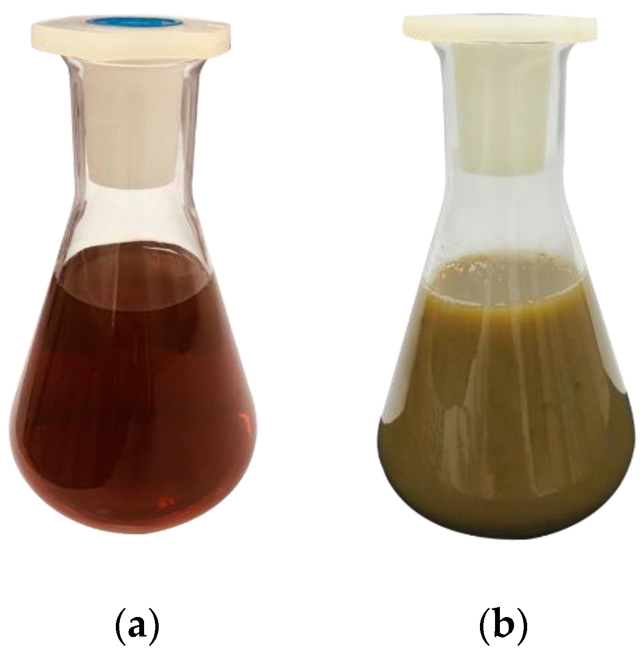
3.3.1. Visual Observation
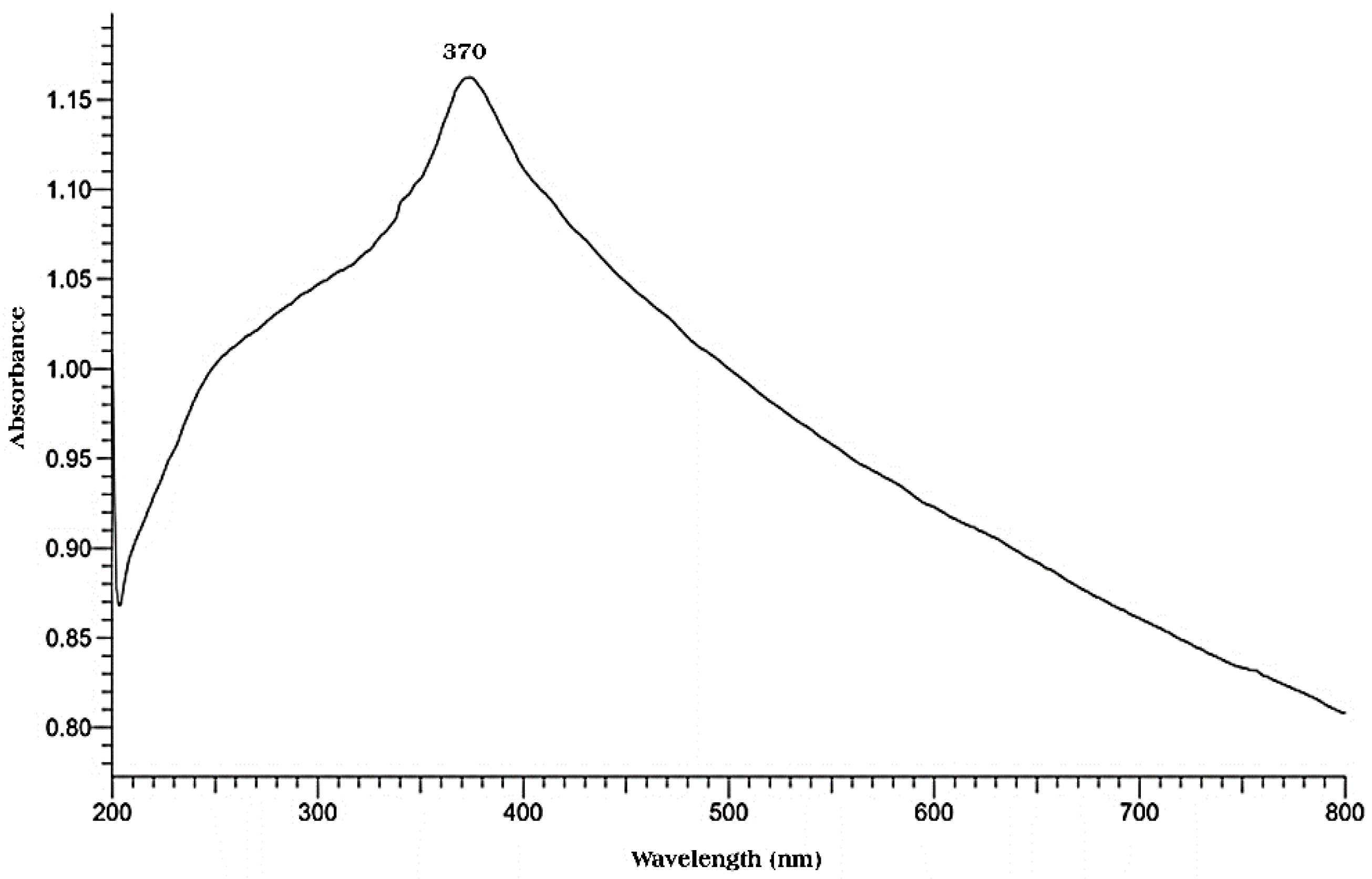
3.3.2. UV-Vis Spectroscopy
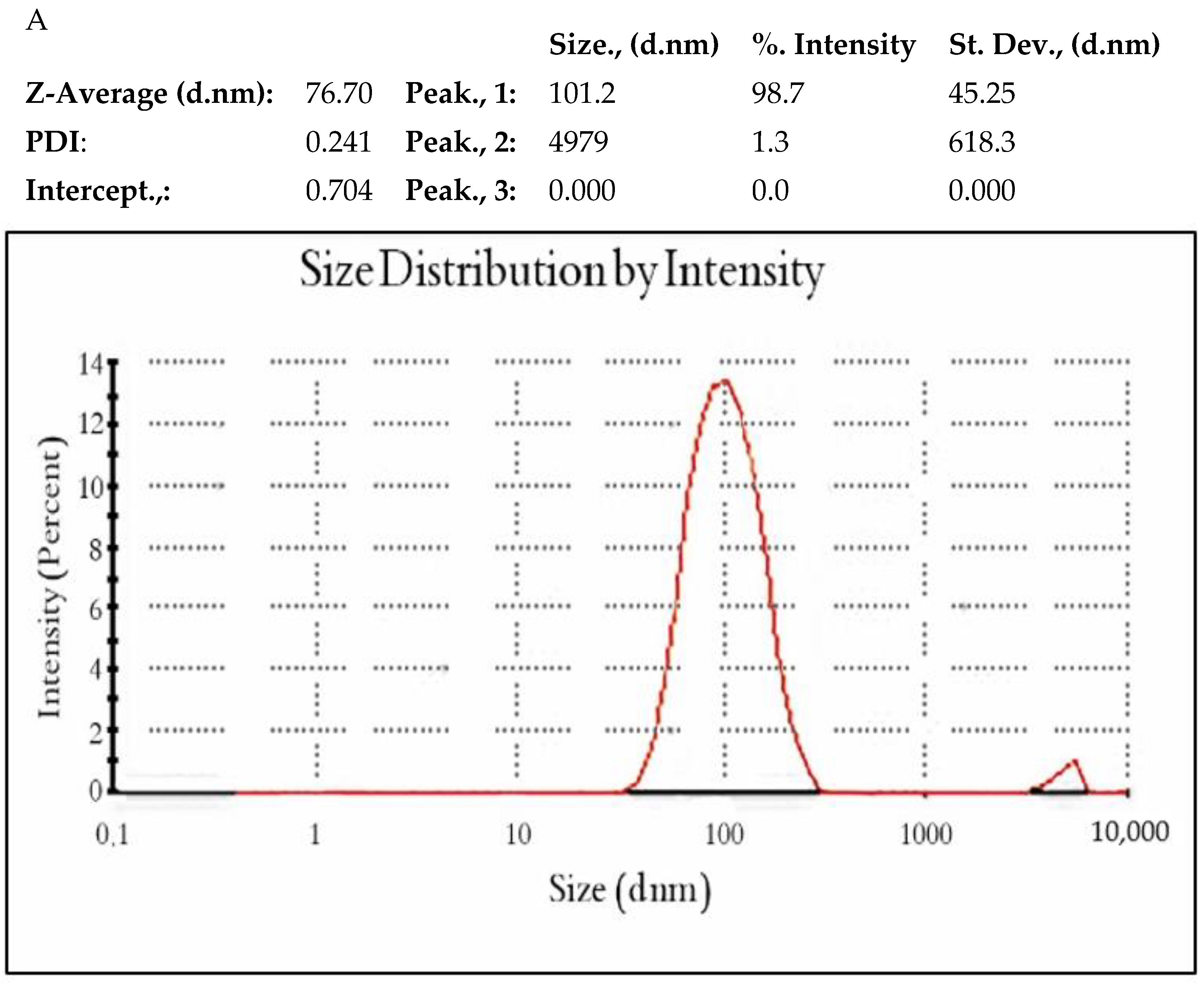
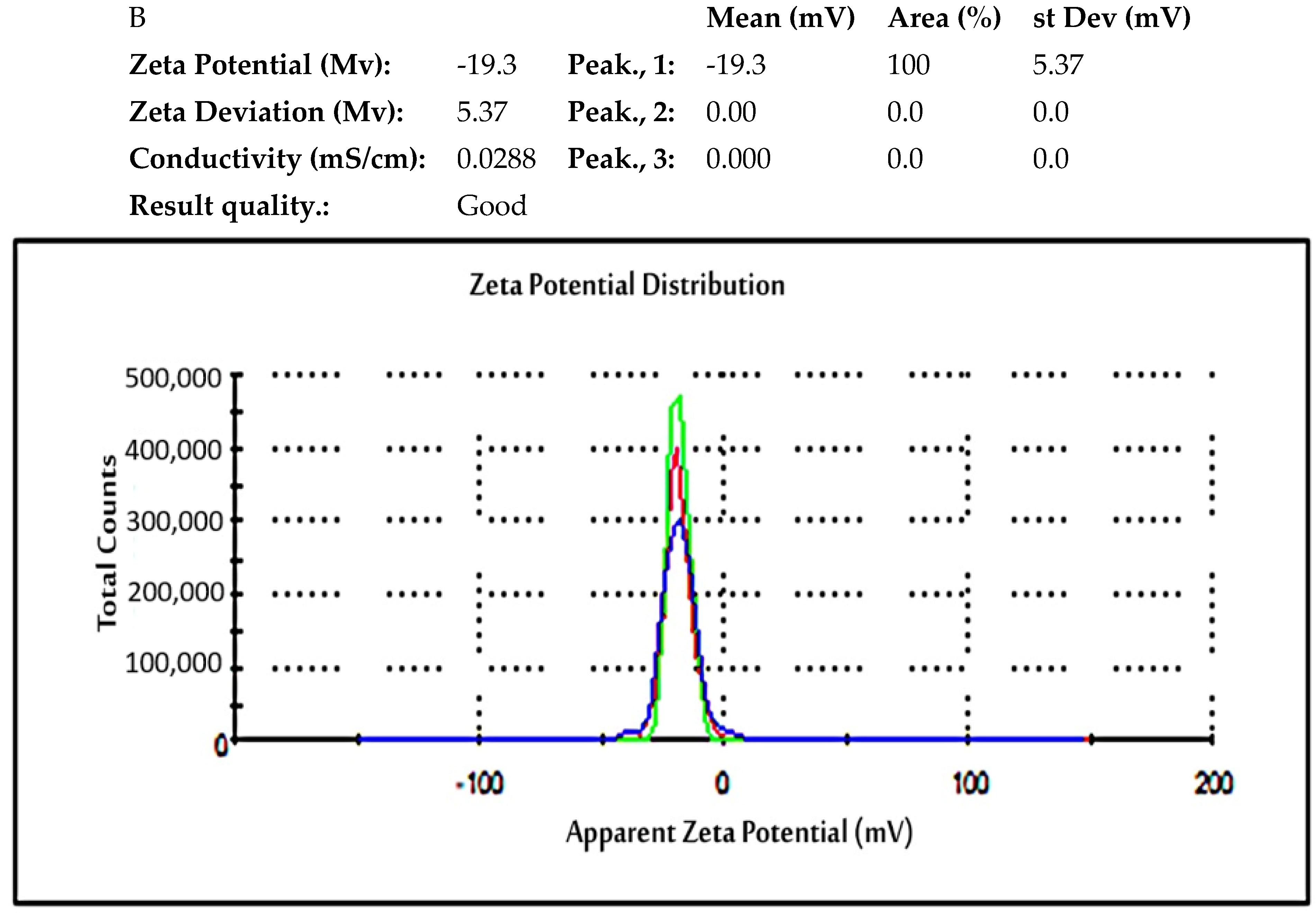
3.3.3. Dynamic Light Scattering (DLS)
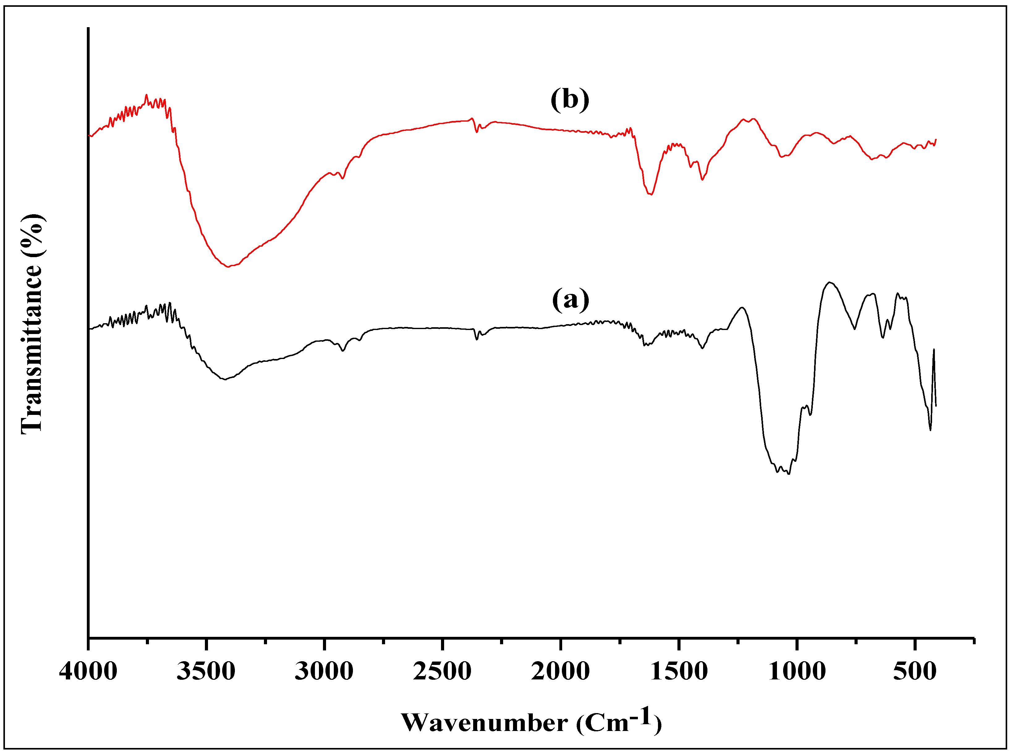
3.3.4. FTIR Analysis of Biosynthesized ZnO NPs and P. odoratissimum ALE
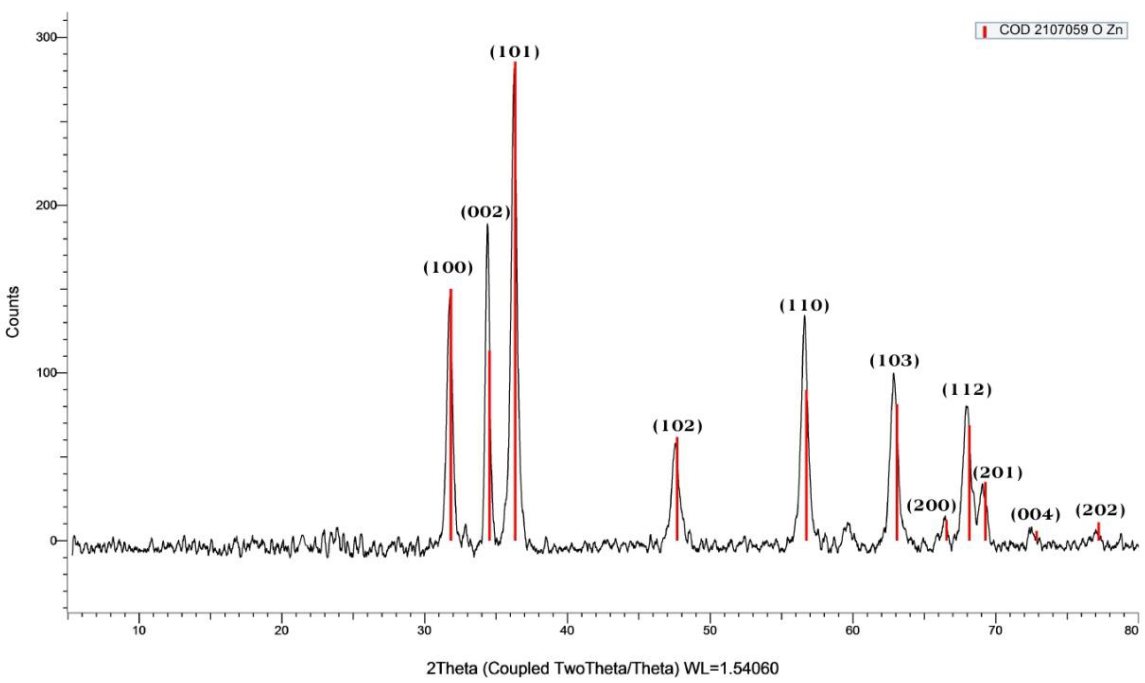
3.3.5. X-ray Diffraction (XRD) Analysis of ZnO NPs
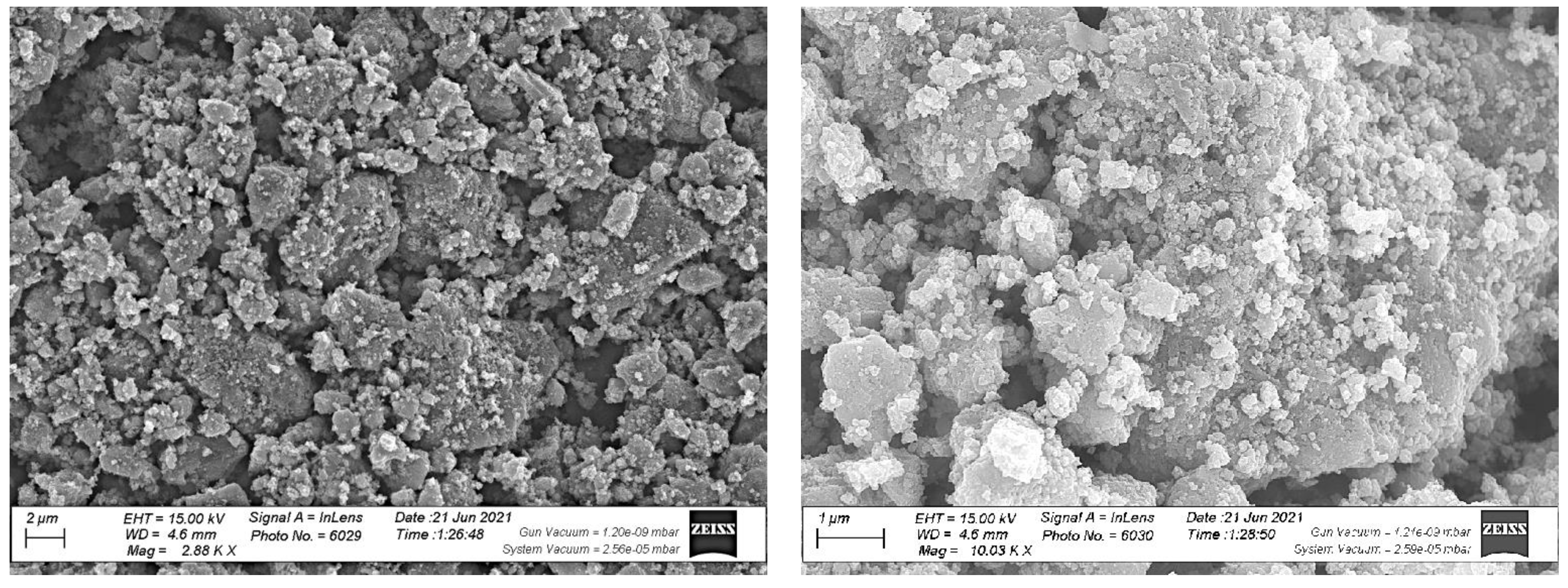
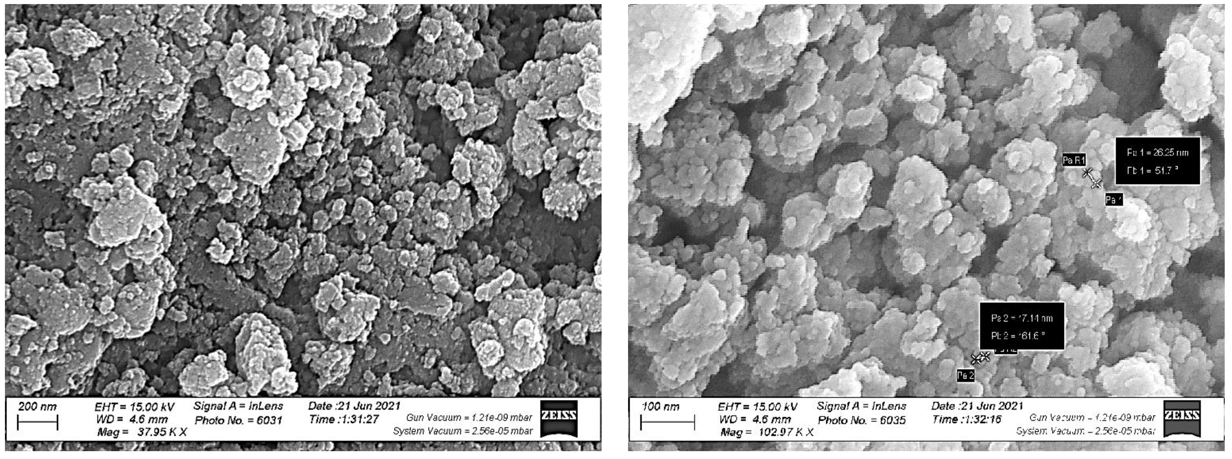
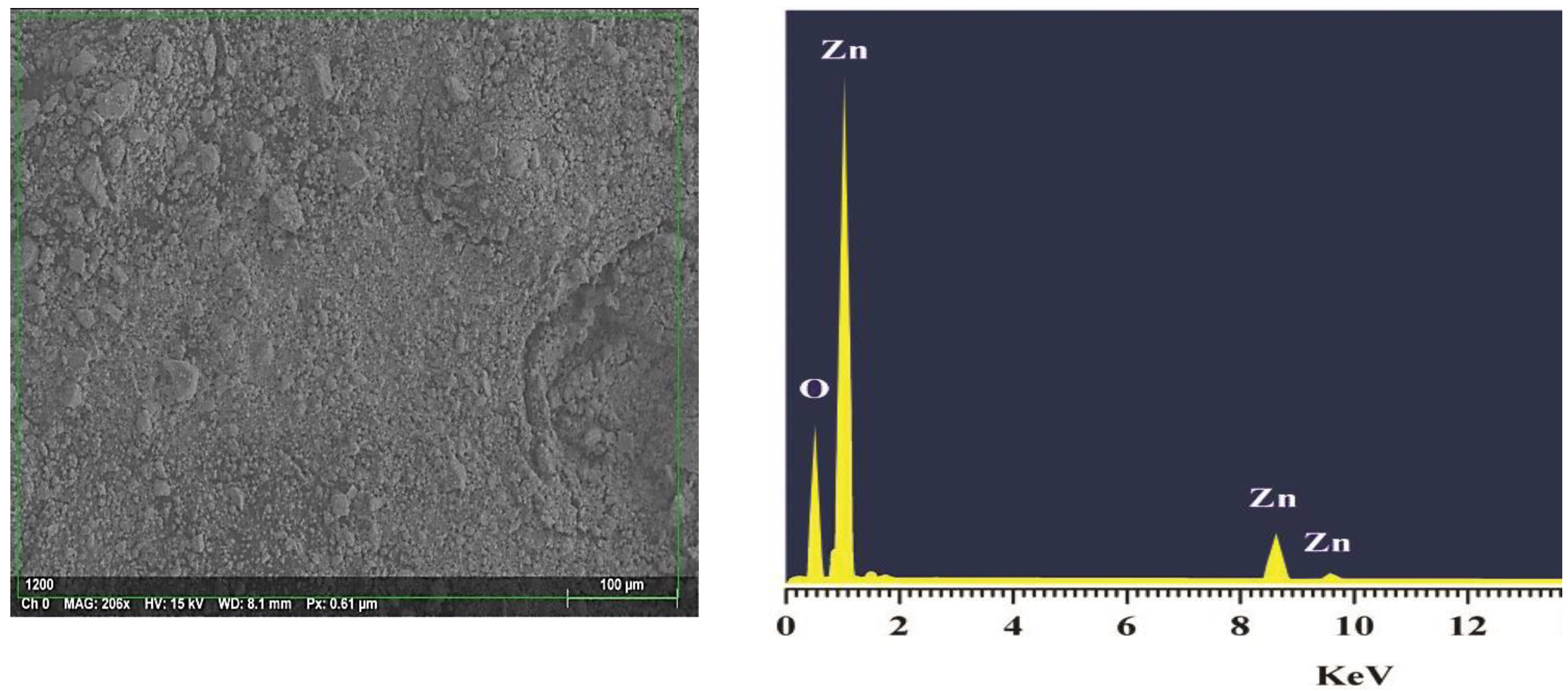
3.3.6. FE-SEM of ZnO NPs
3.3.7. Energy Dispersive X-ray Analysis (EDX) Spectrum of ZnO NPs
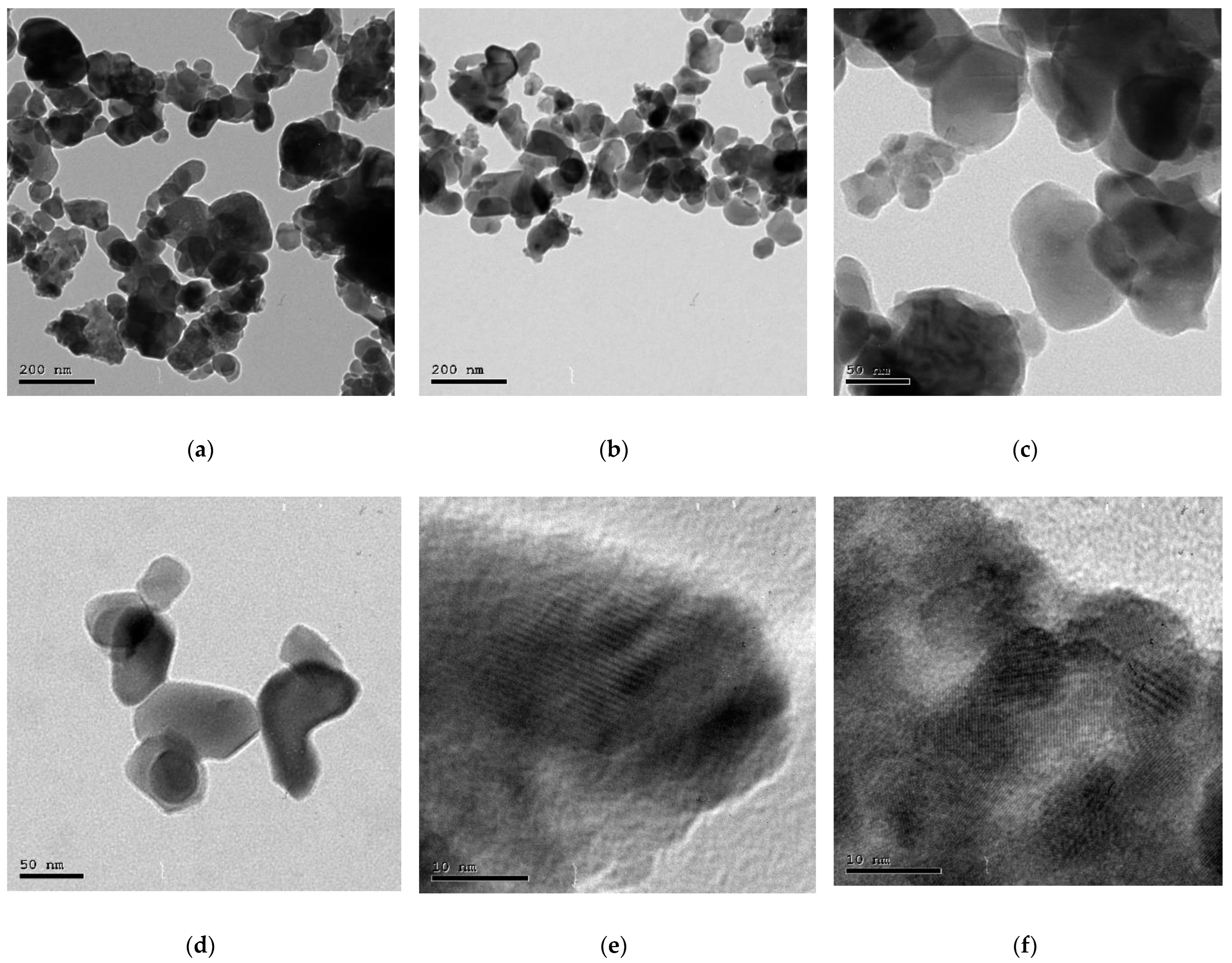
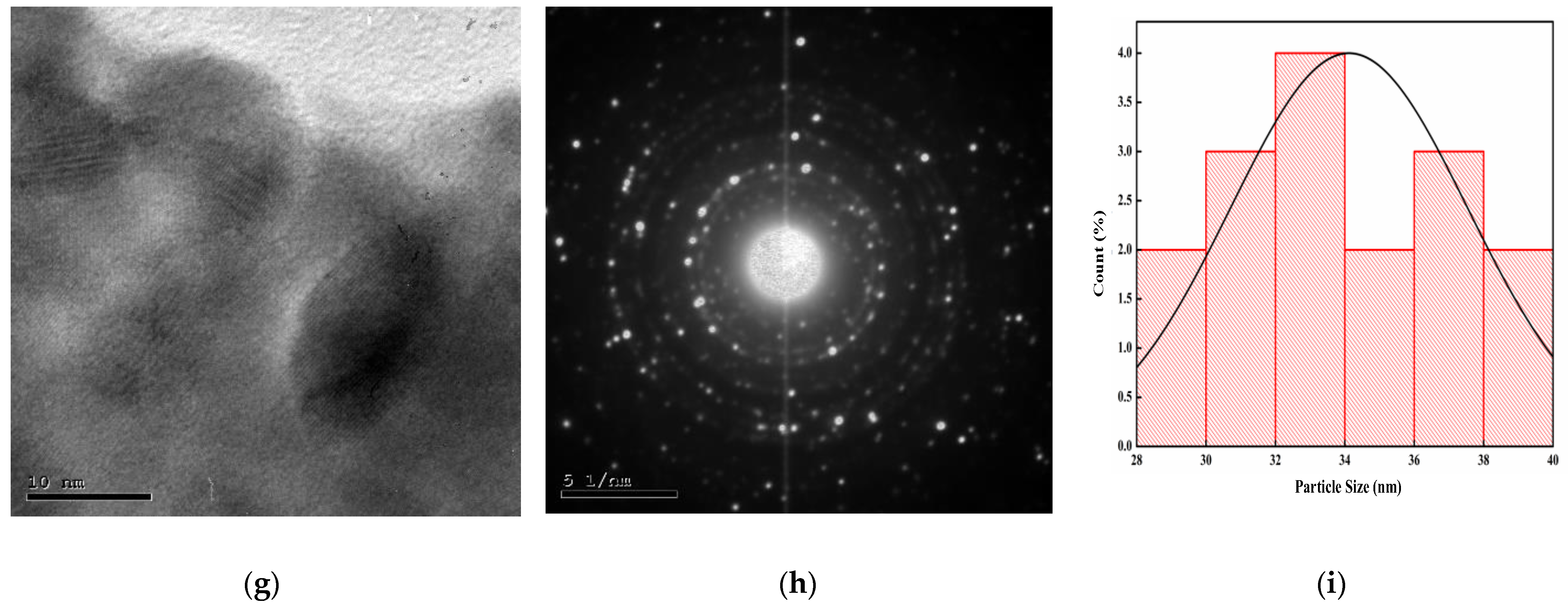
3.3.8. HR-TEM of ZnO NPs
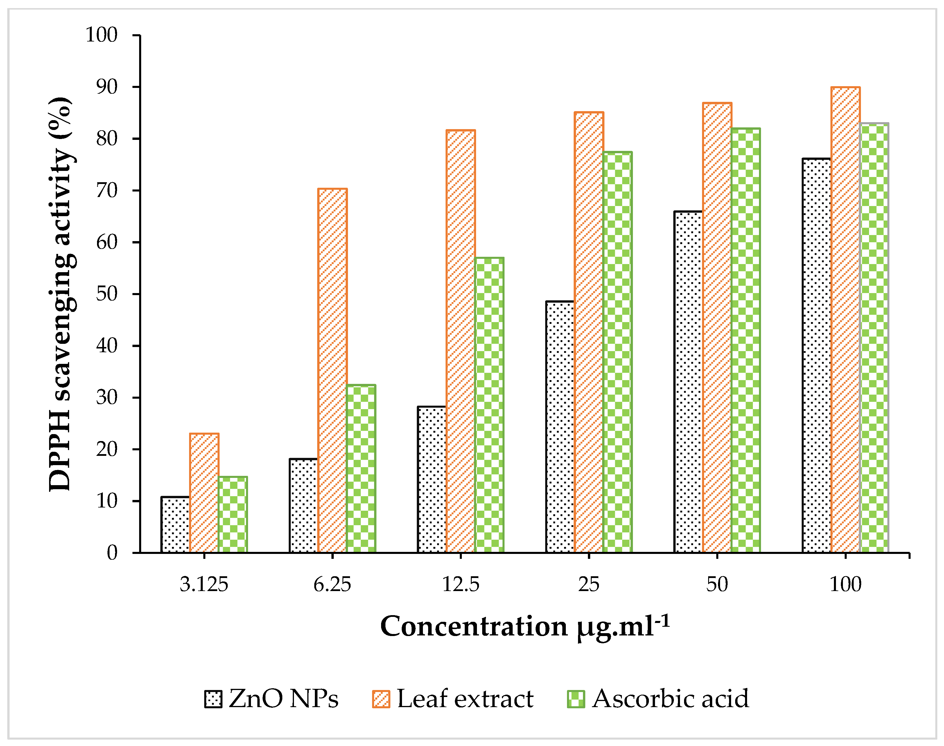
3.4. Antioxidant Activity
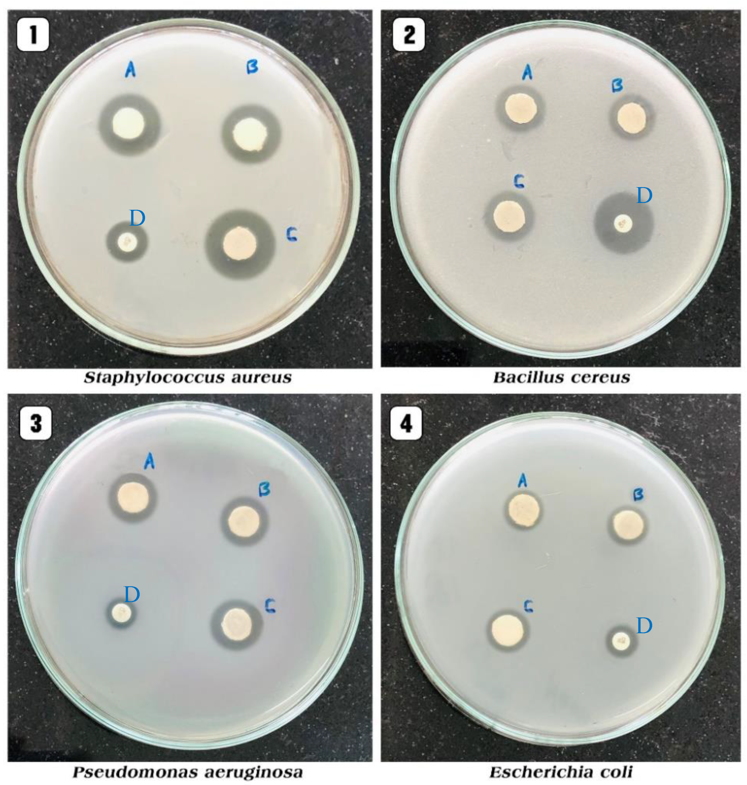
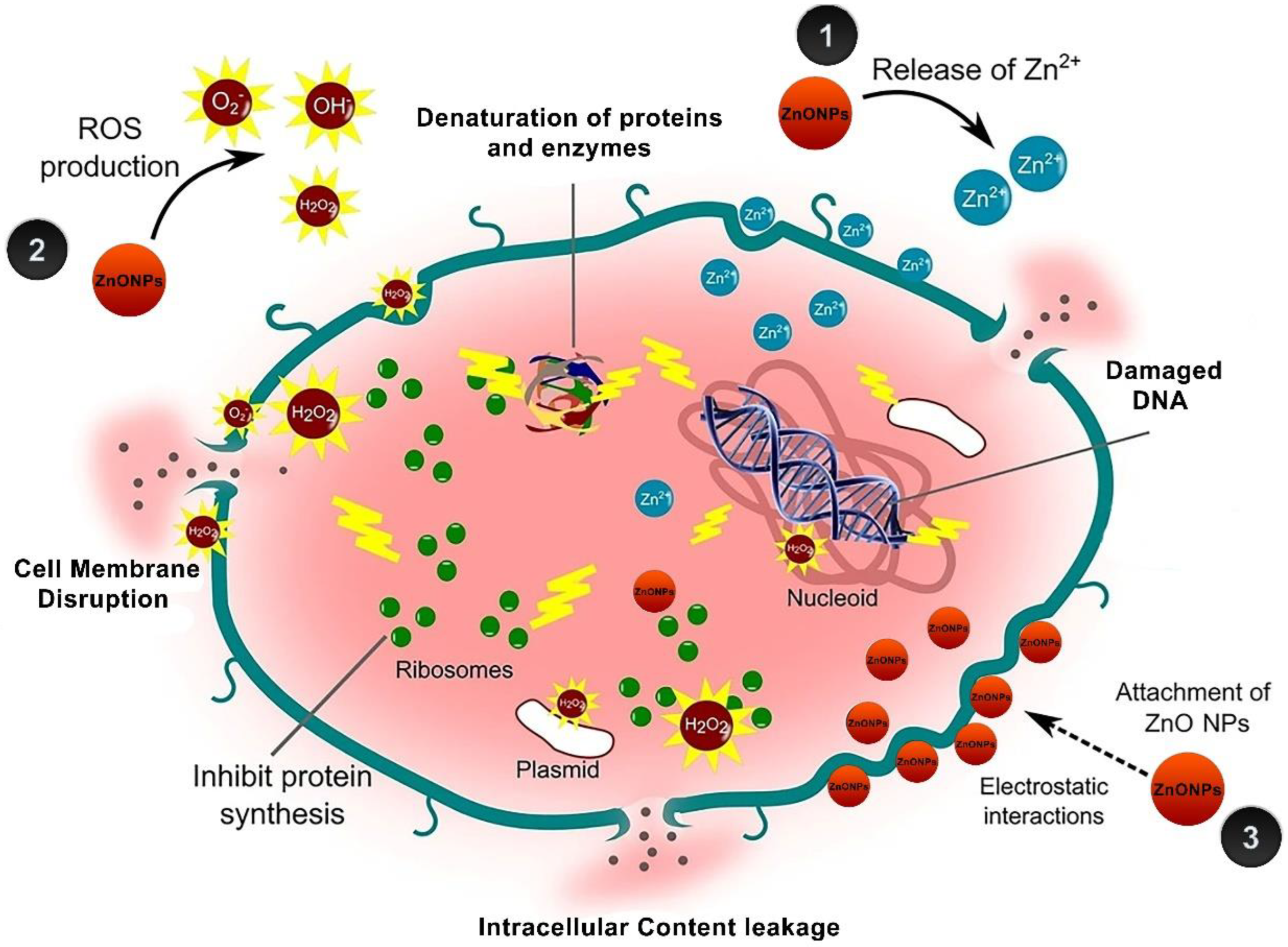
3.5. Antibacterial Activity
3.6. Anti-inflammatory Activity
4. Conclusions
Author Contributions
Funding
Institutional Review Board Statement
Informed Consent Statement
Data Availability Statement
Conflicts of Interest
References
- Bhardwaj, A.; Sharma, G.; Gupta, S. Nanotechnology Applications and Synthesis of Graphene as Nanomaterial for Nanoelectronics. In Nanomaterials and Environmental Biotechnology; Bhushan, I., Singh, V., Tripathi, D., Eds.; Springer: Cham, Switzerland, 2020; pp. 251–269. [Google Scholar]
- Dash, D.K.; Panik, R.K.; Sahu, A.K.; Tripathi, V. Role of Nanobiotechnology in Drug Discovery Development and Molecular Diagnostic. In Applications of Nanobiotechnology; Stoytcheva, M., Zlatev, R., Eds.; IntechOpen: London, UK, 2020; pp. 37–43. [Google Scholar]
- Patil, M.P.; Kim, G.D. Eco-friendly approach for nanoparticles synthesis and mechanism behind antibacterial activity of silver and anticancer activity of gold nanoparticles. Appl. Microbiol. Biotechnol. 2017, 101, 79–92. [Google Scholar] [CrossRef] [PubMed]
- Phull, A.R.; Abbas, Q.; Ali, A.; Raza, H.; Zia, M.; Haq, I.U. Antioxidant cytotoxic and antimicrobial activities of green synthesized silver nanoparticles from crude extract of Bergenia ciliata. J. Pharm. Sci. 2016, 2, 31–36. [Google Scholar] [CrossRef]
- Khan, I.; Saeed, K.; Khan, I. Nanoparticles: Properties applications and toxicities. Arab. J. Chem. 2019, 12, 908–931. [Google Scholar] [CrossRef]
- Nilavukkarasi, M.; Vijayakumar, S.; Prathipkumar, S. Capparis zeylanica mediated bio-synthesized ZnO nanoparticles as antimicrobial photocatalytic and anti-cancer applications. Mater. Sci. Technol. 2020, 3, 335–343. [Google Scholar] [CrossRef]
- Zheng, X.; Yuhui, W.; Ling, S.; Arunachalam, C.; Sulaiman, A.; Liwei, F. Anticarcinogenic effect of zinc oxide nanoparticles synthesized from Rhizoma paridis saponins on Molt-4 leukemia cells. J. King Saud Univ. Sci. 2020, 32, 1865–1871. [Google Scholar]
- Waseem, A.; Divya, K. Green synthesis characterization and anti-microbial activities of ZnO nanoparticles using Euphorbia hirta leaf extract. J. King Saud Univ. Sci. 2020, 32, 2358–2364. [Google Scholar]
- Bekele, E.T.; Gonfa, B.A.; Zelekew, O.A.; Belay, H.H.; Sabir, F.K. Synthesis of titanium oxide nanoparticles using root extract of kniphofia foliosa as a template characterization and its application on drug resistance bacteria. J. Nanomater. 2020, 2020, 2817037. [Google Scholar] [CrossRef]
- Vijayakumar, S.; Vaseeharan, B.; Malaikozhundan, B.; Shobiya, M. Laurus nobilis leaf extract mediated green synthesis of ZnO nanoparticles: Characterization and biomedical applications. Biomed. Pharm. 2016, 84, 1213–1222. [Google Scholar] [CrossRef]
- Mauricio, M.D.; Guerra-Ojeda, S.; Marchio, P.; Valles, S.L.; Aldasoro, M.; Escribano-Lopez, I.; Herance, J.R.; Rocha, M.; Vila, J.M.; Victor, V.M. Nanoparticles in medicine: A focus on vascular oxidative stress. Oxid. Med. Cell Longev. 2018, 2018, 6231482. [Google Scholar] [CrossRef] [Green Version]
- Seyyed, M.; Tabrizi, H.M.; Behrouz, E.; Vahid, J. Biosynthesis of pure zinc oxide nanoparticles using Quince seed mucilage for photocatalytic dye degradation. J. Alloys Compd. 2020, 821, 153519–153527. [Google Scholar]
- Rasli, N.I.; Basri, H.; Harun, Z. Zinc oxide from Aloe vera extract: Two-level factorial screening of biosynthesis parameters. Heliyon 2020, 6, e03156. [Google Scholar] [CrossRef] [PubMed] [Green Version]
- Vijayakumar, S.; Arulmozhi, P.; Kumar, N.; Sakthivel, B.; Prathip Kumar, S.; Praseetha, P.K. Acalypha fruticose L. leaf extract mediated synthesis of ZnO nanoparticles: Characterization and antimicrobial activities. Mater. Today Proc. 2020, 23, 73–80. [Google Scholar] [CrossRef]
- Muthuvel, A.; Jothibas, M.; Manoharan, C. Effect of chemically synthesis compared to biosynthesized ZnO-NPs using Solanum nigrum leaf extract and their photocatalytic antibacterial and in vitro antioxidant activity. J. Environ. Chem. Eng. 2020, 8, 103705. [Google Scholar] [CrossRef]
- Niranjan, B.; Saha, S.; Chakraborty, M.; Das, S.; Basub, R.; Nandyc, P. Green synthesis of zinc oxide nanoparticles using Hibiscus subdariffa leaf extract: Effect of temperature on synthesis anti-bacterial activity and anti-diabetic activity. RSC Adv. 2015, 5, 4993–5003. [Google Scholar]
- Yusof, H.M.; Mohamad, R.; Zaidan, U.H.; Rahman, N.A.A. Microbial synthesis of zinc oxide nanoparticles and their potential application as an antimicrobial agent and a feed supplement in animal industry: A review. J. Anim. Sci. Biotechnol. 2019, 10, 1–22. [Google Scholar]
- Bandeira, M.; Giovanela, M.; Roesch-Ely, M.; Devine, D.; da Silva Crespo, J. Green synthesis of zinc oxide nanoparticles: A review of the synthesis methodology and mechanism of formation. Sustain. Chem. Pharm. 2020, 15, 100223. [Google Scholar] [CrossRef]
- Davar, F.; Majedi, A.; Mirzaei, A. Green synthesis of ZnO nanoparticles and its application in the degradation of some dyes. J. Am. Ceram. Soc. 2015, 98, 1739–1746. [Google Scholar] [CrossRef]
- Prasad, K.S.; Prasad, S.K.; Ansari, M.A.; Alzohairy, M.A.; Alomary, M.N.; AlYahya, S.; Shivamallu, C. Tumoricidal and bactericidal properties of ZnONPs synthesized using Cassia auriculata leaf extract. Biomolecules 2020, 10, 982. [Google Scholar] [CrossRef]
- Chaudhary, A.; Kumar, N.; Kumar, R.; Kumar, R. Antimicrobial activity of zinc oxide nanoparticles synthesized from Aloe vera peel extract. SN Appl. Sci. 2019, 1, 136. [Google Scholar] [CrossRef] [Green Version]
- Shekhawat, M.S.; Ravindran, C.P.; Manokari, M. Biogenic production of zinc oxide nanoparticles from aqueous extracts of Duranta erecta L. World Sci. News 2016, 28, 30. [Google Scholar]
- Mohammad, A.A.; Mahadevamurthy, M.; Daruka, P. Cinnamomum verum bark extract mediated green synthesis of ZnO nanoparticles and their antibacterial potentiality. Biomolecules 2020, 10, 134–336. [Google Scholar]
- Sharmila, G.; Muthukumaran, C.; Sandiya, K.S. Biosynthesis characterization and antibacterial activity of zinc oxide nanoparticles derived from Bauhinia tomentosa leaf extract. J. Nanostructure Chem. 2018, 8, 293–299. [Google Scholar] [CrossRef] [Green Version]
- Elumalai, K.; Velmurugan, S.; Ravi, K. Bio-approach: Plant mediated synthesis of ZnO nanoparticles and their catalytic reduction of methylene blue and antimicrobial activity. Adv. Powder Technol. 2015, 26, 1639–1651. [Google Scholar] [CrossRef]
- Elumalai, K.; Velmurugan, S.; Ravi, S.; Kathiravan, V.; Ashokkumar, S. Green synthesis of Zinc oxide nanoparticles using Moringa oleifera leaf extract and evaluation of its antimicrobial activity. Spectrochim. Spectrochim. Acta Part A Mol. Biomol. Spectrosc. 2015, 143, 158–164. [Google Scholar] [CrossRef]
- Handago, D.T.; Enyew, A.Z.; Bedasa, A.G. Effects of Azadirachta indica leaf extract capping agents on the synthesis of pure and Cu doped ZnO-nanoparticles: A green approach and microbial activity. Open Chem. J. 2019, 17, 246–465. [Google Scholar] [CrossRef]
- Haque, M.J.; Bellah, M.M.; Hassan, M.R.; Rahman, S. Synthesis of ZnO nanoparticles by two different methods & comparison of their structural antibacterial photocatalytic and optical properties. Nano Express 2020, 1, 010007. [Google Scholar]
- Suresh, D.; Shobharani, R.M.; Nethravathi, P.C.; Kumar, M.A.P.; Nagabhushana, H.; Sharma, S. Artocarpus gomezianus aided green synthesis of ZnO nanoparticles: Luminescence photocatalytic and antioxidant properties. Spectrochim. Spectrochim. Acta Part A Mol. Biomol. Spectrosc. 2015, 141, 128–164. [Google Scholar] [CrossRef]
- Solabomi, O.O.; Yasmine, A.; Muchen, Z. Green synthesis of zinc oxide nanoparticles using different plant extracts and their antibacterial activity against Xanthomonas oryzae pv. Oryzae. Artif. Cells Nanomed Biotechnol. 2019, 47, 341–352. [Google Scholar]
- Nash, K.M.; Ahmed, S. Nanomedicine in the ROS-Mediated Pathophysiology: Applications and Clinical Advances. Nanomed. Nanotechnol. Biol. Med. 2015, 11, 2033–2040. [Google Scholar] [CrossRef] [Green Version]
- Cragg, G.M.; Newman, D.J.; Snader, K.M. Natural Products in Drug Discovery and Development. J. Nat. Prod. 1997, 60, 52–60. [Google Scholar] [CrossRef]
- Wang, L.; Hu, C.; Shao, L. The Antimicrobial Activity of Nanoparticles: Present Situation and Prospects for the Future. Int. J. Nanomed. 2017, 12, 1227. [Google Scholar] [CrossRef] [PubMed] [Green Version]
- Bown, D. The Royal Horticultural Society Encyclopedia of Herbs & Their Uses; Dorling Kindersley Limited: London, UK, 1995. [Google Scholar]
- Moutaouafiq, S.; Farah, A.; Ez zoubi, Y.; Ghanmi, M.; Satrani, B.; Bousta, D. Antifungal activity of Pelargonium graveolens essential oil and its fractions against wood decay fungi. J. Essent. Oil-Bear Plants 2019, 22, 1104–1114. [Google Scholar] [CrossRef]
- Carmen, G.; Hancu, G. Antimicrobial and antifungal activity of Pelargonium roseum essential oils. Adv. Pharm. Bull. 2014, 4, 511. [Google Scholar] [PubMed]
- Ben Slima, A.; Ali, M.B.; Barkallah, M.; Traore, A.I.; Boudawara, T.; Allouche, N.; Gdoura, R. Antioxidant properties of Pelargonium graveolens L’Her essential oil on the reproductive damage induced by deltamethrin in mice as compared to alpha-tocopherol. Lipids Health Dis. 2013, 12, 30. [Google Scholar] [CrossRef] [Green Version]
- Mnif, W.; Dhifi, W.; Jelali, N.; Baaziz, H.; Hadded, A.; Hamdi, N. Characterization of leaves essential oil of Pelargonium graveolens originating from Tunisia: Chemical composition antioxidant and biological activities. J. Essent. Oil-Bear Plants 2011, 14, 761–769. [Google Scholar] [CrossRef]
- Kolodziej, H. Traditionally used Pelargonium species: Chemistry and biological activity of umckaloabo extracts and their constituents. Curr. Top. Phytochem. 2000, 3, 77–93. [Google Scholar]
- Abdelbaky, A.S.; Diab, Y.M. Effect of various extraction methods and solvent types on yield phenolic and flavonoid content and antioxidant activity of Spathodea nilotica leaves. Egypt J. Chem. 2021, 64, 7483–7489. [Google Scholar] [CrossRef]
- Harbone, J.B. Phytochemical Methods; Chapman and Hall: London, UK, 1998; pp. 117–119. [Google Scholar]
- Farnsworth, N.R. Biological and phytochemical screening of plants. J. Pharm. Sci. 1966, 55, 225–276. [Google Scholar] [CrossRef]
- Rangari, V.D. Pharmacognosy and Phytochemistry; Carrier Publication: Nashik, Indian, 2002; p. 132. [Google Scholar]
- Yu, L.; Haley, S.; Perret, J.; Harris, M.; Wilson, J.; Qian, M. Free radical scavenging properties of wheat extracts. J. Agric. Food Chem. 2002, 50, 1619–1624. [Google Scholar] [CrossRef]
- Lamaison, J.L.C.; Carnet, A. Contents in main flavonoid compounds of Crataegus monogyna Jacq. and Crataegus laevigata (Poiret) D.C. flowers and leaves at different plant development stages. Pharm. Acta Helv. 1990, 65, 315–320. [Google Scholar]
- Brand-Williams, W.; Cuvelier, M.E.; Berset, C. Use of a free radical method to evaluate antioxidant activity. Lebensm Wissenchaft Technol. 1995, 28, 25–30. [Google Scholar] [CrossRef]
- Bauer, A.W.; Kirby, M.M.; Sherris, J.C.; Turck, M. Antibiotic susceptibility testing by a standardized single disk method. Amr. J. Clin. Pathol. 1966, 45, 493–496. [Google Scholar] [CrossRef]
- Anosike, C.A.; Obidoa, O.; Ezeanyika, L.U. Membrane stabilization as a mechanism of the anti-inflammatory activity of methanol extract of garden egg (Solanum aethiopicum). DARU J. Pharm. Sci. 2012, 20, 76. [Google Scholar] [CrossRef] [PubMed] [Green Version]
- Iravani, S.; Korbekandi, H.; Mirmohammadi, S.V.; Zolfaghari, B. Synthesis of silver nanoparticles: Chemical physical and biological methods. Res. Pharm. Sci. 2014, 9, 385–406. [Google Scholar] [PubMed]
- Abdelbaky, A.S.; Mohamed, A.M.H.A.; Alharthi, S.S. Antioxidant and Antimicrobial Evaluation and Chemical Investigation of Rosa Gallica var. aegyptiaca Leaf Extracts. Molecules 2021, 26, 6498. [Google Scholar] [CrossRef]
- Senthilkumar, N.; Nandhakumar, E.; Priya, P.; Soni, D.; Vimalan, M.; Potheher, I.V. Synthesis of ZnO nanoparticles using leaf extract of Tectona grandis (L.) and their anti-bacterial anti-arthritic anti-oxidant and in vitro cytotoxicity activities. New J. Chem. 2017, 41, 10347–10356. [Google Scholar] [CrossRef]
- Zak, A.K.; Majid, W.A.; Mahmoudian, M.R.; Darroudi, M.; Yousefi, R. Starch-stabilized synthesis of ZnO nanopowders at low temperature and optical properties study. Adv. Powder Technol. 2013, 24, 618–624. [Google Scholar]
- Zak, A.K.; Yousefi, R.; Abd Majid, W.H.; Muhamad, M.R. Facile synthesis and X-ray peak broadening studies of Zn1− xMgxO nanoparticles. Ceram Int. 2012, 38, 2059–2064. [Google Scholar]
- Suresh, D.; Nethravathi, P.C.; Rajanaika, H.; Nagabhushana, H.; Sharma, S.C. Green synthesis of multifunctional zinc oxide (ZnO) nanoparticles using Cassia fistula plant extract and their photodegradative antioxidant and antibacterial activities. Mater. Sci. Semicond. Process. 2015, 31, 446–454. [Google Scholar] [CrossRef]
- Akbar, N.; Aslam, Z.; Siddiqui, R.; Shah, M.R.; Khan, N.A. Zinc oxide nanoparticles conjugated with clinically-approved medicines as potential antibacterial molecules. AMB Express 2021, 11, 104. [Google Scholar] [CrossRef]
- Badran, M. Formulation and in vitro evaluation of flufenamic acid loaded deformable liposome for improved skin delivery. Dig. J. Nanomater. Biostruct. 2014, 9, 83–91. [Google Scholar]
- Chen, M.; Liu, X.; Fahr, A. Skin penetration and deposition of carboxyfluorescein and temoporfin from different lipid vesicular systems: In vitro study with finite and infinite dosage application. Int. J. Pharm. 2011, 408, 223–234. [Google Scholar] [CrossRef] [PubMed]
- Putri, D.C.; Dwiastuti, R.; Marchaban, M.; Nugroho, A.K. Optimization of mixing temperature and sonication duration in liposome preparation. J. Pharm. Sci. Commun. 2017, 14, 79–85. [Google Scholar] [CrossRef] [Green Version]
- Kätzel, U.; Vorbau, M.; Stintz, M.; Gottschalk-Gaudig, T.; Barthel, H. Dynamic light scattering for the characterization of polydisperse fractal systems: II. Relation between structure and DLS results. Part. Part. Syst. Charact. 2008, 25, 19–30. [Google Scholar] [CrossRef]
- Kokila, K.; Elavarasan, N.; Sujatha, V. Diospyros montana leaf extract-mediated synthesis of selenium nanoparticles and their biological applications. New J. Chem. 2017, 41, 7481–7490. [Google Scholar] [CrossRef]
- Jafarirad, S.; Mehrabi, M.; Divband, B.; Kosari-Nasab, M. Biofabrication of zinc oxide nanoparticles using fruit extract of Rosa canina and their toxic potential against bacteria: A mechanistic approach. Mater. Sci. Eng. C 2016, 59, 296–302. [Google Scholar] [CrossRef] [PubMed]
- Awwad, A.M.; Albiss, B.; Ahmad, A.L. Green synthesis characterization and optical properties of zinc oxide nanosheets using Olea europea leaf extract. Adv. Mater. Lett. 2014, 5, 520–524. [Google Scholar] [CrossRef]
- El-Belely, E.F.; Farag, M.; Said, H.A.; Amin, A.S.; Azab, E.; Gobouri, A.A.; Fouda, A. Green synthesis of zinc oxide nanoparticles (ZnO-NPs) using Arthrospira platensis (Class: Cyanophyceae) and evaluation of their biomedical activities. Nanomater 2021, 11, 95. [Google Scholar] [CrossRef]
- Pavithra, G. Leonotis nepetifolia mediated eco-friendly synthesis of zno nps: Photocatalytic antioxidant activities and their applications to nano-composite electrode material for supercapacitor. Heliyon 2021. preprint. [Google Scholar] [CrossRef]
- Jamdagni, P.; Poonam, K.; Rana, J.S. Green synthesis of zinc oxide nanoparticles using flower extract of Nyctanthes arbortristis and their antifungal activity. J. King Saud Univ. Sci. 2018, 30, 168–175. [Google Scholar] [CrossRef] [Green Version]
- Yuvakkumar, R.; Suresh, J.; Saravanakumar, B.; Nathanael, A.J.; Hong, S.I.; Rajendran, V. Rambutan peels promoted biomimetic synthesis of bioinspired zinc oxide nanochains for biomedical applications. Spectrochim. Spectrochim. Acta Part A Mol. Biomol. Spectrosc. 2015, 137, 250–258. [Google Scholar] [CrossRef] [PubMed]
- Alamdari, S.; Sasani Ghamsari, M.; Lee, C.; Han, W.; Park, H.H.; Tafreshi, M.J.; Ara, M.H.M. Preparation and characterization of zinc oxide nanoparticles using leaf extract of Sambucus ebulus. Appl. Sci. 2020, 10, 3620. [Google Scholar] [CrossRef]
- Nagarajan, S.; Arumugam Kuppusamy, K. Extracellular synthesis of zinc oxide nanoparticle using seaweeds of gulf of Mannar. India J. Nanobiotechnol. 2013, 11, 39. [Google Scholar] [CrossRef] [PubMed] [Green Version]
- Khari, S.; Jamzad, M.; Kabiri Fard, H. Green synthesis of zinc oxide nanoparticles: A comparison. Green Chem. Lett. Rev. 2019, 12, 19–24. [Google Scholar]
- Buazar, F.; Bavi, M.; Kroushawi, F.; Halvani, M.; Khaledi-Nasab, A.; Hossieni, S.A. Potato extract as reducing agent and stabilizer in a facile green one-step synthesis of ZnO nanoparticles. J. Exp. Nanosci. 2016, 11, 175–184. [Google Scholar] [CrossRef] [Green Version]
- Zhang, G.; Shen, X.; Yang, Y. Facile synthesis of monodisperse porous ZnO spheres by a soluble starch-assisted method and their photocatalytic activity. J. Phys. Chem. C 2011, 115, 7145–7152. [Google Scholar] [CrossRef]
- Albertsson, J.; Abrahams, S.C.; Kvick, Å. Atomic displacement anharmonic thermal vibration expansivity and pyroelectric coefficient thermal dependences in ZnO. Acta Crystallogr. B Struct. Sci. 1989, 45, 34–40. [Google Scholar] [CrossRef]
- Venkateasan, A.; Prabakaran, R.; Sujatha, V. Phytoextract-mediated synthesis of zinc oxide nanoparticles using aqueous leaves extract of Ipomoea pescaprae (L). R. br revealing its biological properties and photocatalytic activity. Nanotechnol. Environ. Eng. 2017, 2, 8. [Google Scholar] [CrossRef]
- Patterson, A.L. The Scherrer formula for X-ray particle size determination. Phys. Rev. 1939, 56, 978. [Google Scholar] [CrossRef]
- Barzinjy, A.A.; Azeez, H.H. Green synthesis and characterization of zinc oxide nanoparticles using Eucalyptus globulus Labill. leaf extract and zinc nitrate hexahydrate salt. SN Appl. Sci. 2020, 2, 991. [Google Scholar] [CrossRef]
- Khoshhesab, Z.M.; Sarfaraz, M.; Asadabad, M.A. Preparation of ZnO nanostructures by chemical precipitation method. Synth. React. Inorg. M 2011, 41, 814–819. [Google Scholar] [CrossRef]
- Boukhris, M.; Simmonds, M.S.; Sayadi, S.; Bouaziz, M. Chemical composition and biological activities of polar extracts and essential oil of rose-scented geranium Pelargonium graveolens. Phytother. Res. 2013, 27, 1206–1213. [Google Scholar] [CrossRef] [PubMed]
- Luo, F.; Yang, F.; Chen, D.; Megharaj, Z.; Naidu, R. One-step green synthesis of bimetallic Fe/Pd nanoparticles used to degrade Orange II. J. Hazard Mater. 2016, 303, 145–153. [Google Scholar] [CrossRef]
- Weng, X.; Guo, M.; Luo, F.; Chen, Z. One-step green synthesis of bimetallic Fe/Ni nanoparticles by eucalyptus leaf extract: Biomolecules identification characterization and catalytic activity. Chem. Eng. J. 2017, 308, 904–911. [Google Scholar] [CrossRef]
- Mittal, A.K.; Chisti, Y.; Banerjee, U.C. Synthesis of metallic nanoparticles using plant extracts. Biotechnol. Adv. 2013, 31, 346–356. [Google Scholar] [CrossRef]
- Aoki, K.; Chen, J.; Yang, N.; Nagasawa, H. Charge-transfer reactions of silver stearate-coated nanoparticles in suspensions. Langmuir 2003, 19, 9904–9909. [Google Scholar] [CrossRef]
- Sathiyavimal, S.; Vasantharaj, S.; Bharathi, D.; Saravanan, M.; Manikandan, E.; Kumar, S.S.; Pugazhendhi, A. Biogenesis of copper oxide nanoparticles (CuONPs) using Sida acuta and their incorporation over cotton fabrics to prevent the pathogenicity of gram negative and gram positive bacteria. J. Photochem. Photobiol. B Biol. 2018, 188, 126–134. [Google Scholar] [CrossRef]
- Bharathi, D.; Vasantharaj, S.; Bhuvaneshwari, V. Green synthesis of silver nanoparticles using Cordia dichotoma fruit extract and its enhanced antibacterial anti-biofilm and photo catalytic activity. Mater. Res. Express 2018, 5, 055404. [Google Scholar] [CrossRef]
- Getie, S.; Belay, A.; Chandra Reddy, A.R.; Belay, Z. Synthesis and characterizations of zinc oxide nanoparticles for antibacterial applications. J. Nanomed. Nanotechno. 2017, 8, 71–80. [Google Scholar]
- Gunalan, S.; Sivaraj, R.; Rajendran, V. Green synthesized ZnO nanoparticles against bacterial and fungal pathogens. Prog. Nat. Sci. 2012, 22, 693–700. [Google Scholar] [CrossRef] [Green Version]
- Yang, Z.; Xie, C. Zn2+ release from zinc and zinc oxide particles in simulated uterine solution. Colloids Surf. B 2006, 47, 140–145. [Google Scholar] [CrossRef]
- Soren, S.; Kumar, S.; Mishra, S.; Jena, P.K.; Verma, S.K.; Parhi, P. Evaluation of antibacterial and antioxidant potential of the zinc oxide nanoparticles synthesized by aqueous and polyol method. Microb. Pathog. 2018, 119, 145–151. [Google Scholar] [CrossRef] [PubMed]
- Nair, S.; Sasidharan, A.; Divya Rani, V.V.; Menon, D.; Nair, S.; Manzoor, K.; Raina, S. Role of size scale of ZnO nanoparticles and microparticles on toxicity toward bacteria and osteoblast cancer cells. J. Mater. Sci. Mater. Med. 2009, 20, 235–241. [Google Scholar] [CrossRef]
- Agarwal, H.; Menon, S.; Kumar, S.V.; Rajeshkumar, S. Mechanistic study on antibacterial action of zinc oxide nanoparticles synthesized using green route Chem-Biol. Interact 2018, 286, 60–70. [Google Scholar] [CrossRef] [PubMed]
- Jayaseelan, C.; Rahuman, A.A.; Kirthi, A.V.; Marimuthu, S.; Santhoshkumar, T.; Bagavan, A.; Rao, K.B. Novel microbial route to synthesize ZnO nanoparticles using Aeromonas hydrophila and their activity against pathogenic bacteria and fungi. Spectrochim. Acta Part A Mol. Biomol. Spectrosc. 2012, 90, 78–84. [Google Scholar] [CrossRef] [PubMed]
- Mounnissamy, V.M.; Kavimani, S.; Balu, V.; Quine, S.D. Evaluation of Anti-inflammatory and Membrane stabilizing property of Ethanol Extract of Cansjera rheedii J. Gmelin. (Opiliaceae). Iran J. Pharmacol. Ther. 2007, 6, 235–237. [Google Scholar]
- Ferrali, M.; Signorini, C.; Ciccoli, L.; Comporti, M. Iron release and membrane damage in erythrocytes exposed to oxidizing agents phenylhydrazine divicine and isouramil. Biochem. J. 1992, 285, 295–301. [Google Scholar] [CrossRef] [Green Version]
- Halliwell, B.; Whiteman, M. Measuring reactive species and oxidative damage in vivo and in cell culture: How should you do it and what do the results mean. Br. J. Pharmacol. 2004, 142, 231–255. [Google Scholar] [CrossRef] [Green Version]
- Chaitanya, R.; Sandhya, S.; David, B.; Vinod, K.R.; Murali, S. HRBC Membrane Stabilizing Property of Roor Stem and Leaf of Glochidion velutinum. Int. J. Res. Pharmaceut. Biomed. Sci. 2011, 2, 256–259. [Google Scholar]
- Middleton, J.R.E.; Kandaswami, C. Effects of flavonoids on immune and inflammatory cell functions. Biochem. Pharmacol. 1992, 43, 1167–1179. [Google Scholar] [CrossRef]
- Read, M.A. Flavonoids: Naturally occurring anti-inflammatory agents. Am. J. Pathol. 1995, 147, 235. [Google Scholar] [PubMed]
- Halliwell, B.; Rafter, J.; Jenner, A. Health promotion by flavonoids tocopherols tocotrienols and other phenols: Direct or indirect effects? Antioxidant or not? Am. J. Clin. Nutr. 2005, 81, 268S–276S. [Google Scholar] [CrossRef] [PubMed] [Green Version]
- Owoyele, V.B.; Oloriegbe, Y.Y.; Balogun, E.A.; Soladoye, A.O. Analgesic and anti-inflammatory properties of Nelsonia canescens leaf extract. J. Ethnopharmacol. 2005, 99, 153–156. [Google Scholar] [CrossRef] [PubMed]
- Metowogo, K.; Agbonon, A.; Eklu-Gadegbeku, K.; Aklikokou, A.K.; Gbeassor, M. Anti-ulcer and anti-inflammatory effects of hydroalcohol extract of Aloe buettneri A. Berger (Lilliaceae). Trop. J. Pharm. Res. 2008, 7, 907–912. [Google Scholar] [CrossRef] [Green Version]


| Phytoconstituents | Name of Detection Test | Inference |
|---|---|---|
| Saponins | Frothing | + |
| Steroids | Liebermann | − |
| Triterpenoids | Salkowski | − |
| Phenolics and tannins | FeCl3 | + |
| Flavonoids | Lead (II) acetate | + |
| Alkaloids | Wagner’s | − |
| Carbohydrates | Molisch’s | + |
| Proteins | Biuret | + |
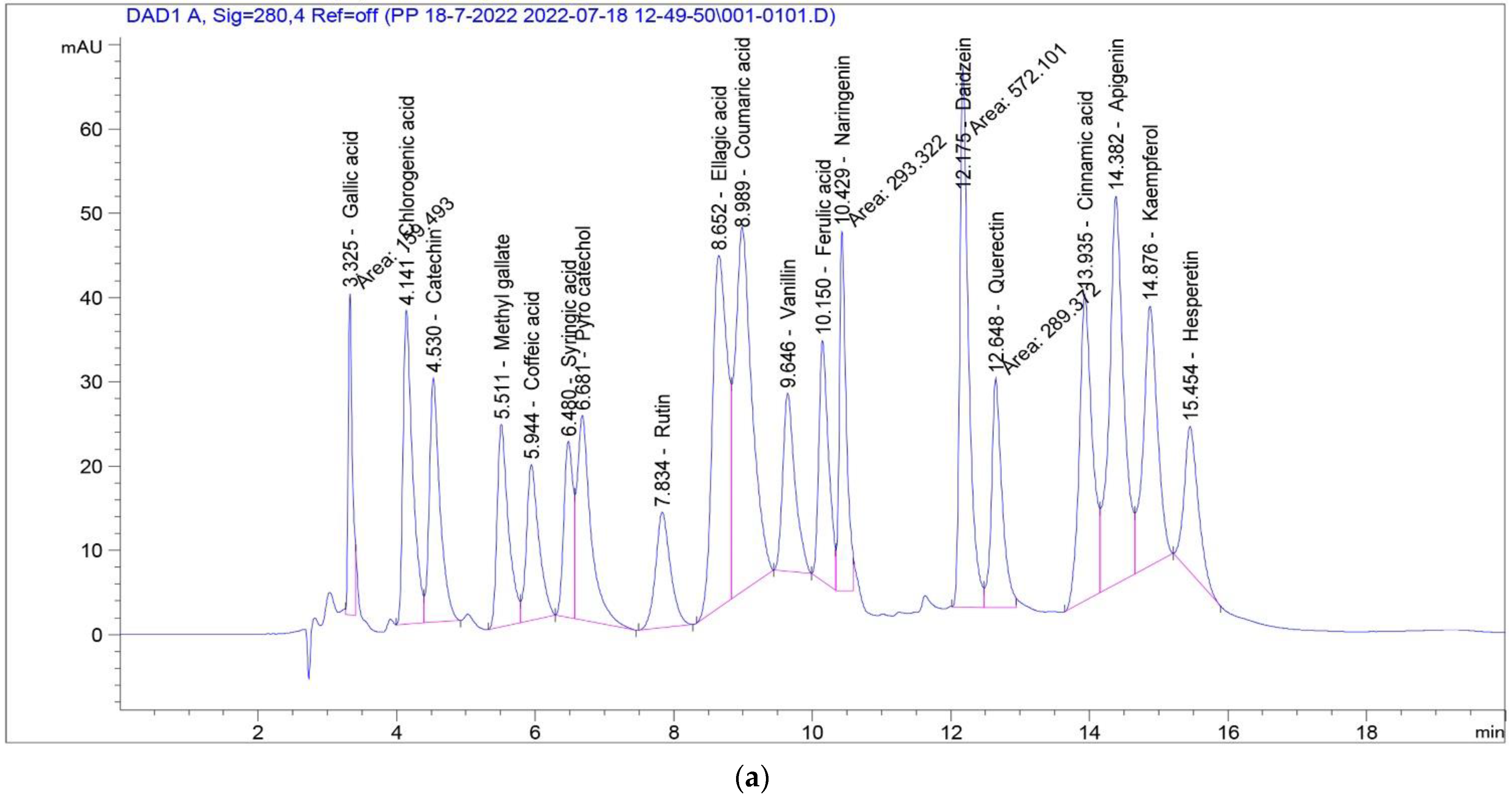
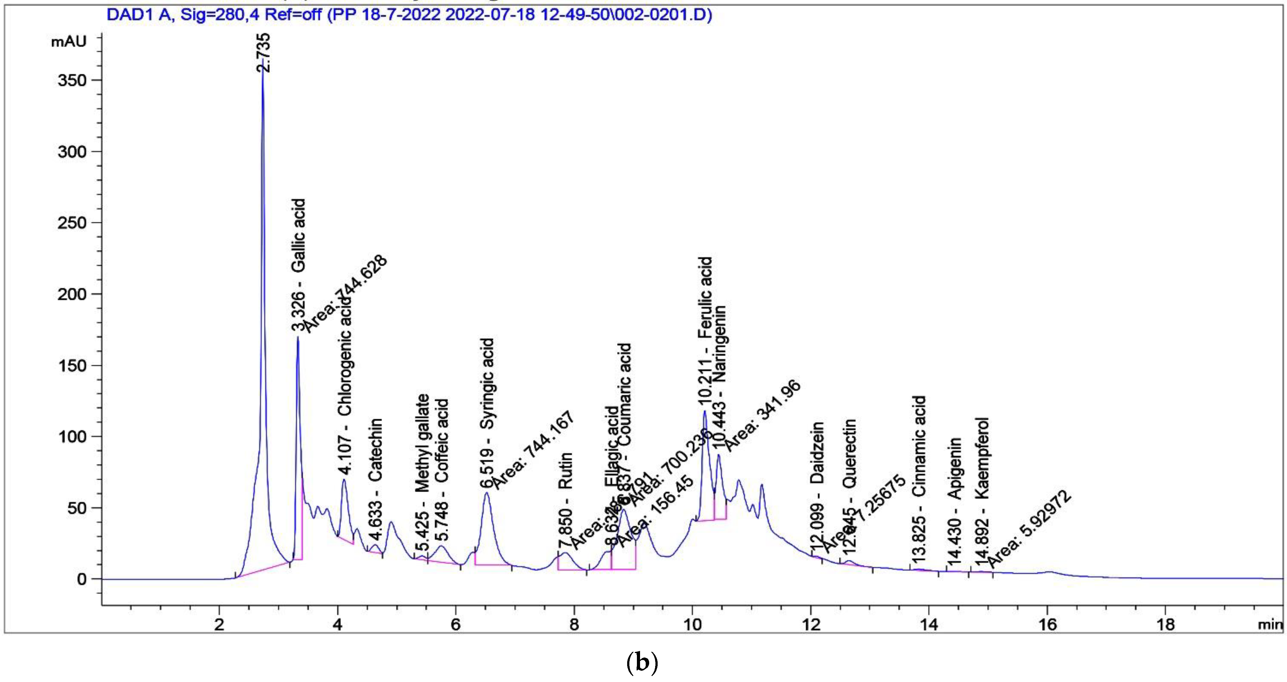
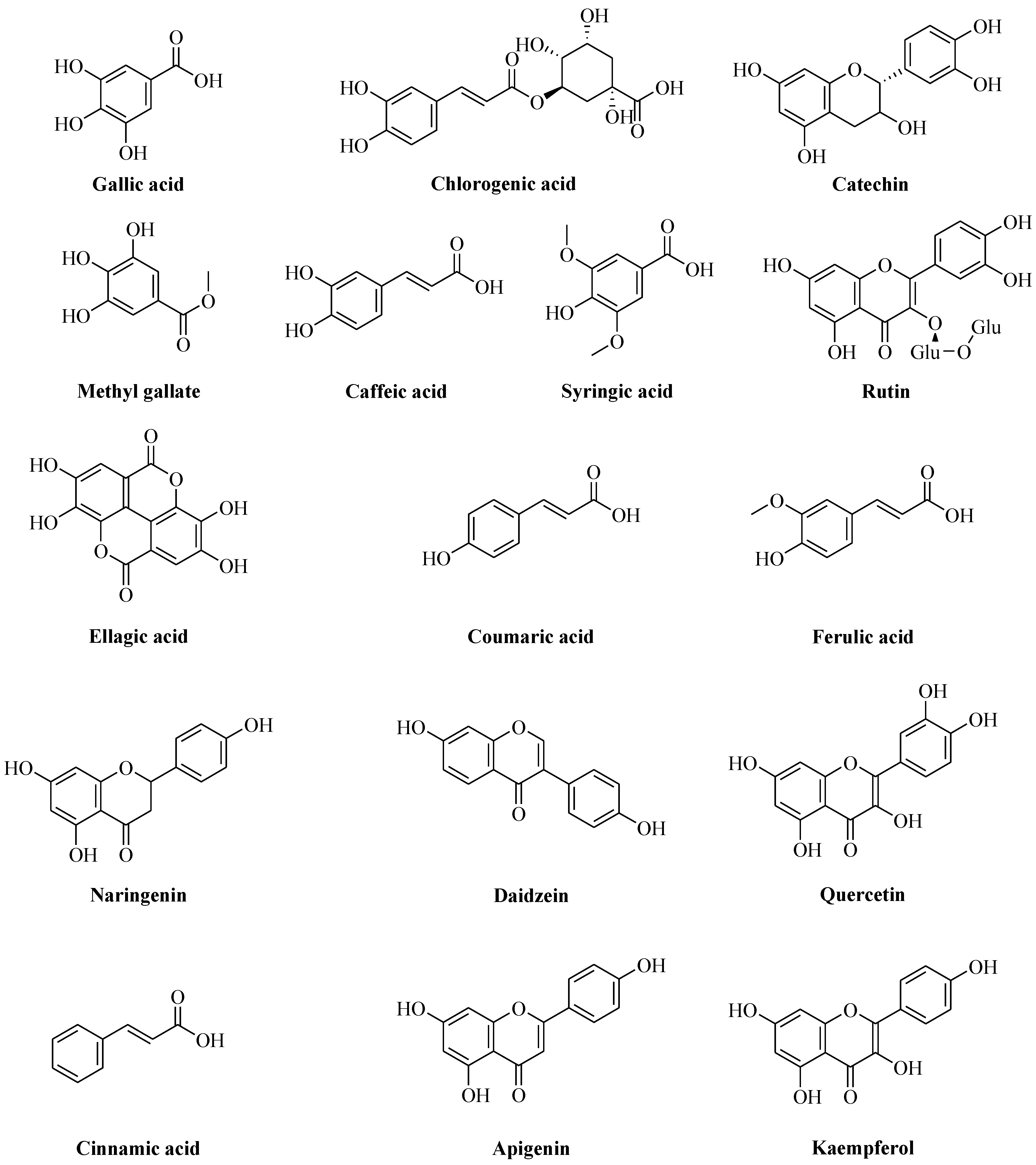
| Compound | Conc. (µg/g) | Compound | Conc. (µg/g) |
|---|---|---|---|
| Gallic acid | 3744.95 | Vanillin | 0.00 |
| Chlorogenic acid | 2523.29 | Ferulic acid | 2507.38 |
| Catechin | 586.08 | Naringenin | 1870.30 |
| Methyl gallate | 63.94 | Daidzein | 23.74 |
| Caffeic acid | 754.77 | Quercetin | 208.25 |
| Syringic acid | 3513.87 | Cinnamic acid | 11.21 |
| Pyro catechol | 0.00 | Apigenin | 13.56 |
| Rutin | 1268.87 | Kaempferol | 21.50 |
| Ellagic acid | 1573.64 | Hesperetin | 0.00 |
| Coumaric acid | 1008.72 |
| Functional Groups | Absorption Bands in ZnO NPs (cm−1) | Absorption Bands in P. odoratissimum ALE (cm−1) |
|---|---|---|
| -OH stretch | 3417 | 3409 |
| -C-H stretch | 2920 | 2923 |
| O=C=O stretch | 2356 | 2356 |
| C=C stretch | 1621 | 1616 |
| C-N stretch | 1403 | 1400 |
| C-O stretch | 1072 | 1068 |
| -C-H stretch (aromatics) | 855 | 852 |
| Zn-O | 435 | - |
| Element | Weight (%) | Atom (%) |
|---|---|---|
| Zn | 80.71 | 50.58 |
| O | 19.29 | 49.42 |
| Total | 100 | 100 |
| Treatment | DPPH IC50 (µg/mL) | TPC (mg GAE/g Dry Leaf Extract) | TFC (mg RE/g Dry Leaf Extract) |
|---|---|---|---|
| ALE | 04.56 ± 0.02 a | 21.93 ± 0.01 | 17.11 ± 0.001 |
| ZnO NPs | 28.11 ± 0.01 c | n.d. | n.d. |
| L-ascorbic acid | 11.50 ± 0.03 b | n.d. | n.d. |
| Pathogenic Bacteria | Diameter of Inhibition Zones (mm) | Positive Control Gentamycin (10 μg mL−1) | Aqueous Leaf Extract (20 μg mL−1) | ||
|---|---|---|---|---|---|
| ZnO NPs | |||||
| 10 μg mL−1 | 20 μg mL−1 | 30 μg mL−1 | |||
| S. aureus | 23 ± 0.70 c | 25 ± 1.41 b | 28 ± 0.35 a | 13 ± 0.28 j | - |
| B. cereus | 17 ± 0.35 g | 18 ± 0.56 f | 24 ± 0.14 f | 22 ± 0.70 d | - |
| E. coli | 13 ± 0.72 j | 15 ± 0.07 i | 16 ± 0.21 h | 12 ± 0.42 k | - |
| P. aeruginosa | 18 ± 1.06 f | 20 ± 0.70 e | 21 ± 0.28 d | 13 ± 0.14 j | - |
| Mean of ZnO NPs | 17.75 ± 3.7 C | 19.5 ± 3.5 B | 22.25 ± 3.5 A | 15 ± 4.00 D | - |
| Sample | Conc. (ug/mL) | Mean Absorbance ± SD | Hemolysis Inhibition % | |
|---|---|---|---|---|
| Hypotonic Solution | Isotonic Solution | |||
| Control | 1.326 ± 0.1 | 0.001 ± 0.01 | ||
| ZnO NPs | 1000 | 0.158 ± 0.004 b | 0.095 ± 0.00 | 95.6 |
| 800 | 0.189 ± 0.003 c | 0.071 ± 0.00 | 91.8 | |
| 600 | 0.264 ± 0.006 d | 0.061 ± 0.00 | 85.9 | |
| 400 | 0.381 ± 0.005 f | 0.054 ± 0.00 | 77.3 | |
| 200 | 0.475 ± 0.002 h | 0.035 ± 0.00 | 69.5 | |
| 100 | 0.583 ± 0.012 j | 0.020 ± 0.00 | 61.0 | |
| ALE | 1000 | 0.198 ± 0.007 c | 0.081 ± 0.00 | 91.9 |
| 800 | 0.329 ± 0.006 e | 0.065 ± 0.00 | 81.7 | |
| 600 | 0.426 ± 0.005 g | 0.035 ± 0.00 | 72.9 | |
| 400 | 0.474 ± 0.007 h | 0.031 ± 0.00 | 69.3 | |
| 200 | 0.544 ± 0.005 i | 0.027 ± 0.00 | 64.1 | |
| 100 | 0.660 ± 0.003 k | 0.022 ± 0.00 | 55.7 | |
| Indomethacin | 1000 | 0.059 ± 0.002 a | 0.035 ± 0.01 | 98.1 |
Publisher’s Note: MDPI stays neutral with regard to jurisdictional claims in published maps and institutional affiliations. |
© 2022 by the authors. Licensee MDPI, Basel, Switzerland. This article is an open access article distributed under the terms and conditions of the Creative Commons Attribution (CC BY) license (https://creativecommons.org/licenses/by/4.0/).
Share and Cite
Abdelbaky, A.S.; Abd El-Mageed, T.A.; Babalghith, A.O.; Selim, S.; Mohamed, A.M.H.A. Green Synthesis and Characterization of ZnO Nanoparticles Using Pelargonium odoratissimum (L.) Aqueous Leaf Extract and Their Antioxidant, Antibacterial and Anti-inflammatory Activities. Antioxidants 2022, 11, 1444. https://doi.org/10.3390/antiox11081444
Abdelbaky AS, Abd El-Mageed TA, Babalghith AO, Selim S, Mohamed AMHA. Green Synthesis and Characterization of ZnO Nanoparticles Using Pelargonium odoratissimum (L.) Aqueous Leaf Extract and Their Antioxidant, Antibacterial and Anti-inflammatory Activities. Antioxidants. 2022; 11(8):1444. https://doi.org/10.3390/antiox11081444
Chicago/Turabian StyleAbdelbaky, Ahmed S., Taia A. Abd El-Mageed, Ahmad O. Babalghith, Samy Selim, and Abir M. H. A. Mohamed. 2022. "Green Synthesis and Characterization of ZnO Nanoparticles Using Pelargonium odoratissimum (L.) Aqueous Leaf Extract and Their Antioxidant, Antibacterial and Anti-inflammatory Activities" Antioxidants 11, no. 8: 1444. https://doi.org/10.3390/antiox11081444