Article

Optimization of a Method for Detecting Intracellular Sulfane Sulfur Levels and Evaluation of Reagents That Affect the Levels in Escherichia coli

1
State Key Laboratory of Microbial Technology, Shandong University, 72 Binhai Road, Qingdao 266237, China
2
School of Molecular Biosciences, Washington State University, Pullman, WA 99164-7520, USA
*
Authors to whom correspondence should be addressed.
Antioxidants 2022, 11(7), 1292; https://doi.org/10.3390/antiox11071292
Submission received: 1 June 2022 / Revised: 22 June 2022 / Accepted: 24 June 2022 / Published: 29 June 2022

Abstract

:
Sulfane sulfur is a class of compounds containing zero-valent sulfur. Most sulfane sulfur compounds are reactive and play important signaling roles. Key enzymes involved in the production and metabolism of sulfane sulfur have been characterized; however, little is known about how to change intracellular sulfane sulfur (iSS) levels. To accurately measure iSS, we optimized a previously reported method, in which reactive iSS reacts with sulfite to produce thiosulfate, a stable sulfane sulfur compound, before detection. With the improved method, several factors were tested to influence iSS in Escherichia coli. Temperature, pH, and osmotic pressure showed little effect. At commonly used concentrations, most tested oxidants, including hydrogen peroxide, tert-butyl hydroperoxide, hypochlorous acid, and diamide, did not affect iSS, but carbonyl cyanide m-chlorophenyl hydrazone increased iSS. For reductants, 10 mM dithiothreitol significantly decreased iSS, but tris(2-carboxyethyl)phosphine did not. Among different sulfur-bearing compounds, NaHS, cysteine, S2O32− and diallyl disulfide increased iSS, of which only S2O32− did not inhibit E. coli growth at 10 mM or less. Thus, with the improved method, we have identified reagents that may be used to change iSS in E. coli and other organisms, providing tools to further study the physiological functions of iSS.

1. Introduction

Sulfide (H2S and HS) is considered the third gaso-transmitter in mammals, participating in various physiological functions [1,2]. Recent reports show that sulfide signaling is usually via intracellular sulfane sulfur (iSS) [3,4]. Sulfane sulfur, containing zero-valence sulfur, comes in several forms, such as inorganic and organic polysulfide (HSn, RSn, and RSSnR; n ≥ 2) and elemental sulfur [5]. It modifies protein cysteine (Cys) thiols to form persulfide (S-sulfhydration), which alters protein configurations and amends the catalytic or regulatory activities [6]. Glyceraldehyde-3-phosphate dehydrogenases in Escherichia coli and Staphylococcus aureus are inhibited after their active site Cys residues are S-sulfhydrated [7,8], but the enzyme activity from the mouse liver is enhanced after S-sulfhydration [9]. Several bacterial gene regulators have been identified to respond to iSS. MgrA senses iSS after sulfide-stress to activate the expression of virulent factors in S. aureus [8]. The iSS level in Pseudomonas aeruginosa is high in the late log phase and early stationary phase of growth, and it activates MexR to turn on the expression of a multiple drug efflux pump MexAB when cells enter the stationary phase [10]. The high levels of iSS in the late log phase and early stationary phase of growth also significantly enhance the activity of the master quorum-sensing activator LasR in P. aeruginosa [11]. The multidrug repressor MarR is inactivated by increased iSS in the early stationary phase of growth of E. coli [12]. When iSS is high, it modifies the oxidative stress regulator OxyR that activates the expression of genes involved in lowing iSS in E. coli [13]. Hence, iSS in bacteria also plays a critical role in regulating different physiological processes.
The iSS is unstable, as it can be oxidized by oxygen, reduced by thiols, and decomposed under acid conditions [14,15]. Therefore, the growth conditions and oxidative and reducing agents may interfere with iSS in bacteria. The growth conditions include osmolarity, pH, temperature, and dissolved oxygen [16,17,18,19]. Reactive oxygen species (ROS) cause oxidative stress [20,21], damaging proteins, nucleic acids, and lipids [22]. Superoxide radicals (O2•−), hydrogen peroxide (H2O2), and hydroxyl radicals (•OH) are common ROS, and they are produced via the electron transport chain of aerobic respiration [23]. Reagents, such as hypochlorous acid and carbonyl cyanide m-chlorophenyl hydrazone (CCCP), are often used to induce oxidative stress in cells. Hypochlorous acid is a strong oxidant, and it reacts with DNA, protein, and lipids [24]. CCCP is an uncoupling agent that inhibits oxidative phosphorylation and promotes ROS production [25], which induces oxidative stress in cells [26]. The reagents diamide and tert-butyl hydroperoxide (tBH) are common thiol oxidants [27]. Commonly used reducing agents include dithiothreitol (DTT) and tris(2-carboxyethyl)phosphine (TCEP). How these factors influence iSS levels in bacteria is unclear.
The production of iSS also affects its homeostasis. The metabolism of Cys is the main source of sulfane sulfur in Escherichia coli [28]. Cys is either present in a rich medium or is produced from sulfate [29]. Sulfate is reduced via assimilatory sulfate reduction to sulfide before being incorporated into Cys. The yeast Saccharomyces cerevisiae can convert thiosulfate to iSS which is then reduced to sulfide for Cys biosynthesis [30]. Further, the yeast Schizosaccharomyces pombe directly uses sulfane sulfur to produce Cys [31]. Garlic oil is rich in sulfane sulfur-containing compounds with diallyl disulfide (DADS) and diallyl trisulfide being prevalent [32]. The supply of these sulfur compounds in the growth medium may affect iSS.
Recently, we have developed a simple and sensitive method to detect total iSS from biological samples. SO32− is used to react with unstable iSS to produce stable S2O32− under hot and alkaline conditions (pH = 9.5, 95 °C). S2O32 is then derived with mono-bromobimane (mBBr) to form bimane-S2O32−, which is detected by using high-performance liquid chromatography (HPLC) with a fluorescence detector. The thiosulfate content of the samples incubated in the control buffer without sulfite is the blank sample that is subtracted from the test samples [33]. Thiosulfate is also a sulfane sulfur-containing compound [1], but it does not react with cellular thiols under physiological conditions [34,35]. This method was named as sulfite-dependent sulfane sulfur detection method (SdSS), and it is sensitive to detect active sulfane sulfur in bacteria, plants, and animals [10,31,33,36] as well as in wine [37].
However, after extensive use, we noticed several shortcomings that affect the accuracy and sensitivity of this method. The peaks of mBBr derivatized S2O32− and glutathione (GSH) partially overlapped, affecting the detection sensitivity of bimane-S2O32−; the washing process of preparing the bacterium samples might cause iSS loss; a fraction of iSS was converted to thiosulfate without sulfite during heating. Here, we addressed these issues and revised the method. With this improved method, we investigated the factors that could affect iSS in E. coli. The key findings included that thiosulfate is a good reagent to increase iSS without affecting bacterial growth.

2. Materials and Methods

2.1. Bacterial Strains, Culture Conditions, and Reagents

Escherichia coli BL21(DE3) was grown in the lysogeny broth (LB) at 37 °C. Sodium sulfite, sodium thiosulfate, sodium sulfate, GSH, Cys, NaHS, DTT, mBBr, tBH, DADS, TCEP, diamide (CAS: 10465-78-8), and CCCP were purchased from Sigma-Aldrich (Burlington, MA, USA). Diethylenetriaminepentaacetic acid (DTPA), FeSO4·7H2O, acetic acid, sodium hypochlorite, and H2O2 were purchased from Bio Basic Inc (Markham, ON, Canada). TritonX-100 was purchased from Sangon Biotech (Shanghai, China). Rosup is a compound mixture of oxidative reagents, and it is used as a positive control in the Reactive Oxygen Species Assay Kit (Beyotime Biotech Inc., Shanghai, China).

2.2. Sample Preparations for iSS Detection

Colonies of E. coli BL21(DE3) were picked up and cultured in LB medium overnight. The cultures were transferred into a fresh medium at an initial OD600 of 0.05 for aerobic cultures or at an initial OD600 of 0.02 for anaerobic cultures. Cells were at defined time intervals, and the OD600 value was measured with the spectrophotometer UV1800 (Shimadzu, Kyoto, Japan). Three different procedures were used to harvest cells. Option 1 (No-wash): E. coli cells were transferred into a microfuge tube and harvested by centrifuging at 3300× g for 5 min at 4 °C. The supernatants were carefully removed with a pipettor, and the collected cells were used for iSS measurement. Option 2 (Double-centrifugation): after the cells were harvested and the supernatant was removed, the tubes with pellets were centrifuged again at 3300× g for 1 min. The residual supernatant was carefully removed with a pipettor. Option 3 (Wash-once): after the cells were harvested and the supernatant was removed, the collected pellets were washed once with 1 mL 100 mM Tris-HCl buffer (pH = 7.4) to remove the residual supernatant. Wash-once is the reported procedure [33]. No-wash and Double-centrifugation were two selected procedures for comparison. Double-centrifugation was found to be the optimal procedure and was adapted for iSS detection.

2.3. The Optimized iSS Detection Method

The previously reported SdSS method is designed to measure iSS [33]. The SdSS method was optimized. Briefly, the reaction buffer was prepared with 50 mM Tris-HCl buffer (pH = 9.5) containing 1% TritonX-100, 50 μM DTPA, and 1 mM sulfite, and the control buffer contained no sulfite but 0.5 mM DTT. DTT was introduced to reduce sulfate sulfur to H2S, avoiding the spontaneous oxidation of sulfane sulfur to thiosulfate during subsequent steps. One mL of cells at OD600 of 1.0 was harvested with double-centrifugation and resuspended in 100 μL of the reaction buffer or the control buffer. The resuspended cells were incubated at 95 °C for 10 min. The samples were then centrifuged at 15,700× g for 3 min; 50 μL of the supernatant was mixed with 5 μL of 25 mM mBBr and incubated in the dark at room temperature for 25 min to convert S2O32− into bimane-S2O32− adduct. 110 μL of an acetic acid and acetonitrile mixture (v/v, 1:9) was added to stop the reaction and denature proteins. The mixtures were centrifuged to precipitate cell debris and denatured proteins at 15,700× g for 3 min.
The bimane-S2O32− adduct in the supernatant was determined by using HPLC (LC-20A, Shimadzu, Kyoto, Japan) with a fluorescence detector (RF20A, Shimadzu, Kyoto, Japan). The gain value and sensitivity of this fluorescence detector were set as “medium” and “×16”, respectively. Briefly, 5 μL supernatant was injected onto a reverse-phase C18 column (VP-ODS, 150 × 4 mm, Shimadzu, Kyoto, Japan) with a guard column (Inertsil ODS-SP 5 μm 5020-19006, Shimadzu, Kyoto, Japan) through an autosampler (SIL-20A, Shimadzu, Kyoto, Japan). The column was maintained at 38 °C in a column thermostat (CTO-20A, Kyoto, Japan), and eluted with a gradient solution A (0.25% acetic acid and 10% methanol in distilled water, with pH being adjusted to 3.9 by using NaOH) and solution B (0.25% acetic acid and 90% methanol in distilled water) from 8% B to 40% B in 7 min, 40% B for 5 min, 40% B to 100% in 0.1 min, 100% B for 6 min at a flow rate of 0.8 mL/min. The adduct was detected by a fluorescence detector with an optimized excitation wavelength (Ex) and emission wavelength (Em) at 380 nm and 466 nm, respectively. The bimane-S2O32− adduct was normally detected at a retention time of 13.0 min.

2.4. The Effect of Growth Conditions and Reagents on E. coli iSS

The cells of E. coli BL21(DE3) were transferred into a fresh LB medium and incubated at 37 °C until OD600 reached about 1.0. The cultures were aliquoted and incubated under various conditions, including different temperatures, pH, and osmolarities, or spiked with sulfur-containing compounds (NaHS, sulfite, sulfate, Cys, thiosulfate, DADS, and GSH), oxidants (H2O2, tBH, CCCP, diamide, sodium hypochlorite, Rosup, and Fenton’s reagent) and reductants (DTT and TCEP). DADS and CCCP were dissolved in absolute ethanol and DMSO, respectively. The other reagents were dissolved in distilled water. Fenton reagent was prepared by mixing 10 mM H2O2 with 10 mM FeSO4·7H2O. The final concentration of each reagent was given in the results. After incubation for 30 min or as specified in the text [33], the cells were collected by using the double-centrifugation method and analyzed with our optimized SdSS method.
The iSS content in E. coli cells was also assayed with cells cultured under aerobic and anaerobic conditions. For aerobic growth, the cells were cultured in 50 mL of LB medium. For anaerobic growth, the cells were cultured in 100 mL serum bottles sealed with butyl rubber stoppers. 70 mL nitrogen deoxygenated LB medium was filled into the bottles before sterilization. The cells were inoculated in the bottles by using a syringe. Cells were taken at defined time intervals given in the results. The iSS content was detected by using our optimized method.

3. Results

The detection sensitivity for sulfane sulfur was improved by optimizing HPLC conditions.
Sulfane sulfur is converted to S2O32−, which is derivatized with mBBr to bimane-S2O32− for detection [33]; however, the peaks of mBBr-derivatized S2O32− and GSH partially overlapped within the chromatogram (Figure 1A). The HPLC elution program was optimized to separate the two peaks (Figure 1B). The maximal Ex and Em of bimane-S2O32− were determined to be 380 nm and 466 nm, respectively (Figure 2A,B). When the maximal wavelengths were used, the peak area of bimane-S2O32− increased about 20.0% over that obtained with the reported Ex and Em (Figure 2C). Nine combinations of the settings of gain value and sensitivity of the fluorescence detector were tested to increase the signal-to-noise ratio (SNR) for the bimane-S2O32− adduct (Table S1), and the highest value of the SNR was when the gain value was “×16” and the sensitivity was “medium”.
By using the optimized HPLC conditions, the standard curves of bimane-S2O32− ranging from 0.5 μM to 5 μM in 50 mM Tris-HCl buffer (pH = 9.5) with or without E. coli BL21(DE3) at OD600 of 1.0 was determined. With the cells, the baseline was increased, but the detection of added S2O32− was not affected (Figure 3A). Further, the detection limit of bimane-S2O32− in the Tris-HCl buffer was improved to 10 nM by using the optimized conditions (Figure 3B).

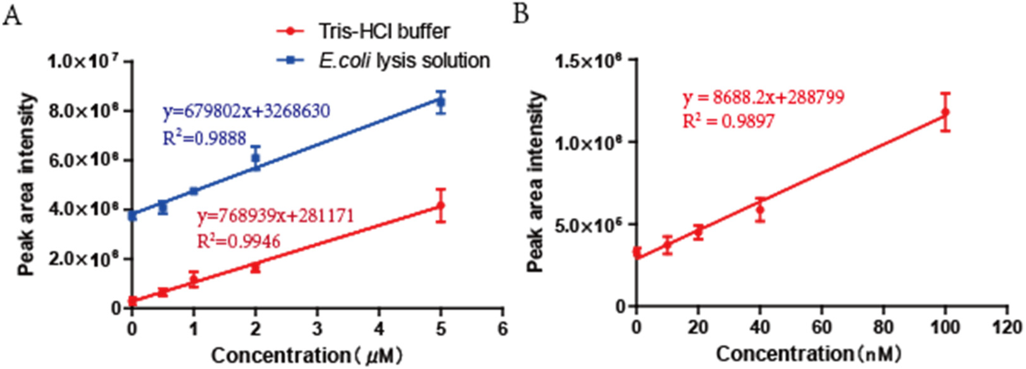
3.1. The Optimization of Bacterial Samples Preparation

E. coli iSS was tested by suspending the cells in the sample buffer (with SO32−) and the control buffer (without SO32−), and heating was used to convert iSS and SO32− to S2O32− [33]. Whether iSS was self-oxidized to S2O32− during the heating process was tested by using DTT to reduce iSS to H2S, as DTT readily reduces reactive sulfane sulfur, such as organic persulfide, to the corresponding thiol and H2S [38]. E. coli cells were treated in the control buffer. When 0.2 mM DTT or more was added into the control buffer, S2O32− in the control group decreased from 8.5 × 10⁻² nmol/mL/OD to 1.5 × 10⁻² nmol/mL/OD (Figure 4A). When S2O32− was added to the control buffer, DTT did not reduce it after heating (Figure 4B). Thus, 0.5 mM DTT was subsequently included in the control buffer without SO32− to prevent S2O32− formation from the autoxidation of iSS during the heating process.
Whether it is necessary to remove the residual culture supernatant before iSS determination was tested. Sulfane sulfur in the supernatant fluctuated around 23 µM without significant changes during the growth of E. coli BL21(DE3) in the LB medium (Figure S1). When the harvested cells by centrifugation were washed once with Tris-HCl buffer (50 mM, pH = 7.4) as reported [33], iSS was 226.5 (10−3 nmol/mL/OD) (Figure 5A). When the harvested cells were directly measured without washing, iSS was 460.9 (10−3·nmol/mL/OD) (Figure 5A). The unwashed cell pellet contained several microliters of the culture supernatant, which contributed to the increased iSS. The residual supernatant was removed by second centrifugation to collect the liquid on the wall of the microfuge tube and pipetting removal (double-centrifugation step). With the double-centrifugation step, iSS was 281.1 (10−3·nmol/mL/OD) (Figure 5A). This double-centrifugation step was adopted because it minimizes the culture supernatant and prevents the loss of iSS during washing.
Various volumes of an E. coli BL21(DE3) culture at OD600 were used to detect iSS contents. With the revised method, iSS concentrations could be accurately detected with 1 mL of E. coli BL21(DE3) cells at OD600 of 1.0, but the accuracy was reduced with sample volume smaller than 1 mL (Figure 5B). This optimized SdSS method was used in the following tests.

3.2. The Effects of Different Stress Factors on iSS Content of E. coli

When cell cultures of E. coli BL21(DE3) in LB medium were incubated at different temperatures for up to 30 min, the iSS contents were not significantly changed (Figure S2A). When cells were resuspended in LB or Tris-HCl buffer at different pH values, the iSS content in E. coli cells was not significantly changed (Figure S2B,C). 1–8% NaCl was added directly into the cultures to change the osmotic pressure, but it did not change the iSS content of cells, either (Figure S2D).
To test the effect of oxygen on iSS, the iSS content in E. coli was tested when the bacterium was cultured at different growth phases under both aerobic and anaerobic conditions. The iSS content under aerobic conditions rose to the highest level rapidly in the mid-log phase and remained high till the early stationary phase. It significantly decreased during the stationary phase (Figure 6A). However, under anaerobic conditions, the iSS content gradually increased during the entire log phase and reached its maximum in the early stationary phase. The high level of iSS content was relatively stable during the stationary phase (Figure 6B).

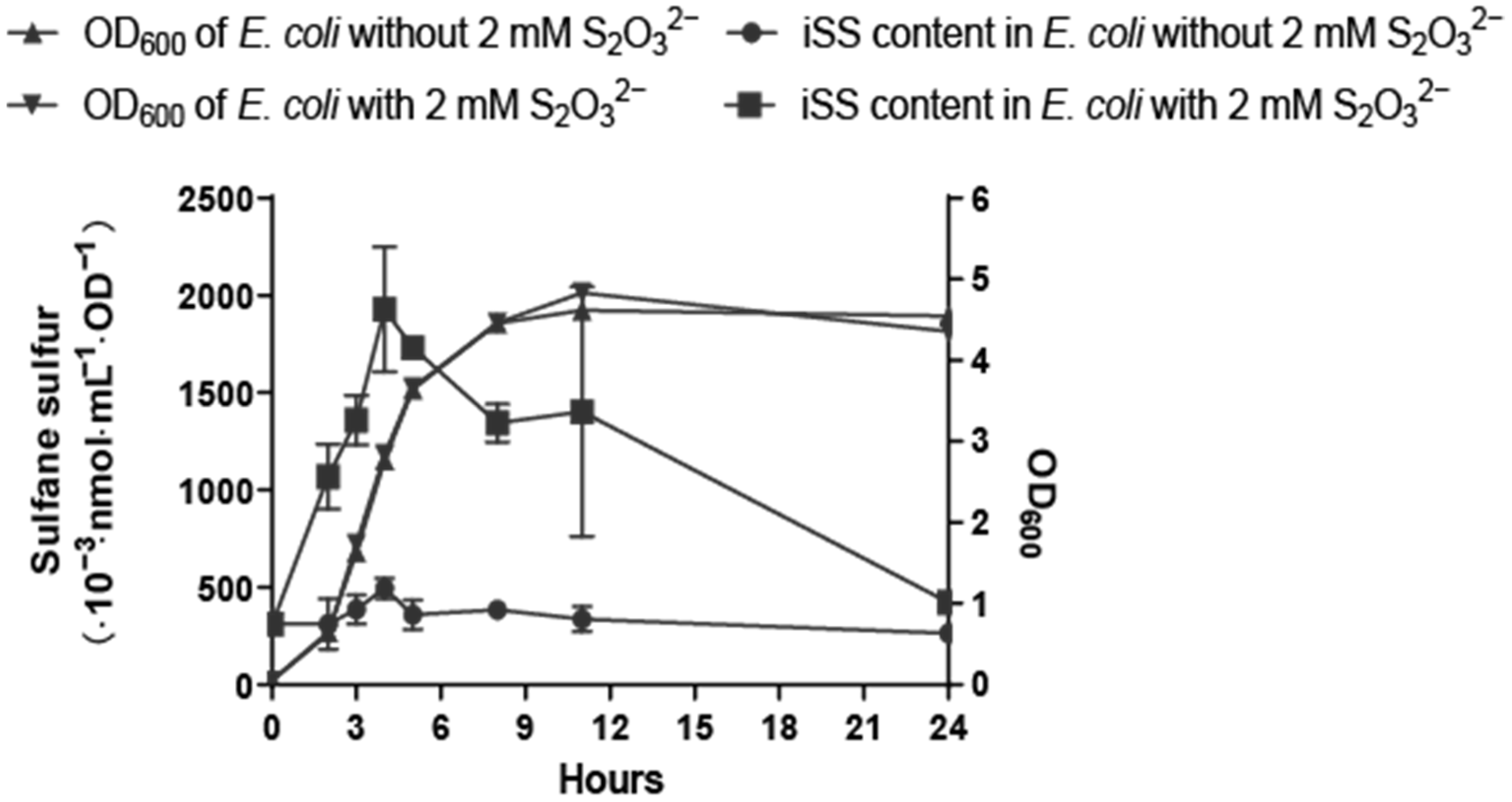
3.3. Effects of Cellular Redox Balance on E. coli iSS

Different compounds that could disturb the intracellular redox potential were used to test if they could change the homeostasis of sulfane sulfur in cells after 30-min treatment. DTT, TCEP, tBH, H2O2, sodium hypochlorite, diamide, and Fenton’s reagent were tested at 1, 0.2, 0.25, 0.1, 0.1, 1, and 0.1 mM, respectively. CCCP and Rosup were used at 10 μM and 50 μg/mL. They are the reported concentrations in the literature and kit instruction [39,40,41,42,43,44,45,46]. Only CCCP and Rosup promoted iSS (Figure 7A). When tested at high concentrations, 10 mM Rosup, 10 mM diamide, and 5 mM Fenton’s reagent increased iSS, but 10 mM H2O2 and tBH did not (Figure S3). For reductants, 1 mM DTT and 0.2 mM TCEP did not significantly change iSS (Figure 7A). When tested at high concentrations, 10 mM DTT reduced iSS, but 10 mM TCEP did not (Figure S3).

3.4. Effects of Exogenous Sulfur-Bearing Compounds on E. coli iSS

Different sulfur-bearing compounds at 10 mM were added into E. coli BL21(DE3) cultures to test if they affected iSS. Cys, NaHS, DADS, and S2O32− increased the iSS content of E. coli, but GSH, SO32− and SO42− did not (Figure 7B). Low concentrations of S2O32−, DADS, Cys, and NaHS were further tested, and they still increased iSS but at reduced magnitudes (Figure S4A–D). S2O32− was still the most effective, and it significantly increased iSS even at 0.5 mM.
The sulfur-bearing compounds which could promote iSS content were added to BL21(DE3) culture to observe whether they were toxic to cells at different concentrations. In a closed environment, all of these sulfur compounds except S2O32− repressed cell growth of E. coli to different degrees (Figure S5A–D). S2O32− at 10 mM or less did not show apparent inhibition. The DADS and NaHS showed more severe inhibition than Cys. In an open environment, the inhibition effect of DADS and NaHS was largely relieved (Figure S5F–H), likely because H2S and DADS were volatile and evaporated. Again, S2O32− did not affect cell growth (Figure S5E).
Nontoxic S2O32− at 2 mM was added into the cell cultures of E. coli in LB medium to observe the change of iSS during growth. The iSS content greatly increased in comparison with the control (Figure 8). The iSS content reached the highest level at the end of logarithmic growth. Then it gradually decreased to a level similar to that of the control group after 24 h of growth.

4. Discussion

The previously reported SdSS method for iSS detection in bacteria was optimized. DTT is a key factor for optimization. DTT is a common reducing agent, and it reduces sulfane sulfur to H2S [47]. The addition of DTT in the control buffer prevented the oxidation of iSS to thiosulfate, which interferes with the SdSS method. The inclusion of DTPA in both the reaction buffer and the control buffer is to chelate transition metals that catalyze sulfur oxidation [48]. Another improvement is the revised HPLC elution method. The revised method separates bimane-S2O32− and bimane-GS so that the high concentrations of bimane-GS derived from E. coli will not interfere with bimane-S2O32− detection (Figure 1B). Third, we optimized the fluorescence detection settings (Figure 1B and Figure 2C and Table S1), and the detection threshold for bimane-S2O32− was improved from 200 nM to 10 nM (Figure 3B). Finally, the double-centrifugation method to prepare cells before iSS detection was recommended (Figure 5A), as it minimizes culture supernatant and prevents the loss of iSS during washing with a buffer that changes the culturing environment of the tested cells.
We had expected a decrease in iSS when ROS was added to cell suspensions, as sulfane sulfur is known to react with ROS [49,50,51]; however, several tested reagents that induce oxidative stress increased iSS (Figure 7A). A possible explanation is that intracellular acid-labile sulfur is also oxidized by the addition of these reagents. The acid-labile sulfur, including Fe-S clusters, is sulfide [52,53]. When O2•− and HO• oxidize Fe-S clusters and release Fe2+ [54,55,56], the sulfur in the cluster is theoretically oxidized to sulfane sulfur (zero valences), as sulfide reacts with ROS to produce sulfane sulfur [57]. H2O2 and tBH were unable to increase iSS (Figure 7A), perhaps partly because they are rapidly metabolized by E. coli cells [58], partly because they react with sulfane sulfur at a relatively slow rate [40], and partly because they do not directly damage Fe-S clusters [59].
Sulfane sulfur, ROS, and Reactive chlorine species (RCS) are signaling molecules, and their signaling pathways may overlap [53,60,61]. OxyR is a major global regulator of E. coli in response to oxidative stress [62], and it also senses iSS through the persulfidation of Cys199 [13]. Other redox-based transcriptional regulators also used Cys residues for signal sensing [61,63]. Two members of the MarR (multiple drug-resistant regulators) families that repress multiple drug efflux pumps have been shown to use Cys residues to sense both H2O2 and iSS [10,12]. Further, our data show that ROS and RCS significantly increased iSS in E. coli cells (Figure 7A). The results imply that iSS may participate in the signaling transduction induced by ROS or RCS. Further studies are needed to understand whether sulfane sulfur is involved in the signaling induced by ROS and RCS.
S2O32− is the only tested sulfur donor that could increase the iSS content without affecting bacterial growth (Figure S5 and Figure 8), and it may be used to increase iSS in E. coli or other organisms to evaluate the effect of elevated iSS on cells. Although S2O32− may be used with other organisms to increase iSS, its concentration should be tested. For example, Saccharomyces cerevisiae is partially inhibited by 10 mM S2O32−, as high concentrations of S2O32− directly inhibit cytochrome c oxidase of the electron transport chain in the yeast mitochondria [35]. E. coli is not inhibited by 10 mM S2O32− (Figure 8), likely because it does not use cytochrome c oxidase in its electron transport chain [64]. There are two known metabolic pathways of S2O32− in E. coli: one is catalyzed by CysM that uses S2O32− to produce Cys [65,66], and Cys is then converted to sulfane sulfur [28]; the other is catalyzed by rhodanese (RHOD) GlpE that directly converts S2O32− to sulfane sulfur [65]. E. coli contains eight proteins carrying RHOD domains [67]. Further studies are necessary to identify whether CysM or one or more RHODs are responsible to convert S2O32− to sulfane sulfur.
In summary, we optimized the SdSS method and used E. coli as a model to extensively investigate the effects of different stress factors and reagents on iSS homeostasis. This work not only provides a better method for analyzing iSS in E. coli and possibly other biological samples but also investigated several factors possibly affecting iSS homeostasis, which facilitates further studies of the physiological functions of iSS.

Supplementary Materials

The following supporting information can be downloaded at: https://www.mdpi.com/article/10.3390/antiox11071292/s1, Figure S1: Sulfane sulfur concentrations in the supernatant of E. coli cultures growing in LB medium; Figure S2: The influence of temperature, pH, and osmolarity on iSS; Figure S3: Changes in the iSS content of E. coli cells under different oxidants; Figure S4: The iSS contents of E. coli BL21 (DE3) cells with varying concentrations of sulfur-bearing compounds; Figure S5: Growth curves of E. coli BL21(DE3) when incubated with different sulfur-bearing compounds; Table S1: Screening for best signal to noise ratio (SNR) of bimane-S2O32− with the combinations of sensitivity and gain values.

Author Contributions

Conceptualization, Y.X. and L.X.; methodology, Y.X. and H.L.; investigation, Q.Y.; writing -original draft preparation, Q.Y.; validation, M.R.; formal analysis and sources, Y.Y.; project administration, Y.X. and L.X.; funding acquisition, Y.X. and L.X.; supervision, L.X. All authors have read and agreed to the published version of the manuscript.

Funding

The work was financially supported by grants from the National Natural Science Foundation of China (31870085, 91951202, 31961133015).

Institutional Review Board Statement

Not applicable.

Informed Consent Statement

Not applicable.

Data Availability Statement

All data are reported in the main text or Supplementary Data.

Conflicts of Interest

The authors declare no conflict of interest.

References

  1. Mishanina, A.V.; Libiad, M.; Banerjee, R. Biogenesis of reactive sulfur species for signaling by hydrogen sulfide oxidation pathways. Nat. Chem. Biol. 2015, 11, 457–464. [Google Scholar] [CrossRef] [Green Version]
  2. Kimura, H.; Shibuya, N.; Kimura, Y. Hydrogen Sulfide Is a Signaling Molecule and a Cytoprotectant. Antioxid. Redox Signal. 2012, 17, 45–57. [Google Scholar] [CrossRef] [Green Version]
  3. Filipovic, M.R.; Zivanovic, J.; Alvarez, B.; Banerjee, R. Chemical Biology of H2S Signaling through Persulfidation. Chem. Rev. 2018, 118, 377–461. [Google Scholar] [CrossRef]
  4. Kimura, H. Hydrogen Sulfide (H2S) and Polysulfide (H2Sn) Signaling: The First 25 Years. Biomolecules 2021, 11, 896. [Google Scholar] [CrossRef]
  5. Nagy, P.; Doka, E.; Ida, T.; Akaike, T. Measuring Reactive Sulfur Species and Thiol Oxidation States: Challenges and Cautions in Relation to Alkylation-Based Protocols. Antioxid. Redox Signal. 2020, 33, 1174–1189. [Google Scholar] [CrossRef]
  6. Castelblanco, M.; Nasi, S.; Pasch, A.; So, A.; Busso, N. The role of the gasotransmitter hydrogen sulfide in pathological calcification. Brit. J. Pharmacol. 2020, 177, 778–792. [Google Scholar] [CrossRef] [Green Version]
  7. Jarosz, A.P.; Wei, W.L.; Gauld, J.W.; Auld, J.; Ozcan, F.; Aslan, M.; Mutus, B. Glyceraldehyde 3-phosphate dehydrogenase (GAPDH) is inactivated by S-sulfuration in vitro. Free Radic. Bio. Med. 2015, 89, 512–521. [Google Scholar] [CrossRef]
  8. Peng, H.; Zhang, Y.X.; Palmer, L.D.; Kehl-Fie, T.E.; Skaar, E.P.; Trinidad, J.C.; Giedroc, D.P. Hydrogen Sulfide and Reactive Sulfur Species Impact Proteome S-Sulfhydration and Global Virulence Regulation in Staphylococcus aureus. ACS Infect. Dis. 2017, 3, 744–755. [Google Scholar] [CrossRef] [Green Version]
  9. Mustafa, A.K.; Gadalla, M.M.; Sen, N.; Kim, S.; Mu, W.T.; Gazi, S.K.; Barrow, R.K.; Yang, G.D.; Wang, R.; Snyder, S.H. H2S Signals Through Protein S-Sulfhydration. Sci. Signal. 2009, 2, ra72. [Google Scholar] [CrossRef] [Green Version]
  10. Xuan, G.; Lü, C.; Xu, H.; Chen, Z.; Li, K.; Liu, H.; Liu, H.; Xia, Y.; Xun, L. Sulfane Sulfur is an intrinsic signal activating MexR-regulated antibiotic resistance in Pseudomonas aeruginosa. Mol. Microbiol. 2020, 114, 1038–1048. [Google Scholar] [CrossRef]
  11. Xuan, G.; Lv, C.; Xu, H.; Li, K.; Liu, H.; Xia, Y.; Xun, L. Sulfane Sulfur Regulates LasR-Mediated Quorum Sensing and Virulence in Pseudomonas aeruginosa PAO1. Antioxidants 2021, 10, 1498. [Google Scholar] [CrossRef]
  12. Xu, H.; Xuan, G.; Liu, H.; Xia, Y.; Xun, L. Sulfane Sulfur Is a Strong Inducer of the Multiple Antibiotic Resistance Regulator MarR in Escherichia coli. Antioxidants 2021, 10, 1778. [Google Scholar] [CrossRef]
  13. Hou, N.K.; Yan, Z.Z.; Fan, K.L.; Li, H.J.; Zhao, R.; Xia, Y.Z.; Xun, L.Y.; Liu, H.W. OxyR senses sulfane sulfur and activates the genes for its removal in Escherichia coli. Redox Biol. 2019, 26, 101293. [Google Scholar] [CrossRef]
  14. Ni, X.; Kelly, S.S.; Xu, S.; Xian, M. The Path to Controlled Delivery of Reactive Sulfur Species. Acc. Chem. Res. 2021, 54, 3968–3978. [Google Scholar] [CrossRef]
  15. Xu, S.; Hamsath, A.; Neill, D.L.; Wang, Y.Y.; Yang, C.T.; Xian, M. Strategies for the Design of Donors and Precursors of Reactive Sulfur Species. Chem. Eur. J. 2019, 25, 4005–4016. [Google Scholar] [CrossRef] [PubMed]
  16. Gregory, G.J.; Boyd, E.F. Stressed out: Bacterial response to high salinity using compatible solute biosynthesis and uptake systems, lessons from Vibrionaceae. Comput. Struct. Biotec. 2021, 19, 1014–1027. [Google Scholar] [CrossRef]
  17. Fang, F.C.; Frawley, E.R.; Tapscott, T.; Vazquez-Torres, A. Bacterial Stress Responses during Host Infection. Cell Host Microbe 2016, 20, 133–143. [Google Scholar] [CrossRef] [PubMed] [Green Version]
  18. Reniere, M.L. Reduce, Induce, Thrive: Bacterial Redox Sensing during Pathogenesis. J. Bacteriol. 2018, 200, e00128-18. [Google Scholar] [CrossRef] [PubMed] [Green Version]
  19. Lu, Z.; Imlay, J.A. When anaerobes encounter oxygen: Mechanisms of oxygen toxicity, tolerance and defence. Nat. Rev. Microbiol. 2021, 19, 774–785. [Google Scholar] [CrossRef]
  20. Khademian, M.; Imlay, J.A. How Microbes Evolved to Tolerate Oxygen. Trends Microbiol. 2021, 29, 428–440. [Google Scholar] [CrossRef]
  21. Reichmann, D.; Voth, W.; Jakob, U. Maintaining a Healthy Proteome during Oxidative Stress. Mol. Cell 2018, 69, 203–213. [Google Scholar] [CrossRef] [PubMed] [Green Version]
  22. Zabel, M.; Nackenoff, A.; Kirsch, W.M.; Harrison, F.E.; Perry, G.; Schrag, M. Markers of oxidative damage to lipids, nucleic acids and proteins and antioxidant enzymes activities in Alzheimer’s disease brain: A meta-analysis in human pathological specimens. Free Radic. Biol. Med. 2018, 115, 351–360. [Google Scholar] [CrossRef] [PubMed]
  23. Jakubczyk, K.; Dec, K.; Kaldunska, J.; Kawczuga, D.; Kochman, J.; Janda, K. Reactive oxygen species—Sources, functions, oxidative damage. Pol. Merkur Lek. 2020, 48, 124–127. [Google Scholar]
  24. Ashby, L.V.; Springer, R.; Hampton, M.B.; Kettle, A.J.; Winterbourn, C.C. Evaluating the bactericidal action of hypochlorous acid in culture media. Free Radic. Biol. Med. 2020, 159, 119–124. [Google Scholar] [CrossRef] [PubMed]
  25. Calleja, L.F.; Yoval-Sanchez, B.; Hernandez-Esquivel, L.; Gallardo-Perez, J.C.; Sosa-Garrocho, M.; Marin-Hernandez, A.; Jasso-Chavez, R.; Macias-Silva, M.; Rodriguez-Zavala, J.S. Activation of ALDH1A1 by omeprazole reduces cell oxidative stress damage. FEBS J. 2021, 288, 4064–4080. [Google Scholar] [CrossRef] [PubMed]
  26. Corona, F.; Martinez, J.L.; Nikel, P.I. The global regulator Crc orchestrates the metabolic robustness underlying oxidative stress resistance in Pseudomonas aeruginosa. Environ. Microbiol. 2019, 21, 898–912. [Google Scholar] [CrossRef] [PubMed] [Green Version]
  27. Di Simplicio, P.; Lupis, E.; Rossi, R. Different mechanisms of formation of glutathione-protein mixed disulfides of diamide and tert-butyl hydroperoxide in rat blood. Biochim. Biophys. Acta 1996, 1289, 252–260. [Google Scholar] [CrossRef]
  28. Li, K.; Xin, Y.; Xuan, G.; Zhao, R.; Liu, H.; Xia, Y.; Xun, L. Escherichia coli Uses Separate Enzymes to Produce H2S and Reactive Sulfane Sulfur From L-cysteine. Front. Microbiol. 2019, 10, 298. [Google Scholar] [CrossRef] [Green Version]
  29. Pester, M.; Knorr, K.H.; Friedrich, M.W.; Wagner, M.; Loy, A. Sulfate-reducing microorganisms in wetlands—Fameless actors in carbon cycling and climate change. Front. Microbiol. 2012, 3, 72. [Google Scholar] [CrossRef] [Green Version]
  30. Olson, K.R. H2S and polysulfide metabolism: Conventional and unconventional pathways. Biochem. Pharmacol. 2018, 149, 77–90. [Google Scholar] [CrossRef]
  31. Zhang, X.; Xin, Y.; Chen, Z.; Xia, Y.; Xun, L.; Liu, H. Sulfide-quinone oxidoreductase is required for cysteine synthesis and indispensable to mitochondrial health. Redox Biol. 2021, 47, 102169. [Google Scholar] [CrossRef] [PubMed]
  32. Chiang, Y.H.; Jen, L.N.; Su, H.Y.; Lii, C.K.; Sheen, L.Y.; Liu, C.T. Effects of garlic oil and two of its major organosulfur compounds, diallyl disulfide and diallyl trisulfide, on intestinal damage in rats injected with endotoxin. Toxicol. Appl. Pharm. 2006, 213, 46–54. [Google Scholar] [CrossRef] [PubMed]
  33. Ran, M.; Wang, T.; Shao, M.; Chen, Z.; Liu, H.; Xia, Y.; Xun, L. Sensitive Method for Reliable Quantification of Sulfane Sulfur in Biological Samples. Anal. Chem. 2019, 91, 11981–11986. [Google Scholar] [CrossRef] [PubMed]
  34. Kruithof, P.D.; Lunev, S.; Lozano, S.P.A.; Batista, F.D.; Al-dahmani, Z.M.; Joles, J.A.; Dolga, A.M.; Groves, M.R.; van Goor, H. Unraveling the role of thiosulfate sulfurtransferase in metabolic diseases. BBA Mol. Basis Dis. 2020, 1866, 165716. [Google Scholar] [CrossRef] [PubMed]
  35. Chen, Z.; Xia, Y.; Liu, H.; Liu, H.; Xun, L. The Mechanisms of Thiosulfate Toxicity against Saccharomyces cerevisiae. Antioxidants 2021, 10, 646. [Google Scholar] [CrossRef] [PubMed]
  36. Lu, T.; Cao, Q.; Pang, X.; Xia, Y.; Xun, L.; Liu, H. Sulfane sulfur-activated actinorhodin production and sporulation is maintained by a natural gene circuit in Streptomyces coelicolor. Microb. Biotechnol. 2020, 13, 1917–1932. [Google Scholar] [CrossRef]
  37. Müller, N.; Rauhut, D.; Tarasov, A. Sulfane Sulfur Compounds as Source of Reappearance of Reductive Off-Odors in Wine. Fermentation 2022, 8, 53. [Google Scholar] [CrossRef]
  38. Kabil, O.; Banerjee, R. Redox Biochemistry of Hydrogen Sulfide. J. Biol. Chem. 2010, 285, 21903–21907. [Google Scholar] [CrossRef] [Green Version]
  39. Park, Y.S.; Choi, S.E.; Koh, H.C. PGAM5 regulates PINK1/Parkin-mediated mitophagy via DRP1 in CCCP-induced mitochondrial dysfunction. Toxicol. Lett. 2018, 284, 120–128. [Google Scholar] [CrossRef]
  40. Wang, Q.; Chen, Z.; Zhang, X.; Xin, Y.; Xia, Y.; Xun, L.; Liu, H. Rhodanese Rdl2 produces reactive sulfur species to protect mitochondria from reactive oxygen species. Free Radic. Biol Med. 2021, 177, 287–298. [Google Scholar] [CrossRef]
  41. Huang, B.; Zhao, Z.; Huang, C.; Zhao, M.; Zhang, Y.; Liu, Y.; Liao, X.; Huang, S.; Zhao, Y. Role of metal cations and oxyanions in the regulation of protein arginine phosphatase activity of Yw1E from Bacillus subtilis. Biochim. Biophys. Acta Gen. Subj. 2020, 1864, 129698. [Google Scholar] [CrossRef]
  42. Panmanee, W.; Charoenlap, N.; Atichartpongkul, S.; Mahavihakanont, A.; Whiteside, M.D.; Winsor, G.; Brinkman, F.S.L.; Mongkolsuk, S.; Hassett, D.J. The OxyR-regulated phnW gene encoding 2-aminoethylphosphonate: Pyruvate aminotransferase helps protect Pseudomonas aeruginosa from tert-butyl hydroperoxide. PLoS ONE 2017, 12, e0189066. [Google Scholar] [CrossRef] [PubMed] [Green Version]
  43. Imlay, J.A. Where in the world do bacteria experience oxidative stress? Environ. Microbiol. 2019, 21, 521–530. [Google Scholar] [CrossRef] [PubMed]
  44. Parker, B.W.; Schwessinger, E.A.; Jakob, U.; Gray, M.J. The RclR Protein Is a Reactive Chlorine-specific Transcription Factor in Escherichia coli. J. Biol. Chem. 2013, 288, 32574–32584. [Google Scholar] [CrossRef] [PubMed] [Green Version]
  45. Bakovic, J.; Yu, B.Y.K.; Silva, D.; Baczynska, M.; Peak-Chew, S.Y.; Switzer, A.; Burchell, L.; Wigneshweraraj, S.; Vandanashree, M.; Gopal, B.; et al. Redox Regulation of the Quorum-sensing Transcription Factor AgrA by Coenzyme, A. Antioxidants 2021, 10, 841. [Google Scholar] [CrossRef] [PubMed]
  46. Spear, N.; Aust, S.D. Effects of glutathione on Fenton reagent-dependent radical production and DNA oxidation. Arch. Biochem. Biophys. 1995, 324, 111–116. [Google Scholar] [CrossRef]
  47. Miyamoto, R.; Koike, S.; Takano, Y.; Shibuya, N.; Kimura, Y.; Hanaoka, K.; Urano, Y.; Ogasawara, Y.; Kimura, H. Polysulfides (H2Sn) produced from the interaction of hydrogen sulfide (H2S) and nitric oxide (NO) activate TRPA1 channels. Sci. Rep. 2017, 7, 45995. [Google Scholar] [CrossRef] [Green Version]
  48. Stevens, R.; Stevens, L.; Price, N.C. The Stabilities of Various Thiol Compounds Used in Protein Purifications. Biochem. Educ. 1983, 11, 70. [Google Scholar] [CrossRef]
  49. Olson, K.R. Hydrogen sulfide, reactive sulfur species and coping with reactive oxygen species. Free Radical Bio. Med. 2019, 140, 74–83. [Google Scholar] [CrossRef]
  50. Hancock, J.T.; Whiteman, M. Hydrogen sulfide signaling: Interactions with nitric oxide and reactive oxygen species. Ann. N. Y. Acad. Sci. 2016, 1365, 5–14. [Google Scholar] [CrossRef]
  51. Fukuto, J.M.; Ignarro, L.J.; Nagy, P.; Wink, D.A.; Kevil, C.G.; Feelisch, M.; Cortese-Krott, M.M.; Bianco, C.L.; Kumagai, Y.; Hobbs, A.J.; et al. Biological hydropersulfides and related polysulfides—A new concept and perspective in redox biology. FEBS Lett. 2018, 592, 2140–2152. [Google Scholar] [CrossRef] [PubMed] [Green Version]
  52. Predmore, B.L.; Lefer, D.J.; Gojon, G. Hydrogen Sulfide in Biochemistry and Medicine. Antioxid. Redox Signal. 2012, 17, 119–140. [Google Scholar] [CrossRef] [PubMed] [Green Version]
  53. Iciek, M.; Bilska-Wilkosz, A.; Gorny, M. Sulfane sulfur—New findings on an old topic. Acta Biochim. Pol. 2019, 66, 533–544. [Google Scholar] [CrossRef]
  54. Bruska, M.K.; Stiebritz, M.T.; Reiher, M. Binding of Reactive Oxygen Species at Fe-S Cubane Clusters. Chem. A Eur. J. 2015, 21, 19081–19089. [Google Scholar] [CrossRef]
  55. Lu, Z.; Imlay, J.A. A conserved motif liganding the [4Fe-4S] cluster in [4Fe-4S] fumarases prevents irreversible inactivation of the enzyme during hydrogen peroxide stress. Redox Biol. 2019, 26, 101296. [Google Scholar] [CrossRef]
  56. Rydz, L.; Wrobel, M.; Jurkowska, H. Sulfur Administration in Fe-S Cluster Homeostasis. Antioxidants 2021, 10, 1738. [Google Scholar] [CrossRef] [PubMed]
  57. Bian, J.S.; Olson, K.R.; Zhu, Y.C. Hydrogen Sulfide: Biogenesis, Physiology, and Pathology. Oxid. Med. Cell. Longev. 2016, 2016, 6549625. [Google Scholar] [CrossRef] [PubMed]
  58. Mishra, S.; Imlay, J. Why do bacteria use so many enzymes to scavenge hydrogen peroxide? Arch. Biochem. Biophys 2012, 525, 145–160. [Google Scholar] [CrossRef] [PubMed] [Green Version]
  59. Flint, D.H.; Tuminello, J.F.; Emptage, M.H. The inactivation of Fe-S cluster containing hydro-lyases by superoxide. J. Biol. Chem. 1993, 268, 22369–22376. [Google Scholar] [CrossRef]
  60. Gray, M.J.; Wholey, W.Y.; Jakob, U. Bacterial Responses to Reactive Chlorine Species. Annu. Rev. Microbiol. 2013, 67, 141–160. [Google Scholar] [CrossRef] [Green Version]
  61. Sevilla, E.; Bes, M.T.; Gonzalez, A.; Peleato, M.L.; Fillat, M.F. Redox-Based Transcriptional Regulation in Prokaryotes: Revisiting Model Mechanisms. Antioxid. Redox Signal. 2019, 30, 1651–1696. [Google Scholar] [CrossRef] [PubMed]
  62. Chiang, S.M.; Schellhorn, H.E. Regulators of oxidative stress response genes in Escherichia coli and their functional conservation in bacteria. Arch. Biochem. Biophys. 2012, 525, 161–169. [Google Scholar] [CrossRef] [PubMed]
  63. Lee, I.G.; Lee, B.J. How Bacterial Redox Sensors Transmit Redox Signals via Structural Changes. Antioxidants 2021, 10, 502. [Google Scholar] [CrossRef]
  64. Unden, G.; Bongaerts, J. Alternative respiratory pathways of Escherichia coli: Energetics and transcriptional regulation in response to electron acceptors. BBA Bioenergetics 1997, 1320, 217–234. [Google Scholar] [CrossRef] [Green Version]
  65. Kawano, Y.; Onishi, F.; Shiroyama, M.; Miura, M.; Tanaka, N.; Oshiro, S.; Nonaka, G.; Nakanishi, T.; Ohtsu, I. Improved fermentative L-cysteine overproduction by enhancing a newly identified thiosulfate assimilation pathway in Escherichia coli. Appl. Microbiol. Biot. 2017, 101, 6879–6889. [Google Scholar] [CrossRef]
  66. Sirko, A.; Hryniewicz, M.; Hulanicka, D.; Bock, A. Sulfate and Thiosulfate Transport in Escherichia-Coli K-12—Nucleotide-Sequence and Expression of the Cystwam Gene-Cluster. J. Bacteriol. 1990, 172, 3351–3357. [Google Scholar] [CrossRef] [PubMed] [Green Version]
  67. Cheng, H.; Donahue, J.L.; Battle, S.E.; Ray, W.K.; Larson, T.J. Biochemical and Genetic Characterization of PspE and GlpE, Two Single-domain Sulfurtransferases of Escherichia coli. Open Microbiol. J. 2008, 2, 18–28. [Google Scholar] [CrossRef]
Figure 1. Partial HPLC chromatograms with two programs. The E. coli BL21(DE3) was cultured until the OD600 reached around 1.0. The cells equivalent to 3 mL at OD600 = 1 were collected and the iSS contents were determined with the SdSS method. After derivation with mBBr, the derivatives were determined with two different HPLC programs. (A) The previous used HPLC elution. The bimane-S2O32− (at 9.2 min) formed a peak overlapped with bimane-GSH (at 8.9 min). The blue curve was the mixture of 10 μM bimane-S2O32− and 10 μM bimane-GSH, which was routinely used as standard. The yellow curve was the mBBr-derivatives of an E. coli cell lysate. The large peak of bimane-GSH from the cell lysate overlapped with that of bimane-S2O32−. (B) The optimized HPLC elution. The optimized elution separated bimane-GSH (at 10.5 min, not shown) and bimane-S2O32− (at 13.0 min).
Antioxidants 11 01292 g001
Figure 2. The optimal excitation and emission wavelengths of bimane-S2O32−. 0.05 mM S2O32− was reacted with 0.25 mM mBBr to ensure S2O32− was converted to bimane-S2O32−. This reaction solution was used to search for the optimal excitation and emission wavelengths. (A) Fixed Em at 485 nm to screen the Ex from 300 nm to 440 nm. (B) Fixed Ex at 370 nm to screen the Em from 410 nm to 550 nm. (C) The comparison of the peak area of bimane-S2O32− detected with the optimized and original fluorescence detection parameters, respectively. Three parallel experiments were performed to obtain the averages and standard deviations (n = 3). A t-test was performed to calculate the p-value. Symbol * was shown when p < 0.05.
Antioxidants 11 01292 g002
Figure 3. The standard curves of bimane-S2O32− range at different concentrations. The standard curves of bimane-S2O32− were established ranging from 0 to 5 μM (A) and 0 to 0.1 μM (B), respectively. The curve in blue color is plotted with cell lysate of E. coli BL21(DE3) as a complex background, and the curve in red color is plotted with Tris-HCl buffer as a plain background. Three parallel experiments were performed to obtain the averages and standard deviations (n = 3).
Antioxidants 11 01292 g003
Figure 4. The addition of DTT in the control buffer prevented iSS formation from self-oxidation. (A) S2O32− detected from 3 mL of E. coli BL21(DE3) cells at OD600 of 1.0 resuspended in the control buffer with different amounts of DTT. (B) DTT did not reduce the amount of S2O32−. 10 μM S2O32− was dissolved in the control buffer with different amounts of DTT and heated at 95 °C for 10 min. The quantity of S2O32− was determined after mBBr derivatization. Three parallel experiments were performed to obtain the averages and standard deviations (n = 3). The one-way ANOVA method was performed to calculate the p-values (Not significance (ns), p ≥ 0.05; ***, p < 0.001).
Antioxidants 11 01292 g004
Figure 5. Effects of different treatment conditions on iSS content in E. coli cells. (A) The harvested cells were treated in three different ways. No-wash, the harvested cells directly proceeded for SdSS analysis without washing; wash-once, the harvested cells were washed once with 50 mM Tris-HCl (pH = 7.4); double-centrifugation, the cell pellets were re-centrifuged and the residual supernatant was removed. (B) Different volumes of cell suspension at OD600 of 1.0 were harvested and analyzed by using the optimized method. Three parallel experiments were performed to obtain the averages and standard deviations (n = 3). The one-way ANOVA method was used to calculate the p-values (ns, p ≥ 0.05; ***, p < 0.001).
Antioxidants 11 01292 g005
Figure 6. The iSS content of E. coli cells cultured under aerobic and anaerobic conditions. The E. coli BL21(DE3) cells were incubated in an LB medium and cultured under aerobic (A) and anaerobic conditions (B). The OD600 and iSS content was detected at defined time intervals. Three parallel experiments for each condition were performed to obtain the averages and standard deviations (n = 3).
Antioxidants 11 01292 g006
Figure 7. Changes in the iSS content of E. coli cells under different oxidants, reductants, and sulfur-bearing compounds. (A) E. coli BL21(DE3) cells were cultured until their OD600 reached 1.0. Then, the cells were aliquoted, and different oxidants and reductants were added and incubated with them for 30 min before iSS detection. The DTT, TCEP, tBH, H2O2, sodium hypochlorite, diamide, Fenton’s reagent, CCCP, and Rosup were used at 1, 0.2, 0.25, 0.1, 0.1, 1, 0.1 mM, 10 μM, and 50 μg/mL, respectively. (B) Different sulfur-bearing compounds at 10 mM were added and incubated with the cell cultures for 30 min before iSS detection. Three parallel experiments were performed to obtain the averages and standard deviations (n = 3). The One-way ANOVA method was performed to calculate the p-values (ns, p ≥ 0.05; *, p < 0.05; **, p < 0.01; ***, p < 0.001).
Antioxidants 11 01292 g007
Figure 8. The addition of S2O32− increased iSS contents in E. coli during growth in LB medium. E. coli BL21(DE3) cells were inoculated into fresh LB medium with or without 2 mM S2O32− and cultured at 37 °C. The cells at defined time intervals were taken, and the OD600 and iSS contents were determined. Three parallel experiments were performed to obtain the averages and standard deviations (n = 3).
Antioxidants 11 01292 g008
Publisher’s Note: MDPI stays neutral with regard to jurisdictional claims in published maps and institutional affiliations.

