Article

Recovery of Lignins with Potent Antioxidant Properties from Shells of Edible Nuts by a Green Ball Milling/Deep Eutectic Solvent (DES)-Based Protocol

Department of Chemical Sciences, University of Naples “Federico II”, Via Cintia 4, I-80126 Naples, Italy
*
Author to whom correspondence should be addressed.
Antioxidants 2022, 11(10), 1860; https://doi.org/10.3390/antiox11101860
Submission received: 26 August 2022 / Revised: 14 September 2022 / Accepted: 16 September 2022 / Published: 21 September 2022

Abstract

:
Lignins are phenolic polymers endowed with potent antioxidant properties that are finding increasing applications in a variety of fields. Consequently, there is a growing need for easily available and sustainable sources, as well as for green extraction methodologies of these compounds. Herein, a ball milling/deep eutectic solvent (DES)-based treatment is reported as an efficient strategy for the recovery of antioxidant lignins from the shells of edible nuts, namely chestnuts, hazelnuts, peanuts, pecan nuts, and pistachios. In particular, preliminarily ball-milled shells were treated with 1:2 mol/mol choline chloride:lactic acid at 120 °C for 24 h, and the extracted material was recovered in 19–27% w/w yields after precipitation by the addition of 0.01 M HCl. Extensive spectroscopic and chromatographic analysis allowed for confirmation that the main phenolic constituents present in the shell extracts were lignins, accompanied by small amounts (0.9% w/w) of ellagic acid, in the case of chestnut shells. The recovered samples exhibited very promising antioxidant properties, particularly in the 2,2-diphenyl-1-picrylhydrazyl (DPPH) assay (EC50 values ranging from 0.03 to 0.19 mg/mL). These results open new perspectives for the valorization of nut shells as green sources of lignins for applications as antioxidants, e.g., in the biomedical, food, and/or cosmetic sector.

1. Introduction

Natural phenolic polymers from plant sources have been emerging in the field of material sciences as versatile, stable, and multifunctional additives [1,2,3]. A prominent role in this context is played by lignin, a heterogeneous polymer derived from peroxidase- or laccase-mediated oxidation of coniferyl, sinapyl, and p-coumaryl alcohol. Lignins are endowed with potent antioxidant and other functional properties that have prompted their applications in photocatalysis, stimuli-responsive materials, food packaging, wound healing, drug delivery, and tissue regeneration [4,5,6,7,8,9,10,11]. Given also the increasing interest in environmentally-friendly production processes, several studies have been directed to the exploitation of novel, easily available, and sustainable sources of these compounds, such as byproducts and wastes from the agri-food industry. Among these, shells from edible nuts that are produced in very large amounts such as peanut (11 million tons per year) [12], pecan nut (>420,000 tons per year) [13], hazelnut (>350,000 tons per year) [14], pistachio (>335,000 tons per year) [15], and chestnut (18,000 tons per year) [16,17] shells represent a most valuable alternative. Particular attention has been devoted in the past few years to green extraction methodologies, allowing for the efficient recovery of functional compounds such as phenols from natural sources through, e.g., the use of solvents with a low environmental impact. In this context, deep eutectic solvents (DESs) are emerging as eco-friendly and non-toxic media for application in the extraction not only of low-molecular-weight phenolic compounds, but also of phenolic polymers including lignin [18,19,20,21,22]. DESs are easily prepared by mixing, at a suitable temperature, a hydrogen bond acceptor (HBA) and a hydrogen bond donor (HBD), resulting in the formation of a eutectic mixture characterized by a melting point lower than that of the individual constituents. The ability of DESs to form hydrogen bonds or to donate and accept protons confers them good dissolution properties toward phenolic compounds, as recently explored in the case of agri-food wastes. A remarkable example is represented by 1:2 mol/mol chlorine chloride/lactic acid (ChCl:LA2), which has been reported as one of the most efficient solvents for lignin recovery [23,24,25,26].
Based on these observations, we report herein the exploitation of ChCl:LA2 for the recovery of lignin from peanut, pecan nut, hazelnut, chestnut, and pistachio shells. The recovered samples were characterized by electron paramagnetic resonance (EPR), attenuated total reflectance (ATR)-FTIR, 1H NMR, and UV-Vis spectroscopy as well as by chemical degradation experiments. The antioxidant properties of the samples were also determined by widely used chemical assays.

2. Materials and Methods

2.1. General Experimental Methods

Pistachios, chestnuts, peanuts, hazelnuts, and pecan nuts were purchased at a local supermarket and shells were recovered by hand peeling. Benzyl mercaptan, 30% w/v hydrogen peroxide, 2,2-diphenyl-1-picrylhydrazyl (DPPH), 2,4,6-tris(2-pirydyl)-s-triazine (TPTZ), (±)-6-hydroxy-2,5,7,8-tetramethylchromane-2-carboxylic acid (Trolox), Folin–Ciocalteu reagent, ellagic acid, gallic acid, iron (III) chloride, syringic acid, vanillic acid, and vanillin were obtained from Sigma-Aldrich (Milan, Italy).
UV-Vis spectra were recorded using a Jasco (Cremella, Lecco, Italy) V-730 spectrophotometer.
ATR-FTIR spectra were recorded as an average of 128 scans in the range 4000−450 cm−1 (4 cm−1 resolution) on a Nicolet 5700 Thermo Fisher Scientific (Milan, Italy) instrument.
1H NMR spectra were recorded in DMSO-d6 at 400 MHz on a Bruker (Milan, Italy) instrument.
HPLC analyses were performed on an instrument equipped with a UV-Vis detector (Agilent, Cernusco sul Naviglio, Milan, Italy); a Phenomenex (Castel Maggiore, Bologna, Italy) Sphereclone ODS column (250 × 4.60 mm, 5 µm) was used, and a gradient elution was performed using 0.1% formic acid (solvent A)/methanol (solvent B): 5% B, 0–10 min; from 5 to 80% B, 10–47.5 min. The flow rate was 1.0 mL/min, and the detection wavelength was 254 nm.
LC-MS analysis were run on an Agilent (Cernusco sul Naviglio, Milan, Italy) LC-MS ESI-TOF 1260/6230DA instrument operating in positive ionization mode. The following conditions were adopted: capillary voltage 3500 V; drying gas (nitrogen) 5 L/min, 325 °C; fragmentor voltage 175 V; nebulizer pressure 35 psig. An Agilent (Cernusco sul Naviglio, Milan, Italy) Eclipse Plus ODS column (150 × 4.6 mm, 5 µm) was used, with the same eluant as above at a flow rate of 0.4 mL/min.
Electron paramagnetic resonance (EPR) measurements were performed using a Bruker (Milan, Italy) Elexys E-500 spectrometer equipped with a superhigh sensitivity probe head. The samples were transferred in weighed amounts to a flame-sealed glass capillaries, which were then coaxially inserted in a 4 mm quartz sample tube. Spectra were recorded at room temperature under the following instrumental settings: sweep width 140 G; resolution 1024 points; scan time 20.97 s. The amplitude of the field modulation (1.0 G) was preventively checked to be low enough to avoid detectable signal overmodulation. To avoid microwave saturation of the resonance absorption curve, the microwave power was also optimized. A total of 16 scans were accumulated in order to improve the signal-to-noise ratio. For power saturation experiments, the microwave power was gradually incremented from 0.02 to 164 mW. The g value and the spin density were evaluated using Mn2+-doped MgO [27] as an internal standard. Since sample hydration was not controlled during the measurements, spin density values must be considered as order of magnitude estimates [28].

2.2. Pretreatment of Nut Shells

Shells were manually broken into small pieces and roughly shredded using a common blender. The resulting powders were then finely minced by treatment in a Fritsch Pulverisette 23 ball mill (Emme 3, Lainate, Milan, Italy) for 15 min at 50 osc/s.

2.3. DES Preparation

A literature-reported procedure was followed [29]. Briefly, choline chloride and lactic acid were mixed at 1:2 mol/mol ratio and heated at 50 °C under stirring until a homogeneous liquid was formed. The DES thus obtained, indicated as ChCl:LA2, was stored in the dark at ambient temperature. No crystal precipitation was observed over the period of use.

2.4. Treatment of Nut Shells with DES

A previously reported procedure was followed, with slight modifications [23]. Briefly, 10 g of each shell were added to 100 g of ChCl:LA2 containing 20% w/w water, and the mixture was taken under stirring in a Pyrex glass bottle at 120 °C for 24 h. After cooling at room temperature, the mixture was centrifuged (7000 rpm, 4 °C, 10 min) and the solid residue and supernatant were separated. Subsequently, the solid residue was washed three times with 50 mL of ethanol (7000 rpm, 4 °C, 10 min) and the collected washings were dried using a rotary evaporator. The obtained residue was added to the supernatant collected from the initial centrifugation, which was then poured into 1 L of 0.01 M HCl and kept at 4 °C until the precipitation of a brown solid was observed. The precipitate was then recovered by centrifugation (7000 rpm, 10 min, 4 °C), washed twice with 0.01 M HCl and once with distilled water, and lyophilized.

2.5. DPPH Assay

Samples were added (final dose 0.02−0.4 mg/mL) to a 0.2 mM DPPH solution in ethanol [30,31]. After stirring at room temperature for 10 min, the absorbance of the solution at 515 nm was measured. Trolox was used as a reference antioxidant. Experiments were run in triplicate.

2.6. Ferric Reducing/Antioxidant Power (FRAP) Assay

Samples were added (final dose 0.025–0.2 mg/mL) to a solution of FeCl3 (1.7 mM) and TPTZ (0.83 mM) in 0.3 M acetate buffer (pH 3.6) [32]. After stirring at room temperature for 10 min, the absorbance of the solution at 593 nm was measured. Results were expressed as Trolox equivalents (eqs). Experiments were run in triplicate.

2.7. Total Phenolic Content (TPC) Assay

Samples were added at a final dose of 0.0125−0.4 mg/mL to a mixture of Folin–Ciocalteu reagent, 75 g/L Na2CO3, and water in a 1:3:14 v/v/v ratio [33]. After incubation for 30 min at 40 °C, the absorbance at 765 nm was measured. Gallic acid was used as a reference compound. Experiments were run in triplicate.

2.8. Vanillin-HCl Assay

Samples were added (final dose 0.0625–2 mg/mL) to 1 mL of 1% w/v vanillin solution in methanol. Subsequently, 1 mL of 9 M HCl was added, and the mixture was incubated at 30 °C for 10 min [34]. Finally, the absorbance at 500 nm was measured. Catechin was used as a reference compound. Experiments were run in triplicate.

2.9. Alkaline Hydrogen Peroxide Degradation

Each sample (10 mg) was suspended in 1mL of 1 M NaOH and treated with 50 µL of 30% hydrogen peroxide [35]. The mixture was kept under vigorous stirring at room temperature for 24 h and then treated with 5% w/v Na2S2O5, taken to pH 3 with 6 M HCl, filtered on a 0.45 µm PVDF filter, and analyzed using HPLC.

2.10. Acid Degradation

Each sample (50 mg) was placed in a Pyrex tube, followed by 5 mL of 4 M HCl. The mixture was vortexed for 1 min and then incubated for 24 h in an oven at 90 °C [36]. After cooling to room temperature, the pH of the mixture was adjusted to 2.5 by addition of 6 M NaOH. The mixture was centrifuged at 7000 rpm for 10 min, the supernatant was recovered, taken to 10 mL by addition of water, and analyzed by HPLC after filtration on a 0.45 µm PVDF filter, whereas the solid residue was dissolved in 10 mL of DMSO/methanol 1:1 v/v and then analyzed by HPLC as well.

2.11. Thiolysis

Each sample (8 mg) was treated in methanol (2 mL) with 50 μL of benzyl mercaptan and 20 μL of 37% HCl at 40 °C under stirring [37]. After 1 h, the mixture was diluted in 1:1 v/v methanol/water (5 mL) and analyzed by HPLC.

3. Results and Discussion

3.1. Lignin Extraction from Nut Shells

Based on the previously reported results [23], a protocol based on the use of ChCl:LA2 as DES was adopted for the recovery of lignin from the selected nut shells. Before extraction, the latter were finely minced with the aid of a ball mill with the aim of improving the extraction yields by both increasing the surface area and cleaving glycosidic bonds and β-O-4 bonds of lignin components [38,39,40,41], also resulting in the liberation of phenolic hydroxyl groups and hence, boosting of the antioxidant properties. Subsequent extraction was performed at 120 °C for 24 h, allowing for the recovery lignins in the form of brown/black powders by simple precipitation, centrifugation, and lyophilization.
Table 1 reports the extraction and recovery yields obtained from the different nut shells, calculated with respect to the amount of starting or DES-dissolved material, respectively. The highest values were observed in the case of hazelnut and pistachio shells. In particular, the hazelnut shell lignin yields were higher than those reported in literature using different extraction procedures [42,43], whereas no data have apparently been reported for pistachio and pecan nut shells. A ca. 36% w/w yield has been recently reported for peanut shell lignin using 6:4 v/v ethanol/water as solvent at 180 °C for 100 min [44]. Though the yield is comparable to that obtained here, it should be noted that in the present paper a higher, and hence more cost effective and environmentally friendly, solid-to-solvent ratio was adopted. Chestnut shells exhibited the lowest extraction/recovery yields, although comparable to those reported in literature using different solvents [45]. In any case, it is noteworthy that to the best of our knowledge, there are no data available in the literature on the combined use of ball milling and DES for lignin recovery from edible nut shells.

3.2. Structural Characterization of Nut Shell-Derived Lignins

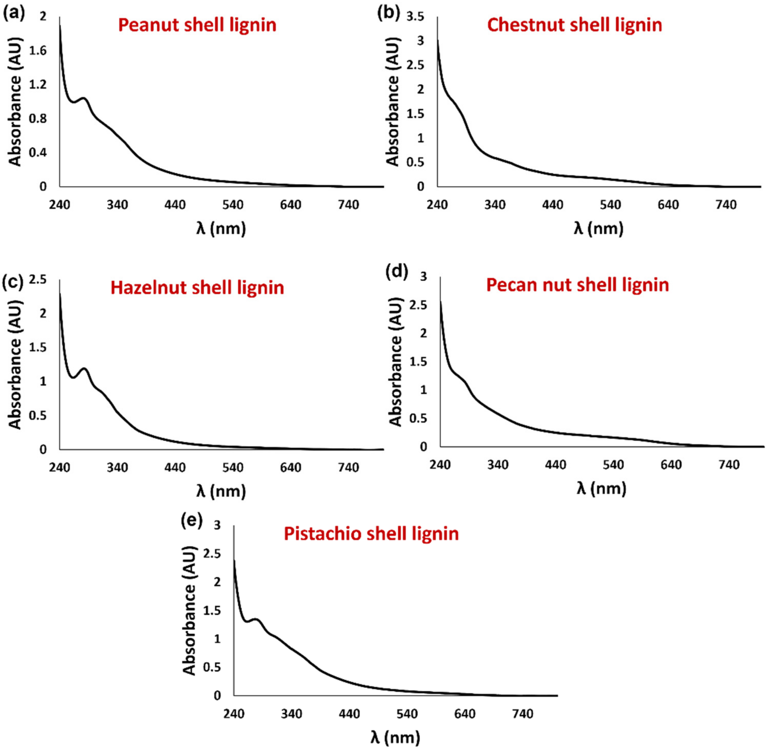
3.2.1. Spectroscopic Analysis

For the structural characterization of the recovered lignins, EPR spectroscopy was initially exploited, since it has been widely proven to be a sensitive and specific technique for the identification of phenolic polymers, which are known to be characterized by the presence of intrinsic free radical centers [1,23,46,47,48]. The EPR spectra of all samples showed a singlet (Figure 1a and Figure S1) at g values typical of other lignin-rich samples and consistent with the presence of carbon-centered radicals (Table 2) [23,49]. Despite the fact that EPR signals could appear featureless, a quantification of their intensity and a thorough analysis of their line shape (in terms of signal broadness and Gaussian or Lorentzian line shape) furnish a wealth of information on the nature, molecular structure, and supramolecular organization of the radical species present in a sample [46]. Indeed, while the starting nut shells exhibited apparently asymmetric spectra (Figure S1), likely as the result of the superposition of signals arising from different radical species, all the recovered lignins exhibited a symmetric signal (Figure 1a) with an intermediate line shape between the derivatives of the Gaussian and Lorentzian functions (Table 1). Moreover, recovered lignins showed higher spin density and lower signal amplitude (ΔB) values with respect to the starting material (Table 1), highlighting the efficacy of DES-based extraction in providing samples enriched in the lignin component and characterized by a more homogenous free radical population and a relatively higher extent of π-electron delocalization [23,46]. In addition, in the case of chestnut, hazelnut, and pecan nut shell lignins, the normalized power saturation curves (Figure 1b) showed a well-pronounced intensity decrease at high microwave power, indicating a strong homogeneous relaxation behavior and confirming a low degree of variety in the free radical population of the DES-recovered samples [23].
ATR-FTIR spectra evidenced for all the DES-recovered samples the presence of two sharp peaks in the 2950–2850 cm−1 region, typically associated to the aromatic C−H bond stretching vibration of lignins (Figure S2) [1,50].
The UV-Vis spectra of the DMSO-soluble fraction of the recovered lignins were also recorded (Figure 2). DMSO was chosen as the solvent based on its ability to dissolve a wide range of most polar and non-polar natural phenolic compounds. All the samples exhibited a broadband absorption in the range 240–640 nm, with a hint of absorption at around 280 and 310 nm typical of lignin moieties [51,52], which was particularly evident for the samples deriving from peanut, hazelnut, and pistachio shells.
Finally, the recovered lignins were analyzed by 1H NMR spectrometry (Figures S3–S5) using DMSO-d6 as solvent. For all the samples, a very broad complex signal at 6.0–7.4 ppm, indicative of the presence of a heterogeneous phenolic polymer such as lignin [23,53], was observed. Notably, in the case of chestnut shell lignin, a singlet at 7.48 ppm was also observed, likely due to the presence of a small amount of ellagic acid [54], in line with what previously reported for DES-based extractions of chestnut shells [55,56]. Chestnut shell lignin, and to a lesser extent pecan nut shell lignin, were also the only samples to exhibit a well-detectable broad signal in the range of 8.5–9.5 ppm, likely due to the presence of a significant number of phenolic OH groups.

3.2.2. Chromatographic Analysis

To acquire additional data on the composition of the DES-recovered samples from the different nut shells, the DMSO-soluble fractions were analyzed also by HPLC after proper dilution in methanol. In agreement with what was observed by 1H NMR analysis, the elutographic profiles showed no significant peaks for any samples, suggesting the absence of phenolic compounds with a low molecular weight. The only exception was the chestnut shell-derived sample, showing a peak around 36 min (Figure 3), which was attributed to ellagic acid by LC-MS analysis and comparison of the chromatographic properties with those of an authentic standard. This compound was found to be present at a ca. 0.9% w/w amount in the chestnut shell DES-extracted sample.
In order to obtain more information on the non-cromatographable components of the different samples, chemical degradation treatments commonly used for the qualitative and quantitative analysis of phenolic polymers were performed. These involved thiolysis, acid degradation, and alkaline hydrogen peroxide degradation. The first is commonly used to analyse condensed tannins [57], whereas acid degradation is employed to detect extractable and non-extractable ellagitannins in plant materials [36]. Alkaline hydrogen peroxide degradation enables the analysis of insoluble and structurally complex phenolic polymers such as melanin pigments and lignins, and is based on the identification of chromatographable, low-molecular weight markers deriving from the oxidative breakdown of the polymer [35].
HPLC analysis of the thiolysis mixtures showed the presence of some low-intensity peaks for all of the recovered lignins (not shown). However, the results of the spectrophotometric vanillin-HCl assay [34,57] indicated the absence of detectable amounts of condensed tannins in all the samples.
In the case of acid degradation mixtures, no products were detected for any samples, indicating the absence of significant amounts of hydrolyzable tannins in all of them.
Finally, the HPLC profiles of the alkaline hydrogen peroxide degradation mixtures showed, among others, two main peaks eluted at 25.0 and 26.7 min (Figure S6), identified as vanillic and syringic acid, respectively, based on LC-MS analysis and on the comparison of the chromatographic properties with those of authentic standards. In particular, both these compounds, which are indicative of the presence of guaiacyl and syringyl units in the lignin samples, respectively, were found in the elutographic profiles of the degradation mixtures of hazelnut, peanut, and pistachio shell lignins. On the other hand, only small amounts of syringic acid were observed in chestnut shell lignin, whereas in the case of pecan nut shell lignin, only vanillic acid was observed.
Based on the results of both spectroscopic and chromatographic analysis, it can be concluded that the ball milling/DES-based extraction approach adopted in this study allowed for the acquisition of structurally diverse lignin-rich samples of good purity from all of the selected nut shells.

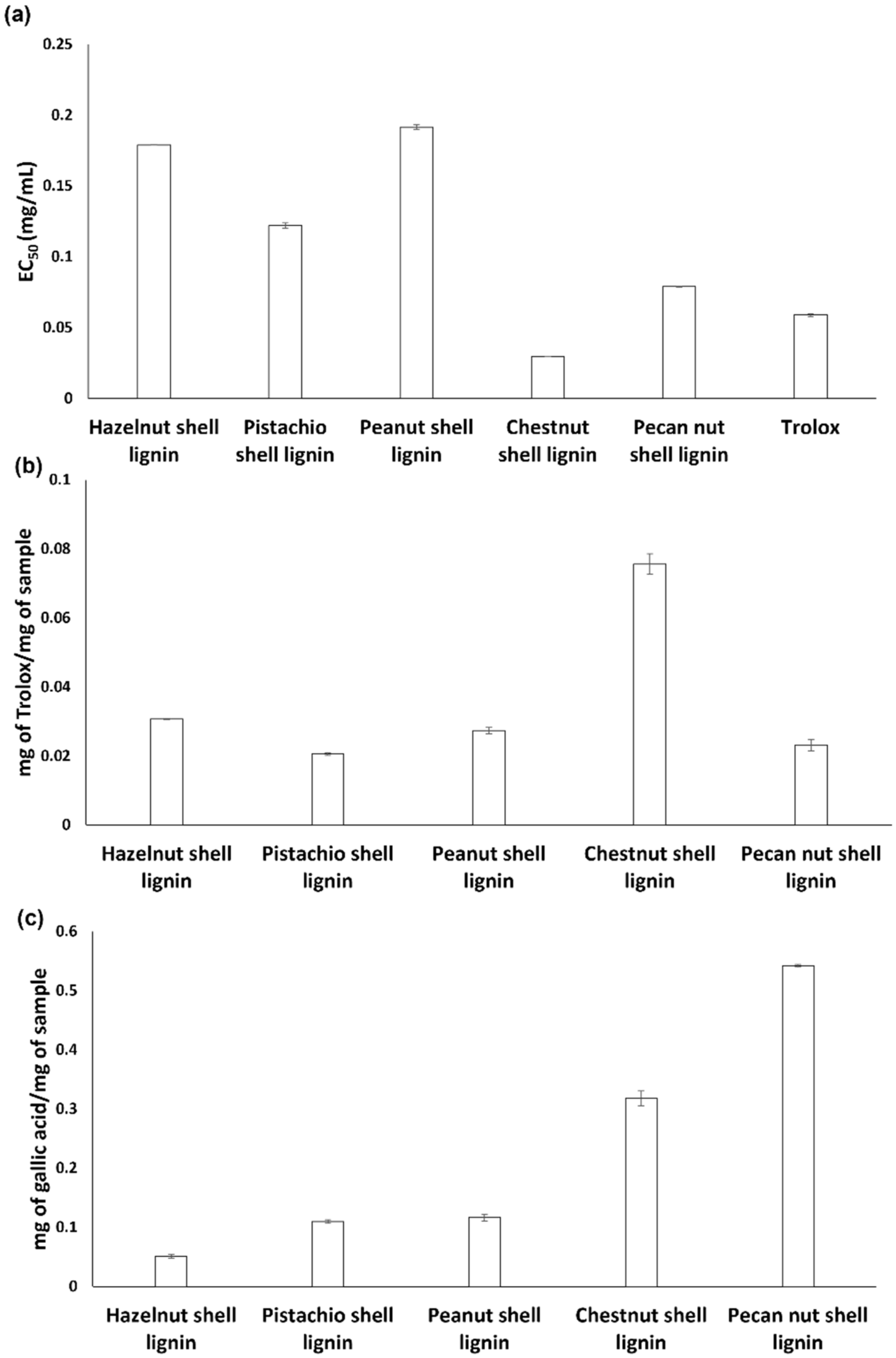
3.3. Antioxidant Properties of the Nut Shell-Derived Lignins

In a last series of experiments, the recovered lignins were characterized for their antioxidant properties and TPC with commonly used spectrophotometric assays.
In the DPPH assay, chestnut shell lignin was the most active material, exhibiting an EC50 value lower than that determined for the reference antioxidant compound Trolox (Figure 4a). Good DPPH-reducing properties were also observed for pecan nut shell lignin, confirming the high efficiency of this agri-food byproduct as a source of antioxidant compounds [1,58]. On the other hand, peanut, hazelnut and pistachio shell lignins were characterized by EC50 values significantly higher than that of chestnut and pecan nut shell lignins.
Chestnut shell lignin also exhibited the highest reducing properties in the FRAP assay, whereas all the other samples were characterized by comparable Trolox equivalent values (Figure 4b).
The superior antioxidant properties of chestnut shell lignin in both the DPPH and FRAP assay would be in line with the presence of the relatively high number of phenolic OH groups detected by 1H NMR analysis. It is also noteworthy that based on the amount of EA present in the chestnut shell lignin and the antioxidant properties reported for pure EA [23], this compound accounts for only 5–12% of the DPPH and Fe3+-reducing properties of the chestnut shell-derived sample, highlighting the real antioxidant effectiveness of chestnut lignin.
In agreement with the results of the antioxidant assays, the highest TPCs as determined by the Folin–Ciocalteu assay were observed for pecan nut shell and chestnut lignins (Figure 4c).
The discrepancy in the sample ranking emerging from the three assays is likely the result of the different properties monitored by each of them. Indeed, while the FRAP assay measures the electron transfer capacity of an antioxidant, the DPPH assay is a mixed-mode assay, as DPPH can be reduced through both an electron transfer and a hydrogen atom transfer mechanism [59]. On the other hand, the very high working pH required by the Folin–Ciocalteu assay may lead to an overestimation of the phenolic content and hence, of the antioxidant properties [60]. Of course, the relative solubility in the assay medium might also affect the antioxidant efficiency of the samples.
Notably, a remarkable direct linear correlation (R2 = 0.94) was found between the EC50 values from the DPPH assay and the ΔB values of the EPR spectra (Figure 5). This is in agreement with what has been previously reported for synthetic, biomimetic phenolic polymers [46,61], and would highlight the importance of π-electron spin delocalization, one of the main factors driving EPR signal narrowing, as a structural determinant of the antioxidant properties of phenolic polymers, including lignin.

4. Conclusions

In conclusion, a green protocol for the recovery of lignins from the shells of edible nuts based on ball milling/DES extraction is reported. The proposed approach allowed for the recovery in good yields of lignin-rich samples of good purity, characterized by very efficient antioxidant properties, comparable or even higher than those of the reference antioxidant Trolox, particularly in the DPPH assay. This is likely the result of the efficacy of the ball milling pre-treatment and the acidic nature of the DES employed in inducing cleavage of glycosidic and β-O-4 bonds of the lignin components, resulting in a higher availability of antioxidant phenolic OH groups. Although the potential of chestnut and pecan nut shells as sources of antioxidant compounds is well documented, the results of this study highlight the possibility of exploiting other widely produced nut shells to recover lignin samples for application as antioxidant additives in the biomedical, food, and/or cosmetic sectors, from a green chemistry perspective. In addition, this work provides, for the first time, a comparative and systematic evaluation of both the antioxidant and structural properties of lignins recovered from edible nut shells, highlighting the importance of the structural features (as investigated by spectroscopic and chromatographic techniques) in determining the functional properties of natural, complex phenolic polymers.

Supplementary Materials

The following supporting information can be downloaded at: https://www.mdpi.com/article/10.3390/antiox11101860/s1, Figure S1. Solid state EPR spectra of starting nut shells. Figure S2. ATR-FTIR spectra of (a) peanut, (b) chestnut, (c) hazelnut, (d) pecan nut and (e) pistachio shell lignins. Figure S3. 1H NMR spectra (DMSO-d6) of (a) peanut and (b) chestnut shell lignins. Figure S4. 1H NMR spectra (DMSO-d6) of (a) hazelnut and (b) pecan nut shell lignins. Figure S5. 1H NMR spectrum (DMSO-d6) of pistachio shell lignin. Figure S6. HPLC profiles of the alkaline hydrogen peroxide degradation mixtures of (a) peanut, (b) chestnut, (c) hazelnut, (d) pecan nut, and (e) pistachio shell lignins.

Author Contributions

Conceptualization, L.P.; data curation, R.A., F.M. and R.E.; investigation, R.A., F.M. and R.E.; methodology, R.A., F.M., G.D. and L.P.; supervision, G.D., L.P. and A.N.; validation, G.D. and L.P.; writing—original draft, R.A. and F.M.; writing—review and editing, G.D., L.P. and A.N. All authors have read and agreed to the published version of the manuscript.

Funding

This research received no external funding.

Institutional Review Board Statement

Not applicable.

Informed Consent Statement

Not applicable.

Data Availability Statement

All of the data is contained within the article.

Acknowledgments

The authors wish to thank the European Union (FSE, PON Ricerca e Innovazione 2014–2020, Azione I.1 “Dottorati Innovativi con caratterizzazione Industriale”) for funding a PhD grant to Federica Moccia. The financial support to Lucia Panzella provided by Italian MIUR, PRIN 2017 2017YJMPZN is acknowledged. Gerardino D’Errico wishes to thank project PON ARS01_00755 ProFood-IV.

Conflicts of Interest

The authors declare no conflict of interest.

References

  1. Moccia, F.; Agustin-Salazar, S.; Verotta, L.; Caneva, E.; Giovando, S.; D’Errico, G.; Panzella, L.; d’Ischia, M.; Napolitano, A. Antioxidant properties of agri-food byproducts and specific boosting effects of hydrolytic treatments. Antioxidants 2020, 9, 438. [Google Scholar] [CrossRef] [PubMed]
  2. Panzella, L.; Napolitano, A. Natural phenol polymers: Recent advances in food and health applications. Antioxidants 2017, 6, 30. [Google Scholar] [CrossRef] [PubMed]
  3. Han, H.; Lee, K. Systematic approach to mimic phenolic natural polymers for biofabrication. Polymers 2022, 14, 1282. [Google Scholar] [CrossRef] [PubMed]
  4. Khan, A.; Nair, V.; Colmenares, J.C.; Gläser, R. Lignin-based composite materials for photocatalysis. Top. Curr. Chem. 2018, 376, 20. [Google Scholar] [CrossRef]
  5. Rico-García, D.; Ruiz-Rubio, L.; Pérez-Alvarez, L.; Hernández-Olmos, S.L.; Guerrero-Ramírez, G.L.; Vilas-Vilela, J.L. Lignin-based hydrogels: Synthesis and applications. Polymers 2020, 12, 81. [Google Scholar] [CrossRef]
  6. Tao, J.; Li, S.; Ye, F.; Zhou, Y.; Lei, L.; Zhao, G. Lignin—An underutilized, renewable and valuable material for food industry. Crit. Rev. Food Sci. Nutr. 2020, 60, 2011–2033. [Google Scholar] [CrossRef]
  7. Verdini, F.; Gaudino, E.C.; Canova, E.; Tabasso, S.; Behbahani, P.J.; Cravotto, G. Lignin as a natural carrier for the efficient delivery of bioactive compounds: From waste to health. Molecules 2022, 27, 3598. [Google Scholar] [CrossRef]
  8. Pei, W.; Deng, J.; Wang, P.; Wang, X.; Zheng, L.; Zhang, Y.; Huang, C. Sustainable lignin and lignin-derived compounds as potential therapeutic agents for degenerative orthopaedic diseases: A systemic review. Int. J. Biol. Macromol. 2022, 212, 547–560. [Google Scholar] [CrossRef]
  9. Sreejaya, M.M.; Sankar, R.J.; Ramanunni, K.; Pillai, N.P.; Ramkumar, K.; Anuvinda, P.; Meenakshi, V.S.; Sadanandan, S. Lignin-based organic coatings and their applications: A review. Mater. Today Proc. 2022, 60, 494–501. [Google Scholar] [CrossRef]
  10. Lu, X.; Gu, X.; Shi, Y. A review on lignin antioxidants: Their sources, isolations, antioxidant activities and various applications. Int. J. Biol. Macromol. 2022, 210, 716–741. [Google Scholar] [CrossRef]
  11. Nan, N.; Hu, W.; Wang, J. Lignin-based porous biomaterials for medical and pharmaceutical applications. Biomedicines 2022, 10, 747. [Google Scholar] [CrossRef] [PubMed]
  12. Perea-Moreno, M.-A.; Manzano-Agugliaro, F.; Hernandez-Escobedo, Q.; Perea-Moreno, A.-J. Peanut shell for energy: Properties and its potential to respect the environment. Sustainability 2018, 10, 3254. [Google Scholar] [CrossRef]
  13. Agustin-Salazar, S.; Cerruti, P.; Medina-Juárez, L.Á.; Scarinzi, G.; Malinconico, M.; Soto-Valdez, H.; Gamez-Meza, N. Lignin and holocellulose from pecan nutshell as reinforcing fillers in poly (lactic acid) biocomposites. Int. J. Biol. Macromol. 2018, 115, 727–736. [Google Scholar] [CrossRef] [PubMed]
  14. Marius, C.; Sepperer, T.; Tudor, E.M.; Petutschnigg, A. Walnut and hazelnut shells: Untapped industrial resources and their suitability in lignocellulosic composites. Appl. Sci. 2020, 10, 6340. [Google Scholar] [CrossRef]
  15. Pistachio Production in 2018, Crops/Regions/World List/Production Quantity (Pick Lists). UN Food Agric. Organ. Corp. Stat. Database. 2019. Available online: https://www.fao.org/faostat/en/#home (accessed on 3 July 2022).
  16. Silva, V.; Falco, V.; Dias, M.I.; Barros, L.; Silva, A.; Capita, R.; Alonso-Calleja, C.; Amaral, J.S.; Igrejas, G.; Ferreira, I.C.F.R.; et al. Evaluation of the phenolic profile of Castanea sativa Mill. by-products and their antioxidant and antimicrobial activity against multiresistant bacteria. Antioxidants 2020, 9, 87. [Google Scholar] [CrossRef]
  17. Lucarini, M.; Durazzo, A.; Bernini, R.; Campo, M.; Vita, C.; Souto, E.B.; Lombardi-Boccia, G.; Ramadan, M.F.; Santini, A.; Romani, A. Fruit wastes as a valuable source of value-added compounds: A collaborative perspective. Molecules 2021, 26, 6338. [Google Scholar] [CrossRef] [PubMed]
  18. Liu, J.; Li, X.; Row, H.K. Development of deep eutectic solvents for sustainable chemistry. J. Mol. Liq. 2022, 362, 119654. [Google Scholar] [CrossRef]
  19. Ling, J.K.U.; Hadinoto, K. Deep eutectic solvent as green solvent in extraction of biological macromolecules: A review. Int. J. Mol. Sci. 2022, 23, 3381. [Google Scholar] [CrossRef]
  20. Ijardar, S.P.; Singh, V.; Gardas, R.L. Revisiting the physicochemical properties and applications of deep eutectic solvents. Molecules 2022, 27, 1368. [Google Scholar] [CrossRef]
  21. Chen, Z.; Ragauskas, A.; Wan, C. Lignin extraction and upgrading using deep eutectic solvents. Ind. Crops Prod. 2020, 147, 112241. [Google Scholar] [CrossRef]
  22. Dheyab, A.S.; Abu Bakar, M.F.; AlOmar, M.; Sabran, S.F.; Hanafi, A.F.M.; Mohamad, A. Deep eutectic solvents (DESs) as green extraction media of beneficial bioactive phytochemicals. Separation 2021, 8, 176. [Google Scholar] [CrossRef]
  23. Moccia, F.; Gallucci, N.; Giovando, S.; Zuorro, A.; Lavecchia, R.; D’Errico, G.; Panzella, L.; Napolitano, A. A tunable deep eutectic solvent-based processing for valorization of chestnut wood fiber as a source of ellagic acid and lignin. J. Environ. Chem. Eng. 2022, 10, 107773. [Google Scholar] [CrossRef]
  24. Francisco, M.; Van Den Bruinhorst, A.; Kroon, M.C. New natural and renewable low transition temperature mixtures (LTTMs): Screening as solvents for lignocellulosic biomass processing. Green Chem. 2012, 14, 2153–2157. [Google Scholar] [CrossRef]
  25. Ci, Y.H.; Yu, F.; Zhou, C.X.; Mo, H.E.; Li, Z.Y.; Ma, Y.Q.; Zang, L.H. New ternary deep eutectic solvents for effective wheat straw deconstruction into its high-value utilization under near-neutral conditions. Green Chem. 2020, 22, 8713–8720. [Google Scholar] [CrossRef]
  26. Provost, V.; Dumarcay, S.; Ziegler-Devin, I.; Boltoeva, M.; Trébouet, D.; Villain-Gambier, M. Deep eutectic solvent pretreatment of biomass: Influence of hydrogen bond donor and temperature on lignin extraction with high β-O-4 content. Bioresour. Technol. 2022, 349, 126837. [Google Scholar] [CrossRef]
  27. Muñoz-García, A.B.; Sannino, F.; Vitiello, G.; Pirozzi, D.; Minieri, L.; Aronne, A.; Pernice, P.; Pavone, M.; D’Errico, G. Origin and electronic features of reactive oxygen species at hybrid zirconia-acetylacetonate interfaces. ACS Appl. Mater. Interfaces 2015, 7, 21662–21667. [Google Scholar] [CrossRef]
  28. Mostert, A.B.; Hanson, G.R.; Sarna, T.; Gentle, I.R.; Powell, B.J.; Meredith, P. Hydration-controlled X-band EPR spectroscopy: A tool for unravelling the complexities of the solid-state free radical in eumelanin. J. Phys. Chem. B 2013, 117, 4965–4972. [Google Scholar] [CrossRef]
  29. Li, C.; Huang, C.; Zhao, Y.; Zheng, C.; Su, H.; Zhang, L.; Luo, W.; Zhao, H.; Wang, S.; Huang, L.-J. Effect of choline-based deep eutectic solvent pretreatment on the structure of cellulose and lignin in bagasse. Processes 2021, 9, 384. [Google Scholar] [CrossRef]
  30. Goupy, P.; Dufour, C.; Loonis, M.; Dangles, O. Quantitative kinetic analysis of hydrogen transfer reactions from dietary polyphenols to the DPPH radical. J. Agric. Food Chem. 2003, 51, 615–622. [Google Scholar] [CrossRef]
  31. Gökmen, V.; Serpen, A.; Fogliano, V. Direct measurement of the total antioxidant capacity of foods: The “QUENCHER” approach. Trends Food Sci. Technol. 2009, 20, 278–288. [Google Scholar] [CrossRef]
  32. Benzie, I.F.F.; Strain, J.J. The ferric reducing ability of plasma (FRAP) as a measure of ‘“antioxidant power”’: The FRAP assay. Anal. Biochem. 1996, 239, 70–76. [Google Scholar] [CrossRef] [PubMed]
  33. Seifzadeh, N.; Sahari, M.A.; Barzegar, M.; Gavlighi, H.A.; Calani, L.; Del Rio, D.; Galaverna, G. Evaluation of polyphenolic compounds in membrane concentrated pistachio hull extract. Food Chem. 2019, 277, 398–406. [Google Scholar] [CrossRef] [PubMed]
  34. Herald, T.J.; Gadgil, P.; Perumal, R.; Bean, S.R.; Wilson, J.D. High-throughput micro-plate HCl-vanillin assay for screening tannin content in sorghum grain. J. Sci. Food Agric. 2014, 94, 2133–2136. [Google Scholar] [CrossRef] [PubMed]
  35. Panzella, L.; Eidenberger, T.; Napolitano, A.; d’Ischia, M. Black sesame pigment: DPPH assay-guided purification, antioxidant/antinitrosating properties, and identification of a degradative structural marker. J. Agric. Food Chem. 2012, 60, 8895–8901. [Google Scholar] [CrossRef]
  36. Garcia-Villalba, R.; Espín, J.C.; Kroon, P.A.; Alasalvar, C.; Heinonen, M.; Voorspoels, S.; Tomas-Barberan, F. A validated method for the characterization and quantification of extractable and non-extractable ellagitannins after acid hydrolysis in pomegranate fruits, juices, and extracts. J. Agric. Food Chem. 2015, 63, 6555–6566. [Google Scholar] [CrossRef]
  37. Gea, A.; Stringano, E.; Brown, R.H.; Mueller-Harvey, I. In Situ analysis and structural elucidation of sainfoin (Onobrychis viciifolia) tannins for high-throughput germplasm screening. J. Agric. Food Chem. 2011, 59, 495–503. [Google Scholar] [CrossRef]
  38. Yang, G.; An, X.; Yang, S. The effect of ball milling time on the isolation of lignin in the cell wall of different biomass. Front. Bioeng. Biotechnol. 2022, 9, 807625. [Google Scholar] [CrossRef]
  39. Guo, X.; Junna, X.; Wolcott, M.P.; Zhang, J. Mechanochemical oleation of lignin through ball milling and properties of its blends with PLA. Sustain. Chem. 2016, 1, 3449–3454. [Google Scholar] [CrossRef]
  40. Zinovyev, G.; Sumerskii, I.; Rosenau, T.; Balakshin, M.; Potthast, A. Ball milling’s effect on pine milled wood lignin’s structure and molar mass. Molecules 2018, 23, 2223. [Google Scholar] [CrossRef]
  41. Dabral, S.; Wotruba, H.; Hernandez, J.G.; Bolm, C. Mechanochemical oxidation and cleavage of lignin β-O-4 model compounds and lignin. ACS Sustain. Chem. Eng. 2018, 6, 3242–3254. [Google Scholar] [CrossRef]
  42. Schiavi, D.; Ronchetti, R.; Di Lorenzo, V.; Salustri, M.; Petrucci, C.; Vivani, R.; Giovagnoli, S.; Camaioni, E.; Balestra, G.M. Circular hazelnut protection by lignocellulosic waste valorization for nanopesticides development. Appl. Sci. 2022, 12, 2604. [Google Scholar] [CrossRef]
  43. Gordobil, O.; Olaizola, P.; Banales, J.M.; Labidi, J. Lignins from agroindustrial by-products as natural ingredients for cosmetics: Chemical structure and in vitro sunscreen and cytotoxic activities. Molecules 2020, 25, 1131. [Google Scholar] [CrossRef]
  44. Du, B.; Wang, X.; Chai, L.; Wang, X.; Pan, Z.; Chen, X.; Zhou, J.; Sun, R.-C. Fabricating lignin-based carbon nanofibers as versatile supercapacitors from food wastes. Int. J. Biol. Macromol. 2022, 194, 632–643. [Google Scholar] [CrossRef] [PubMed]
  45. Sogut, E.; Seydim, A.C. Utilization of chestnut shell lignin in alginate films. J. Sci. Food Agric. 2022. [Google Scholar] [CrossRef] [PubMed]
  46. Panzella, L.; D’Errico, G.; Vitiello, G.; Perfetti, M.; Alfieri, M.L.; Napolitano, A.; d’Ischia, M. Disentangling structure-dependent antioxidant mechanisms in phenolic polymers by multiparametric EPR analysis. Chem. Commun. 2018, 54, 9426–9429. [Google Scholar] [CrossRef] [PubMed]
  47. Patil, S.V.; Argyropoulos, D.S. Stable organic radicals in lignin: A review. ChemSusChem 2017, 10, 3284–3303. [Google Scholar] [CrossRef] [PubMed]
  48. Bährle, C.; Nick, T.U.; Bennati, M.; Jeschke, G.; Vogel, F. High-field electron paramagnetic resonance and density functional theory study of stable organic radicals in lignin: Influence of the extraction process, botanical origin, and protonation reactions on the radical g tensor. J. Phys. Chem. A 2015, 119, 6475–6482. [Google Scholar] [CrossRef]
  49. Kocheva, L.S.; Karmanov, A.P.; Mironov, M.V.; Belyy, V.A.; Polina, I.N.; Pokryshkin, S.A. Characteristics of chemical structure of lignin biopolymer from Araucaria relict plant. Questions and answers of evolution. Int. J. Biol. Macromol. 2020, 159, 896–903. [Google Scholar] [CrossRef]
  50. Panzella, L.; Cerruti, P.; Ambrogi, V.; Agustin-Salazar, S.; D’Errico, G.; Carfagna, C.; Goya, L.; Ramos, S.; Martín, M.A.; Napolitano, A.; et al. A superior all-natural antioxidant biomaterial from spent coffee grounds for polymer stabilization, cell protection, and food lipid preservation. ACS Sustain. Chem. Eng. 2016, 4, 1169–1179. [Google Scholar] [CrossRef]
  51. Piccinino, D.; Capecchi, E.; Tomaino, E.; Gabellone, S.; Gigli, V.; Avitabile, D.; Saladino, R. Nano-structured lignin as green antioxidant and UV shielding ingredient for sunscreen applications. Antioxidants 2021, 10, 274. [Google Scholar] [CrossRef]
  52. Panzella, L.; Pérez-Burillo, S.; Pastoriza, S.; Martín, M.Á.; Cerruti, P.; Goya, L.; Ramos, S.; Rufián-Henares, J.Á.; Napolitano, A.; d’Ischia, M. High antioxidant action and prebiotic activity of hydrolyzed spent coffee grounds (HSCG) in a simulated digestion-fermentation model: Toward the development of a novel food supplement. J. Agric. Food Chem. 2017, 65, 6452–6459. [Google Scholar] [CrossRef] [PubMed]
  53. Miki, K.; Kamitakahara, H.; Yoshinaga, A.; Tobimatsu, Y.; Takano, T. Methylation-triggered fractionation of lignocellulosic biomass to afford cellulose-, hemicellulose-, and lignin-based functional polymers via click chemistry. Green Chem. 2020, 22, 2909–2928. [Google Scholar] [CrossRef]
  54. Goriparti, S.; Harish, M.N.K.; Sampath, S. Ellagic acid—A novel organic electrode material for high capacity lithium ion batteries. Chem. Commun. 2013, 49, 7234–7236. [Google Scholar] [CrossRef] [PubMed]
  55. González-Rivera, J.; Mero, A.; Husanu, E.; Mezzetta, A.; Ferrari, C.; D’Andrea, F.; Bramanti, E.; Pomelli, C.S.; Guazzelli, L. Combining acid-based deep eutectic solvents and microwave irradiation for improved chestnut shell waste valorization. Green Chem. 2021, 23, 10101–10115. [Google Scholar] [CrossRef]
  56. Husanu, E.; Mero, A.; Rivera, J.G.; Mezzetta, A.; Ruiz, J.C.; D’Andrea, F.; Pomelli, C.S.; Guazzelli, L. Exploiting deep eutectic solvents and ionic liquids for the valorization of chestnut shell waste. ACS Sustain. Chem. Eng. 2020, 8, 18386–18399. [Google Scholar] [CrossRef]
  57. Panzella, L.; Napolitano, A. Condensed tannins, a viable solution to meet the need for sustainable and effective multifunctionality in food packaging: Structure, sources, and properties. J. Agric. Food Chem. 2022, 70, 751–758. [Google Scholar] [CrossRef]
  58. Moccia, F.; Agustin-Salazar, S.; Berg, A.; Setaro, B.; Micillo, R.; Pizzo, E.; Weber, F.; Gamez-Meza, N.; Schieber, A.; Cerruti, P.; et al. Pecan (Carya illinoinensis (Wagenh.) K. Koch) nut shell as an accessible polyphenol source for active packaging and food colorant stabilization. ACS Sustain. Chem. Eng. 2020, 8, 6700–6712. [Google Scholar] [CrossRef]
  59. Prior, R.L.; Wu, X.; Shaich, K. Standardized methods for the determination of antioxidant capacity and phenolics in foods and dietary supplements. J. Agric. Food Chem. 2005, 53, 4290–4302. [Google Scholar] [CrossRef]
  60. Mustafa, O.; Gu, K.; Esra, C. Antioxidant activity/capacity measurement. 1. Classification, physicochemical principles, mechanisms, and electron transfer (ET)-based assays. J. Agric. Food Chem. 2016, 64, 997–1027. [Google Scholar] [CrossRef]
  61. Alfieri, M.L.; Moccia, F.; D’Errico, G.; Panzella, L.; d’Ischia, M.; Napolitano, A. Acid treatment enhances the antioxidant activity of enzymatically synthesized phenolic polymers. Polymers 2020, 12, 2544. [Google Scholar] [CrossRef]
Figure 1. (a) Solid-state EPR spectra and (b) power saturation profiles of lignins recovered from the different nut shells.
Antioxidants 11 01860 g001
Figure 2. UV-Vis spectra of the DMSO-soluble fraction of (a) peanut, (b) chestnut, (c) hazelnut, (d) pecan nut, and (e) pistachio shell lignins.
Antioxidants 11 01860 g002
Figure 3. HPLC profile of the DMSO-soluble fraction of chestnut shell lignin.
Antioxidants 11 01860 g003
Figure 4. Antioxidant properties and TPC of nut shell-derived lignins. (a) DPPH assay; (b) FRAP assay; (c) Folin-Ciocalteu assay. Reported are the mean ± SD values from at least three experiments.
Antioxidants 11 01860 g004
Figure 5. Correlation between antioxidant properties (expressed as EC50 values determined in the DPPH assay) and ΔB values (determined by EPR spectroscopy) of nut shell lignins (EC50 = (−0.58 ± 0.10) + (1.15 ± 0.02) ΔB, R2 = 0.94).
Antioxidants 11 01860 g005
Table 1. Lignin extraction and recovery yields from selected nut shells.
ShellExtraction Yield (% w/ws) 1Recovery Yield (% w/wd) 1
Peanut1934
Chestnut1927
Hazelnut2536
Pecan nut1934
Pistachio2738
1ws: weight of starting material; wd: weight of the DES-dissolved material.
Table 2. EPR data for nut shells and related lignins.
SampleSpin/g
Concentration
ΔB
(±0.2)
Gauss Fraction (±0.05) 1g-Factor (±0.0003)
Peanut shellStarting2.6 × 10165.6-2.0038
Lignin2.2 × 10175.10.422.0030
Chestnut
shell
Starting1.6 × 10165.9-2.0031
Lignin3.5 × 10174.20.702.0031
Hazelnut
shell
Starting2.1 × 10167.0-2.0031
Lignin6.6 × 10165.30.212.0036
Pecan nut
shell
Starting1.4 × 10164.8-2.0026
Lignin4.6 × 10174.50.482.0030
Pistachio
shell
Starting5.9 × 10156.8-2.0037
Lignin2.5 × 10174.70.432.0027
1 For the spectra of the starting material showing the superposition of signals, the Gaussian fraction was not determined.
Publisher’s Note: MDPI stays neutral with regard to jurisdictional claims in published maps and institutional affiliations.

