The Role of Antioxidants Supplementation in Clinical Practice: Focus on Cardiovascular Risk Factors

 ,
,  , , , , , and
Abstract
:1. Introduction
2. Biomarkers of Oxidative Stress in Clinical Practice
2.1. Advanced Glycation End Products (AGEs)
2.2. Oxidized Low-Density Lipoprotein (oxLDL)
2.3. Protein Oxidation Is Advanced Oxidation Protein Products (AOPP)
2.4. Lipid Oxidation Products
2.5. 8-Hydroxy-2′-Deoxyguanosine (8-OHdG)
2.6. Hydrogen Peroxide (H2O2)
2.7. NOX2 Activity (sNOX2-dp)
3. Antioxidants Supplementations: Which Are the Most Effective in Clinical Practice?
3.1. Vitamins E and C
3.2. Omega-3 and Omega-6 Fatty Acids
3.3. Polyphenols
3.4. Non-Flavonoids
3.5. Flavonoids
3.6. Carotenoids
3.7. Selenium
3.8. Lipoic Acid
3.9. Coenzyme Q10
4. Biomarkers of Oxidative Stress in Patients with Cardiovascular Risk Factors
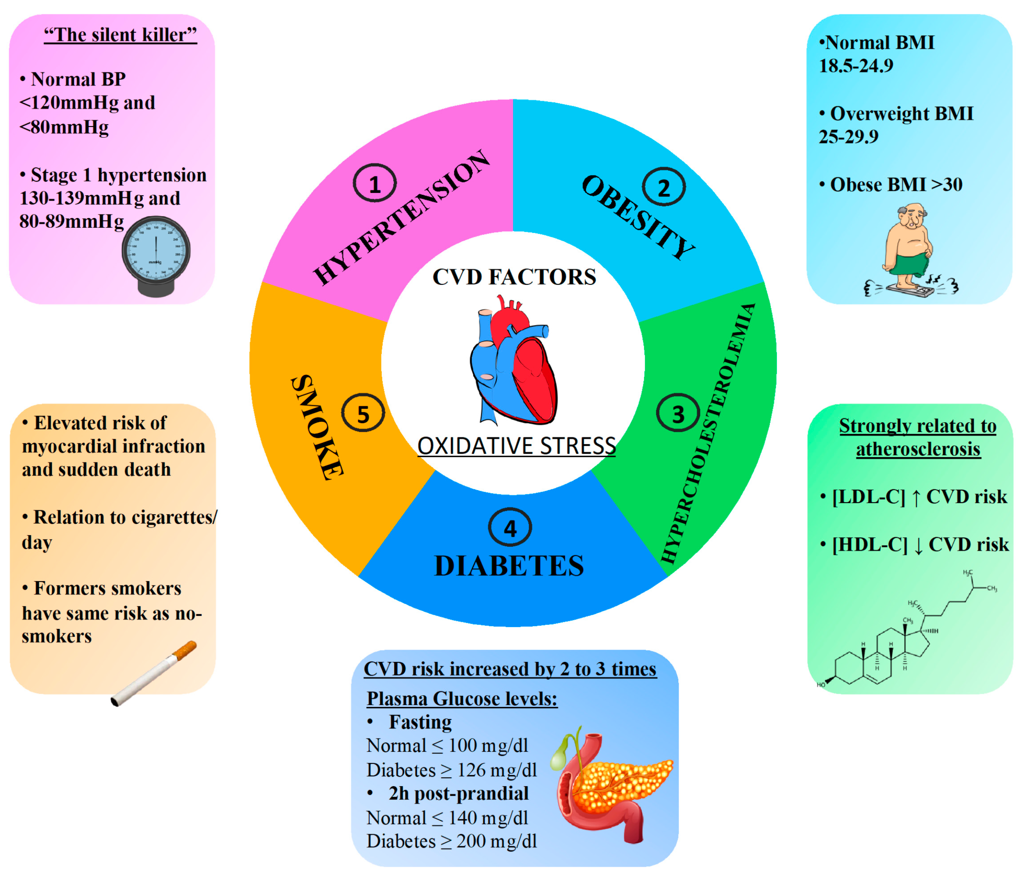
4.1. Hypertension
4.2. Diabetes
4.3. Hypercholesterolemia
4.4. Obesity
4.5. Smoking
5. Antioxidant Supplementation in Patients with Cardiovascular Risk Factors
5.1. Hypertension
5.2. Diabetes
5.3. Hypercholesterolemia
5.4. Obesity
5.5. Smoke
6. Conclusions
Author Contributions
Funding
Acknowledgments
Conflicts of Interest
References
- Phaniendra, A.; Jestadi, D.B.; Periyasamy, L. Free radicals: Properties, sources, targets, and their implication in various diseases. Indian J. Clin. Biochem. 2015, 30, 11–26. [Google Scholar] [CrossRef] [PubMed] [Green Version]
- Valko, M.; Leibfritz, D.; Moncol, J.; Cronin, M.T.D.; Mazur, M.; Telser, J. Free radicals and antioxidants in normal physiological functions and human disease. Int. J. Biochem. Cell Biol. 2007, 39, 44–84. [Google Scholar] [CrossRef] [PubMed]
- Katerji, M.; Filippova, M.; Duerksen-Hughes, P. Approaches and Methods to Measure Oxidative Stress in Clinical Samples: Research Applications in the Cancer Field. Oxid. Med. Cell. Longev. 2019, 2019. [Google Scholar] [CrossRef] [PubMed] [Green Version]
- El Hawary, R.; Meshaal, S.; Deswarte, C.; Galal, N.; Abdelkawy, M.; Alkady, R.; Elaziz, D.A.; Freiberger, T.; Ravcukova, B.; Litzman, J.; et al. Role of Flow Cytometry in the Diagnosis of Chronic Granulomatous Disease: The Egyptian Experience. J. Clin. Immunol. 2016, 36, 610–618. [Google Scholar] [CrossRef] [PubMed]
- Gomes, A.; Fernandes, E.; Lima, J.L.F.C. Fluorescence probes used for detection of reactive oxygen species. J. Biochem. Biophys. Methods 2005, 65, 45–80. [Google Scholar] [CrossRef]
- Drummen, G.P.C.; Van Liebergen, L.C.M.; Op den Kamp, J.A.F.; Post, J.A. C11-BODIPY581/591, an oxidation-sensitive fluorescent lipid peroxidation probe: (Micro)spectroscopic characterization and validation of methodology. Free Radic. Biol. Med. 2002, 33, 473–490. [Google Scholar] [CrossRef]
- Weber, D.; Milkovic, L.; Bennett, S.J.; Griffiths, H.R.; Zarkovic, N.; Grune, T. Measurement of HNE-protein adducts in human plasma and serum by ELISA-Comparison of two primary antibodies. Redox Biol. 2013, 1, 226–233. [Google Scholar] [CrossRef] [Green Version]
- Mehta, K.; Patel, V.B. Measurement of 4-Hydroxynonenal (4-HNE) protein adducts by ELISA. Methods Mol. Biol. 2019, 1990, 43–52. [Google Scholar]
- Khoschsorur, G.A.; Winklhofer-Roob, B.M.; Rabl, H.; Auer, T.; Peng, Z.; Schaur, R.J. Evaluation of a sensitive HPLC method for the determination of malondialdehyde, and application of the method to different biological materials. Chromatographia 2000, 52, 181–184. [Google Scholar] [CrossRef]
- Dreißigacker, U.; Suchy, M.T.; Maassen, N.; Tsikas, D. Human plasma concentrations of malondialdehyde (MDA) and the F2-isoprostane 15(S)-8-iso-PGF2α may be markedly compromised by hemolysis: Evidence by GC-MS/MS and potential analytical and biological ramifications. Clin. Biochem. 2010, 43, 159–167. [Google Scholar] [CrossRef]
- Smith, K.A.; Shepherd, J.; Wakil, A.; Kilpatrick, E.S. A comparison of methods for the measurement of 8-isoPGF2α: A marker of oxidative stress. Ann. Clin. Biochem. 2011, 48, 147–154. [Google Scholar] [CrossRef] [PubMed]
- Buss, H.; Chan, T.P.; Sluis, K.B.; Domigan, N.M.; Winterbourn, C.C. Protein carbonyl measurement by a sensitive ELISA method. Free Radic. Biol. Med. 1997, 23, 361–366. [Google Scholar] [CrossRef]
- Taylor, E.L.; Armstrong, K.R.; Perrett, D.; Hattersley, A.T.; Winyard, P.G. Optimisation of an advanced oxidation protein products assay: Its application to studies of oxidative stress in diabetes mellitus. Oxid. Med. Cell. Longev. 2015, 2015. [Google Scholar] [CrossRef] [Green Version]
- Dizdaroglu, M.; Jaruga, P.; Birincioglu, M.; Rodriguez, H. Free radical-induced damage to DNA: Mechanisms and measurement. Free Radic. Biol. Med. 2002, 32, 1102–1115. [Google Scholar] [CrossRef]
- Garaycoechea, J.I.; Crossan, G.P.; Langevin, F.; Mulderrig, L.; Louzada, S.; Yang, F.; Guilbaud, G.; Park, N.; Roerink, S.; Nik-Zainal, S.; et al. Alcohol and endogenous aldehydes damage chromosomes and mutate stem cells. Nature 2018, 553, 171–177. [Google Scholar] [CrossRef]
- Weydert, C.J.; Cullen, J.J. Measurement of superoxide dismutase, catalase and glutathione peroxidase in cultured cells and tissue. Nat. Protoc. 2010, 5, 51–66. [Google Scholar] [CrossRef] [Green Version]
- Marrocco, I.; Altieri, F.; Peluso, I. Measurement and Clinical Significance of Biomarkers of Oxidative Stress in Humans. Oxid. Med. Cell. Longev. 2017, 2017. [Google Scholar] [CrossRef]
- Kaplan, L.A.; Miller, J.A.; Stein, E.A.; Stampfer, M.J. Simultaneous, high-performance liquid chromatographic analysis of retinol, tocopherols, lycopene, and α- and β-carotene in serum and plasma. Methods Enzymol. 1990, 189, 155–167. [Google Scholar] [CrossRef]
- Lettieri-Barbato, D.; Tomei, F.; Sancini, A.; Morabito, G.; Serafini, M. Effect of plant foods and beverages on plasma non-enzymatic antioxidant capacity in human subjects: A meta-analysis. Br. J. Nutr. 2013, 109, 1544–1556. [Google Scholar] [CrossRef] [Green Version]
- Arauz, J.; Ramos-Tovar, E.; Muriel, P. Redox state and methods to evaluate oxidative stress in liver damage: From bench to bedside. Ann. Hepatol. 2016, 15, 160–173. [Google Scholar]
- Ogino, K.; Wang, D.H. Biomarkers of oxidative/nitrosative stress: An approach to disease prevention. Acta Med. Okayama 2007, 61. [Google Scholar] [CrossRef]
- Dikalov, S.; Griendling, K.K.; Harrison, D.G. Measurement of reactive oxygen species in cardiovascular studies. Hypertension 2007, 49, 717–727. [Google Scholar] [CrossRef] [PubMed] [Green Version]
- Forkink, M.; Smeitink, J.A.M.; Brock, R.; Willems, P.H.G.M.; Koopman, W.J.H. Detection and manipulation of mitochondrial reactive oxygen species in mammalian cells. Biochim. Biophys. Acta Bioenerg. 2010, 1797, 1034–1044. [Google Scholar] [CrossRef] [PubMed] [Green Version]
- Winterbourn, C.C. The challenges of using fluorescent probes to detect and quantify specific reactive oxygen species in living cells. Biochim. Biophys. Acta 2014, 1840, 730–738. [Google Scholar] [CrossRef] [PubMed]
- Esterbauer, H.; Cheeseman, K.H. Determination of aldehydic lipid peroxidation products: Malonaldehyde and 4-hydroxynonenal. Methods Enzymol. 1990, 186, 407–421. [Google Scholar] [CrossRef]
- Lapenna, D.; Ciofani, G.; Pierdomenico, S.D.; Giamberardino, M.A.; Cuccurullo, F. Reaction conditions affecting the relationship between thiobarbituric acid reactivity and lipid peroxides in human plasma. Free Radic. Biol. Med. 2001, 31, 331–335. [Google Scholar] [CrossRef]
- Yu, R.; Zhao, G.; Christman, J.W.; Xiao, L.; Van Breemen, R.B. Method development and validation for ultra-high pressure liquid chromatography/tandem mass spectrometry determination of multiple prostanoids in biological samples. J. AOAC Int. 2013, 96, 67–76. [Google Scholar] [CrossRef] [Green Version]
- Yan, L.J.; Forster, M.J. Chemical probes for analysis of carbonylated proteins: A review. J. Chromatogr. B Anal. Technol. Biomed. Life Sci. 2011, 879, 1308–1315. [Google Scholar] [CrossRef] [Green Version]
- Bigagli, E.; Lodovici, M. Circulating oxidative stress biomarkers in clinical studies on type 2 diabetes and its complications. Oxid. Med. Cell. Longev 2019, 1, 1–17. [Google Scholar] [CrossRef]
- Broedbaek, K.; Siersma, V.; Henriksen, T.; Weimann, A.; Petersen, M.; Andersen, J.T.; Jimenez-Solem, E.; Hansen, L.J.; Henriksen, J.E.; Bonnema, S.J.; et al. Urinary markers of nucleic acid oxidation and cancer in type 2 diabetes. Redox Biol. 2015, 4, 34–39. [Google Scholar] [CrossRef] [Green Version]
- Voulgaridou, G.P.; Anestopoulos, I.; Franco, R.; Panayiotidis, M.I.; Pappa, A. DNA damage induced by endogenous aldehydes: Current state of knowledge. Mutat. Res.-Fundam. Mol. Mech. Mutagen. 2011, 711, 13–27. [Google Scholar] [CrossRef] [PubMed]
- Carnevale, R.; Nocella, C.; Pignatelli, P.; Bartimoccia, S.; Stefanini, L.; Basili, S.; Novo, M.; D’Amico, A.; Cammisotto, V.; Pastori, D.; et al. Blood hydrogen peroxide break-down activity in healthy subjects and in patients at risk of cardiovascular events. Atherosclerosis 2018, 274, 29–34. [Google Scholar] [CrossRef] [PubMed] [Green Version]
- Strimbu, K.; Tavel, J.A. What are biomarkers? Curr. Opin. HIV AIDS 2010, 5, 463–466. [Google Scholar] [CrossRef] [PubMed]
- Magalhães, P.M.; Appell, H.J.; Duarte, J.A. Involvement of advanced glycation end products in the pathogenesis of diabetic complications: The protective role of regular physical activity. Eur. Rev. Aging Phys. Act. 2008, 5, 17–29. [Google Scholar] [CrossRef] [Green Version]
- Dhama, K.; Latheef, S.K.; Dadar, M.; Samad, H.A.; Munjal, A.; Khandia, R.; Karthik, K.; Tiwari, R.; Yatoo, M.I.; Bhatt, P.; et al. Biomarkers in stress related diseases/disorders: Diagnostic, prognostic, and therapeutic values. Front. Mol. Biosci. 2019, 6, 91. [Google Scholar] [CrossRef]
- Stirban, A.; Gawlowski, T.; Roden, M. Vascular effects of advanced glycation endproducts: Clinical effects and molecular mechanisms. Mol. Metab. 2014, 3, 94–108. [Google Scholar] [CrossRef] [PubMed]
- Gil, L.; Siems, W.; Mazurek, B.; Gross, J.; Schroeder, P.; Voss, P.; Grune, T. Age-associated analysis of oxidative stress parameters in human plasma and erythrocytes. Free Radic. Res. 2006, 40, 495–505. [Google Scholar] [CrossRef] [PubMed]
- Greilberger, J.; Koidl, C.; Greilberger, M.; Lamprecht, M.; Schroecksnadel, K.; Leblhuber, F.; Fuchs, D.; Oettl, K. Malondialdehyde, carbonyl proteins and albumin-disulphide as useful oxidative markers in mild cognitive impairment and Alzheimer’s disease. Free Radic. Res. 2008, 42, 633–638. [Google Scholar] [CrossRef]
- Totan, Y.; Yağcı, R.; Bardak, Y.; Özyurt, H.; Kendir, F.; Yılmaz, G.; Şahin, Ş.; Şahin Tığ, U. Oxidative Macromolecular Damage in Age-Related Macular Degeneration. Curr. Eye Res. 2009, 34, 1089–1093. [Google Scholar] [CrossRef]
- Tsimikas, S. Oxidized low-density lipoprotein biomarkers in atherosclerosis. Curr. Atheroscler. Rep. 2006, 8, 55–61. [Google Scholar] [CrossRef]
- Gabai, G.; De Luca, E.; Miotto, G.; Zin, G.; Stefani, A.; Da Dalt, L.; Barberio, A.; Celi, P. Relationship between Protein Oxidation Biomarkers and Uterine Health in Dairy Cows during the Postpartum Period. Antioxidants 2019, 8, 21. [Google Scholar] [CrossRef] [PubMed] [Green Version]
- Kaneda, H.; Taguchi, J.; Ogasawara, K.; Aizawa, T.; Ohno, M. Increased level of advanced oxidation protein products in patients with coronary artery disease. Atherosclerosis 2002, 162, 221–225. [Google Scholar] [CrossRef]
- Martín-Gallán, P.; Carrascosa, A.; Gussinyé, M.; Domínguez, C. Biomarkers of diabetes-associated oxidative stress and antioxidant status in young diabetic patients with or without subclinical complications. Free Radic. Biol. Med. 2003, 34, 1563–1574. [Google Scholar] [CrossRef]
- Negre-Salvayre, A.; Auge, N.; Ayala, V.; Basaga, H.; Boada, J.; Brenke, R.; Chapple, S.; Cohen, G.; Feher, J.; Grune, T.; et al. Pathological aspects of lipid peroxidation. Free Radic. Res. 2010, 44, 1125–1171. [Google Scholar] [CrossRef]
- Milne, G.L.; Sanchez, S.C.; Musiek, E.S.; Morrow, J.D. Quantification of F2-isoprostanes as a biomarker of oxidative stress. Nat. Protoc. 2007, 2, 221–226. [Google Scholar] [CrossRef]
- Davies, S.S.; Roberts, L.J. F2-isoprostanes as an indicator and risk factor for coronary heart disease. Free Radic. Biol. Med. 2011, 50, 559–566. [Google Scholar] [CrossRef] [Green Version]
- Ito, F.; Sono, Y.; Ito, T. Measurement and clinical significance of lipid peroxidation as a biomarker of oxidative stress: Oxidative stress in diabetes, atherosclerosis, and chronic inflammation. Antioxidants 2019, 8, 72. [Google Scholar] [CrossRef] [Green Version]
- Miller, E.R.; Appel, L.J.; Jiang, L.; Risby, T.H. Association between cigarette smoking and lipid peroxidation in a controlled feeding study. Circulation 1997, 96, 1097–1101. [Google Scholar] [CrossRef]
- Walter, M.F.; Jacob, R.F.; Jeffers, B.; Ghadanfar, M.M.; Preston, G.M.; Buch, J.; Mason, R.P. Serum levels of thiobarbituric acid reactive substances predict cardiovascular events in patients with stable coronary artery disease: A longitudinal analysis of the PREVENT study. J. Am. Coll. Cardiol. 2004, 44, 1996–2002. [Google Scholar] [CrossRef] [Green Version]
- Ho, E.; Karimi Galougahi, K.; Liu, C.C.; Bhindi, R.; Figtree, G.A. Biological markers of oxidative stress: Applications to cardiovascular research and practice. Redox Biol. 2013, 1, 483–491. [Google Scholar] [CrossRef] [Green Version]
- Kroese, L.J.; Scheffer, P.G. 8-Hydroxy-2′-Deoxyguanosine and Cardiovascular Disease: A Systematic Review. Curr. Atheroscler. Rep. 2014, 16, 1–8. [Google Scholar] [CrossRef]
- Di Minno, A.; Turnu, L.; Porro, B.; Squellerio, I.; Cavalca, V.; Tremoli, E.; Di Minno, M.N.D. 8-Hydroxy-2-Deoxyguanosine Levels and Cardiovascular Disease: A Systematic Review and Meta-Analysis of the Literature. Antioxid. Redox Signal. 2016, 24, 548–555. [Google Scholar] [CrossRef] [PubMed] [Green Version]
- Nocella, C.; Cammisotto, V.; Bartimoccia, S.; Castellani, V.; Loffredo, L.; Pastori, D.; Pignatelli, P.; Sanguigni, V.; Violi, F.; Carnevale, R. A novel role of MMP2 in regulating platelet NOX2 activation. Free Radic. Biol. Med. 2020, 152, 355–362. [Google Scholar] [CrossRef] [PubMed]
- Pignatelli, P.; Carnevale, R.; Di Santo, S.; Bartimoccia, S.; Sanguigni, V.; Lenti, L.; Finocchi, A.; Mendolicchio, L.; Soresina, A.R.; Plebani, A.; et al. Inherited Human gp91phox deficiency is associated with impaired isoprostane formation and platelet dysfunction. Arterioscler. Thromb. Vasc. Biol. 2011, 31, 423–434. [Google Scholar] [CrossRef] [PubMed] [Green Version]
- Carnevale, R.; Silvestri, R.; Loffredo, L.; Novo, M.; Cammisotto, V.; Castellani, V.; Bartimoccia, S.; Nocella, C.; Violi, F. Oleuropein, a component of extra virgin olive oil, lowers postprandial glycaemia in healthy subjects. Br. J. Clin. Pharmacol. 2018, 84, 1566–1574. [Google Scholar] [CrossRef] [PubMed] [Green Version]
- Pignatelli, P.; Pastori, D.; Carnevale, R.; Farcomeni, A.; Cangemi, R.; Nocella, C.; Bartimoccia, S.; Vicario, T.; Saliola, M.; Lip, G.Y.H.; et al. Serum NOX2 and urinary isoprostanes predict vascular events in patients with atrial fibrillation. Thromb. Haemost. 2015, 113, 617–624. [Google Scholar] [CrossRef] [Green Version]
- Lubrano, V. Enzymatic antioxidant system in vascular inflammation and coronary artery disease. World J. Exp. Med. 2015, 5, 218. [Google Scholar] [CrossRef]
- Pisoschi, A.M.; Pop, A. The role of antioxidants in the chemistry of oxidative stress: A review. Eur. J. Med. Chem. 2015, 97, 55–74. [Google Scholar] [CrossRef]
- Infusino, F.; Marazzato, M.; Mancone, M.; Fedele, F.; Mastroianni, C.M.; Severino, P.; Ceccarelli, G.; Santinelli, L.; Cavarretta, E.; Marullo, A.G.M.; et al. Diet supplementation, probiotics, and nutraceuticals in SARS-CoV-2 infection: A scoping review. Nutrients 2020, 12, 1718. [Google Scholar] [CrossRef]
- Nocella, C.; Cammisotto, V.; Pigozzi, F.; Borrione, P.; Fossati, C.; D’amico, A.; Cangemi, R.; Peruzzi, M.; Gobbi, G.; Ettorre, E.; et al. Impairment between oxidant and antioxidant systems: Short- and Long-Term implications for athletes’ health. Nutrients 2019, 11, 1353. [Google Scholar] [CrossRef] [Green Version]
- Carnevale, R.; Loffredo, L.; Pignatelli, P.; Nocella, C.; Bartimoccia, S.; Di Santo, S.; Martino, F.; Catasca, E.; Perri, L.; Violi, F. Dark chocolate inhibits platelet isoprostanes via NOX2 down-regulation in smokers. J. Thromb. Haemost. 2012, 10, 125–132. [Google Scholar] [CrossRef] [PubMed]
- Aboul-Enein, H.Y.; Kruk, I.; Kładna, A.; Lichszteld, K.; Michalska, T. Scavenging effects of phenolic compounds on reactive oxygen species. Biopolymers 2007, 86, 222–230. [Google Scholar] [CrossRef] [PubMed]
- Li, Y.; Cao, Z.; Zhu, H. Upregulation of endogenous antioxidants and phase 2 enzymes by the red wine polyphenol, resveratrol in cultured aortic smooth muscle cells leads to cytoprotection against oxidative and electrophilic stress. Pharmacol. Res. 2006, 53, 6–15. [Google Scholar] [CrossRef]
- Miller, E.R.; Pastor-Barriuso, R.; Dalal, D.; Riemersma, R.A.; Appel, L.J.; Guallar, E. Meta-Analysis: High-Dosage Vitamin E Supplementation May Increase All-Cause Mortality. Ann. Intern. Med. 2005, 142, 37. [Google Scholar] [CrossRef] [Green Version]
- Loffredo, L.; Perri, L.; Di Castelnuovo, A.; Iacoviello, L.; De Gaetano, G.; Violi, F. Supplementation with vitamin E alone is associated with reduced myocardial infarction: A meta-analysis. Nutr. Metab. Cardiovasc. Dis. 2015, 25, 354–363. [Google Scholar] [CrossRef] [PubMed]
- Schwingshackl, L.; Boeing, H.; Stelmach-Mardas, M.; Gottschald, M.; Dietrich, S.; Hoffmann, G.; Chaimani, A. Dietary Supplements and risk of cause-specific death, cardiovascular disease, and cancer: A systematic review and meta-analysis of primary prevention trials. Adv. Nutr. 2017, 8, 27–39. [Google Scholar] [CrossRef]
- Ye, Y.; Li, J.; Yuan, Z. Effect of Antioxidant Vitamin Supplementation on Cardiovascular Outcomes: A Meta-Analysis of Randomized Controlled Trials. PLoS ONE 2013, 8. [Google Scholar] [CrossRef] [Green Version]
- Chambial, S.; Dwivedi, S.; Shukla, K.K.; John, P.J.; Sharma, P. Vitamin C in disease prevention and cure: An overview. Indian J. Clin. Biochem. 2013, 28, 314–328. [Google Scholar] [CrossRef] [Green Version]
- Ashor, A.W.; Lara, J.; Mathers, J.C.; Siervo, M. Effect of vitamin C on endothelial function in health and disease: A systematic review and meta-analysis of randomised controlled trials. Atherosclerosis 2014, 235, 9–20. [Google Scholar] [CrossRef]
- Shi, R.; Li, Z.H.; Chen, D.; Wu, Q.C.; Zhou, X.L.; Tie, H.T. Sole and combined vitamin C supplementation can prevent postoperative atrial fibrillation after cardiac surgery: A systematic review and meta-analysis of randomized controlled trials. Clin. Cardiol. 2018, 41, 871–878. [Google Scholar] [CrossRef] [Green Version]
- Uzun, A.; Yener, U.; Cicek, O.F.; Yener, O.; Yalcinkaya, A.; Diken, A.; Ozkan, T.; Turkvatan, A.; Ulas, M. Does vitamin C or its combination with vitamin e improve radial artery endothelium-dependent vasodilatation in patients awaiting coronary artery bypass surgery? Cardiovasc. J. Afr. 2013, 24, 255–259. [Google Scholar] [CrossRef] [PubMed]
- Deckelbaum, R.J.; Torrejon, C. The Omega-3 Fatty Acid Nutritional Landscape: Health Benefits and Sources. J. Nutr. 2012, 142, 587S–591S. [Google Scholar] [CrossRef] [PubMed] [Green Version]
- Lange, K.W.; Nakamura, Y.; Gosslau, A.M.; Li, S. Are there serious adverse effects of omega-3 polyunsaturated fatty acid supplements? J. Food Bioact. 2019, 7. [Google Scholar] [CrossRef] [Green Version]
- Heydari, B.; Abdullah, S.; Pottala, J.V.; Shah, R.; Abbasi, S.; Mandry, D.; Francis, S.A.; Lumish, H.; Ghoshhajra, B.B.; Hoffmann, U.; et al. Effect of omega-3 acid ethyl esters on left ventricular remodeling after acute myocardial infarction. Circulation 2016, 134, 378–391. [Google Scholar] [CrossRef] [PubMed]
- Nosaka, K.; Miyoshi, T.; Iwamoto, M.; Kajiya, M.; Okawa, K.; Tsukuda, S.; Yokohama, F.; Sogo, M.; Nishibe, T.; Matsuo, N.; et al. Early initiation of eicosapentaenoic acid and statin treatment is associated with better clinical outcomes than statin alone in patients with acute coronary syndromes: 1-year outcomes of a randomized controlled study. Int. J. Cardiol. 2017, 228, 173–179. [Google Scholar] [CrossRef] [PubMed] [Green Version]
- Casanova, M.A.; Medeiros, F.; Trindade, M.; Cohen, C.; Oigman, W.; Neves, M.F. Omega-3 fatty acids supplementation improves endothelial function and arterial stiffness in hypertensive patients with hypertriglyceridemia and high cardiovascular risk. J. Am. Soc. Hypertens. 2017, 11, 10–19. [Google Scholar] [CrossRef]
- Alfaddagh, A.; Elajami, T.K.; Ashfaque, H.; Saleh, M.; Bistrian, B.R.; Welty, F.K. Effect of eicosapentaenoic and docosahexaenoic acids added to statin therapy on coronary artery plaque in patients with coronary artery disease: A randomized clinical trial. J. Am. Heart Assoc. 2017, 6. [Google Scholar] [CrossRef] [Green Version]
- Bhatt, D.L.; Steg, P.G.; Miller, M.; Brinton, E.A.; Jacobson, T.A.; Ketchum, S.B.; Doyle, R.T.; Juliano, R.A.; Jiao, L.; Granowitz, C.; et al. Cardiovascular Risk Reduction with Icosapent Ethyl for Hypertriglyceridemia. N. Engl. J. Med. 2019, 380, 11–22. [Google Scholar] [CrossRef]
- Miller, M.; Ballantyne, C.M.; Bays, H.E.; Granowitz, C.; Doyle, R.T.; Juliano, R.A.; Philip, S. Effects of Icosapent Ethyl (Eicosapentaenoic Acid Ethyl Ester) on Atherogenic Lipid/Lipoprotein, Apolipoprotein, and Inflammatory Parameters in Patients With Elevated High-Sensitivity C-Reactive Protein (from the ANCHOR Study). Am. J. Cardiol. 2019, 124, 696–701. [Google Scholar] [CrossRef] [Green Version]
- Maki, K.C.; Palacios, O.M.; Bell, M.; Toth, P.P. Use of supplemental long-chain omega-3 fatty acids and risk for cardiac death: An updated meta-analysis and review of research gaps. J. Clin. Lipidol. 2017, 11, 1152–1160.e2. [Google Scholar] [CrossRef] [Green Version]
- Kris-Etherton, P.M.; Harris, W.S.; Appel, L.J. Omega-3 fatty acids and cardiovascular disease: New recommendations from the American Heart Association. Arterioscler. Thromb. Vasc. Biol. 2003, 23, 151–152. [Google Scholar] [CrossRef] [PubMed] [Green Version]
- Quiñones, M.; Miguel, M.; Aleixandre, A. Beneficial effects of polyphenols on cardiovascular disease. Pharmacol. Res. 2013, 68, 125–131. [Google Scholar] [CrossRef] [PubMed]
- Shaito, A.; Posadino, A.M.; Younes, N.; Hasan, H.; Halabi, S.; Alhababi, D.; Al-Mohannadi, A.; Abdel-Rahman, W.M.; Eid, A.H.; Nasrallah, G.K.; et al. Potential adverse effects of resveratrol: A literature review. Int. J. Mol. Sci. 2020, 21, 2084. [Google Scholar] [CrossRef] [PubMed] [Green Version]
- Cottart, C.H.; Nivet-Antoine, V.; Beaudeux, J.L. Review of recent data on the metabolism, biological effects, and toxicity of resveratrol in humans. Mol. Nutr. Food Res. 2014, 58, 7–21. [Google Scholar] [CrossRef] [PubMed]
- Ramírez-Garza, S.L.; Laveriano-Santos, E.P.; Marhuenda-Muñoz, M.; Storniolo, C.E.; Tresserra-Rimbau, A.; Vallverdú-Queralt, A.; Lamuela-Raventós, R.M. Health effects of resveratrol: Results from human intervention trials. Nutrients 2018, 10, 1892. [Google Scholar] [CrossRef] [PubMed] [Green Version]
- Panche, A.N.; Diwan, A.D.; Chandra, S.R. Flavonoids: An overview. J. Nutr. Sci. 2016, 5, 1–15. [Google Scholar] [CrossRef] [Green Version]
- Parmenter, B.H.; Croft, K.D.; Hodgson, J.M.; Dalgaard, F.; Bondonno, C.P.; Lewis, J.R.; Cassidy, A.; Scalbert, A.; Bondonno, N.P. An overview and update on the epidemiology of flavonoid intake and cardiovascular disease risk. Food Funct. 2020, 11, 6777–6806. [Google Scholar] [CrossRef]
- Tapas, A.; Sakarkar, D.; Kakde, R. Flavonoids as Nutraceuticals: A Review. Trop. J. Pharm. Res. 2008, 7, 1089–1099. [Google Scholar] [CrossRef]
- Skibola, C.F.; Smith, M.T. Potential health impacts of excessive flavonoid intake. Free Radic. Biol. Med. 2000, 29, 375–383. [Google Scholar] [CrossRef]
- Bhatt, T.; Patel, K. Carotenoids: Potent to Prevent Diseases Review. Nat. Prod. Bioprospect. 2020, 10, 109–117. [Google Scholar] [CrossRef]
- Kulczyński, B.; Gramza-Michałowska, A.; Kobus-Cisowska, J.; Kmiecik, D. The role of carotenoids in the prevention and treatment of cardiovascular disease—Current state of knowledge. J. Funct. Foods 2017, 38, 45–65. [Google Scholar] [CrossRef]
- Toti, E.; Oliver Chen, C.Y.; Palmery, M.; Valencia, D.V.; Peluso, I. Non-provitamin A and provitamin A carotenoids as immunomodulators: Recommended dietary allowance, therapeutic index, or personalized nutrition? Oxid. Med. Cell. Longev. 2018, 1, 1–20. [Google Scholar] [CrossRef] [PubMed]
- Alpha-Tocopherol Beta Carotene Cancer Prevention Study Group. The Effect of Vitamin E and Beta Carotene on the Incidence of Lung Cancer and Other Cancers in Male Smokers. N. Engl. J. Med. 1994, 330, 1029–1035. [Google Scholar] [CrossRef] [PubMed]
- MacFarquhar, J.K.; Broussard, D.L.; Melstrom, P.; Hutchinson, R.; Wolkin, A.; Martin, C.; Burk, R.F.; Dunn, J.R.; Green, A.L.; Hammond, R.; et al. Acute selenium toxicity associated with a dietary supplement. Arch. Intern. Med. 2010, 170, 256–261. [Google Scholar] [CrossRef] [PubMed] [Green Version]
- Flores-Mateo, G.; Navas-Acien, A.; Pastor-Barriuso, R.; Guallar, E. Selenium and coronary heart disease: A meta-analysis. Am. J. Clin. Nutr. 2006, 84, 762–773. [Google Scholar] [CrossRef] [Green Version]
- Raygan, F.; Behnejad, M.; Ostadmohammadi, V.; Bahmani, F.; Mansournia, M.A.; Karamali, F.; Asemi, Z. Selenium supplementation lowers insulin resistance and markers of cardio-metabolic risk in patients with congestive heart failure: A randomised, double-blind, placebo-controlled trial. Br. J. Nutr. 2018, 120, 33–40. [Google Scholar] [CrossRef] [Green Version]
- Rees, K.; Hartley, L.; Day, C.; Flowers, N.; Clarke, A.; Stranges, S. Selenium supplementation for the primary prevention of cardiovascular disease. Cochrane Database Syst. Rev. 2013, 2013, 1–43; [Google Scholar] [CrossRef] [Green Version]
- Salehi, B.; Berkay Yılmaz, Y.; Antika, G.; Boyunegmez Tumer, T.; Fawzi Mahomoodally, M.; Lobine, D.; Akram, M.; Riaz, M.; Capanoglu, E.; Sharopov, F.; et al. Insights on the use of α-lipoic acid for therapeutic purposes. Biomolecules 2019, 9, 256. [Google Scholar] [CrossRef] [Green Version]
- Crane, F.L. Biochemical Functions of Coenzyme Q10. J. Am. Coll. Nutr. 2001, 20, 591–598. [Google Scholar] [CrossRef]
- Hidaka, T.; Fujii, K.; Funahashi, I.; Fukutomi, N.; Hosoe, K. Safety assessment of coenzyme Q10 (CoQ10). BioFactors 2008, 32, 99–208. [Google Scholar] [CrossRef]
- Soni, A.; Verma, M.; Kaushal, V.; Ghalaut, V. Coenzyme Q10 therapy in current clinical practice. Int. J. Res. Med. Sci. 2015, 3, 817. [Google Scholar] [CrossRef] [Green Version]
- Mortensen, A.L.; Rosenfeldt, F.; Filipiak, K.J. Effect of coenzyme q10 in Europeans with chronic heart failure: A sub-group analysis of the Q-SYMBIO randomized double-blind trial. Cardiol. J. 2019, 26, 147–156. [Google Scholar] [CrossRef] [PubMed] [Green Version]
- Fotino, A.D.; Thompson-Paul, A.M.; Bazzano, L.A. Effect of coenzyme Q10 supplementation on heart failure: A meta-analysis. Am. J. Clin. Nutr. 2013, 97, 268–275. [Google Scholar] [CrossRef] [PubMed] [Green Version]
- Jorat, M.V.; Tabrizi, R.; Mirhosseini, N.; Lankarani, K.B.; Akbari, M.; Heydari, S.T.; Mottaghi, R.; Asemi, Z. The effects of coenzyme Q10 supplementation on lipid profiles among patients with coronary artery disease: A systematic review and meta-analysis of randomized controlled trials. Lipids Health Dis. 2018, 17, 230. [Google Scholar] [CrossRef] [Green Version]
- Piepoli, M.F.; Hoes, A.W.; Agewall, S.; Albus, C.; Brotons, C.; Catapano, A.L.; Cooney, M.-T.; Corrà, U.; Cosyns, B.; Deaton, C.; et al. ESC Scientific Document Group 2016 European Guidelines on cardiovascular disease prevention in clinical practice. Eur. Heart J. 2016, 37, 2315–2381. [Google Scholar] [CrossRef]
- Singh, S.S.; Pilkerton, C.S.; Shrader, C.D.; Frisbee, S.J. Subclinical atherosclerosis, cardiovascular health, and disease risk: Is there a case for the Cardiovascular Health Index in the primary prevention population? BMC Public Health 2018, 18, 429. [Google Scholar] [CrossRef]
- Śladowska-Kozłowska, J.; Litwin, M.; Niemirska, A.; Płudowski, P.; Wierzbicka, A.; Skorupa, E.; Wawer, Z.T.; Janas, R. Oxidative stress in hypertensive children before and after 1 year of antihypertensive therapy. Pediatr. Nephrol. 2012, 27, 1943–1951. [Google Scholar] [CrossRef] [Green Version]
- Guo, G.; Sun, W.; Liu, G.; Zheng, H.; Zhao, J. Comparison of oxidative stress biomarkers in hypertensive patients with or without hyperhomocysteinemia. Clin. Exp. Hypertens. 2018, 40, 262–266. [Google Scholar] [CrossRef]
- Draganovic, D.; Lucic, N.; Jojic, D. Oxidative Stress Marker and Pregnancy Induced Hypertension. Med. Arch. (Sarajevo, Bosnia Herzegovina) 2016, 70, 437–440. [Google Scholar] [CrossRef] [Green Version]
- Pawluk, H.; Pawluk, R.; Robaczewska, J.; Kędziora-Kornatowska, K.; Kędziora, J. Biomarkers of antioxidant status and lipid peroxidation in elderly patients with hypertension. Redox Rep. 2017, 22, 542–546. [Google Scholar] [CrossRef] [Green Version]
- Yu, Y.; Gao, Q.; Xia, W.; Zhang, L.; Hu, Z.; Wu, X.; Jia, X. Association between Physical Exercise and Biomarkers of Oxidative Stress among Middle-Aged and Elderly Community Residents with Essential Hypertension in China. Biomed. Res. Int. 2018, 2018. [Google Scholar] [CrossRef] [PubMed]
- Madoglio, R.J.; Rugolo, L.M.S.S.; Kurokawa, C.S.; Sá, M.P.A.; Lyra, J.C.; Antunes, L.C.O. Inflammatory and oxidative stress airway markers in premature newborns of hypertensive mothers. Braz. J. Med. Biol. Res. 2016, 49. [Google Scholar] [CrossRef] [PubMed] [Green Version]
- Turpin, C.A.; Sakyi, S.A.; Owiredu, W.K.B.A.; Ephraim, R.K.D.; Anto, E.O. Association between adverse pregnancy outcome and imbalance in angiogenic regulators and oxidative stress biomarkers in gestational hypertension and preeclampsia. BMC Pregnancy Childbirth 2015, 15, 189. [Google Scholar] [CrossRef] [PubMed] [Green Version]
- Huguenin, G.V.B.; Oliveira, G.M.M.; Moreira, A.S.B.; Saint’Pierre, T.D.; Gonçalves, R.A.; Pinheiro-Mulder, A.R.; Teodoro, A.J.; Luiz, R.R.; Rosa, G. Improvement of antioxidant status after Brazil nut intake in hypertensive and dyslipidemic subjects. Nutr. J. 2015, 14, 54. [Google Scholar] [CrossRef] [Green Version]
- Verma, M.K.; Jaiswal, A.; Sharma, P.; Kumar, P.; Singh, A.N. Oxidative stress and biomarker of TNF-α, MDA and FRAP in hypertension. J. Med. Life 2019, 12, 253–259. [Google Scholar] [CrossRef]
- Dantas, F.F.O.; Brasileiro-Santos, M. d.S.; Batista, R.M.F.; do Nascimento, L.S.; Castellano, L.R.C.; Ritti-Dias, R.M.; Lima, K.C.; da Santos, A.C. Effect of Strength Training on Oxidative Stress and the Correlation of the Same with Forearm Vasodilatation and Blood Pressure of Hypertensive Elderly Women: A Randomized Clinical Trial. PLoS ONE 2016, 11, e0161178. [Google Scholar] [CrossRef]
- Reis, G.S.; Augusto, V.S.; Silveira, A.P.C.; Jordão, A.A.; Baddini-Martinez, J.; Poli Neto, O.; Rodrigues, A.J.; Evora, P.R.B. Oxidative-stress biomarkers in patients with pulmonary hypertension. Pulm. Circ. 2013, 3, 856–861. [Google Scholar] [CrossRef] [Green Version]
- Anderson, C.; Milne, G.L.; Park, Y.-M.M.; Sandler, D.P.; Nichols, H.B. Cardiovascular disease risk factors and oxidative stress among premenopausal women. Free Radic. Biol. Med. 2018, 115, 246–251. [Google Scholar] [CrossRef]
- Nowicki, G.J.; Ślusarska, B.; Prystupa, A.; Polak, M.; Czubaj-Kowal, M.; Rudnicka-Drożak, E. Oxidative/antioxidative status in patients after myocardial infarction and in those without cardiovascular event depending on anthropometric factors defining body weight. Int. J. Environ. Res. Public Health 2019, 16, 4077. [Google Scholar] [CrossRef] [Green Version]
- Thomas, M.C.; Woodward, M.; Li, Q.; Pickering, R.; Tikellis, C.; Poulter, N.; Cooper, M.E.; Marre, M.; Zoungas, S.; Chalmers, J. Relationship Between Plasma 8-OH-Deoxyguanosine and Cardiovascular Disease and Survival in Type 2 Diabetes Mellitus: Results From the ADVANCE Trial. J. Am. Heart Assoc. 2018, 7. [Google Scholar] [CrossRef] [Green Version]
- Jamilian, M.; Mirhosseini, N.; Eslahi, M.; Bahmani, F.; Shokrpour, M.; Chamani, M.; Asemi, Z. The effects of magnesium-zinc-calcium-vitamin D co-supplementation on biomarkers of inflammation, oxidative stress and pregnancy outcomes in gestational diabetes. BMC Pregnancy Childbirth 2019, 19, 107. [Google Scholar] [CrossRef] [PubMed]
- Odegaard, A.O.; Jacobs, D.R.; Sanchez, O.A.; Goff, D.C.; Reiner, A.P.; Gross, M.D. Oxidative stress, inflammation, endothelial dysfunction and incidence of type 2 diabetes. Cardiovasc. Diabetol. 2016, 15. [Google Scholar] [CrossRef] [PubMed] [Green Version]
- Broedbaek, K.; Køster-Rasmussen, R.; Siersma, V.; Persson, F.; Poulsen, H.E.; de Fine Olivarius, N. Urinary albumin and 8-oxo-7,8-dihydroguanosine as markers of mortality and cardiovascular disease during 19 years after diagnosis of type 2 diabetes—A comparative study of two markers to identify high risk patients. Redox Biol. 2017, 13, 363–369. [Google Scholar] [CrossRef]
- Shahid, S.U.; Humphries, S. The SNP rs10911021 is associated with oxidative stress in coronary heart disease patients from Pakistan. Lipids Health Dis. 2018, 17. [Google Scholar] [CrossRef] [PubMed] [Green Version]
- Segre, C.A.W.; Hueb, W.; Garcia, R.M.R.; Rezende, P.C.; Favarato, D.; Strunz, C.M.C.; Sprandel, M.d.C.O.; Roggério, A.; Carvalho, A.L.D.O.; Maranhão, R.C.; et al. Troponin in diabetic patients with and without chronic coronary artery disease. BMC Cardiovasc. Disord. 2015, 15. [Google Scholar] [CrossRef] [Green Version]
- Su, G.; Zhang, T.; Yang, H.X.; Dai, W.L.; Wang, T.; Tian, L.; Mi, S.H. Association of isoprostanes-related oxidative stress with vulnerability of culprit lesions in diabetic patients with acute coronary syndrome. Int. Heart J. 2019, 60, 271–279. [Google Scholar] [CrossRef]
- Carnevale, R.; Pastori, D.; Nocella, C.; Cammisotto, V.; Baratta, F.; Del Ben, M.; Angelico, F.; Sciarretta, S.; Bartimoccia, S.; Novo, M.; et al. Low-grade endotoxemia, gut permeability and platelet activation in patients with impaired fasting glucose. Nutr. Metab. Cardiovasc. Dis. 2017, 27, 890–895. [Google Scholar] [CrossRef]
- Carnevale, R.; Iuliano, L.; Nocella, C.; Bartimoccia, S.; Trapè, S.; Russo, R.; Gentile, M.C.; Cangemi, R.; Loffredo, L.; Pignatelli, P.; et al. IPINET group Relationship between platelet and urinary 8-Iso-PGF2α levels in subjects with different degrees of NOX2 regulation. J. Am. Heart Assoc. 2013, 2, e000198. [Google Scholar] [CrossRef] [Green Version]
- Cangemi, R.; Pignatelli, P.; Carnevale, R.; Nigro, C.; Proietti, M.; Angelico, F.; Lauro, D.; Basili, S.; Violi, F. Platelet isoprostane overproduction in diabetic patients treated with aspirin. Diabetes 2012, 61, 1626–1632. [Google Scholar] [CrossRef] [Green Version]
- Rahman, T.; Hamzan, N.S.; Mokhsin, A.; Rahmat, R.; Ibrahim, Z.O.; Razali, R.; Thevarajah, M.; Nawawi, H. Enhanced status of inflammation and endothelial activation in subjects with familial hypercholesterolaemia and their related unaffected family members: A case control study. Lipids Health Dis. 2017, 16. [Google Scholar] [CrossRef] [Green Version]
- Augusti, P.R.; Ruviaro, A.R.; Quatrin, A.; Somacal, S.; Conterato, G.M.M.; Vicentini, J.T.; Duarte, M.M.F.; Emanuelli, T. Imbalance in superoxide dismutase/thioredoxin reductase activities in hypercholesterolemic subjects: Relationship with low density lipoprotein oxidation. Lipids Health Dis. 2012, 11, 79. [Google Scholar] [CrossRef] [PubMed] [Green Version]
- Pavadhgul, P.; Bumrungpert, A.; Harjani, Y.; Kurilich, A. Oat porridge consumption alleviates markers of inflammation and oxidative stress in hypercholesterolemic adults. Asia Pac. J. Clin. Nutr. 2019, 28, 260–265. [Google Scholar] [CrossRef] [PubMed]
- Martínez-López, S.; Sarriá, B.; Mateos, R.; Bravo-Clemente, L. Moderate consumption of a soluble green/roasted coffee rich in caffeoylquinic acids reduces cardiovascular risk markers: Results from a randomized, cross-over, controlled trial in healthy and hypercholesterolemic subjects. Eur. J. Nutr. 2019, 58, 865–878. [Google Scholar] [CrossRef] [PubMed] [Green Version]
- Bumrungpert, A.; Lilitchan, S.; Tuntipopipat, S.; Tirawanchai, N.; Komindr, S. Ferulic acid supplementation improves lipid profiles, oxidative stress, and inflammatory status in hyperlipidemic subjects: A randomized, double-blind, placebo-controlled clinical trial. Nutrients 2018, 10, 713. [Google Scholar] [CrossRef] [Green Version]
- Barale, C.; Cavalot, F.; Frascaroli, C.; Bonomo, K.; Morotti, A.; Guerrasio, A.; Russo, I. Association between high on-aspirin platelet reactivity and reduced superoxide dismutase activity in patients affected by type 2 diabetes mellitus or primary hypercholesterolemia. Int. J. Mol. Sci. 2020, 21, 4983. [Google Scholar] [CrossRef]
- Hernandez-Mijares, A.; Bañuls, C.; Rovira-Llopis, S.; Diaz-Morales, N.; Escribano-Lopez, I.; de Pablo, C.; Alvarez, A.; Veses, S.; Rocha, M.; Victor, V.M. Effects of simvastatin, ezetimibe and simvastatin/ezetimibe on mitochondrial function and leukocyte/endothelial cell interactions in patients with hypercholesterolemia. Atherosclerosis 2016, 247, 40–47. [Google Scholar] [CrossRef]
- Loffredo, L.; Pignatelli, P.; Martino, F.; Carnevale, R.; Bartimoccia, S.; Catasca, E.; Colantoni, C.; Zanoni, C.; Perri, L.; Violi, F. Early increase of NOX2-derived oxidative stress in children: Relationship with age. Pediatr. Res. 2013, 73, 788–793. [Google Scholar] [CrossRef]
- Loffredo, L.; Martino, F.; Carnevale, R.; Pignatelli, P.; Catasca, E.; Perri, L.; Calabrese, C.M.; Palumbo, M.M.; Baratta, F.; Del Ben, M.; et al. Obesity and hypercholesterolemia are associated with NOX2 generated oxidative stress and arterial dysfunction. J. Pediatr. 2012, 161, 1004–1009. [Google Scholar] [CrossRef]
- Pignatelli, P.; Carnevale, R.; Cangemi, R.; Loffredo, L.; Sanguigni, V.; Stefanutti, C.; Basili, S.; Violi, F. Atorvastatin inhibits gp91phox circulating levels in patients with hypercholesterolemia. Arterioscler. Thromb. Vasc. Biol. 2010, 30, 360–367. [Google Scholar] [CrossRef] [Green Version]
- Moutzouri, E.; Liberopoulos, E.N.; Tellis, C.C.; Milionis, H.J.; Tselepis, A.D.; Elisaf, M.S. Comparison of the effect of simvastatin versus simvastatin/ezetimibe versus rosuvastatin on markers of inflammation and oxidative stress in subjects with hypercholesterolemia. Atherosclerosis 2013, 231, 8–14. [Google Scholar] [CrossRef]
- Lechuga-Sancho, A.M.; Gallego-Andujar, D.; Ruiz-Ocaña, P.; Visiedo, F.M.; Saez-Benito, A.; Schwarz, M.; Segundo, C.; Mateos, R.M. Obesity induced alterations in redox homeostasis and oxidative stress are present from an early age. PLoS ONE 2018, 13. [Google Scholar] [CrossRef] [PubMed]
- Selvaraju, V.; Ayine, P.; Fadamiro, M.; Babu, J.R.; Brown, M.; Geetha, T. Urinary Biomarkers of Inflammation and Oxidative Stress Are Elevated in Obese Children and Correlate with a Marker of Endothelial Dysfunction. Oxid. Med. Cell. Longev. 2019, 2019. [Google Scholar] [CrossRef] [PubMed]
- Accattato, F.; Greco, M.; Pullano, S.A.; Caré, I.; Fiorillo, A.S.; Pujia, A.; Montalcini, T.; Foti, D.P.; Brunetti, A.; Gulletta, E. Effects of acute physical exercise on oxidative stress and inflammatory status in young, sedentary obese subjects. PLoS ONE 2017, 12. [Google Scholar] [CrossRef] [PubMed] [Green Version]
- Herranz, B.; Álvarez, M.D.; Pérez-Jiménez, J. Association of plasma and urine viscosity with cardiometabolic risk factors and oxidative status. A pilot study in subjects with abdominal obesity. PLoS ONE 2018, 13, e0204075. [Google Scholar] [CrossRef] [PubMed]
- Guo, L.; Zhang, X.M.; Zhang, Y.B.; Huang, X.; Chi, M.H. Asociación del hydroxynonenal 4 con las adipoquinas clásicas y la resistencia a la insulin en una población China obesa no diabética. Nutr. Hosp. 2017, 34, 363–368. [Google Scholar] [CrossRef]
- Wang, H.; Steffen, L.M.; Vessby, B.; Basu, S.; Steinberger, J.; Moran, A.; Jacobs, D.R.; Hong, C.-P.; Sinaiko, A.R. Obesity Modifies the Relations Between Serum Markers of Dairy Fats and Inflammation and Oxidative Stress Among Adolescents. Obesity 2011, 19, 2404–2410. [Google Scholar] [CrossRef] [Green Version]
- Rhyu, H.-S.; Park, K.-S. Effects of Telephone Follow-Up Intervention on %Body Fat, Inflammatory Cytokines, and Oxidative Stress in Obese Hispanic Children. Int. J. Environ. Res. Public Health 2019, 16, 2854. [Google Scholar] [CrossRef] [Green Version]
- Roh, H.-T.; Cho, S.-Y.; So, W.-Y. Effects of Regular Taekwondo Intervention on Oxidative Stress Biomarkers and Myokines in Overweight and Obese Adolescents. Int. J. Environ. Res. Public Health 2020, 17, 2505. [Google Scholar] [CrossRef] [Green Version]
- Abbasihormozi, S.; Babapour, V.; Kouhkan, A.; Naslji, A.N.; Afraz, K.; Zolfaghary, Z.; Shahverdi, A. Stress hormone and oxidative stress biomarkers link obesity and diabetes with reduced fertility potential. Cell J. 2019, 21, 307–313. [Google Scholar] [CrossRef]
- Blanton, C.; Gordon, B. Effect of morning vs. Evening turmeric consumption on urine oxidative stress biomarkers in obese, middle-aged adults: A feasibility study. Int. J. Environ. Res. Public Health 2020, 17, 4088. [Google Scholar] [CrossRef]
- Zalewska, A.; Kossakowska, A.; Taranta-Janusz, K.; Zięba, S.; Fejfer, K.; Salamonowicz, M.; Kostecka-Sochoń, P.; Wasilewska, A.; Maciejczyk, M. Dysfunction of Salivary Glands, Disturbances in Salivary Antioxidants and Increased Oxidative Damage in Saliva of Overweight and Obese Adolescents. J. Clin. Med. 2020, 9, 548. [Google Scholar] [CrossRef] [PubMed] [Green Version]
- Rowicka, G.; Dyląg, H.; Ambroszkiewicz, J.; Riahi, A.; Weker, H.; Chełchowska, M. Total Oxidant and Antioxidant Status in Prepubertal Children with Obesity. Oxid. Med. Cell. Longev. 2017, 2017. [Google Scholar] [CrossRef] [PubMed] [Green Version]
- Aslan, M.; Duzenli, U.; Esen, R.; Soyoral, Y.U. Serum prolidase enzyme activity in obese subjects and its relationship with oxidative stress markers. Clin. Chim. Acta 2017, 473, 186–190. [Google Scholar] [CrossRef] [PubMed]
- Hsia, D.S.; Zhang, D.J.; Beyl, R.S.; Greenway, F.L.; Khoo, C. Effect of Daily Consumption of Cranberry Beverage on Insulin Sensitivity and Modification of Cardiovascular Risk Factors in Obese Adults: A Pilot Randomized Placebo-Controlled Study. Br. J. Nutr. 2020, 124. [Google Scholar] [CrossRef] [PubMed] [Green Version]
- McElroy, J.P.; Carmella, S.G.; Heskin, A.K.; Tang, M.K.; Murphy, S.E.; Reisinger, S.A.; Jensen, J.A.; Hatsukami, D.K.; Hecht, S.S.; Shields, P.G. Effects of cessation of cigarette smoking on eicosanoid biomarkers of inflammation and oxidative damage. PLoS ONE 2019, 14, e0218386. [Google Scholar] [CrossRef]
- Moheimani, R.S.; Bhetraratana, M.; Yin, F.; Peters, K.M.; Gornbein, J.; Araujo, J.A.; Middlekauff, H.R. Increased Cardiac Sympathetic Activity and Oxidative Stress in Habitual Electronic Cigarette Users. JAMA Cardiol. 2017, 2, 278. [Google Scholar] [CrossRef] [Green Version]
- Aksoy, S.; Cam, N.; Gurkan, U.; Oz, D.; Özden, K.; Altay, S.; Durmus, G.; Agirbasli, M. Oxidative stress and severity of coronary artery disease in young smokers with acute myocardial infarction. Cardiol. J. 2012, 19, 381–386. [Google Scholar] [CrossRef]
- Liu, J.; Liang, Q.; Frost-Pineda, K.; Muhammad-Kah, R.; Rimmer, L.; Roethig, H.; Mendes, P.; Sarkar, M. Relationship between biomarkers of cigarette smoke exposure and biomarkers of inflammation, oxidative stress, and platelet activation in adult cigarette smokers. Cancer Epidemiol. Biomark. Prev. 2011, 20, 1760–1769. [Google Scholar] [CrossRef] [Green Version]
- Mastrangeli, S.; Carnevale, R.; Cavarretta, E.; Sciarretta, S.; Peruzzi, M.; Marullo, A.G.M.; De Falco, E.; Chimenti, I.; Valenti, V.; Bullen, C.; et al. Predictors of oxidative stress and vascular function in an experimental study of tobacco versus electronic cigarettes: A post hoc analysis of the SUR-VAPES 1 Study. Tob. Induc. Dis. 2018, 16. [Google Scholar] [CrossRef]
- Carnevale, R.; Sciarretta, S.; Violi, F.; Nocella, C.; Loffredo, L.; Perri, L.; Peruzzi, M.; Marullo, A.G.M.; De Falco, E.; Chimenti, I.; et al. Acute Impact of Tobacco vs Electronic Cigarette Smoking on Oxidative Stress and Vascular Function. Chest 2016, 150, 606–612. [Google Scholar] [CrossRef]
- Frati, G.; Carnevale, R.; Nocella, C.; Peruzzi, M.; Marullo, A.G.M.; De Falco, E.; Chimenti, I.; Cammisotto, V.; Valenti, V.; Cavarretta, E.; et al. Profiling the Acute Effects of Modified Risk Products: Evidence from the SUR-VAPES (Sapienza University of Rome-Vascular Assessment of Proatherosclerotic Effects of Smoking) Cluster Study. Curr. Atheroscler. Rep. 2020, 22, 1–11. [Google Scholar] [CrossRef] [PubMed]
- Upadhyay, R.K. Emerging Risk Biomarkers in Cardiovascular Diseases and Disorders. J. Lipids 2015, 2015, 1–50. [Google Scholar] [CrossRef] [PubMed]
- Joseph, J.J.; Golden, S.H. Type 2 diabetes and cardiovascular disease: What next? Curr. Opin. Endocrinol. Diabetes Obes. 2014, 21, 109–120. [Google Scholar] [CrossRef] [PubMed] [Green Version]
- Giacco, F.; Brownlee, M. Oxidative stress and diabetic complications. Circ. Res. 2010, 107, 1058–1070. [Google Scholar] [CrossRef] [PubMed] [Green Version]
- Soran, H.; Dent, R.; Durrington, P. Evidence-based goals in LDL-C reduction. Clin. Res. Cardiol. 2017, 106, 237–248. [Google Scholar] [CrossRef] [PubMed] [Green Version]
- Naqvi, T.Z.; Shah, P.K.; Ivey, P.A.; Molloy, M.D.; Thomas, A.M.; Panicker, S.; Ahmed, A.; Cercek, B.; Kaul, S. Evidence that high-density lipoprotein cholesterol is an independent predictor of acute platelet-dependent thrombus formation. Am. J. Cardiol. 1999, 84, 1011–1017. [Google Scholar] [CrossRef]
- Barale, C.; Bonomo, K.; Frascaroli, C.; Morotti, A.; Guerrasio, A.; Cavalot, F.; Russo, I. Platelet function and activation markers in primary hypercholesterolemia treated with anti-PCSK9 monoclonal antibody: A 12-month follow-up. Nutr. Metab. Cardiovasc. Dis. 2019. [Google Scholar] [CrossRef]
- Marseglia, L.; Manti, S.; D’Angelo, G.; Nicotera, A.; Parisi, E.; Di Rosa, G.; Gitto, E.; Arrigo, T. Oxidative stress in obesity: A critical component in human diseases. Int. J. Mol. Sci. 2015, 16, 378–400. [Google Scholar] [CrossRef] [Green Version]
- Francula-Zaninovic, S.; Nola, I.A. Management of Measurable Variable Cardiovascular Disease’ Risk Factors. Curr. Cardiol. Rev. 2018, 14, 153–163. [Google Scholar] [CrossRef]
- Pearson, T.A. Public policy approaches to the prevention of heart disease and stroke. Circulation 2011, 124, 2560–2571. [Google Scholar] [CrossRef] [Green Version]
- Samet, J.M. Smoking cessation: Benefits versus risks of using pharmacotherapy to quit. Circulation 2014, 129, 8–10. [Google Scholar] [CrossRef] [PubMed] [Green Version]
- Biondi-Zoccai, G.; Sciarretta, S.; Bullen, C.; Nocella, C.; Violi, F.; Loffredo, L.; Pignatelli, P.; Perri, L.; Peruzzi, M.; Marullo, A.G.M.; et al. Acute Effects of Heat-Not-Burn, Electronic Vaping, and Traditional Tobacco Combustion Cigarettes: The Sapienza University of Rome-Vascular Assessment of Proatherosclerotic Effects of Smoking (SUR-VAPES) 2 Randomized Trial. J. Am. Heart Assoc. 2019, 8. [Google Scholar] [CrossRef] [PubMed] [Green Version]
- Barnoya, J.; Glantz, S.A. Cardiovascular effects of secondhand smoke: Nearly as large as smoking. Circulation 2005, 111, 2684–2698. [Google Scholar] [CrossRef] [PubMed] [Green Version]
- Fearon, M.I.; Phillips, G.; Carr, T.; Taylor, M.; Breheny, D.; Faux, P.S. The Role of Oxidative Stress in Smoking-Related Diseases. Mini. Rev. Org. Chem. 2012, 8, 360–371. [Google Scholar] [CrossRef]
- Tousoulis, D.; Bouras, G.; Antoniades, C.; Marinou, K.; Miliou, A.; Papageorgiou, N.; Chatzis, G.; Tentolouris, C.; Tsioufis, C.; Stefanadis, C. The activation of endothelin-1 pathway during methionine-induced homocysteinemia mediates endothelial dysfunction in hypertensive individuals. J. Hypertens. 2010, 28, 925–930. [Google Scholar] [CrossRef]
- Rodrigo, R.; Miranda-Merchak, A.; Grau, R.V.; Bachler, J.P.; Vergara, L. Modulation of (Na,K)-ATPase activity by membrane fatty acid composition: Therapeutic implications in human hypertension. Clin. Exp. Hypertens. 2014, 36, 17–26. [Google Scholar] [CrossRef]
- Van Den Eynde, M.D.G.; Geleijnse, J.M.; Scheijen, J.L.J.M.; Hanssen, N.M.J.; Dower, J.I.; Afman, L.A.; Stehouwer, C.D.A.; Hollman, P.C.H.; Schalkwijk, C.G. Quercetin, but not epicatechin, decreases plasma concentrations of methylglyoxal in adults in a randomized, double-blind, placebo-controlled, crossover trial with pure flavonoids. J. Nutr. 2018, 148, 1911–1916. [Google Scholar] [CrossRef]
- Saarenhovi, M.; Salo, P.; Scheinin, M.; Lehto, J.; Lovró, Z.; Tiihonen, K.; Lehtinen, M.J.; Junnila, J.; Hasselwander, O.; Tarpila, A.; et al. The effect of an apple polyphenol extract rich in epicatechin and flavan-3-ol oligomers on brachial artery flow-mediated vasodilatory function in volunteers with elevated blood pressure. Nutr. J. 2017, 16. [Google Scholar] [CrossRef] [Green Version]
- Nesami, N.B.; Mozaffari-Khosravi, H.; Najarzadeh, A.; Salehifar, E. The effect of coenzyme Q10 supplementation on pro-inflammatory factors and adiponectin in mildly hypertensive patients: A randomized, double-blind, placebo-controlled trial. Int. J. Vitam. Nutr. Res. 2015, 85, 156–164. [Google Scholar] [CrossRef]
- Young, J.M.; Florkowski, C.M.; Molyneux, S.L.; McEwan, R.G.; Frampton, C.M.; Nicholls, M.G.; Scott, R.S.; George, P.M. A randomized, double-blind, placebo-controlled crossover study of coenzyme Q 10 therapy in hypertensive patients with the metabolic syndrome. Am. J. Hypertens. 2012, 25, 261–270. [Google Scholar] [CrossRef] [Green Version]
- Cazeau, R.M.; Huang, H.; Bauer, J.A.; Hoffman, R.P. Effect of vitamins C and e on endothelial function in type 1 diabetes mellitus. J. Diabetes Res. 2016, 2016. [Google Scholar] [CrossRef] [PubMed] [Green Version]
- El-Aal, A.A.; El-Ghffar, E.A.A.; Ghali, A.A.; Zughbur, M.R.; Sirdah, M.M. The effect of vitamin C and/or E supplementations on type 2 diabetic adult males under metformin treatment: A single-blinded randomized controlled clinical trial. Diabetes Metab. Syndr. Clin. Res. Rev. 2018, 12, 483–489. [Google Scholar] [CrossRef] [PubMed]
- Baumgartner, S.; Mensink, R.P.; Haenen, G.R.; Bast, A.; Binder, C.J.; Bekers, O.; Husche, C.; Lütjohann, D.; Plat, J. The effects of Vitamin E or lipoic acid supplementation on oxyphytosterols in subjects with elevated oxidative stress: A randomized trial. Sci. Rep. 2017, 7. [Google Scholar] [CrossRef]
- Rafraf, M.; Bazyun, B.; Sarabchian, M.A.; Safaeiyan, A.; Gargari, B.P. Vitamin E Improves Serum Paraoxonase-1 Activity and Some Metabolic Factors in Patients with Type 2 Diabetes: No Effects on Nitrite/Nitrate Levels. J. Am. Coll. Nutr. 2016, 35, 521–528. [Google Scholar] [CrossRef] [PubMed]
- Ceriello, A.; Novials, A.; Ortega, Em.; Canivell, Si.; Sala, L. La; Pujadas, G.; Bucciarelli, L.; Rondinelli, M.; Genovese, S. Vitamin C further improves the protective effect of glucagon-like peptide-1 on acute hypoglycemia-induced oxidative stress, inflammation, and endothelial dysfunction in type 1 diabetes. Diabetes Care 2013, 36, 4104–4108. [Google Scholar] [CrossRef] [PubMed] [Green Version]
- Sattarinezhad, A.; Roozbeh, J.; Shirazi Yeganeh, B.; Omrani, G.R.; Shams, M. Resveratrol reduces albuminuria in diabetic nephropathy: A randomized double-blind placebo-controlled clinical trial. Diabetes Metab. 2019, 45, 53–59. [Google Scholar] [CrossRef]
- Seyyedebrahimi, S.S.; Khodabandehloo, H.; Nasli Esfahani, E.; Meshkani, R. The effects of resveratrol on markers of oxidative stress in patients with type 2 diabetes: A randomized, double-blind, placebo-controlled clinical trial. Acta Diabetol. 2018, 55, 341–353. [Google Scholar] [CrossRef]
- Bo, S.; Togliatto, G.; Gambino, R.; Ponzo, V.; Lombardo, G.; Rosato, R.; Cassader, M.; Brizzi, M.F. Impact of sirtuin-1 expression on H3K56 acetylation and oxidative stress: A double-blind randomized controlled trial with resveratrol supplementation. Acta Diabetol. 2018, 55, 331–340. [Google Scholar] [CrossRef] [Green Version]
- Imamura, H.; Yamaguchi, T.; Nagayama, D.; Saiki, A.; Shirai, K.; Tatsuno, I. Resveratrol ameliorates arterial stiffness assessed by cardio-ankle vascular index in patients with type 2 diabetes mellitus. Int. Heart J. 2017, 58, 577–583. [Google Scholar] [CrossRef] [Green Version]
- Brasnyó, P.; Molnár, G.A.; Mohás, M.; Markó, L.; Laczy, B.; Cseh, J.; Mikolás, E.; Szijártó, I.A.; Mérei, Á.; Halmai, R.; et al. Resveratrol improves insulin sensitivity, reduces oxidative stress and activates the Akt pathway in type 2 diabetic patients. Br. J. Nutr. 2011, 106, 383–389. [Google Scholar] [CrossRef] [Green Version]
- Asemi, Z.; Alizadeh, S.A.; Ahmad, K.; Goli, M.; Esmaillzadeh, A. Effects of beta-carotene fortified synbiotic food on metabolic control of patients with type 2 diabetes mellitus: A double-blind randomized cross-over controlled clinical trial. Clin. Nutr. 2016, 35, 819–825. [Google Scholar] [CrossRef] [PubMed]
- Bahmani, F.; Kia, M.; Soleimani, A.; Mohammadi, A.A.; Asemi, Z. The effects of selenium supplementation on biomarkers of inflammation and oxidative stress in patients with diabetic nephropathy: A randomised, double-blind, placebo-controlled trial. Br. J. Nutr. 2016, 116, 1222–1228. [Google Scholar] [CrossRef] [PubMed]
- Farrokhian, A.; Bahmani, F.; Taghizadeh, M.; Mirhashemi, S.M.; Aarabi, M.; Raygan, F.; Aghadavod, E.; Asemi, Z. Selenium Supplementation Affects Insulin Resistance and Serum hs-CRP in Patients with Type 2 Diabetes and Coronary Heart Disease. Horm. Metab. Res. 2016, 48, 263–268. [Google Scholar] [CrossRef] [PubMed] [Green Version]
- Derosa, G.; D’Angelo, A.; Romano, D.; Maffioli, P. A clinical trial about a food supplement containing α-lipoic acid on oxidative stress markers in type 2 diabetic patients. Int. J. Mol. Sci. 2016, 17, 1802. [Google Scholar] [CrossRef] [PubMed] [Green Version]
- Glycemic and Oxidative Status of Patients with Type 2 Diabetes Mellitus following Oral Administration of Alpha-lipoic Acid: A Randomized Double-blinded Placebo-controlled Study. Available online: https://pubmed.ncbi.nlm.nih.gov/22374556/ (accessed on 13 September 2020).
- α-Lipoic Acid Treatment of Aged Type 2 Diabetes Mellitus Complicated with Acute Cerebral Infarction. Available online: https://pubmed.ncbi.nlm.nih.gov/25535146/ (accessed on 19 September 2020).
- Carrepeiro, M.M.; Rogero, M.M.; Bertolami, M.C.; Botelho, P.B.; Castro, N.; Castro, I.A. Effect of n-3 fatty acids and statins on oxidative stress in statin-treated hypercholestorelemic and normocholesterolemic women. Atherosclerosis 2011, 217, 171–178. [Google Scholar] [CrossRef]
- Doenyas-Barak, K.; Berman, S.; Abu-Hamad, R.; Golik, A.; Rahimi-Levene, N.; Efrati, S. N-3 fatty acid supplementation to routine statin treatment inhibits platelet function, decreases patients’ daytime blood pressure, and improves inflammatory status. Eur. J. Clin. Pharmacol. 2012, 68, 1139–1146. [Google Scholar] [CrossRef] [PubMed]
- Apostolidou, C.; Adamopoulos, K.; Iliadis, S.; Kourtidou-Papadeli, C. Alterations of antioxidant status in asymptomatic hypercholesterolemic individuals after resveratrol intake. Int. J. Food Sci. Nutr. 2016, 67, 541–552. [Google Scholar] [CrossRef]
- Murer, S.B.; Aeberli, I.; Braegger, C.P.; Gittermann, M.; Hersberger, M.; Leonard, S.W.; Taylor, A.W.; Traber, M.G.; Zimmermann, M.B. Antioxidant Supplements Reduced Oxidative Stress and Stabilized Liver Function Tests but Did Not Reduce Inflammation in a Randomized Controlled Trial in Obese Children and Adolescents. J. Nutr. 2014, 144, 193–201. [Google Scholar] [CrossRef] [Green Version]
- Batista, G.M.S.; Rocha, H.N.M.; Storch, A.S.; Garcia, V.P.; Teixeira, G.F.; Mentzinger, J.; Gomes, E.A.C.; Velasco, L.L.; Nóbrega, A.C.L.; Rocha, N.G. Ascorbic acid inhibits vascular remodeling induced by mental stress in overweight/obese men. Life Sci. 2020, 250, 117554. [Google Scholar] [CrossRef]
- Ebrahimi-Mameghani, M.; Jamali, H.; Mahdavi, R.; Kakaei, F.; Abedi, R.; Kabir-Mamdooh, B. Conjugated linoleic acid improves glycemic response, lipid profile, and oxidative stress in obese patients with non-alcoholic fatty liver disease: A randomized controlled clinical trial. Croat. Med. J. 2016, 57, 331–342. [Google Scholar] [CrossRef] [Green Version]
- Most, J.; Warnke, I.; Boekschoten, M.V.; Jocken, J.W.E.; de Groot, P.; Friedel, A.; Bendik, I.; Goossens, G.H.; Blaak, E.E. The effects of polyphenol supplementation on adipose tissue morphology and gene expression in overweight and obese humans. Adipocyte 2018, 7, 190–196. [Google Scholar] [CrossRef] [PubMed]
- De Groote, D.; Van Belleghem, K.; Devire, J.; Van Brussel, W.; Mukaneza, A.; Amininejad, L. Effect of the intake of resveratrol, resveratrol phosphate, and catechin-rich grape seed extract on markers of oxidative stress and gene expression in adult obese subjects. Ann. Nutr. Metab. 2012, 61, 15–24. [Google Scholar] [CrossRef] [PubMed]
- Wong, R.H.X.; Berry, N.M.; Coates, A.M.; Buckley, J.D.; Bryan, J.; Kunz, I.; Howe, P.R.C. Chronic resveratrol consumption improves brachial flow-mediated dilatation in healthy obese adults. J. Hypertens. 2013, 31, 1819–1827. [Google Scholar] [CrossRef]
- Wong, R.H.X.; Howe, P.R.C.; Buckley, J.D.; Coates, A.M.; Kunz, I.; Berry, N.M. Acute resveratrol supplementation improves flow-mediated dilatation in overweight/obese individuals with mildly elevated blood pressure. Nutr. Metab. Cardiovasc. Dis. 2011, 21, 851–856. [Google Scholar] [CrossRef] [PubMed] [Green Version]
- McNeilly, A.M.; Davison, G.W.; Murphy, M.H.; Nadeem, N.; Trinick, T.; Duly, E.; Novials, A.; McEneny, J. Effect of α-lipoic acid and exercise training on cardiovascular disease risk in obesity with impaired glucose tolerance. Lipids Health Dis. 2011, 10, 217. [Google Scholar] [CrossRef] [Green Version]
- Xiao, C.; Giacca, A.; Lewis, G.F. Short-term oral α-lipoic acid does not prevent lipid-induced dysregulation of glucose homeostasis in obese and overweight nondiabetic men. Am. J. Physiol.-Endocrinol. Metab. 2011, 301. [Google Scholar] [CrossRef] [Green Version]
- Zhang, Y.; Han, P.; Wu, N.; He, B.; Lu, Y.; Li, S.; Liu, Y.; Zhao, S.; Liu, L.; Li, Y. Amelioration of Lipid Abnormalities by α-Lipoic acid Through Antioxidative and Anti-Inflammatory Effects. Obesity 2011, 19, 1647–1653. [Google Scholar] [CrossRef]
- Karkabounas, S.; Papadopoulos, N.; Anastasiadou, C.; Gubili, C.; Peschos, Di.; Daskalou, T.; Fikioris, N.; Simos, Y.V.; Kontargiris, E.; Gianakopoulos, X.; et al. Effects of α-Lipoic Acid, Carnosine, and Thiamine Supplementation in Obese Patients with Type 2 Diabetes Mellitus: A Randomized, Double-Blind Study. J. Med. Food 2018, 21, 1197–1203. [Google Scholar] [CrossRef]
- Mah, E.; Pei, R.; Guo, Y.; Ballard, K.D.; Barker, T.; Rogers, V.E.; Parker, B.A.; Taylor, A.W.; Traber, M.G.; Volek, J.S.; et al. γ-Tocopherol-rich supplementation additively improves vascular endothelial function during smoking cessation. Free Radic. Biol. Med. 2013, 65, 1291–1299. [Google Scholar] [CrossRef]
- Mah, E.; Pei, R.; Guo, Y.; Masterjohn, C.; Ballard, K.D.; Taylor, B.A.; Taylor, A.W.; Traber, M.G.; Volek, J.S.; Bruno, R.S. Greater γ-tocopherol status during acute smoking abstinence with nicotine replacement therapy improved vascular endothelial function by decreasing 8-iso-15(S)-prostaglandin F2α. Exp. Biol. Med. 2015, 240, 527–533. [Google Scholar] [CrossRef] [Green Version]
- Guertin, K.A.; Grant, R.K.; Arnold, K.B.; Burwell, L.; Hartline, J.A.; Goodman, P.J.; Minasian, L.M.; Lippman, S.M.; Klein, E.; Cassano, P.A. Effect of long-term vitamin E and selenium supplementation on urine F2-isoprostanes, a biomarker of oxidative stress. Free Radic. Biol. Med. 2016, 95, 349–356. [Google Scholar] [CrossRef] [PubMed] [Green Version]
- Sadeghi-Ardekani, K.; Haghighi, M.; Zarrin, R. Effects of omega-3 fatty acid supplementation on cigarette craving and oxidative stress index in heavy-smoker males: A double-blind, randomized, placebo-controlled clinical trial. J. Psychopharmacol. 2018, 32, 995–1002. [Google Scholar] [CrossRef] [PubMed]
- Bo, S.; Ciccone, G.; Castiglione, A.; Gambino, R.; De Michieli, F.; Villois, P.; Durazzo, M.; Cavallo-Perin, P.; Cassader, M. Anti-Inflammatory and Antioxidant Effects of Resveratrol in Healthy Smokers A Randomized, Double-Blind, Placebo-Controlled, Cross-Over Trial. Curr. Med. Chem. 2013, 20, 1323–1331. [Google Scholar] [CrossRef] [PubMed]
- Carnevale, R.; Pastori, D.; Peruzzi, M.; De Falco, E.; Chimenti, I.; Biondi-Zoccai, G.; Greco, E.; Marullo, A.G.M.; Nocella, C.; Violi, F.; et al. Total adiponectin is inversely associated with platelet activation and CHA2DS2-VASc score in anticoagulated patients with atrial fibrillation. Mediat. Inflamm. 2014, 2014. [Google Scholar] [CrossRef] [PubMed]
- Sutherland, W.H.F.; Manning, P.J.; Walker, R.J.; De Jong, S.A.; Ryalls, A.R.; Berry, E.A. Vitamin E supplementation and plasma 8-isoprostane and adiponectin in overweight subjects. Obesity 2007, 15, 386–391. [Google Scholar] [CrossRef] [PubMed] [Green Version]


| Type of Biomarkers | Direct/Indirect Measurement | Method of Detection | Type of Sample | References |
|---|---|---|---|---|
| DCFH-DA | Direct | Flow-cytometer | Platelets and leukocytes | [3] |
| DHR123 | Direct | Flow-cytometer | Leukocytes | [3] |
| DAF-2-DA | ||||
| DAF-FM | ||||
| D-Rooms | Direct | Flow-cytometer | Serum | [4,5] |
| C11-BODIPY581/591 | Direct | Flow-cytometer | Platelets, leukocytes granulocytes | [6] |
| 4-HNE | Indirect: lipid oxidation | ELISA | Urine and plasma | [7,8] |
| MDA | Indirect: lipid oxidation | HPLC | Urine and plasma | [9] |
| TBARS | ELISA | |||
| F2-IsoPs | Indirect: lipid oxidation | Gas-chromatography | Biological fluids | [10,11] |
| ELISA | ||||
| DNPH | Indirect: protein damage | Colorimetric | Biological fluids | [12,13] |
| AOPP | ||||
| 8-OHdG | Indirect: DNA damage | ELISA | Blood and urine | [14,15] |
| SOD Catalase | Indirect: enzymatic antioxidants | Colorimetric | Biological samples | [16,17] |
| Western blots | ||||
| GPx | Activity assays | |||
| GSTs | ||||
| Endogenous and nutritional elements (glutathione, Vitamins A, C, E) | Indirect: enzymatic antioxidants | Colorimetric, HPLC, Gas-chromatography | Plasma/serum and tissue samples | [18,19] |
| Subjects and Healthy Status | Type of Sampling | Type of Biomarkers | References |
|---|---|---|---|
| Hypertension | |||
| N 86 | Plasma, Erythrocytes | TBARS | [107] |
| Children PH | oxLDL | ||
| GPX and GSH activity | |||
| N 100 | Serum | SOD | [108] |
| CAT | |||
| GSH-Px | |||
| MDA | |||
| 8-iso-PGF2α | |||
| Hypertension | |||
| N 100 | Fresh whole blood | TBARS | [109] |
| Pregnant women with hypertension | |||
| N 49 | Serum | GSH | [110] |
| GPx | |||
| SOD | |||
| Plasma | CAT | ||
| Erythrocytes | TBARS | ||
| Hypertension | |||
| N 402 | Plasma | SOD | [111] |
| MDA | |||
| Hypertension | 4-HNE | ||
| N 32 | Tracheal aspirate | TBARS | [112] |
| Pregnant with hypertension | |||
| N 150 | Serum | 8-epi-PGF2α | [113] |
| TOAC | |||
| Pregnant with hypertension | FRAP | ||
| N 91 | Plasma | GPx activity | [114] |
| TAC | |||
| oxLDL | |||
| 8-epi-PGF2α | |||
| Hypertension | |||
| N 30 | Serum | MDA | [115] |
| Hypertension | TAC | ||
| N 25 | Plasma | MDA | [116] |
| Hypertension | TAC | ||
| N 12 | Plasma | MDA | [117] |
| GSH | |||
| Vitamin A and Vitamin E | |||
| Hypertension | |||
| N 897 | Urine | 8-epi-PGF2α | [118] |
| Hypertension | |||
| N 54 | Serum | TOS/TOC | [119] |
| TAS/TAC | |||
| Hypertension | oxLDL | ||
| Diabetes | |||
| N 3766 | Plasma | 8-oxo-2′-dG | [120] |
| T2DM | |||
| N 60 | Plasma | TAC | [121] |
| GSH | |||
| GDM | MDA | ||
| N 2339 | Plasma | F2-isoprostanes | [122] |
| Carotenoid | |||
| T2D | Serum | Tocopherol | |
| N 1381 | Urine | 8-oxoGuo | [123] |
| T2D | |||
| N 275 | Red cell hemolysate | TBARS | [124] |
| GSH | |||
| MDA | |||
| T2D | GSSG | ||
| N 95 | Plasma | MDA oxLDL | [125] |
| T2D | |||
| N 79 | Urine | 8-iso-PGF2α | [126] |
| T2D | |||
| N 35 | Serum | oxLDL | [127] |
| IGF | |||
| N 121 | Urine | 8-iso-PGF2α | [128] |
| T2DM | Serum | sNOX2 dp | |
| N 19 | Serum | TOS/TOC | [119] |
| TAS/TAC | |||
| Diabetes | oxLDL | ||
| N 50 | Urine | 8-iso-PGF2α | [129] |
| sNOX2 dp | |||
| T2DM | Platelets | ROS | |
| Hypercholesterolemia | |||
| N 131 | Serum | oxLDL | [130] |
| MDA | |||
| FH | 8-iso-PGF2α | ||
| N 43 | Serum | TBARS | [131] |
| oxLDL | |||
| LDL > 160 mg/dL | PON1 | ||
| N 24 | Plasma | ORAC | [132] |
| FRAP | |||
| Hypercholesterolemia | MDA | ||
| N 27 | Plasma | ORAC | [133] |
| FRAP | |||
| Hypercholesterolemia | MDA | ||
| N 48 | Serum | TBARS | [134] |
| H2O2 | |||
| Hypercholesterolemia | oxLDL | ||
| N 61 | Plasma | SOD | [135] |
| FH | Urine | 8-iso-PGF2α | |
| N 39 | Serum | ROS | [136] |
| GSH | |||
| SOD | |||
| Hypercholesterolemia | Plasma | CAT | |
| N 125 | Serum | sNOX2-dp | [137] |
| Hypercholesterolemia | oxLDL | ||
| N 40 | Serum | 8-iso-PGF2α | [138] |
| sNOX2-dp | |||
| Hypercholesterolemia | Urine | oxLDL | |
| N 30 | Serum | sNOX2-dp | [139] |
| Hypercholesterolemia | Urine | 8-iso-PGF2α | |
| N 153 | Plasma | 8-iso-PGF2α | [140] |
| Hypercholesterolemia | oxLDL | ||
| Obesity | |||
| N 10 | Plasma | TBARS | [141] |
| MDA | |||
| TAC | |||
| CAT | |||
| Obese children | Erythrocytes | 8-iso-PGF2α | |
| Urine | |||
| N 88 | Urine | 8-iso-PGF2α | [142] |
| Obese children | 8-OHdG | ||
| N 30 | Plasma | GSH | [143] |
| GPx | |||
| SOD | |||
| Obese Adult | Erythrocyte lysate | TAS | |
| N 20 | Plasma | FRAP | [144] |
| Obese | Urine | Polyphenol content | |
| N 160 | Plasma | 4-HNE | [145] |
| Obese | |||
| N 113 | Urine | 15-keto-dihydro-PGF2α | [146] |
| Overweight | 8-iso-PGF2α | ||
| N 65 | Serum | 8-OHdG | [147] |
| Obese | TAS | ||
| N 20 | Plasma | MDA | [148] |
| SOD | |||
| GPx | |||
| Overweight and obese adolescents | TAC | ||
| N 75 | Serum | TAC | [149] |
| Obese | ROS | ||
| N not applicable | Urine | TBARS | [150] |
| Obese | 8-iso-PGF2α | ||
| N 40 | Salivary | SOD and CAT | [151] |
| TAC and TOS | |||
| GSH | |||
| AGE | |||
| MDA | |||
| 8-OHdG and 4-HNE | |||
| Overweight and obese adolescents | Plasma | ||
| N 62 | Serum | TOC and TAC | [152] |
| OSI | |||
| Obese | oxLDL | ||
| N 27 | Serum | TAC | [153] |
| TOS | |||
| Obese | OSI | ||
| N 20 | Serum | 8-iso-PGF2α | [138] |
| sNOX2-dp | |||
| Hypercholesterolemia | Urine | oxLDL | |
| N 35 | Urine | 8-iso-PGF2α | [154] |
| Obese | |||
| Smoke | |||
| N 15 | Urine | 8-iso-PGF2α | [155] |
| Smokers | |||
| N 23 | Plasma | oxLDL | [156] |
| Habitual e-cigarette users | |||
| N 33 | Serum | LOOH | [157] |
| TAS | |||
| TOS | |||
| OSI | |||
| PON | |||
| Smokers | |||
| N 3585 | Urine | 8-iso-PGF2α | [158] |
| Smokers | |||
| N 20 | Serum | sNOX2-dp | [159] |
| 8-iso-PGF2α | |||
| Vitamin E | |||
| Smokers | |||
| N 20 | Serum | sNOX2-dp | [61] |
| 8-iso-PGF2α | |||
| Smokers | Platelets | ROS | |
| N 20 | Serum | sNOX2-dp | [160] |
| 8-iso-PGF2α | |||
| Smokers | Vitamin E | ||
| N 20 | Serum | sNOX2-dp | [161] |
| 8-iso-PGF2α | |||
| H2O2 | |||
| HBA | |||
| Vitamin E | |||
| Smokers | |||
| N 25 | Serum | TOS/TOC | [119] |
| TAS/TAC | |||
| Smokers | oxLDL | ||
| Groups of Supplementation | Dose | Subjects and Healthy Status | Study Design, Duration | Markers | References | |
|---|---|---|---|---|---|---|
| Hypertension | ||||||
| Vitamin C, Vitamin E Vs Placebo | 2 g vitamin C and 800IU vitamin E | 39 subjects Hypertension | Double-blind, placebo-controlled study | Lipid hydroperoxides ↓ Methionine-induced homocysteinemia EDD ↔ | [175] | |
| Vitamin C, Vitamin E Vs Placebo | 1 g/day Vitamin C + 400 IU/day Vitamin E | 120 subjects Hypertension | Double-blind, randomized, placebo-controlled study 8 weeks | 8-iso-PGF2α ↓ Antioxidant capacity ↑ (Na, K)-ATPase ↑ | [176] | |
| (-)-epicatechin | 100 mg/day | 37 Pre-hypertension | Randomized, controlled study 4 weeks | MGO ↔ Advanced glycation end products ↔ | [177] | |
| Quercetin 3-glucoside | 160 mg/day | 37 Pre-hypertension | Randomized, controlled study 4 weeks | MGO ↓ Advanced glycation end products ↔ | [177] | |
| Epicatechin Vs Placebo | 100 mg epicatechin | 60 (26 men and 34 female) Borderline or mild hypertension | Repeated-dose, double-blind, placebo-controlled, crossover study 4 weeks | FMD ↑ | [178] | |
| Coenzyme Q10 Vs Placebo | 100 mg/day | 60 Mildly hypertension | Randomized, double-blind, placebo-controlled clinical trial | Adiponectin ↑ hs-CRP ↓ IL6 ↓ | [179] | |
| Coenzyme Q10 Vs Placebo | 100 mg twice daily | 30 Hypertension and metabolic syndrome | Randomized, double-blind, placebo-controlled crossover trial 12 weeks | Blood pressure ↔ | [180] | |
| Diabetes | ||||||
| Vitamin C and vitamin E | 100 IU of vitamin E, 250 mg of vitamin C or 200 IU of vitamin E, 500 mg of vitamin C or 300 IU of vitamin E, 750 mg of vitamin C | 9 T1DM children and adolescents | Open-label antioxidant supplementation 6 weeks | TAOC ↔ Endothelial function ↔ | [181] | |
| Vitamin C and vitamin E Vs Placebo | 500 mg of vitamin C twice daily 400 mg of vitamin E twice daily 500 mg Vitamin C plus 400 mg Vitamin E | 40 T2DM | Single-blinded randomized controlled clinical trial 90 days | Fasting blood sugar ↓ Lipid profile ↓ HOMA-IR ↓ GSH ↑ MDA ↔ | [182] | |
| Vitamin E Vs Placebo | 804 mg/day | 20 subjects with IGT or T2DM | Randomized placebo-controlled crossover design 4 weeks | Oxyphytosterol ↔ oxycholesterol ↔ MDA↔ GSH/GSSG ↔ uric acid ↔ | [183] | |
| Vitamin E Vs Placebo | 400 IU/day | 83 T2DM | Double-blind, randomized, controlled clinical trial | Fasting blood sugar ↓ Insulin ↓ Insulin resistance ↓ Paraoxonase-1 activity ↑ TAS ↑ MDA ↔ NOx ↔ | [184] | |
| Vitamin C Vs GLP1 | 30 mg/min infusion Vs 0.4 pmol/kg/min | 20 T2DM | Randomized study | 8-iso-PGF2α ↓ | [185] | |
| Resveratrol Vs Placebo | 500 mg/day | 60 T2DM patients | Two-arm randomized, double-blind, placebo-controlled clinical trial 3 months | NO ↑ SOD ↑ GSH-Px ↑ CAT ↑ MDA ↓ Insulin ↓ HOMA-IR ↓ | [186] | |
| Resveratrol Vs Placebo | 400 mg/ twice daily | 48 T2DM patients | Randomized, placebo-controlled, double-blind clinical trial | MDA ↔ DCFH-DA ↓ FRAP ↑ HOMA-IR ↓ | [187] | |
| Resveratrol Vs Placebo | 500 mg/day or 40 mg/day | 192 T2DM patients | Randomized trial 6 months | TAS ↑ | [188] | |
| Resveratrol Vs Placebo | 100 mg/day | 50 T2DM patients | Double-blind, randomized, placebo-controlled study 12 weeks | d-ROMs ↓ Cardio-ankle vascular index ↓ | [189] | |
| Resveratrol Vs Placebo | 5 mg twice daily | 19 T2DM patients | Randomized, placebo-controlled, double-blind clinical trial 4 weeks | Ortho-tyrosine level ↓ HOMA-IR ↓ | [190] | |
| Beta-carotene Vs Control Food | 0.05 g three times a day | 51 T2DM | Randomized double-blinded placebo-controlled crossover clinical trial 6 weeks | NO ↑ GSH ↑ Insulin ↓ HOMA-IR ↓ | [191] | |
| Selenium Vs Placebo | 200 µg/day | 60 Diabetic nephropathy patients | Randomized, double-blind, placebo-controlled clinical trial | MDA ↓ GSH ↔ NO ↔ | [192] | |
| Selenium Vs Placebo | 200 μg | 60 T2DM | Randomized, double-blind, placebo-controlled trial 8 weeks | TAC ↑ Insulin ↓ HOMA-IR ↓ | [193] | |
| α-lipoic acid Vs Placebo | 600 mg | 105 T2DM | Randomized trial 3 months | SOD ↑ GSH-Px ↑ MDA ↓ Fasting plasma glucose ↓ HbA1c↓ FPI ↓ HOMA-IR ↓ | [194] | |
| α-lipoic acid Vs Placebo | 300, 600, 900 or 1200 mg/day | 38 T2DM | Randomized double-blind placebo-controlled clinical trial 6 months | 8-iso-PGF2α ↔ 8-OHdG ↔ Glucose ↓ HbA1c ↓ | [195] | |
| R-lipoic acid Vs Placebo | R-lipoic acid 600 mg/day | 20 subjects with IGT or T2DM | Randomized placebo-controlled crossover design 4 weeks | Oxyphytosterol ↔ oxycholesterol ↔ MDA↔ GSH/GSSG ↔ uric acid ↔ | [183] | |
| α-lipoic acid Vs Vitamin C | 600 mg injection Vs 3.0 g Vitamin C injection | 90 T2DM | Randomized study 3 weeks | SOD ↑ GSH-Px ↑ MDA ↓ Glucose ↓ HOMA-IA ↓ | [196] | |
| Hypercolesterolemia | ||||||
| N-3 fatty acids | 2.4 g/day of a mixture of EPA and DHA | 23 women HC | Randomized, controlled, cross over study 6 weeks | SOD ↓ CAT ↑ Cholesterol ↓ | [197] | |
| N-3 fatty acids | 1.9 g/day of a mixture of EPA and DHA | 32 subjects HC | Sequential self-controlled trial 23 weeks | Endothelial function ↔ Platelet function ↓ STAT-8-Isoprostane ↔ | [198] | |
| Resveratrol Vs Placebo | 150 mg/day | 18 subjects HC | Randomized study 4 weeks | TAC ↔ Vitamin E ↑ Total cholesterol ↔ | [199] | |
| Obesity | ||||||
| Vitamin C, Vitamin E, Selenium Vs Placebo | 500 mg of Vitamin C, 400 IU of Vitamin E and 50 μg of selenium 7 d/wk | 44 children and adolescents Overweight or obese | Randomized, placebo-controlled, single-masked intervention | 8-iso-PGF2α ↓ MDA ↓ Antioxidant status ↑ | [200] | |
| Vitamin C | 3 g | 14 men Overweight/obese grade I | 5 min | Protein carbonylation TBARS SOD | [201] | |
| N-6 fatty acids | 1000 mg conjugated linoleic acid supplementation 400 IU vitamin E | 38 patients Obeses NAFLD | Randomized, controlled clinical trial 8 weeks | MDA ↓ Insulin ↓ HbA1c ↓ | [202] | |
| Epigallocatechin-gallate and resveratrol Vs Placebo | 282 mg/d EGCG, 80 mg/d resveratrol | 25 (15 male and 10 women) Overweight and obese | Randomized, placebo-controlled study | Oxidative stress ↓ Inflammation ↓ Adipogenesis ↓ | [203] | |
| Resveratrol | 1 capsule/day | 32 Obeses | Randomized Controlled Trial 28 days | Redox-related genes modulation | [204] | |
| Resveratrol | 75 mg | 28 Obeses | Double-blind crossover supplementation trial 6 weeks | FMD ↑ | [205] | |
| Resveratrol | 30, 90 and 270 mg | 19 (14 male and 5 female) Overweight/obese | Double-blind, randomized crossover study 1 h | FMD ↑ | [206] | |
| α-lipoic acid (+ exercise training) | 1.0 g per day | 24 (12 male and 12 female) Obeses | Randomized controlled trial 12 weeks | TOS ↓ TAC ↑ Ox-LDL ↓ | [207] | |
| α-lipoic acid | 1800 mg/day | 8 male Overweight and obese | Randomized Controlled Trial 2 weeks | Insulin resistance ↔ | [208] | |
| α-lipoic acid | 600 mg intravenously once daily | 13 obese subjects with IGT (obese-IGT) | Randomized study 2 weeks | ox-LDL-Chol ↓ MDA ↓ 8-iso-PGF2α ↓ | [209] | |
| α-lipoic acid, carnosine, and thiamine Vs Placebo | 7 mg ALA/kg body weight, 6 mg carnosine/kg body weight, and 1 mg thiamine/kg body weight | 82 subjects Obeses type 2 diabetic | Randomized double-blind placebo-controlled trial 8 weeks | Glucose ↓ HbA1c ↓ HOMA-IR ↑ Hydroperoxide ↓ | [210] | |
| Smoke | ||||||
| γ-Tocopherol-rich supplementation Vs Placebo | 500 mg/day | 16 healthy subjects Smokers | Randomized, double-blind, placebo-controlled study 7 days | FMD ↑ MPO ↓ | [211] | |
| γ-Tocopherol-rich supplementation Vs Placebo | 500 mg/day | 25 healthy subjects Healthy male and female cigarette smokers (more than 10 cigarettes/day; more than 1 year) | Randomized, double-blind, placebo-controlled study 24 h | FMD ↑ 8-iso-15(S)-PGF2a ↓ | [212] | |
| Vitamin E Vs Placebo | 400 IU/day | 312 male healthy subjects Current smokers | Randomized placebo-controlled trial 36 months | 8-iso-PGF2α ↓ | [213] | |
| Fish-oil-derived omega-3 fatty acid supplements | 80 mg of eicosapentenoic acid and 120 mg of docosahexanoic acid | 54 Healthy subjects Heavy-smoker males (smoking ⩾20 cigarettes per day) | Double-blind, randomized clinical trial 3 months | TAS ↑ TOS ↓ | [214] | |
| Resveratrol | 500 mg resveratrol/day, | 25 healthy subjects Smokers | Randomized, double-blind, crossover trial 30 days | TAS ↑ Triglicerides ↓ | [215] | |
| Selenium Vs Placebo | 200 µg/day | 312 male Current smokers | Randomized placebo-controlled trial 36 months | 8-iso-PGF2α ↔ | [213] | |
Publisher’s Note: MDPI stays neutral with regard to jurisdictional claims in published maps and institutional affiliations. |
© 2021 by the authors. Licensee MDPI, Basel, Switzerland. This article is an open access article distributed under the terms and conditions of the Creative Commons Attribution (CC BY) license (http://creativecommons.org/licenses/by/4.0/).
Share and Cite
Cammisotto, V.; Nocella, C.; Bartimoccia, S.; Sanguigni, V.; Francomano, D.; Sciarretta, S.; Pastori, D.; Peruzzi, M.; Cavarretta, E.; D’Amico, A.; et al. The Role of Antioxidants Supplementation in Clinical Practice: Focus on Cardiovascular Risk Factors. Antioxidants 2021, 10, 146. https://doi.org/10.3390/antiox10020146
Cammisotto V, Nocella C, Bartimoccia S, Sanguigni V, Francomano D, Sciarretta S, Pastori D, Peruzzi M, Cavarretta E, D’Amico A, et al. The Role of Antioxidants Supplementation in Clinical Practice: Focus on Cardiovascular Risk Factors. Antioxidants. 2021; 10(2):146. https://doi.org/10.3390/antiox10020146
Chicago/Turabian StyleCammisotto, Vittoria, Cristina Nocella, Simona Bartimoccia, Valerio Sanguigni, Davide Francomano, Sebastiano Sciarretta, Daniele Pastori, Mariangela Peruzzi, Elena Cavarretta, Alessandra D’Amico, and et al. 2021. "The Role of Antioxidants Supplementation in Clinical Practice: Focus on Cardiovascular Risk Factors" Antioxidants 10, no. 2: 146. https://doi.org/10.3390/antiox10020146