Article

Physiological Functions of Thiol Peroxidases (Gpx1 and Prdx2) during Xenopus laevis Embryonic Development

1
BK21 FOUR KNU Creative BioResearch Group, School of Life Sciences, Kyungpook National University, Daegu 41566, Korea
2
Department of Immunology, School of Medicine, Keimyung University, Daegu 41566, Korea
3
Department of Biological Sciences, College of Information-Bio Convergence, Ulsan National Institute of Science and Technology (UNIST), Ulsan 44919, Korea
4
Department of Biomedical Engineering, College of Information-Bio Convergence, Ulsan National Institute of Science and Technology (UNIST), Ulsan 44919, Korea
*
Authors to whom correspondence should be addressed.
All these authors contributed equally.
Antioxidants 2021, 10(10), 1636; https://doi.org/10.3390/antiox10101636
Submission received: 29 September 2021 / Revised: 13 October 2021 / Accepted: 14 October 2021 / Published: 17 October 2021
(This article belongs to the Special Issue Physiological and Pathological Significance of Peroxiredoxins)

Abstract

:
Glutathione peroxidase 1 (Gpx1) and peroxiredoxin 2 (Prdx2) belong to the thiol peroxidase family of antioxidants, and have been studied for their antioxidant functions and roles in cancers. However, the physiological significance of Gpx1 and Prdx2 during vertebrate embryogenesis are lacking. Currently, we investigated the functional roles of Gpx1 and Prdx2 during vertebrate embryogenesis using Xenopus laevis as a vertebrate model. Our investigations revealed the zygotic nature of gpx1 having its localization in the eye region of developing embryos, whereas prdx2 exhibited a maternal nature and were localized in embryonic ventral blood islands. Furthermore, the gpx1-morphants exhibited malformed eyes with incompletely detached lenses. However, the depletion of prdx2 has not established its involvement with embryogenesis. A molecular analysis of gpx1-depleted embryos revealed the perturbed expression of a cryba1-lens-specific marker and also exhibited reactive oxygen species (ROS) accumulation in the eye regions of gpx1-morphants. Additionally, transcriptomics analysis of gpx1-knockout embryos demonstrated the involvement of Wnt, cadherin, and integrin signaling pathways in the development of malformed eyes. Conclusively, our findings indicate the association of gpx1 with a complex network of embryonic developmental pathways and ROS responses, but detailed investigation is a prerequisite in order to pinpoint the mechanistic details of these interactions.

1. Introduction

Aerobic metabolism results in cellular energy production along with the generation of a toxic oxygen intermediate termed “reactive oxygen species” (ROS), which in excess amounts causes cellular homeostasis disturbance [1]. Among the different ROS, peroxides (e.g., H2O2) are generated by a diverse variety of metabolic processes, such as superoxide anion dismutation, or as oxidase byproducts [2]. Organic peroxides are also produced in the cells by cyclooxygenase and lipoxygenase actions or due to polyunsaturated fatty acid oxidation [2]. ROS are important signaling molecules at physiological or low levels in the cells, activating/deactivating important signaling pathways [3], and they play significant roles in embryonic development [4]. However, excess amounts of ROS damage cells, and the critical balance of ROS is crucial for the body’s proper functioning and embryonic development [5,6]. The homeostatic ROS balance is maintained through various enzymatic and non-enzymatic quenchers [7].
Thiol peroxidases play an essential role in the intracellular metabolism of peroxides [8] and comprise the glutathione peroxidase (GPx) and peroxiredoxin (Prdx) families of enzymatic proteins [9]. GPxs and Prdxs are mechanistically distinct from each other, but they share overlapping substrate specificities [9]. Both depend on intracellular electron donors for their peroxidase activity against H2O2 and organic peroxides [8]. GPxs containing selenocysteine at their active site depend on glutathione to reduce hydro-peroxides [10]. Mammalian cells contain at least four types of GPxs, but GPx1 is the only glutathione peroxidase present in the erythrocytes [11]. Prdxs are a large and highly conserved family of peroxiredoxins that catalyze the reduction of peroxides by conserved cysteine residue, termed peroxidatic cysteine, serving as the oxidation site for peroxides [12,13]. In mammals, there are six types of Prdxs, among which only Prdx2 is abundant in erythrocytes [14,15]. They play an essential role in the antioxidant defense of erythrocytes, along with catalase. In this way, these two thiol peroxidases, i.e., GPx1 and Prdx2, share a similarity in their occurrence. Studies have shown that Gpx1 overexpression leads to enhanced protection against oxidative stress, but Gpx1-overexpressing mice exhibit obesity, insulin resistance, hyperinsulinemia, and hyperglycemia [16]. Gpx1 has also been known for its significant roles in cancer [17,18]. Reduced mRNA expression and the DNA promoter methylation of Gpx1 have been detected in gastric cancer cell lines [19]. Furthermore, Gpx1 significance has been observed in breast cancer [20]. It is reported that mice lacking Prdx2 develop hemolytic anemia [21], but they possess fully functional catalase and glutathione peroxidase [14]. This implies the specific and non-redundant functions of erythrocyte Prdx2 in peroxide removal and the protection of erythrocytes against oxidative damage [14]. Prdx2 has been reported to have a high expression level in various cancers, including in lung, colorectal, and gastric cancers [22]. Moreover, Prdx2 has been reported to be involved in the progression of arthrosclerosis [23].
The critical balance of ROS is indispensable for embryonic development, and the members of thiol peroxidases have been studied for their essential roles in early vertebrate embryogenesis, for example, Prdx1 plays a crucial role in pronephros development during embryogenesis [24], and Prdx5 has been found to control vertebrate ciliogenesis [25]. In addition to Prdxs, GPx3 is involved in posterior development during vertebrate embryonic development [26]. Based on the pronounced roles of the members of thiol peroxidases during vertebrate embryonic development, we sought to investigate the physiological significance of erythrocyte’s thiol peroxidases, i.e., GPx1 and Prdx2, during early embryogenesis.
In this study, we employed Xenopus laevis as a vertebrate model to investigate the developmental significance of Gpx1 and Prdx2. The spatiotemporal expression of gpx1 indicated its zygotic nature, exhibiting its localization in the eye region, particularly in the developing lenses, whereas prdx2 was expressed as a maternal gene exhibiting its expression in the embryonic ventral blood islands (VBI). Loss-of-function studies using gpx1 and prdx2 morpholino oligonucleotide (MOs) revealed incompletely detached lenses with a smaller diameter, perturbed expression of lens-specific markers, and increased ROS levels in gpx1-morphant embryos, but the knockdown of prdx2 did not induce any significant malformation during Xenopus embryogenesis. gpx1 depletion led to interference with the Wnt and cadherin signaling pathways associated with normal eye development. These findings clearly showed the prerequisite functions of gpx1 in vertebrate eye development, but no particular significance of prdx2 was observed during Xenopus embryogenesis.

2. Materials and Methods

2.1. Xenopus Growth Conditions and In Vitro Fertilization

The Korean Xenopus Resource Center for Research provided the adult Xenopus laevis, and they were kept at 18 °C under a 12 h photoperiod in containers recommended by the Institutional Review Board of the Kyungpook National University, Daegu, Korea. The Xenopus females were injected with 1000 IU of human chorionic gonadotropins into the dorsal lymph sac in the evening before the experiment for ovulation induction. The following morning, the females were transferred to 1× high salt solution for egg harvesting. For fertilization, the male Xenopus were kept in 1× benzocaine solution for 5–15 min and were then sacrificed to isolate the testis. Isolated testes were kept in 1× MBS at 4 °C. The eggs were washed with 0.1× MBS thrice and then fertilized with sperm suspension solution derived from the isolated testes. After successful fertilization, the embryos were de-jellied by swirling in 2% L-cysteine and were then washed with 0.5× MBS five times. The unfertilized and dead eggs were removed by observing under a light stereomicroscope. The healthy and fertilized eggs were cultured at 15−18 °C in 0.5× MBS containing 2% Ficoll 400 (GE Healthcare, Little Chalfont, UK).

2.2. Plasmids and mRNA Synthesis

The cDNA was synthesized from the total RNA of the tailbud stage embryos. Based on the gpx1 (Accession no. NM_001095427) and prdx2 (Accession no. NM_001091945) sequence in NCBI and Xenbase, primers were designed to clone gpx1 and prdx2. Flag-tagged gpx1 mRNA and prdx2 mRNA were generated by PCR. Plasmids were constructed using the T-easy vector and pCS107 vector for gpx1 and prdx2, respectively, and the mRNAs were synthesized using SP6 mMessage mMachine kit (Ambion, Woodward Street, Austin, TX, USA).

2.3. Morpholino Oligonucleotides Design and Xenopus Embryos Microinjection

The gpx1 MO (Gene Tools, Philomath, USA) used was 25 nucleotides and was designed to bind the translation initiation region of the gpx1 mRNA; the sequence of the X. laevis gpx1 translation-blocking MO was 5′-ATGCGTTCAGCTATGGTTTCTCGCA-3′. The splice-blocking gpx1 MOs were as follows, 5′-CAGTTGTTCCCTTGTCACTCACCTG-3′. The prdx2 morpholino (MO) (Gene Tools) used was 25 nucleotides long and had the following base composition, 5′-AATTTCACATC (ATG) GCGTGTCCTGTG-3′. The MOs were injected into the animal pole of two-cell stage embryos, and green fluorescent proteins mRNAs were coinjected as a lineage tracer.
The HyPer-cyto gene was obtained from pHyPer-cyto (Evrogen, Moscow, Russia) and was subcloned into the pCS2+ vector. HyPer-pCS2+ was linearized using the NotI restriction enzyme. The SP6 mMessage mMachine kit was used to synthesize mRNA (Ambion) [26].

2.4. Whole-Mount In Situ Hybridization (WISH)

gpx1 and prdx2 MOs were injected into one or both blastomeres of two cells staged with Xenopus embryos individually, and the MO-injected embryos at the desired stage were fixed in MEMFA (4% paraformaldehyde, 0.1-M MOPS (pH7.4), 1-mM MgSO4, 2-mM EGTA) overnight at 4 °C and then dehydrated before storage in 100% methanol at −20 ℃. To prepare the antisense DIG-labeling probes of hba3 (Accession no. NM_001086328), mpo (Accession no. NM_001087639), and cryba1 (Accession no. NM_001094493), DNA templates were linearized using appropriate restriction enzymes. The probes were synthesized using SP6 or T7 RNA polymerase (Ambion) and were detected using an alkaline phosphatase-labeled anti-digoxigenin antibody (1:1000, Roche, Basel, Switzerland) and NBT/BCIP (nitro blue tetrazolium/5-bromo-4-chloro-3indolyl phosphate) [27].

2.5. RT-PCR (Reverse Transcription–Polymerase Chain Reaction)

The total RNA was extracted from Xenopus laevis embryos using Isol-RNA lysis reagent (5 Prime GmbH, Hilden, Germany). cDNA was synthesized using a PrimeScript™ first-strand cDNA synthesis kit (Takara, Kusatsu, Japan) using the total RNA extracted from Xenopus embryos. PCR was conducted using customized primers, and the products were loaded on 1% agarose gels. The images were captured using WiseCapture I-1000 (Daihan Scientific, Wonju, Korea).

2.6. Transcriptomic Analysis

The RNA sequencing library was constructed using the total RNA extracted from each sample using polyA enrichment, according to the manufacturer’s instructions (Illumina, San Diego, CA, USA). To estimate the mRNA abundance, X. laevis cDNA sequence reads were mapped from the genome project consortium using bwa (version 0.7.15), and then, edgeR (version 3.20.7) was used to analyze differentially expressed genes (DEGs). Genes with > 4-fold change and a false discovery rate (FDR) < 0.01 in the exact test were considered to show a significant differential expression. Over-represented biological processes in these DEGs were tested using Fisher’s test provided by the PANTHER database (released 20171205) with human orthologous genes based on best hits using BLASTP search. Raw data for RNA-seq are available in the ENA database (accession numbers: ERS4819529 and ER4819530 for the control and ERS4819534 and ERS4819535 for the gpx1 MOs) [28].

2.7. Terminal Deoxynucleotidal Transferase dUTP Nick End Labeling (TUNEL) Staining

Xenopus embryos were fixed for 24 h in MEMFA (4% paraformaldehyde, 0.1-M MOPS pH 7.4, 1-mM MgSO4, and 2-mM EGTA) at 4 °C and were bleached using bleaching solution (3% H2O2, 5% formamide, and 5X SSC) after PBS washing. The embryos were processed for TUNEL [26,27].

2.8. In Vivo Imaging of ROS

HyPer cyto (HyPer) was used to detect H2O2 related ROS. HyPer mRNAs (10 ng) were injected into the D.1.2. blastomere of 16 cell stage embryos. Images were taken using live anesthetized embryos using 1:1000 diluted benzocaine and an Olympus FV1200 confocal microscope. Images were analyzed by ImageJ (NIH; http://imagej.nih.gov; accessed on 10 June 2021) [26].

2.9. Statistical Analysis

Data from WISH and RT-PCR were analyzed using ImageJ software (NIH; http://imagej.nih.gov; accessed on 10 June 2021). The results were presented as the mean ± standard error from at least three independent experiments and were interpreted by paired and unpaired t-test using GraphPad Prism 7 software (GraphPad Software Inc., La Jolla, CA, USA). P-values of less than 0.05 were considered statistically significant and were represented by an asterisk (*).

3. Results

3.1. Spatiotemporal Expression Pattern of gpx1 and prdx2 during Xenopus Embryogenesis

To investigate the specific roles of gpx1 and prdx2 during embryogenesis, we determined their expression pattern by conducting RT-PCR and WISH analyses. RT-PCR indicated the zygotic nature of gpx1 as it exhibited its expression at the tadpole stage (Nieuwkoop and Faber; St. 30) of Xenopus embryonic development (Figure 1A) and maintained its expression until NF. St. 40 of embryogenesis (Figure 1A). The highest expression level was observed at NF. St. 35 of the embryonic development (Figure 1A). The temporal gpx1 expression at NF. St. 30 of embryogenesis made us speculate about the involvement of gpx1 in eye development during Xenopus embryogenesis because optic vesicle formation starts at the NF. St. 25 of embryonic development, followed by lens induction at NF. St. 30, lens detachment at NF. St. 35, and finally cornea formation at NF. St. 40 of embryonic development (Figure 1B). To evaluate our speculation, we conducted a WISH analysis to determine the spatial expression of gpx1 during Xenopus embryogenesis. Our results showed that gpx1 is expressed explicitly in the lens region of developing embryos at NF. St. 30 of embryogenesis, and its expression is sustained until NF. St. 40 in the lens region of developing embryos (Figure 1C). Based on the spatiotemporal expression of gpx1, it is evident that it may have critical roles during the eye development stage of embryogenesis.
In case of prdx2, we examined the spatiotemporal pattern of prdx2, and the temporal expression pattern of prdx2 indicated the maternal nature of the gene as it exhibited its expression from the initial stages of embryogenesis until stage 40 of embryonic development (Figure 1D). The enhanced expression of prdx2 was observed at NF. St 20 (late neurula) of Xenopus embryonic development (Figure 1D). The spatial expression of prdx2 as investigated by the WISH analysis indicated its localization in the embryonic VBI (Figure 1E), and it was in the same position as where the posterior VBI and myeloid cells are distributed. Based on the spatial expression of prdx2, we speculated prdx2 involvement in embryonic blood cell development.

3.2. gpx1 Knockdown Leads to Malformed Eyes, but prdx2 Morphant Embryos Exhibited No Specific Malformation during Xenopus Embryonic Development

To evaluate our speculation of gpx1 involvement in eye development and the prdx2 association with embryonic blood cell development during embryogenesis, we performed loss-of-function studies using antisense gpx1 and prdx2 MOs. For gpx1, we used translational blocking and splice blocking (sp) MOs. The animal poles of two cells staged developing Xenopus embryos were microinjected with 20-ng gpx1 MO and 5-ng spMO to repress the gpx1 expression. Microinjection of these MOs resulted in malformed eyes in injected embryos compared with the control MO-injected embryos (Figure 2A). It was observed that more than 70% of embryos showed malformed eyes after injection with translational blocking gpx1, while more than 50% of malformed embryos were observed after injecting with gpx1 spMO (Figure 2B), although similar abnormalities were observed in both groups of embryos compared with the control embryos (Figure 2A). Furthermore, the malformed embryos were cryosectioned to observe the anatomical details of the eye, and the analysis of these sections showed incomplete detachment of the lens in gpx1-morphant embryos (Figure 2A).
To further elaborate the role of gpx1 in eye development and to determine the specificity of gpx1 MOs, rescue experiments were conducted using ebselen, a selenium-based compound that mimics glutathione peroxidases, with free-radical and singlet oxygen-quenching properties [29]. It catalyzes the reduction of hydrogen peroxide at the expense of thiols, just like glutathione peroxidases [29]. Ebselen was used for the rescue experiments because of the non-availability of gpx1 specific antibodies in X. laevis. To rescue the malformed phenotypes induced by gpx1 MOs, embryos were injected with gpx1 MOs and then transferred to 0.5-μM ebselen solution. The ebselen-grown embryos recovered the malformations just like the control embryos, both in the case of translational blocking gpx1 MO-injected embryos, as well as gpx1 spMO-injected embryos (Figure 2C). Around 40–50% of embryos injected with gpx1 MOs rescued the malformed eyes after being grown in ebselen (Figure 2D,E). Altogether, our data show the significance of gpx1 during eye development and confirm the specificity of gpx1 MOs.
To evaluate the significance of prdx2 during Xenopus embryogenesis, 40 ng of prdx2 MO and 1 ng of wild type prdx2 mRNA were injected into two-cell stage Xenopus embryos. The prdx2 MO injection did not induce any phenotypic malformations in morphant embryos and the prdx2 morphant embryos were morphologically similar to the embryos injected with wild type prdx2 mRNA, exhibiting no phenotypical malformations, just like the control embryos (Figure 2E). As the spatial expression of prdx2 exhibited its expression in embryonic VBI, we speculated its involvement in embryonic blood cell development. To investigate the prdx2 knockdown effect on embryonic blood cell development, we conducted a WISH analysis using blood cell markers, i.e., hba3 (blood cell marker) and mpo (myeloid cell marker). The results of the WISH analysis showed that the expression of these markers was not affected by prdx2 depletion, and similar expression and distribution of these markers were observed in the control embryos, prdx2 morphant embryos, and embryos injected with prdx2 mRNA (Figure 2F). Altogether, our results using gpx1 and prdx2 MO indicated the association of gpx1 during embryogenesis, but no particular significance of prdx2 was observed during Xenopus embryonic development.

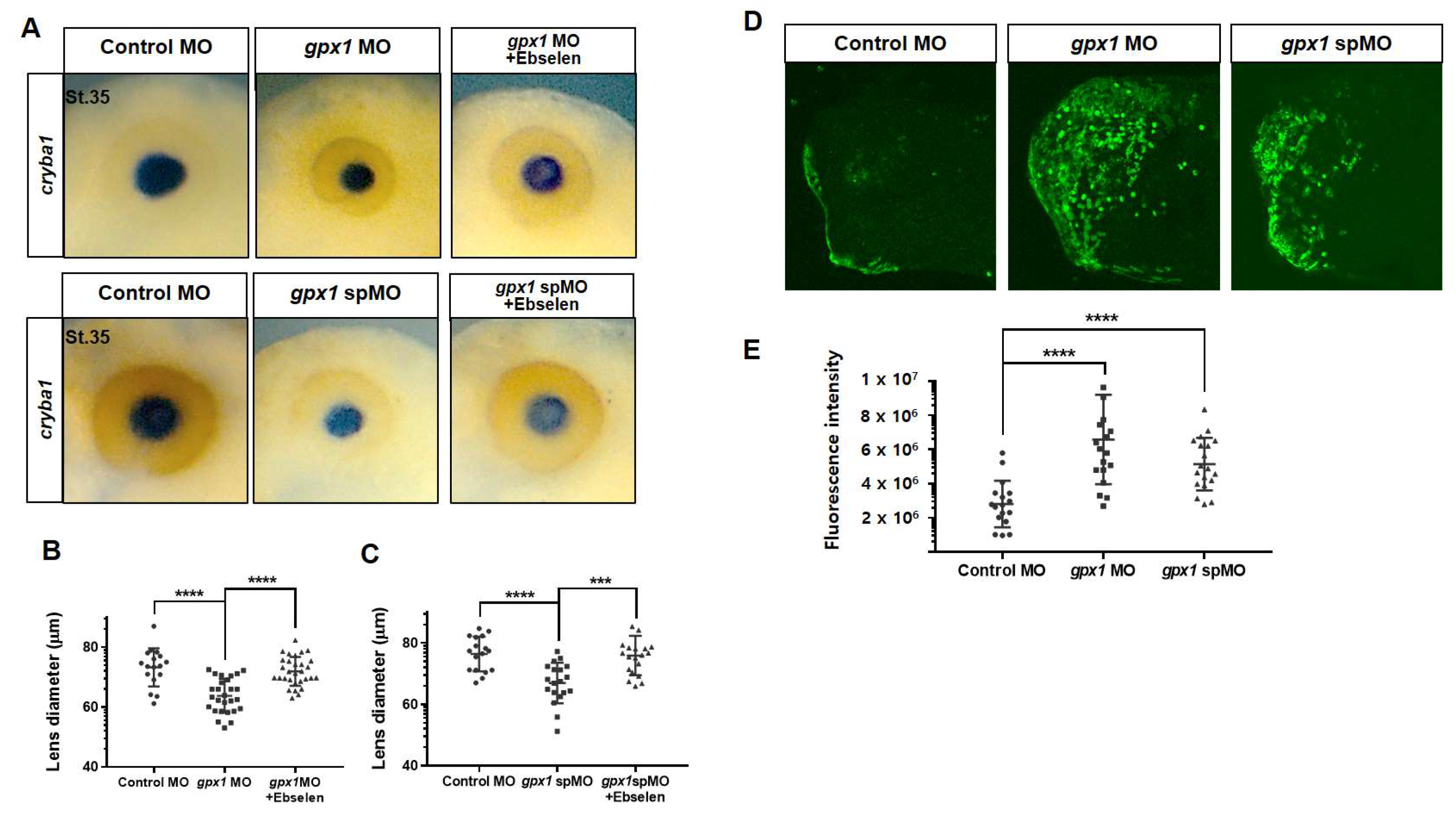
3.3. Gpx1 Interferes with Lens Development at the Molecular Level and Regulates ROS Levels in the Eye Regions during Embryogenesis

As the cross-sectional analysis of the eye in gpx1-morphant embryos revealed an incompletely detached lens compared to the control embryos in the case of both groups of MO-injected embryos, i.e., translational blocking gpx1 MO and gpx1 spMO (Figure 2A), to investigate the molecular details of gpx1 knockdown during lens development, we performed WISH using the lens-specific marker crystalline beta A1 (cryba1) [30] to analyze the epithelial stem cells in the anterior lens pole.
Our WISH analysis revealed that upon gpx1 depletion by injecting both types of gpx1 MOs (translational block and spMO), the cryba1 expression was downregulated and small-sized lenses were observed in gpx1-morphant embryos (Figure 3A). The lens diameter was reduced to less than 70 μm in the gpx1 MO-injected group and approximately 75 μm in the gpx1 spMO-injected group compared with 80−85 μm in the control group (Figure 3B). To confirm that the smaller lens and downregulated expression of cryba1 is particularly due to gpx1 knockdown, we injected the embryos with gpx1 MOs (translational block and splice block) and transferred the injected embryos to ebselen. The ebselen-grown embryos significantly rescued the cryba1 expression and lens size, just like the control embryos (Figure 3A,B). Altogether, our results demonstrate that gpx1 plays a significant role in eye development, affecting the lens during embryogenesis.
To assess the gpx1 antioxidant function during eye development, we microinjected HyPer mRNA into the D.1.2 blastomere of 16 cell-staged embryos followed by gpx1 MO injection (translational block and splice block). As expected, the fluorescence intensity was significantly increased in gpx1 MO-injected embryos in the eye region compared with control embryos (Figure 3D). More intense fluorescence was observed in the translational block MO-injected group compared with the splice MO-injected group (Figure 3E). However, both groups showed a significantly higher intensity compared with the control group (Figure 3D,E). Our data clearly show that gpx1 knockdown induced ROS accumulation in the eye region and evidently confirmed that the gpx1 antioxidant activity is critical for embryonic eye development, along with its influence on specific eye markers.

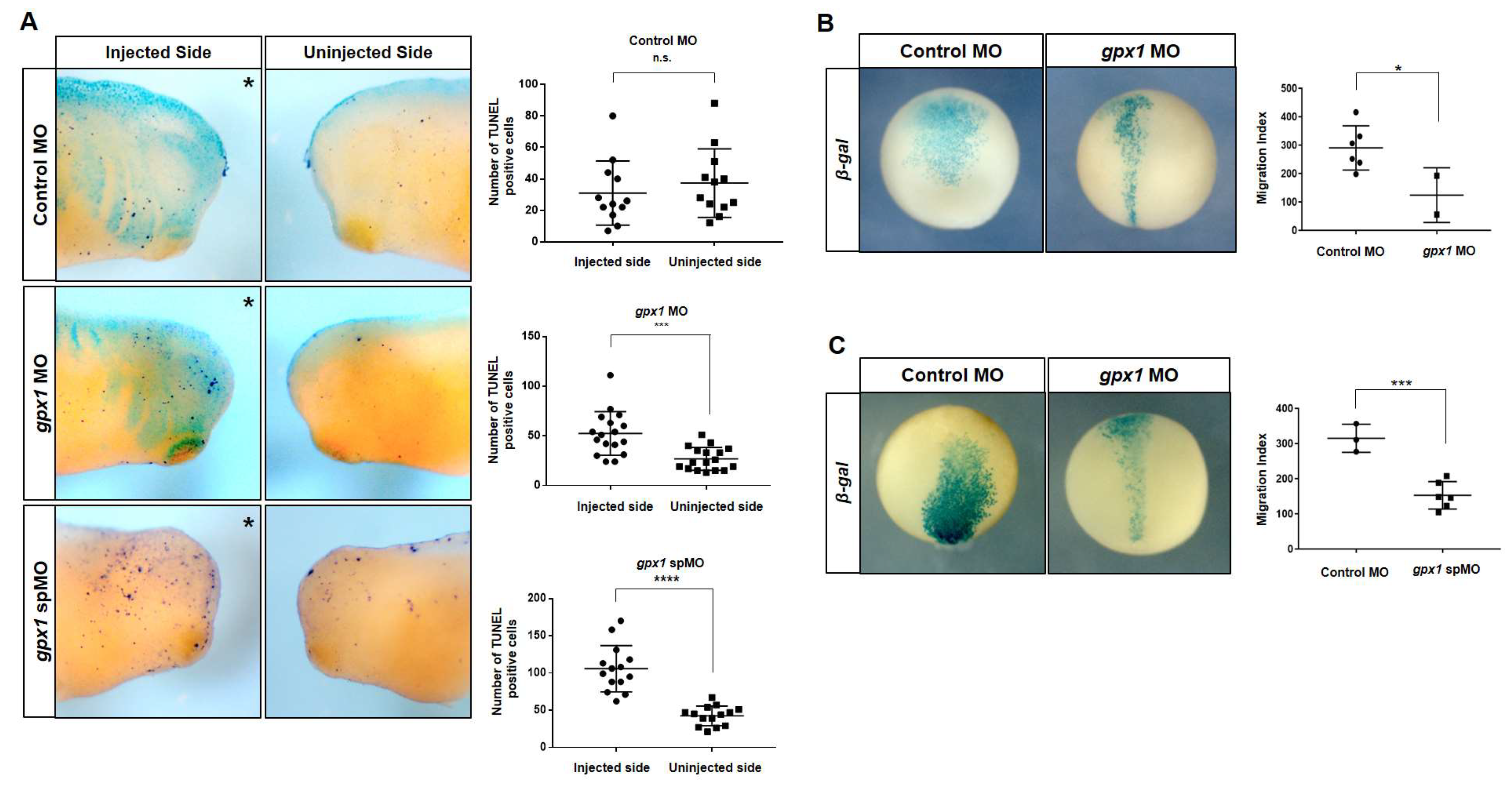
3.4. Gpx1 Regulates Cell Death and Interferes with Cell Migration during Xenopus Embryogenesis

Cell death plays significant roles in determining the organ size and particular organ structure. Different mechanisms maintain the proper size and structure of specific organs, such as apoptosis. Cell death is crucial in the development of the embryonic eye during embryogenesis. To elucidate whether eye malformations observed after gpx1 knockdown involve the perturbation of cell death, a TUNEL assay was conducted. Our results of TUNEL staining indicated that TUNEL-positive cells were significantly increased on the gpx1 MO injected side of gpx1-morphant embryos as compared with the uninjected side of the embryos (Figure 4A). The statistical analysis clearly showed the increase in TUNEL positive cells on the injected side of embryos as compared with the uninjected side of the embryos (Figure 4A), indicating the association of gpx1 in cell death regulation.
To further investigate the effects of gpx1 knockdown on cell migration during X. laevis embryogenesis, we microinjected β-galactosidase mRNA with gpx1 MO and gpx1 spMO into blastomere D.1.2. The results of this injection showed a broadened migration pattern for the control embryos, while for gpx1 morphants, the cells were concentrated in the midline regions of developing embryos (Figure 4B). The migration index calculated by measuring the width of cells from the ventral to dorsal side clearly exhibited a decrease in migration as a result of gpx1 depletion. The similar cell concentration in the midline of developing embryos was observed for gpx1 spMO injected embryos and the migration index was considerably reduced as compared with the control embryos (Figure 4C). These results showed gpx1 involvement in cell migration during early embryogenesis.

3.5. Gpx1 Interferes with Wnt and Cadherin Signaling during Embryonic Eye Development in X. laevis

To pinpoint the specific genes and associated pathways and to investigate the mechanism of eye development by gpx1, we conducted a transcriptomics analysis of gpx1-morphant embryos. The total RNA from gpx1-depleted embryos was extracted and processed for transcriptomics. Our transcriptomic analysis exhibited remarkable difference in the overall transcript expression between the control embryos and gpx1-morphant embryos (Figure 5A). Gpx1 knockdown led to an upregulation of the pathways associated with oxygen transport, fibrinolysis, gas transport, and cytolysis, showing a fold-enrichment of approximately ten, while gpx1 downregulation also induced the upregulation of the hydrogen peroxide catabolic process, cellular oxide detoxification, and response to hydrogen peroxide, but with a low fold-enrichment, i.e., less than five (Figure 5B). The most plausible reason for these upregulated biological responses associated with hydrogen peroxide is that ROS accumulation in gpx1-depleted embryos activated other antioxidant mechanisms to maintain a homeostatic balance [31]. In addition, depletion led to an upregulation of the cell killing (GO-0031341) pathway and it supported the increased number of TUNEL positive cells observed in gpx1 morphant embryos (Figure 4A). Furthermore, the increased cell death after gpx1 depletion was also supported by previously published data [32,33].
Because gpx1 knockdown induced eye phenotypes and lens detachment, we analyzed the transcriptomic data for pathways associated with eye development, and our analysis revealed the downregulation of Wnt, cadherin, and integrin signaling pathways (Figure 5C); these three pathways are critical for eye organogenesis as well as in lens cell differentiation and lens development, and not the protection of the lens epithelial cell phenotype [34,35,36]. In the case of Wnt signaling pathway-associated genes, WNT10b, WNT7b, and WNT6 [37] showed RPKM values of <2 in gpx1 morphants compared with the control embryos, showing RPKM values of approximately four (Figure 5D). Furthermore, genes associated with cadherin signaling, i.e., CDH20 and PCDHGB5 [38], exhibited RPKM values of less than two, whereas PCDH19 had RPKM values of less than three in the gpx1-depleted embryos as compared with the control embryos with RPKM values of >3 in CDH20 and PCDHGB5, and approximately 8 in PCDH19. In addition to the above genes associated with Wnt and cadherin signaling pathways, gpx1 morphants exhibited a downregulation of RBP1 [39], ALDH1A3 [40], and VAX2 [41], with RPKM values of <2 compared with the control embryos showing RPKM values of ≥4 (Figure 5D).
The downregulation of the Wnt and cadherins pathways (Figure 5C) in gpx1-depleted embryos not only provide information about the eye phenotypes observed in gpx1 morphant embryos, but also support the cell migration defects observed in gpx1 morphant embryos (Figure 4B,C). Wnt and cadherin signaling play an essential role in vertebrate embryonic development by tightly controlling the cell migration, differentiation, and cell−cell adhesion [34,42].
Altogether, our transcriptomics data support gpx1 involvement during embryogenesis, giving insight into the molecular pathways associated with embryonic eye development and indicating that gpx1 knockdown led to interfering with the signaling pathways, which in turn led to defects in cell migration and differentiation. These cell migration defects might be the possible reason for the ocular abnormalities observed after gpx1 depletion.

4. Discussion

Gpx1 and Prdx2 belonging to the thiol peroxidase class of antioxidants are the most abundant erythrocyte proteins to protect from oxidative damage. Both Gpx1 and Prdx2 have been studied for their antioxidant functions and have roles in different diseases [31]. Studies have implicated the role of Gpx1 in some cancers and cardiovascular diseases in humans [17,18,19,20]. Moreover, it is reported that Gpx1 is related to diabetes, and Gpx1 overexpressed mice exhibited insulin resistance, hyperinsulinemia, obesity and hyperglycemia [43,44]. Prdx2 has a high expression level in various cancers, such as lung, colorectal, and gastric cancers [22]. Moreover, Prdx2 has been reported to be involved in arthrosclerosis progression [23]. However, the embryonic significance of Gpx1 and Prdx2 has not yet been elucidated. In this study, we sought to explore the physiological roles of Gpx1 and Prdx2 during Xenopus embryonic development.
X. laevis is an excellent model for the study of embryonic development, as the gene expression can be easily manipulated by RNA and DNA microinjection in fertilized eggs; subsequently, the gene expression can be analyzed through different stages of embryogenesis [27,45]. In this study, we systematically observed the spatiotemporal expression of gpx1 and prdx2 during Xenopus embryonic development. RT-PCR analysis revealed the zygotic nature of gpx1 and the maternal nature of prdx2 (Figure 1A,D). WISH analysis revealed gpx1 localization in the eye regions, specifically in the lens of the developing embryos (Figure 1C), and prdx2 was localized in embryonic VB1 (Figure 1E). The functional significance of gpx1 and prdx2 during embryonic development was analyzed by microinjecting gpx1 and prdx2 into two-cell stage Xenopus embryos individually. Loss-of-function studies showed that gpx1 is essential for eye development during embryogenesis. Gpx1 morphants showed malformed eyes with incompletely detached lenses (Figure 2), indicating the critical roles of gpx1 in embryonic eye development. However, prdx2 depletion has no effect on developing embryos’ phenotypes, and prdx2 morphant embryos appeared to be normal without any significant malformation (Figure 2E,F).
Embryonic development is a complicated process that relies on the interplay of different cellular processes and is regulated by various factors [46]. The intricate balance of ROS is a prerequisite for proper embryonic development [6]. Thus, we investigated the involvement of ROS in the abnormal eye phenotypes induced by gpx1 knockdown. The HyPer mRNA injection in Xenopus embryos followed by gpx1 MOs injection resulted in increased fluorescence intensity in gpx1 morphants compared with the control embryos (Figure 3C). The increased fluorescence intensity in gpx1 morphants supports ROS accumulation in the malformed eyes, which agrees with previous reports showing ROS involvement in poor eye development.
Eye development is a critical step in vertebrate embryogenesis that is initiated during gastrulation through the emergence of crescent-shaped eye fields in the anterior neural plate [47]. Embryonic eye development is regulated by different genes and transcriptiontional factors involved in lens cell differentiation and lens development, cornea and retinal formation, and finally proper eye development. As gpx1 knockdown resulted in detached lenses in the morphant embryos, we investigated the expression of cryba1 in gpx1 morphants. The downregulated expression of cryba1 and the decreased lens diameter (Figure 3A,B) in gpx1 morphants indicated the interference of gpx1 with molecular markers associated with lens development. Furthermore, the decreased lens diameter was supported by increased cell death in gpx1-depleted embryos (Figure 4A). The increased apoptotic cell death after gpx1 depletion is in agreement with previously reported data [33]. These findings confirm that gpx1 plays a critical role in eye development during Xenopus embryogenesis by regulating the cell growth and eye-related transcription factors.
The process of vertebrate eye development is regulated by different signaling pathways, and the malformed eyes observed in gpx1 morphants may involve the perturbation of these signaling pathways [48]. The transcriptomic analysis revealed the downregulation of the Wnt, cadherin, and integrin signaling pathways. Wnt signaling plays an essential role in eye organogenesis and is active in the periocular surface ectoderm and lens epithelium during lens development [34,49]. gpx1 knockdown resulted in the downregulation of Wnt signaling-associated genes, WNT10b, WNT7b, and WNT6 (Figure 5C,D), indicating the possibility of the involvement of gpx1 in Wnt signaling.
Cadherin is significant for epithelial cell formation and retinal pigment epithelium is required for normal vision [35]. gpx1 depletion resulted in the downregulation of the CDH20, PCDH19, and PCDHGB5 genes associated with cadherin signaling, showing the plausible association of gpx1 with cadherin signaling (Figure 5C,D). Integrin plays an important role in lens development [36], and gpx1 knockdown induced the downregulation of the integrin signaling pathway (Figure 5C). The downregulation of Wnt and cadherin signaling pathways supports the cell migration defects observed in gpx1 depleted embryos. On the basis of transcriptomic analysis, we propose that gpx1 interfered with Wnt and cadherin signaling pathways in developing embryos, which in turn led to the cell migration defects, and these migration defects ultimately led to ocular abnormalities in developing embryos.

5. Conclusions

Thiol peroxidases (Gpx1 and Prdx2) have essential antioxidant functions, but physiologically, gpx1 is a prerequisite for normal embryonic development. Alternatively, prdx2 does not appear to be associated with embryogenesis. The association of gpx1 with lens-specific markers and key genes involved in eye development provides valuable information about the complex network involved in vertebrate eye development. gpx1 perturbs normal eye development by regulating ROS levels, as well as Wnt, cadherin, and integrin signaling pathways. However, further detailed investigation is required to determine the regulatory pathways followed by gpx1 to control embryonic eye development. To the best of our knowledge, this is the first study indicating the association of gpx1 with embryogenesis, and it has emphasized the significance of antioxidants in embryonic development involving a complex network of signaling pathways.

Author Contributions

Conceptualization, H.-S.L., T.J.P. and T.K.; methodology, H.L., N.Y.L. and Y.K.; formal analysis, H.L., Y.K., N.Y.L. and H.-S.C.; investigation, H.-Y.R., T.K. and H.-S.L.; writing—original draft preparation, T.I., D.-H.C., Z.Y.R. and H.-S.L.; writing—review and editing, H.-S.L., D.-S.L., T.K.K., Y.K. and T.I.; visualization, H.L., N.Y.L. and H.-S.L.; supervision, H.-S.L. and T.K.; funding acquisition, H.-S.L. All authors have read and agreed to the published version of the manuscript.

Funding

This research was supported by the National Research Foundation of Korea funded by the Ministry of Science and ICT (2020R1A4A1018280 and 2021R1A2C1010408).

Institutional Review Board Statement

Experiments were strictly conducted according to the guidelines of the Animal Care and Use Committee, consistent with international laws and policies (National institute of health-NIH Guide for the Care and Use of Laboratory Animals, NIH publication no. 85-23, 1985). The Institutional Review Board of Kyungpook National University in Korea approved the experimental use of amphibians (2021-0017). All members of the research group were trained for the appropriate care and use of experimental organisms. There were no unexpected deaths of adult Xenopus during this study.

Informed Consent Statement

Not applicable.

Data Availability Statement

The data presented in this study are available in the manuscript.

Conflicts of Interest

The authors declare no conflict of interest.

References

  1. Cadenas, E.; Davies, K.J. Mitochondrial free radical generation, oxidative stress, and aging. Free Radic. Biol. Med. 2000, 29, 222–230. [Google Scholar] [CrossRef]
  2. Dringen, R.; Pawlowski, P.G.; Hirrlinger, J. Peroxide detoxification by brain cells. J. Neurosci. Res. 2005, 79, 157–165. [Google Scholar] [CrossRef]
  3. Schieber, M.; Chandel, N.S. ROS function in redox signaling and oxidative stress. Curr. Biol. 2014, 24, R453–R462. [Google Scholar] [CrossRef] [Green Version]
  4. Timme-Laragy, A.R.; Hahn, M.E.; Hansen, J.M.; Rastogi, A.; Roy, M.A. Redox stress and signaling during vertebrate embryonic development: Regulation and responses. Semin. Cell Dev. Biol. 2018, 80, 17–28. [Google Scholar] [CrossRef]
  5. Imlay, J.A. Pathways of oxidative damage. Annu. Rev. Microbiol. 2003, 57, 395–418. [Google Scholar] [CrossRef] [PubMed]
  6. Covarrubias, L.; Hernández-García, D.; Schnabel, D.; Salas-Vidal, E.; Castro-Obregón, S. Function of reactive oxygen species during animal development: Passive or active? Dev. Biol. 2008, 320, 1–11. [Google Scholar] [CrossRef] [PubMed] [Green Version]
  7. Valko, M.; Leibfritz, D.; Moncol, J.; Cronin, M.T.; Mazur, M.; Telser, J. Free radicals and antioxidants in normal physiological functions and human disease. Int. J. Biochem. Cell Biol. 2007, 39, 44–84. [Google Scholar] [CrossRef]
  8. Bindoli, A.; Fukuto, J.M.; Forman, H.J. Thiol chemistry in peroxidase catalysis and redox signaling. Antioxid. Redox Signal. 2008, 10, 1549–1564. [Google Scholar] [CrossRef] [PubMed]
  9. Flohé, L.; Toppo, S.; Cozza, G.; Ursini, F. A comparison of thiol peroxidase mechanisms. Antioxid. Redox Signal. 2011, 15, 763–780. [Google Scholar] [CrossRef]
  10. Flohé, L.; Brigelius-Flohé, R.; Hatfield, D. Selenium: Its Molecular Biology and Role in Human Health; Selenoproteins Glutathione System; Kluwer: Norwel, MA, USA, 2001; pp. 157–178. [Google Scholar]
  11. Low, F.M.; Hampton, M.B.; Winterbourn, C.C. Peroxiredoxin 2 and peroxide metabolism in the erythrocyte. Antioxid. Redox Signal. 2008, 10, 1621–1630. [Google Scholar] [CrossRef]
  12. Hall, A.; Nelson, K.; Poole, L.B.; Karplus, P.A. Structure-based insights into the catalytic power and conformational dexterity of peroxiredoxins. Antioxid. Redox Signal. 2011, 15, 795–815. [Google Scholar] [CrossRef] [Green Version]
  13. Rhee, S.G.; Woo, H.A.; Kil, I.S.; Bae, S.H. Peroxiredoxin functions as a peroxidase and a regulator and sensor of local peroxides. J. Biol. Chem. 2012, 287, 4403–4410. [Google Scholar] [CrossRef] [Green Version]
  14. Lee, T.H.; Kim, S.U.; Yu, S.L.; Kim, S.H.; Park, D.S.; Moon, H.B.; Dho, S.H.; Kwon, K.S.; Kwon, H.J.; Han, Y.H.; et al. Peroxiredoxin II is essential for sustaining life span of erythrocytes in mice. Blood 2003, 101, 5033–5038. [Google Scholar] [CrossRef] [Green Version]
  15. Rhee, S.G.; Kang, S.W.; Chang, T.S.; Jeong, W.; Kim, K. Peroxiredoxin, a novel family of peroxidases. IUBMB Life 2001, 52, 35–41. [Google Scholar] [CrossRef]
  16. Ho, Y.S.; Magnenat, J.L.; Bronson, R.T.; Cao, J.; Gargano, M.; Sugawara, M.; Funk, C.D. Mice deficient in cellular glutathione peroxidase develop normally and show no increased sensitivity to hyperoxia. J. Biol. Chem. 1997, 272, 16644–16651. [Google Scholar] [CrossRef] [PubMed] [Green Version]
  17. Cheng, Y.; Xu, T.; Li, S.; Ruan, H. GPX1, a biomarker for the diagnosis and prognosis of kidney cancer, promotes the progression of kidney cancer. Aging 2019, 11, 12165–12176. [Google Scholar] [CrossRef] [PubMed]
  18. Wei, R.; Qiu, H.; Xu, J.; Mo, J.; Liu, Y.; Gui, Y.; Huang, G.; Zhang, S.; Yao, H.; Huang, X.; et al. Expression and prognostic potential of GPX1 in human cancers based on data mining. Ann. Transl. Med. 2020, 8, 124. [Google Scholar] [CrossRef] [PubMed]
  19. Min, S.Y.; Kim, H.S.; Jung, E.J.; Jee, C.D.; Kim, W.H. Prognostic significance of glutathione peroxidase 1 (GPX1) down-regulation and correlation with aberrant promoter methylation in human gastric cancer. Anticancer Res. 2012, 32, 3169–3175. [Google Scholar] [PubMed]
  20. Hu, J.; Zhou, G.W.; Wang, N.; Wang, Y.J. GPX1 Pro198Leu polymorphism and breast cancer risk: A meta-analysis. Breast Cancer Res. Treat 2010, 124, 425–431. [Google Scholar] [CrossRef] [PubMed]
  21. Han, Y.H.; Kim, S.U.; Kwon, T.H.; Lee, D.S.; Ha, H.L.; Park, D.S.; Woo, E.J.; Lee, S.H.; Kim, J.M.; Chae, H.B.; et al. Peroxiredoxin II is essential for preventing hemolytic anemia from oxidative stress through maintaining hemoglobin stability. Biochem. Biophys. Res. Commun. 2012, 426, 427–432. [Google Scholar] [CrossRef]
  22. Wang, W.; Wei, J.; Zhang, H.; Zheng, X.; Zhou, H.; Luo, Y.; Yang, J.; Deng, Q.; Huang, S.; Fu, Z. PRDX2 promotes the proliferation of colorectal cancer cells by increasing the ubiquitinated degradation of p53. Cell Death Dis. 2021, 12, 605. [Google Scholar] [CrossRef]
  23. Li, J.; Wang, C.; Wang, W.; Liu, L.; Zhang, Q.; Zhang, J.; Wang, B.; Wang, S.; Hou, L.; Gao, C.; et al. PRDX2 Protects Against Atherosclerosis by Regulating the Phenotype and Function of the Vascular Smooth Muscle Cell. Front. Cardiovasc. Med. 2021, 8, 624796. [Google Scholar] [CrossRef]
  24. Chae, S.; Lee, H.-K.; Kim, Y.-K.; Jung Sim, H.; Ji, Y.; Kim, C.; Ismail, T.; Park, J.-W.; Kwon, O.-S.; Kang, B.-S.; et al. Peroxiredoxin1, a novel regulator of pronephros development, influences retinoic acid and Wnt signaling by controlling ROS levels. Sci. Rep. 2017, 7, 8874. [Google Scholar] [CrossRef] [PubMed] [Green Version]
  25. Ji, Y.; Chae, S.; Lee, H.K.; Park, I.; Kim, C.; Ismail, T.; Kim, Y.; Park, J.W.; Kwon, O.S.; Kang, B.S.; et al. Peroxiredoxin5 Controls Vertebrate Ciliogenesis by Modulating Mitochondrial Reactive Oxygen Species. Antioxid. Redox Signal. 2019, 30, 1731–1745. [Google Scholar] [CrossRef] [PubMed]
  26. Lee, H.; Ismail, T.; Kim, Y.; Chae, S.; Ryu, H.Y.; Lee, D.S.; Kwon, T.K.; Park, T.J.; Kwon, T.; Lee, H.S. Xenopus gpx3 Mediates Posterior Development by Regulating Cell Death during Embryogenesis. Antioxidants 2020, 9, 1265. [Google Scholar] [CrossRef]
  27. Kim, Y.; Jeong, Y.; Kwon, K.; Ismail, T.; Lee, H.K.; Kim, C.; Park, J.W.; Kwon, O.S.; Kang, B.S.; Lee, D.S.; et al. Physiological effects of KDM5C on neural crest migration and eye formation during vertebrate development. Epigenet. Chromatin 2018, 11, 72. [Google Scholar] [CrossRef] [PubMed]
  28. Robinson, M.D.; McCarthy, D.J.; Smyth, G.K. edgeR: A Bioconductor package for differential expression analysis of digital gene expression data. Bioinformatics 2010, 26, 139–140. [Google Scholar] [CrossRef] [Green Version]
  29. Nakamura, Y.; Feng, Q.; Kumagai, T.; Torikai, K.; Ohigashi, H.; Osawa, T.; Noguchi, N.; Niki, E.; Uchida, K. Ebselen, a glutathione peroxidase mimetic seleno-organic compound, as a multifunctional antioxidant. Implication for inflammation-associated carcinogenesis. J. Biol. Chem. 2002, 277, 2687–2694. [Google Scholar] [CrossRef] [Green Version]
  30. Zigler, J.S., Jr.; Sinha, D. βA3/A1-crystallin: More than a lens protein. Prog. Retin. Eye Res. 2015, 44, 62–85. [Google Scholar] [CrossRef]
  31. Lubos, E.; Loscalzo, J.; Handy, D.E. Glutathione peroxidase-1 in health and disease: From molecular mechanisms to therapeutic opportunities. Antioxid. Redox Signal. 2011, 15, 1957–1997. [Google Scholar] [CrossRef] [Green Version]
  32. Flentjar, N.J.; Crack, P.J.; Boyd, R.; Malin, M.; de Haan, J.B.; Hertzog, P.; Kola, I.; Iannello, R. Mice lacking glutathione peroxidase-1 activity show increased TUNEL staining and an accelerated inflammatory response in brain following a cold-induced injury. Exp. Neurol. 2002, 177, 9–20. [Google Scholar] [CrossRef]
  33. Ufer, C.; Wang, C. The roles of glutathione peroxidases during embryo development. Front. Mol. Neurosci. 2011, 4, 12. [Google Scholar] [CrossRef] [Green Version]
  34. Fujimura, N. WNT/β-Catenin Signaling in Vertebrate Eye Development. Front. Cell Dev. Biol. 2016, 4, 138. [Google Scholar] [CrossRef] [Green Version]
  35. Yang, X.; Chung, J.-Y.; Rai, U.; Esumi, N. Cadherins in the retinal pigment epithelium (RPE) revisited: P-cadherin is the highly dominant cadherin expressed in human and mouse RPE in vivo. PLoS ONE 2018, 13, e0191279. [Google Scholar] [CrossRef] [Green Version]
  36. Walker, J.; Menko, A.S. Integrins in lens development and disease. Exp. Eye Res. 2009, 88, 216–225. [Google Scholar] [CrossRef] [Green Version]
  37. Fokina, V.M.; Frolova, E.I. Expression patterns of Wnt genes during development of an anterior part of the chicken eye. Dev. Dyn. Off. Publ. Am. Assoc. Anat. 2006, 235, 496–505. [Google Scholar] [CrossRef] [Green Version]
  38. Etzrodt, J.; Krishna-K, K.; Redies, C. Expression of classic cadherins and delta-protocadherins in the developing ferret retina. BMC Neurosci. 2009, 10, 153. [Google Scholar] [CrossRef] [Green Version]
  39. Flach, H.; Basten, T.; Schreiner, C.; Dietmann, P.; Greco, S.; Nies, L.; Roßmanith, N.; Walter, S.; Kühl, M.; Kühl, S.J. Retinol binding protein 1 affects Xenopus anterior neural development via all-trans retinoic acid signaling. Dev. Dyn. 2021, 250, 1096–1112. [Google Scholar] [CrossRef] [PubMed]
  40. Abouzeid, H.; Favez, T.; Schmid, A.; Agosti, C.; Youssef, M.; Marzouk, I.; El Shakankiry, N.; Bayoumi, N.; Munier, F.L.; Schorderet, D.F. Mutations in ALDH1A3 represent a frequent cause of microphthalmia/anophthalmia in consanguineous families. Hum. Mutat. 2014, 35, 949–953. [Google Scholar] [CrossRef] [PubMed]
  41. Alfano, G.; Conte, I.; Caramico, T.; Avellino, R.; Arnò, B.; Pizzo, M.T.; Tanimoto, N.; Beck, S.C.; Huber, G.; Dollé, P.; et al. Vax2 regulates retinoic acid distribution and cone opsin expression in the vertebrate eye. Development 2011, 138, 261–271. [Google Scholar] [CrossRef] [PubMed] [Green Version]
  42. Halbleib, J.M.; Nelson, W.J. Cadherins in development: Cell adhesion, sorting, and tissue morphogenesis. Genes Dev. 2006, 20, 3199–3214. [Google Scholar] [CrossRef] [PubMed] [Green Version]
  43. Liu, D.; Liu, L.; Hu, Z.; Song, Z.; Wang, Y.; Chen, Z. Evaluation of the oxidative stress-related genes ALOX5, ALOX5AP, GPX1, GPX3 and MPO for contribution to the risk of type 2 diabetes mellitus in the Han Chinese population. Diabetes Vasc. Dis. Res. 2018, 15, 336–339. [Google Scholar] [CrossRef] [Green Version]
  44. Mohammedi, K.; Patente, T.A.; Bellili-Muñoz, N.; Driss, F.; Le Nagard, H.; Fumeron, F.; Roussel, R.; Hadjadj, S.; Corrêa-Giannella, M.L.; Marre, M.; et al. Glutathione peroxidase-1 gene (GPX1) variants, oxidative stress and risk of kidney complications in people with type 1 diabetes. Metabolism 2016, 65, 12–19. [Google Scholar] [CrossRef]
  45. Blum, M.; Beyer, T.; Weber, T.; Vick, P.; Andre, P.; Bitzer, E.; Schweickert, A. Xenopus, an ideal model system to study vertebrate left-right asymmetry. Dev. Dyn. 2009, 238, 1215–1225. [Google Scholar] [CrossRef] [PubMed]
  46. Shaping embryonic development. Nat. Chem. Biol. 2017, 13, 559. [CrossRef] [Green Version]
  47. Donner, A.L.; Lachke, S.A.; Maas, R.L. Lens induction in vertebrates: Variations on a conserved theme of signaling events. Semin. Cell Dev. Biol. 2006, 17, 676–685. [Google Scholar] [CrossRef]
  48. Seigfried, F.A.; Cizelsky, W.; Pfister, A.S.; Dietmann, P.; Walther, P.; Kühl, M.; Kühl, S.J. Frizzled 3 acts upstream of Alcam during embryonic eye development. Dev. Biol. 2017, 426, 69–83. [Google Scholar] [CrossRef] [PubMed]
  49. Van Raay, T.J.; Vetter, M.L. Wnt/frizzled signaling during vertebrate retinal development. Dev. Neurosci. 2004, 26, 352–358. [Google Scholar] [CrossRef] [PubMed]
Figure 1. Spatiotemporal expression pattern of gpx1 and prdx2 during Xenopus embryogenesis. (A) The transcriptase-polymerase chain reaction (RT-PCR) analysis indicated the zygotic nature of gpx1 during Xenopus embryonic development. The temporal expression of gpx1 started at NF. St. 30 of embryogenesis and proceeded until NF. St. 40. The highest expression level was observed at NF. St. 35 of Xenopus embryogenesis. Ornithine decarboxylase (odc) was used as an internal loading control. (B) The general scheme for Xenopus eye development in the tadpole stage indicates eye vesicle formation at NF. St. 25, followed by lens induction at NF. St. 30., while lens detachment begins at NF. St. 35, leading to cornea formation at NF. St 40 of Xenopus embryonic development. (C) The spatial expression of gpx1 as analyzed by whole-mount in situ hybridization (WISH) indicated its specific expression in the lens after eye vesicle formation at NF. St 30 embryonic development with an increased expression level in the lens region at NF. St. 35 and it proceeded until NF. St. 40 of Xenopus embryogenesis. (D) RT-PCR analysis revealed the maternal nature of prdx2 during Xenopus embryogenesis. The highest level of prdx2 expression was observed at the late neurula and tailbud stages of embryogenesis. Ornithine decarboxylase (odc) was used as the internal loading control. (E) WISH analysis indicated the localization of prdx2 in embryonic VBIs during Xenopus embryonic development.
Antioxidants 10 01636 g001
Figure 2. gpx1 loss-of-function results in malformed eyes with incompletely detached lenses, while prdx2 morphant embryos have no phenotypic malformations. (A) Two types of gpx1 morpholino oligonucleotides (MOs), i.e., translational blocking MO (gpx1 MO; 20 ng) and splicing blocking MO (gpx1 spMO; 5 ng), were microinjected into animal poles of two-cell stage Xenopus embryos, and then the embryos were fixed at NF. St. 40. Knockdown of gpx1 in both groups of MOs resulted in malformed eyes, as shown in the enlarged images. Additionally, the cross-sectional analysis indicated the incompletely detached lens in both groups of gpx1 morphants. (B) The statistical analysis of gpx1 morphants indicated that more than 70% of the embryos exhibited malformed eyes in the case of gpx1 MO microinjection, while more than 50% of embryos showed eye malformations in the case of gpx1 spMO injection compared with the control embryos. ** p ≤ 0.01, *** p ≤ 0.001. (C) Rescue experiments were conducted by transferring the gpx1 morpholino injected embryos to 0.5-μΜ ebselen, a Gpx mimic, and then fixed at NF. St. 40. The eye malformations observed in both groups of gpx1-depleted embryos were effectively recovered after ebselen treatment. (D) A graphical representation of rescued embryos showed that more than 30% of embryos recovered the malformed eyes observed due to gpx1 MO and gpx1 spMO microinjection after ebselen treatment. * p ≤ 0.05 ** p ≤ 0.01, *** p ≤ 0.001, **** p ≤ 0.0001. (E) prdx2 MO (40 ng) and wildtype prdx2 mRNA (1 ng) were microinjected into the two cell stage developing embryos and then the embryos were fixed at NF. St. 40. The embryos developed normally after prdx2 MO injection and there were no phenotypic differences between prdx2 morphant embryos and embryo injected with wildtype prdx2 mRNA. Both group of embryos were morphologically similar to the control MO injected embryos. (F) Xenopus embryos were injected with prdx2 MO and were processed for WISH analysis using blood cell markers, i.e., hba3 and mpo. The knockdown of prdx2 did not affect the expression pattern of the blood cell-specific markers.
Antioxidants 10 01636 g002
Figure 3. Loss of gpx1 results in a perturbed expression of the lens-specific marker and an accumulation of ROS in developing eyes during Xenopus embryonic development. Here, (A) 20-ng gpx1 MO and 5-ng gpx1 spMO were microinjected into the animal pole of two-cell stage embryos, and the embryos were processed for WISH analysis using the lens-specific marker cryba1. The expression of cryba1 was remarkably reduced in both groups of gpx1-morphant embryos. Moreover, the downregulated expression of cryba1 was effectively rescued in gpx1 morphants after ebselen treatment. (B) A graphical representation of gpx1 MO-injected embryos revealed that gpx1-morphant embryos exhibited a lens diameter of 70 μm compared with >80 μm in the control embryos, and it was effectively recovered to 80 μm in the gpx1-depleted embryos after ebselen treatment. **** p ≤ 0.0001. (C) Statistical analysis of gpx1 spMO-injected embryos indicated that gpx1-depleted embryos showed a lens diameter of approximately 75 μm as compared with >80 μm in the control embryos. The smaller size of the lens was effectively rescued to 80 μm after ebselen treatment in the gpx1 spMO-injected embryos. *** p ≤ 0.001, **** p ≤ 0.0001. (D) Both gpx1 MOs with HyPer mRNA (10 ng) were injected into the D.1.2. blastomeres of 16 cell stage Xenopus embryos and the gpx1-depleted embryos exhibited increased fluorescence intensity in the eye regions compared with the control embryos. (E) HyPer fluorescence intensity quantification clearly showed significant intense fluorescence in the gpx1 MO and gpx1 spMO injected embryos as compared with the control embryos. **** p ≤ 0.0001.
Antioxidants 10 01636 g003
Figure 4. gpx1 depletion activates cell apoptosis and inhibits cell proliferation, as well as affects cell migration during Xenopus embryogenesis. (A) The depletion of gpx1 increased TUNEL-positive cells in the eye region of developing gpx1 morphants compared with the control embryos. n.s. > 0.05, *** p ≤ 0.001, **** p ≤ 0.0001. (B) gpx1 MO was coinjected with β-galactosidase mRNA into the D.1.2 blastomere of 16-cell stage Xenopus embryos. The staining showed that the cells were concentrated in the midline of the developing gpx1-depleted embryos compared with the widely distributed cells in the control embryos. The migration index was considerably reduced after gpx1 MO injection. * p ≤ 0.05 (C) The injection of gpx1 spMO into the D.1.2. blastomere also resulted in a cell concentration in the midline of developing embryos and decreased the migration index as compared with the control embryos. *** p ≤ 0.001.
Antioxidants 10 01636 g004
Figure 5. Transcriptomic analyses provide insight into the mechanism of eye development by gpx1 during Xenopus embryogenesis. (A) The heat map shows the remarkable difference between the control embryos and gpx1-morphant embryos. (B) Transcriptome enrichment analysis using PANTHER software revealed the upregulation of the biological pathways associated with the response to reactive oxygen species, oxygen transport, hydrogen peroxide, hydrogen peroxide catabolic process, and gas transport. (C) Transcriptomic analysis using PANTHER software demonstrated the downregulation of signaling pathways, i.e., Wnt, Cadherin, and integrin associated with eye development in gpx1-depleted embryos. (D) Genes associated with Wnt signaling (Wnt10b, Wnt 7b, and Wnt6) showed significantly low RPKM values in gpx1-depleted embryos compared with the control embryos. A similar case was observed for cadherin signaling-associated genes (CDH20, PCDH19, and PCDHGB5), showing considerably low RPKM values compared with the control embryos. Additionally, genes associated with eye development, such as RBP1, ALDH1A3, and VAX2, exhibited remarkably low RPKM values after gpx1 knockout compared with the control embryos.
Antioxidants 10 01636 g005
Publisher’s Note: MDPI stays neutral with regard to jurisdictional claims in published maps and institutional affiliations.

