Review

Transcranial Direct Current Stimulation Enhances Cognitive Function in Patients with Mild Cognitive Impairment and Early/Mid Alzheimer’s Disease: A Systematic Review and Meta-Analysis

Department of Geriatrics, Tongji Hospital, Tongji Medical College, Huazhong University of Science and Technology, Wuhan 430074, China
*
Author to whom correspondence should be addressed.
Brain Sci. 2022, 12(5), 562; https://doi.org/10.3390/brainsci12050562
Submission received: 27 March 2022 / Revised: 18 April 2022 / Accepted: 22 April 2022 / Published: 27 April 2022
(This article belongs to the Section Social Cognitive and Affective Neuroscience)

Abstract

:
Transcranial direct current stimulation (tDCS) i a non-invasive brain stimulation which is considered to have the potential to improve cognitive impairment in patients with mild cognitive impairment (MCI) and Alzheimer’s disease (AD). However, previous studies have been controversial on the therapeutic effect of tDCS. This meta-analysis aimed to evaluate the effects of tDCS on cognitive impairment in patients with MCI and mild-to-moderate AD. Five databases, namely PubMed, EMBASE, MEDLINE, Web of Science and The Cochrane Library, were searched with relative terms to extract the cognitive function changes measured by an objective cognitive scale in the included studies. The meta-analysis results showed that, compared with sham tDCS treatment, the overall cognitive function of patients with AD and MCI was significantly improved (weighted mean difference = 0.99; 95% confidence interval, 0.32 to 1.66; p = 0.004) after tDCS treatment, but the behavioral symptoms, recognition memory function, attention and executive function were not significantly improved. The subgroup analysis showed that the treatment would be more efficacious if the temporal-lobe-related brain areas were stimulated, the number of stimulations was greater than or equal to 10 and the current density was 2.5 mA/cm2. Among them, AD patients benefited more than MCI patients. No cognitive improvement was observed in patients with MCI or AD at different follow-up times after treatment. Our meta-analysis provided important evidence for the cognitive enhancement of tDCS in patients with MCI and mild-to-moderate AD and discussed its underlying mechanisms.

1. Introduction

Alzheimer’s disease (AD) [1] is a common neurodegenerative disease in the elderly and the main cause of dementia; its core symptom is progressive memory loss. With the progression of the disease, patients may also experience aphasia, executive function, and other cognitive impairment, as well as anxiety, depression, irritability, hallucinations, and other neuropsychiatric symptoms [2]. Mild cognitive impairment (MCI) is a state between normal aging and dementia and is considered the preclinical stage of Alzheimer’s disease, with about 5 to 10 percent of patients with mild cognitive impairment possibly developing into dementia each year [3,4,5]. As the population ages, the number of dementia patients is increasing, but drugs, including cholinesterase inhibitors and memantine, cannot effectively improve the cognitive ability of patients [6,7,8]. There is increasing evidence that transcranial direct-current stimulation (tDCS) may be an effective alternative therapy [9].
TDCS is a non-invasive brain stimulation that usually involves placing electrodes on the scalp to apply a weak direct current to modulate cortical function [10,11]. The stimulation of tDCS can be divided into anode stimulation and cathode stimulation; the stimulation current is mostly 1–2 mA, and each stimulation time is tens of minutes, which is considered to be a safe range of stimulation [12,13]. Stimulations of different polarities have diverse effects on the cortex. The anodic tDCS depolarized the resting membrane potential of neurons and increased the excitability of the cortex by increasing the frequency of spontaneous firing of neurons, while the cathodic tDCS hyperpolarized the resting membrane potential of neurons and inhibited the excitability of the cortex by decreasing the firing frequency of neurons [14,15]. However, it has also been observed that, compared with cathodic stimulation of 1 mA, cathodic stimulation of 2 mA can enhance the excitability of the cortex [16]. Changes in cortical excitability caused by tDCS leads to corresponding changes in cortical function and activation [17], that is, changes in synaptic plasticity [18]. Increased cortical excitability and neuroplasticity are considered to be important mechanisms for improving clinical and cognitive abilities in neurodegenerative diseases [19]. In addition, some studies suggest that the cognitive improvement of tDCS may be related to the neural noise produced by TDCS [20,21]. Numerous studies have shown that tDCS can produce varying degrees of therapeutic effects on a variety of neurodegenerative diseases, including Parkinson’s disease, AD, and primary progressive aphasia [22,23,24,25].
Studies have found that tDCS can improve learning and memory disorders in AD-model mice [26,27]. A recent meta-analysis found that tDCS can significantly improve the cognitive function of AD patients, especially when using a low current density [28]. Another meta-analysis found that anodic stimulation of tDCS on DLPFC significantly improved cognitive ability, especially at high-current intensity and density [29]. In terms of the maintenance time of the treatment effect, tDCS can improve the memory impairment of patients with MCI and AD in the short term, but this improvement cannot be maintained for a long time [30]. As a promising treatment for cognitive impairment, it has also been noted that tDCS has no significant therapeutic effect on AD [31,32]. These differences may be related to various factors, such as the stimulus parameters used in various studies, the frequency of stimulation, the means of testing the effect of stimulation, and whether it is combined with cognitive training.
The purpose of this meta-analysis was to evaluate the effect of tDCS on improving the cognitive impairment of patients with MCI and mild-to-moderate AD. Meanwhile, the optimal parameters and duration of the effect were explored.

2. Materials and Methods

This study was registered with PROSPERO (CRD42021275672). We followed the stated guidelines for the Preferred Reporting Items for Systematic Reviews and Meta-Analyses (PRISMA) [33]. Two reviewers were independently involved in citation retrieval, study selection, quality assessment, and data extraction. Divergences between reviewers were resolved by consulting the third reviewer.

2.1. Search Strategy

In this study, five databases, namely PubMed, Embase, MEDLINE, Web of Science, and The Cochrane Library, were searched; the retrieval time was from the database construction to 31 August 2021. The key words were (“Alzheimer disease” OR “Alzheimer’s disease” OR “AD” OR “mild cognitive impairment” OR “MCI”) AND (“transcranial direct current stimulation” OR “tDCS” OR “direct current stimulation” OR “TES” OR “transcranial stimulation”). The list of references contained in the study was also searched manually to identify any relevant articles.

2.2. Study Selection

To ensure the comprehensiveness of the included studies, we accepted both the parallel design trials and the crossover design trials. The inclusion criteria for this study were as follows. (1) Study type: randomized controlled trial (RCT), single-blind, double-blind or non-blind. (2) Subjects: patients with mild-to-moderate AD or MCI who met at least one of the following diagnostic criteria: (a) National Institute of Neurological Communicative Disorders and Stroke/Alzheimer disease and Related Disorders Association(NINCDS/ADRDA), (b) the Diagnostic and Statistical Manual-IV(DSM-IV), (c) Petersen’s criteria, (d) Alzheimer’s disease neuroimaging initiative criteria(ADNI), (e) the Diagnostic and Statistical Manual of Mental Disorders, Fifth Edition(DSM-5), and (f) the criteria of the MCI Working Group of the European Consortium on Alzheimer’s disease. (3) Intervention: The experimental group was treated with tDCS alone (anodic or cathode) or a combination of tDCS and other treatments, and the control group was treated with sham tDCS or a combination of sham tDCS and other treatments. (4) Outcome measures: The primary outcome was the change of cognitive function in MCI and AD patients by tDCS, which was measured by objective cognitive scales. The secondary outcome was the duration of tDCS effect on cognitive impairment.
Exclusion criteria: (1) no RCT; (2) non-English studies; and (3) subjects with vascular dementia, Parkinson’s dementia, Lewy body dementia, frontotemporal dementia, or other types of dementia.

2.3. Study Quality Assessment

Methodological quality evaluation was conducted for the included literature, according to the RCT quality evaluation method in Cochrane Systematic Review Manual 5.1.0 [34]. It mainly includes the following six aspects: random sequence generation, allocation concealment, blinding of participants and personnel, blinding of outcome assessment, incomplete outcome data, and selective reporting and other bias. The evaluation results were separated into high risk of bias, low risk of bias, or unclear bias.

2.4. Data Extraction and Analysis

In this study, two researchers independently extracted and input data, developed an information extraction table for the literature, and checked each other. Data extraction contents included basic information about the original study, patient characteristics, intervention measures, stimulation site, stimulation times, evaluation methods, and adverse events. If different research protocols are reported in a single study, the data would be included in the meta-analysis as separate units.
The data utilized in this study were the mean difference (MD) and standard deviation (SD) of the changes in the scores of the two groups of patients after treatment and baseline. If the change values were provided in the original study, they were directly extracted; otherwise, the change values were calculated by using formulas [33]. If only images were used to present the results in the original study, the data would be extracted by using Getdata Graph Digitizer (http://getdata-graph-digitizer.com, accessed on 15 October 2021) [28]. When necessary, we contacted the corresponding author to obtain data. If the above methods are not feasible, this study would be excluded.
The formulas are as follows:
MD = Meanfinal − Meanbaseline;
SD = SD baseline 2 + SD final 2 ( 2 × Corr × SD baseline × SD final )
All data were analyzed by RevMan 5.4 and STATA 16.0. Weighted mean difference (WMD) was used as the effective value for continuous variables, and 95% confidence interval (CI) was used for interval estimation. I2 method was used to determine heterogeneity between studies. When p < 0.1 or I2 > 50%, the random-effects model would be utilized; otherwise, the fixed-effects model was selected. We searched for possible causes of heterogeneity through meta-regression and sensitivity analysis, and we used subgroup analysis to determine the source of heterogeneity. The leave-one-out cross-validation method was used to test the robustness of the primary outcomes [35]. Publication bias was assessed by observing the asymmetric funnel plot of WMD. Depending on the number of included studies, we chose whether to use Egger’s test for interception [36]. A p < 0.05 was considered statistically significant.

3. Results

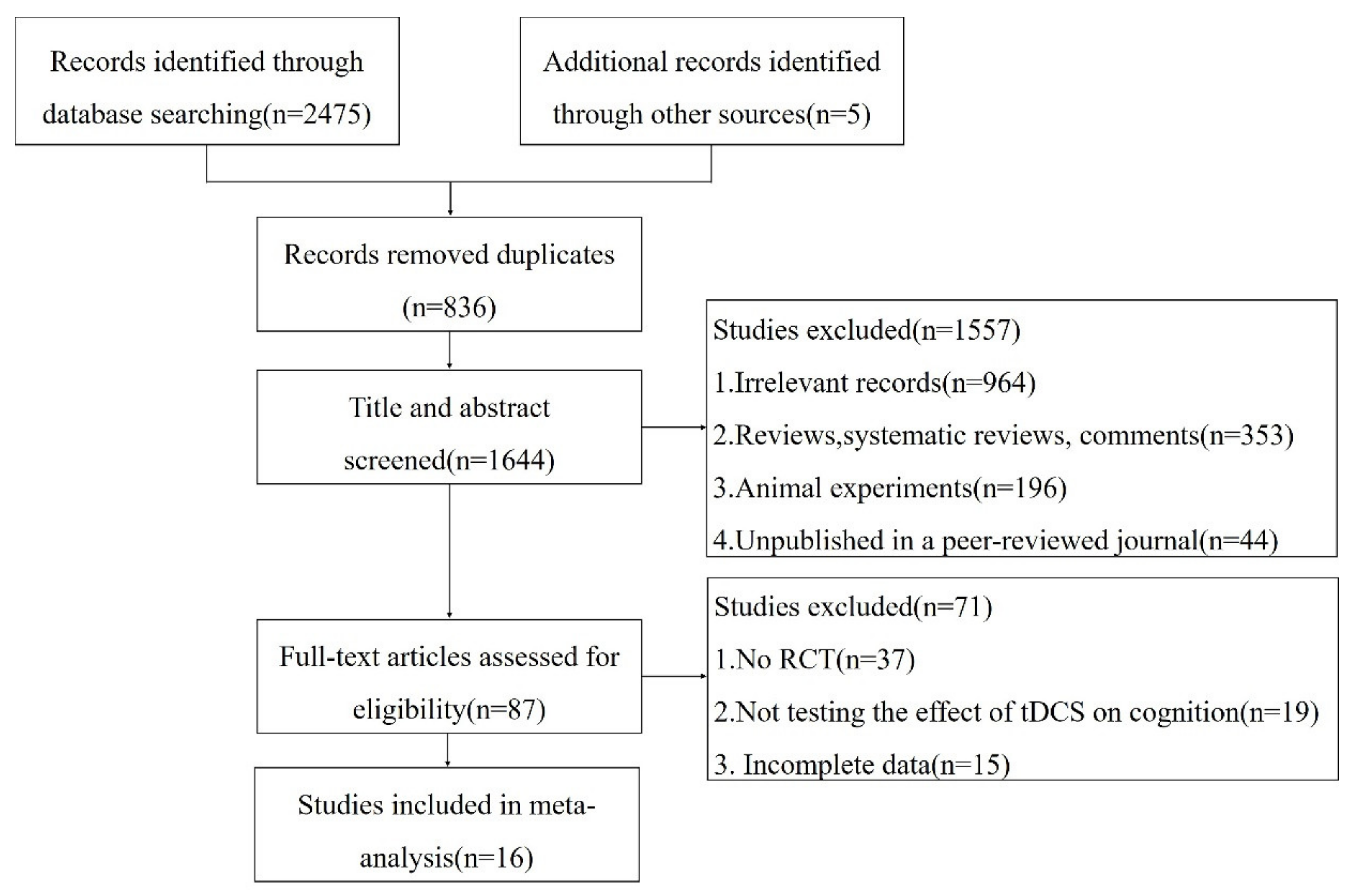
3.1. Study Selection and Characteristics

A total of 2480 relevant documents were retrieved in this study. After removing duplicate studies, excluding irrelevant documents, reviews, systematic reviews, animal experiments, etc., 87 documents remained. After browsing through the full text, we excluded non-RCT, inconsistent outcome indicators, and incomplete data. Finally, sixteen works from the literature were included, including seventeen independent studies, in the meta-analysis. The flow diagram of the literature search and selection is shown in Figure 1.
The basic characteristics of the included studies are presented in Table 1. These studies were published between 2008 and 2021.Among the included studies, fifteen studies [37,38,39,40,41,42,43,44,45,46,47,48,49,50] adopted parallel designs, and two studies [17,51] adopted crossover designs. A total of 616 patients were enrolled, with an average age of 72.2 (SD = 7.88). Participants in six studies [39,41,42,47,48,50] were diagnosed with MCI, and participants in the remaining studies [17,37,38,40,43,44,45,46,49,51] were patients with mild-to-moderate AD. Characteristics of tDCS interventions are shown in Table 2. Five studies [38,39,43,46,47] used a combination of anode tDCS and different cognitive training, and two studies [17,44] used both anodic and cathodic tDCS stimulation. One study [17] used a single stimulation, while the others used multiple stimulations. Eight studies [32,38,41,42,43,44,47,49,50] selected the left DLPFC brain region as the stimulation site; one study [48] selected the right DLPFC brain region as the stimulation site; one study [39] selected the left inferior frontal gyrus; and the remaining seven studies selected the temporal-lobe-related brain area as the stimulation site, including (a) temporal cortex bilaterally [51], (b) left temporal lobe [37], (c) temporal areas bilaterally [17,45], (d) left frontotemporal cortex [40], and (e) left lateral temporal cortex [46]. Eight studies [17,37,43,45,46,47,49,51] used a current density of 0.06 mA/cm2, one study [42] used a current density of 0.07 mA/cm2, five studies [38,41,44,48,50] used a current density of 0.08 mA/cm2, one study [39] used a current density of 0.13 mA/cm2, and the current density used in two studies [40] was 0.25 mA/cm2. All studies evaluated the effect after treatment ends, and ten of them [38,39,41,43,44,46,47,48,49,51] followed up the cognitive function of patients at different time points from one week to six months after the end of treatment.
In terms of cognitive function measurement, different studies used different scales to evaluate the improvement of cognitive function. The detection of overall cognitive function includes Mini-Mental State Examination (MMSE) [37,38,40,41,42,43,44,45,46,51], Alzheimer’s Disease Assessment Scale—Cognitive Subscale (ADAS-Cog) [43,46,49,51], Montreal Cognitive Assessment [45], Cambridge Neuropsychological Test Automated Battery [47,48], Milan Overall Dementia Assessment [40], and Cambridge Cognitive Examination [41]. For the memory domain, the assessment included the word-recall task [41,51], instruction-remembering task [46,51], Rivermead behavioral memory test [38], Rey auditory verbal learning test [38], Tinetti balance scale [38], Tinetti gait scale [38], Word List Memory Test [41], N-back [41], Wechsler Adult Intelligence Scale [41,44], Rey Complex Figure Test [42], Seoul Verbal Learning Test [42], frontal assessment [43], category verbal fluency test [46], digit cancellation task [49], and word-list learning task [49]. The language was assessed by using the Boston Naming Test [41,42], picture-naming task [38], and Battery for Analysis of Aphasic Deficits [38]. Neuropsychiatric Inventory (NPI) [38,46,49] was used to assess the behavioral symptoms, while the word-recognition task [17,49,51] was used to assess recognition memory function. The verbal fluency was assessed by using the Semantic Verbal Fluency test [41], the processing speed was assessed by Symbol Digit Modalities Task [47], and the subjective cognitive function was assessed by Cognitive Failures Questionnaire [47]. Attention was assessed by using the Forward Digit Span Test (FDS) [41,42,46] and Backward Digit Span Test (BDS) [41,42,46]. Visual recognition memory was assessed by using the Visual Recognition Task [51] and Visual Attention Task [17,51]. Verbal memory function was assessed by the California Verbal Learning Test–Second Edition (CVLT-II) [37,39,47]. Trail Making Test part A (TMT-A) [37,38,41,46] measures sustained attention, while Trail Making Test part B (TMT-B) [37,38,41,46], Test of Strategic Learning [39], Delis–Kaplan executive function system [39], Clock Drawing Test [37,41,42,45], Contrasting Program [42], Go-no go Test [42], Controlled Oral Word Association Test [42], and Stroop Test [42] assessed executive function. The Face–Name Association Memory Task [38] was used to assess the patient’s associative memory, and the subjective memory perception was assessed by using Multifactorial Memory Questions [39,50].

3.2. Quality Assessment

Among the 17 experiments included in this study, Gangemi et al. [40] conducted two independent experiments. The quality evaluation results of the included literature are shown in Table 3. Most experiments described the random sequence generation method in detail, and seven experiments did not describe the specific situation of allocation concealment. The overall methodological quality of the included experiments was good.

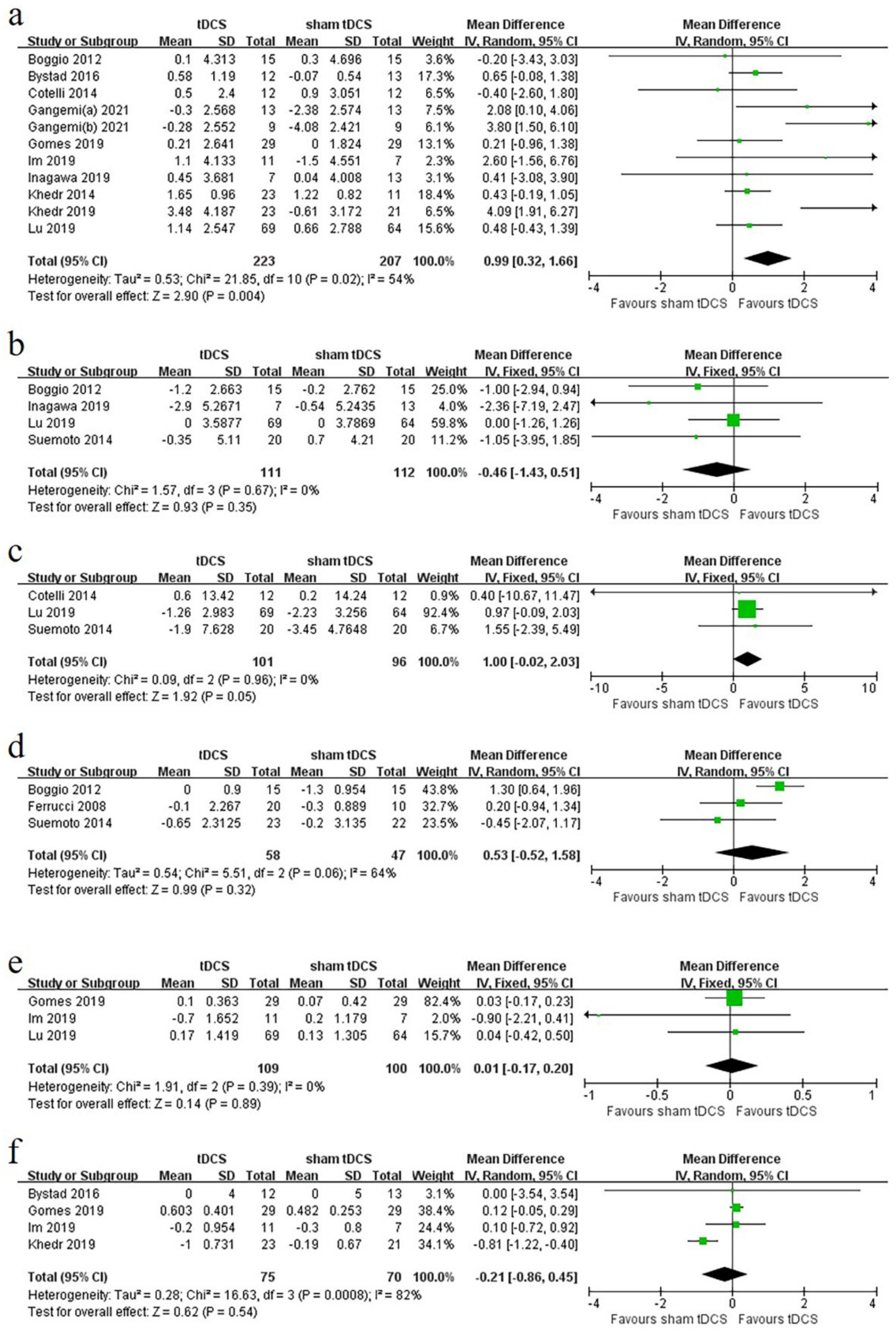
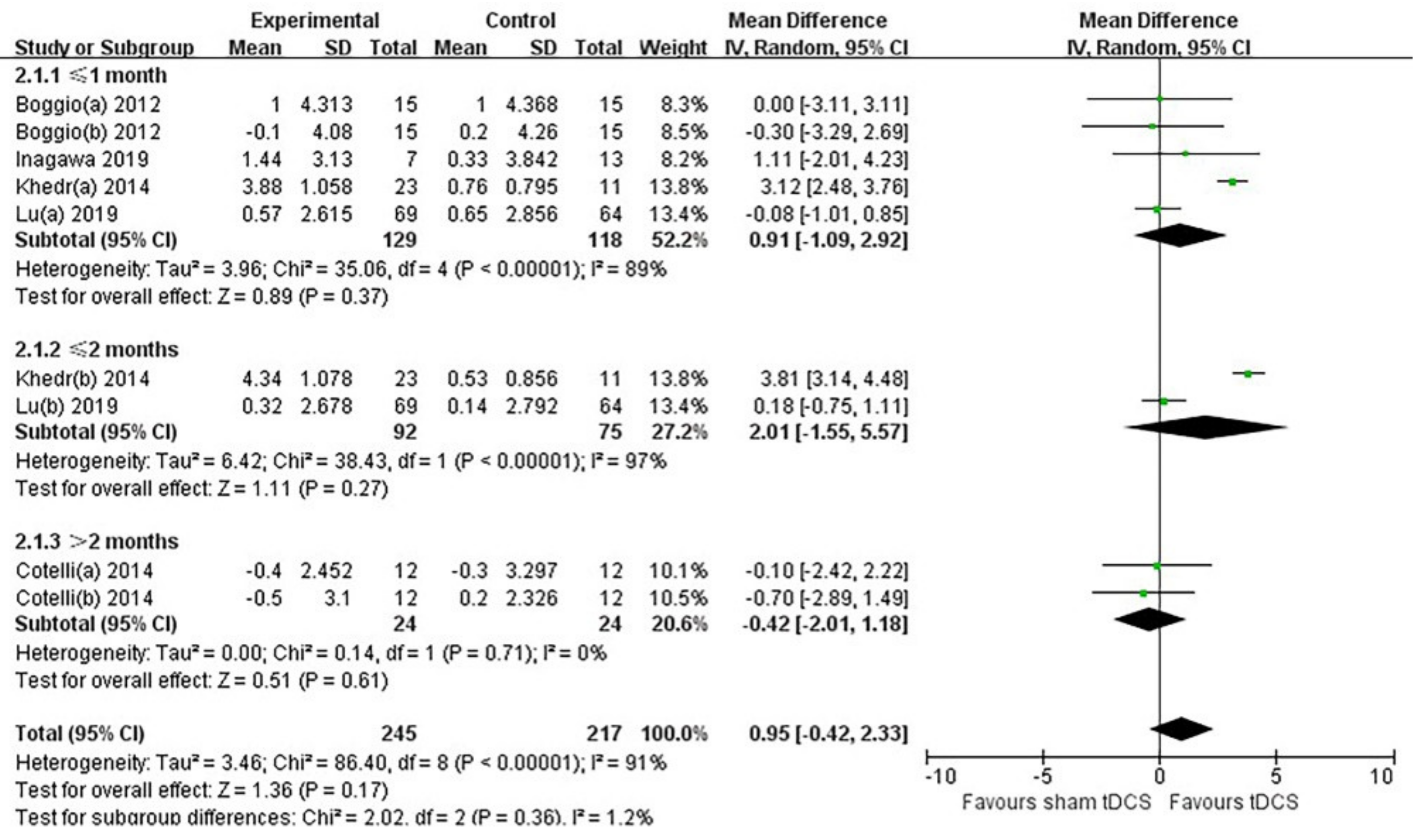
3.3. Primary Outcome

Among the 17 experiments included in the study, objective cognitive score scales used in at least three experiments were selected as outcome indicators: MMSE, ADAS-Cog, NPI, word-recognition task, FDS, BDS, CVLT-II, TMT-A, TMT-B, and Clock Drawing Test. Among them, the baseline score of BDS, TMT-A, and TMT-B was not provided in one study [46]; the data of CVLT-II was insufficient in one study [39]; and the version used in TMT-A and TMT-B in one study [41] was different from other studies, so the above scores were excluded. Finally, we analyzed the six scores, namely the MMSE, ADAS-Cog, NPI, FDS, word-recognition task, and Clock Drawing Test. Their forest plots are shown in Figure 2.

3.4. MMSE

Eleven experiments [35,36,38,39,40,41,42,43,44,49] were included, with 223 patients in the experimental group and 207 patients in the control group. The heterogeneity test indicated that a random-effects model could be used (I2 = 54%, p < 0.1). The meta-analysis showed that, compared with the control group, tDCS treatment significantly improved the overall cognitive function assessed by the MMSE of the experimental group, with a combined WMD of 0.99 (95% CI, 0.32 to 1.66; p = 0.004; Figure 2a). If studies involving cognitive training were ignored, tDCS significantly improved the overall cognitive function, as assessed by MMSE in the experimental group compared with the control group, with a combined WMD of 1.34 (95% CI, 0.45 to 2.23; p = 0.003; Supplementary Figure S1).

3.5. ADAS-Cog

Four experiments [32,46,49,51] were included, with 111 patients in the experimental group and 112 patients in the control group. The heterogeneity test indicated that fixed-effects model could be used (I2 = 0%, p > 0.1). The meta-analysis showed that tDCS treatment failed to significantly improve overall cognitive function, as assessed by ADAS-Cog in the experimental group, with a combined WMD of −0.46 (95% CI, −1.43 to 0.51; p = 0.35; Figure 2b).

3.6. NPI

Three experiments [38,46,49] were included, with 101 patients in the experimental group and 96 patients in the control group. The heterogeneity test indicated that the fixed-effects model could be used (I2 = 0%, p > 0.1). The meta-analysis showed that the tDCS treatment failed to significantly improve behavioral symptoms in the treatment group compared with the control group, with a combined WMD of 1.00 (95% CI, −0.02 to 2.03; p = 0.05; Figure 2c).

3.7. Word-Recognition Task

Three experiments [17,49,51] were included, with 58 patients in the experimental group and 47 patients in the control group. The heterogeneity test indicated that the random-effects model could be used (I2 = 64%, p < 0.1). The meta-analysis showed that the tDCS treatment did not significantly improve recognition memory function in the experimental group compared with the control group, with a combined WMD of 0.53 (95% CI, −0.52 to 1.58; p = 0.32; Figure 2d).

3.8. FDS

Three experiments [41,42,46] were included, with 109 patients in the experimental group and 100 patients in the control group. The heterogeneity test indicated that the fixed-effects model could be used (I2 = 0%, p > 0.1). The meta-analysis showed that the tDCS treatment did not significantly improve attention in the treatment group compared with the control group, with a combined WMD of 0.01 (95% CI, −0.17 to 0.20; p = 0.89; Figure 2e).

3.9. Clock Drawing Test

Four experiments [37,41,42,45] were included, with 75 patients in the experimental group and 70 patients in the control group. The heterogeneity test showed that the random-effects model could be used (I2 = 82%, p < 0.1). The meta-analysis showed that tDCS treatment did not significantly improve the executive function in the treatment group compared with the control group, with a combined WMD of −0.21 (95% CI, −0.86 to 0.45; p = 0.54; Figure 2f).

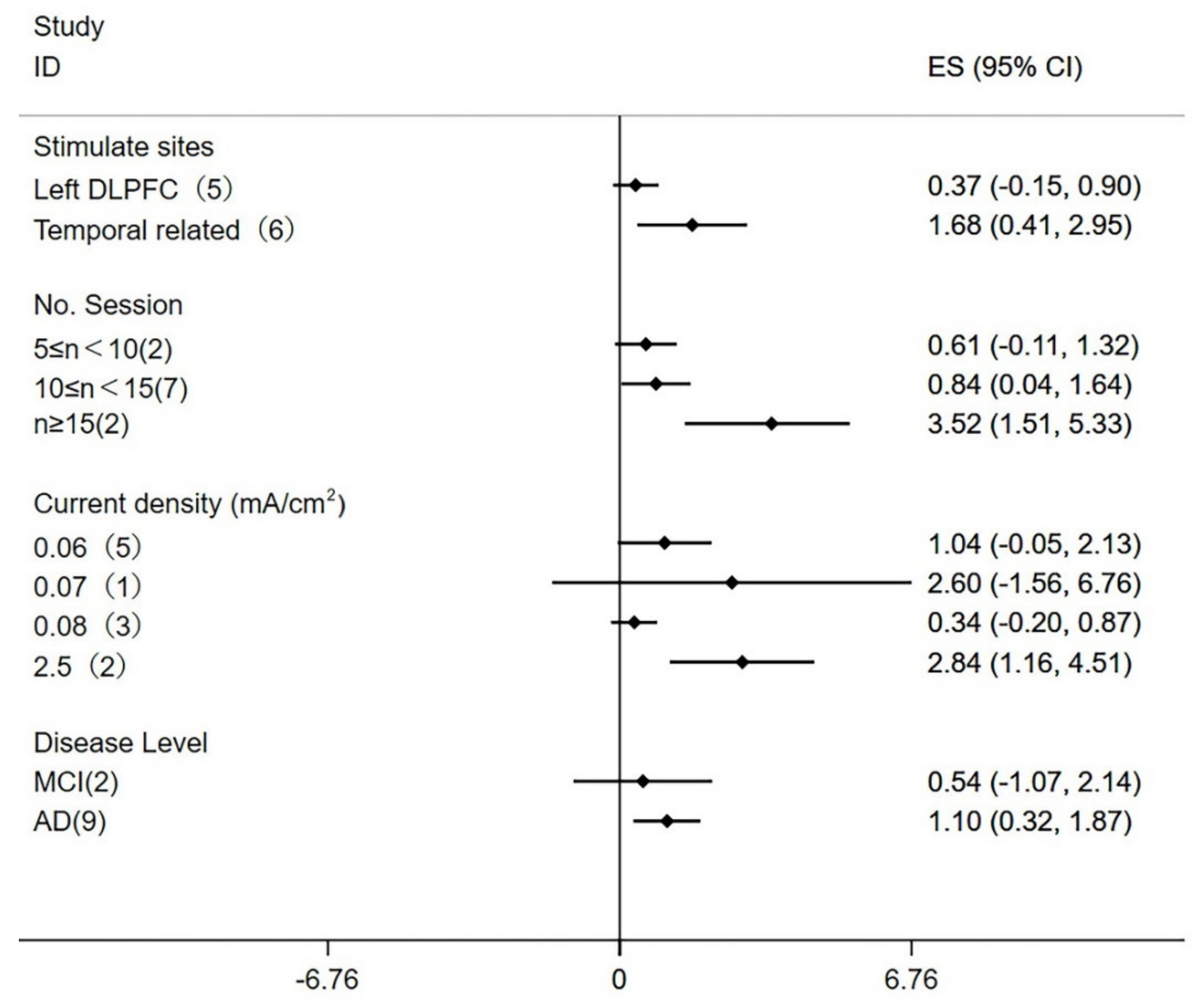
3.10. Subgroup Analysis and Meta-Regression

A subgroup analysis was used to identify variables that might affect the heterogeneity for the MMSE score (Figure 3). In view of the complexity of tDCS parameters in each study, the factors most likely to affect heterogeneity were selected for subgroup analysis, including stimulate sites, number of sessions, current density, and disease level. Among them, the number of sessions was comprehensively analyzed according to the number of tDCS treatments and the days required to complete the treatment, and the current density was divided according to the included study conditions. The results showed that the stimulation of the left DLPFC (WMD = 0.37; 95% CI, −0.15 to 0.90; p = 0.16) did not significantly improve the MMSE score, and stimulated temporal-lobe-related brain areas (WMD = 1.68; 95% CI, 0.41 to 2.95; p = 0.009) can significantly improve the MMSE score. The subgroup analysis of the number of stimulations showed that the number of stimulations that ranged from 5 to 10 (WMD = 0.61; 95% CI, −0.11 to 1.32; p = 0.10) had no improvement in cognitive function; meanwhile, the number of stimulations that ranged from 10 to 15 (WMD = 0.84; 95% CI, 0.04 to 1.64; p = 0.04) improved cognitive function, and the number of stimulations that was greater than or equal to 15 times (WMD = 3.52; 95% CI, 1.51 to 5.53; p = 0.0006) significantly improved cognitive function. The subgroup analysis of current density showed that the current density was 0.06 mA/cm2 (WMD = 1.04; 95% CI, −0.05 to 2.13; p = 0.06), 0.07 mA/cm2 (WMD = 2.60; 95% CI, −1.56 to 6.76; p = 0.22), or 0.08 mA/cm2 (WMD = 0.34; 95% CI, −0.20 to 0.87; p = 0.22) and did not improve cognitive function, while 2.5 mA/cm2 (WMD = 2.84; 95% CI, 1.16 to 4.51; p = 0.0009) significantly improved cognitive function. Compared with MCI patients (WMD = 0.54; 95% CI, −1.07 to 2.14; p = 0.51), AD patients benefited more in regard to cognitive function after tDCS stimulation (WMD = 1.10; 95% CI, 0.32 to 1.87; p = 0.005).
A subgroup meta-regression analysis (Supplementary Figure S2) was conducted to explore the influence of different study characteristics on the MMSE score, and the overall results were relatively robust.

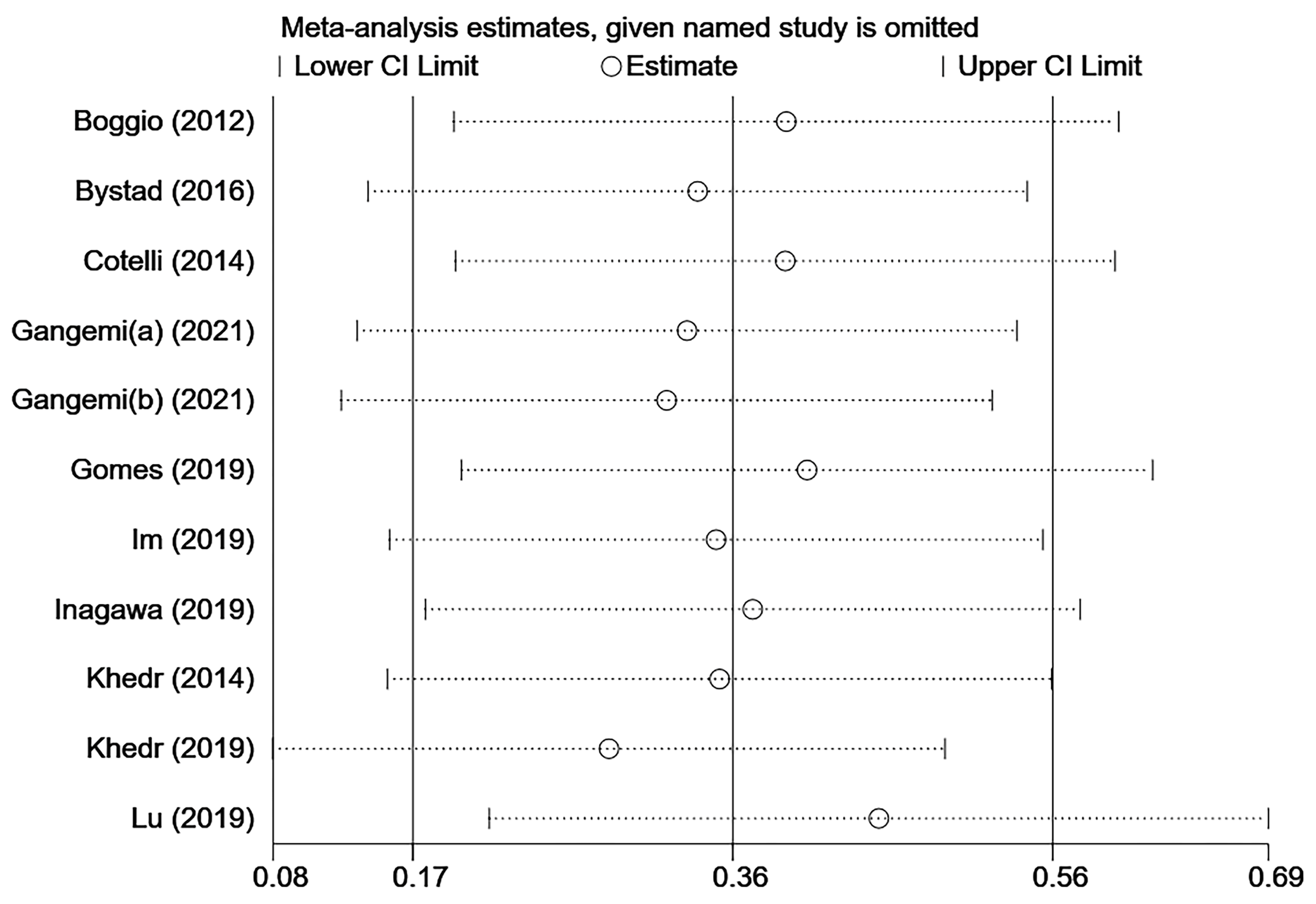
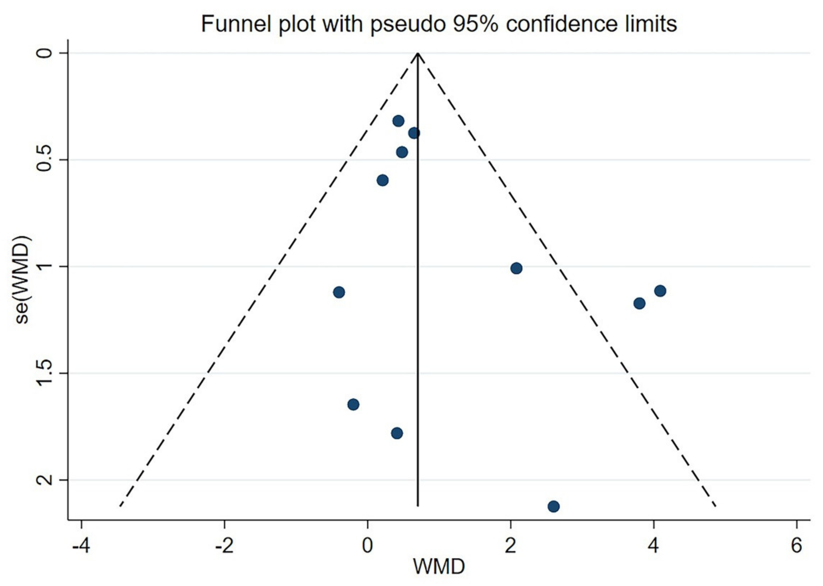
3.11. Sensitivity Analysis and Publication Bias

The heterogeneity of the MMSE score included in the study is relatively high (I2 = 54%), so a sensitivity analysis was performed (Figure 4). When we ignored any of the studies, the overall results were not significantly different. Therefore, the results remain stable and robust. There was no suggestion of a small study effect based on visual inspection of the funnel plot (Figure 5). The results of the Egger’s test (p = 0.144) and Begg’s test (p = 0.350) showed that there was no potential publication bias (Supplementary Figure S3).

3.12. Secondary Results

Effect at different time points after tDCS stimulation:
To evaluate the effects of tDCS on MCI or AD patients at different time points after treatment, we divided the MMSE score into three subgroups (≤1 month, ≤2 months, and >2 months) according to different follow-up times. The forest plot (Figure 6) shows that the total effective value at different follow-up times after the end of treatment was 0.95 (95% CI, −0.42 to 2.33; p = 0.17). Among them, the WMD of the ≤1 month group was 0.91 (95% CI, −1.09 to 2.92; p = 0.37), the WMD of the ≤2 months group was 2.01 (95% CI, −1.55 to 5.57; p = 0.27), and the >2 months group WMD was −0.42 (95% CI, −2.01 to 1.18; p = 0.61).

4. Discussion

This study systematically evaluated the effect of tDCS stimulation on the cognitive function of patients with MCI and mild-to-moderate AD and the effect at different time points after treatment, including 16 studies. The results showed that tDCS significantly improved the overall cognitive function of patients with MCI and mild-to-moderate AD evaluated by MMSE, but it had no significant improvement on the ADAS-Cog score. Meanwhile, tDCS treatment failed to significantly improve the behavioral symptoms, recognition memory function, attention, and executive function of patients with MCI and mild-to-moderate AD. The results of the subgroup analysis showed that the stimulation of temporal-lobe-related brain regions, the number of stimulations ≥10, and the current density of 2.5 mA/cm2 were better than for the stimulation of left DLPFC; the number of stimulations was between 5 and 10; the current density was 0.06, 0.07, or 0.08 mA/cm2. Moreover, compared with MCI patients, patients with mild-to-moderate AD might benefit more from tDCS treatment. After the end of the tDCS treatment, there were no statistically significant differences in MMSE score changes at the follow-ups after 1 month, 2 months, and more than 2 months.
After receiving tDCS treatment, the overall cognitive function of patients with MCI and mild-to-moderate AD evaluated by MMSE had been significantly improved, which was similar to the results of a previous meta-analysis [28]. The MMSE score is the most commonly used screening tool to measure cognitive impairment in clinical practice [52]. The research selection, data extraction, and bias-risk assessment were conducted by an independent reviewer in this study, and this helped us avoid the selective reporting of specific results and further strengthened the effectiveness of our meta-analysis. The ADAS-Cog score did not change significantly after tDCS treatment in this study; this outcome might be linked to the small number of trials included in the score. In addition to the impairment of overall cognitive function, AD patients also have defects in working memory, executive function, attention, language fluency, etc. [53,54]. Currently, studies have reported that tDCS has a significant therapeutic effect on speech–motor learning [55], seizure-related attention deficit [56], executive dysfunction [57], etc. However, we did not find a significant therapeutic effect with MCI and mild-to-moderate AD.
The effects of tDCS stimulation in different brain regions might be different. Our results revealed that the stimulation of the temporal-lobe-related brain regions had better cognitive improvement than the left DLPFC stimulation. Numerous studies have shown that the temporal lobe is related to short-term and long-term memory storage [58,59], and the DLPFC is not only involved in cortical and subcortical functional connectivity, but also plays an important role in maintaining executive memory and cognition and working memory [60,61]. Therefore, non-invasive transcranial stimulation often uses these two brain regions as the stimulation site. It has been found that transcranial random noise stimulation in the lateral temporal lobe can significantly improve epileptic-related memory deficits [62]. This may mean that tDCS can also improve cognitive function by producing neural noise in the temporal lobe. Transcranial magnetic stimulation (rTMS), another non-invasive brain stimulation modality that works by altering cortical excitability, was considered to be effective in cognitive improvement by stimulating the right DLPFC in a meta-study [63]. All the experiments that stimulate DLPFC included in this study used the left side, and this might be the reason for its insignificant effect. In addition to the two stimulation sites mentioned above, this study also included one experiment stimulating right DLPFC and one stimulating the inferior frontal gyrus (IFG). The cognitive evaluation scale of the two experiments was less than three experiments, so no meta-analysis was conducted. Increasing the exploration of brain regions where tDCS might be effective and unifying the evaluation criteria for cognitive function might give us more insight into the effect of tDCS on cognitive improvement.
Multiple studies have shown that tDCS treatment has a cumulative effect [64,65,66] and repeated tDCS treatment may be more effective than a single treatment. In this study, we found no significant improvement in MMSE scores when the total number of stimulations was between 5 and 10. When the number of stimulations was between 10 and 15, the MMSE score increased significantly. When the number of stimulations was more than 15, the MMSE score improved more significantly. Studies have also shown that tDCS stimulation at different time intervals may affect its therapeutic effect. The cumulative effect was obvious when tDCS was applied continuously, but there was no obvious cumulative effect when tDCS was applied every 2 days or weekly [67,68]. This may be related to the current state of the brain, as studies have found that fewer active neurons are more likely to be promoted by subsequent external stimulation (tDCS) [69]. Exploring the underlying mechanism of the cumulative effect of tDCS and finding the best time to produce the cumulative effect will be an important research direction for tDCS to improve the cognitive function of patients with MCI and mild-to-moderate AD.
The current density is the ratio of the current to the size of Montage, which is an important parameter in tDCS treatment. In our study, the current densities of 0.06, 0.07, and 0.08 mA/cm2 could not improve the MMSE score, but 2.5 mA/cm2 could produce significant cognitive improvement. Among them, 0.06 and 0.08 mA/cm2 are commonly used parameters in clinical studies. As is well-known, the effect induced by 0.08 mA/cm2 is generally greater than the effect induced by 0.06 mA/cm2 [70], which is slightly different from our conclusions. It has been found that tDCS in MCI patients may produce smaller current density in the brain regions targeted by tDCS than in healthy elderly people when given the same current density of tDCS, due to greater brain atrophy [71]. Therefore, it is possible that only a relatively large current density can produce significant effects on tDCS treatment. In terms of the selection of Montage size, Foerster et al. found that the stimulation of a small Montage size was more specific than that of a large Montage size at the same current density [72]. In this study, when the current density was 2.5 mA/cm2, the size of Montage was only 0.8 cm2, which might be the reason for the significant improvement in cognitive function. However, 2.5 mA/cm2 was the first time to be applied to patients with AD; although that study included two independent experiments, the small number of subjects included might reduce the credibility of the conclusions. Therefore, more studies with different current densities are required to determine the best parameters for tDCS to improve the cognitive function of patients with MCI and mild-to-moderate AD.
Among patients at different disease degrees, tDCS significantly improved the cognitive function of AD patients but not MCI patients. A recent meta-analysis [73] found that high-frequency rTMS improved cognitive function in patients with AD, but had no significant effect on cognitive function in patients with MCI. The cognitive improvement of tDCS was also greater only in patients with AD than in patients with MCI [73,74]. This might be related to the ceiling effect of MMSE [75]; that is, it is difficult for MCI patients to detect large changes in MMSE scores. However, there were only two experiments of MCI patients included in this study, and we need to interpret the results more cautiously.
How long the cognitive benefit would be of tDCS in patients of MCI and AD is another important issue that people care about. In the current meta-analysis, we did not find that the improvement effect of tDCS on cognitive function could be maintained for a long time. In addition to the acute effects on brain functions, specific tDCS protocols have been reported to induce long-lasting alterations of cortical excitability and activity [18]. Therefore, tDCS treatment has the potential for lasting benefits. Given the small number of trials with follow-up records included in this study, the current results need to be interpreted with caution, and more studies are needed to provide evidence for the long-term effects of tDCS.

5. Limitations

This meta-analysis also has certain limitations. On the one hand, constrained by the inclusion criteria and the different evaluation criteria between the studies, the final sample size for meta-analysis was small; thus, it might have some limitations on the statistics of tDCS treatment effects. On the other hand, this study also included the experiment of tDCS alone and the experiment of combining tDCS with other cognitive training. The cognitive training methods of each study are different and may have a certain impact on the results. In terms of heterogeneity test, there was no subgroup analysis of treatment time, follow-up time, race, blindness of study design, and other factors, due to the lack of parameters, thus resulting in insufficient detailed parameters for the tDCS treatment.

6. Conclusions

In summary, the existing evidence showed that tDCS can significantly improve the overall cognitive function of patients with MCI and mild-to-moderate AD, especially in the stimulation of temporal-lobe-related brain regions; the number of stimulations was greater than or equal to 10 times, and the current density is 2.5 mA/cm2. Meanwhile, AD patients might benefit more than MCI patients. In terms of behavioral symptoms, recognition memory function, attention, and executive function, tDCS treatment did not bring significant benefits. Finally, the therapeutic effect of tDCS was only obvious at the end of the tDCS, and no effect of tDCS on improving cognitive function was found in the follow-up of 1 month, 2 months, and more than 2 months.

Supplementary Materials

The following supporting information can be downloaded at https://www.mdpi.com/article/10.3390/brainsci12050562/s1. Figure S1: Forest plots of MMSE score mean change from baseline after studies involving cognitive training were ignored. Figure S2: Meta-regression analyses of MMSE. Figure S3: Potential publication bias detection results.

Author Contributions

K.Z. conceived and supervised the study; J.C., Z.W. and Q.C. screened the literature and data extraction; J.C. and Z.W. performed statistical analyses; J.C., Z.W. and Y.F. wrote the manuscript; Q.C. edited the manuscript. All authors have read and agreed to the published version of the manuscript.

Funding

This research was funded by the Fundamental Research Funds for the Central Universities (item number: 2019kfyXKJC055), and the National Key Research and Development Program of China (Project No. 2019YFC2004805).

Institutional Review Board Statement

Not required, since this a systematic review.

Informed Consent Statement

This study is a review study and informed consent is not required.

Data Availability Statement

https://doi.org/10.5061/dryad.dr7sqvb1j (accessed on 18 April 2022).

Conflicts of Interest

The authors declare no conflict of interest.

References

  1. Nance, C.; Ritter, A.; Miller, J.B.; Lapin, B.; Banks, S.J. The Pathology of Rapid Cognitive Decline in Clinically Diagnosed Alzheimer’s Disease. J. Alzheimer’s Dis. 2019, 70, 983–993. [Google Scholar] [CrossRef] [PubMed]
  2. Lyketsos, C.G.; Carrillo, M.C.; Ryan, J.M.; Khachaturian, A.S. Neuropsychiatric symptoms in Alzheimer’s disease. Alzheimer’s Dement. 2011, 7, 532–539. [Google Scholar] [CrossRef] [PubMed] [Green Version]
  3. Kirova, A.M.; Bays, R.B.; Lagalwar, S. Working memory and executive function decline across normal aging, mild cognitive impairment, and Alzheimer’s disease. Biomed. Res. Int. 2015, 2015, 748212. [Google Scholar] [CrossRef] [PubMed] [Green Version]
  4. Knopman, D.S.; Beiser, A.; Machulda, M.M.; Fields, J.; Roberts, R.O.; Pankratz, V.S.; Aakre, J.; Cha, R.H.; Rocca, W.A.; Mielke, M.M.; et al. Spectrum of cognition short of dementia: Framingham Heart Study and Mayo Clinic Study of Aging. Neurology 2015, 85, 1712–1721. [Google Scholar] [CrossRef] [Green Version]
  5. Roberts, R.O.; Knopman, D.S.; Mielke, M.M.; Cha, R.H.; Pankratz, V.S.; Christianson, T.J.; Geda, Y.E.; Boeve, B.F.; Ivnik, R.J.; Tangalos, E.G.; et al. Higher risk of progression to dementia in mild cognitive impairment cases who revert to normal. Neurology 2014, 82, 317–325. [Google Scholar] [CrossRef] [Green Version]
  6. Birks, J. Cholinesterase inhibitors for Alzheimer’s disease. Cochrane Database Syst. Rev. 2006, 1, CD00559. [Google Scholar] [CrossRef]
  7. Arvanitakis, Z.; Shah, R.C.; Bennett, D.A. Diagnosis and Management of Dementia: Review. JAMA 2019, 322, 1589–1599. [Google Scholar] [CrossRef]
  8. Marucci, G.; Buccioni, M.; Ben, D.D.; Lambertucci, C.; Volpini, R.; Amenta, F. Efficacy of acetylcholinesterase inhibitors in Alzheimer’s disease. Neuropharmacology 2021, 190, 108352. [Google Scholar] [CrossRef]
  9. Gonsalvez, I.; Baror, R.; Fried, P.; Santarnecchi, E.; Pascual-Leone, A. Therapeutic Noninvasive Brain Stimulation in Alzheimer’s Disease. Curr. Alzheimer Res. 2017, 14, 362–376. [Google Scholar] [CrossRef]
  10. Woods, A.J.; Antal, A.; Bikson, M.; Boggio, P.S.; Brunoni, A.R.; Celnik, P.; Cohen, L.G.; Fregni, F. A technical guide to tDCS, and related non-invasive brain stimulation tools. Clin. Neurophysiol. 2016, 127, 1031–1048. [Google Scholar] [CrossRef] [Green Version]
  11. Yokoi, Y.; Narita, Z.; Sumiyoshi, T. Transcranial Direct Current Stimulation in Depression and Psychosis: A Systematic Review. Clin. EEG Neurosci. 2018, 49, 93–102. [Google Scholar] [CrossRef]
  12. Nitsche, M.A.; Schauenburg, A.; Lang, N.; Liebetanz, D.; Exner, C.; Paulus, W.; Tergau, F. Facilitation of implicit motor learning by weak transcranial direct current stimulation of the primary motor cortex in the human. J. Cogn. Neurosci. 2003, 15, 619–626. [Google Scholar] [CrossRef]
  13. Wagner, T.; Valero-Cabre, A.; Pascual-Leone, A. Noninvasive human brain stimulation. Annu. Rev. Biomed. Eng. 2007, 9, 527–565. [Google Scholar] [CrossRef] [Green Version]
  14. Philip, N.S.; Nelson, B.G.; Frohlich, F.; Lim, K.O.; Widge, A.S.; Carpenter, L.L. Low-Intensity Transcranial Current Stimulation in Psychiatry. Am. J. Psychiatry 2017, 174, 628–639. [Google Scholar] [CrossRef] [Green Version]
  15. Nitsche, M.A.; Paulus, W. Excitability changes induced in the human motor cortex by weak transcranial direct current stimulation. J. Physiol. 2000, 527 Pt 3, 633–639. [Google Scholar] [CrossRef]
  16. Batsikadze, G.; Moliadze, V.; Paulus, W.; Kuo, M.F.; Nitsche, M.A. Partially non-linear stimulation intensity-dependent effects of direct current stimulation on motor cortex excitability in humans. J. Physiol. 2013, 591, 1987–2000. [Google Scholar] [CrossRef]
  17. Ferrucci, R.; Mameli, F.; Guidi, I.; Mrakic-Sposta, S.; Vergari, M.; Marceglia, S.; Cogiamanian, F.; Barbieri, S.; Scarpini, E.; Priori, A. Transcranial direct current stimulation improves recognition memory in Alzheimer disease. Neurology 2008, 71, 493–498. [Google Scholar] [CrossRef] [Green Version]
  18. Stagg, C.J.; Antal, A.; Nitsche, M.A. Physiology of Transcranial Direct Current Stimulation. J. ECT 2018, 34, 144–152. [Google Scholar] [CrossRef]
  19. Pellicciari, M.C.; Miniussi, C. Transcranial Direct Current Stimulation in Neurodegenerative Disorders. J. ECT 2018, 34, 193–202. [Google Scholar] [CrossRef]
  20. Beheshti, I.; Ko, J.H. Modulating brain networks associated with cognitive deficits in Parkinson’s disease. Mol. Med. 2021, 27, 24. [Google Scholar] [CrossRef]
  21. De Berker, A.O.; Bikson, M.; Bestmann, S. Predicting the behavioral impact of transcranial direct current stimulation: Issues and limitations. Front. Hum. Neurosci. 2013, 7, 613. [Google Scholar] [CrossRef] [Green Version]
  22. Gervits, F.; Ash, S.; Coslett, H.B.; Rascovsky, K.; Grossman, M.; Hamilton, R. Transcranial direct current stimulation for the treatment of primary progressive aphasia: An open-label pilot study. Brain Lang. 2016, 162, 35–41. [Google Scholar] [CrossRef] [Green Version]
  23. Dagan, M.; Herman, T.; Harrison, R.; Zhou, J.; Giladi, N.; Ruffini, G.; Manor, B.; Hausdorff, J.M. Multitarget transcranial direct current stimulation for freezing of gait in Parkinson’s disease. Mov. Disord. 2018, 33, 642–646. [Google Scholar] [CrossRef]
  24. Broeder, S.; Nackaerts, E.; Heremans, E.; Vervoort, G.; Meesen, R.; Verheyden, G.; Nieuwboer, A. Transcranial direct current stimulation in Parkinson’s disease: Neurophysiological mechanisms and behavioral effects. Neurosci. Biobehav. Rev. 2015, 57, 105–117. [Google Scholar] [CrossRef]
  25. Liu, C.S.; Herrmann, N.; Gallagher, D.; Rajji, T.K.; Kiss, A.; Vieira, D.; Lanctot, K.L. A Pilot Study Comparing Effects of Bifrontal versus Bitemporal Transcranial Direct Current Stimulation in Mild Cognitive Impairment and Mild Alzheimer Disease. J. ECT 2020, 36, 211–215. [Google Scholar] [CrossRef]
  26. Luo, Y.; Yang, W.; Li, N.; Yang, X.; Zhu, B.; Wang, C.; Hou, W.; Wang, X.; Wen, H.; Tian, X. Anodal Transcranial Direct Current Stimulation Can Improve Spatial Learning and Memory and Attenuate Abeta42 Burden at the Early Stage of Alzheimer’s Disease in APP/PS1 Transgenic Mice. Front. Aging Neurosci. 2020, 12, 134. [Google Scholar] [CrossRef]
  27. Cocco, S.; Rinaudo, M.; Fusco, S.; Longo, V.; Gironi, K.; Renna, P.; Aceto, G.; Mastrodonato, A.; Li Puma, D.D.; Podda, M.V.; et al. Plasma BDNF Levels Following Transcranial Direct Current Stimulation Allow Prediction of Synaptic Plasticity and Memory Deficits in 3xTg-AD Mice. Front. Cell Dev. Biol. 2020, 8, 541. [Google Scholar] [CrossRef]
  28. Cai, M.; Guo, Z.; Xing, G.; Peng, H.; Zhou, L.; Chen, H.; McClure, M.A.; He, L.; Xiong, L.; He, B.; et al. Transcranial Direct Current Stimulation Improves Cognitive Function in Mild to Moderate Alzheimer Disease: A Meta-Analysis. Alzheimer Dis. Assoc. Disord. 2019, 33, 170–178. [Google Scholar] [CrossRef]
  29. Dedoncker, J.; Brunoni, A.R.; Baeken, C.; Vanderhasselt, M.A. A Systematic Review and Meta-Analysis of the Effects of Transcranial Direct Current Stimulation (tDCS) Over the Dorsolateral Prefrontal Cortex in Healthy and Neuropsychiatric Samples: Influence of Stimulation Parameters. Brain Stimul. 2016, 9, 501–517. [Google Scholar] [CrossRef] [Green Version]
  30. Cruz Gonzalez, P.; Fong, K.N.K.; Chung, R.C.K.; Ting, K.H.; Law, L.L.F.; Brown, T. Can Transcranial Direct-Current Stimulation Alone or Combined with Cognitive Training Be Used as a Clinical Intervention to Improve Cognitive Functioning in Persons with Mild Cognitive Impairment and Dementia? A Systematic Review and Meta-Analysis. Front. Hum. Neurosci. 2018, 12, 416. [Google Scholar] [CrossRef] [PubMed] [Green Version]
  31. Liu, C.S.; Rau, A.; Gallagher, D.; Rajji, T.K.; Lanctot, K.L.; Herrmann, N. Using transcranial direct current stimulation to treat symptoms in mild cognitive impairment and Alzheimer’s disease. Neurodegener. Dis. Manag. 2017, 7, 317–329. [Google Scholar] [CrossRef] [PubMed]
  32. Inagawa, T.; Narita, Z.; Sugawara, N.; Maruo, K.; Stickley, A.; Yokoi, Y.; Sumiyoshi, T. A Meta-Analysis of the Effect of Multisession Transcranial Direct Current Stimulation on Cognition in Dementia and Mild Cognitive Impairment. Clin. EEG Neurosci. 2019, 50, 273–282. [Google Scholar] [CrossRef] [PubMed]
  33. Moher, D.; Liberati, A.; Tetzlaff, J.; Altman, D.G.; Group PRISMA. Preferred reporting items for systematic reviews and meta-analyses: The PRISMA statement. BMJ 2009, 339, b2535. [Google Scholar] [CrossRef] [PubMed] [Green Version]
  34. Deeks, J.J.; Higgins, J.P.T.; Altman, D.G. Chapter 10: Analysing data and undertaking meta-analyses. In Cochrane Handbook for Systematic Reviews of Interventions; Version 6.2 (Updated February 2021); Higgins, J.P.T., Thomas, J., Chandler, J., Cumpston, M., Li, T., Page, M.J., Welch, V.A., Eds.; Cochrane: London, UK, 2021; Available online: https://www.training.cochrane.org/handbook (accessed on 25 September 2021).
  35. Higgins, J.P.; Thompson, S.G.; Deeks, J.J.; Altman, D.G. Measuring inconsistency in meta-analyses. BMJ 2003, 327, 557–560. [Google Scholar] [CrossRef] [Green Version]
  36. Egger, M.; Davey Smith, G.; Schneider, M.; Minder, C. Bias in meta-analysis detected by a simple, graphical test. BMJ 1997, 315, 629–634. [Google Scholar] [CrossRef] [Green Version]
  37. Bystad, M.; Gronli, O.; Rasmussen, I.D.; Gundersen, N.; Nordvang, L.; Wang-Iversen, H.; Aslaksen, P.M. Transranial direct current stimulation as a memory enhancer in patients with Alzheimer’s disease: A randomized, placebo-controlled trial. Alzheimer’s Res. Ther. 2016, 8, 13. [Google Scholar] [CrossRef] [Green Version]
  38. Cotelli, M.; Manenti, R.; Brambilla, M.; Petesi, M.; Rosini, S.; Ferrari, C.; Zanetti, O.; Miniussi, C. Anodal tDCS during face-name associations memory training in Alzheimer’s patients. Front. Aging Neurosci. 2014, 6, 38. [Google Scholar] [CrossRef]
  39. Das, N.; Spence, J.S.; Aslan, S.; Vanneste, S.; Mudar, R.; Rackley, A.; Quiceno, M.; Chapman, S.B. Cognitive Training and Transcranial Direct Current Stimulation in Mild Cognitive Impairment: A Randomized Pilot Trial. Front. Neurosci. 2019, 13, 307. [Google Scholar] [CrossRef] [Green Version]
  40. Gangemi, A.; Colombo, B.; Fabio, R.A. Effects of short- and long-term neurostimulation (tDCS) on Alzheimer’s disease patients: Two randomized studies. Aging Clin. Exp. Res. 2021, 33, 383–390. [Google Scholar] [CrossRef]
  41. Gomes, M.A.; Akiba, H.T.; Gomes, J.S.; Trevizol, A.P.; de Lacerda, A.L.T.; Dias, A.M. Transcranial direct current stimulation (tDCS) in elderly with mild cognitive impairment: A pilot study. Dement. Neuropsychol. 2019, 13, 187–195. [Google Scholar] [CrossRef]
  42. Im, J.J.; Jeong, H.; Bikson, M.; Woods, A.J.; Unal, G.; Oh, J.K.; Na, S.; Park, J.S.; Knotkova, H.; Song, I.U.; et al. Effects of 6-month at-home transcranial direct current stimulation on cognition and cerebral glucose metabolism in Alzheimer’s disease. Brain Stimul. 2019, 12, 1222–1228. [Google Scholar] [CrossRef]
  43. Inagawa, T.; Yokoi, Y.; Narita, Z.; Maruo, K.; Okazaki, M.; Nakagome, K. Safety and Feasibility of Transcranial Direct Current Stimulation for Cognitive Rehabilitation in Patients with Mild or Major Neurocognitive Disorders: A Randomized Sham-Controlled Pilot Study. Front. Hum. Neurosci. 2019, 13, 273. [Google Scholar] [CrossRef]
  44. Khedr, E.M.; Gamal, N.F.; El-Fetoh, N.A.; Khalifa, H.; Ahmed, E.M.; Ali, A.M.; Noaman, M.; El-Baki, A.A.; Karim, A.A. A double-blind randomized clinical trial on the efficacy of cortical direct current stimulation for the treatment of Alzheimer’s disease. Front. Aging Neurosci. 2014, 6, 275. [Google Scholar] [CrossRef]
  45. Khedr, E.M.; Salama, R.H.; Abdel Hameed, M.; Abo Elfetoh, N.; Seif, P. Therapeutic Role of Transcranial Direct Current Stimulation in Alzheimer Disease Patients: Double-Blind, Placebo-Controlled Clinical Trial. Neurorehabil. Neural Repair 2019, 33, 384–394. [Google Scholar] [CrossRef]
  46. Lu, H.; Chan, S.S.M.; Chan, W.C.; Lin, C.; Cheng, C.P.W.; Linda Chiu Wa, L. Randomized controlled trial of TDCS on cognition in 201 seniors with mild neurocognitive disorder. Ann. Clin. Transl. Neurol. 2019, 6, 1938–1948. [Google Scholar] [CrossRef] [Green Version]
  47. Martin, D.M.; Mohan, A.; Alonzo, A.; Gates, N.; Gbadeyan, O.; Meinzer, M. A Pilot Double-Blind Randomized Controlled Trial of Cognitive Training Combined with Transcranial Direct Current Stimulation for Amnestic Mild Cognitive Impairment. J. Alzheimer’s Dis. 2019, 71, 503–512. [Google Scholar] [CrossRef]
  48. Stonsaovapak, C.; Hemrungroj, S.; Terachinda, P.; Piravej, K. Effect of Anodal Transcranial Direct Current Stimulation at the Right Dorsolateral Prefrontal Cortex on the Cognitive Function in Patients with Mild Cognitive Impairment: A Randomized Double-Blind Controlled Trial. Arch. Phys. Med. Rehabil. 2020, 101, 1279–1287. [Google Scholar] [CrossRef]
  49. Suemoto, C.K.; Apolinario, D.; Nakamura-Palacios, E.M.; Lopes, L.; Leite, R.E.; Sales, M.C.; Nitrini, R.; Brucki, S.M.; Morillo, L.S.; Magaldi, R.M.; et al. Effects of a non-focal plasticity protocol on apathy in moderate Alzheimer’s disease: A randomized, double-blind, sham-controlled trial. Brain Stimul. 2014, 7, 308–313. [Google Scholar] [CrossRef]
  50. Yun, K.; Song, I.U.; Chung, Y.A. Changes in cerebral glucose metabolism after 3 weeks of noninvasive electrical stimulation of mild cognitive impairment patients. Alzheimer’s Res. Ther. 2016, 8, 49. [Google Scholar] [CrossRef] [Green Version]
  51. Boggio, P.S.; Ferrucci, R.; Mameli, F.; Martins, D.; Martins, O.; Vergari, M.; Tadini, L.; Scarpini, E.; Fregni, F.; Priori, A. Prolonged visual memory enhancement after direct current stimulation in Alzheimer’s disease. Brain Stimul. 2012, 5, 223–230. [Google Scholar] [CrossRef]
  52. Arevalo-Rodriguez, I.; Smailagic, N.; Roque, I.F.M.; Ciapponi, A.; Sanchez-Perez, E.; Giannakou, A.; Pedraza, O.L.; Bonfill Cosp, X.; Cullum, S. Mini-Mental State Examination (MMSE) for the detection of Alzheimer’s disease and other dementias in people with mild cognitive impairment (MCI). Cochrane Database Syst. Rev. 2015, 3, CD010783. [Google Scholar]
  53. Mueller, K.D.; Hermann, B.; Mecollari, J.; Turkstra, L.S. Connected speech and language in mild cognitive impairment and Alzheimer’s disease: A review of picture description tasks. J. Clin. Exp. Neuropsychol. 2018, 40, 917–939. [Google Scholar] [CrossRef] [PubMed]
  54. Pei, H.; Ma, L.; Cao, Y.; Wang, F.; Li, Z.; Liu, N.; Liu, M.; Wei, Y.; Li, H. Traditional Chinese Medicine for Alzheimer’s Disease and Other Cognitive Impairment: A Review. Am. J. Chin. Med. 2020, 48, 487–511. [Google Scholar] [CrossRef] [PubMed]
  55. Buchwald, A.; Calhoun, H.; Rimikis, S.; Lowe, M.S.; Wellner, R.; Edwards, D.J. Using tDCS to facilitate motor learning in speech production: The role of timing. Cortex 2019, 111, 274–285. [Google Scholar] [CrossRef]
  56. Hameed, M.Q.; Dhamne, S.C.; Gersner, R.; Kaye, H.L.; Oberman, L.M.; Pascual-Leone, A.; Rotenberg, A. Transcranial Magnetic and Direct Current Stimulation in Children. Curr. Neurol. Neurosci. Rep. 2017, 17, 11. [Google Scholar] [CrossRef]
  57. Dubreuil-Vall, L.; Chau, P.; Ruffini, G.; Widge, A.S.; Camprodon, J.A. tDCS to the left DLPFC modulates cognitive and physiological correlates of executive function in a state-dependent manner. Brain Stimul. 2019, 12, 1456–1463. [Google Scholar] [CrossRef]
  58. Jeneson, A.; Squire, L.R. Working memory, long-term memory, and medial temporal lobe function. Learn. Mem. 2012, 19, 15–25. [Google Scholar] [CrossRef] [Green Version]
  59. Lara, A.H.; Wallis, J.D. The Role of Prefrontal Cortex in Working Memory: A Mini Review. Front. Syst. Neurosci. 2015, 9, 173. [Google Scholar] [CrossRef] [Green Version]
  60. Liang, P.; Wang, Z.; Yang, Y.; Jia, X.; Li, K. Functional disconnection and compensation in mild cognitive impairment: Evidence from DLPFC connectivity using resting-state fMRI. PLoS ONE 2011, 6, e22153. [Google Scholar]
  61. Yang, Y.; Liang, P.; Lu, S.; Li, K.; Zhong, N. The role of the DLPFC in inductive reasoning of MCI patients and normal agings: An fMRI study. Sci. China C Life Sci. 2009, 52, 789–795. [Google Scholar] [CrossRef]
  62. Meisenhelter, S.; Jobst, B.C. Neurostimulation for Memory Enhancement in Epilepsy. Curr. Neurol. Neurosci. Rep. 2018, 18, 30. [Google Scholar] [CrossRef]
  63. Liao, X.; Li, G.; Wang, A.; Liu, T.; Feng, S.; Guo, Z.; Tang, Q.; Jin, Y.; Xing, G.; McClure, M.A.; et al. Repetitive Transcranial Magnetic Stimulation as an Alternative Therapy for Cognitive Impairment in Alzheimer’s Disease: A Meta-Analysis. J. Alzheimer’s Dis. 2015, 48, 463–472. [Google Scholar] [CrossRef]
  64. Shariatirad, S.; Vaziri, A.; Hassani-Abharian, P.; Sharifi Fardshad, M.; Molavi, N.; Fitzgerald, P.B. Cumulative and booster effects of tdcs sessions on drug cravings, lapse, and cognitive impairment in methamphetamine use disorder: A case study report. Am. J. Addict. 2016, 25, 264–266. [Google Scholar] [CrossRef]
  65. Christova, M.; Rafolt, D.; Gallasch, E. Cumulative effects of anodal and priming cathodal tDCS on pegboard test performance and motor cortical excitability. Behav. Brain Res. 2015, 287, 27–33. [Google Scholar] [CrossRef]
  66. Besson, P.; Perrey, S.; Teo, W.P.; Muthalib, M. Commentary: Cumulative effects of anodal and priming cathodal tDCS on pegboard test performance and motor cortical excitability. Front. Hum. Neurosci. 2016, 10, 70. [Google Scholar] [CrossRef] [Green Version]
  67. Boggio, P.S.; Nunes, A.; Rigonatti, S.P.; Nitsche, M.A.; Pascual-Leone, A.; Fregni, F. Repeated sessions of noninvasive brain DC stimulation is associated with motor function improvement in stroke patients. Restor. Neurol. Neurosci. 2007, 25, 123–129. [Google Scholar]
  68. Alonzo, A.; Brassil, J.; Taylor, J.L.; Martin, D.; Loo, C.K. Daily transcranial direct current stimulation (tDCS) leads to greater increases in cortical excitability than second daily transcranial direct current stimulation. Brain Stimul. 2012, 5, 208–213. [Google Scholar] [CrossRef]
  69. Silvanto, J.; Muggleton, N.; Walsh, V. State-dependency in brain stimulation studies of perception and cognition. Trends Cogn. Sci. 2008, 12, 447–454. [Google Scholar] [CrossRef]
  70. Bastani, A.; Jaberzadeh, S. Differential modulation of corticospinal excitability by different current densities of anodal transcranial direct current stimulation. PLoS ONE 2013, 8, e72254. [Google Scholar] [CrossRef]
  71. Mahdavi, S.; Towhidkhah, F. Computational human head models of tDCS: Influence of brain atrophy on current density distribution. Brain Stimul. 2018, 11, 104–107. [Google Scholar] [CrossRef]
  72. Foerster, Á.S.; Rezaee, Z.; Paulus, W.; Nitsche, M.A.; Dutta, A. Effects of Cathode Location and the Size of Anode on Anodal Transcranial Direct Current Stimulation Over the Leg Motor Area in Healthy Humans. Front. Neurosci. 2018, 12, 443. [Google Scholar] [CrossRef]
  73. Chu, C.S.; Li, C.T.; Brunoni, A.R.; Yang, F.C.; Tseng, P.T.; Tu, Y.K.; Stubbs, B.; Carvalho, A.F.; Thompson, T.; Rajji, T.K.; et al. Cognitive effects and acceptability of non-invasive brain stimulation on Alzheimer’s disease and mild cognitive impairment: A component network meta-analysis. J. Neurol. Neurosurg. Psychiatry 2021, 92, 195–203. [Google Scholar] [CrossRef]
  74. Teselink, J.; Bawa, K.K.; Koo, G.K.; Sankhe, K.; Liu, C.S.; Rapoport, M.; Oh, P.; Marzolini, S.; Gallagher, D.; Swardfager, W.; et al. Efficacy of non-invasive brain stimulation on global cognition and neuropsychiatric symptoms in Alzheimer’s disease and mild cognitive impairment: A meta-analysis and systematic review. Ageing Res. Rev. 2021, 72, 101499. [Google Scholar] [CrossRef]
  75. Spencer, R.J.; Wendell, C.R.; Giggey, P.P.; Katzel, L.I.; Lefkowitz, D.M.; Siegel, E.L.; Waldstein, S.R. Psychometric limitations of the mini-mental state examination among nondemented older adults: An evaluation of neurocognitive and magnetic resonance imaging correlates. Exp. Aging Res. 2013, 39, 382–397. [Google Scholar] [CrossRef]
Figure 1. Flow diagram of the screening process of the literature.
Brainsci 12 00562 g001
Figure 2. Forest plots of mean change from baseline, based on different scales: (a) Mini-Mental State Examination (MMSE), the order of references is [51,37,38,40,40,41,42,44,45,46]. (b) Alzheimer’s Disease Assessment Scale–Cognitive Subscale (ADAS-Cog), the order of references is [51,43,46,49]. (c) Neuropsychiatric Inventory (NPI), the order of references is [38,46,49]. (d) word-recognition task, the order of references is [51,17,49]. (e) Forward Digital Span (FDS), the order of references is [41,42,46]. (f) Clock Drawing Test, the order of references is [37,41,42,45]. Independent studies in the same literature are distinguished by (a,b). Error bars are 95% confidential intervals.
Brainsci 12 00562 g002
Figure 3. Subgroup analyses of MMSE.
Brainsci 12 00562 g003
Figure 4. Sensitivity analyses of MMSE. The order of references is [51,37,38,40,40,41,42,43,44,45,46].
Brainsci 12 00562 g004
Figure 5. Funnel plot of MMSE.
Brainsci 12 00562 g005
Figure 6. Forest plots of the effects of tDCS on MCI or mild-to-moderate AD patients at different time points after treatment. The order of references is [51,37,38,40,40,41,42,43,44,45,46]. The results of different follow-up times in the same study are represented by (a) and (b).
Brainsci 12 00562 g006
Table 1. The basic characteristics of the included studies.
Study (Time)Sample Size DesignDiagnosisGender
(M/F)
Age (y)Education (y)Duration of
Disease(y)
Outcomes for Cognition Function
Boggio et al. (2012) [51] NE:15
NC:15
CrossoverAD8/778.95 ± 8.0714.42 ± 3.654.39 ± 1.88MMSE, VAT, ADAS-Cog, Word recall, Word recognition, Instruction remembering, VRT
Bystad et al. (2016) [37]NE:12
NC:13
ParallelAD7/5
7/6
70.0 ± 8.0
75.0 ± 8.7
NRNRCVLT-II, MMSE, Clock-drawing test, TMT-A, TMT-B
Cotelli et al. (2014) [38]NE:12
NC:12
ParallelAD2/10
3/9
76.6 ± 4.6
74.7 ± 6.1
5.5 ± 2.4
8.9 ± 5.1
NRFNAT, MMSE, Tinetti balance scale, Tinetti gait scale, NPI, Picture naming task, BADA, Rivermead behavioral memory test, Rey auditory verbal learning test, TMT-A, TMT-B
Das et al. (2019) [39]NE:12
NC:10
ParallelMCI8/4
8/2
62.58 ± 8.43
63.30 ± 7.38
17.92 ± 3.94
16.20 ± 1.75
NRTOSL, DKEFS, CVLT, MMQ
Ferrucci et al. (2008) [17]NEa:10
NEb:10
NC:10
CrossoverAD3/775.2 ± 7.310.9 ± 4.8NRWord recognition task, VAT
Gangemi(a) et al. (2021) [40]NE:13
NC:13
ParallelADNR67.5 ± 2.8
69.01 ± 3.1
6.5 ± 2.0
6.1 ± 2.1
NRMMSE, MODA
Gangemi(b) et al. (2021) [40]NE:9
NC:9
ParallelADNR68.5 ± 2.8
68.7 ± 3.1
6.7 ± 2.0
6.2 ± 2.7
NRMMSE, MODA
Gomes et al. (2019) [41]NE:29
NC:29
ParallelMCI9/20
7/22
73.0 ± 9.2
71.6 ± 7.9
NRNRCAMCOG, MMSE, TMT-A, TMT-B, SVF, BNT, Clock-drawing test, WLMT, WAIS, N-back, FDS, BDS
Im et al. (2019) [42]NE:11
NC:7
ParallelMCI1/10
2/5
71.9 ± 9.2
74.9 ± 5.0
6.3 ± 3.8
5.4 ± 5.9
NRMMSE, FDS, BDS, BNT, SVLT, COWAT, RCFT, Contrasting Program, Go-no go Test, Stroop Test, Clock-drawing test
Inagawa et al. (2019) [43]NE:7
NC:13
ParallelAD3/4
7/6
76.6 ± 5.7
76.2 ± 7.7
NR0.9 ± 1.2
1.2 ± 1.5
ADAS-Cog, MMSE, FAB
Khedr et al. (2014) [44]NEa:11
NEb:12
NC:11
ParallelAD6/5
8/4
5/6
68.5 ± 7.2
70.7 ± 5.4
67.3 ± 5.9
NR3.0 ± 2.6
2.9 ± 1.9
3.5 ± 1.7
MMSE, WAIS
Khedr et al. (2019) [45]NE:23
NC:21
ParallelAD13/10
13/8
64.22 ± 3.64
65.23 ± 4.52
1.17 ± 0.48
1.17 ± 0.39
4.04 ± 2.83
3.52 ± 1.96
MMSE, Clock-drawing test, MoCA
Lu et al. (2019) [46]NE:69
NC:64
ParallelAD21/42
17/36
74.2 ± 6.7
74.5 ± 6.6
7.3 ± 4.8
6.5 ± 4.3
NRADAS-Cog, MMSE, NPI, CVFT, FDS, BDS, TMT-A, TMT-B
Martin et al. (2019) [47]NE:33
NC:35
ParallelMCI13/20
10/25
71.8 ± 6.39
71.6 ± 6.35
14.5 ± 3.51
14.9 ± 3.23
NRCVLT-II, CANTAB, SDMT, CFQ
Stonsaovapak et al. (2020) [48]NE:23
NC:22
ParallelMCI2/21
2/20
68.39 ± 8.37
69.68 ± 7.60
NRNRCANTAB
Suemoto et al. (2014) [49]NE:20
NC:20
ParallelAD5/15
7/13
79.4 ± 7.1
81.6 ± 8.0
5 ± 4.2
4.5 ± 3.9
NRNPI, ADAS-Cog, Digit cancellation task, Word list learning task, Word recognition task
Yun et al. (2016) [50]NE:8
NC:8
ParallelMCI3/5
2/6
74.75 ± 7.47
73.12 ± 4.25
8.06 ± 4.93
5.56 ± 2.41
NRMMQ
Data are expressed as mean ± SD. Independent studies in the same literature are distinguished by (a) and (b). AD, Alzheimer disease; MCI, mild cognitive impairment; M, male; F, female; NR, not reported; MMSE, Mini-Mental State Examination; VAT, Visual Attention Task; ADAS-Cog, Alzheimer’s Disease Assessment Scale—Cognitive Subscale; VRT, Visual Recognition Task; CVLT-II, California Verbal Learning Test—Second Edition; TMT-A, Trail Making Test parts A; TMT-B, Trail Making Test parts B; FNAT, Face–Name Association Memory Task; NPI, Neuropsychiatric Inventory; BADA, Battery for Analysis of Aphasic Deficits; TOSL, Test of Strategic Learning; DKEFS, Delis–Kaplan executive function system; MMQ, Multifactorial Memory Questions; MODA, Milan Overall Dementia Assessment; CAMCOG, Cambridge Cognitive Examination; SVF, Semantic Verbal Fluency test; BNT, Boston Naming Test; SVLT, Seoul Verbal Learning Test; COWAT, Controlled Oral Word Association Test; RCFT, Rey Complex Figure Test; FAB, frontal assessment battery; WAIS, Wechsler Adult Intelligence Scale; MoCA, Montreal Cognitive Assessment; FDS, Forward Digit Span Test; BDS, Backward Digit Span Test; CVFT, category verbal fluency test; CANTAB, Cambridge Neuropsychological Test Automated Battery; SDMT, Symbol Digit Modalities Task; CFQ, Cognitive Failures.
Table 2. The characteristics of tDCS interventions.
Study (Time)Type of StimulationNumber of SessionsDuration
(min)
Stimulation SiteCurrent
(mA)
Montage Size
(cm2)
Stimulation ModelAdverse Effects
Boggio et al. (2012) [51]Anode
Sham
per day for 5 consecutive days30Temporal cortex bilaterally235OfflineNo adverse effects were recorded after five daily tDCS sessions
Bystad et al. (2016) [37]Anode
Sham
6 sessions for 10 days30Left temporal lobe235OfflineNo adverse effects were reported
Cotelli et al. (2014) [38]Anode+ICMT
Sham+ICMT
5 sessions per week for 2 weeks25Left DLPFC225OnlineNR
Das et al. (2019) [39]Anode+SMART
Sham+SMART
8 sessions for 4 weeks20Left IFG215OfflineNR
Ferrucci et al. (2008) [17]Anodal
Cathodal
Sham
1 session15Temporoparietal areas bilaterally1.525OfflineNR
Gangemi(a) et al. (2021) [40]Anode
Sham
Daily, for 10 days20Left frontotemporal cortex20.8OfflineNR
Gangemi(b) et al. (2021) [40]Anode
Sham
10 sessions each month for 8 months20Left frontotemporal cortex20.8OfflineNR
Gomes et al. (2019) [41]Anode
Sham
Twice per week for 5 weeks30Left DLPFC 225OfflineNR
Im et al. (2019) [42]Anode
Sham
Daily, for 6 months30Left DLPFC 228OfflineNR
Inagawa et al. (2019) [43]Anode+CT|
Sham+CT
2 sessions per day for 5 consecutive days20Left DLPFC235OnlineNeither severe adverse events nor the need for medications caused by adverse events
Khedr et al. (2014) - [44]Anodal
Cathodal
Sham
Daily, for 10 days25Left DLPFC224OfflineTwo patients under active stimulation recorded itching, headache, and dizziness that were disappear after few hours
Khedr et al. (2019) [45]Anode
Sham
5 sessions per week for 2 consecutive weeks20 (each side)Left TP lobe and right TP lobe235OfflineAll the patients tolerated tDCS well without major adverse effects
Lu et al. (2019) [46]Anode+WMT
Sham+WMT
3 sessions per week for 4 weeks20Left LTC235Offlinethree cases had skin lesions under the cathodal electrode during the repeated sessions of tDCS
Martin et al. (2019) [47]Anode+CT
Sham+CT
3 sessions per week for 5 weeks30Left DLPFC235OnlineNo adverse effects were reported
Stonsaovapak et al. (2020) [48]Anode
Sham
3 times per week for 4 weeks20Right DLPFC225OfflineDizziness was found in one participant from the atDCS group. All side effects disappeared within 24 hours
Suemoto et al. (2014) [49]Anode
Sham
3 sessions per week for 2 weeks20Left DLPFC 235OfflineTDCS was well tolerated and not associated with significant adverse effects
Yun et al. (2016) [50]Anode
Sham
3 sessions per week for 3 weeks30Left DLPFC225OfflineNo patient reported adverse effects
NR, not reported; CT, cognitive training; ICMT, individualized computerized memory training; SMART, strategic memory and advanced reasoning training; WMT, working-memory training; DLPFC, dorsolateral prefrontal cortex; IFG, inferior frontal gyrus; LTC, lateral temporal cortex; TP, temporoparietal.
Table 3. Assessment of risk of bias for included studies.
StudySequence GenerationAllocation ConcealmentBlinding of ParticipantsPersonnel and Outcomes AssessorsIncomplete
Outcome Data
Selective Outcomes ReportingBaseline
Characteristics
Boggio (2012) [51]???++?-
Bystad (2016) [37]++??++?
Cotelli (2014) [38]??++?++
Das (2019) [39]++++?+?
Ferrucci (2008) [17]+++++??
Gangemi(a) (2021) [40]??++?+?
Gangemi(b) (2021) [40]??++?+?
Gomes (2019) [41]???+?+-
Im (2019) [42]++?++++
Inagawa (2019) [43]++?++++
Khedr (2014) [44]+?++++?
Khedr (2019) [45]+?++++?
Lu (2019) [46]+?+++++
Martin (2019) [47]++?+?++
Stonsaovapak (2020) [48]+++++++
Suemoto (2014) [49]+++++++
Yun (2016) [50]++++++?
Note: + low, - high, and ? uncertain.
Publisher’s Note: MDPI stays neutral with regard to jurisdictional claims in published maps and institutional affiliations.

