Article

Episodic Memory Encoding and Retrieval in Face-Name Paired Paradigm: An fNIRS Study

1
Exercise Psychophysiology Laboratory, Institute of KEEP Collaborative Innovation, School of Psychology, Shenzhen University, Shenzhen 518060, China
2
Swiss Center for Affective Sciences, University of Geneva, 1205 Geneva, Switzerland
3
Laboratory for the Study of Emotion Elicitation and Expression (E3Lab), Department of Psychology, FPSE, University of Geneva, 1205 Geneva, Switzerland
4
MOE Key Laboratory for Neuroinformation, The Clinical Hospital of Chengdu Brain Science Institute, School of Life Science and Technology, University of Electronic Science and Technology of China, Xiyuan Ave 2006, Chengdu 611731, China
5
Department of Neurology, Medical Faculty, Otto von Guericke University, Leipziger Street 44, 39120 Magdeburg, Germany
6
German Center for Neurodegenerative Diseases (DZNE), Research Group Neuroprotection, Leipziger Street 44, 39120 Magdeburg, Germany
7
Department of Psychology, The Education University of Hong Kong, Tai Po, Hong Kong, China
8
UMR 9193-SCALab-Sciences Cognitives et Sciences Affectives, Université de Lille, F-59000 Lille, France
9
Department of Health, Exercise Science, and Recreation Management, The University of Mississippi, Oxford, MS 38677, USA
*
Author to whom correspondence should be addressed.
Brain Sci. 2021, 11(7), 951; https://doi.org/10.3390/brainsci11070951
Submission received: 6 May 2021 / Revised: 28 June 2021 / Accepted: 9 July 2021 / Published: 19 July 2021
(This article belongs to the Section Social Cognitive and Affective Neuroscience)

Abstract

:
Background: Episodic memory (EM) is particularly sensitive to pathological conditions and aging. In a neurocognitive context, the paired-associate learning (PAL) paradigm, which requires participants to learn and recall associations between stimuli, has been used to measure EM. The present study aimed to explore whether functional near-infrared spectroscopy (fNIRS) can be employed to determine cortical activity underlying encoding and retrieval. Moreover, we examined whether and how different aspects of task (i.e., novelty, difficulty) affects those cortical activities. Methods: Twenty-two male college students (age: M = 20.55, SD = 1.62) underwent a face-name PAL paradigm under 40-channel fNIRS covering fronto-parietal and middle occipital regions. Results: A decreased activity during encoding in a broad network encompassing the bilateral frontal cortex (Brodmann areas 9, 11, 45, and 46) was observed during the encoding, while an increased activity in the left orbitofrontal cortex (Brodmann area 11) was observed during the retrieval. Increased HbO concentration in the superior parietal cortices and decreased HbO concentration in the inferior parietal cortices were observed during encoding while dominant activation of left PFC was found during retrieval only. Higher task difficulty was associated with greater neural activity in the bilateral prefrontal cortex and higher task novelty was associated with greater activation in occipital regions. Conclusion: Combining the PAL paradigm with fNIRS provided the means to differentiate neural activity characterising encoding and retrieval. Therefore, the fNIRS may have the potential to complete EM assessments in clinical settings.

1. Introduction

Episodic memory (EM) refers to the process of currently retrieving events from the past [1,2]. EM incorporates two main phases: (a) an encoding stage, which contributes to the formation of a new memory trace, and (b) a retrieval stage, which refers to the conscious remembering of past events. EM is an essential process for various higher cortical functions such as judgment and decision making [1,2]. EM performance has been reported to be particularly sensitive to aging and pathological conditions such as amnesia, mild cognitive impairment (MCI), and Alzheimer’s disease (AD) [3,4]. Notably, a recent study indicated subjective memory complaints in 14% of younger adults (N = 4425) aged between 18 and 39 [5]. The progressive decline of EM has detrimental effects on multiple daily life outcomes including educational success and work performance [6,7]. Furthermore, because of the associated medical cost, EM impairment has been shown to increase financial pressures on families of affected people [8,9,10].
Over the last two decades, the paired-associate learning (PAL) paradigm has become one of the most widely used paradigms to assess EM performance [11,12,13]. PAL paradigm usually involves recollection of objects after learning a series of associations between stimuli (e.g., faces and names). Given that the difficulty and novelty of the paradigm can be easily manipulated by means of item–item associations, it can be adapted to the functional levels of targeted clinical populations. In addition, the paradigm does not include complex language requirements, and it has been widely used to examine EM performances in both healthy participants and individuals with apparent memory impairments [14,15].
Based on the results of previous functional magnetic resonance imaging (fMRI) researches and lesion studies, specific brain regions (i.e., prefrontal, parietal and occipital cortices, and temporal hippocampal areas) have been suggested to be engaged in the process of EM [16,17,18]. According to the transfer-appropriate processing (TAP) theory, retrieval success depends on the successful encoding. Specifically, the extent of encoding-related activation in regions engaged during both encoding and retrieval has been associated with subsequent memory success [19]. However, this reengagement phenomenon in EM has not been well proved by neuroimaging evidence. Moreover, it remains largely elusive whether the novelty (repetition times; repeated pairs vs. novel pairs) and difficult levels (number of characters in a name; two-word pairs vs. three-word pairs) of stimulus can influence the accuracy rate of retrieval. To elucidate the neural mechanisms underlying the retrieval successes, the use of functional near-infrared spectroscopy (fNIRS) has been recently suggested as it provides the means to identify brain functioning to investigate the central and autonomous nervous system in more ecological and clinically usable contexts [20].
Recently, the combination of behavioral assessment with fNIRS recordings has been broadly and efficiently applied in the exploration of cognitive performances and associated brain correlates [21]. Compared with electroencephalogram (EEG) and fMRI, fNIRS is less sensitive to movement artifacts and has fewer contraindications (e.g., allow the participation of individuals with metallic implants such as braces). Thus, fNIRS technology enables researchers to monitor the brain activity of children, healthy adults, as well as clinical population (e.g., MCI, AD) with less stress, higher ecological validity, and fewer costs, thus allowing a better integration into the clinical practice. Regarding the PAL paradigm, fNIRS allows investigators to record the verbal answers and EM-related cortical brain activity simultaneously [22]. Given its advantages, we aimed to combine the PAL paradigm with fNIRS to examine differences in oxygenated–hemoglobin concentrations associated with different aspects of paradigm (i.e., memory phases, novelty, and difficulty level of face-name pairs) among the male college students. Specifically, we hypothesized that the encoding and retrieval should be characterized by different brain responses among the frontal and parietal cortices in spite of partial activation overlap [23,24,25], allowing to differentiate the two phases of EM. As more cognitive resources and efforts are required when performing a novel task [26] and with increasing difficulty levels [27,28,29], we also hypothesized that greater activation would be observed among pairs of trials presenting contrasting novelty (vs. repeated/same pair) and difficulty levels.

2. Methods

2.1. Participants

Based on a priori sample size calculation (α = 0.05, effective size dz = 0.8) [22], 22 male students from Shenzhen University (China; age: M = 20.55, SD = 1.62) participated in this study. Inclusion criteria were (a) normal visual, hearing, and physical function to complete the experiment; (b) right-handedness; (c) absence of mental health disorders or cognitive impairments; (d) no dependence on alcohol, nicotine, coffee, or drugs. Written informed consent forms were signed by all individuals before participation. This study was approved by Shenzhen University Institutional Review Board, with protocol (PN-2020-034) performed in accordance with the latest revision of the Declaration of Helsinki.

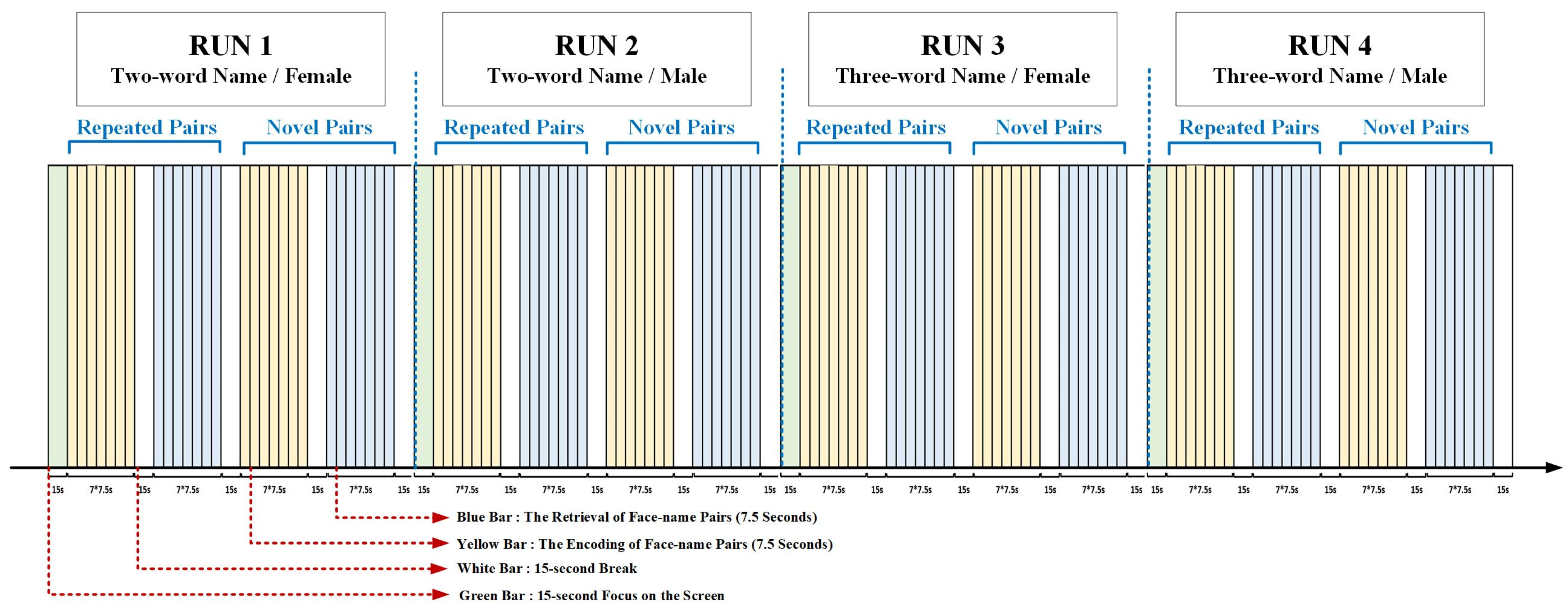
2.2. Experimental Procedure

We adopted the face-name PAL paradigm from a previous fMRI study (Figure 1) [30]. In the present study, the neutral faces were acquired from the CAS-PEAL Large Scale Chinese Face Database and were presented on the lab computer. There were 4 runs in the present paradigm. In the first two runs, two-word names were presented, with (a) run 1: female face-name pairs and (b) run 2: male face-name pairs. In the last two runs, three-word names were presented, with (a) run 3: female face-name pairs and (b) run 4: male face-name pairs. Each run started with a 15-s baseline, and consisted of 4 blocks presented following the order of encoding (repeated), retrieval (repeated), encoding (novel), and retrieval (novel) face-name pairs. In each block, 7 faces were randomly presented on the white background, each lasting for 4.5 s and with 3-s intervals between faces. Each block was followed by a 15-s reset period. In the novel trials, 7 different face-name pairs were randomly presented; in the repeated trials, 2 different face-name pairs were randomly represented (at least 3 times for each pair). The two-word Chinese names and the three-word Chinese names (higher difficult level) represented different difficulty levels in our study. In the encoding phase, the participants were asked to try their best to remember the face-name pairs. In the retrieval phase, the faces which appeared in the encoding phases could be presented, and the participants were required to report the names associated with the presented faces within 4.5 s. The accuracy (accuracy = the number of correct answers/the number of total face-name pairs * 100%) was noted by investigators.
Before the experiment, the entire procedure was explained in detail by the investigator and the participants were given specific instructions on how to remember the face-name matches for later testing. During the experiment, the participants were required to sit in front of the computer (45 cm) without extraneous movements for about 24 min and the fNIRS data were collected simultaneously. At the beginning, a white fixation cross (+) was presented in the center of the visual field on a black background to ensure the concentration of participants on paradigm stimuli. During the encoding runs, seven faces with names printed underneath were shown, and the participants were asked to memorize as accurately as possible the face-name pairs presented on the screen. In the retrieval runs, a face without a name appeared on the screen, and the participants were required to report the paired name within the given time (7.5 s). During the rest periods, a 15-s black screen was displayed. The participants’ responses were recorded for further analysis.

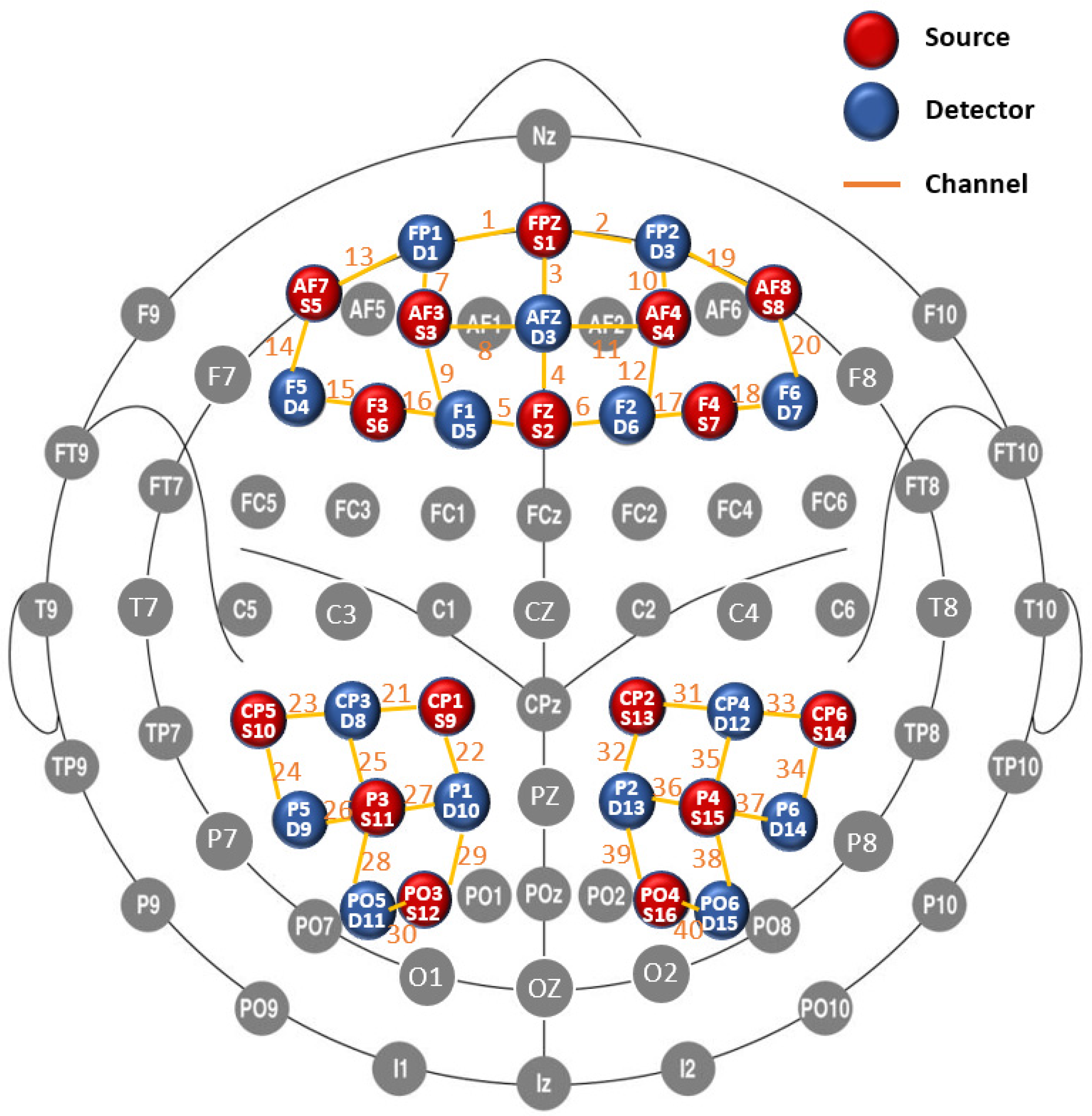
2.2.1. fNIRS Data Acquisition

A 40-channel fNIRS system (Danyang Huichuang Medical Equipment Co. Ltd., China) was used to measure the relative change in oxygenated hemoglobin (HbO). Beta values were computed within the bilateral prefrontal, superior parietal, inferior parietal, and middle occipital cortices. The fNIRS system operated at two wavelengths (750 and 850 nm) and recorded changes at a sample rate of 7 Hz. In total, 16 sources and 15 detectors were used and the optodes were placed in the cap based on the international 10–5 system for EEG electrode placement (Figure 2), with a black overcap covering all the optodes to avoid interference from environmental light (Babak S et al., 2010). The uniform distance between neighboring optodes was 3 cm. The 3D positions (MNI standard coordinates) of the sources and detectors, as well as the reference optodes, were calculated via a 3D digitizer (PATRIOT, Polhemus), which is based on a four-point positioning algorithm in 3D space relying on received signal strength indication.
We mapped the fNIRS channels to the corresponding brain regions according to their MNI coordinates, as shown in Figure 2 (prefrontal cortex: channel 1–20; superior parietal cortex: channel 22, 32; inferior parietal cortex: channel 21–27, 29, 31–37, 39; middle occipital cortex: channel 28, 30, 38, 40). Additionally, the coordinates were checked using the fNIRS Optodes’ Location Decider (fOLD) [31].

2.2.2. fNIRS Data Preprocessing

The raw fNIRS data were recorded during the face-name PAL paradigm using the NIRSmart acquisition software and then processed via the NIRSpark analyzing software. During the data preprocessing, the unrelated time intervals and artifacts induced by motion and environment were eliminated (automatic motion correction of NIRSpark analyzing software; the standard deviation of threshold = 6.0; the amplitude of threshold = 0.5). The light intensities were converted to optical densities and blood oxygen concentrations through the modified Beer–Lambert law. A bandpass filter (0.01–0.1 Hz) was then applied to remove both noise and interference signals (heart rate, breathing rate, and Mayer waves) [32]. The initial time of the hemodynamic response function (HRF) was set to −2 s (i.e., baseline state) and the end time to 50 s (i.e., task state for a single block). The oxygenated HRF was averaged for each channel across the four blocks.

2.3. Data Analysis

The generalized linear model (GLM) was employed to analyze channel-wise hemodynamic responses induced by the face-name PAL paradigm. The GLM established a canonical HRF for each experiment condition and each participant, and calculate the degree of matching between the experimental and ideal HRF values. GLM models were used to analyze HbO concentration, with the stage (encoding/retrieval), novelty (novel/repeated), and difficult level of face-name pairs (two-word name/three-word name) as independent measures. The beta value represented how much a regressor (e.g., experimental manipulation) contributed to the fNIRS signal and whether the HRF model was reliable (Pinti P et al., 2017). Diffuse optical topography (Figure 3) of beta value was generated by EasyTopo [33,34], a toolbox running through MatLab software. Additionally, mean activation of hemodynamic state (HbO concentration) was also computed and exported for postprocessing analyses. In this study, only HbO signals calculated by the modified Beer–Lambert law were used. HbO is less influenced by physiological noise and better reflect changes in the regional cerebral blood oxygenation induced by stimuli when compared to deoxygenated hemoglobin (Hb) [35]. The path length factor was set at 6 for both 740 nm and 850 nm waves.
For the exported data (HbO concentration and beta value), the analysis was performed using IBM SPSS (v.23.0). The normality distribution of data was checked by the Shapiro–Wilk test, as recommended for small sample sizes [36,37]. Then, the paired-sample t-tests (e.g., encoding vs. retrieval, repeated pairs vs. novel pairs (novelty), two-word pairs vs. three-word pairs (difficulty level)) were used for data of normal distribution, and the Wilcoxon signed-rank tests were used for the data of non-normal distribution, with a significance level set at α = 0.05. As recommended for fNIRS studies, the false discovery rate (FDR) was used to account for the multiple comparison problem (i.e., type I error accumulation) [38,39]. For the behavioral data, Pearson’s correlation was used to test the associations between the accuracy of cognitive paradigm (number of correct answers) and beta value [40]. We rated the correlation coefficients as follows: 0 to 0.19: no correlation; 0.2 to 0.39: low correlation, 0.40 to 0.59: moderate correlation; 0.60 to 0.79: moderately high correlation; ≥0.80: high correlation.

3. Results

Table 1 shows the significances of HbO concentration among channels in the different task conditions. The channels with significant differences in beta values (baseline vs. task condition) are shown in Table 2. Finally, the visualization of beta values in the encoding and retrieval phases is illustrated in Figure 3.

3.1. Encoding and Retrieval Phases

During the encoding, HbO concentration showed significant increases from baseline in the left superior parietal cortex (channel 22; FDR-corrected p-value = 0.038); and the significant decreases were observed in bilateral prefrontal cortices and left inferior parietal cortex (channel 1, 3, 17, 18, 20, 25, 26; FDR-corrected p-values = 0.048, 0.048, 0.012, 0.048, 0.029, 0.000, 0.012). During the retrieval phase, a significant increase from baseline of HbO concentration was only observed in the left prefrontal cortex (channel 1; FDR-corrected p-value = 0.048).
In the phase of encoding, the highest beta value was 0.611 located in the left superior parietal cortex, and the lowest beta value was reported in the middle occipital cortex (left: −0.147; right: 0.020). For the retrieval phase, the highest beta value (0.231) was found in the left prefrontal cortex while the lowest one (−0.183) in the left superior parietal cortex. Compared with retrieval, significantly higher beta values during encoding were observed in the right prefrontal cortex (channel 10,12; FDR-corrected p-values = 0.022, 0.022), while significantly lower beta values were observed in the left prefrontal cortex (channel 1; FDR-corrected p-value = 0.021) and right middle occipital cortex (FDR-corrected p-value = 0.021).

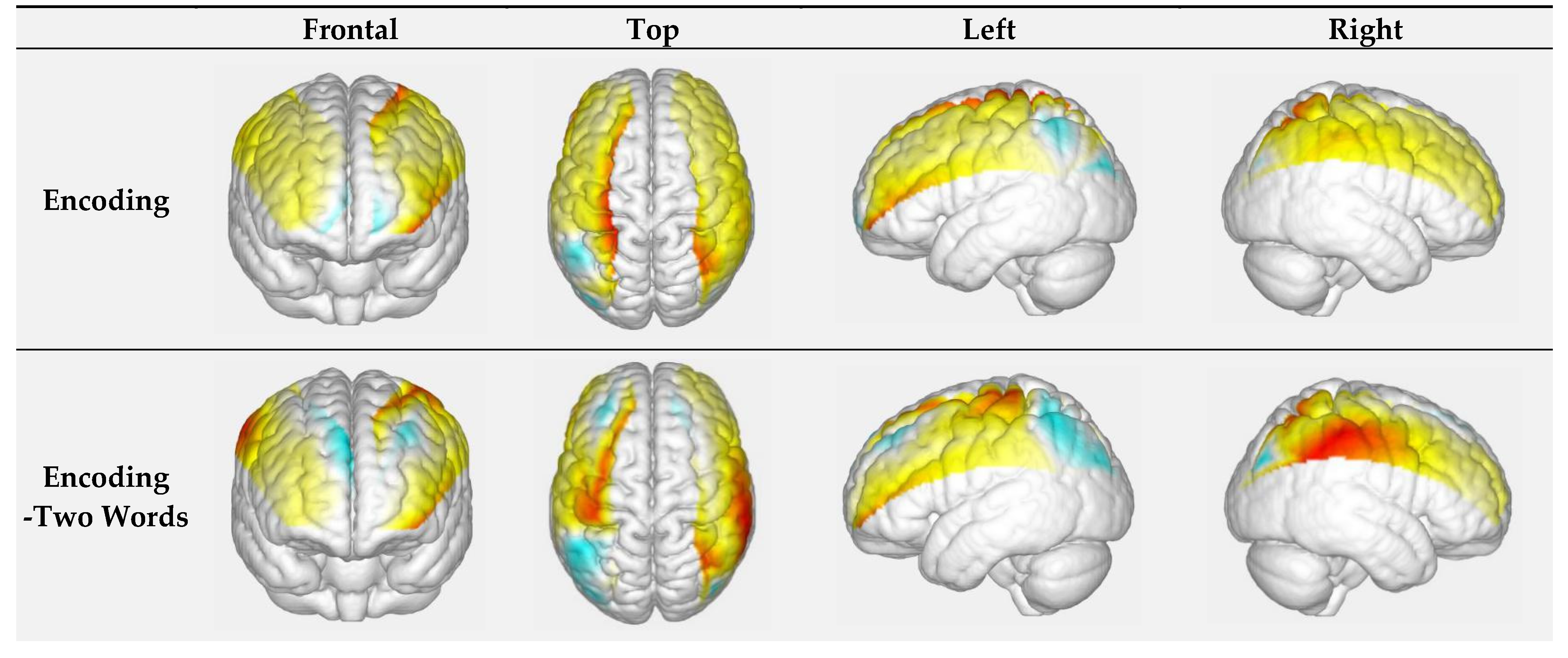
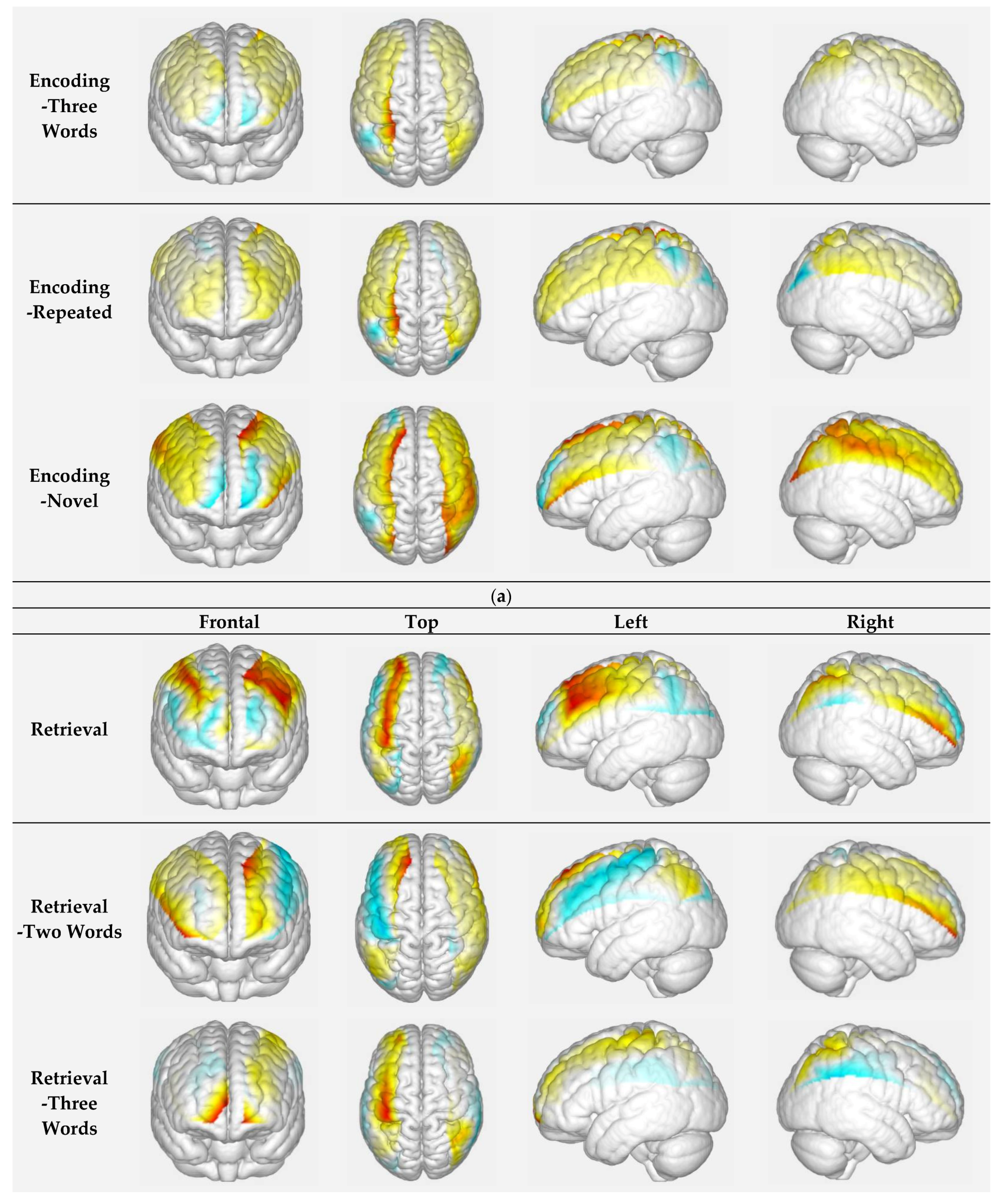
3.1.1. Encoding of Faces with Different Difficulty-Level Names: Two-Word Names vs. Three-Word Names

During the encoding of faces with two-word names, the significant decreases of HbO concentration were observed in bilateral prefrontal cortices, left inferior parietal cortex, and left middle occipital cortex (channel 15, 17, 25, 26, 30; FDR-corrected p-values = 0.026, 0.034, 0.010, 0.012, 0.048). For the encoding of faces with three-word names, significant differences of HbO concentration were shown in the right prefrontal cortex and bilateral inferior parietal cortices (increase from baseline: channel 33; FDR-corrected p-value = 0.035) (decreases from baseline: channel 19, 20, 23, 37; FDR-corrected p-values = 0.035, 0.035, 0.010, 0.048). Compared with the encoding of faces with two-word names, the encoding of faces with three-word names showed a significant increase of beta value in the bilateral prefrontal cortex (channel 4; FDR-corrected p-values = 0.032).

3.1.2. Encoding of Face at Different Novelty Levels: Repeated Faces vs. Novel Faces

During the encoding of repeated faces, HbO concentration showed a significant increase in the left superior parietal cortex (Channel 22; FDR-corrected p-value = 0.035) and decreases in right prefrontal cortex and left inferior parietal cortex (Channel 18, 25; FDR-corrected p-values = 0.026, 0.010). For the encoding of novel faces, significant decreases were observed in the right prefrontal cortex and left inferior parietal cortex (Channel 2,17, 23, 26; FDR-corrected p-values = 0.048, 0.012, 0.026, 0.035). Compared with the encoding with repeated faces, significant differences of beta values were observed in the right prefrontal cortex (lower beta value in channel 11; FDR-corrected p-value = 0.022) and right middle occipital cortex (higher beta value in channel 38; FDR-corrected p-value = 0.034) during the encoding with novel faces.

3.1.3. Retrieval of Faces with Different Difficulty-Level Names: Two-Word Names vs. Three-Word Names

During the retrieval of faces with two-word names, there was no significant decrease or increase from baseline in HbO concentration. During the retrieval of faces with three-word names, HbO showed significant increases in the Brodmann area 11 of bilateral prefrontal cortices (channel 7, 19; FDR-corrected p-values = 0.031, 0.035) and decrease in the Brodmann area 46 of the right prefrontal cortex (channel 20; FDR-corrected p-value = 0.031). Between these two task conditions, no significant difference of beta value was observed in any brain regions.

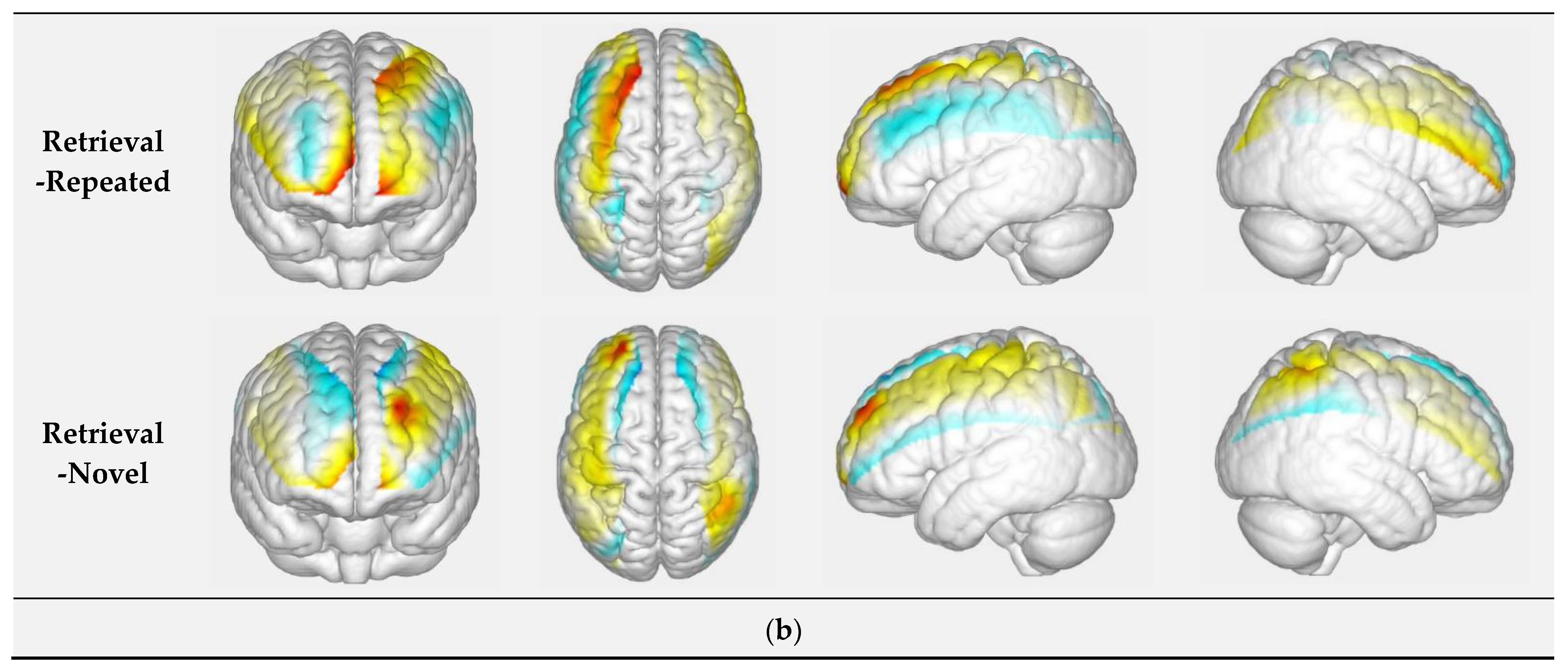
3.1.4. Retrieval of Face at Different Novelty Levels: Repeated Faces vs. Novel Faces

During the retrieval of repeated and novel faces, no channel showed a significant increase or decrease of HbO concentration in any brain region. Compared with the retrieval with repeated faces, significant differences of beta values were observed in the left prefrontal cortex (channel 5; FDR-corrected p-value = 0.021), left middle occipital cortex (channel 30; FDR-corrected p-value = 0.036), right inferior parietal cortex (channel 31; FDR-corrected p-value = 0.046) and right superior parietal cortex (channel 32; FDR-corrected p-value = 0.022) during the retrieval of novel faces.

3.2. Correlations between Memory Performance and Brain Activation (Beta Value)

No correlation was found between the accuracy of retrieval and brain activation during encoding (Table S1). During the retrieval of novel faces, negative correlations were observed between the accuracy of retrieval and beta values (right superior parietal cortex (channel 32): FDR-corrected p-values = 0.020; r = −0.573).

4. Discussion

The present study used fNIRS to examine the brain mechanisms underlying memory encoding and retrieval in a sample of male college students. Compared to baseline, the fNIRS results showed differential changes in brain activation in the PFC between the encoding and retrieval processes of episodic memory (EM). In particular, the encoding process was characterized by decreases in HbO, suggesting deactivations in broader subregions of the bilateral PFC (Brodmann area 9, 11, 45, 46). In contrast, the retrieval process was characterized by increases in HbO in the left orbitofrontal cortex (Brodmann area 11). Compared with retrieval, significantly higher beta values during encoding were observed in the right prefrontal cortex while significantly lower beta values were observed in the left prefrontal cortex and right middle occipital cortex. These outcomes were partially consistent with previous fMRI [16,17,18] and fNIRS [22] studies. Overall, our findings indicate the fNIRS can be a useful tool to model brain activation patterns during memory-related tasks.
Previous fMRI studies reported increased activation of PFC during encoding [41,42,43]. However, a recent fNIRS study [22] reported decreased prefrontal activity during encoding, which was consistent with our outcomes. The differences between fMRI and fNIRS can be explained by the fact that fMRI measures the deeper brain regions, including those in the prefrontal cortex. Regarding the dominant role of left PFC in the retrieval, at least two factors can explain this phenomenon. First, the activation of the left PFC–precuneus network resulted from item–item associations [44]. Secondly, the contribution of left PFC to acquire semantic knowledge was used in the face-name PAL paradigm [45,46,47].
The HbO status during encoding was opposite in the superior (increased activation) and inferior (Brodmann area 39, 40; decreased activation) parietal cortices. Notably, the activated superior parietal cortex is an essential component of the dorsal attention network (DAM) and contributes to the top-down modulation of sensory [48]. Likewise, the deactivated angular gyrus (i.e., Brodmann area 39), which is part of the default mode network (DMN), has been reported to be involved mainly in passive states [48,49,50]. Based on these past findings, it could be argued that memory encoding may require higher attentional processes and thereby may inhibit the function of DMN. Importantly, accumulating evidence pinpoint the important role of parietal DMN nodes in EM retrieval. Specifically, it has been suggested that the task-evoked activity first induces the close coupling of intra-DMN, para-hippocampal, and medial temporal regions and then activates the extra-DMN nodes to make a final decision [51,52]. Therefore, it can be drawn that the parietal cortex differentially responds to memory phases and is in coordination with intra/extra-DMN areas for different purposes.
In this study, regarding faces with different levels of naming difficulty, compared with the blocks with two-word names, the ones with three-word names generated greater beta values and HbO concentration levels in the bilateral prefrontal cortices (Brodmann 9, 10, 11) during both the encoding and retrieval processes. Additionally, during retrieval, the left hemisphere activation was observed to be more dominant among pairs with higher difficulty levels. These results are in line with previous neuroimaging studies [51,52] and fit well within the “cortical asymmetry of reflective activity (CARA)” hypothesis [53], demonstrating that the left prefrontal cortex is more likely to engage in the more complex and reflectively demanding test. The complexity and reflective demand of the task include detailed and deliberative evaluation, analysis, and maintenance of the input information as well as the self-cueing initiation for additional information. Furthermore, in the more difficult level of the task, the selection of relevant information and the inhibition of irrelevant information also requires the participation of the left prefrontal cortex [51,54,55,56].
A successful EM retrieval relies on the rapid reactivation of sensory (i.e., visual) information presented in the encoding process, a process that relies on the occipital cortex [57], which plays a key role in visual processing [57]. In the present study, the repeated pairs generated lower activation in the occipital cortex, compared to the novel pairs. This result is in agreement with the findings of a previous fMRI study observing that the repetition of items suppressed the occipital visual cortex activity [58,59]. It is reported that such repetition suppression arises from stimulus-specific expectations. In particular, the visual cortex (i.e., visual area 1) is likely to produce a weaker response for the expected stimulus relative to unexpected things [60].
According to the transfer-appropriate processing (TAP) theory, people tend to deal with a task faster and more efficiently if the associated stimulus has been experienced before. In terms of memory, this phenomenon can be explained as follows: the success of retrieval depends on the successful encoding processing [61,62,63]. It is hypothesized that key brain areas during encoding can reengage the retrieval process [64]. Our results partially matched with the TAP hypothesis. During the encoding and retrieval of three-word names, Brodmann area 46 was deactivated in a systematic fashion. Likewise, we found that participants did better on the repeated trials only in cases where the degree of activation (or deactivation) in several regions was similar in the encoding and retrieval phases.

Limitations

Due to the limitation of fNIRS penetration depth, the brain activities from the temporal hippocampal area were not available for observation, as noted in previous fMRI studies. Due to the limitation in optode number, we could not assess the activation of the whole occipital lobe. However, the primary visual cortex (visual area 1; Brodmann area 17) and partial extrastriate areas (visual area 2–3; Brodmann area 18) were well covered by the channel setup in this study, which enables us to explore the basic role of the visual system in EM. Additionally, we did not observe the correlation between accuracy of retrieval and brain activation during encoding in the present study. In addition, future studies using fNIRS to investigate EM-related brain activation patterns should use data processing techniques to remove artifacts arising from systemic physiological changes (e.g., short-separation channel regression to account for changes in superficial blood flow) in order to reduce the likelihood of false-positive findings [65,66]. Moreover, considering the limitation of small sample size, as well as a single-sex and narrow-age group (male university students) in this study, a larger sample and wider-scale population will be required to test the present outcomes in the further studies.

5. Conclusions

In conclusion, the results of the present study demonstrated that the encoding and retrieval of EM are associated with distinct patterns of prefrontal, parietal, and middle occipital activation/deactivation. In addition, the paradigm difficulty and the novelty play modulating roles in the engagement of brain substrates. The opposite brain responses of superior and inferior parietal cortices during encoding and the dominant activation of left PFC during retrieval allow us to differentiate the two phases of EM using fNIRS technology during a face-name PAL paradigm. Moreover, these cerebral correlates findings were reinforced by the observation of significant correlations between brain activation patterns and behavioral performance (i.e., accuracy) during the retrieval process that was reinforced. These findings suggest that fNIRS may represent a valuable tool to investigate the brain processes engaged in EM.

Supplementary Materials

The following are available online at https://www.mdpi.com/article/10.3390/brainsci11070951/s1, Table S1: Accuracy in Retrieval Phase.

Author Contributions

Conceptualization, Q.Y. and L.Z.; methodology, Q.Y., B.C., B.B. and F.H.; software, Q.Y., F.H.; validation, L.Z.; formal analysis, Q.Y.; investigation, F.H.; resources, L.Z.; data curation, Q.Y.; writing—original draft preparation, Q.Y. and F.H; writing—review and editing, C.C.H.C., Y.N.D.-T., S.M.R.G., P.L. and N.M.; visualization, C.C.H.C., Y.N.D.-T., S.M.R.G., P.L. and N.M.; supervision, L.Z.; project administration, L.Z.; funding acquisition, L.Z. and B.C. All authors have read and agreed to the published version of the manuscript.

Funding

This study is supported by Start-up Research Grant of Shenzhen University (20200807163056003), Start-Up Research Grant (Peacock Plan: 20191105534C), and Ambizione grant (PZ00P1_180040) from the Swiss National Science Foundation (SNSF).

Institutional Review Board Statement

This study was approved by Shenzhen University Institutional Review Board, with protocol (PN-2020-034) performed in accordance with the latest revision of the Declaration of Helsinki.

Informed Consent Statement

All procedures were conducted with approval from the ethics committee of Shenzhen University (Guangdong, China).

Data Availability Statement

Data of this study were collected at Shenzhen University.

Conflicts of Interest

The authors declare that they have no conflict of interest.

References

  1. Schacter, D.L.; Gilbert, D.T.; Wegner, D.M. Semantic and Episodic Memory, Psychology. 2009. Worth, New York. Available online: http://www.worthpublishers.com/Catalog/product/psychology-firstedition-s... (accessed on 19 July 2021).
  2. Tulving, E. Elements of Episodic Memory; Oxford University Press: Oxford, UK, 1983. [Google Scholar]
  3. Anderson, K.E.; Lynch, K.; Zarahn, E.; Scarmeas, N.; Van Heertum, R.; Sackeim, H.; Moeller, J.R.; Stern, Y. H215O pet study of impairment of nonverbal recognition with normal aging. J. Neuropsychiatry Clin. Neurosci. 2005, 17, 192–200. [Google Scholar] [CrossRef] [PubMed]
  4. Tromp, D.; Dufour, A.; Lithfous, S.; Pebayle, T.; Després, O. Episodic memory in normal aging and alzheimer disease: Insights from imaging and behavioral studies. Ageing Res. Rev. 2015, 24, 232–262. [Google Scholar] [CrossRef] [PubMed]
  5. Chen, S.T.; Siddarth, P.; Ercoli, L.M.; Merrill, D.; Torres-Gil, F.; Small, G.W. Modifiable risk factors for alzheimer disease and subjective memory impairment across age groups. PLoS ONE 2014, 9, e98630. [Google Scholar] [CrossRef] [Green Version]
  6. Conway, M.A.; Gardiner, J.M.; Perfect, T.J.; Anderson, S.J.; Cohen, G.M. Changes in memory awareness during learning: The acquisition of knowledge by psychology undergraduates. J. Exp. Psychol. Gen. 1997, 126, 393–413. [Google Scholar] [CrossRef] [PubMed]
  7. Herbert, D.B.M.; Burt, J.S. What do students remember? Episodic memory and the development of schematization. Appl. Cogn. Psychol. 2004, 18, 77–88. [Google Scholar] [CrossRef]
  8. Irish, M.; Lawlor, B.A.; Coen, R.F.; O’Mara, S.M. Everyday episodic memory in amnestic mild cognitive impairment: A preliminary investigation. BMC Neurosci. 2011, 12, 80. [Google Scholar] [CrossRef] [PubMed] [Green Version]
  9. Paradise, M.; McCade, D.; Hickie, I.B.; Diamond, K.; Lewis, S.; Naismith, S. Caregiver burden in mild cognitive impairment. Aging Ment. Health 2015, 19, 72–78. [Google Scholar] [CrossRef]
  10. Wong, W. Economic burden of alzheimer disease and managed care considerations. Am. J. Manag. Care. 2020, 26, S177–S183. [Google Scholar] [CrossRef]
  11. Blackwell, A.D.; Sahakian, B.; Vesey, R.; Semple, J.M.; Robbins, T.W.; Hodges, J.R. Detecting dementia: Novel neuropsychological markers of preclinical alzheimer’s disease. Dement. Geriatr. Cogn. Disord. 2004, 17, 42–48. [Google Scholar] [CrossRef]
  12. De Jager, C.; Blackwell, A.D.; Budge, M.M.; Sahakian, B.J. Predicting cognitive decline in healthy older adults. Am. J. Geriatr. Psychiatry 2005, 13, 735–740. [Google Scholar] [CrossRef]
  13. Fowler, K.S.; Saling, M.M.; Conway, E.L.; Semple, J.M.; Louisp, W.J. Paired associate performance in the early detection of dat. J. Int. Neuropsychol. Soc. 2002, 8, 58–71. [Google Scholar] [CrossRef]
  14. Crystal, J.D. Elements of episodic-like memory in animal models. Behav. Process. 2009, 80, 269–277. [Google Scholar] [CrossRef]
  15. Eichenbaum, H.; Alvarez, P.; Ramus, S.J. Animal Models of Amnesia; Elsevier: Amsterdam, The Netherlands, 2000. [Google Scholar]
  16. Glahn, D.C.; Robinson, J.L.; Tordesillas-Gutierrez, D.; Monkul, E.S.; Holmes, M.K.; Green, M.; Bearden, C. Fronto-temporal dysregulation in asymptomatic bipolar i patients: A paired associate functional mri study. Hum. Brain Mapp. 2010, 31, 1041–1051. [Google Scholar] [CrossRef] [Green Version]
  17. Köhler, S.; Paus, T.; Buckner, R.L.; Milner, B. Effects of left inferior prefrontal stimulation on episodic memory formation: A two-stage fmri-rtms study. J. Cogn. Neurosci. 2004, 16, 178–188. [Google Scholar] [CrossRef] [PubMed] [Green Version]
  18. Sidhu, M.K.; Stretton, J.; Winston, G.; Bonelli, S.; Centeno, M.; Vollmar, C.; Symms, M.; Thompson, P.J.; Koepp, M.J.; Duncan, J.S. A functional magnetic resonance imaging study mapping the episodic memory encoding network in temporal lobe epilepsy. Brain 2013, 136, 1868–1888. [Google Scholar] [CrossRef] [PubMed]
  19. Rugg, M.D.; Johnson, J.D.; Uncapher, M.R. Encoding and Retrieval in Episodic Memory: Insights from Fmri; Wiley Blackwell: Hoboken, NJ, USA, 2015. [Google Scholar]
  20. Adorni, R.; Gatti, A.; Brugnera, A.; Sakatani, K.; Compare, A. Could fNIRS Promote Neuroscience Approach in Clinical Psychology? Front. Psychol. 2016, 7, 456. [Google Scholar] [CrossRef] [PubMed] [Green Version]
  21. Yücel, M.A.; Lühmann, A.V.; Scholkmann, F.; Gervain, J.; Dan, I.; Ayaz, H.; Boas, D.; Cooper, R.J.; Culver, J.; Elwell, C.E.; et al. Best practices for fnirs publications. Neurophotonics 2021, 8, 012101. [Google Scholar] [CrossRef]
  22. Jahani, S.; Fantana, A.L.; Harper, D.; Ellison, J.M.; Boas, D.A.; Forester, B.P.; Yücel, M.A. Fnirs can robustly measure brain activity during memory encoding and retrieval in healthy subjects. Sci. Rep. 2017, 7, 9533. [Google Scholar] [CrossRef] [PubMed]
  23. Cabeza, R.; Nyberg, L. Imaging cognition ii: An empirical review of 275 pet and fmri studies. J. Cogn. Neurosci. 2000, 12, 1–47. [Google Scholar] [CrossRef] [PubMed]
  24. Gui, P.; Ku, Y.; Li, L.; Li, X.; Bodner, M.; Lenz, F.A.; Wang, L.; Zhou, Y.-D. Neural correlates of visuo-tactile crossmodal paired-associate learning and memory in humans. Neuroscience 2017, 362, 181–195. [Google Scholar] [CrossRef]
  25. Preston, A.R.; Eichenbaum, H. Interplay of hippocampus and prefrontal cortex in memory. Curr. Biol. 2013, 23, R764–R773. [Google Scholar] [CrossRef] [Green Version]
  26. Hunkin, N.M.; Mayes, A.R.; Gregory, L.J.; Nicholas, A.K.; Nunn, J.A.; Brammer, M.J.; Bullmore, E.T.; Williams, S.C. Novelty-related activation within the medial temporal lobes. Neuropsychologia 2002, 40, 1456–1464. [Google Scholar] [CrossRef]
  27. Nyberg, L.; Tulving, E.; Habib, R.; Nilsson, L.G.; Kapur, S.; Houle, S.; Cabeza, R.; McIntosh, A.R. Functional brain maps of retrieval mode and recovery of episodic information. Neuroreport 1995, 7, 249–252. [Google Scholar] [CrossRef]
  28. Schacter, D.L.; Reiman, E.; Uecker, A.; Roister, M.R.; Yun, L.S.; Cooper, L.A. Brain regions associated with retrieval of structurally coherent visual information. Nature 1995, 376, 587–590. [Google Scholar] [CrossRef]
  29. Tulving, E.; Markowitsch, H.J.; Craik, F.I.M.; Habib, R.; Houle, S. Novelty and familiarity activations in pet studies of memory encoding and retrieval. Cereb. Cortex. 1996, 6, 71–79. [Google Scholar] [CrossRef] [Green Version]
  30. Sperling, R.A.; Bates, J.F.; Cocchiarella, A.J.; Schacter, D.L.; Rosen, B.R.; Albert, M.S. Encoding novel face-name associations: A functional mri study. Hum. Brain Mapp. 2001, 14, 129–139. [Google Scholar] [CrossRef]
  31. Zimeo Morais, G.A.; Balardin, J.B.; Sato, J.R. fNIRS Optodes’ Location Decider (fOLD): A toolbox for probe arrangement guided by brain regions-of-interest. Sci. Rep. 2018, 8, 3341. [Google Scholar] [CrossRef] [PubMed] [Green Version]
  32. Pinti, P.; Scholkmann, F.; Hamilton, A.; Burgess, P.; Tachtsidis, I. Current status and issues regarding pre-processing of fnirs neuroimaging data: An investigation of diverse signal filtering methods within a general linear model framework. Front. Hum. Neurosci. 2018, 12, 505. [Google Scholar] [CrossRef] [PubMed] [Green Version]
  33. Tian, F.; Kozel, F.A.; Yennu, A.S.; Croarkin, P.E.; McClintock, S.M.; Mapes, K.S.; Husain, M.M.; Liu, H. Test-retest assessment of cortical activation induced by repetitive transcranial magnetic stimulation with brain atlas-guided optical topography. J. Biomed. Opt. 2012, 17, 116020. [Google Scholar] [CrossRef] [PubMed]
  34. Tian, F.; Lin, Z.J.; Liu, H. Easytopo: A toolbox for rapid diffuse optical topography based on a standard template of brain atlas. In Proceedings of the SPIE. 8578: 85782J, San Francisco, CA, USA, 25 March 2013. [Google Scholar] [CrossRef]
  35. Strangman, G.; Culver, J.P.; Thompson, J.H.; Boas, D.A. A quantitative comparison of simultaneous bold fmri and nirs recordings during functional brain activation. Neuroimage 2002, 17, 719–731. [Google Scholar] [CrossRef] [PubMed]
  36. Ghasemi, A.; Zahediasl, S. Normality tests for statistical analysis: A guide for non-statisticians. Int. J. Endocrinol. Metab. 2012, 10, 486–489. [Google Scholar] [CrossRef] [Green Version]
  37. Shapiro, S.S.; Wilk, M.B.; Chen, H.J. A comparative study of various tests for normality. J. Am. Stat. Assoc. 1968, 63, 1343–1372. [Google Scholar] [CrossRef]
  38. Benjamini, Y.; Hochberg, Y. Controlling the false discovery rate: A practical and powerful approach to multiple testing. J. R. Stat. Soc. Ser. B (Methodol.) 1995, 57, 289–300. [Google Scholar] [CrossRef]
  39. Singh, A.K.; Dan, I. Exploring the false discovery rate in multichannel nirs. Neuroimage 2006, 33, 542–549. [Google Scholar] [CrossRef] [PubMed]
  40. Zhu, W. P < 0.05, < 0.01, < 0.001, < 0.0001, < 0.00001, < 0.000001, or < 0.0000001. J. Sport Health Sci. 2016, 5, 77–79. [Google Scholar] [CrossRef] [PubMed] [Green Version]
  41. Blumenfeld, R.S.; Ranganath, C. Prefrontal cortex and long-term memory encoding: An integrative review of findings from neuropsychology and neuroimaging. Neuroscientist 2007, 13, 280–291. [Google Scholar] [CrossRef] [PubMed]
  42. Demb, J.B.; Desmond, J.E.; Wagner, A.D.; Vaidya, C.J.; Glover, G.H.; Gabrieli, J.D. Semantic encoding and retrieval in the left inferior prefrontal cortex: A functional mri study of task difficulty and process specificity. J. Neurosci. 1995, 15, 5870–5878. [Google Scholar] [CrossRef]
  43. Opitz, B.; Mecklinger, A.; Friederici, A.D. Functional asymmetry of human prefrontal cortex: Encoding and retrieval of verbally and nonverbally coded information. Learn. Mem. 2000, 7, 85–96. [Google Scholar] [CrossRef] [PubMed] [Green Version]
  44. Lundstrom, B.N.; Petersson, K.M.; Andersson, J.; Johansson, M.; Fransson, P.; Ingvar, M. Isolating the retrieval of imagined pictures during episodic memory: Activation of the left precuneus and left prefrontal cortex. Neuroimage 2003, 20, 1934–1943. [Google Scholar] [CrossRef] [PubMed]
  45. Friederici, A.D. Towards a neural basis of auditory sentence processing. Trends Cogn. Sci. 2002, 6, 78–84. [Google Scholar] [CrossRef] [Green Version]
  46. Hagoort, P.; Hald, P.; Petersson, K.M. Semantic vs. world knowledge integration during sentence comprehension. J. Cogn. Neurosci. Suppl. 2002, 159. [Google Scholar]
  47. Wagner, A.D.; Paré-Blagoev, E.J.; Clark, J.; Poldrack, R.A. Recovering meaning: Left prefrontal cortex guides controlled semantic retrieval. Neuron 2001, 31, 329–338. [Google Scholar] [CrossRef] [Green Version]
  48. Sestieri, C.; Shulman, G.L.; Corbetta, M. The contribution of the human posterior parietal cortex to episodic memory. Nat. Rev. Neurosci. 2017, 18, 183–192. [Google Scholar] [CrossRef] [PubMed] [Green Version]
  49. Buckner, R.L.; Andrews-Hanna, J.R.; Schacter, D.L. The brain’s default network: Anatomy, function, and relevance to disease. Ann. N. Y. Acad. Sci. 2008, 1124, 1–38. [Google Scholar] [CrossRef] [Green Version]
  50. White, T.P.; Jansen, M.; Doege, K.; Mullinger, K.; Park, S.B.; Liddle, E.B.; Gowland, P.; Francis, S.T.; Bowtell, R.; Liddle, P.F. Theta power during encoding predicts subsequent-memory performance and default mode network deactivation. Hum. Brain Mapp. 2013, 34, 2929–2943. [Google Scholar] [CrossRef]
  51. Sestieri, C.; Corbetta, M.; Romani, G.L.; Shulman, G.L. Episodic memory retrieval, parietal cortex, and the default mode network: Functional and topographic analyses. J. Neurosci. 2011, 31, 4407–4420. [Google Scholar] [CrossRef] [PubMed]
  52. Wagner, A.D.; Shannon, B.J.; Kahn, I.; Buckner, R.L. Parietal lobe contributions to episodic memory retrieval. Trends Cogn. Sci. 2005, 9, 445–453. [Google Scholar] [CrossRef]
  53. Nolde, S.F.; Johnson, M.K.; Raye, C.L. The role of prefrontal cortex during tests of episodic memory. Trends Cogn. Sci. 1998, 2, 399–406. [Google Scholar] [CrossRef]
  54. Fletcher, P.C.; Henson, R.N. Frontal lobes and human memory: Insights from functional neuroimaging. Brain 2001, 124, 849–881. [Google Scholar] [CrossRef] [Green Version]
  55. Pinti, P.; Merla, A.; Aichelburg, C.; Lind, F.; Power, S.; Swingler, E.; Hamilton, A.; Gilbert, S.; Burgess, P.W.; Tachtsidis, I. A novel glm-based method for the automatic identification of functional events (aide) in fnirs data recorded in naturalistic environments. Neuroimage 2017, 155, 291–304. [Google Scholar] [CrossRef] [PubMed]
  56. Thompson-Schill, S.L.; D’Esposito, M.; Kan, I.P. Effects of repetition and competition on activity in left prefrontal cortex during word generation. Neuron 1999, 23, 513–522. [Google Scholar] [CrossRef] [Green Version]
  57. Huff, T.; Mahabadi, N.; Tadi, P. Neuroanatomy, Visual Cortex; StatPearls: Treasure Island, FL, USA, 2020. [Google Scholar]
  58. Qiao, F.; Zheng, L.; Li, L.; Zhu, L.; Wang, Q. Reduced repetition suppression in the occipital visual cortex during repeated negative chinese personality-trait word processing. Scand. J. Psychol. 2014, 55, 533–537. [Google Scholar] [CrossRef]
  59. Grill-Spector, K.; Henson, R.; Martin, A. Repetition and the brain: Neural models of stimulus-specific effects. Trends Cogn. Sci. 2006, 10, 14–23. [Google Scholar] [CrossRef]
  60. Utzerath, C.; John-Saaltink, E.S.; Buitelaar, J.; De Lange, F.P. Repetition suppression to objects is modulated by stimulus-specific expectations. Sci. Rep. 2017, 7, 8781. [Google Scholar] [CrossRef] [PubMed] [Green Version]
  61. Franks, J.J.; Bilbrey, C.W.; Lien, K.G.; McNamara, T.P. Transfer-appropriate processing (tap) and repetition priming. Mem. Cognit. 2000, 28, 1140–1151. [Google Scholar] [CrossRef] [Green Version]
  62. Morris, C.D.; Bransford, J.D.; Franks, J.J. Levels of processing versus transfer appropriate processing. J. Verbal Learn. Verbal Behav. 1977, 16, 519–533. [Google Scholar] [CrossRef]
  63. Tulving, E.; Thomson, D.M. Encoding specificity and retrieval processes in episodic memory. Psychol. Rev. 1973, 80, 353–373. [Google Scholar] [CrossRef] [Green Version]
  64. Morcom, A.M. Re-engaging with the past: Recapitulation of encoding operations during episodic retrieval. Front. Hum. Neurosci. 2014, 8, 351. [Google Scholar] [CrossRef] [Green Version]
  65. Herold, F.; Wiegel, P.; Scholkmann, F.; Müller, N.G. Applications of functional near-infrared spectroscopy (fnirs) neuroimaging in exercise-cognition science: A systematic, methodology- focused review. J. Clin. Med. 2018, 7, 466. [Google Scholar] [CrossRef] [Green Version]
  66. Tachtsidis, I.; Scholkmann, F. False positives and false negatives in functional near-infrared spectroscopy: Issues, challenges, and the way forward. Neurophotonics 2016, 3, 031405. [Google Scholar] [CrossRef] [PubMed] [Green Version]
Figure 1. face-name PAL Paradigm.
Brainsci 11 00951 g001
Figure 2. Location of sources, detectors and channels on the 2D Plane. Note 1. MNI coordinates. channel 01 (−13, 70, −12), channel 02 (14, 71, −12), channel 03 (0, 68, 8), channel 04 (1, 55, 40), channel 05 (−10, 44, 54), channel 06 (13, 44, 54), channel 07 (−27, 68, 0), channel 08 (−14, 70, 19), channel 09 (−23, 58, 35), channel 10 (28, 69, −1), channel 11 (16, 71, 19), channel 12 (27, 57, 33), channel 13 (−37, 61, −11), channel 14 (−50, 47, 3), channel 15 (−47, 39, 30), channel 16 (−30, 41, 46), channel 17 (33, 41, 47), channel 18 (48, 40, 31), channel 19 (39, 64, −10), channel 20 (51, 49, 5), channel 21 (−38, −32, 71), channel 22 (−22, −46, 76), channel 23 (−64, −36, 46), channel 24 (−64, −58, 22), channel 25 (−50, −53, 57), channel 26 (−51, −75, 33), channel 27 (−33, −66, 63), channel 28 (−40, −87, 30), channel 29 (−24, −80, 54), channel 30 (−28, −98, 19), channel 31 (42, −30, 70), channel 32 (29, −48, 74), channel 33 (65, −32, 49), channel 34 (65, −53, 31), channel 35 (53, −49, 57), channel 36 (38, −65, 61), channel 37 (53, −69, 40), channel 38 (44, −83, 32), channel 39 (31, −82, 48), channel 40 (33, −97, 14). Note 2. Location. prefrontal cortex: channel 1–20; superior parietal cortex: channel 22, 32; inferior parietal cortex: channel 21–27, 29, 31–37, 39; middle occipital cortex: channel 28, 30, 38, 40.
Brainsci 11 00951 g002
Figure 3. (a) Visualization of beta values in the encoding condition; (b) visualization of beta values in the retrieval. The warmer color represents a higher beta value (positive), whereas the cooler represents a lower beta value (negative).
Brainsci 11 00951 g003aBrainsci 11 00951 g003bBrainsci 11 00951 g003c
Table 1. The significance of HbO concentration in different conditions (i.e., baseline vs. task).
ConditionsChannelBrodmann AreaLocationBaseline (Rest)TaskFDR Corrected p-Value
MeanSDMeanSD
EncodingC111Left Prefrontal Cortex−0.0010.011−0.0520.3370.048
C3/Prefrontal Cortex−0.0020.012−0.0500.3140.048
C179Right Prefrontal Cortex−0.0010.016−0.1020.4510.012
C18450.0000.009−0.0690.4550.048
C20460.0000.008−0.0490.2580.029
C22/Left Superior Parietal Cortex0.0030.0280.3452.0700.038
C2540Left Inferior Parietal Cortex−0.0020.014−0.2190.8150.000
C2639−0.0020.013−0.1980.8680.012
Encoding
-Two Words
C1545Left Prefrontal Cortex0.0000.008−0.1350.4680.026
C179Right Prefrontal Cortex0.0000.009−0.1040.4250.034
C2540Left Inferior Parietal Cortex0.0000.012−0.2680.7480.010
C2639−0.0040.012−0.3050.8950.012
C3018Left Middle Occipital Cortex−0.0020.012−0.1840.8420.048
Encoding
-Three Words
C1911Right Prefrontal Cortex0.0000.005−0.0120.2980.035
C20460.0000.004−0.0420.2830.035
C2340Left Inferior Parietal Cortex0.0010.018−0.0451.7450.010
C3340Right Inferior Parietal Cortex0.0020.0220.2222.2510.035
C37390.0000.016−0.0231.4600.048
Encoding
-Repeated
C1845Right Prefrontal Cortex0.0000.008−0.1060.3830.026
C22/Left Superior Parietal Cortex0.0060.0280.5582.2240.035
C2540Left Inferior Parietal Cortex0.0000.013−0.2520.7300.010
Encoding
-Novel
C211Right Prefrontal Cortex−0.0020.011−0.0850.3830.048
C179−0.0030.020−0.1350.3940.012
C2340Left Inferior Parietal Cortex−0.0040.018−0.1990.6920.026
C2639−0.0040.015−0.2330.9300.035
RetrievalC111Left Prefrontal Cortex0.0020.0180.2371.5510.048
Retrieval
-Two words
/ //////
Retrieval
-Three words
C711Left Prefrontal Cortex0.0010.0120.1210.4560.031
C1911Right Prefrontal Cortex0.0000.0060.0910.3740.035
C2046−0.0010.0050.0840.3200.031
Retrieval
-Repeated
/ //////
Retrieval
-Novel
/ //////
Prefrontal cortex: channel 1–20; superior parietal cortex: channel 22, 32; inferior parietal cortex: channel 21–27, 29, 31–37, 39; middle occipital cortex: channel 28, 30, 38, 40. FDR = false discovery rate.
Table 2. The significance of beta values in different conditions.
(1) Encoding vs. Retrieval
ChannelLocationEncodingRetrievalFDR Corrected p-Value
MeanSDMeanSD
C1Left Prefrontal Cortex−0.1790.9260.8811.9360.021
C10Right Prefrontal Cortex0.2170.291−0.2060.7230.022
C120.1810.283−0.1990.5490.022
C40Right Middle Occipital Cortex0.0660.8430.1090.8180.021
(2) Encoding (Two Words) vs. Encoding (Three Words)
ChannelLocationEncoding
(Two Words)
Encoding
(Three Words)
FDR Corrected p-Value
MeanSDMeanSD
C4Prefrontal Cortex−0.3901.1740.4561.0020.032
(3) Encoding (Repeated) vs. Encoding (Novel)
ChannelLocationEncoding
(Repeated)
Encoding
(Novel)
FDR Corrected p-Value
MeanSDMeanSD
C11Right Prefrontal Cortex0.2230.476−0.0020.2740.022
C38Right Middle Occipital Cortex−0.7131.5300.3421.1990.034
(4) Retrieval (Two Words) vs. Retrieval (Three Words)
ChannelLocationRetrieval
(Two Words)
Retrieval
(Three Words)
FDR Corrected p-Value
MeanSDMeanSD
///////
(5) Retrieval (Repeated) vs. Retrieval (Novel)
ChannelLocationRetrieval
(Repeated)
Retrieval
(Novel)
FDR Corrected p-Value
MeanSDMeanSD
C5Left Prefrontal Cortex0.9562.210−0.6182.2420.021
C30Left Middle Occipital Cortex−0.2731.0050.2890.8260.036
C31Right Inferior Parietal Cortex−0.0520.3440.1780.3100.046
C32Right Superior Parietal Cortex−0.1501.1830.1201.0150.022
FDR = false discovery rate.
Publisher’s Note: MDPI stays neutral with regard to jurisdictional claims in published maps and institutional affiliations.

