Article

Molecular Characterization of an Endo-β-1,4-Glucanase, CelAJ93, from the Recently Isolated Marine Bacterium, Cellulophaga sp. J9-3

1
Biological and Genetic Resource Assessment Division, National Institute of Biological Resource, Incheon 17058, Korea
2
Department of Bioscience and Bioinformatics, Myongji-Ro 116, Yongin, Gyeoggido 17058, Korea
*
Authors to whom correspondence should be addressed.
Appl. Sci. 2019, 9(19), 4061; https://doi.org/10.3390/app9194061
Submission received: 5 September 2019 / Revised: 23 September 2019 / Accepted: 24 September 2019 / Published: 28 September 2019
(This article belongs to the Special Issue Improvement and Application of Microbial Hydrolytic Enzymes)

Abstract

:

Featured Application

CelAJ93 is a novel thermostable endo-β-1,4-glucanase that can be used to process cellulosic material for various purposes.

Abstract

A novel cellulase was characterized from a newly isolated marine bacterium, strain J9-3. Phylogenetic analysis based on the 16S rRNA gene revealed that strain J9-3 belonged to the genus Cellulophaga, and thus, it was named Cellulophaga sp. J9-3. An extracellular cellulase was purified from cell-free culture broth of J9-3 cultured in Marine Broth containing 0.2% carboxymethylcellulose. Sodium dodecyl sulfate-polyacrylamide gel electrophoresis of the protein revealed a single band with an apparent molecular weight (Mw) of 35 kDa. Based on the NH2-terminal amino acid sequence (N-N-T-E-Q-T-V-V-D-A-Y-G), the gene (named celAJ93) encoding the protein was identified from J9-3 genomic sequencing data. CelAJ93 was expected to be translated into a premature protein (359 amino acids) and then processed to a mature protein (307 amino acids, Mw = 34,951 Da), which is consistent with our results. CelAJ93 had high homology with many uncharacterized putative glycosyl hydrolases of the genus Cellulophaga and it was highly specific for carboxymethylcellulose and cellooligosaccharides under optimum conditions (pH 7.5, 60 °C). Co2+ completely recovered CelAJ93 activity that was severely inhibited by ethylenediaminetetraacetic acid (EDTA), indicating that CelAJ93 required Co2+ as a cofactor. Thus, CelAJ93 is a Co2+-dependent endo-β-1,4-glucanase that can hydrolyze carboxymethylcellulose and cellooligosaccharides into cellobiose at a relatively high temperature.

1. Introduction

Cellulose is a homopolymer in which D-glucose units are linked by β-1,4 glycosidic bonds [1]. Cellulose, together with hemicellulose, are major constituents of the plant cell wall, which maintains the shape of plant cells and plays a role in supporting trees to stand firmly against gravity. Cellulose is the most abundant biomass on earth and is a renewable energy source that can be continuously biosynthesized using solar energy. Therefore, developing an optimal process for effectively treating and utilizing inexpensive carbon sources, such as cellulose and cellulose-containing waste material, has become of great interest to humankind, as it will help enable a fossil fuel-independent future [2].
The breakdown of cellulose is relatively simple compared to that of hemicellulose. It is mainly broken down by cellulase enzymes produced by bacteria and fungi. Rather than acting alone, cellulases synergize with at least three kinds of enzymes, endo-β-1,4-glucanase (carboxymethylcellulase, E.C. 3.2.1.4), cellobiohydrolase (avicelase, E.C. 3.2.1.91), and β-D-glucosidase (cellobiases, E.C. 3.2.1.21), which can effectively hydrolyze cellulose polymers into glucose units [3]. Among these enzymes, endoglucanases have long been used for a variety of purposes in the textile, detergent, leather, food, and pulp industries [4,5]. Moreover, their use has recently been reported in fine chemical manufacturing processes, such as biomass fermentation, fiber processing, and pharmaceutical [6] and bioethanol production [7]. The application of these enzymes to these industrial fields requires the development of stable enzymes that are active at wide pH ranges and at high temperatures [8,9]. Most research on cellulase production has been performed in fungi, such as Trichoderma reesei [2,10]. However, there is currently much research on cellulase production in bacteria because of their faster growth rate compared with fungi [6].
Many bacteria that can hydrolyze insoluble crystalline cellulose have been identified from soil, rotten wood, and industrial waste, such as molasses [6,11]. In this context, we recently isolated a bacterium, Cellulophaga sp. J9-3, from sea water and found that it can hydrolyze cellulose (glucan) as well as agar (galactan). The unique biochemical properties of the cellulase produced by Cellulophaga sp. J9-3 are described in this article.

2. Materials and Methods

2.1. Chemicals

DNA modification and restriction enzymes, including Taq polymerase, were purchased from DyneBio (Seongnam, Republic of Korea). Polymerase chain reaction (PCR) primers were provided by Genotech Company (Incheon, Republic of Korea). Cellobiose, cellotriose, cellotetraose, cellopentaose, cellohexaose, and beechwood xylan were purchased from Megazyme (Bray, Ireland). Other fine chemicals, if not otherwise noted, were purchased from Sigma Chemical Co. (St. Louis, MO, USA).

2.2. Isolation of Microorganisms

In July 2015, seawater was collected from around Pyoseon-ri, Jeju Island, Republic of Korea. Seawater samples were diluted 10−1–10−5 times in sterile sea water and then plated on Marine Agar 2216 (MA) medium (Difco, Franklin Lakes, NJ, USA) and incubated at 30 °C for 1 day. Colonies grown on MA were transferred to fresh MA medium and incubated at 37 °C for 2 days to confirm pure cultures. Each colony was inoculated on an MA plate containing 0.2% Azurine-cross-linked (AZCL)-cellulose (Megazyme) and the strains forming a blue color around the colonies were selected as cellulase-producing strains. AZCL substrates turn blue when they are degraded by specific enzymes, which make them suitable for observing specific enzymatic activity secreted from microorganisms. In this way, J9-3 strains with excellent AZCL-cellulose-degradation activity were selected.

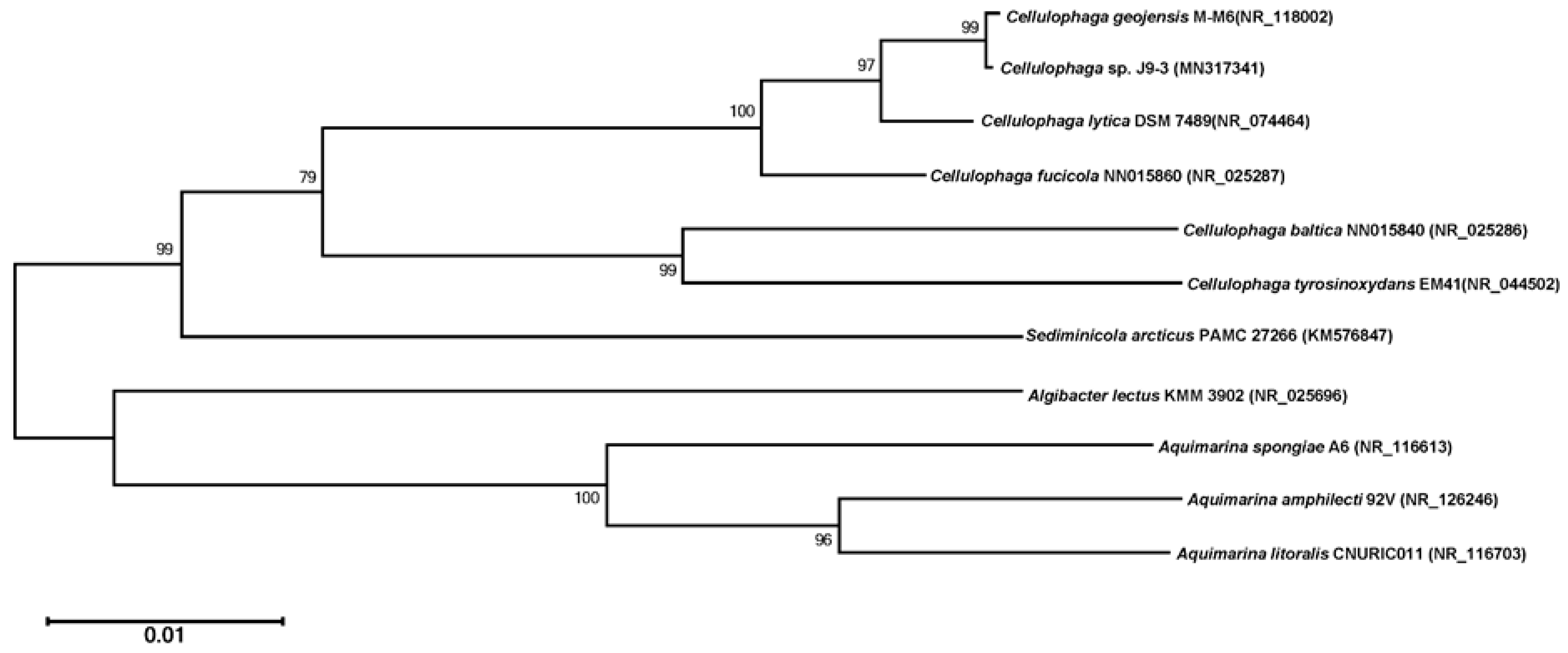
2.3. Phylogenetic Analysis through 16S rRNA Gene and Whole-Genome Sequencing

The 16S rRNA gene of the J9-3 strain was amplified by PCR using universal bacterial primers (785F; 5’-GGA TTA GAT ACC CTG GTA-3’ and 907R; 5’-CCG TCA ATT CCT TTR AGT TT-3’) [12]. Amplified double-stranded DNA was cloned into the pGEM-T easy vector (Promega Co., Madison, WI, USA) and transformed into Escherichia coli JM109 cells. E. coli was maintained on Luria-Bertani (LB) medium at 37 °C [13]. Nucleotide sequencing was performed by Macrogen Inc. (Seoul, Korea). The resulting 16S rRNA gene sequence was searched for homology with sequences from the GenBank database using the BlastN program [14] of the National Center for Biotechnology Information (NCBI). The EzTaxon database (http://www.ezbiocloud.net/) was used to analyze 16S rRNA gene sequence homology with the type strain [15]. A phylogenetic analysis was performed based on the 16S rRNA gene sequences of the type strains provided by the EzTaxon database, using the neighbor-joining (N-J) method [16] of the Mega 6 program (https://www.megasoftware.net/). Bootstrap analysis was performed to evaluate the tree topology of N-J results, by performing 1000 re-samplings and marking the branching points. Total genomic DNA samples were extracted from the J9-3 strain, using a bacterial genomic DNA isolation kit (Davinch-K, Seoul, Korea). Genomic DNA samples were submitted to Macrogen Inc. for sequencing on a PacBio RS II system (Pacific Biosciences, Menlo Park, CA, USA).

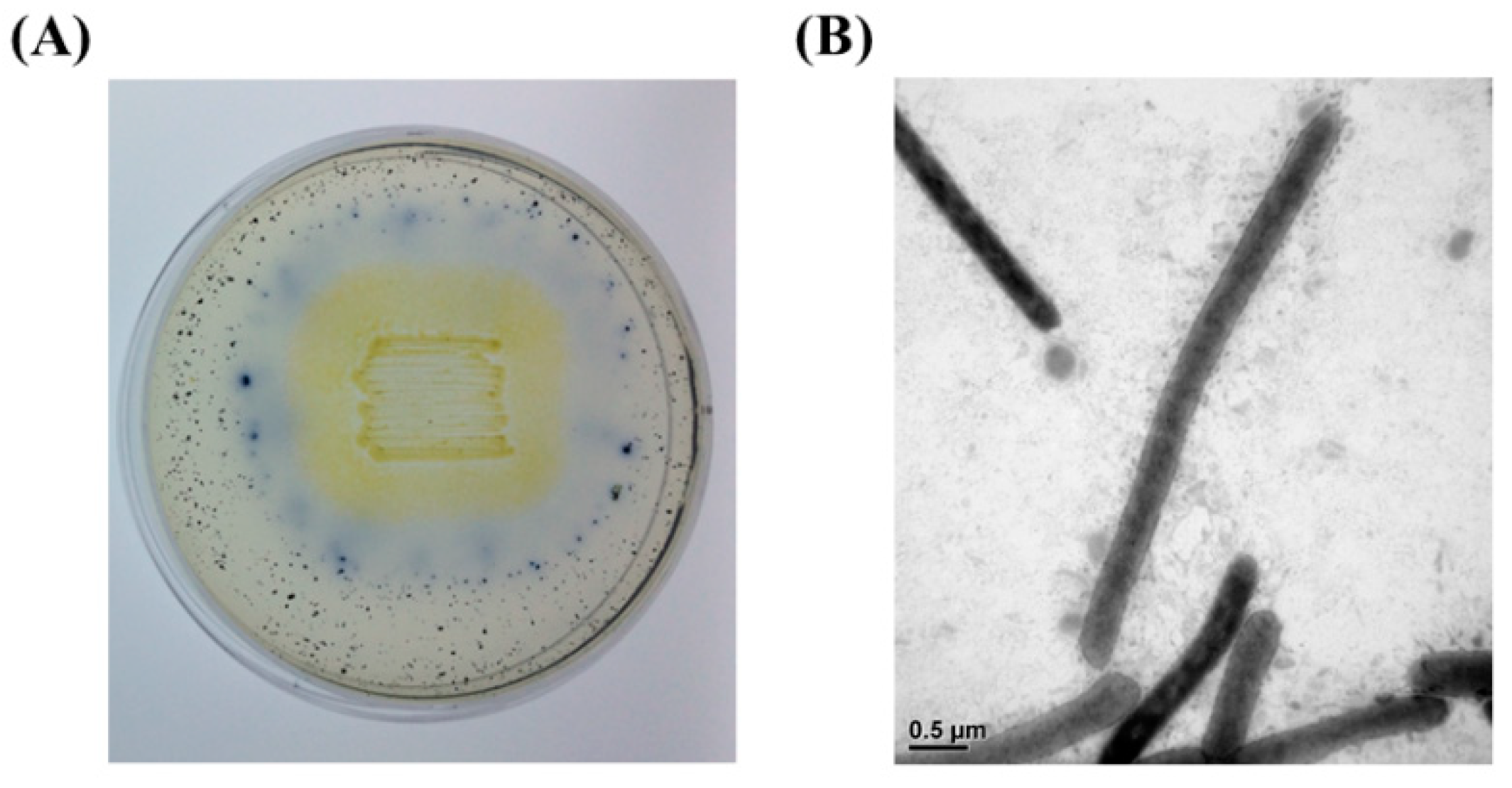
2.4. Morphological Characterization

J9-3 strains were incubated at 30 °C for 1 day on MA medium for morphological characterization. The cells were stained using a Gram Stain Kit (Becton Dickinson, Franklin Lakes, NJ, USA) and observed by light microscopy. Detailed morphological observations of J9-3 cells were performed by transmission electron microscopy (JEM1010; JEOL, Tokyo, Japan) after staining with 1% phosphotungstic acid.

2.5. Cellulase Production by the J9-3 Strain in Liquid Culture

The J9-3 strain was inoculated in 50 mL (250 mL baffled flask) of MB medium and incubated at 30 °C for 1 day with vigorous shaking. Five milliliters of cultured broth was used to inoculate 100 mL (500 mL baffled flask) of fresh MB or MB containing 0.2% (w/v) carboxymethylcellulose (CMC), which was then cultured for 3 days under the same conditions. Two milliliters of culture broth was sampled at 12 h intervals and used for measuring cell density at 600 nm (A600) by spectrophotometry. Cells were removed by centrifugation at 10,000 × g and 4 °C for 20 min and cellulase activity was determined in the supernatant using the 3′, 5′-dinitrosalicylic acid (DNS) method, with absorbance measurements taken at 540 nm (A540).

2.6. Measurement of Cellulase Activity by the DNS Method

The cellulase activity of each sample was determined using the DNS method, which measures the total amount reducing sugars released by polysaccharide hydrolysis [17]. Briefly, CMC was used as the main substrate and its degradation was assayed in a reaction mixture containing 0.1 mL of enzyme solution and 0.2 mL of 0.3% CMC substrate in buffer A (50 mM Tris-Cl, pH 7.5) at 37 °C for 30 min. After the reaction, the enzyme activity was determined by A540 measurement using a Synergy HT microplate reader (Biotek, Winooski, VT, USA), after reacting with the DNS reagent, as previously described [18]. One unit (U) of cellulase was defined as the amount of enzyme that produced 1 μmol of D-glucose per min under the assay conditions. D-glucose was used as a reference reducing sugar for preparing the standard curve.

2.7. Purification of Cellulase

Strain J9-3 was inoculated in 50 mL of MB and incubated at 30 °C for 1 day. The entire culture solution was then inoculated into 1 L of MB medium containing 0.2% CMC and incubated for 2 days, followed by centrifugation at 10,000 × g for 20 min to remove cells. Ammonium sulfate (final concentration of 75%) was added to the supernatant and, after standing in the refrigerator for 12 h, the pellet was recovered by centrifugation at 20,000 × g for 60 min. The pellet was suspended in buffer A, dialyzed at 4 °C for 12 h with buffer A, and centrifuged at 20,000 × g for 30 min. The supernatant was then collected and filtered through a 0.22 μm syringe filter (Millipore, Burlington, MA, USA).
All subsequent protein purification procedures were performed at 4 °C, unless otherwise noted. The filtrate was adsorbed onto a diethylaminoethyl (DEAE) Sepharose Fast Flow (GE Healthcare, Chicago, IL, USA) anion exchange resin (2 mL, 50 × 10 mm column) equilibrated with buffer A and subjected to open column chromatography. Protein elution was performed using a stepwise gradient containing 100–500 mM NaCl (100 mM intervals) in buffer A. Cellulase enzyme activity in each fraction was measured using the DNS method. The active fractions were concentrated with an Amicon ultracentrifugal filter (10 kDa cut off, Millipore) and subjected to Superdex 75 (GE Healthcare) gel permeation chromatography. Buffer A, containing 100 mM NaCl, was used as the mobile phase, at a flow rate of 0.5 mL/min, and 24 fractions (1 mL/fraction) were collected. Protein purity of the active fractions was confirmed by 0.1% sodium dodecyl sulfate–12% polyacrylamide gel electrophoresis (SDS-PAGE). The purified protein was transferred to a polyvinylidene difluoride (PVDF) membrane (Millipore) in transfer buffer (glycine 14.4 g, Tris base 3.03 g, MeOH 200 mL, per L) for 48 h at 48 mA and the NH2-terminus of the protein was analyzed by the Edman degradation method.

2.8. Biochemical Characterization of Cellulase

To investigate the substrate specificity of the enzyme, enzyme reactions were performed using 0.3% agarose, beechwood xylan, starch, or carboxylmethylcellulose as substrates. Enzyme activity was measured using the DNS method, after reactions were performed for 30 min at pH 7.5 and 37 °C.
All subsequent reactions were performed with 0.3% CMC as a substrate for 30 min, unless otherwise noted. To determine the optimum temperature of the enzyme, reactions were performed at 30–70 °C (5 °C intervals) at pH 7.5. To evaluate temperature stability, the enzyme solution was incubated at 30–70 °C (5 °C intervals) for 60 min and then used in reactions with the substrate at pH 7.5 and 37 °C.
The optimum pH of the enzyme was determined after enzyme reactions at 37 °C and a pH range of 6.0–10.0 (pH 0.5 intervals). The buffers used were 50 mM MOPS (pH 6.0–7.0), 50 mM Tris-Cl (pH 7.0–9.0), and 50 mM glycine-NaOH (pH 9.0–10.0).
The effects of metal ions, ethylenediaminetetraacetic acid (EDTA), and surfactants on the enzymatic reaction were studied by adding 1 mM of Ca2+, Co2+, Cu2+, Mg2+, Mn2+, Fe2+, Zn2+, Na+, K+, EDTA, or 1% SDS to the reaction solution and performing reactions at pH 7.5 and 37 °C.

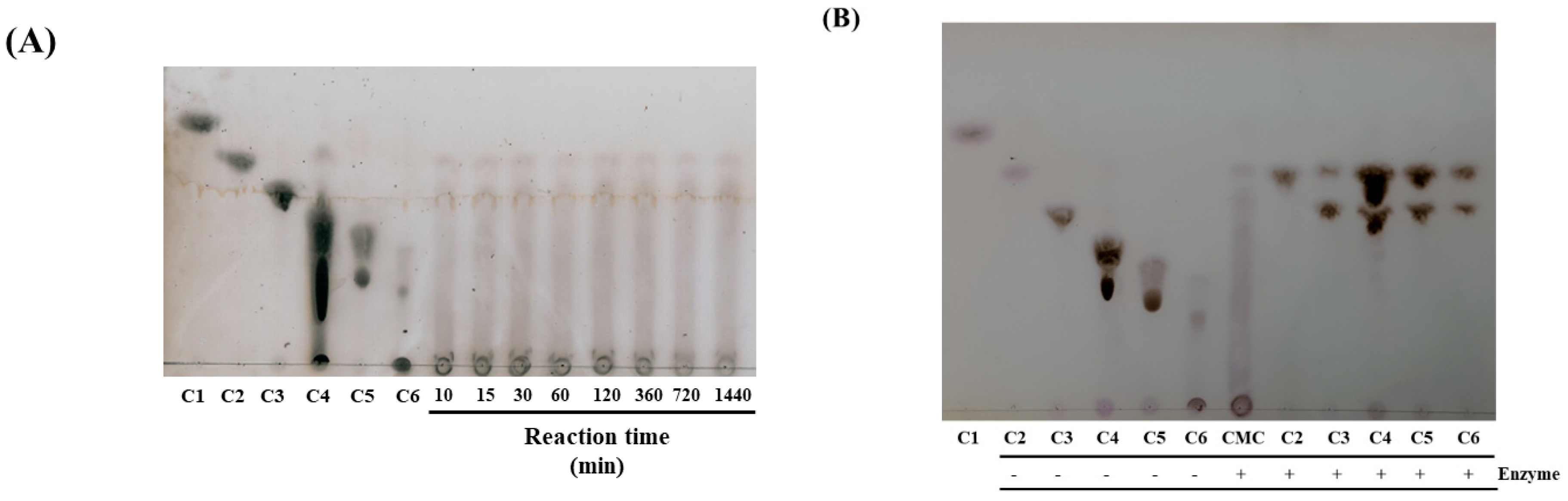
2.9. Thin Layer Chromatography (TLC) Analysis of Enzyme Reaction

To analyze the cellulose hydrolysate produced by the purified protein, 20 μL of enzyme solution and 0.3% CMC solution in buffer A were mixed (total volume of 300 μL) and reacted for 24 h at 37 °C. Then, 5 µL of the reaction mix was spotted onto a TLC plate (60G F254, Merck, Kenilworth, NJ, USA). As standards, 2 µL of 5 mM glucose, cellobiose, cellotriose, cellotetraose, cellopentaose, and cellohexaose were also spotted onto the TLC plate. The mobile phase consisted of n-butanol:acetic acid:distilled water, at a ratio of 2:1:2. Spots were visualized by spraying a color-developing reagent (ethanol:sulfuric acid, 9:1) and then heating them at 120 °C.
To assess the enzyme’s ability to hydrolyze cellooligosaccharides, 20 µL of purified cellulase enzyme was mixed with a 0.3% substrate solution of each cellooligosaccharide (cellobiose, cellotriose, cellotetraose, cellopentaose, or cellohexaose) in a total volume of 300 μL. After reacting at 37 °C for 24 h, 5 µL of each reaction mix was spotted onto a TLC plate for analysis.

3. Results

3.1. Identification of Strain J9-3

One bacterial strain showing high AZCL-cellulose-hydrolyzing activity was finally selected and designated as strain J9-3 (Figure 1A). Strain J9-3 cells were long, rod-shaped, Gram-negative cells, with no flagella, as observed by electron microscopy (Figure 1B). The 16S rRNA gene sequence was determined for J9-3 and was registered in NCBI GenBank (accession no. MN317341). Based on 16S rRNA gene sequences, J9-3 was shown to have the highest homology with the type-strains, Cellulophaga geojensis M-M6 (99.93%) [19], Cellulophaga lytica DSM7489 (99.19%) [20], Cellulophaga fucicola NN015860 (98.35%) [20], Cellulophaga tyrosinoxydans EM41 (93.9%) [21], and Cellulophaga baltica NN015840 (93.81%) [20].
The phylogenetic correlation analysis of 16S rRNA gene sequences using the N-J method showed that J9-3 had the closest association with C. geojensis M-M6, forming a distinct clade from other strains (Figure 2). Based on 16S rRNA gene sequence homology and phylogenetic analyses, J9-3 was determined to be a species of the genus Cellulophaga, and thus, it was named Cellulophaga sp. J9-3.

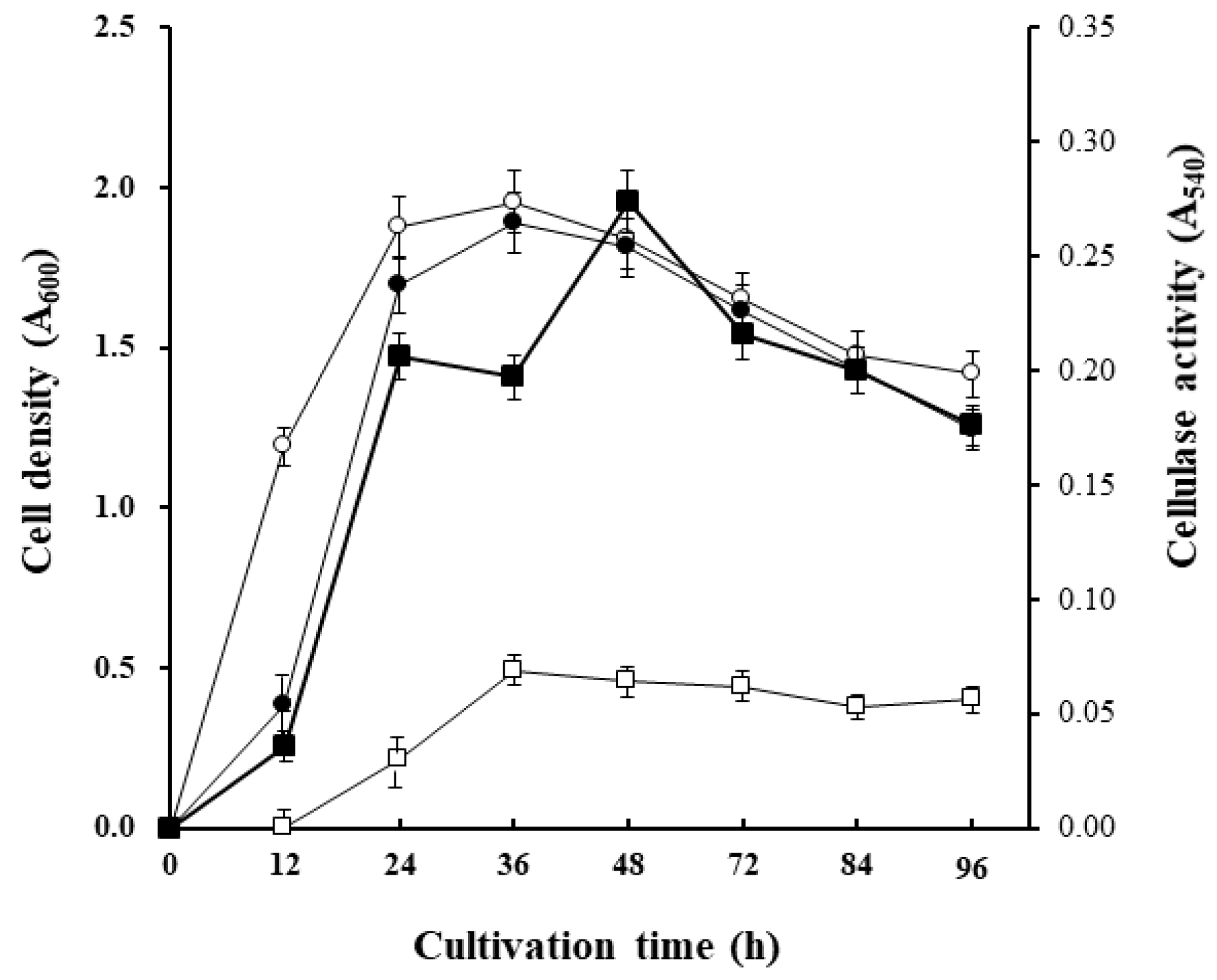
3.2. Cellulase Production by Cellulophaga sp. J9-3 in Liquid Culture

Cellulophga sp. J9-3 decomposed the substrate in AZCL-cellulose-containing MA medium to give a blue color (Figure 1A). In addition, cellulose-degradation activity was detected, using the DNS method, in the supernatant of MB medium cultures (Figure 3). From these results, J9-3 was expected to secrete cellulose-degrading enzymes extracellularly.
When J9-3 was incubated in MB medium, the addition of 0.2% CMC did not affect its growth. Cell growth (A600) peaked at 36 h of cultivation, and then gradually decreased due to depletion of limiting nutrients (probably carbon sources) in both media. Although enzyme activity was weakly detected (A540 = 0.064) in MB medium, the addition of CMC to the medium resulted in a rapid increase in enzyme activity, reaching 4.3 times higher than the activity in MB medium alone (A540 = 0.274) in 48 h (Figure 3). This result suggested that enzyme production can be actively induced by the addition of the substrate, CMC.

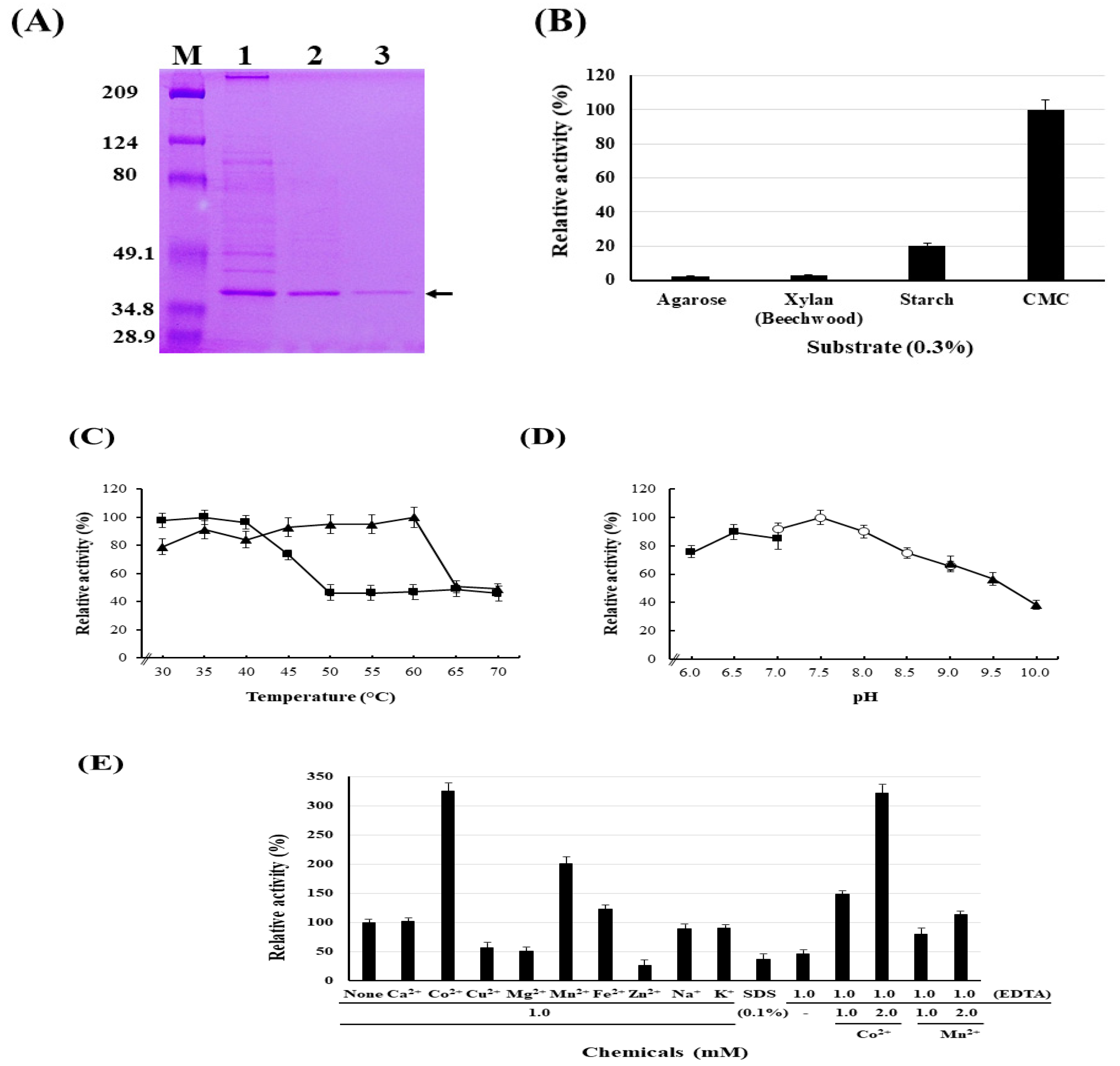
3.3. Purification of Cellulase

In DEAE Sepharose Fast Flow chromatography of the protein sample prepared by ammonium sulfate precipitation of the J3-9 culture broth, most of the enzyme activity was detected in the fraction eluted with 100 mM NaCl. The active fractions were concentrated from 15 mL to 1 mL and applied to a Superdex 75 column. The two fractions (fraction no. 11 and 12) that showed the highest enzyme activity and a single protein band on SDS-PAGE were combined (Figure 4A) and further analyzed. The purification yields for each step are summarized in Table 1.

3.4. Identification of Cellulase Enzymes and their Genes

The NH2-terminal amino acid sequence of the purified protein was determined to be N-N-T-E-Q-T-V-V-D-A-Y-G. Genomic sequencing analysis of J9-3 revealed that the bacterium had a circular chromosome composed of 3,806,639 bp, with 32.08% G+C content. From the 3300 coding sequences, we identified one gene encoding a polypeptide that was perfectly matched with the NH2-terminal amino acid sequence of the purified protein. This gene was registered in GenBank under accession no. MN33592. According to its nucleotide sequence, the corresponding protein was composed of 359 amino acids (Mw = 40,738 Da). Moreover, the NH2-terminal amino acid sequence revealed that the premature form of the protein was cleaved between Ser-52 and Asn-53, thus yielding a mature protein with 307 amino acids (Mw = 34,951 Da), which was consistent with our SDS-PAGE results (Figure 4A).
Based on amino acid sequences, several homologous proteins with more than 90% identity were retrieved using the BlastP program. These proteins were all listed as putative cellulases (endoglucanase) belonging to glycosyl hydrolase family 5 (GH5) of Carbohydrate-Active enZYmes Database [22]. They included Cellulophaga sp. W5C (99% identity with WP_077398709.1), Cellulophaga lytica (95% identity with WP_013619852.1), Cellulophaga sp. RHA_52 (95% identity with WP_144709822.1), Cellulophaga geojensis (95% identity with WP_034644593.1), Cellulophaga sp. QY3 (92% identity with ADB80152.1), and Cellulophaga fucicola (90% identity with WP_072302686.1). However, the enzymatic properties of these proteins have not been characterized. Therefore, studies on the enzymatic properties of the purified cellulase were performed.

3.5. Biochemical Characterization of the Cellulase

Among the tested substrates, the purified protein showed CMC-hydrolyzing activity, but no activity against agarose, xylan, or starch. This result strongly indicated that the protein was a cellulase, and thus, it was named CelAJ93 (Figure 4B).
Using CMC as a substrate, CelAJ93 showed maximum cellulase activity at 60 °C and maintained more than 80% of its maximum activity at a temperature range of 30 to 60 °C (Figure 4C). However, at temperatures above 65 °C, enzyme activity rapidly decreased. CelAJ93 maintained enzyme activity after incubation at 40 °C for 60 min, but started to decrease after incubation at 45 °C and maintained only about 40% of normal activity after incubation at 50 °C.
CelAJ93 showed maximal cellulase activity near pH 7.5, and then gradually decreased with increasing pH (Figure 4D). From these results, it was concluded that CelAJ93 is a thermophilic enzyme with activity at neutral pH conditions.
EDTA treatment severely inhibited enzyme activity, indicating that CelAJ93 needs a metallic cofactor (Figure 4E). Metal ions (Cu2+, Mg2+, Zn2+), EDTA at 1 mM, and SDS at 0.1% (w/v) significantly inhibited enzyme activity by more than 50%. However, Mn2+ and Co2+ at 1 mM resulted in a marked increase in enzymatic activity by 200% and 325%, respectively. In particular, addition of Co2+ completely circumvented the inhibitory effect by EDTA in a concentration-dependent manner, indicating that Co2+ is a cofactor for the endoglucanase activity of CelAJ93.

3.6. TLC Analysis of the Hydrolysate Produced by CelAJ93

TLC analysis of the CMC hydrolysate produced by CelAJ93 revealed that the enzyme can decompose cellulose into cellobiose (Figure 5A). Cellooligosaccharides larger than cellobiose were also generated by CelAJ93, indicating that the enzyme is an endo-type β-glucanase, i.e., endo-β-1,4-glucanase.
Among the commercially available cellooligosaccharides tested, CelAJ93 could not hydrolyze cellobiose but decompose cellotriose into cellobiose. Moreover, it decomposed cellotetraose, cellopentaose, and cellohexaose to produce common products cellobiose (major product) and cellotriose (minor product). From these, CelAJ93 is expected to break down cellooligosaccharides to produce cellobiose (major) and cellotriose (minor), and subsequently to break down cellotriose into cellobiose and glucose. However, spots corresponding to D-glucose produced by the degradation of cellotriose on TLC were detected very weakly. These results are consistent with the results reported in several scientific papers [23,24], and are interpreted to be attributed to the relatively low degradation activity of the enzyme toward cellotriose (Figure 5B). In addition, the enzyme activity against CMC was very weak compared to the enzyme activity against cellooligosaccharides, probably because the three-dimensional structure of crystalline cellulose, such as CMC, makes it difficult for the enzyme to access the substrate. These results clearly indicate that CelAJ93 is endo-β-1,4-glucanase that can hydrolyze CMC and cellooligosaccharides into cellobiose.

3.7. Strain Deposit and GenBank Accession Number

The strain Cellulophaga sp. J9-3 was deposited in the Korean Agricultural Culture Collection (KACC) with an accession number of KACC92201P. The GenBank accession number for the 16S rRNA and celAJ93 gene sequences of strain J9-3 are MN317341 and MN33592, respectively.

4. Discussion

Cellulase is one of the most important industrial enzymes that allows the utilization of the vast amount of biomass, especially lignocellulose-based biomass, on earth. To date, a lot of cellulases have been reported; however, there are many limitations in developing technologies that can effectively apply cellulase in biomass decomposition [8,9]. In the case of lignocellulosic substrates, since the lignin-hemicellulose-cellulose forms a tight complex, it is difficult for the enzyme to access the substrate for its action. In addition, cellulase enzyme activity can be influenced by various factors in the process such as inhibition by the final product, inactivation by high temperature, solvent or pH change, nonspecific binding with lignin, and irreversible adsorption on heterogeneous substrates. Therefore, it is necessary to screen as many cellulases as possible, with various biochemical characteristics, to broaden their application in degrading lignocellulosic materials.
In this study, we identified a marine bacterium, Cellulophaga sp. J9-3, with an ability to degrade cellulose and we characterized a cellulase (endo-β-1,4-glucanase) from its culture broth. According to the SignalP 5.0 program (http://www.cbs.dtu.dk/services/SignalP/) [25] the protein, CelAJ93, was expected, at a probability of 0.9994, to have a lipoprotein signal peptide (Sec/SPII) [26] with a cleavage site between Ser-19 and Cys-20. However, NH2-terminal amino acid sequencing revealed that the mature protein was cleaved between Ser-52 and Asn-53, indicating a unique pattern of cleavage.
There are currently two reports of cellulases from the genus Cellulophaga in PubMed (https://www.ncbi.nlm.nih.gov/pubmed/). Wang et al. [27] reported a novel endoglucanase, Celal_2753 (Mw = 44 kDa), from the psychrophilic bacterium, Cellulophaga algicola IC166(T). It showed maximum activity at 40 °C and pH 6. It was resistant to various detergent treatments, including SDS, and could be stimulated by NaCl and KCl. Moreover, it maintained 55% of its initial activity after boiling for 5 min. Thus, it will be useful for the degradation of hemicelluloses in very harsh conditions. Another study reported only CMCase production by Cellulophaga lytica LBH-14 using rice bran, without any information regarding enzymatic properties [28]. Although many genes have been annotated as GH5 cellulases in the genus Cellulophaga, only one cellulase (Celal_2753), which shared 51% identity in its amino acid sequence with CelAJ93, was identified. CelAJ93 was quite distinct from Celal_2753, especially in that it had a higher optimum temperature (60 °C), it was highly sensitive to EDTA and SDS, and it required Co2+ as a cofactor.
Although most cellulases are not affected by the presence of the Co2+ ion, some have been reported to be stimulated by Co2+. The enzyme activity of CelA, a thermostable and pH-stable cellobiohydrolase from Neocallimastix patriciarum J11, was greatly enhanced by Co2+ [29]. RuCelA, encoding a bifunctional xylanase/endoglucanase, was reported in yak rumen microorganisms [30]. The presence of Co2+ greatly enhanced the endoglucanase activity of RuCelA, but inhibited its xylanase activity. However, further investigation is required to confirm that those activities are dependent on Co2+.
CelAJ93 contains a well-conserved glycosyl hydrolase family 5 cellulase domain (pfam00150) [31], spanning Asn-79 and Ile-316, with an E-value of 1.04 × e−57. Among the proteins sharing high homology with CelAJ93, chain A endoglucanase (5IHS_A) reported in Cytophaga hutchinsonii [32] has the highest identity (54%) at the amino acid sequence level. This strain is unique in that it does not have a gene encoding a cellobiohydrolase, and thus, the chain A endoglucanase is thought to be responsible for the initial digestion of cellulose fibers in amorphous regions, facilitating the generation of oligomers to be transported into the periplasm for further hydrolysis. In contrast to chain A endoglucanase, CelAJ93 acts more efficiently on cellulooligosaccharides than CMC, suggesting that it is not the main enzyme responsible for the initial digestion of cellulose fibers. We found two candidate genes for endo-β-1,4-glucanase and two candidate genes for β-D-glucosidase, but none for cellobiohydrolase in the genomic sequence of Cellulophaga sp. J9-3, which is similar to the genome of C. hutchinsonii [32]. Therefore, a more precise understanding of the cellulose degradation system of Cellulophaga sp. J9-3 at the molecular level is required to broaden our knowledge to use cellulose.
CelAJ93 is the second biochemically characterized enzyme in the cellulase family produced by the genus Cellulophaga. It has a unique cleavage sequence for protein maturation and requires Co2+ as a cofactor. Therefore, this report will be helpful in understanding the molecular and biochemical characteristics of many enzymes listed in GenBank with high homology to CelAJ93. Moreover, CelAJ93 has an optimal temperature of 60 °C, which will be an advantage for its application in cellulose degradation processes.

Author Contributions

Conceptualization, W.-J.C. and S.-K.H.; Methodology, D.S.K. and W.-J.C.; Investigation, D.S.K.; Resources, W.-J.C.; Data Curation, S.-K.H.; Writing—Original Draft Preparation, W.-J.C.; Writing—Review & Editing, S.-K.H.; Funding Acquisition, W.-J.C.

Funding

This research was funded by a grant from the National Institute of Biological Resources (NIBR), funded by the Ministry of Environment (MOE) of the Republic of Korea (NIBR201920101).

Conflicts of Interest

The authors declare no conflict of interest.

References

  1. Klemm, D.; Heublein, B.; Fink, H.-P.; Bohn, A. Cellulose: Fascinating Biopolymer and Sustainable Raw Material. Angew. Chem. Int. Ed. 2005, 44, 3358–3393. [Google Scholar] [CrossRef] [PubMed]
  2. Shida, Y.; Furukawa, T.; Ogasawara, W. Deciphering the Molecular Mechanisms behind Cellulase Production in Trichoderma reesei, the Hyper-Cellulolytic Filamentous Fungus. Biosci. Biotechnol. Biochem. 2016, 80, 1712–1729. [Google Scholar] [CrossRef] [PubMed]
  3. Bhat, M.K. Cellulases and Related Enzymes in Biotechnology. Adv. Biotechnol. 2000, 1, 355–383. [Google Scholar] [CrossRef]
  4. Perez, J.; Munoz-Dorado, J.; de la Rubia, T.; Martinez, J. Biodegradation and Biological Treatments of Cellulose, Hemicellulose and Lignin: An overview. Int. Microbiol. 2002, 5, 53–63. [Google Scholar] [CrossRef] [PubMed]
  5. Cavaco-Paulo, A. Mechanism of Cellulose Action in Textile Processes. Carbohydr. Polym. 1998, 37, 273–277. [Google Scholar] [CrossRef]
  6. Thomas, L.; Joseph, A.; Gottumukkala, L.D. Xylanase and Cellulase Systems of Clostridium sp.: An Insight on Molecular Approaches for Strain Improvement. Bioresour. Technol. 2014, 158, 343–350. [Google Scholar] [CrossRef] [PubMed]
  7. Wei, G.Y.; Gao, W.; Jin, I.H.; Yoo, S.Y.; Lee, J.H.; Chung, C.H.; Lee, J.W. Pretreatment and Saccharification of Rice Hulls for the Production of Fermentable Sugars. Biotechnol. Bioprocess Eng. 2009, 14, 828–834. [Google Scholar] [CrossRef]
  8. Cherry, J.R.; Fidantsef, A.L. Directed Evolution of Industrial Enzymes: An Update. Curr. Opin. Biotechnol. 2003, 14, 438–443. [Google Scholar] [CrossRef]
  9. Dos Santos, Y.Q.; de Veras, B.O.; de França, A.F.J.; Gorlach-Lira, K.; Velasques, J.; Migliolo, L.; Dos Santos, E.A. A New Salt-Tolerant Thermostable Cellulase from a Marine Bacillus sp. Strain. J. Microbiol. Biotechnol. 2018, 28, 1078–1085. [Google Scholar] [CrossRef]
  10. Zou, Z.; Zhao, Y.; Zhang, T.; Xu, J.; He, A.; Deng, Y. Efficient Isolation and Characterization of a Cellulase Hyperproducing Mutant Strain of Trichoderma reesei. J Microbiol. Biotechnol. 2018, 28, 1473–1481. [Google Scholar] [CrossRef]
  11. Nakamura, K.; Kappamura, K. Isolation and Identification of Crystalline Cellulose Hydrolyzing Bacterium and its Enzymatic Properties. J. Ferment. Technol. 1982, 60, 343–348. [Google Scholar]
  12. Galkiewicz, J.P.; Kellogg, C.A. Cross-kingdom Amplification Using Bacteria-specific Primers: Complications for Studies of Coral Microbial Ecology. Appl. Environ. Microbiol. 2008, 74, 7828–7831. [Google Scholar] [CrossRef] [PubMed]
  13. Green, M.R.; Sambrook, J. Molecular Cloning. A Laboratory Manual, 4th ed.; Cold Spring Harbor Laboratory Press: Cold Spring Harbor, NY, USA, 2012. [Google Scholar]
  14. Altschul, S.F.; Madden, T.L.; Schaffer, A.A.; Zhang, J.; Zhang, A.; Miller, W.; Lipman, D.J. Gapped BLAST and PSI-BLAST: A New Generation of Protein Data Base. Nucleic Acids Res. 1997, 25, 3389–3402. [Google Scholar] [CrossRef] [PubMed]
  15. Chun, J.; Lee, J.H.; Jung, Y.Y.; Kim, M.J.; Kim, S.I.; Kim, B.K.; Lim, Y.W. ExTaxon: A Web-Based Tool for the Identification of Prokaryotes Based on 16S Ribosomal RNA Gene Sequences. Int. J. Syst. Evol. Microbiol. 2007, 57, 2259–2261. [Google Scholar] [CrossRef]
  16. Saitou, N.; Nei, M. The Neighbor-joining Method: A New Method for Reconstructing Phylogenetic Trees. Mol. Biol. Evol. 1987, 4, 406–425. [Google Scholar] [PubMed]
  17. Ghose, T.K. Measurement of Cellulase Activities. Pure Appl. Chem. 1987, 59, 257–268. [Google Scholar] [CrossRef]
  18. Lim, J.H.; Lee, C.R.; Dhakshnamoorthy, V.; Park, J.S.; Hong, S.K. Molecular Characterization of Streptomyces coelicolor A(3) SCO6548 as a Cellulose 1,4-β-Cellobiosidase. FEMS Microbiol. Lett. 2016, 363, fnv245. [Google Scholar] [CrossRef]
  19. Park, S.; Oh, K.H.; Lee, S.Y.; Oh, T.K.; Yoon, J.H. Cellulophaga geojensis sp. nov., a member of the family Flavobacteriaceae isolated from marine sand. Int. J. Syst. Evol. Microbiol. 2012, 62 Pt 6, 1354–1358. [Google Scholar] [CrossRef]
  20. Johansen, J.E.; Nielsen, P.; Sjøholm, C. Description of Cellulophaga baltica gen. nov., sp. nov. and Cellulophaga fucicola gen. nov., sp. nov. and reclassification of [Cytophaga] lytica to Cellulophaga lytica gen. nov., comb. nov. Int. J. Syst. Bacteriol. 1999, 49 Pt 3, 1231–1240. [Google Scholar] [CrossRef]
  21. Kahng, H.Y.; Chung, B.S.; Lee, D.H.; Jung, J.S.; Park, J.H.; Jeon, C.O. Cellulophaga tyrosinoxydans sp. nov., a tyrosinase-producing bacterium isolated from seawater. Int. J. Syst. Evol. Microbiol. 2009, 59 Pt 4, 654–657. [Google Scholar] [CrossRef]
  22. Lombard, V.; Golaconda Ramulu, H.; Drula, E.; Coutinho, P.M.; Henrissat, B. The carbohydrate-active enzymes database (CAZy) in 2013. Nucleic Acids Res. 2014, 42, D490–D495. [Google Scholar] [CrossRef] [PubMed]
  23. Han, S.O.; Yukawa, H.; Inui, M.; Doi, R.H. Molecular cloning and transcriptional and expression analysis of engO, encoding a new noncellulosomal family 9 enzyme, from Clostridium cellulovorans. J. Bacteriol. 2005, 187, 4884–4889. [Google Scholar] [CrossRef] [PubMed]
  24. Park, J.D.; Kim, Y.A.; Yoon, K.-H. Properties of a Bacillus licheniformis cellulase produced by recombinant Escherichia coli. Korean J. Microbiol. 2009, 45, 257–262. [Google Scholar]
  25. Almagro Armenteros, J.J.; Tsirigos, K.D.; Sønderby, C.K.; Petersen, T.N.; Winther, O.; Brunak, S.; von Heijne, G.; Nielsen, H. SignalP 5.0 improves signal peptide predictions using deep neural networks. Nat. Biotechnol. 2019, 37, 420–423. [Google Scholar] [CrossRef] [PubMed]
  26. Schneewind, O.; Missiakas, D. Sec-secretion and sortase-mediated anchoring of proteins in Gram-positive bacteria. Biochim. Biophys. Acta. 2014, 1843, 1687–1697. [Google Scholar] [CrossRef] [PubMed]
  27. Wang, Y.; Yu, W.; Han, F. Expression and Characterization of a Cold-adapted, Thermotolerant and Denaturant-stable GH5 Endoglucanase Celal_2753 that Withstands Boiling from the Psychrophilic Bacterium Cellulophaga algicola IC166(T). Biotechnol. Lett. 2016, 38, 285–290. [Google Scholar] [CrossRef] [PubMed]
  28. Gao, W.; Lee, E.J.; Lee, S.U.; Li, J.; Chung, C.H.; Lee, J.W. Enhanced Carboxymethylcellulase Production by a Newly Isolated Marine Bacterium, Cellulophaga lytica LBH-14, using rice bran. J. Microbiol. Biotechnol. 2012, 22, 1412–1422. [Google Scholar] [CrossRef] [PubMed]
  29. Wang, H.C.; Chen, Y.C.; Huang, C.T.; Hseu, R.S. Cloning and Characterization of a Thermostable and pH-stable Cellobiohydrolase from Neocallimastix patriciarum J11. Protein Expr. Purif. 2013, 90, 153–159. [Google Scholar] [CrossRef]
  30. Chang, L.; Ding, M.; Bao, L.; Chen, Y.; Zhou, J.; Lu, H. Characterization of a Bifunctional Xylanase/Endoglucanase from Yak Rumen Microorganisms. Appl. Microbiol. Biotechnol. 2011, 90, 1933–1942. [Google Scholar] [CrossRef]
  31. Finn, R.D.; Bateman, A.; Clements, J.; Coggill, P.; Eberhardt, R.Y.; Eddy, S.R.; Heger, A.; Hetherington, K.; Holm, L.; Mistry, J.; et al. Pfam: The protein families database. Nucleic Acids Res. 2014, 42, D222–D230. [Google Scholar] [CrossRef]
  32. Zhu, Y.; Han, L.; Hefferon, K.L.; Silvaggi, N.R.; Wilson, D.B.; McBride, M.J. Periplasmic Cytophaga hutchinsonii Endoglucanases Are Required for Use of Crystalline Cellulose as the Sole Source of Carbon and Energy. Appl. Environ. Microbiol. 2016, 82, 4835–4845. [Google Scholar] [CrossRef] [PubMed]
Figure 1. Characteristic features of the bacterial isolate, J9-3. (A) Detection of cellulase activity on Marine Agar plate containing AZCL-cellulose; (B) scattering electron microscopy of J9-3 cells after staining with 1% phosphotungstic acid.
Applsci 09 04061 g001
Figure 2. Neighbor-joining phylogenic tree based on 16S rRNA gene sequences between strain J9-3 and other species. Nucleotide sequence accession numbers are given in parentheses. Scale bar, 0.01 substitutions per 100 nucleotides.
Applsci 09 04061 g002
Figure 3. Cell growth and cellulase production by Cellulophaga sp. J9-3 in Marine Broth containing carboxymethylcellulose at different cultivation times. Cell density (A600) in Marine Broth (MB, ○) and MB + 0.2% carboxymethylcellulose (CMC, ●), cellulase activity (A540) in MB (□) and MB + 0.2% CMC (■).
Applsci 09 04061 g003
Figure 4. Purification and biochemical characterization of an extracellular cellulase, CelAJ93, from the culture broth of Cellulophaga sp. J9-3. (A) Sodium dodecyl sulfate-polyacrylamide gel electrophoresis of protein samples during purification. Lane M, protein molecular weight markers; 1, 75% ammonium sulfate precipitate; 2, sample after DEAE Sepharose Fast Flow column chromatography; 3, sample after Superdex 75 column chromatography; (B) substrate specificity of CelAJ93 toward various polysaccharides; (C) effect of temperature on CelAJ93 enzyme activity toward carboxymethylcellulose. ▲, optimum temperature; ■, thermostability; (D) effect of pH on CelAJ93 enzyme activity toward carboxymethylcellulose. ■, 50 mM MOPS (pH 6.0–7.0); ○, 50 mM Tris-Cl (pH 7.0–9.0); ▲, 50 mM glycine-NaOH (pH 9.0–10.0); (E) effect of metal ions, ethylenediaminetetraacetic acid (EDTA), and sodium dodecyl sulfate (SDS) on CelAJ93 enzyme activity toward carboxymethylcellulose. The final concentration of each chemical used is indicated under the figure. The maximum enzyme activity in (B)–(D) and the enzyme activity without chemicals in (E) was set to 100% when calculating relative activities. In (B)–(E), all data shown are mean values from at least three replicate experiments.
Applsci 09 04061 g004
Figure 5. Analysis of CelAJ93 enzyme hydrolysates of various substrates by thin layer chromatography. (A) Chromatogram of the carboxymethylcellulose hydrolysate produced by CelAJ93 according to reaction time at pH 7.5 and 37 °C. The presence of smeared bands under the cellobiose band indicates that CelAJ93 is acting as an endo-type glucanase, leading to cellobiose; (B) chromatogram of various cellooligosaccharide hydrolysates produced by CelAJ93 after reacting for 24 h at pH 7.5 and 37 °C. Cellooligosaccharides larger than cellobiose gave the common product of cellobiose, indicating that CelAJ93 can hydrolyze substrates to cellobiose. C1, D-glucose; C2, cellobiose; C3, cellotriose; C4, cellotetraose; C5, cellopentaose; C6, cellohexaose. CMC; carboxymethylcellulose.
Applsci 09 04061 g005
Table 1. Summary of purification for CelAJ93.
Purification StepVolume (mL)Total Protein (mg)Total Activity (U)Specific Activity (U/mg)Purification Fold
Culture supernatant8802551.12110,588.5743.351.00
Ammonium sulfate precipitation75279.6814,705.3652.581.21
Diethylaminoethyl sepharose Fast Flow chromatography1514.855721.98385.328.89
Superdex 75 chromatography21.921838.03957.3122.98

