Article

Increased Anti-Inflammatory Effects on LPS-Induced Microglia Cells by Spirulina maxima Extract from Ultrasonic Process

1
Department of Medical Biomaterials Engineering, Kangwon National University, Chuncheon 200-701, Korea
2
R&BD Center, Korea Yakult Co. Ltd., Seoul 17086, Korea
3
The Jeju International Marine Science Center for Research & Education, Korea Institute of Ocean Science and Technology (KIOST), Ansan 63349, Korea
4
Department of Food Science and Engineering, Seowon University, Cheongju 361-742, Korea
*
Author to whom correspondence should be addressed.
Appl. Sci. 2019, 9(10), 2144; https://doi.org/10.3390/app9102144
Submission received: 5 April 2019 / Revised: 19 May 2019 / Accepted: 22 May 2019 / Published: 26 May 2019
(This article belongs to the Special Issue Eco-Novel Food and Feed)

Abstract

:
The Spirulina maxima exact from a non-thermal ultrasonic process (UE) contains 17.5 mg/g of total chlorophyll, compared to 6.24 mg/g of chlorophyll derived from the conventional 70% ethanol extraction at 80 °C for 12 h (EE). The UE also showed relatively low cytotoxicity against murine microglial cells (BV-2) and inhibited the production of the inflammatory mediators, NO and PGE2. The UE also effectively suppresses both mRNA expression and the production of pro-inflammatory cytokines, such as TNF-α, IL-6 and IL-1β, in a concentration-dependent manner. Notably, TNF-α gene and protein production were most strongly down-regulated, while IL-6 was the least affected by all ranges of treatment concentrations. This work first demonstrated a quantitative correlation between mRNA expression and the production of cytokines, showing that suppression of TNF-α gene expression was most significantly correlated with its secretion. These results clearly proved that the anti-inflammatory effects of Spirulina extract from a nonthermal ultrasonic process, which yielded high concentrations of intact forms of chlorophylls, were increased two-fold compared to those of conventional extracts processed at high temperature.

1. Introduction

Recently, studies on the discovery and treatment of new diseases have been accelerated by the dramatic development of medical technologies. As our understanding of the human brain has widened, studies on the causes and treatments of diseases related to brain diseases have been intensively conducted [1,2,3]. Studies on the treatment and prevention of dementia, which is among the most threatening diseases to humans both socially and medically and seriously deteriorates patients’ quality of life, have been intensively conducted, substantially advancing our mechanistic understanding of dementia and the development of diverse methods and drugs to treat dementia [3,4,5]. Specifically, many studies on cognitive function deterioration and preventive drugs have widely accepted the theory that a major cause of cognitive dysfunction is brain nerve cell death, due to persistent exogenous stress or accumulation of oxidants, which lead to cognitive function deterioration and consequent brain diseases, such as dementia [6,7,8]. Therefore, many studies on cognitive and memory impairments, such as dementia and Parkinson’s disease, have concentrated on mechanisms that protect brain nerve cells by preventing the accumulation of metabolic oxidants through antioxidant activity or rapid decomposition of gene oxidants and the development of drugs for such mechanisms [9,10,11]. Therefore, drugs, such as donepezil, rivastigmine, and galantamine have been developed to control and/or enhance the neuronal signaling pathway linked to p-ERK/p-CREB/BDNF. However, after extended use, these synthetic drugs show adverse side effects, such as vomiting, hand tremors, and heart disease [12,13,14]. Accordingly, remarkable findings have been reported from studies on natural products that protect cranial nerves by exerting antioxidant activity, and these products have relatively fewer side effects [15,16,17].
Most of these studies have fundamentally focused on suppressing cognitive function and memory loss through antioxidant-mediated cranial nerve protection. However, rather than exhibiting intense antioxidative effects in the brain nerve cells and their activation sites, natural products suppress the deterioration of cognitive function resulting from overall antioxidative activity throughout the body, so the actual effects have been remarkably lower than expected in most cases [18,19]. Among the recent studies on suppressing cognitive function and memory loss, those featuring natural products with functions beyond simple antioxidant activity to directly suppress memory deterioration by inhibiting inflammation in brain nerve cells have attracted substantial attention [20]. In particular, the brain damage-induced activation of pro-inflammatory cytokines, such as TNF-α and IL-6, can evoke brain inflammation, which results in various neurodegenerative diseases like ischemia, trauma, infection, Alzheimer’s disease and Parkinson’s disease [20,21,22]. There is recent strong evidence that continuous stress and diverse inflammation in the brain could be a major and direct cause of dementia and memory impairment, due to the continuous accumulation of inflammatory cytokines in the brain. While many efforts have been made to reduce the production of these inflammatory cytokines to indirectly attenuate degenerative brain diseases, more detailed studies on the mechanism underlying delayed neurodegeneration should be carried out based on our understanding of the blood-brain barrier [23,24]. Therefore, the development of functional natural materials that more efficiently prevent and treat dementia is possible if the materials can selectively inhibit brain nerve cell inflammation and exert antioxidative activity [25,26].
To this end, a variety of natural resources have been developed recently, including Spirulina which has attracted much attention. Spirulina has been consumed by humans for thousands of years without side effects and has been selected as space food by the US National Aeronautics and Space Administration (NASA). Since Spirulina has diverse active substances, such as C-phycocyanin, beta-carotene, chlorophyll, and functional fatty acids, it has been reported to have excellent anti-cancer, immunity enhancing, skin improving, and anti-inflammatory effects and to be useful in treating hypertension, diabetes, and diseases related to metabolism, such as liver dysfunctions [27,28,29]. In particular, Spirulina contains at least 50–100% more beta-carotene and chlorophyll, which are highly antioxidative, than other natural products [27,30]. Therefore, many study findings have been reported promising outcomes of Spirulina on brain nerve cell protection from these strong antioxidants, and many positive effects of Spirulina in cranial nerve protection are associated with the strong antioxidant activities of chlorophylls [31,32]. Interestingly, Spirulina is also known to contain at least 70–90% pure chlorophyll a, which has relatively higher activity than the other subtypes chlorophyll b and c which normally exist in a mixture in most plants. Therefore, studies using the antioxidant activity of Spirulina are expected to have great potential [33,34]. However, most of these studies have focused on the antioxidant activity and regulation of antioxidative signaling pathways in the body [32,35,36], while studies on enhancing memory by suppressing brain nerve cell inflammation via chlorophyll derived from Spirulina are extremely rare.
Although studies on brain cell anti-inflammation using chlorophylls from Spirulina are promising, obtaining a high concentration of chlorophylls from Spirulina using existing conventional extraction processes is difficult because chlorophylls are extremely vulnerable to heat and fat solubility [37,38]. Low-temperature extraction is essential to obtain high-concentration chlorophyll that maintains its activity. However, it is difficult to obtain chlorophyll at a sufficient concentration by ordinary low-temperature extraction processes, due to very low extraction yields and longer process time [37,38]. To overcome this difficulty, an ultrasonic low-temperature extraction process will be an excellent alternative to treat heat-sensitive Spirulina extract. The ultrasonic process (UE) is a typical low-temperature extraction process that does not apply heat and can efficiently extract heat-sensitive active components through efficient destruction of the cell walls using air cavities generated by ultrasonic vibration even at room temperature or temperatures below 40 °C [39,40]. Therefore, this study investigated the anti-inflammatory mechanism of brain nerve cells associated with the antioxidant activity of chlorophylls in the Spirulina extract obtained by low-temperature ultrasonic treatment.

2. Materials and Methods

2.1. Preparation of the S. maxima Extracts

100 g of dried S. maxima (cultivated in Korea Institute of Ocean Science and Technology, Jeju Center, Korea) was pretreated with 70% ethanol (w/w) at a ratio of 1:10 (w/w) for 8 h using an ultrasonicator (AUG-R3-900, ASIA ULTRASONIC, Gyeonggi-do, Korea) installed with a temperature controller at 40 kHz with 800 W of input power and constant room temperature. Then, the pretreated S. maxima was further extracted by a reflux condenser (TL-6Point, Misung Scientific Co., Yangjoo, Korea) at 65 °C for 4 h. For the conventional ethanol extraction, 100 g of dried S. maxima were extracted with 1 L of 70% ethanol (w/w) at 80 °C for 12 h by a reflux condensing extractor. Then, the extracts from each experiment were concentrated using a rotary vacuum evaporator (N-N series, EYELA, Rikakikai Co., Tokyo, Japan) and freeze-dried with a freeze-dyer (PVTFA 10AT, ILSIN, Suwon, Korea) before the use. The extract from each process was expressed as an ultrasonic extract (UE) from the ultrasonic pretreated process at room temperature and ethanol extract (EE) from conventional hot ethanol extraction process at 80 °C, respectively.

2.2. Measurement of the Chlorophyll Contents in the Extracts

The amounts of total chlorophylls in the extracts were estimated by the following method [41]: First, 1.4 g of sodium oxide was dissolved in 40 mL of distilled water and 16.6 g of pyridine was added to make 100 mL of the final volume. Then, 10 mg of the extract was placed in a foil-covered test tube and added with 10 mL of distilled water, and further incubated for 30 min. Thereafter, 2 mL of the suspension was mixed with 5 mL of an alkaline pyridine solution and incubated at 60 °C with aluminum foiled cover, and then allowed to stand for 15 min. After that, the solution was centrifuged at 3000× g for 3 min. and the supernatant was transferred to a 10 mL brown flask and stored in a cool place. Then, the absorbance of the sample solution was compared with that of alkaline pyridine solution by a UV spectrophotometer (UTX-20M, Biostep co., Burkhardsdorf, Germany) at 419 nm and 454 nm of wavelengths. The actual concentrations in the extracts were estimated by the following Equation (1):
Total chlorophyll (mg/100 g) = C/S × 100.
  • C: Chlorophyll (mg/L) = 8.970 × (7.19 × A419 nm + 3.33 × A454 nm)
  • S: Weight (mg) of the extracted sample in 2 mL of alkaline pyridine solution
  • A419 nm: Absorbance at a wavelength of 419 nm
  • A454 nm: Absorbance at a wavelength of 454 nm

2.3. Measurement of Cell Cytotoxicity and the Production of Nitric Oxide (NO) and Prostaglandin E2 (PGE2)

The cytotoxicity of the Spirulina extracts to mouse microglial cells (BV-2, ATCC, Manassas, VA, USA) was observed with the following method [42]. First, BV-2 cells were seeded in a 24-well plate at a concentration of 1 × 10⁵ viable cells/mL with Dulbecco’s modified Eagle’s Medium (DMEM, Gibco, Carlsbad, CA, USA) enriched with 10% fetal bovine serum (FBS, Gibco, Carlsbad, CA, USA), and 100 U/mL penicillin and 100 µg/mL streptomycin at 37 °C with 5% CO2. Then, the cells were treated with various concentrations of two samples (UE and EE) with or without 1 µg/mL of lipopolysaccharide (LPS, L2630, Sigma, St. Louis, MO, USA), as well as with no treatment as a control, and cultured for one more day [43]. Next, the culture medium was removed and 1 mg/mL 3-(4,5-methylthiazol-2-yl)-2,5-diphenyltetrazolium bromide (MTT, Sigma) solution was added. The cells were cultured again for 4 h at 37 °C with 5% CO2 with minimal light. The MTT solution was then removed, 200 µl of dimethyl sulfoxide (DMSO, Sigma) was added to the wells, and the wells were incubated for 30 min in the dark. After that, the absorbance was measured at a wavelength of 570 nm by a microplate ELISA reader (Thermo Fisher Scientific, Waltham, MA, USA). The cytotoxicity was estimated as the ratio of the absorbance of the untreated group (Au) to that of the sample group (As), as shown in the following Equation (2) [44]:
Cytotoxicity (%) = (1 − Au/As) × 100.
The concentrations of NO and PGE2 secreted from BV-2 cells were measured with the following method. To measure NO production from BV-2 cells, the BV-2 cells were seeded into 96-well plates at a concentration of 1.0 × 10⁵ cells/mL, certain concentrations of each of the samples were added to the wells, and the cells were incubated for 4 h. Then 1 µg/mL of LPS was added, and the cells were cultured for 24 h at 37 °C in a CO2 incubator (CB150, Binder, Bohemia, NY, USA) with 5% CO2. Next, 50 µL of Griess reagent and 50 µL of cell culture supernatant were mixed, added to the cells, and allowed to react for 5 min in 96-well plates. The amount of NO in the medium was measured at 540 nm using an ELISA Reader (Thermo Fisher Scientific, Waltham, MA, USA) with NaNO2 as a standard [45]. To measure PGE2, a PGE2 EIA kit (R&D Systems, Minneapolis, MN, USA) was used by adding the supernatants from BV-2 cells treated with the same procedures described above for NO experiments. The resulting culture media were used to measure the amounts of PGE2 using an ELISA Reader (Thermo Fisher Scientific, Waltham, MA, USA) according to the manufacturer’s protocols [46].

2.4. Reverse Transcription Polymerase Chain Reaction (RT-PCR)

To measure the expression of tumor necrosis factor-α (TNF-α), Interleukin-6 (IL-6) and Interleukin-1β (IL-1β) in murine microglial cells, 1 × 10⁵ cells/mL of BV-2 cells were cultured with or without varying concentrations of the Spirulina along with 1 µg/mL of LPS, following the same procedures in the NO and PGE2 measurement experiments. After incubation, RNA was isolated from BV-2 cells using the RNeasy mini kit (Qiagen, Hilden, Germany) according to the manufacturer’s protocols. Then 1 μg of total RNA was synthesized into cDNA by a cDNA synthesis kit (RevertAid First Strand Kit, Fermentas, ON, Canada) with incubations at 25 °C for 5 min., 42 °C for 60 min., and finally 85 °C for 5 min. Then, 40 μl of cDNA was mixed with 0.5 μl of each target gene primer for mouse TNF- α, IL-6, IL-1β and β-actin. Forward and reverse primers were provided by the manufacture (Bioneer, Inc., Seoul, Korea) as premade primers as follows: TNF- α (N-4015, 300 bp), IL-6 (N-4013, 155 bp), IL-1β (N4009, 291 bp) and β-actin (N4021, 395 bp), respectively. The mixture was amplified with an RNA PCR kit (Takara, Shiga, Japan) at 94 °C for 30 s, 55 °C for 30 s and 72 °C for 1 min for 35 cycles using a PCR equipment (XP Thermal Cycler, TC-XP, BIOER Tech. Co., Hangzhou, China). Then, the PCR products were analyzed on ethidium bromide (EB, Sigma)-stained 1.2% agarose gels (BioRad co., Berkeley, CA, USA) through electrophoresis with 1-5 V/cm of the applied voltage. The amount of mRNA corresponding to each gene was quantified by Image Processing Analysis in Java (Image J, National Institute of Mental Health, Bethesda, MD, USA) by normalizing to the housekeeping gene β-actin; the values were expressed as the relative amounts (%).

2.5. Measurement of the Secretioin of Pro-Inflammatory Cytokines from BV-2 Cells

The amounts of three different Cytokines, tumor necrosis factor-α (TNF-α), Interleukin 6 (IL-6), and Interleutin-1β (IL-1β) secreted from mouse microglial cells were measured with TNF-α, IL-6 and IL-1β ELISA kits (R&D Systems) as follows [47]. First, BV-2 cells (1 × 10⁵ cells/mL) were inoculated into a 96-well plate and cultured for one day at 37 °C with 5% CO2. Then, various concentrations of the Spirulina extracts and 1 μM of vitamin D3 (cholecalciferol, Sigma) dissolved in 0.1% ethanol were added to 1 μg/mL of LPS for one additional day of culturing. Following the ELISA kit manuals, 50 μL of the assay diluent was added to the wells, and the standard samples were also added to the wells. Then the plate was shaken for one week and left unattended for 2 h at room temperature in the dark. Next, the plate was washed with a wash buffer, 100 μL of the substrate solution was added to the plate, and the plate was incubated for 30 min at room temperature in the dark. After blocking the reaction with a stop solution, the concentrations of the cytokines were measured at 450 nm with an ELISA reader (Thermo Fisher Scientific).

2.6. Statistical Analysis

All experimental data were repeated three times, and statistical significance was analyzed by one-way Analysis of Variance (ANOVA) using the Statistical Analysis System program (SAS, Cary, NC, USA). The difference between the significance levels was set to p < 0.05.

3. Results and Discussion

3.1. Total Chlorophyll Concentrations in the Extracts from Two Different Extraction Processes

Table 1 shows the comparison of the total chlorophyll in the extracts and the difference in extraction yields between the conventional 70% ethanol at 80 °C for 12 h extraction and the UE extraction. The amount of chlorophyll in the UE extract was 17.56 mg/g, which is at least three-fold higher than the concentration obtained through extraction with 70% ethanol at 80 °C which was 6.24 mg/g. This result clearly proved that most of the chlorophyll in the extract was destroyed at temperatures exceeding 60 °C resulting in extremely low chlorophyll content. Therefore, this result reconfirms that heat-sensitive substances, ‘such as chlorophyll can be extracted’ in their intact forms with the least loss of their activities only when they are extracted at low temperatures associated with UE. Additionally, low-temperature extraction does not affect the activity of the target substance, and the extraction yield is reduced to below half of that of the UE. This confirms that simple low-temperature extractions cannot extract substances with the targeted activity; however, the UE extraction produces higher concentrations than the concentrations ranging from 8 to 15 mg/g reported in other works using chlorophyll extractions with other solvents, process temperatures, or complex processes in Spirulina [33,38]. Therefore, we could conclude that Spirulina extract from UE results in relatively high extract yields, and the activity of the targeted bioactive substances is maintained. These results were similar to other studies that mentioned the excellence of UE in low-temperature extraction processes [39,48].
As shown in Table 1, the extracts from the UE are expected to contain intact forms of chlorophylls at high concentrations and have many beneficial biological activities. In particular, 70–80% of chlorophyll in Spirulina is known to be in a form of chlorophyll a, which reportedly has stronger antioxidant activity than other chlorophylls [33,34]. Therefore, the extract obtained through this process is expected to have strong antioxidative activity. The extract has also been reported in various to protect cranial nerves. However, most of these studies showed that the antioxidant activity amplifies the p-ERK/p-CREB/BDNF signaling pathway and enhances cognitive functions through inhibition of the acetylcholinesterase (AChE) enzyme [10,32,35]. Furthermore, studies on the anti-inflammatory action of the Spirulina extracts, or their chlorophylls on the resultant protection of nerve cells, are very rare. Therefore, the effects of the Spirulina extracts, obtained from the ultrasonic process on suppressing the inflammation of mouse nerve cells through antioxidation, are shown as follows.

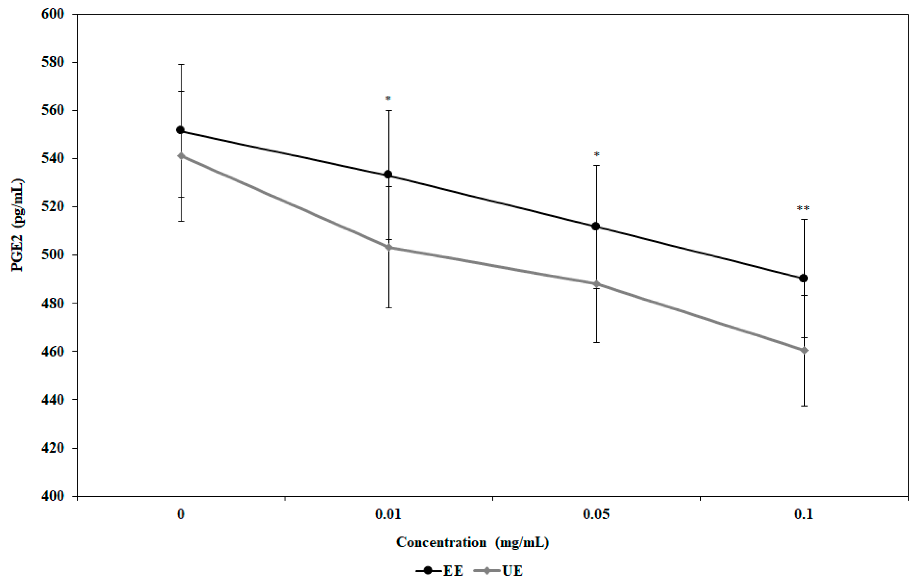
3.2. Inhibition of Nitric Oxide (NO) and Prostaglandin E2 (PGE2) Production by the Extracts

Prior to carrying out the anti-inflammatory experiments, in Figure 1 the cell cytotoxicity of the samples against mouse microglial cells was compared by treating the UE or EE (0.01 to 0.1 mg/mL) with (black bars) or without 1 µg/mL of LPS (white bars).
Interestingly, the EE from the hot 70% ethanol extraction process generally had higher cytotoxicity than the UE both with and without LPS, which was presumably attributable to the high heat-mediated degeneration of chlorophyll and resultant increases in toxic substances, such as pheophorbide. The effects of the samples on cell death also showed concentration dependency. The highest cell cytotoxicity (approximately 14%) was observed upon treatment of 0.1 mg/mL EE with LPS, compared to approximately 12% upon treatment of the UE at the same concentration; approximately 9% and 5% of the cells died upon treatment of 0.01 mg/mL UE and EE with LPS, respectively. In general, less cell cytotoxicity was observed in both samples without LPS, but the difference in the cytotoxicities of extracts treated with and without LPS was not significant. Therefore, the extracts from both processes could be used to investigate their anti-inflammatory effects because ca. 90% and 86% of the lowest survival rates were observed at the maximum dosage of 0.1 mg/mL without and with LPS, respectively. Figure 2 and Figure 3 demonstrate the NO and PGE2 measurements after treatment with the two extracts in BV-2 cells. In Figure 2, the amount of NO produced in the untreated group not treated with LPS was extremely small at 5.21 μM, but the amount of NO produced in the LPS-treated group sharply increased to 40.31 μM. In contrast, when BV-2 cells were treated with 70% ethanol extract together with LPS, 35.6 μM NO was generated at the 0.01 mg/mL EE concentration, while 20.3 μM of NO was generated at the maximum concentration of 0.1 mg/mL. Additionally, when BV-2 cells were treated with UE extracts, 31.4 μM NO was generated at the low concentration, and 19.6 μM NO was generated at the high concentration, indicating that the extracts obtained from the UE had greater ability to suppress NO generation than extracts from conventional extraction processes. Additionally, both extracts suppressed the generation of NO in a concentration-dependent manner, demonstrating that UEs exert remarkable anti-inflammatory effects on brain nerve cells and that 70% EEs also exhibit anti-inflammatory activity in brain nerve cells. These results are consistent with the findings of other studies [49], indicating that chlorophyll has anti-inflammatory effects and strongly suggesting that the anti-inflammatory effect of Spirulina extract is attributable to chlorophyll; similar results regarding the antioxidant effect of chlorophyll were also reported in other studies [37,50]. Additionally, when the extracts were administered alone at high concentrations, NO was minimally produced to levels similar to the control group not treated with LPS (data not shown).
Figure 3 demonstrates the production of PGE2, a major product of phlogogenic mechanisms.
As shown in Figure 3, 88.4 pg/mL PGE2 was produced in the control without any treatment, while 1580.9 pg/mL PGE2 was produced when BV2 cells were treated with LPS, indicating that inflammation was induced. When the extracts were administered at concentrations ranging from 0.01 to 0.1 mg/mL, PGE2 production decreased in a concentration-dependent manner in response to both extracts. Approximately 826.5 pg/mL PGE2 was produced when the UE was administered at a maximum concentration of 0.1 mg/mL. In general, the amount of PGE2 produced decreased according to concentration in the same pattern as that of NO production. The ultrasonic extract had a more dramatic reduction compared to the extract obtained by the general extraction process, reconfirming that ultrasound extracts have stronger anti-inflammatory effects. When compared to Coridalyis bungeana [51], Coptids rhizome [52], and Royal jelly [53], the Spirulina extracts from this UE showed anti-inflammatory effects selectively on brain nerve cells similar to or higher than those of the other substances. The extract containing large amounts of the useful bioactive substance in intact forms without the destruction of activity obtained through ultrasonic extraction has better anti-inflammatory properties, therefore confirming the excellence of ultrasonic low-temperature extraction once again. In particular, NO is known to be excessively produced by macrophages in response to stimulation with substances, such as LPS and amyloid-beta which are toxic intracellular substances to cause cytotoxicity and inflammation [54]. PEG2 is synthesized by COX-2 to mediate the pain and fever on damaged tissues or cell regions and is known to be involved in the induction of Parkinson’s and Alzheimer’s disease at high levels [55]. Therefore, effective inhibition of these substances may reduce factors that cause inflammation in brain nerve cells, thereby improving cognitive functions through anti-inflammatory effects. This information can be understood in the same context as previously reported findings, indicating that Spirulina can protect brain nerve cells and improve cognitive function and memory [31,32,35].

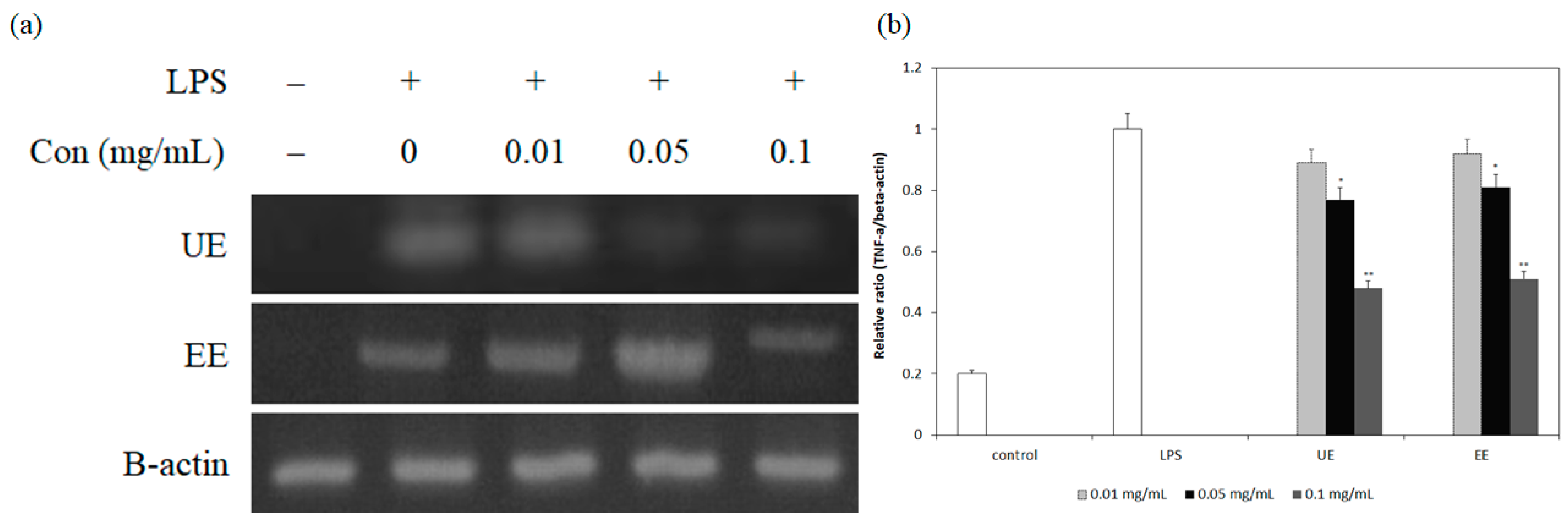
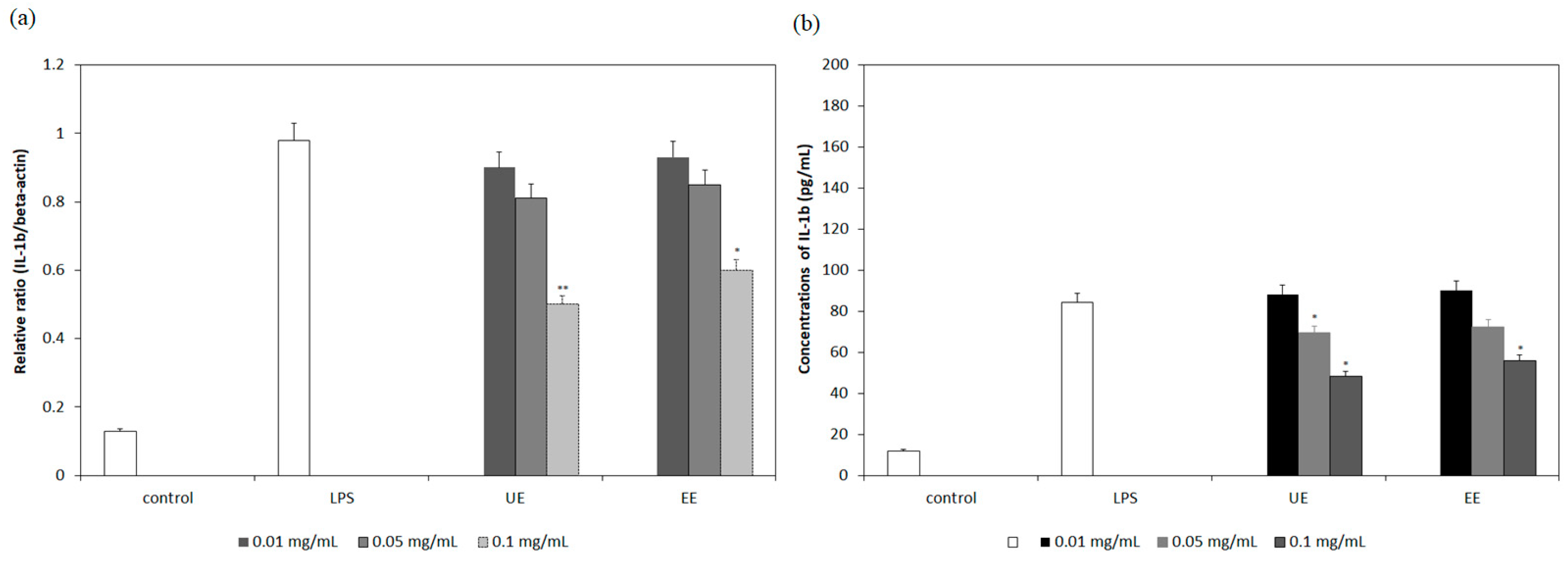
3.3. Inhibition of mRNA Expression and Secretion of Pro-Inflammatory Cytokines by the Extracts

Because the Spirulina extracts proved to be capable of suppressing the production of mediators that directly affect inflammation, Figure 4, Figure 5, Figure 6 and Figure 7 show the effects of the two extracts on the mRNA expression of TNF-α, IL-6 and IL-1β, which are pro-inflammatory cytokines that directly affect the inflammation of brain nerve cells. These experiments also assess the degree to which the extracts suppress the actual production of these cytokines in mouse nerve cells.
Figure 4 shows RT-PCR products that reflect the expression of mRNA in the cells treated with only LPS, cells treated with both LPS and extracts, and the control with no not treatment. As shown in Figure 4a, the amount of TNF-α increased sharply in the mouse microglial cells treated with LPS alone compared to the control without any treatment. In contrast, when LPS and Spirulina extracts were administered together, TNF-α expression decreased in a concentration-dependent manner. Similar to the effects on NO and PGE2 production, shown in Figure 2 and Figure 3, the ultrasonic extract had greater inhibitory effects on expression than the extract obtained through the general extraction process. To quantitatively compare the electrophoretic bands, shown in Figure 4a, the sizes of individual bands were normalized to the beta-actin band, which is a house-keeping gene, using a program that quantitatively compares the band sizes, as shown in Figure 4b. Similar to the pattern in Figure 4a, mRNA expression relative to beta-actin decreased as the concentration of the extracts administered increased. Additionally, the ultrasonic extract had much greater inhibitory activity than the general extract, as shown in Figure 4a. In particular, the difference in TNF-α expression inhibition between the two extracts was larger higher extract concentrations of 0.1 mg/mL than at lower concentrations, possibly, due to higher amounts of chlorophylls in the extracts. Therefore, this result indicates once again that extraction of bioactive substances sensitive to heat requires a low-temperature extraction process and that the UE is the most efficient extraction process in such cases.
As shown in Figure 5, the secretion of TNF-α from BV-2 cells was observed to confirm whether down-regulation of mRNA expression actually reflects the suppression of TNF-α secretion in mouse central nervous system (CNS) nerve cells. Similar to the pattern of the transcription of mRNA in Figure 4, the secretion of TNF-α was also decreased as a concentration-dependent manner with the greatest reductions at high concentrations. Unusually, although the number of general extracts produced concentration-dependent reductions, the reductions were relatively smaller compared to the ultrasonic extract. Although mRNA expression suppression, as shown in Figure 4, appeared to be concentration-dependent for both extractions, the degree of TNF-α suppression was lower in the general extract treatment. This means that mRNA expression is more sensitive to the useful components in the extract, but dramatic effects occur only when the concentration of useful substances is high enough in the following transcription stage. Therefore, we hypothesize that to anticipate intracellular inhibition or enhance functions using natural products along with gene expression, the natural products should exist at least at the critical concentration necessary to affect the production of the target substance. Figure 6 and Figure 7 show the quantification of IL-6 and IL-1β gene expression, which was determined using the Image J program with the IL-6 and IL-1β RT-PCR product bands, as shown in Figure 4a, to more easily compare the suppression levels of these genes than that allowed by the pictures of electrophoresis bands themselves. Figure 6a shows the quantitative comparison of the RT-PCR-amplified bands reflecting mRNA levels of the target and housekeeping genes after treatment with LPS and the two extracts at different concentrations. Figure 7a shows the comparison of IL-1β mRNA gene expression levels.
These two graphs do not show the electrophoretic bands, as shown in Figure 4a, to avoid redundancy, as the graphs in Figure 6a and Figure 7a show the quantitative comparison of the bands obtained by electrophoresis. Examination of IL-6 and IL-1β gene expression after extract administration in Figure 6a and Figure 7a revealed that the degree of IL-1β inhibition is generally higher than that of IL-6 for both extracts, although they are similar. This suggests that Spirulina extract may act more selectively on IL-1β than on IL-6 transcript levels. According to this difference of IL-6 and IL-1β protein secretion, shown in Figure 6b and Figure 7b, the amount of IL-6 secreted, which was 680.5 pg/mL when 0.01 (mg/mL) of the ultrasonic extract was administered, decreased by 29.4% to 480.3 pg/mL when 0.1 mg/mL was administered. The amount of IL-1β secretion decreased by approximately 47% (from 94.5 pg/mL to 48.2 pg/mL), indicating that the extract from UE is more effective at inhibiting IL-1β and TNF-α production among the pro-inflammatory cytokines. Moreover, similar to the trend for the suppression of TNF-α production, the UE showed higher inhibitory effects than the EE from conventional hot ethanol extraction processes, demonstrating for the first time that ultrasonic extracts are generally more effective at inhibiting inflammation of mouse nerve cells than the extract from the hot extraction process. Although the inhibitory activity was generally lower than that of the ultrasonic extracts, the Spirulina extract from conventional 70% ethanol extraction also inhibited the secretion of pro-inflammatory cytokines from mouse nerve cells to levels similar to or higher than those from other natural products [5,9,15,16].

3.4. Correlation Between Gene Down-Regulation and Secretion of Pro-Inflammatory Cytokines

The above-mentioned results suggest that the extracts down-regulate the expression of the genes involved in pro-inflammatory cytokine production, thereby inhibiting the secretion of the relevant cytokines. Many other studies have reported similar results. However, there are no published data on the quantitative correlation between gene regulation and protein secretion, since it is considered to be natural that the amounts of the cytokines secreted from BV-2 cells measured by ELISA analysis only support the results of gene expression from PCR analysis.
Therefore, Figure 8 shows the comparison of the down-regulation of the expression of TNF-α (Figure 8a), IL-6 (Figure 8b), and Il-1β (Figure 8c) genes, which are the cytokines most closely involved in the inflammation of brain nerve cells and resultant secretion of cytokines according to two different extraction processes, such as ultrasonic and conventional extraction processes. As shown in Figure 8a, for TNF-α, gene expression and protein production decrease proportionally according to the concentrations of extracts administered with high correlations. In particular, the degree of downregulation of gene expression and TNF-α production is constant at all concentrations of the extracts without any large difference among concentrations. This means that the extracts act on gene regulation the most directly leads to the control of TNF-α production. Therefore, it can be inferred that Spirulina extracts inhibit TNF-α production directly from transcription to induce anti-inflammatory effects. In contrast, for IL-6 (Figure 8b), although gene expression and IL-6 production inhibition occurred, the decreasing rates were not significantly proportional to each other. In particular, the analysis indicated that the degree of IL-6 production inhibition was higher than the degree of gene expression inhibition indicating the possibility that inhibition of IL-6 production from BV-2 cells occurs through pathways than gene expression or compositely unlike the TNF-α. Unlike the previous examples, IL-1β shows higher correlations between gene expression and inhibition of IL-6 production, but it has lower correlations than TNF-α. Therefore, it can be assumed that the extracts first control the amplification of TNF-α and IL-1β genes thereby directly inhibiting the production of cytokines. Additionally, the production of IL-6 is more effectively reduced because of the effects on the IL-6-producing gene and the reduction of TNF-α and IL-1β production, which have already been inhibited. The basic data on these mutual relationships have been presented for the first time in this study, and more detailed studies on the mutual relationships between Spirulina extracts and the three pro-inflammatory cytokines are needed to confirm the findings. However, the data shows for the first time that although Spirulina extracts are involved in inhibiting the secretion of three cytokines, rather than acting simultaneously, there are differences in the degree of inhibition mechanisms, and order of action. Therefore, these results suggest that rather than uniformly influencing the secretion of all pro-inflammatory cytokines, the natural extracts selectively affect the production. Future studies on the anti-inflammatory effects of the extracts are necessary to investigate more subdivided inhibition of target cytokines. In particular, stimulation by LPS is most directly involved in inflammatory diseases, such as septic shock, rheumatoid arthritis, insulin resistance, and cachexia, by maximizing TNF-α production from macrophages [55,56]. Therefore, since the ultrasonic extract inhibits TNF-α secretion most effectively, the anti-inflammatory effects of these extracts are very high. In particular, the effects of the Spirulina extracts, containing chlorophyll on the inflammatory mediator and inflammatory cytokine production inhibition, were shown. This strongly implies that in addition to the antioxidant effects of chlorophyll and Spirulina extracts that are already known, the extracts also have an ability to protect against nerve cell inflammation. Therefore, the neuroprotective effects of Spirulina extracts were caused by inhibiting the inflammation of mouse nerve cells via antioxidant properties, potentially improving cognitive activities. The results would suggest another possible mechanism of the Spirulina extracts on its neuroprotection process.

4. Conclusions

This work first showed that the Spirulina extract from the UE enhanced anti-inflammation activities in mouse microglial cells associated with its antioxidant effects, compared with that from the conventional 70% ethanol extract at high temperature. The non-thermal ultrasonic extract contained more than two-fold higher amounts of extremely heat-sensitive chlorophylls than the conventional extract and exhibited a two-fold higher extraction yield. Specifically, the extract effectively inhibited the production of inflammation mediators, such as NO and PGE2, while the conventional extract also showed a relatively high ability to inhibit their production. Strong anti-inflammatory effects of the extract from the UE were also demonstrated by the significant down-regulation of mRNA expression of the pro-inflammatory cytokines, TNF-α, IL-6 and IL-1β. Moreover, we quantitatively demonstrated that inhibition of the gene expression was closely related to the suppression of pro-inflammatory cytokine secretion even though ELISA analysis supports PCR results in general; in particular, TNF-α had a very strong relationship while IL-6 demonstrated the least correlation between the down-regulation of gene expression and the suppression of cytokine secretion. Notably, the effect on TNF-α expression was greater than that on IL-1β and IL-6, which changed the least. This result suggests that the extract first controls TNF-α secretion and later inhibits IL-6, but this hypothesis should be further validated with a more detailed mechanism of the anti-inflammation cascade within nerve cells. However, we clearly showed that the extract from the ultrasonic process was more effective at suppressing the secretion of both inflammatory mediators and pro-inflammatory cytokines, as reflected by its neuroprotective activities. Conclusively, the high anti-inflammatory effects of the extracts were closely correlated with the high amounts of chlorophylls, which have strong antioxidant activities, obtained only through a non-thermal ultrasonic process. The high anti-inflammatory effects of the Spirulina extract with mouse microglia cell line could be employed to develop a possible mechanism for in vivo anti-inflammation effects along with the results from primary nerve cells. However, the expression of anti-inflammatory cytokines, such as IL-10 and TGF-β, should also be studied to further elucidate more exact anti-inflammation effects of this extract. This study also provides useful information for developing functional foods from heat-labile natural resources.

Author Contributions

W.Y.C., J.-H.S., J.-Y.L. and D.H.K. carried out all of the experiments and H.Y.L. designed the whole experiments and drafted the manuscript. All authors read and approved the final manuscript.

Funding

This work has been supported by a research grant from the Marine Biotechnology Program funded by the Ministry of Oceans and Fisheries (PM61400), and all of the authors deeply appreciate their financial support.

Conflicts of Interest

The authors declare no conflict of interest.

References

  1. Bartus, R.T.; Dean, R.L.; Beer, D.; Lipa, A.S. The cholinergic hypothesis of geriatric memory dysfunction. Science 1982, 217, 408–417. [Google Scholar] [CrossRef]
  2. Citron, M. Alzheimer’s disease: Treatments in discovery and development. Nat. Neurosci. 2002, 5, 1055–1057. [Google Scholar] [CrossRef]
  3. Squire, L.R. Memory systems of the brain: A brief history and current perspective. Neurobiol. Learn. Mem. 2004, 82, 71–77. [Google Scholar] [CrossRef] [PubMed]
  4. Minghetti, L. Role of inflammation in neurodegenerative diseases. Curr. Opin. Neurol. 2005, 18, 315–321. [Google Scholar] [CrossRef]
  5. Asai, M.; Iwat, N.; Yoshikawa, A.; Aizaki, Y.; Ishiura, S.; Saido, T.C.; Maruyana, K. Berberine alters the processing of Alzheimer’s amyloid precursor protein to decrease Abeta secretion. Bioichem. Biophys. Res. Commun. 2007, 352, 498–502. [Google Scholar] [CrossRef] [PubMed]
  6. Floyd, R.A. Antioxidants, oxidative stress and degenerative neurological disorders. Proc. Soc. Exp. Biol. Med. 2001, 222, 236–245. [Google Scholar] [CrossRef]
  7. Halliwell, B.; Gutterridge, J.M. Role of free radicals and catalytic metal ions in human diseases: An overview. Methods Enzymol. 1990, 186, 1–85. [Google Scholar] [CrossRef] [PubMed]
  8. Boille, S.; Vande Vele, C.; Cleveland, D.W. ALS: A disease of motor neurons and their nonneuronal neighbors. Neuron 2006, 52, 130–144. [Google Scholar] [CrossRef]
  9. Talhouk, R.S.; Karam, C.; Fostok, S.; El-Jouni, W.; Barbour, E.K. Anti-inflammatory bioactivities in plant extracts. J. Med. Food 2007, 10, 1–10. [Google Scholar] [CrossRef] [PubMed]
  10. Jenkins, S.M.; Barone, S. The neurotoxicant trimethyltin induces apoptosis via caspase activation, p38 protein kinase and oxidative stress in PC12 cells. Toxicol. Lett. 2004, 147, 63–72. [Google Scholar] [CrossRef] [PubMed]
  11. Ali, S.F.; LeBel, C.P.; Bondy, S.C. Reactive oxygen species formation as a biomarker of methylmercury and trimethyltin neurotoxicity. Neurotoxicology 1992, 13, 637648. [Google Scholar] [PubMed]
  12. Corvino, V.; Marches, E.; Michetti, F.; Geloso, M.C. Neuroprotective strategies in hippocampal neurodegeneration induced by the neurotoxicant trimethyltin. Neurochem. Res. 2013, 38, 240–253. [Google Scholar] [CrossRef] [PubMed]
  13. Chen, A.; Xiong, L.J.; Tong, Y.; Mao, M. The neuroprotective roles of BDNF in hypoxic ischemic brain injury. Biomed. Rep. 2013, 1, 167–176. [Google Scholar] [CrossRef] [PubMed]
  14. Dawson, G.R.; Iversen, S.D. The effects of novel cholinesterase inhibitors and selective muscarinic receptor agonists in tests of reference and working memory. Behav. Brain Res. 1993, 57, 143–153. [Google Scholar] [CrossRef]
  15. Zhang, W.; Zhang, X.; Zou, K.; Wang, J.L.; Wang, Y. Seabackthorn berry polysaccharide protects against carbon tetrachloride-induced hepatoxicity in mice via anti-oxidative and anti-inflammatory activities. Food Funct. 2017, 8, 3130–3138. [Google Scholar] [CrossRef]
  16. Yuk, H.J.; Lee, J.W.; Park, H.A.; Seo, K.S.; Oh, S.R.; Ryu, H.W. Protective effects of coumestrol on lipopolysaccharide-induced acute lung injury via the inhibition of pro-inflammatory mediators and NF-kB activation. J. Funct. Food 2017, 34, 181–188. [Google Scholar] [CrossRef]
  17. Young, I.S.; Woodside, J.V. Antioxidants in health and disease. J. Clin. Pathol. 2001, 54, 176–186. [Google Scholar] [CrossRef] [Green Version]
  18. Bennett, P.B.; Marquis, R.E.; Demchenko, I. High Pressure Biology and Medicine; Bennett, P.B., Ed.; University of Rochester Press: New York, NY, USA, 1998; pp. 1–23. [Google Scholar]
  19. Lim, G.P.; Chu, T.; Yang, F.; Beech, W.; Frautschy, S.A.; Cole, G.M. The curry spice curcumin reduces oxidative damage and amyloid pathology in an Alzheimer transgenic mouse. J. Neurosci. 2001, 21, 8370–8377. [Google Scholar] [CrossRef]
  20. Murabe, Y.; Sano, Y.; Roisen, F.J. Morphological studies on neuralgia. VI. Postnatal development of microglial cell. Tissue Res. 1982, 225, 469–485. [Google Scholar] [CrossRef]
  21. Nakajima, K.; Kohsaka, D.; Raquel, E.G. Functional roles of microglia in the brain. Neurosci. Res. 1993, 17, 187–203. [Google Scholar] [CrossRef]
  22. Gonzalez-Scarano, F.; Baltuch, G. Microglia as mediators of inflammatory and degenerative diseases. Annl. Rev. Neurosci. 1999, 22, 219–240. [Google Scholar] [CrossRef]
  23. Witkamp, R.; Monshouwer, M. Signal transduction in inflammatory processes, current and future therapeutic targets: A mini review. Vet. Q. 2002, 22, 11–16. [Google Scholar] [CrossRef] [PubMed]
  24. Eikelenboom, P.; van Gool, W.A. Neuroinflammatory perspectives on the two faces of Alzheimer’s disease. J. Neural Transm. 2004, 11, 281–294. [Google Scholar] [CrossRef] [PubMed]
  25. Liu, J.S.; Hong, J.S. Role of microglia in inflammation-mediated neurodegenerative diseases: Mechanisms and strategies for therapeutic intervention. J. Pharmacol. Exp. Ther. 2003, 304, 1–7. [Google Scholar] [CrossRef]
  26. Gao, H.M.; Liu, B.; Zhang, W.; Hong, J.S. Novel anti-inflammatory therapy for Parkinson’s disease. Trends Pharmacol. Sci. 2003, 24, 395–401. [Google Scholar] [CrossRef]
  27. Saeid, A.; Chojnacka, K.; Korczyński, M.; Korniewicz, D.; Dobrzański, Z. Biomass of Spirulina maxima enriched by biosorption process as a new feed supplement for swine. J. Appl. Phycol. 2013, 25, 667–675. [Google Scholar] [CrossRef]
  28. Son, M.H.; Park, K.H.; Choi, A.R.; Yoo, G.; In, M.J.; Kim, D.H.; Chae, H.J. Investigation of biological activities of enzymatic hydrolysate of Spirulina. J. Korean Soc. Food Sci. Nutr. 2009, 38, 136–141. [Google Scholar] [CrossRef]
  29. Hernandez, A.C.; Nieves, I.; Meckes, M.; Chamorro, G.; Barron, B.L. Antiviral activity of Spirulina maxima against Herpes simplex virus type 2. Antivir. Res. 2002, 56, 279–285. [Google Scholar] [PubMed]
  30. Zao, X.; Yang, Y.H.; Cho, Y.S.; Chun, H.K.; Song, K.B.; Kim, M.R. Quality characteristics of Spirulina maxima added salad dressing. East Asian Soc. Diet. Life 2005, 15, 292–299. [Google Scholar]
  31. Deng, R.; Chow, T.J. Hypolipidemic, antioxidant, and anti-inflammatory activities of microalgae Spirulina. Cardiovasc. Ther. 2010, 28, 33–45. [Google Scholar] [CrossRef]
  32. Koh, E.J.; Seo, Y.J.; Choi, J.; Lee, H.Y.; Kang, D.H.; Kim, K.J.; Lee, B.Y. Spirulina maxima extract prevents neurotoxicity via promoting activation of BDNF/CREB signaling pathways in neuronal cells and mice. Molecules 2017, 22, 1363. [Google Scholar] [CrossRef] [PubMed]
  33. Bhat, S. R Chlorophyll: The wonder pigment. Sci Rep. 2005, 42, 29–32. [Google Scholar]
  34. Hosikian, A.; Lim, S.; Halim, R.; Danquah, M.K. Chlorophyll Extraction from Microalgae: A Review on the Process Engineering Aspects. Int. J. Chem. Eng. 2010, 29, 1–11. [Google Scholar] [CrossRef]
  35. Koh, E.J. The Improvement of Cognitive Impairment and Neuroprotection Effects of Spirulina maxima Extract. Ph.D. Thesis, Cha University, Pangyo, Korea, 2018; pp. 1–125. [Google Scholar]
  36. Kay, R.A.; Barton, L.L. Microalgae as food and supplement. Crit. Rev. Food Sci. Nutr. 1991, 30, 555–573. [Google Scholar] [CrossRef] [PubMed]
  37. McCarty, M.F. The chlorophyll metabolite phytanic acid is a natural rexinoid—Potential for treatment and prevention of diabetes. Med. Hypotheses 2001, 56, 217–219. [Google Scholar] [CrossRef]
  38. Lee, Y.J.; Kim, S.H.; Kim, J.S.; Han, J.A.; Seo, H.J.; Lim, H.J.; Choi, S.Y. Studies on Simultaneous Determination of Chlorophyll a and b, Pheophorbide a, and β-Carotene in Chlorella and Spirulina Products. J. Food Hyg. Saf. 2005, 20, 141–146. [Google Scholar]
  39. Kim, W.I.; Chung, K.W.; Hong, I.K.; Park, K.A. Ultrasound Energy Effect on Solvent Extraction of Amaranth Seed Oil. J. Korean Ind. Eng. Chem. 2001, 12, 307–311, OSTI ID:20268268. [Google Scholar]
  40. Oh, S.H.; Ahn, J.H.; Kang, D.H.; Lee, H.Y. The effect of ultrasonificated extracts of Spirulina maxima on the anticancer activity. Mar. Biotechnol. 2011, 13, 205–214. [Google Scholar] [CrossRef]
  41. Lee, D.H.; Hong, J.H. Antioxidant activities of chlorella extracts and physicochemical characteristics of spray-dried Chlorella powders. Korean J. Food Preserv. 2015, 22, 591–597. [Google Scholar] [CrossRef]
  42. Mosmann, T. Rapid colorimetric assay for cellular growth and survival: Application to proliferation and cytotoxicity assays. J. Immunol. Met. 1983, 65, 55–63. [Google Scholar] [CrossRef]
  43. Lee, Y.H.; Jeon, S.H.; Kim, S.H.; Kim, C.Y.; Lee, S.J.; Koh, D.S.; Lim, Y.H.; Ha, K.S.; Shin, S.Y. A new synthetic chalcone derivative, 2-hydroxy-3′,5,5′-trimethoxychalcone (DK-139), suppresses the Toll-like receptor 4-mediated inflammatory response through inhibition of the Akt/NF-κB pathway in BV-2 microglial cells. Exp. Mol. Med. 2012, 44, 369–377. [Google Scholar] [CrossRef]
  44. Kim, J.S.; Seo, Y.C.; Choi, W.Y.; Kim, H.S.; Kim, B.H.; Shin, D.H.; Lee, H.Y. Enhancement of antioxidant activities and whitening effect of Acer mono Sap through nano-encapsulation processes. Korean J. Med. Corp. Sci. 2011, 19, 191–197. [Google Scholar] [CrossRef]
  45. Green, L.C.; Wagner, D.A.; Glogowski, J.; Skipper, P.L.; Wishnok, J.S.; Tannenbaum, S.R. Analysis of nitrate, nitrite, and [15N] nitrate in biological fluids. Anal. Biochem. 1982, 126, 131–138. [Google Scholar] [CrossRef] [PubMed]
  46. Deck, L.M.; Hunsaker, L.A.; Gonzales, A.M.; Orlando, R.A.; Vander, D.L. Substituted trans-stilbenes can inhibit or enhance the TPA-induced up-regulation of activator protein-1. BMC Pharmacol. 2008, 8, 19–25. [Google Scholar] [CrossRef]
  47. Kim, M.R.; Kang, O.H.; Kim, S.B.; Kang, H.J.; Kim, J.E.; Hwang, H.C.; Kim, I.W.; Kwon, D.Y. The study of anti-inflammatory effect of Hwanggeumjakyak-tang extract in RAW 264.7 Macrophage. Korean J. Herbol. 2013, 28, 43–50. [Google Scholar] [CrossRef]
  48. Zheng, H.; Yin, J.; Gao, Z.; Huang, H.; Ji, X.; Dou, C. Disruption of Chlorella vulgaris Cells for the release of biodiesel-producing lipids: A Comparison of grinding, ultrasonication, bead milling, enzymatic lysis, and microwaves. Appl. Biochem. Biotechnol. 2011, 164, 1215–1224. [Google Scholar] [CrossRef]
  49. Okamoto, K.; Kihira, T.; Kobashi, G.; Sasaki, S.; Yokoyama, T.; Inaba, Y.; Nagai, M. Fruit and vegetable intake and risk of amyotrophic lateral sclerosis in Japan. Neuroepidemiology 2009, 3, 251–258. [Google Scholar] [CrossRef]
  50. Gruskin, B. Chlorophyll—Its therapeutic place in acute and suppurative disease: Preliminary report of clinical use and rationale. Am. J. Surg. 1940, 49, 49–55. [Google Scholar] [CrossRef]
  51. Ju, M.K. Protorpine, and Alkaloid Isolated from Croydalis bugeana Turca, Attenuate Neuro-Inflammation in LPS-Induced BV-2 Microglia Cells. Master’s Thesis, Kyungbuk National University, Daegu, Korea, 2017; pp. 1–56. [Google Scholar]
  52. Akhter, M.H.; Sabir, M.; Bhide, N.K. Anti-inflammatory effect of berberine in rats injected locally with cholera toxin. Indian J. Med. Res. 1977, 65, 133–141. [Google Scholar] [PubMed]
  53. Yu, M.M.; Chen, Y.F.; Pan, Y.M.; Liu, Y.C.; Tu, J.; Wang, K.; Hu, F.L. Royal jelly attenuate LPS-induced inflammation in BV-2 microglia cells through modulating NF-kB and p38/JNK signaling pathways. Med. Inflamm. 2018, 2018, 7834381. [Google Scholar] [CrossRef]
  54. Abramson, S.B.; Attur, M.; Amin, A.R.; Clancy, R. Nitric oxide and inflammatory mediators in the perpetuation of osteoarthritis. Curr. Rheumatol. Rep. 2001, 3, 535–564. [Google Scholar] [CrossRef] [PubMed]
  55. Kuo, C.L.; Chi, C.W.; Liu, T.Y. The anti-inflammatory potential of berberine in vitro and in vivo. Cancer Lett. 2004, 203, 127–137. [Google Scholar] [CrossRef] [PubMed]
  56. Jhun, B.S.; Jin, Q.; Kin, Y.T.; Kong, S.S.; Cho, Y.; Ha, Y.H.; Baik, H.H.; Kang, I. 5-aminoinidazole-4-carboximide riboside suppresses LPS-induced TNF-alpha production through inhibition of posphophatidylinositol-3-kinase/A activation in RAW264.7 murine macrophage. Biochem. Biophys. Res. Commun. 2004, 318, 372–380. [Google Scholar] [CrossRef] [PubMed]
Figure 1. Cytotoxicity of the S. maxima extracts with (black bars) and without (white bars) adding 1 µg/mL of lipopolysaccharide (LPS) against BV-2 cells. EE, 70% ethanol extraction at 80 °C for 12 h, UE, ultrasonic pretreatment with 70 % ethanol at 40 kHz and room temperature for 8 h, and further extraction at 65 °C for 4 h. Values are presented as means ±SD; * p < 0.05 and ** p < 0.01 compared with the non-treatment group.
Applsci 09 02144 g001
Figure 2. Secretion of nitric oxide from BV-2 cells by the treatment of various concentrations of the S. maxima extracts. EE, 70% ethanol extraction at 80 °C for 12 h, UE, ultrasonic pretreatment with 70% ethanol at 40 kHz and room temperature for 8 h, and further extraction at 65 °C for 4 h. Values are presented as means ±SD; * p < 0.05 and ** p < 0.01 compared with the LPS group.
Applsci 09 02144 g002
Figure 3. Comparison of PGE2 secretion from BV-2 cells by the treatment of various concentrations of the S.maxima extracts. EE, 70% ethanol extraction at 80 °C for 12 h, UE, ultrasonic pretreatment with 70% ethanol at 40 kHz and room temperature for 8 h, and further extraction at 65 °C for 4 h. Values are presented as means ±SD; * p < 0.05 and ** p < 0.01 compared with the non-treatment group.
Applsci 09 02144 g003
Figure 4. Down-regulation of mRNA expression of TNF-a from LPS-induced BV-2 cells (a) and the relative ratio of the gene expression by normalizing with beta-actin as a house-keeping gene (b) by the treatment of various concentrations of the Spirulina extracts along with the untreated control. EE, 70% ethanol extraction at 80 °C for 12 h, UE, ultrasonic pretreatment with 70% ethanol at 40 kHz and room temperature for 8 h, and further extraction at 65 °C for 4 h. Values are presented as means ±SD; * p < 0.05 and ** p < 0.01 compared with the LPS group.
Applsci 09 02144 g004
Figure 5. The secretion of TNF-a from LPS-induced BV-2 cells by the treatment of various concentrations of the Spirulina extracts. EE, 70% ethanol extraction at 80 °C for 12 h, UE, ultrasonic pretreatment with 70% ethanol at 40 kHz and room temperature for 8 h, and further extraction at 65 °C for 4 h. Values are presented as means ±SD; * p < 0.05 and ** p < 0.01 compared with the LPS group.
Applsci 09 02144 g005
Figure 6. Relative ratios of mRNA expression of IL-6 by normalizing with beta-actin as a house-keeping gene (a) and secretion of IL-6 (b) from LPS-induced BV-2 cells by the treatment of various concentrations of the Spirulina extracts. EE, 70% ethanol extraction at 80 °C for 12 h, UE, ultrasonic pretreatment with 70% ethanol at 40 kHz and room temperature for 8 h, and further extraction at 65 °C for 4 h. Values are presented as means ±SD; * p < 0.05 and ** p < 0.01 compared with the LPS group.
Applsci 09 02144 g006
Figure 7. Relative ratios of mRNA expression of IL-1beta by normalizing with beta-actin as a house-keeping gene (a) and secretion of IL-6 (b) from LPS-induced BV-2 cells by the treatment of various concentrations of the Spirulina extracts. EE, 70% ethanol extraction at 80 °C for 12 h, UE, ultrasonic pretreatment with 70% ethanol at 40 kHz and room temperature for 8 h, and further extraction at 65 °C for 4 h. Values are presented as means ±SD; * p < 0.05 and ** p < 0.01 compared with the LPS group.
Applsci 09 02144 g007
Figure 8. Quantitative comparison of the secretion and mRNA expression of TNF-a (a), IL-6 (b) and IL-1beta (c). EE, 70% ethanol extraction at 80 °C for 12 h, UE, ultrasonic pretreatment with 70% ethanol at 40 kHz and room temperature for 8 h, and further extraction at 65 °C for 4 h. Values are presented as means ±SD; * p < 0.05 and ** p < 0.01 compared with the non-treatment group.
Applsci 09 02144 g008
Table 1. Estimation of the concentrations of total chlorophylls and extraction yields of the Spirulina extracts from two different extraction processes.
Extraction ProcessTotal Chlorophyll Contents (mg/g)Extraction Yields (%)
Conventional ethanol extraction (EE) *6.24 ± 0.928.9 ± 1.66
Ultrasonic extraction (UE) **17.56 ± 1.8611.6 ± 2.02
* EE, 70% ethanol extraction at 80 °C for 12 h. ** UE, ultrasonic pretreatment with 70% ethanol at 40 kHz and room temperature for 8 h, and further extraction at 65 °C for 4 h.

