Review

The Use of Free Weight Squats in Sports: A Narrative Review—Terminology and Biomechanics

1
Center of Excellence for Sport Science and Coach Education, Department of Sport, Exercise, Recreation and Kinesiology, East Tennessee State University, Johnson City, TN 37614, USA
2
School of Sport Sciences, College of Applied Human Sciences, West Virginia University, Morgantown, WV 26505, USA
3
Peak Force International, Taichung 406, Taiwan
4
School of Kinesiology, Louisiana State University, Shreveport, LA 71115, USA
5
Strength and Power Research Group, School of Medical and Health Sciences, Edith Cowan University, Joondalup, WA 6065, Australia
*
Author to whom correspondence should be addressed.
Appl. Sci. 2024, 14(5), 1977; https://doi.org/10.3390/app14051977
Submission received: 22 January 2024 / Revised: 14 February 2024 / Accepted: 23 February 2024 / Published: 28 February 2024
(This article belongs to the Special Issue Advances in Sport Science: Athlete Development and Performance)

Abstract

:
There is substantial evidence indicating that increased maximum strength as a result of training with squats, particularly full and parallel squats, is associated with superior athletic capabilities, such as sprinting, jumping and agility. Although full and parallel squats have been strongly associated with sport performance, there is also some evidence that the use of partial squats may provide angle specific adaptations that are likely advantageous for specific sporting activities. Partial squats may be particularly advantageous when trained in conjunction with full or parallel squats, as this practice results in a greater training effect. There is a paucity of evidence that squatting is associated with excessive injuries to the knees, lower back, or other structures. Evidence does indicate that squatting, including full squats, can be undertaken safely, provided an appropriate training methodology is applied. Indeed, based on scientific data, the cost/benefit ratio indicates that squats should be recommended and should be a central strength training exercise for the preparation of athletes in most sports, particularly those requiring strong and powerful whole body and lower body movements.

1. Introduction

In the past and presently, most strength and conditioning coaches and sport scientists have considered the squat to be an essential and primary tool for the complete development of athletic potential [1,2,3,4,5,6,7]. The overall importance that the squat plays in strength and conditioning programs can be ascertained from the fact that this category of exercise is commonly identified as one of the most important and frequently prescribed exercises used by strength and conditioning coaches in many sports, reflecting its importance in strength and conditioning programs. For example, the squat and weightlifting movements have been reported to be the most commonly used exercises in professional sports including Major League Baseball (MLB) [8], the National Basketball Association (NBA) [9], the National Football League (NFL) [10], the National Hockey League (NHL) [11] and Major League Soccer (MLS) [12,13].
Additionally, the squat exercise has been considered to be a very important training exercise used in United States high school strength and conditioning programs [14]. Outside of the United States, the squat is one of the most frequently used exercises in the preparation of athletes in Spanish handball, basketball, volleyball, and football [15], Argentinian rugby [16], and for rowers in the United Kingdom [3]. Ultimately, when examining coaching and scientific literature, squats are widely regarded as a critical exercise group for developing lower body strength in athletic populations [17].
While the squat is often considered a primary exercise in the development of athletes [1,2,4,5], several authors have suggested that squats, especially deep squatting, may increase the risk of knee injury [18,19,20,21]. Deep squats are those in which during the descent, the top of the thigh reaches parallel or a below parallel position, i.e., parallel and full squats. Typical rationales for the avoidance of squatting movements primarily deal with degenerative changes within the tendofemoral complex and increased potential for the occurrence of chondromalacia, osteoarthritis, and osteochondritis when deep squats are performed [18,22]. However, based upon the existing body of evidence, deep squatting performed with the correct technique has been shown to actually stimulate training-induced adaptations that can provide a protective effect for the knee joint [22,23,24]. Indeed, a review by Hartmann et al. [22] presented compelling evidence that deep squatting does not place the knee, hip, or vertebral column at an increased risk for injury. In fact, the review suggested that deep squatting may actually create a protective adaptive scenario for the lower extremity. In support of Hartmann et al., a more recent work by Case et al. [25] revealed that examined athletes who had a higher relative maximum parallel back squat strength displayed a substantially lower injury risk. In fact, based on these data, male athletes who squat >2.2 × body mass and female athletes who squat >1.6 × body mass had a significantly lower injury risk [25].
The squat is a closed kinetic chain, large muscle mass, multi-joint exercise that stresses the upper body and mid-section musculature, particularly the erector spinae (primarily isometric) and the leg and hip extensors (dynamically). From the performance perspective, evidence indicates that squatting movements, particularly deeper squats, have a substantial potential to increase performance capacity [23,26,27,28]. This perspective is supported by improvements in traditional performance-based tests such as the vertical jump and sprinting assessments, as well as in sport performance. For example, Speranza et al. [29,30] noted that alterations in absolute and relative 1RM squat strength were statistically related to changes in tackling ability in rugby players. Additionally, based on several studies and reviews [23,27,31], deeper squats stimulate greater improvements in muscle cross-sectional area (CSA) and produce superior improvements in vertical jumping performance when compared to partial squats.
Several review papers [17,22,27,32,33] have been published in the scientific literature that explore various aspects of squatting motions. Since the publication of these reviews on squatting, there has been a large volume of research and critical analysis of existing research published exploring the physiological, biomechanical, and performance effects of squatting movements. Additionally, some of this newer research has examined contraindications and risks associated with squatting movements.
Although scientific inquiry in this area has increased, a primary factor limiting research and its interpretation relates to common terminology use. A principle purpose of this review is to establish common terminology related to the different variants of the back and front squat, particularly concerning depth and foot placement. The authors strongly believe that establishment of a common vernacular is critical for setting the foundation for the development of a better understanding of squatting movements. The second area of emphasis will center on the basic biomechanics of the squat in order to establish an understanding of the kinetic and kinematic aspects of the movement. Collectively, these key areas of inquiry can provide a strong foundation for a comprehensive understanding of squatting movements.

2. Methods

Literature, for this narrative, was gathered from Google Scholar, ResearchGate, and PubMed. With few exceptions, literature was confined primarily to those studies using free weights. Literature cited was limited to back squats and front squats and their derivatives (e.g., partial and speed squats). Other squat exercises such as barbell overhead squats, squat exercises using different types of bars/devices, and squat jumps are not included in this review paper.
The primary focus of this review paper is to create a standardization of squat-related terminology as well as gathering enough evidence-based data for the back and front squat to justify this terminology. Some squat exercises have not been used for training studies, and as a result, there is not enough well-defined evidence to include in this review paper. Free weights and the back/front squat types were chosen for consideration as these are commonly used in the training of athletes worldwide. Key words and phrases (English) used in the search included “squats”, “strength”, “strength-endurance”, “power endurance”, and “high intensity exercise”.

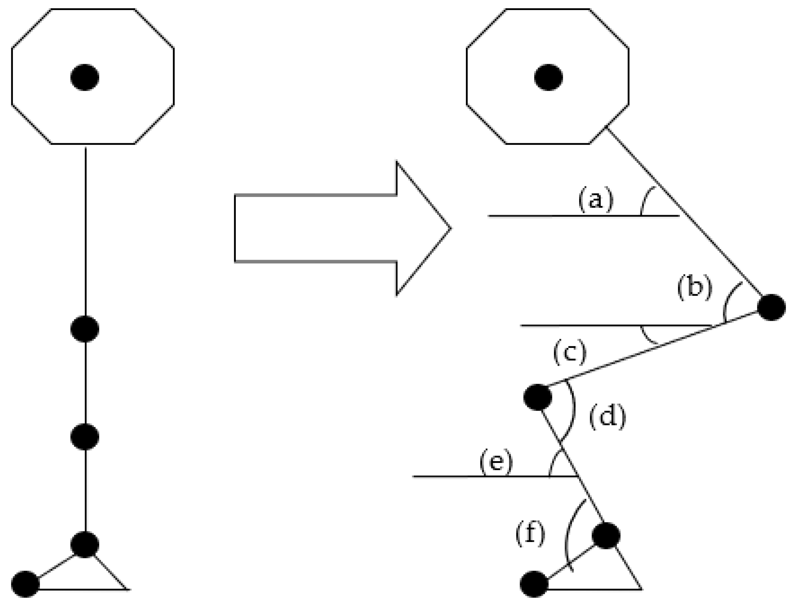
3. Basic Terminology Related to Squatting Movements

In a discussion dealing with the efficacy and safety of the squat for sport, it is essential that basic terminology can be used to describe the different variants of the squat, squatting depth, and foot placements. Establishment of common terms and descriptors sets a recognizable foundation that allows for a more complete understanding of how various squatting positions and techniques impact both kinetic and kinematic variables and how these variables might relate to various sports performance factors.
Defining the Barbell Squat: Squatting movements can be classified into several types or categories that can either be performed bilaterally or unilaterally depending upon which variant of the exercise is being implemented. Typically, the bilateral variants of the squatting movement include the back squat [2,4,6,34], front squat [17,35], and jump squat [36]. While the unilateral variant (one-legged squat) has been less well defined, it is often used in training [37,38,39]. Although these various squat types are commonly discussed in coaching and scientific literature, there is often confusion about the basic concepts related to the performance of these exercises. Therefore, it is essential to establish clear definitions and descriptions of the various squatting motions, which can be applied in either practical or scientific settings. Consequently, this review will use the following terminology as the framework for describing each of the various squatting movements:
Back squat: The free weight back squat is the most commonly performed squatting movement [40,41] and is considered to have the greatest potential to increase lower body strength [27,42]. Because of its ability to impact overall strength, the back squat and its variants have been considered to be one of the cornerstones of sports performance-based strength training interventions used by both professional and amateur athletes [9,10,13,14,15,43]. As a whole, there are two basic back squatting techniques, the high bar and low bar squat [4,16,33,44], that are often used in sports performance-based strength and conditioning programs. The high bar squat, sometimes termed the Olympic-style squat, is performed with the bar centered across the shoulders, just below the spinous process of the seventh cervical vertebrae (C7), while the low bar squat, or powerlifting squat, is performed with the bar approximately 5–6 cm further down the back, across the top of the scapula [33,45]. The low bar squat is most commonly associated with the sport of powerlifting, while the high bar squat is more typically associated with the sport of weightlifting [35,45]. Generally, the low bar squat is characterized by a larger degree of hip flexion and a hip movement that may be greater than the knee movement [44,45,46]. Conversely, the high bar squat is performed in a more upright position resulting in a more equal distribution of joint movement forces between the knee and the hip joints [44,45]. It should also be noted that squats, particularly high bar full and partial back squats, can be performed explosively, using plantar flexion, and rising on the balls of the feet [47].
An additional derivative of the free weight back squat that is often performed by powerlifters is the box squat [41,48]. The box squat requires the athlete to perform an eccentric phase (i.e., descent) followed by sitting on a box, typically for a minimum of 1 s, prior to executing the concentric phase (i.e., ascent) of the movement [41,48]. The box squat can be performed in a full movement, in parallel movements, or in partial movements. By sitting on the box, the athlete removes much of the effect of the stretch shortening cycle [48]. Generally, when athletes perform the box squat, a wider foot stance is selected compared to traditional back squats [41,48], resulting in different muscle activation patterns [49]. It has been advised that box squats should only be used to improve low bar squat performance, specifically for those competing in powerlifting [41]. However, more scientific inquiry into the efficacy of box squats is necessary.
Another back or front squat derivative, with full, parallel, and partial variants, is the speed squat [50], where the velocity of movement throughout the concentric phase is intentionally maximized to create a ballistic or semi-ballistic high power movement [51,52,53]. Speed squats are often timed for a specific number of repetitions and load; the lifter tries to decrease the exercise time with training, thus raising the average power of the movements. Often this squat variant is performed in a manner in which the athlete plantar flexes and rises onto the balls of their feet [47].
Front squat: The front squat is a derivation that is not as frequently used in training as the traditional back squat exercise [32,52,54,55]. However, the importance of the front squat cannot be underestimated because of its relationship to the clean and power clean movements [13,48], which are commonly part of comprehensive strength and conditioning programs [9,10,14,15,53]. The front squat exercise is typically performed with a pronated grip with the barbell held across the anterior portion of the deltoids and clavicles [35,54,55]. For athletes who lack wrist and shoulder flexibility, the grip can be modified by either using a cross-armed grip [35,40,54] or by using lifting straps to help hold the barbell [35]. The load that can be lifted during a front squat is typically less than in a back squat [27,55].
Jump squat: The jump squat is a derivative of the high bar back squat, as the bar placement is typically centered across the shoulders just below the spinous process of the C7 vertebra [56]. However, it is sometimes classified as a different exercise group because of its unique power attributes and the various levels of depth that can be used. Typically, the jump squat is considered a ballistic or explosive exercise, as the object of the exercise is to physically jump into the air while under load, thus removing the braking phase that occurs towards the end of the ascent in traditional squatting exercises [57]. Alternative methods for the performance of the jump squat can include movements with either dumbbells or on a restricted lifting device such as a Smith machine, although task specific adaptations may be different [12,17]. Jump squats can be performed as a countermovement exercise or from a static start.
One-leg squat: The one-leg squat, unilateral squat, fore-aft split squat, or “Bulgarian” split squat is performed with one leg designated as the stance leg, while the support leg is often placed at knee height on an adjustable bench located behind the athlete [55,58]. The stance leg’s distance from the bench is adjusted in order to allow the athlete to be able to descend to a position in which the posterior portion of the thigh is parallel to the ground [55,59] or to where the top of thigh is parallel to the ground [58]. An alternative approach would be to not elevate the support leg, keeping both feet in contact with the floor [60]. One-leg squats can also be performed with the stance leg elevated to the side and the support leg on the ground beside the lifter. Step-ups, although typically viewed as auxiliary exercises, are essentially a type of one-leg squats, where the athlete steps up on an adjustable box or bench to vary hip or knee range of motion (RoM). Regardless of which variant of the one-leg squat is used, a barbell can be held in a high bar position [58], or dumbbells can be held in one or each hand [60]. Interestingly, these types of unilateral squats are not typically considered a primary training exercise, as they are often programmed based on the athlete’s maximum back squat strength or in relation to body mass [51,61]. The maximal lifting capacity using these exercises is substantially less than what can be accomplished in a back or front squat.
Other leg and hip exercises: Exercises such as the leg press, knee extension, deadlift variations, and hamstring curl are all typically thought of as auxiliary leg exercises and are not classified as part of the squatting exercise group. All of these exercises use some of the same muscle groups and can be programmed based on maximal bilateral squatting capabilities [43,62]. These exercises should not be used as replacements for squatting movements as they do not affect exactly the same results [56,63]. However, these exercises can be useful as auxiliary exercises and as an alternative to squatting due to mitigating circumstances such as injuries that prevent the performance of appropriate squatting movements.

4. Defining Squat Depth by Knee Angle

When examining any of the various squatting techniques, one of the key areas of discussion revolves around the depth at which squats are performed [1]. This is often a difficult concept because the term depth suggests that a vertical displacement of the barbell is being quantified, when in reality the depth of the squat is a function of the knee and hip angle achieved during the squatting motion and the movement of the bar and body as a system [27,57,64]. Therefore, simply measuring or quantifying barbell displacement as a means of determining depth can be misleading [65] in that this type of measurement does not account for trunk lean or the actual knee and hip angles achieved during the squatting motion. Based upon this idea, a reasonable determination of the depth of any squatting movement should be quantified in relation to a specified knee angle achieved [43,66] and not the vertical displacement of the bar (Table 1). The authors realize that tissue strain is a function of various knee, hip, and ankle angles and is covered elsewhere in this paper; however, it should be noted here that the knee angle and relation of the thigh to the floor offer easily discernable landmarks for standardization of depth. Although hip angle can affect tissue strain, it is quite difficult to measure/estimate during a squat performance. However, reasonable quantification of knee angle is possible both before and during a squatting movement using appropriate measurement tools [43,66]. Furthermore, the authors’ observations and that of other researchers indicate that when squatting to a specific knee angle, the change in hip angle during the movement remains relatively constant in comparison to the change in knee angle throughout the movement(s) [27,57,64]. Additionally, the terminal point of the squat movement can be more easily estimated through observation–for example, for a parallel squat in which the top of the thigh is parallel (or slightly below) with the floor.
A point of confusion in the literature is how to clearly define joint angles. For example, some authors define squat depth in the context of the external knee angle [17,22,74], while others relate squatting depth to an internal knee angle [27,43,64,66,68,70,75]. In order to clearly present and define the various squatting depths, this review will reference the internal knee angle for the quantification of the various squatting depths that have been reported in the scientific literature or used in practical settings. Based upon these knee angles, four generalized squat depths can be established for bilateral movements, which include the full, parallel, half, and quarter squat [1,2,27].
Full squat: A full squat, or deep squat as it is sometimes termed, is classified as being performed until a depth where a 40–45° internal knee angle is achieved [2,45,66,74]. Full squats can be performed with either the high bar back squat or the front squat. Typically, the front squat requires the athlete to go into a deeper squat position and will result in a substantially smaller knee angle and a more upright trunk position when compared to the high bar back squat [2]. Conversely, the low bar back squat typically does not allow for as great a depth to be performed as in the front squat; because of the posterior shifting of the hip and forward trunk lean typically noted in this movement, only a ≈68° internal knee angle can be achieved [41]. As such, the low bar squat typically cannot be performed to a full squat position.
Parallel squat: A parallel squat is performed to an internal knee angle between 60 and 70°, where a straight horizontal line can be drawn between in the inguinal fold and the top of the knee musculature, just proximal to the patella [2,45,67,68,69]. The parallel squat can be performed with either variant of the back squat or the front squat. This depth (parallel) is commonly associated with the low bar or powerlifting squat [41]. While the high bar squat can be performed to parallel, it is often performed to a full squat position. Overall, the parallel squat is one of the most common exercises performed when targeting the development of lower body strength [2,76].
Half squat: A half squat is generally performed to a depth in which an 80–100° internal knee angle is achieved [2,40,66,67,77,78,79]. Half squats can be performed using the back, front, or Smith machine squats [12]. Because of the lesser degree of knee flexion associated with the half squat, substantially higher training loads can be used than loads in either the parallel or full back squat. The ability to handle higher loads in this range of knee angles is related to the higher force production capacities that occur closer to the apex of the ascending strength curve for this movement pattern [80].
Quarter squat: A quarter squat is typically performed at a knee angle between 110–140° [66]. The quarter squat is often performed from a dead stop and would represent a concentric squat movement [70]. This type of squat is commonly used in the creation of strength-power-potentiating complexes (SPPCs) [70]. Because this movement pattern occurs at knee angles that are even closer to the apex of the ascending strength curve, higher forces can be generated allowing for higher training loads compared to the full, parallel, or half squats [80].
Additional measurement considerations: When examining the depth of fore-aft unilateral movements, the knee angles used to describe these types of movements can be slightly different than those used for the bilateral squatting movements [39,58,59]. Due to biomechanical limitations, such as less torso lean or elevated support leg during the one-leg squat, the full movement is typically marked by an internal knee angle of 60–70°, while the parallel squat has a bottom position in which an internal knee angle of 80–100° is achieved [39,58,59].

5. Defining the Foot Position in Squatting

One factor that must always be considered when examining the scientific and practical literature on squatting is how foot placement is defined. Specifically, it is important to establish a description of both the stance width and foot angle used [17,20,81,82,83,84,85,86], as these two characteristics may have an impact on the kinematics and muscle activation patterns associated with the squatting movement [17,83].
Squat stance width: Careful examination of the scientific literature reveals little consensus on how foot placement is described or quantified [17,20,81,82,83,84,85,86]. However, three common squat stances including the (1) narrow, (2) medium or shoulder-width, and (3) wide stance are generally referenced in the scientific and applied literature [20,84]. See Table 2 for a more detailed explanation.
Traditionally, an athlete’s squat stance has been established by using the shoulders as a reference point [20,83,85]. Specifically, the narrow stance has been defined as being between 87 and 118% of shoulder width for the low bar squat [20] or 75% of shoulder width for the high bar squat [84]. The medium stance is typically equal to the athlete’s shoulder width for a high bar squat [84] or between 121 and 153% of shoulder width for the low bar squat [20]. A stance width of 120% of shoulder width is often used to define a wide stance with the high bar squat [84], while a wide stance with a low bar squat would be equal to 158–196% of shoulder width [20].
An alternative method for determining the stance width during a low bar squat is to measure the distance anteriorly between the lateral aspect of the left and right acromion process [85]. Using this method, a narrow stance would have a distance of 40.9 ± 3.8 cm between the left and right ankle centers, while the medium stance would have a distance 59.7 ± 6.6 cm, and the wide stance a distance of 69.6 ± 9.5 cm. When using a high bar squat, the stance can be established in relation to the greater trochanter distance (GTd) with the narrow, medium, and wide stances being 100%, 150%, and 200% of GTd respectively [49].
Squat stance foot angle: The stance utilized in the squat also requires reference to the angle of foot placement [20,81,83,85]. The foot placement is of particular interest because it has been reported to impact muscle activation patterns [83,85]. Typically, in the applied literature, the angle of foot placement is related to a position in which the foot is directly pointing forward, often referred to as the center position. Often, an outward rotation of 25–30° from center is recommended when using a high bar back squat [87]. For example, Escamilla et al. [81] reported that the angle of rotation of the feet during a narrow, medium, or wide stance is 20 ± 5°, 23 ± 3°, and 26 ± 4°, respectively, during a low bar back squat. However, Lorenzetti et al. [83] suggested that a moderate foot placement angle (approximately 20°) in combination with a moderate stance width (with feet approximately shoulder width apart) should be used. The moderate stance and foot rotation would reduce rotational RoMs in the hip and knee, abduction/adduction RoMs in the hip, and flexion RoMs in the knee [83]. Overall, there is limited research on the optimal foot angle from the center position and how these positions impact performance and muscle activation.

6. Basic Squat Biomechanics

In an effort to better understand the underlying mechanisms associated with the squat, the biomechanics of the barbell back squat were originally investigated in elite level athletes nearly five decades ago [88,89]. A major reason for this approach was to obtain normative data on elite strength athletes. At that time (the 1970s), the primary available resource for performing a biomechanical analysis of any human motion was to employ techniques limited to the examination of only one plane of movement. Shortly after the initial video analyses, the integration of force plates in the biomechanical assessments of the squat began to be used [90]. Typically, this approach was undertaken to determine kinetic and kinematic information about the squat. Inverse dynamics were employed when examining joint kinematics [91]. With these approaches, investigators established a baseline of biomechanical characteristics for the barbell back squat.
Although motion capture technology has advanced well over the last forty years, squat mechanics are still being analyzed largely in the sagittal plane because of the anterior-posterior movement that primarily occurs at the trunk and the joints of the lower extremities [19,92,93]. In the back squat, a typical RoM for an experienced lifter would require ankle dorsiflexion of about 20 to 25°, knee flexion to an internal angle of 95° in a half squat and approximately 60° in a parallel squat, and flexing the hip to a range of 85 to 110° as a lifter reaches the peak descent position [34,43,46,66,68,88].
Of importance from both practical and analytical standpoints is squat symmetry. Observation suggests that both sides of the kinematic squat sequence are similar [34,46,88]. Salem et al. [94] examined the squat 30 ± 12 weeks after ACL reconstruction and indicated that during a high bar squat, lower extremity RoM on both legs was relatively similar with less than a 3° difference between each side of the body for the three joints (ankle, knee, and hip) examined. It is possible that a difference of ≥3° in RoM between the right and left sides of the lower extremity joints could be indicative of a potential asymmetry that could be a cause for some concern. However, from a practical performance analytical point of view, a ≤3° difference in RoM between the right and left sides of the body would be difficult to visually capture and is usually considered to be of limited importance during the analytical process.
Another measurement approach is to collect kinetic measures using individual or multiple force plates in order to examine force production and power output during squatting [39,62,95,96]. More detailed analyses of the squatting movement can be performed when synchronizing video (2D or 3D) cameras with a force plate [97,98]. Researchers have used this technique in order to analyze lower extremity joint kinetics and kinematics [19,97]. Indeed, joint kinetics analyses have been undertaken to understand the mechanical loads in the lower extremity joints as well as the lower back region [39,46,82,89,95,97,98]. Typically, the greatest lower extremity joint torque, but least external applied force, occurs shortly after the initiation of the ascent phase of the squat at what has been termed the “sticking point” or “sticking region” [19,34,80,99,100]. Other kinetic variables such as compression and shear forces at the knee have been reported in scientific literature because of their clinical relevance [19,39,81,101]. Obviously, a loaded barbell back squat creates a greater amount of compressive force (and shear force) on the spine as compared to a body weight squat. As a result of placing the loaded barbell on the upper back, the joint force at the lumbar region becomes the largest among the joints in the lower half of the body [102].
Finally, electromyography (EMG) analysis is a necessary approach for examining neural activation of the muscles associated with variations of load, foot position, and stance width that can be used during various types of squatting movements [32,66,82,86]. In recent years, comprehensive studies using various combinations of motion capture videography, force plates, and EMG for squat analysis have increased in number. The increased interest in exploring the biomechanical aspects of the squat suggest that to truly understand the intricacies of squatting movements, a comprehensive understanding of the mechanisms associated with the neuromuscular components of squat performance is required.
The primary focus of these studies has dealt with understanding the nature of the mechanistic contributor(s) during a squat. At the same time, there has been a more recent trend of investigation dealing with the prediction of injuries based on the biomechanical findings. In the following section of this paper, further biomechanical reports dealing with a more detailed discussion of kinematics, kinetics, and EMG are discussed.
Kinematics: Kinematics deals with the characteristics that result from force application (kinetics), such as displacement and velocity. From a human factor standpoint, both joint and segmental kinematics are commonly reported variables in research directly exploring the squat (Figure 1). Typically, the sagittal plane is used as the plane of reference when performing a kinematic evaluation of squatting movements. This type of analysis is typically used to determine joint angles, joint and segmental displacement, and velocity.
Squatting characteristics (e.g., depth, trunk lean etc.) can be markedly affected by differences in anthropometrics and tissue structure. These include characteristics such as sex differences. Examinations of various types of squats have clearly indicated that sex-specific kinematic differences are present [103,104]. Both male and female subjects displayed marked hip, knee, and ankle deviations. However, in the back squat, males, compared to females, displayed smaller hip and knee flexion angles with all external loads (ranging from zero to body mass + 50%) [105]. Furthermore, increasing external loading resulted in ankle joint dorsiflexion to a greater degree in males in order to compensate for the lost RoM from the hip and knee [103]. Additionally, knee displacement was forward and shin angle was diagonal rather than vertical, which ultimately relates to greater ankle dorsiflexion, assuming the entire soles of the feet remain in contact with the floor [103]. An interesting finding from the same study was that females displayed somewhat different squat kinematics demonstrating less knee forward displacement. This may indicate that they were using a marked forward trunk lean, or possibly exhibited greater flexibility in the hip and knee joints, resulting in a minimization of the forward movement of the knee without having a greater degree of dorsiflexion. Additionally, there were sex specific timing differences, with the males reaching maximum forward knee displacement at 84% of their maximum descent, while females achieved maximum knee displacement at 93% of their maximum descent [103]. These differences in joint flexion and extension likely affect the ability to generate forces and subsequent velocity of movement.
When focusing specifically on the lower extremity joints, knee and hip angular velocities influence vertical concentric velocity [20]. If the focus of training is to generate high rates of force development (RFD) and power output during the concentric action, faster knee and hip extension velocities are required in order to create these characteristics. For example, Escamilla et al. [20], using a low bar squat and three different foot stances (107 ± 10, 142 ± 12, and 169 ± 12% of shoulder width), studied concentric velocities at the knee and hip. At near maximum loading, peak knee angular velocity occurred in the range of 110 to 130°∙s−1 and the peak hip angular velocity occurred in the range of 102 to 122°∙s−1 [20]. Importantly, the athletes studied (N = 39) were national powerlifting competitors typically using a low bar technique and compressive gear and represented a “specific” population [100,106]. However, Donnelly et al. [34], using American Football players (N = 10) as the study focus, investigated lower body kinematics during a high bar squat using much lighter loads (25% of 1RM). They reported a similar angular velocity of the knee and hip, ranging from 113 to 120°∙s−1 at the knee and 114 to 117°∙s−1 at the hip. It is important to note that peak ankle, knee, and hip angular velocities or power outputs do not necessarily occur at the same absolute or relative bar load, particularly in ballistic movements such as loaded jumps [107,108]. This observation has important variation ramifications for training loads, suggesting a spectrum of loads be used to ensure “optimum” velocity and power training of all three joints.
The vertical velocity of a system (lifter plus barbell) is often calculated using the center of mass (CoM) displacement. Although concentric velocity (and power) calculated from CoM and the bar are similar, depending upon the sport, velocity (and power) may be more appropriately calculated from the bar, particularly for strength power athletes using implements (e.g., throwers, weightlifters, powerlifters) [109,110]. Indeed, the bar velocity is easier to quantify compared to tracking the movement of CoM of the system for a number of reasons. Typically, the location of the CoM is established with the use of video motion (kinematic) analysis and requires the use of a full-body marker set. While this process is well established, it is time consuming and requires specialized laboratory equipment. However, placing a marker on the barbell during squatting movements allows for sagittal plane analysis of barbell displacement and velocity in a more time-efficient and effective manner, particularly when frequent measurements are necessary. This method of analysis has been used in several studies investigating bar velocity to analyze explosive effort during the concentric phase of the back squat [96,111]. More recently, methods of analyzing bar velocity (and displacement) have included the use of linear position transducers [112], accelerometers, or inertial movement sensors [72,111,113,114] that are attached to the bar or a combination of linear potentiometers, and force plates that can be used to analyze a variety of kinetic and kinematic characteristics and derivations including power output [72,95].
During the ascent phase, average bar velocity typically ranges from approximately 0.97 to 0.54 m s−1 using 20% to 90% of 1RM back squat [96]. Obviously, compared to lighter loads, heavy loads, particularly near maximum loads, can substantially reduce the bar velocity and power output [71,73]. Indeed, a faster bar velocity can markedly influence the average and peak power output of the lift. Additionally, bar velocity can be affected by fatigue and the loss of bar velocity may represent a method of quantifying acute or chronic fatigue [95]. Furthermore, it is important to produce a high RFD in the concentric (ascent) phase to emphasize “explosiveness” during the squat. From this perspective, the importance of analyzing bar velocity and RFD specifically for explosiveness becomes apparent.
Although there is a specific emphasis on pushing the bar as hard as possible during the concentric phase, for heavy loads, relatively constant and relatively slow bar velocity indicates the “control” aspect of the lift in the eccentric (descent) phase. Typically, during the eccentric phase, it is important to produce relatively slow and constant negative bar velocity (negative because the bar moves downward) in order to keep the body position and movement in control, rather than creating a rapid and potentially deleterious descent. This is important especially when instructing novice level lifters that are learning appropriate techniques. However, it should be noted that in some cases, lifters may use a somewhat faster descent or partial descent (near the bottom of the descent) which may augment the eccentric maximum force levels and produce a subsequent higher concentric force and power output [115]. Higher descent velocities may also be used in “speed squats”, in which lighter weight squats are to be completed as fast as possible with good technique. Higher eccentric velocities may offer a task-specific or greater stretch shortening cycle/plyometric training adaptation [115,116].
From both a performance and clinical perspective, frontal/transverse planes of hip and knee movements during squatting have been studied [117,118,119]. Typically, total medial-lateral knee displacement (frontal plane) up to ≈3 cm can be expected in a back squat [117]. Excessive hip adduction and knee valgus characteristics are not considered an optimal movement pattern in the squat and may increase long-term injury potential, particularly of the knee joint [120,121]. Additionally, it is unclear as to how hip adduction alters the typical muscle activation patterns. Although hip adductors show considerable activation, some studies have indicated substantially greater activation of the vastus medialis and gluteus medius [28,122], while others have not [79,123]. However, the degree to which knee valgus motions are related to the bi-lateral squat maximum strength as a result of hip adduction is unclear [77,124]. Valgus motion could be a result of multiple factors such as (1) sub-optimal maximum hip muscle strength, (2) technique preferences, (3) habits from years of training, and (4) abnormal anatomical structure (e.g., excessive Q-angle) [73,77]. There is conflicting information available regarding the relationship between hip muscle strength and lower extremity valgus. A meta-analysis has indicated that lower extremity dynamic valgus was positively, but weakly, associated with hip strength (external rotation) in single leg ballistic tasks among women [124]. While it is reasonable and important to understand the mechanisms producing undesirable mechanics, additional investigation is necessary to understand the impact of these mechanisms on the hip and knee joints, particularly as it effects long-term injury potential.
Focusing specifically on bar kinematics of the squat, an excessive amount of multi-planar bar displacements such as tilting, uneven extension, rotation, and excessive anterior displacement are considered undesirable actions, as this creates an inefficient squat movement pattern. Among trained powerlifters, total anterior displacement of the bar (shoulder), knee, and system CoM has been shown to be somewhat greater during a high bar squat compared to a low bar back squat, while posterior displacement of the hip was larger for the low bar squat, particularly during the eccentric phase [41]. Trunk forward tilt was clearly greater in the low bar squat [41]. Additionally, the peak joint movements during extension of the knee and ankle tended to be higher for the high bar squat, while the peak hip movement is larger during the low bar squat [41].
From a coaching perspective, minimizing the forward lean of the trunk is recommended, particularly during the initial phase of learning how to squat [105,125]. If there is no excessive posterior hip displacement, the forward lean of the trunk leads to anterior displacement of the bar [126]. Aberrant mechanics can limit performance of most “vertical” resistance training exercises. For example, analogies can be made from the sport of weightlifting, in which excessive frontal and transverse plane movements during a pull are considered faulty mechanics [127,128,129]. Several researchers [129,130] have stated that these faulty motions can lead to unsuccessful life attempts, and less barbell displacement asymmetry between the left and right sides increases the chance of a successful lift in weightlifting competitions. Although the movements are different between the squat and weightlifting, minimizing unwanted lateral tilt and rotation of the barbell has been noted as an effective way to improve squat performance [131,132,133].
It should be, again, noted that during powerlifting or the low bar squat, in which the bar is lower on the shoulders/back, there is more anterior lean of the trunk, particularly if the foot stance is relatively narrow [41,46]. This position can place considerably more stress on the lower back compared to the high bar squat [46]. However, lower back stress can be reduced by two factors. Firstly, the bar is lower on the back, which shortens the moment of the arm relative to the lower back, and thus hip torque can be reduced compared to a high bar squat at the same load and degree of trunk tilt. However, lower back stress can be higher in a low bar squat when the two squats are performed in actual training, because many athletes are able to use heavier absolute loads with a low bar squat and there is typically a greater degree of trunk forward tilt with a low bar squat. Secondly, a wider stance can provide a mechanism which would allow the hips to move forward, allowing the lifter to “sit up” and thus creating a more erect trunk and consequently reducing the moment arm and lower back strain [41].
Kinetics: Linear kinetics deals with the causes of change in the state of linear motion. When measuring kinetic values during squatting tasks, ground reaction forces (GRF) are typically measured using a single force plate that allows examination of both legs, or dual force plates allowing examination of forces produced by each side of the body independently [96,110,134]. The ability to assess vertical GRF allows for a more comprehensive examination of the force-time characteristics associated with squatting movements. Logically, GRF (and impulse) will increase with higher loads. Although it is clear that higher loads increase the GRF, few investigators have specifically examined the relationship between the peak or average GRF and various loads [96,110].
System CoM acceleration is reflected by the pattern of concentric GRF during the squat. For example, Zink et al. [96] reported that a double peaked GRF occurred (1) during the initial phase of concentric action and (2) immediately after passing the sticking region during back squats performed with high bar squat loads ranging from 20–90% of 1RM squat. These peaks in force correspond to brief periods of acceleration; an initial acceleration occurs during the transition from the descent (eccentric action) into the ascent (concentric action) phase of the movement, and another acceleration occurs in the latter part of the ascent phase near the finish of the lift. This later acceleration possibly occurs as a result of the hips and knees moving forward, placing the lifter at a stronger mechanical advantage. Both McLaughlin et al. [88] and Escamilla [20] demonstrated a double peak in the same phase of ascent velocity for powerlifters using a low bar squat. Because the barbell velocity is related to GRF and acceleration, it is expected that there would be two velocity peaks during the concentric phase. This appears to be particularly evident when heavy loads are used [96]. Thus, it appears that two GRF peaks occurring during the ascent phase is a normal characteristic of full or parallel squat kinetics.
Understanding the load-power relationship during squatting movements is essential for establishing an understanding of potential optimal load(s) for peak power production, as well as potential injury reduction [135]. As would be expected, the load-power relationship is different between front, back, and partial squats [67]. Although peak power during the concentric phase of back squats at various loads (20–90% of 1RM) have not been shown to be statistically different, the optimal load for peak power output appears to be at loads of about 40–60% of 1RM [96,136,137]. In a few studies [109,137,138], the optimal load was shown to be at ~30% of 1RM, perhaps depending upon the type of athlete. However, peak and mean power output in these studies varied depending upon whether the power output was calculated for the load (bar + plates) [107,136], the system mass [73,96], or relative to the depth of the squat [137]. Additionally, in young women, measured leg press power may be similar or slightly greater than power calculations using the squat bar mass [139]. It seems logical that peak bar or system power production would occur at loads that are under 50–60% of 1RM, because these loads allow generation of greater velocities. However, when examining athletic populations, the optimal load for the bar mass has been shown to range between approximately 40% and 70% of 1RM for bar peak power [136,137], with similar values for system mass [96,110]. Strength-power athletes consistently produced optimum power outputs with higher relative loads [137]. This suggests that the optimum load range may differ with the method of training and level of strength development [110,137]. However, as previously noted, the optimum load for force/velocity and therefore power production may be different at different joints [107,108]. Thus, optimum production of force, velocity, and power appear to be best trained using a variety of loads and with a variety of conditions (non-ballistic versus ballistic).
Linear kinetics, including joint reaction force through the knee (tibio-femoral and patello-femoral), can be a valuable method for analyzing force application in the lower extremities. Investigations concerning the magnitude (and rate) of force directed through anatomical structures may be critical to understanding movement and the potential for injury. Based on limited research, early reviews [19] indicated that greater knee flexion (deeper squat) increases stress in the knee ligaments and is potentially injurious. Although ligament stress does rise with greater knee flexion [140,141], the tension that occurs during a full squat does not necessarily lead to damaged ligaments, such as the anterior cruciate ligament (ACL) and posterior cruciate ligament (PCL), even with high external loads [17,142,143]. More details can be found in Section 7: Safety and Injury factors. Indeed, recent evidence indicates that it is possible that the stress of squatting on the knee, including parallel and full squats, could lead to beneficial alterations. Although the combination of partial and full squats may be advantageous [78], evidence indicates that full squats can result in greater structural and strength adaptations compared to partial squats [23,31,138]. Furthermore, it appears that the stimulus increases with movement velocity [75]. In their review, Hartmann et al. [22] indicated that “concerns about degenerative changes of the tendofemoral complex and the apparent higher risk for chondromalacia, osteoarthritis, and osteochondritis in deep squats are unfounded”. As a final analysis, correct squatting technique, appropriate progression, and properly periodized/programmed training can minimize the risk of knee injury and may strengthen connective tissue including ligaments.
Kinetics (Angular kinetics): Angular kinetics deals with the causes of changes in the state of rotational motion (torques or moments of force). Inverse dynamics is a method for computing force or moments of force (torques), using a rigid body model. Calculation methods using such a model are based on the motion (kinematics) of a body and the body’s inertial properties (mass and moment of inertia). Although 2D approaches, often synchronized with a force plate, have been the norm, recent advances in 3D motion technology have enhanced the accuracy of inverse dynamic calculations. Using 3D video analysis combined with synchronized force plate(s) has resulted in lower extremity joint torques being a more commonly reported variable in the quantification of squatting movements. For example, Flanagan and Salem [99] reported hip and knee joint torque as a main contributor to concentric action during the squat. Depending on the bar position (e.g., high- or low-bar position) and different squat techniques, the contribution of the hip and knee also change as the body position changes. The magnitude of the forward lean of the trunk or the posterior displacement of the hip can all impact the knee and hip joint torques [98,99]. For example, performing a high bar squat (21 kg) during which restricted forward knee movement caused a forward trunk tilt resulted in 302 N·m of hip torque and 117.3 N·m of knee torque [46]. Conversely, when the trunk angle was relatively vertical, and the knee was allowed to freely move forward, there was a hip torque of 28.2 N·m and a knee torque of 15.1 N·m. Fortenbaugh et al. [126] also reported that forward trunk lean is associated with a posterior hip shift during the descent of the squat. This pattern of movement is particularly evident during low bar squats, when restricting forward knee displacement. This technique can result in a larger moment arm for the weight relative to the hip, and greater hip torque at the initiation of the concentric action and results in substantial low back loading [144].
Investigating angular kinetics in different squat tasks and examining bilateral asymmetry between a healthy knee and a repaired or reconstructed knee can provide important insights into injury mechanisms as well as rehabilitation aspects. For example, Salem et al. [94] investigated the bilateral differences in the high bar back squat performed by participants who had reconstructive knee surgery. Peak hip joint moment was markedly greater on the surgery side compared to the healthy side (1.67 Nm·kg−1 vs. 1.12 Nm·kg−1, p = 0.06). Additionally, there was a statistically significant difference in the peak knee joint moment for the surgery side compared to the non-injured side (surgery side: 1.02 Nm·kg−1 vs. non-injured side: 1.28 Nm·kg−1, p = 0.003). Furthermore, the ratio of average peak hip extensor and knee extensor torque was greater on the surgery side by 46.5% (p = 0.02) as compared to the non-injured side. Collectively [94,145,146,147], the effort level at the knee and hip was different between the surgery side and healthy side. Importantly, this asymmetry may persist for several years [145,146]. Conversely, for non-injured athletes, there is no supportive evidence that these types of asymmetries exist in the lower extremities. The general assumption is that non-injured athletes are fairly symmetrical, indicating that asymmetries occur as a result of injury, or post-surgery. Based on current data, from the rehabilitative standpoint, a goal would be to minimize the difference between the two sides during squatting and likely other tasks such as jumping.
Electromyography (EMG): EMG is a method of evaluating the neural activation of muscles by measuring the muscles’ electrical activity. Using EMG for the evaluation of squatting movements and other resistance training exercises has a relatively long history [148]. In previous EMG studies, investigators often targeted the superficial lower extremity muscles [68,86,149,150,151], and have included both “global and local” musculature of the mid-section [152]. However, it is common to measure electrical activity from large superficial muscles instead of using indwelling fine-wired EMG to examine deeper muscles. It is also logical to assume that larger muscles contribute substantially to force production in the squat. In examining the activity of muscles engaged in the squat, regional surface electrodes are typically placed on medial/lateral sides of flexor/extensor muscles of the lower extremity [147].
Based upon the available evidence, squatting depth can play a role in muscle activation. Deeper squats produce greater activity in the quadriceps and gluteus maximus [32,153]. Studies of the back squat, using surface electrodes, indicate that two quadriceps muscles (vastus lateralis and medialis) are not markedly different in activation level and there is a lower activation of the rectus femoris in both the descent and ascent phases of the high bar full back squat [91,151,154]. Some data suggest that a low bar back squat, when performed by powerlifters, may produce somewhat more activation of the quadriceps compared to a high bar squat among weightlifters at 65% of 1 RM [45]. However, these results may have occurred as a result of differences in body mass and training level compared to the weightlifting group [45]. The two antagonist hamstring muscles (lateral and medial) are much less activated than quadriceps muscles in both front and back squats [42]. However, the lateral hamstring (biceps femoris) typically shows a higher activation than the medial hamstring (semitendinosus) [81,151,154]. During the concentric phase, the quadriceps muscles typically show activation of around 50–80% of peak isometric values, while the hamstring group only activates up to about 20% of the maximum voluntary contraction (MVC) [93,154]. Relative activation of the gluteus maximus tends to be somewhat greater in the low bar squat, both eccentrically and concentrically [155], and the quadriceps shows greater activation in the high bar squat [33,44]. The eccentric/concentric action of the squat primarily results from hip (gluteus maximus) and knee extensor (quadriceps) activation, and it is clear that hamstring muscles are not primary contributors for squat tasks [82,119,149,151].
Data indicate that the gluteus maximus activation level is dependent upon the squat depth [66]. Increasing the strength of the gluteus maximus can be paramount for improving performance in many sport activities. For example, in sprinting, major functions of the gluteus maximus include control of trunk flexion on the stance-side and deceleration of the swing leg. Contractions of the stance-side gluteus maximus may also help to control flexion of the hip and to extend the thigh [156]. As such, performing deeper squatting, perhaps periodically including front squats, may be of benefit to those sports in which gluteus maximus’ maximum strength is of particular importance. Examining the EMG of the gluteus maximus is a technique necessary to understand the activation of such a strong hip extensor muscle. However, caution is necessary to minimize measurement error associated with electrode placement and electrical activity readings, which can be related to the muscle’s fiber alignment (diagonal) and the density of adipose tissue surrounding the gluteus maximus. Without proper precautions taken in electrode placement, conductivity factors, etc., high variance between and within subjects is typically reported, and all investigators should be aware of these problems in future studies [18,139]. It is also important to note that the degree of intra-muscular activation may not be consistent throughout a muscle [157,158], and this could influence inhomogeneous adaptions [45,159].
It is apparent that a biomechanical approach to understanding the underlying mechanisms of the squat can be a vital resource for coaches and practitioners. The following sections further elaborate on biomechanical findings to help understand the safety and benefits of performing squat exercises.

7. Safety and Injury Factors

Concerns have arisen that squat exercises are injurious, particularly to the knee and lower back [21,23,160,161]. For example, the knees and lower back appear to be common injury sites among lifters who regularly perform various squat exercises as a part of their training [162,163]. These injuries are believed to be a result of excessive forces on the tissues or repetitive overload without sufficient recovery. Generally, tissues—particularly bone—are regularly exposed to three force types: compression, shear, and angular torque. Compressive forces deal with those directed axially, shear forces are directed transversely and typically cut across the axis of the tissue, and angular forces are those causing a twisting motion around the tissue axis. Even though it was not the primary aim of many past studies, it is logical to believe that compression force as well as shear force in the knee increase with additional external resistance for squatting exercises [82].
Excessive force projected through the joint is believed to be a factor related to greater injury potential. However, researchers question the degree to which squat exercises are actually contributing to acute/chronic pain in, and injuries to, the knee and lower back [22,23]. Although some investigators and clinicians believe that injuries in the knee and lower back may result from performing squat exercises, there is little evidence that the exercise itself is the primary cause of the injury [23,160,164]. Moreover, these types of injuries may be due to overuse often associated with progressing training volume too fast as well as technique errors rather than an inherent problem with the exercise itself. In other words, this suggests that many, perhaps most, squat-related injuries can be prevented if lifters are properly instructed and perform and train the squat correctly. It should also be noted that among athletes, squats are rarely the only exercises being performed impacting the hip and knee joints, thus making it difficult to ascertain the impact of a single exercise on injury potential. In this section, studies and reviews are discussed in order to evaluate the speculation and concerns about pain and injuries in the spine (especially the lower back) and knee potentially resulting from squat exercises, particularly full squats.
Spine: During axial compression of the spine, the vertebral body is typically the first structure to show damage and/or fracture [22,165]. Squatting can place considerable compressive force on the vertebrae, particularly along the lumbar spine. When considering the lumbar spine (L-spine) region, it is logical to believe that as the external load increases during a squat, there will be higher axial compressive forces placed upon the body of the vertebrae, and if the weight is lifted faster, the magnitude of the compressive force increases due to higher acceleration of the CoM [144,166]. In typical training scenarios, it is common to increase the squatting load during a session (or from session to session), which can increase spinal compression.
For example, half and quarter squat exercises using loads of 0.8–1.6 × body weight can result in vertebral compressive forces (L3–4) of 6–10 times body weight at the initiation of the concentric phase (76), so the compression force magnitude likely increases as external load increases [79,167]. While compressive force magnitudes during squatting movements appear to be relatively high, it is important to note, based on cadavers, that the maximal compressive forces that the vertebral body can handle can be as high as 7800 N –10,000 N for typical untrained middle-aged males [23,168]. Although it seems unrealistic to believe that the general population commonly experience compression forces this high, among well-trained male strength-power athletes, compression forces may reach as much as 37,000 N [57,169]. Among females, vertebral CSA and compressive strength is lower than males [22]. Thus, at equivalent external loads, female vertebrae are exposed to a relatively higher compressive force. However, during full squats, females demonstrated a lower range of lumbar flexion and a greater anterior tilt of the sacrum compared to males [22]. Females have also demonstrated a lower stiffness and greater RoM between lumbar spinal segments and may be more capable of developing muscular stabilization of the spine and thus maintaining a lordotic curve during deep squats [80]. Importantly, high compression forces observed among strength-power athletes, including females, especially those engaged in squatting for many years without spinal compression injury, suggests a positive training adaptation [169,170,171].
Axial compression force is not the only concern for lower back issues; loading and body position can create other forces, such as shear force (transverse) and angular torque, that could be associated with lower back issues [21]. From a mechanical perspective, changing the CoM position toward the anterior or posterior can increase or decrease the amount of shear and compressive forces within the lumbar spine. Early research indicated that with spinal flexion, there are greater trunk extensor and lumbar shear force demands, but a reduction in compressive forces [172]. It should be noted that back muscle activation brings about resultant shear forces on the joint that can be maintained at a relatively constant and safe level [90,135]. This indicates that the musculature represents a safety mechanism that is particularly useful for maintaining a lordotic posture during lifting, when the muscles must provide the majority of the support moment [135]. These observations suggest that to minimize shear force in the spine, a relatively erect neutral to lordotic trunk position is recommended. Conversely, when a natural curvature of the lumbar area is maintained, the magnitude of the compressive force in the area can stay the same, but as the magnitude of lordotic curvature increases, the compression force also increases [165,167]. Furthermore, McKean et al., [103] employing real-time collection at 120 Hz using a 3D Magnetic Tracking Device, indicated that during lifting the spine becomes slightly flexed at 50% of body mass (even though the spinal muscles are contracting), particularly in men, partly due to structural differences between men and women. They indicated that the subtle alterations in spinal position would be quite difficult to see visually [103]. Thus, a more neutral spine may be the actual case during lifting, although the lifter should attempt to maintain a lordotic posture. Interestingly, Aasa et al. [173] used IMUs affixed at the spinous processes at T11, L2, and at the sacrum (S2), and these authors reported that when experienced powerlifters and weightlifters performed three repetitions of the squat at approximately 70% 1RM, lumbar spinal alignments were adjusted in all three planes. The three-dimensional spinal alignment adjustments showed low variability and did not reach the outer ranges of lumbar spinal flexion or extension for the squat. It should also be mentioned that the capacity of the human movement system to adapt to tissue loading in order to maintain tissue homoeostasis and function is quite large [174]. Thus, to a point, the more the tissues are loaded, the stronger they become, and it is possible that subtle degrees of spinal flexion-extension may enhance tissue adaptation, making tissues stronger [175]. In any case, the development of good squat technique and the need for qualified instructors and coaches is paramount.
A traditional method believed to help maintain a neutral or lordotic curvature in the spine when performing squat exercises is the use of a belt [161,176]. However, some evidence indicates that wearing a belt has little or no effect [161]. For example, Walsh et al. [161], using 3D motion analysis, studied experienced lifters, and showed little influence on spinal movements from a kinematic perspective when using a belt with increased loading; hyperextension occurs with or without a belt [161]. However, other studies showed contradictory results: a belt may provide some support to back position by increasing the activation of supporting muscles, increasing intra-abdominal pressure, and stiffening the trunk, which may enhance squat performance [177,178,179]. It is well-documented that increased intra-abdominal pressure created by a Valsalva or semi-Valsalva technique can serve as a significant support mechanism for the spine [61,178,180,181,182]. A potentially beneficial function of belts may be to assist in raising intra-abdominal pressure and lowering compression forces on the lumbar vertebrae [178,180]. This is related to the importance of using mid-section musculature to assist in maintaining an appropriate stable spinal alignment in a normal curvature, or in a lordotic position independent of belts, to help minimize anterior-posterior shifts of the trunk during the squat. Although specific mid-section exercises can be beneficial for trunk/spine stabilization, data indicates that large muscle mass, multi-joint exercises transmitting forces through the mid-section, such as squats, likely play an important role in improving the stability of the mid-section [152,164,183]. For example, evidence indicates that large muscle mass, multi-joint exercises can activate a number of mid-section muscles to a greater degree than typically performed isolated or unstable mid-section exercises, such as the superman and pelvic thrust on a stability ball [152,183]. Furthermore, as previously noted, an erect (stable) spine is advantageous for squatting, as excessive trunk flexion creates greater shear forces, and excessive hyperextension or flexion creates an uneven load on the vertebral body in multiple directions [102]. Compression and simultaneous extension or rotation of the spine, particularly the lumbar spine, can cause increased compression in the posterior annulus compared with neutral loading [161]. However, injury potential can be greatly reduced through changes in hip joint angle during the squat [22]. Forward leaning during squatting increases the potential for flexion of the lumber spine [22]. Because of the combination of high axial compression plus shear forces, ventral (forward) flexion can increase the risk of disk herniation [53]. The effects of ventral flexion can be minimized by creating a strong lumbar extension during motion reversal (initiating concentric action). Lumbar extension is created and maintained by increased lumbar erector spinae muscle activity and results in closing of the apophyseal joints and reducing shear force on the discs [22].
Although analyzing cervical spine (C-spine) compression would be valuable, a solid body of literature regarding this topic has not been established. Specifically, when considering the bar placement for a high bar squat, the bar is centered across the shoulders just below the spinous process of the C-7 vertebrae [52,105]. For a low bar squat, the bar sits further down the back, often on the superior portion of the scapula across the middle trapezius and rhomboid muscles regions. Currently, neither placement has been identified as a cause of substantial injury among individuals performing squats regularly.
Considering this discussion, along with proper loading progression, (1) performing squat exercises with correct technique specifically by maintaining a stable spinal position throughout the squat, and (2) appropriate use of a Valsalva or partial Valsalva technique to help maintain appropriate intra-abdominal pressure may assist in preventing back injuries that potentially result from squat exercises. Practitioners should be aware that if one of these factors is deficient or lacking, the risk of developing a lower back injury and pain may increase.
Knee: In the past, the squat was believed to be quite injurious to the knee [184]. Unfortunately, many, particularly in the medical profession, still believe that squatting is injurious to the knee (and other structures). Indeed, excessive tibio-femoral shear and compressive forces are a concern for many clinical professionals when considering the use of squat exercises as a part of a rehabilitative process with their patients. Because of this concern, investigators are often interested in analyzing forces transferred through the knee (i.e., tibio-femoral joint), as well as patella-femoral joints [22,101].
Compression and shear force: Compression forces are those axially directed forces from the “top and bottom” of a structure. Knee compressive forces can be generally divided into tibio-femoral and patello-femoral [22,101]. Shear forces are perpendicular to the axial direction. Several factors can affect knee compressive and shear forces. Increased velocity of movement, squat depth, and loading are related to increases in forces applied to the tissues [75]. Indeed, increased acceleration can increase compression and shear forces to a greater extent than loading alone, particularly in the transition from the descent to the ascent phase of the squat [22,27]. Additionally, lack of proper control of the descent phase can result in ballistic contact between the hamstrings and calf muscles, potentially increasing the stress on knee ligaments [17,34,179]. However, it should be noted that weightlifters routinely use semi-ballistic descents in the catch phase of the snatch and clean and jerk but have not shown excessive knee ligament problems [53,160,163]. Nevertheless, there is a potential for injury with rapid descents or bouncing out at the bottom; therefore, it is logical to perform squat exercises with a controlled speed, particularly in the eccentric phase. For advanced speed-strength training, the squat can be performed explosively with high velocity to maximize the training adaptation, provided bouncing is avoided. However, for novice level and very young lifters, certified/licensed strength and conditioning coaches and personal trainers should teach those lifters to perform a descent that is controlled to a degree that allows maintenance of body control and appropriate positions (e.g., neutral or lordotic spine). As long as reasonable control is maintained, especially in the descent phase of the squat, concerns for damage such as osteoarthritis, ligament tears, and meniscus tears in the knee joint can be minimized. Following is a summary of literature addressing the shear and compressive force issues at the knee joint.
Shear force: Shear force is the result of forces created in opposite directions to one another; during squatting, the greatest forces are projected posteriorly [35,185]. Shear forces can be particularly injurious to knee cartilage and cruciate ligaments, particularly the ACL. Tibio-femoral compression forces stabilize the knee joint and help to reduce shear forces [27,186]. For the knee joint during squatting, shear force is generally calculated in a plane with respect to the alignment of tibia and femur. Shear force reduction depends upon appropriate technique application and proper alignment of body segments during squatting [27,170].
Based on cadavers, tensile strength (load at failure) of the PCL for young men is about 4000 N. The tensile strength is about 2000 N for the ACL and decreases with age [187,188]. Mean posterior shear force (MPSF) creates a tensile load on the PCL when performing a squat. Load values for MPSF range widely depending on external loads and specific joint angles. With similar external loads and similar populations tested, studies have reported similar MPSF load values of 1783 to 1868 N at an internal knee angle of 117° (at a quarter squat depth) and 90° (at a half squat depth) [81,118]. Mean anterior shear force (MASF) creates a tensile load in the ACL when performing a squat. MASF values were found to be up to 500 N at 160° of internal knee flexion with a heavy external load (250 kg) [189]. While shear forces are influenced by squat speed, none of the previous studies reported extensively regarding the relationship between angular knee flexion velocity and the magnitude of shear forces. Specifically for MPSF, knee joint angular velocity seems to be an important factor for predicting load force as previous studies showed different magnitudes of MPSF with a range of external loads (147–250 kg) and internal knee flexion angles from 117° up to 50°, representing approximately 45% of limit tensile strength [81,151,190]. Calculated shear forces on the ACL were found to be considerably lower, typically less than 1000 N and less than 30% limit tensile strength [27,187]. Thus, shear forces developed in the cruciate ligaments are well within tensile strength tolerance. Note that based on evidence from recreational trained subjects, anterior-posterior shear forces may be less in males compared to females, resulting from greater co-activation among males (hamstrings and quadriceps) [104]. Additionally, some evidence suggests that squat training increases co-activation, thus reducing shear forces [76].
Compressive force: Knee compressive forces occur where the femur and tibia (i.e., tibio-femoral joint), and patella and femur (i.e., patello-femoral joint), are in contact. Compressive forces for both joints generally increase as the knee flexes during a low bar squat motion and reach a maximum at the peak knee flexion angle [39]. Escamilla et al. [39], describing the low bar squat, also reported differences in patello-femoral forces at different internal knee angles (120° p = 0.001; 110° p = 0.001; 100° p = 0.002; and 90° p = 0.002), indicating that as the knee flexes, greater forces at the patello-femoral joint are created. However, it is important to note that increases in force at this joint are largely due to alterations in mechanical advantage at the knee joint. As documented by Escamilla et al. [39], underpinning mechanisms of knee pain associated with patello-femoral pain syndrome are poorly understood. Thus, the greater force application at the patello-femoral joint cannot be clearly defined as a primary reason for the pain.
Past studies have indicated that knee compression forces of a powerlifter squatting can be greater than twice body mass. The tibio-femoral compressive force at the internal knee flexion of 50° was approximately 8000 N, at 120° was 5500 N, and at 150° was 3500 N [151,161]. Although these values seem very high, especially at an internal knee flexion of 50° (i.e., equivalent to near a full squat position), it appears that these values are well below the threshold for patellar ligament or tendon failure, indicating a substantial reserve before acute failure occurs. Indeed, the patellar ligament (untrained) can handle up to 15,000 N before failure. Although information is lacking, the quadriceps tendon has a much larger CSA than the patellar ligament and potentially could withstand even greater forces [189,191,192]. Interestingly, an internal knee flexion of approximately 130° seems to be a threshold for compressive and shear forces in the tibio-femoral and patello-femoral joints. After 130°, forces markedly rise with continued flexion, as occurs in deeper squats (or leg presses) [82]. The recommendation was to limit the internal knee flexion angle to 130° for selected cases in rehabilitation [82]. However, this does not mean that internal knee flexion ≥130° is injurious. Considering the data from these studies [189,191,192], proper technique and a reasonable training process still seem to be key factors for minimizing the risk of pain and injury.
As noted, if a lifter squats more than twice body weight, tibio-femoral compressive forces can be quite large (i.e., up to 8000 N) [85]. With the same load, patellar ligament tension can increase up to 6000 N, but patellar ligament tensile strengths (untrained) have been measured at over 10,000 N [34]. The ultimate structural strength for the healthy knee appears to be capable of handling the forces encountered during heavy squatting. Therefore, those forces projecting through the joint are not necessarily a critical element for creating pain and injuries. As previously noted, injury could be due to ineffective lifting technique and improper training progression, such as adding loads too quickly. However, these same forces may be critical stimuli for adaptive mechanisms [27,50].
Menisci and cartilage are also susceptible to mechanical influences, particularly compressive and shear forces. Concerns about degenerative changes of cartilage and the apparent increased risk for chondromalacia, osteoarthritis, and osteochondritis resulting from deep squats are apparently unfounded [27]. Although compressive and shear forces could induce cellular trauma, there are two protective mechanisms that must be considered. First, for articular cartilage (as in the tibio-femoral joint), load is transmitted not by elastic deformation (as might occur with severe impact), but rather through hydrostatic compression [193], which markedly reduces the potential for cellular trauma [194]. Secondly, in deep squats, such as the catch in weightlifting movements or during deep front squats, in which internal knee angles can be ≥40°, posterior thigh and calf contact can diminish knee-joint forces markedly [179,195]. Calculations of forces at deep squat knee angles have not considered soft tissue contact [27,196] and must be examined critically [27]. Additionally, depending upon the CSA of hamstring and calf muscles, thigh-calf contact could begin at larger knee angles, from approximately 60° [23], reducing tibiofemoral [74,113] and patellofemoral joint forces [24,197]. Thus, the belief that deep squats increase the potential for injury and degenerative changes of the knee joint [18,125] appears unsupported by objective evidence [22,30]. As previously noted, knee forces are altered by a number of factors including knee angle. For example, partial squats (80°–100° internal knee angle) should be used with caution because, at the initiation of the concentric phase, these knee joint angles result in the highest patella-femoral [163,198] and compressive stresses [168,193] and occur with only a small tendo-femoral support surface [197,198]. Thus, clinical recommendations for half or quarter squats in order to reduce the potential for degenerative changes in the knee joint may be counterproductive from an adaptive perspective [22,97]. However, with a pre-existing injury or previous reconstruction of the PCL, it may be best to restrict knee flexion to ~130°, which can minimize posterior shear [17,21].
Changes in forces with alternative squat exercises: It has been previously established that knee angles can influence the magnitude of knee compressive and shear forces in squatting. Studying squat variants, Escamilla et al. [39] showed that single-leg squats (knees translated beyond toes at maximum knee flexion) generally produced smaller patello-femoral compressive/shear forces compared to two different types of dynamic wall squats, long (knees over ankles at maximum knee flexion) and short (knees translate beyond toes at maximum knee flexion). It is important to mention that the squatting tasks used in this study (one-leg squat and wall squat) are often used in clinical settings, while more traditional strength and conditioning exercises, such as the back squat, can produce higher patello-femoral compressive and shear forces, often due to greater loading. Compressive forces reached approximately 3100 N in one-leg and wall squats, whereas barbell back squats can result in around 4500 N of patello-femoral compressive force with lifts greater than 140 kg [81]. Escamilla et al. [82] reported that the chosen exercises (i.e., leg press of high and low foot positions and back squat) altered the magnitude of tibio-femoral and patello-femoral compressive forces, and the squat produced higher forces than the leg press. Additionally, it was reported that wide stance low bar squats resulted in higher tibio-femoral and patello-femoral compressive forces compared to narrow stance squats [82]. Based on these findings, it is understandable that the leg press is often used in training and occasionally used to replace the squat during rehabilitation. However, it should be noted that there are marked differences in muscle activation when comparing squats, leg presses, or leg extensions. Clearly the larger muscle mass activated by squats provides advantages in training for sport or daily living activities [93,151]. Thus, if an athlete is training for a better squat performance or in terms of transfer of training to other activities, squats may be an excellent choice [36,93,151].

8. Conclusions and Summary

In the current narrative review, we have offered a common terminology that describes various types of squats. The standardization of terminology is of paramount importance as it provides the foundation for a coherent discussion surrounding the effects of squats. Additionally, it is important to understand the forces and loads that joints are exposed to during squatting movements, especially in the spine and knee, as these forces could be a mechanism for injury. However, based upon the contemporary body of evidence on performing squat-based exercises, even at the highest levels of training or competition, squats result in only a small potential for injury to the structures of the knee, including ligaments and tendons. It is possible and even likely that injuries from squatting are commonly caused by overuse and technique flaws which can be mitigated by sound programming and coaching. Therefore, it is important that athletes focus on the attainment of proper technique under the supervision of certified/accredited/licensed personnel. These factors, as well as following proper training progressions, are considered key components in further reducing the already small injury risk associated with squat exercises.

Author Contributions

Conceptualization, M.H.S., G.G.H., M.E.S. and W.G.H.; Methodology, M.H.S., K.S., S.M. and G.G.H.; Writing—Original Draft Preparation, M.H.S., S.M., K.S. and G.G.H.; Writing—Review and Editing, D.G., M.D., K.M.C., M.W.R., M.E.S. and K.C.P.; Supervision, M.H.S. and W.G.H.; Project Administration, M.H.S. All authors have read and agreed to the published version of the manuscript.

Funding

This research received no external funding.

Conflicts of Interest

The authors declare no conflicts of interest.

References

  1. Chandler, T.J.; Stone, M.H. The squat exercise in athletic conditioning: A position statement and review of the literature. Strength Cond. J. 1991, 13, 51–58. [Google Scholar]
  2. Chandler, T.J.; Wilson, G.D.; Stone, M.H. A Survey: The squat exercise: Attitudes and practices of high school football coaches. Strength Cond. J. 1989, 11, 30–36. [Google Scholar] [CrossRef]
  3. Gee, T.I.; Olsen, P.D.; Berger, N.J.; Golby, J.; Thompson, K.G. Strength and conditioning practices in rowing. J. Strength Cond. Res. 2011, 25, 668–682. [Google Scholar] [CrossRef] [PubMed]
  4. O’Shea, J.P. Sports Performance Series: The parallel squat. Strength Cond. J. 1985, 7, 4–6. [Google Scholar] [CrossRef]
  5. Wilson, G.J.; Newton, R.U.; Murphy, A.J.; Humphries, B.J. The optimal training load for the development of dynamic athletic performance. Med. Sci. Sports Exerc. 1993, 25, 1279–1286. [Google Scholar] [CrossRef] [PubMed]
  6. Wisloff, U.; Castagna, C.; Helgerud, J.; Jones, R.; Hoff, J. Strong correlation of maximal squat strength with sprint performance and vertical jump height in elite soccer players. Br. J. Sports Med. 2004, 38, 285–288. [Google Scholar] [CrossRef] [PubMed]
  7. Zabaleta-Korta, A.; Fernández-Peña, E.; Torres-Unda, J.; Garbisu-Hualde, A.; Santos-Concejero, J. The role of exercise selection in regional Muscle Hypertrophy: A randomized controlled trial. J. Sports Sci. 2021, 39, 2298–2304. [Google Scholar] [CrossRef] [PubMed]
  8. Ebben, W.P.; Hintz, M.J.; Simenz, C.J. Strength and conditioning practices of Major League Baseball strength and conditioning coaches. J. Strength Cond. Res. 2005, 19, 538–546. [Google Scholar]
  9. Simenz, C.J.; Dugan, C.A.; Ebben, W.P. Strength and conditioning practices of National Basketball Association strength and conditioning coaches. J. Strength Cond. Res. 2005, 19, 495–504. [Google Scholar]
  10. Ebben, W.P.; Blackard, D.O. Strength and conditioning practices of the national football league strength and conditioning coaches. J. Strength Cond. Res. 2001, 15, 2001. [Google Scholar]
  11. Ebben, W.P.; Carroll, R.M.; Simenz, C.J. Strength and conditioning practices of National Hockey League strength and conditioning coaches. J. Strength Cond. Res. 2004, 18, 889–897. [Google Scholar]
  12. Bogdanis, G.C.; Papaspyrou, A.; Souglis, A.G.; Theos, A.; Sotiropoulos, A.; Maridaki, M. Effects of two different half-squat training programs on fatigue during repeated cycling sprints in soccer players. J. Strength Cond. Res. 2011, 25, 1849–1856. [Google Scholar] [CrossRef] [PubMed]
  13. Weldon, A.; Duncan, M.J.; Turner, A.; Sampaio, J.; Noo, M.; Wong, D.; Lai, V.W. Contemporary practices of strength and conditioning coaches in professional soccer. Biol. Sport 2021, 38, 377–390. [Google Scholar] [CrossRef]
  14. Duehring, M.D.; Feldmann, C.R.; Ebben, W.P. Strength and conditioning practices of United States high school strength and conditioning coaches. J. Strength Cond. Res. 2009, 23, 2188–2203. [Google Scholar] [CrossRef]
  15. Reverter-Masía, J.; Legaz-Arrese, A.; Munguía-Izquierdo, D.; Barbany, J.R.; Serrano-Ostáriz, E. profile of the resistance training practices of elite Spanish club teams. J. Strength Cond. Res. 2009, 23, 1537–1547. [Google Scholar] [PubMed]
  16. Zabaloy, S.; Tondelli, E.; Pereira, L.A.; Freitas, T.T.; Loturco, I. Training and testing practices of strength and conditioning coaches in Argentinian Rugby Union. Int. J. Sports Sci. Coach. 2022, 17, 1331–1344. [Google Scholar] [CrossRef]
  17. Schoenfeld, B.J. Squatting kinematics and kinetics and their application to exercise performance. J. Strength Cond. Res. 2010, 24, 3497–3506. [Google Scholar] [CrossRef] [PubMed]
  18. Ciccone, T.; Davis, K.; Baagley, J.; Galpin, A. Deep Squats and Knee Health: A Scientific Review; California State University: San Marcos, CA, USA, 2015. [Google Scholar]
  19. Escamilla, R.F. Knee biomechanics of the dynamic squat exercise. Med. Sci. Sports Exerc. 2001, 33, 127–141. [Google Scholar] [CrossRef]
  20. Escamilla, R.F.; Fleisig, G.S.; Lowry, T.M.; Barrentine, S.W.; Andrews, J.R. A three-dimensional biomechanical analysis of the squat during varying stance widths. Med. Sci. Sports Exerc. 2001, 33, 984–998. [Google Scholar] [CrossRef]
  21. Schoenfeld, B.; Williams, M. Are Deep Squats a Safe and Viable Exercise? Strength Cond. J. 2012, 34, 34–36. [Google Scholar] [CrossRef]
  22. Hartmann, H.; Wirth, K.; Klusemann, M. Analysis of the load on the knee joint and vertebral column with changes in squatting depth and weight load. Sports Med. 2013, 43, 993–1008. [Google Scholar] [CrossRef]
  23. Bloomquist, K.; Langberg, H.; Karlsen, S.; Madsgaard, S.; Boesen, M.; Raastad, T. Effect of range of motion in heavy load squatting on muscle and tendon adaptations. Eur. J. Appl. Physiol. 2012, 113, 2133–2142. [Google Scholar] [CrossRef] [PubMed]
  24. Chiu, L.Z.; Vongaza, G.L.; Jean, L.M. Net joint moments and muscle activation in barbell squats without and with restricted anterior leg rotation. J. Sports Sci. 2017, 35, 35–43. [Google Scholar] [CrossRef] [PubMed]
  25. Case, M.J.; Knudson, D.V.; Downey, D.L. Barbell Squat Relative Strength as an Identifier for Lower Extremity Injury in Collegiate Athletes. J. Strength Cond. Res. 2020, 34, 1249–1253. [Google Scholar] [CrossRef] [PubMed]
  26. Augustsson, J.; Esko, A.; Thomee, R.; Svantesson, U. Weight training of the thigh muscles using closed vs. open kinetic chain exercises: A comparison of performance enhancement. J. Orthop. Sports Phys. Ther. 1998, 27, 3–8. [Google Scholar] [CrossRef] [PubMed]
  27. Hartmann, H.; Wirth, K.; Klusemann, M.; Dalic, J.; Matuschek, C.; Schmidtbleicher, D. Influence of squatting depth on jumping performance. J. Strength Cond. Res. 2012, 26, 3243–3261. [Google Scholar] [CrossRef] [PubMed]
  28. Hyong, I.H. Effects of squats accompanied by hip joint adduction on the selective activity of the vastus medialis oblique. J. Phys. Ther. Sci. 2015, 27, 1979–1981. [Google Scholar] [CrossRef]
  29. Speranza, M.J.; Gabbett, T.J.; Johnston, R.D.; Sheppard, J.M. Effect of Strength and Power Training on Tackling Ability in Semiprofessional Rugby League Players. J. Strength Cond. Res. 2016, 30, 336–343. [Google Scholar] [CrossRef]
  30. Speranza, M.J.A.; Gabbett, T.J.; Greene, D.A.; Johnston, R.D.; Sheppard, J.M. Changes in Rugby League Tackling Ability During a Competitive Season: The Relationship with strength and power Qualities. J. Strength Cond. Res. 2017, 31, 3311–3318. [Google Scholar] [CrossRef]
  31. Kubo, K.; Ikebukuro, T.; Yata, H. Effects of squat training with different depths on lower limb muscle volumes. Eur. J. Appl. Physiol. 2019, 119, 1933–1942. [Google Scholar] [CrossRef]
  32. Clark, D.R.; Lambert, M.I.; Hunter, A.M. Muscle activation in the loaded free barbell squat: A brief review. J. Strength Cond. Res. 2012, 26, 1169–1178. [Google Scholar] [CrossRef]
  33. Glassbrook, D.J.; Helms, E.R.; Brown, S.R.; Storey, A.G. A Review of the Biomechanical Differences Between the High-Bar and Low-Bar Back-Squat. J. Strength Cond. Res. 2017, 31, 2618–2634. [Google Scholar] [CrossRef]
  34. Donnelly, D.V.; Berg, W.P.; Fiske, D.M. The effect of the direction of gaze on the kinematics of the squat exercise. J. Strength Cond. Res. 2006, 20, 145–150. [Google Scholar]
  35. Waller, M.; Townsend, R. The front squat and its variations. Strength Cond. J. 2007, 29, 14–19. [Google Scholar] [CrossRef]
  36. Loturco, I.; McGuigan, M.R.; Freitas, T.T.; Valenzuela, P.; Pereira, L.A.; Pareja-Blanco, F. Performance and reference data in the jump squat at different relative loads in elite sprinters, rugby players, and soccer players. Biol. Sport 2021, 38, 219–227. [Google Scholar] [CrossRef]
  37. Eliassen, W.; Saeterbakken, A.H.; van den Tillaar, R. Comparison of bilateral and unilateral squat exercises on barbell kinematics and muscle activation. Int. J. Sports Phys. Ther. 2018, 13, 871–881. [Google Scholar] [CrossRef] [PubMed]
  38. Escamilla, R.F.; Zheng, N.; Imamura, R.; Macleod, T.; Edwards, W.B.; Hreljac, A.; Fleisig, G.; Wilk, K.; Moorman, C.; Andrews, J. Cruciate ligament force during the wall squat and the one-leg squat. Med. Sci. Sports Exerc. 2009, 41, 408–417. [Google Scholar] [CrossRef] [PubMed]
  39. Escamilla, R.F.; Zheng, N.; Macleod, T.D.; Edwards, B.; Imamura, R.; Hreljac, A.; Fleisig, G.; Wilk, K.; Moorman, C.; Andrews, J. Patellofemoral joint force and stress during the wall squat and one-leg squat. Med. Sci. Sports Exerc. 2009, 41, 879–888. [Google Scholar] [CrossRef] [PubMed]
  40. Caruso, J.F.; Olson, N.M.; Taylor, S.T.; McLagan, J.R.; Shepherd, C.M.; Borgsmiller, J.A.; Mason, M.L.; Riner, R.R.; Gilliland, L.; Grisewold, S. Front squat data reproducibility collected with a triple-axis accelerometer. J. Strength Cond. Res. 2012, 26, 40–46. [Google Scholar] [CrossRef] [PubMed]
  41. Swinton, P.A.; Lloyd, R.; Keogh, J.W.; Agouris, I.; Stewart, A.D. A biomechanical comparison of the traditional squat, powerlifting squat, and box squat. J. Strength Cond. Res. 2012, 26, 1805–1816. [Google Scholar] [CrossRef] [PubMed]
  42. Appleby, B.B.; Cormack, S.J.; Newton, R.U. Specificity and Transfer of Lower-Body Strength: Influence of Bilateral or Unilateral Lower-Body Resistance Training. J. Strength Cond. Res. 2019, 33, 318–326. [Google Scholar] [CrossRef]
  43. Wong, D.P.; Tan, E.C.; Chaouachi, A.; Carling, C.; Castagna, C.; Bloomfield, J.; Behm, D.G. Using squat testing to predict training loads for lower-body exercises in elite karate athletes. J. Strength Cond. Res. 2010, 24, 3075–3080. [Google Scholar] [CrossRef]
  44. Glassbrook, D.J.; Brown, S.R.; Helms, E.R.; Duncan, S.; Storey, A.G. The High-Bar and Low-Bar Back-Squats: A Biomechanical Analysis. J. Strength Cond. Res. 2019, 33 (Suppl. S1), S1–S18. [Google Scholar] [CrossRef]
  45. Wretenberg, P.; Feng, Y.; Arborelius, U.P. High- and low-bar squatting techniques during weight-training. Med. Sci. Sports Exerc. 1996, 28, 218–224. [Google Scholar] [CrossRef]
  46. Fry, A.C.; Smith, J.C.; Schilling, B.K. Effect of knee position on hip and knee torques during the barbell squat. J. Strength Cond. Res. 2003, 17, 629–633. [Google Scholar] [PubMed]
  47. Harris, G.R.; Stone, M.H.; O’Bryant, H.S.; Proulx, C.M.; Johnson, R.L. Short-term performance effects of high power, high force, or combined weight-training methods. J. Strength Cond. Res. 2000, 14, 14–20. [Google Scholar]
  48. McBride, J.M.; Skinner, J.W.; Schafer, P.C.; Haines, T.L.; Kirby, T.J. Comparison of kinetic variables and muscle activity during a squat vs. a box squat. J. Strength Cond. Res. 2010, 24, 3195–3199. [Google Scholar] [CrossRef] [PubMed]
  49. Paoli, A.; Marcolin, G.; Petrone, N. The effect of stance width on the electromyographical activity of eight superficial thigh muscles during back squat with different bar loads. J. Strength Cond. Res. 2009, 23, 246–250. [Google Scholar] [CrossRef] [PubMed]
  50. Kirby, T.J.; Erickson, T.; McBride, J.M. Model for Progression of Strength, Power, and Speed Training. Strength Cond. J. 2010, 32, 86–90. [Google Scholar] [CrossRef]
  51. Haff, G.G.; Nimphius, S. Training Principles for Power. Strength Cond. J. 2012, 34, 2–12. [Google Scholar] [CrossRef]
  52. Graham, J. Front Squat. Strength Cond. J. 2002, 24, 75–76. [Google Scholar]
  53. Aasa, U.; Svartholm, I.; Andersson, F.; Berglund, L. Injuries among weightlifters and powerlifters: A systematic review. Br. J. Sports Med. 2017, 51, 211–219. [Google Scholar] [CrossRef]
  54. Baechle, T.R.; Earle, R.W. (Eds.) Essentials of Strength Training and Conditioning; Human Kinetics Publisher: Champaign, IL, USA, 2008. [Google Scholar]
  55. Bird, S.P.; Casey, S. Exploring the Front Squat. Strength Cond. J. 2012, 34, 27–33. [Google Scholar] [CrossRef]
  56. Rossi, F.E.; Schoenfeld, B.J.; Ocetnik, S.; Young, J.; Vigotsky, A.; Contreras, B.; Krieger, J.W.; Miller, M.G.; Cholewa, J. Strength, body composition, and functional outcomes in the squat versus leg press exercises. J. Sports Med. Phys. Fit. 2018, 58, 263–270. [Google Scholar] [CrossRef]
  57. Thompson, S.W.; Lake, J.P.; Rogerson, D.; Ruddock, A.; Barnes, A. Kinetics and Kinematics of the Free-Weight Back Squat and Loaded Jump Squat. J. Strength Cond. Res. 2023, 37, 1–8. [Google Scholar] [CrossRef] [PubMed]
  58. McCurdy, K.W.; Langford, G.A.; Doscher, M.W.; Wiley, L.P.; Mallard, K.G. The effects of short-term unilateral and bilateral lower-body resistance training on measures of strength and power. J. Strength Cond. Res. 2005, 19, 9–15. [Google Scholar]
  59. Jones, M.T.; Ambegaonkar, J.P.; Nindl, B.C.; Smith, J.A.; Headley, S.A. Effects of unilateral and bilateral lower-body heavy resistance exercise on muscle activity and testosterone responses. J. Strength Cond. Res. 2012, 26, 1094–1100. [Google Scholar] [CrossRef] [PubMed]
  60. Graham, J.F. Exercise Technique: Dumbbell Squat, Dumbbell Split Squat, and Barbell Box Step-up. Strength Cond. J. 2011, 33, 76–78. [Google Scholar] [CrossRef]
  61. Haff, G.G.; Whitley, A.; Potteiger, J.A. A Brief Review: Explosive Exercises and Sports Performance. Strength Cond. J. 2001, 23, 13–20. [Google Scholar] [CrossRef]
  62. Ebben, W.P.; Feldmann, C.R.; Dayne, A.; Mitsche, D.; Chmielewski, L.M.; Alexander, P.; Knetgzer, K.J. Using squat testing to predict training loads for the deadlift, lunge, step-up, and leg extension exercises. J. Strength Cond. Res. 2008, 22, 1947–1949. [Google Scholar] [CrossRef]
  63. Weyand, P.G.; Davis, J.A. Running performance has a structural basis. J. Exp. Biol. 2005, 208, 2625–2631. [Google Scholar] [CrossRef]
  64. Bryanton, M.A.; Kennedy, M.D.; Carey, J.P.; Chiu, L.Z. Effect of squat depth and barbell load on relative muscular effort in squatting. J. Strength Cond. Res. 2012, 26, 2820–2828. [Google Scholar] [CrossRef]
  65. Appleby, B.; Newton, R.U.; Cormie, P. Changes in strength over a 2-year period in professional rugby union players. J. Strength Cond. Res. 2012, 26, 2538–2546. [Google Scholar] [CrossRef]
  66. Caterisano, A.; Moss, R.F.; Pellinger, T.K.; Woodruff, K.; Lewis, V.C.; Booth, W.; Khadra, T. The effect of back squat depth on the EMG activity of 4 superficial hip and thigh muscles. J. Strength Cond. Res. 2022, 16, 428–432. [Google Scholar]
  67. Martinez-Cava, A.; Moran-Navarro, R.; Sanchez-Medina, L.; Gonzalez-Badillo, J.J.; Pallares, J.G. Velocity- and power-load relationships in the half, parallel and full back squat. J. Sports Sci. 2019, 37, 1088–1096. [Google Scholar] [CrossRef] [PubMed]
  68. Fry, A.C. Exercise Technique: Coaching Considerations for the Barbell Squat—Part II. Strength Cond. J. 1993, 15, 28–32. [Google Scholar] [CrossRef]
  69. Fry, A.C.; Aero, T.A.; Bauer, J.A.; Kraemer, W.J. A comparison of methods for determining kinematic properties of three barbell squat exercises. J. Hum. Mov. Stud. 1993, 24, 83–95. [Google Scholar]
  70. Crum, A.J.; Kawamori, N.; Stone, M.H.; Haff, G.G. The acute effects of moderately loaded concentric-only quarter squats on vertical jump performance. J. Strength Cond. Res. 2012, 26, 914–925. [Google Scholar] [CrossRef] [PubMed]
  71. Stone, M.H.; O’Bryant, H.S.; McCoy, L.; Coglianese, R.; Lehmkuhl, M.A.; Schilling, B. Power and maximum strength relationships during performance of dynamic and static weighted jumps. J. Strength Cond. Res. 2003, 17, 140–147. [Google Scholar] [PubMed]
  72. Cormie, P.; Deane, R.; McBride, J.M. Methodological concerns for determining power output in the jump squat. J. Strength Cond. Res. 2007, 21, 424–430. [Google Scholar]
  73. Cormie, P.; McCaulley, G.O.; McBride, J.M. Power versus strength-power jump squat training: Influence on the load-power relationship. Med. Sci. Sports Exerc. 2007, 39, 996–1003. [Google Scholar] [CrossRef] [PubMed]
  74. Comfort, P.; Kasim, P. Optimizing squat technique. Strength Cond. J. 2007, 29, 10–13. [Google Scholar] [CrossRef]
  75. Cotter, J.A.; Chaudhari, A.M.; Jamison, S.T.; Devor, S.T. Knee joint kinetics in relation to commonly prescribed squat loads and depths. J. Strength Cond. Res. 2013, 27, 1765–1774. [Google Scholar] [CrossRef] [PubMed]
  76. Arabatzi, F.; Kellis, E. Olympic weightlifting training causes different knee muscle-coactivation adaptations compared with traditional weight training. J. Strength Cond. Res. 2012, 26, 2192–2201. [Google Scholar] [CrossRef] [PubMed]
  77. Alzahrani, A.M.; Alzhrani, M.; Alshahrani, S.N.; Alghamdi, W.; Alqahtani, M.; Alzahrani, H. Is Hip Muscle Strength Associated with Dynamic Knee Valgus in a Healthy Adult Population? A Systematic Review. Int. J. Environ. Res. Public Health 2021, 18, 7669. [Google Scholar] [CrossRef] [PubMed]
  78. Bazyler, C.D.; Sato, K.; Wassinger, C.A.; Lamont, H.S.; Stone, M.H. The efficacy of incorporating partial squats in maximal strength training. J. Strength Cond. Res. 2014, 28, 3024–3032. [Google Scholar] [CrossRef] [PubMed]
  79. Boling, M.; Padua, D.; Blackburn, J.T.; Petschauer, M.; Hirth, C. Hip Adduction Does not Affect VMO EMG Amplitude or VMO:VL Ratios during a Dynamic Squat Exercise. J. Sport Rehabil. 2006, 15, 195–205. [Google Scholar] [CrossRef]
  80. McMaster, D.T.; Cronin, J.; McGuigan, M. Forms of Variable Resistance Training. Strength Cond. J. 2009, 31, 50–64. [Google Scholar] [CrossRef]
  81. Escamilla, R.F.; Fleisig, G.S.; Zheng, N.; Barrentine, S.W.; Wilk, K.E.; Andrews, J.R. Biomechanics of the knee during closed kinetic chain and open kinetic chain exercises. Med. Sci. Sports Exerc. 1998, 30, 556–569. [Google Scholar] [CrossRef]
  82. Escamilla, R.F.; Fleisig, G.S.; Zheng, N.; Lander, J.E.; Barrentine, S.W.; Andrews, J.R.; Bergemann, B.W.; Moorman III, C.T. Effects of technique variations on knee biomechanics during the squat and leg press. Med. Sci. Sports Exerc. 2001, 33, 1552–1566. [Google Scholar] [CrossRef]
  83. Lorenzetti, S.; Ostermann, M.; Zeidler, F.; Zimmer, P.; Jentsch, L.; List, R.; Taylor, W.R.; Schellenberg, F. How to squat? Effects of various stance widths, foot placement angles and level of experience on knee, hip and trunk motion and loading. BMC Sports Sci. Med. Rehabil. 2018, 10, 14. [Google Scholar] [CrossRef]
  84. McCaw, S.T.; Melrose, D.R. Stance width and bar load effects on leg muscle activity during the parallel squat. Med. Sci. Sports Exerc. 1999, 31, 428–436. [Google Scholar] [CrossRef]
  85. Ninos, J.C.; Irrgang, J.J.; Burdett, R.; Weiss, J.R. Electromyographic analysis of the squat performed in self-selected lower extremity neutral rotation and 30 degrees of lower extremity turn-out from the self-selected neutral position. J. Orthop. Sports. Phys. Ther. 1997, 25, 307–315. [Google Scholar] [CrossRef]
  86. Signorile, J.F.; Kwiatkowski, K.; Caruso, J.F.; Robertson, B. Effect of Foot Position on the Electromyographical Activity of the Superficial Quadriceps Muscles During the Parallel Squat and Knee Extension. J. Strength Cond. Res. 1995, 9, 182–187. [Google Scholar]
  87. Everett, G. Olympic Weightlifting (Kindle Edition): A Complete Guide for Athletes and Coaches; Catalyst Athletics: Sunnyvale, CA, USA, 2009. [Google Scholar]
  88. McLaughlin, T.M.; Dillman, C.J.; Lardner, T.J. A kinematic model of performance in the parallel squat by champion powerlifters. Med. Sci. Sports 1977, 9, 128–133. [Google Scholar] [CrossRef]
  89. McLaughlin, T.M.; Lardner, T.J.; Dillman, C.J. Kinetics of the parallel squat. Res. Q. 1978, 49, 175–189. [Google Scholar] [CrossRef]
  90. Delitto, R.S.; Rose, S.J.; Apts, D.W. Electromyographic analysis of two techniques for squat lifting. Phys. Ther. 1987, 67, 1329–1334. [Google Scholar] [CrossRef]
  91. Donohue, M.R.; Ellis, S.M.; Heinbaugh, E.M.; Stephenson, M.L.; Zhu, Q.; Dai, B. Differences and correlations in knee and hip mechanics during single-leg landing, single-leg squat, double-leg landing, and double-leg squat tasks. Res. Sports Med. 2015, 23, 394–411. [Google Scholar] [CrossRef] [PubMed]
  92. Ishida, T.; Samukawa, M.; Endo, D.; Kasahara, S.; Tohyama, H. Effects of Changing Center of Pressure Position on Knee and Ankle Extensor Moments During Double-Leg Squatting. J. Sports Sci. Med. 2022, 21, 341–346. [Google Scholar] [CrossRef] [PubMed]
  93. Stuart, M.J.; Meglan, D.A.; Lutz, G.E.; Growney, E.S.; An, K.N. Comparison of intersegmental tibiofemoral joint forces and muscle activity during various closed kinetic chain exercises. Am. J. Sports Med. 1996, 24, 792–799. [Google Scholar] [CrossRef] [PubMed]
  94. Salem, G.J.; Salinas, R.; Harding, F.V. Bilateral kinematic and kinetic analysis of the squat exercise after anterior cruciate ligament reconstruction. Arch. Phys. Med. Rehabil. 2003, 84, 1211–1216. [Google Scholar] [CrossRef]
  95. Sanchez-Medina, L.; Gonzalez-Badillo, J.J. Velocity loss as an indicator of neuromuscular fatigue during resistance training. Med. Sci. Sports Exerc. 2011, 43, 1725–1734. [Google Scholar] [CrossRef] [PubMed]
  96. Zink, A.J.; Perry, A.C.; Robertson, B.L.; Roach, K.E.; Signorile, J.F. Peak power, ground reaction forces, and velocity during the squat exercise performed at different loads. J. Strength Cond. Res. 2006, 20, 658–664. [Google Scholar] [PubMed]
  97. Flanagan, S.P.; Salem, G.J. Bilateral differences in the net joint torques during the squat exercise. J. Strength Cond. Res. 2007, 21, 1220–1226. [Google Scholar] [CrossRef] [PubMed]
  98. Logar, J.; Kleva, M.; Marušič, U.; Supej, M.; Gerževič, M. Differences in the knee torque between high-and low-bar back squat techniques: A pilot study. Ann. Kinesiol. 2014, 5, 141–151. [Google Scholar]
  99. Flanagan, S.P.; Salem, G.J. Lower extremity joint kinetic responses to external resistance variations. J. Appl. Biomech. 2008, 24, 58–68. [Google Scholar] [CrossRef] [PubMed]
  100. Godawa, T.M.; Credeur, D.P.; Welsch, M.A. Influence of compressive gear on powerlifting performance: Role of blood flow restriction training. J. Strength Cond. Res. 2012, 26, 1274–1280. [Google Scholar] [CrossRef] [PubMed]
  101. Hattin, H.C.; Pierrynowski, M.R.; Ball, K.A. Effect of load, cadence, and fatigue on tibio-femoral joint force during a half squat. Med. Sci. Sports Exerc. 1989, 21, 613–618. [Google Scholar] [CrossRef] [PubMed]
  102. Noyes, F.R.; Butler, D.L.; Grood, E.S.; Zernicke, R.F.; Hefzy, M.S. Biomechanical analysis of human ligament grafts used in knee-ligament repairs and reconstructions. J. Bone Jt. Surg. Am. 1984, 66, 344–352. [Google Scholar] [CrossRef]
  103. McKean, M.R.; Dunn, P.K.; Burkett, B.J. Quantifying the movement and the influence of load in the back squat exercise. J. Strength Cond. Res. 2010, 24, 1671–1679. [Google Scholar] [CrossRef]
  104. Mehls, K.; Grubbs, B.; Jin, Y.; Coons, J. Electromyography Comparison of Sex Differences During the Back Squat. J. Strength Cond. Res. 2022, 36, 310–313. [Google Scholar] [CrossRef] [PubMed]
  105. Pierce, K. Basic back squat. Strength Cond. J. 1997, 19, 20–21. [Google Scholar] [CrossRef]
  106. Blatnik, J.A.; Skinner, J.W.; McBride, J.M. Effect of supportive equipment on force, velocity, and power in the squat. J. Strength Cond. Res. 2012, 26, 3204–3208. [Google Scholar] [CrossRef]
  107. Jandacka, D.; Uchytil, J.; Farana, R.; Zahradnik, D.; Hamill, J. Lower extremity power during the squat jump with various barbell loads. Sports Biomech. 2014, 13, 75–86. [Google Scholar] [CrossRef] [PubMed]
  108. Moir, G.L.; Gollie, J.M.; Davis, S.E.; Guers, J.J.; Witmer, C.A. The effects of load on system and lower-body joint kinetics during jump squats. Sport Biomech. 2012, 11, 492–506. [Google Scholar] [CrossRef] [PubMed]
  109. Kaneko, M.; Fuchimoto, T.; Toji, H.; Suei, K. Training effect of different loads on the force-velocity relationship and mechanical power output in human muscle. Scand. J. Sports Sci. 1983, 5, 50–55. [Google Scholar]
  110. McBride, J.M.; Haines, T.L.; Kirby, T.J. Effect of loading on peak power of the bar, body, and system during power cleans, squats, and jump squats. J. Sports Sci. 2011, 29, 1215–1221. [Google Scholar] [CrossRef]
  111. Merrigan, J.J.; Martin, J.R. Is the OUTPUT Sports Unit Reliable and Valid When Estimating Back Squat and Bench Press Concentric Velocity? J. Strength Cond. Res. 2022, 36, 2069–2076. [Google Scholar] [CrossRef]
  112. Banyard, H.G.; Nosaka, K.; Sato, K.; Haff, G.G. Validity of Various Methods for Determining Velocity, Force, and Power in the Back Squat. Int. J. Sports Physiol. Perform. 2017, 12, 1170–1176. [Google Scholar] [CrossRef]
  113. Clemente, F.M.; Akyildiz, Z.; Pino-Ortega, J.; Rico-González, M. Validity and Reliability of the Inertial Measurement Unit for Barbell Velocity Assessments: A Systematic Review. Sensors 2021, 21, 2511. [Google Scholar] [CrossRef]
  114. Guerriero, A.; Varalda, C.; Piacentini, M.F. The Role of Velocity Based Training in the Strength Periodization for Modern Athletes. J. Funct. Morphol. Kinesiol. 2018, 3, 55. [Google Scholar] [CrossRef]
  115. Handford, M.J.; Bright, T.E.; Mundy, P.; Lake, J.; Theis, N.; Hughes, J.D. The Need for Eccentric Speed: A Narrative Review of the Effects of Accelerated Eccentric Actions During Resistance-Based Training. Sports Med. 2022, 52, 2061–2083. [Google Scholar] [CrossRef]
  116. Kubo, K.; Ikebukuro, T.; Yata, H.; Tsunoda, N.; Kanehisa, H. Time course of changes in muscle and tendon properties during strength training and detraining. J. Strength Cond. Res. 2010, 24, 322–331. [Google Scholar] [CrossRef] [PubMed]
  117. Bailey, C.; Sato, K.; Heise, G. Frontal plane knee displacement in barbell back squat. In Proceedings of the 31st International Society of Biomechanics in Sports, Taipei, Taiwan, 7–11 July 2013. [Google Scholar]
  118. Bell, D.R.; Padua, D.A.; Clark, M.A. Muscle strength and flexibility characteristics of people displaying excessive medial knee displacement. Arch. Phys. Med. Rehabil. 2008, 89, 1323–1328. [Google Scholar] [CrossRef] [PubMed]
  119. Dahlkvist, N.J.; Mayo, P.; Seedhom, B.B. Forces during squatting and rising from a deep squat. Eng. Med. 1982, 11, 69–76. [Google Scholar] [CrossRef] [PubMed]
  120. Powers, C.M. The influence of abnormal hip mechanics on knee injury: A biomechanical perspective. J. Orthop. Sports Phys. Ther. 2010, 40, 42–51. [Google Scholar] [CrossRef] [PubMed]
  121. Wallace, B.J.; Kernozek, T.W.; Mikat, R.P.; Wright, G.A.; Simons, S.Z.; Wallace, K. LA comparison between back squat exercise and vertical jump kinematics: Implications for determining anterior cruciate ligament injury risk. J. Strength Cond. Res. 2008, 22, 1249–1258. [Google Scholar] [CrossRef] [PubMed]
  122. Felício, L.R.; Dias, L.A.; Silva, A.P.; Oliveira, A.S.; Bevilaqua-Grossi, D. Muscular activity of patella and hip stabilizers of healthy subjects during squat exercises. Rev. Bras. Fisioter. 2011, 15, 206–211. [Google Scholar] [CrossRef]
  123. Baffa, A.P.; Felicio, L.R.; Saad, M.C.; Santos, A.; Bevilaqua-Grossi, D. Quantitative MRI of vastus medialis, vastus lateralis and gluteus medius muscle workload after squat exercise: Comparison between squatting with hip adduction and hip abduction. J. Hum. Kinet. 2012, 33, 5–14. [Google Scholar] [CrossRef]
  124. Dix, J.; Marsh, S.; Dingenen, B.; Malliaras, P. The relationship between hip muscle strength and dynamic knee valgus in asymptomatic females: A systematic review. Phys. Ther. Sport 2019, 37, 197–209. [Google Scholar] [CrossRef]
  125. Graham, J.F. Back squat. Strength Cond. J. 2001, 23, 28. [Google Scholar] [CrossRef]
  126. Fortenbaugh, D.; Sato, K.; Hitt, J. The effects of weightlifting shoes on squat kinematics. In Proceedings of the 28 International Conference on Biomechanics in Sports, Marquette, MI, USA, 19–23 July 2010. [Google Scholar]
  127. Cross, T. Motivation: Rationale and Coaching Points for Olympic Style Lifting to Enhance Volleyball Performance. Strength Cond. J. 1993, 15, 59–61. [Google Scholar] [CrossRef]
  128. Isaka, T.; Okada, J.; Funato, K. Kinematic analysis of the barbell during the snatch movement of elite Asian weight lifters. J. Appl. Biomech. 1996, 12, 508–516. [Google Scholar] [CrossRef]
  129. Schilling, B.K.; Stone, M.H.; O’Bryant, H.S.; Fry, A.C.; Coglianese, R.H.; Pierce, K.C. Snatch technique of collegiate national level weightlifters. J. Strength Cond. Res. 2002, 16, 551–555. [Google Scholar] [PubMed]
  130. Rossi, S.J.; Buford, T.W.; Smith, D.B.; Kennel, R.; Haff, E.E.; Haff, G.G. Bilateral comparison of barbell kinetics and kinematics during a weightlifting competition. Int. J. Sports Physiol. Perform. 2007, 2, 150–158. [Google Scholar] [CrossRef] [PubMed]
  131. Kushner, A.M.; Brent, J.L.; Schoenfeld, B.J.; Hugentobler, J.; Lloyd, R.S.; Vermeil, A.; Chu, D.A.; Harbin, J.; McGill, S.M.; Myer, G.D. The Back Squat Part 2: Targeted Training Techniques to Correct Functional Deficits and Technical Factors that Limit Performance. Strength Cond. J. 2015, 37, 13–60. [Google Scholar] [CrossRef] [PubMed]
  132. Myer, G.D.; Kushner, A.M.; Brent, J.L.; Hugentobler, J.; Lloyd, R.S.; Vermeil, A.; Chu, D.A.; Harbin, J.; McGill, S.M. The back squat: A proposed assessment of functional deficits and technical factors that limit performance. Strength Cond. J. 2014, 36, 4–27. [Google Scholar] [CrossRef] [PubMed]
  133. Sato, K.; Heise, G.D. Influence of weight distribution asymmetry on the biomechanics of a barbell back squat. J. Strength Cond. Res. 2012, 26, 342–349. [Google Scholar] [CrossRef] [PubMed]
  134. Painter, K.B.; Haff, G.G.; Ramsey, M.W.; McBride, J.; Triplett, T.; Sands, W.A.; Lamont, H.S.; Stone, M.E.; Stone, M.H. Strength gains: Block versus daily undulating periodization weight training among track and field athletes. Int. J. Sports Physiol. Perform. 2012, 7, 161–169. [Google Scholar] [CrossRef] [PubMed]
  135. Potvin, J.R.; Norman, R.W.; McGill, S.M. Reduction in anterior shear forces on the L 4L 5 disc by the lumbar musculature. Clin. Biomech. 1991, 6, 88–96. [Google Scholar] [CrossRef]
  136. Gantois, P.; Fonseca, F.S.; Nakamura, F.Y.; de Sousa Forte, L.; Fernandez-Fernande, J.; Batista, G.R. Analysis of velocity- and power-load relationships of the free-weight back-squat and hexagonal bar deadlift exercises. Biol. Sport 2023, 40, 201–208. [Google Scholar] [CrossRef] [PubMed]
  137. Izquierdo, M.; Häkkinen, K.; Gonzalez-Badillo, J.J.; Ibáñez, J.; Gorostiaga, E.M. Effects of long-term training specificity on maximal strength and power of the upper and lower extremities in athletes from different sports. Eur. J. Appl. Physiol. 2002, 87, 264–271. [Google Scholar] [CrossRef]
  138. Pallarés, J.G.; Cava, A.M.; Courel-Ibáñez, J.; González-Badillo, J.J.; Morán-Navarro, R. Full squat produces greater neuromuscular and functional adaptations and lower pain than partial squats after prolonged resistance training. Eur. J. Sport Sci. 2020, 20, 115–124. [Google Scholar] [CrossRef]
  139. Thomas, M.; Fiatarone, M.A.; Fielding, R.A. Leg power in young women: Relationship to body composition, strength, and function. Med. Sci. Sports Exerc. 1996, 28, 1321–1326. [Google Scholar] [CrossRef]
  140. Kellis, E.; Arambatzi, F.; Papadopoulos, C. Effects of load on ground reaction force and lower limb kinematics during concentric squats. J. Sports Sci. 2005, 23, 1045–1055. [Google Scholar] [CrossRef]
  141. Li, G.; Zayontz, S.; DeFrate, L.E.; Most, E.; Suggs, J.F.; Rubash, H.E. Kinematics of the knee at high flexion angles: An in vitro investigation. J. Orthop. Res. 2004, 22, 90–95. [Google Scholar] [CrossRef] [PubMed]
  142. Li, G.; Defrate, L.E.; Rubash, H.E.; Gill, T.J. In vivo kinematics of the ACL during weight-bearing knee flexion. J. Orthop. Res. 2005, 23, 340–344. [Google Scholar] [CrossRef] [PubMed]
  143. Panariello, R.A.; Backus, S.I.; Parker, J.W. The effect of the squat exercise on anterior-posterior knee translation in professional football players. Am. J. Sports Med. 1994, 22, 768–773. [Google Scholar] [CrossRef]
  144. Lorenzetti, S.; Gülay, T.; Stoop, M.; List, R.; Gerber, H.; Schellenberg, F.; Stüssi, E. Comparison of the angles and corresponding moments in the knee and hip during restricted and unrestricted squats. J. Strength Cond. Res. 2012, 26, 2829–2836. [Google Scholar] [CrossRef]
  145. Charlton, P.C.; Bryant, A.L.; Kemp, J.L.; Clark, R.A.; Crossley, K.M.; Collins, N.J. Single-Leg Squat Performance is Impaired 1 to 2 Years After Hip Arthroscopy. PM&R 2016, 8, 321–330. [Google Scholar]
  146. Rossi, M.D.; Eberle, T.; Roche, M.; Brunt, D.; Wong, M.; Waggoner, M.; Blake, R.; Burwell, B.; Baxter, A. Use of a squatting movement as a clinical marker of function after total knee arthroplasty. Am. J. Phys. Med. Rehabil. 2013, 92, 53–60. [Google Scholar] [CrossRef] [PubMed]
  147. Sanford, B.A.; Williams, J.L.; Zucker-Levin, A.; Mihalko, W.M. Asymmetric ground reaction forces and knee kinematics during squat after anterior cruciate ligament (ACL) reconstruction. Knee 2016, 23, 820–825. [Google Scholar] [CrossRef] [PubMed]
  148. Basmajian, J.V. Muscles alive. Their functions revealed by electromyography. Acad. Med. 1962, 37, 802. [Google Scholar]
  149. Isear, J.A., Jr.; Erickson, J.C.; Worrell, T.W. EMG analysis of lower extremity muscle recruitment patterns during an unloaded squat. Med. Sci. Sports Exerc. 1997, 29, 532–539. [Google Scholar] [CrossRef] [PubMed]
  150. Signorile, J.F.; Weber, B.; Roll, B.; Caruso, J.F.; Lowensteyn, I.; Perry, A.C. An Electromyographical Comparison of the Squat and Knee Extension Exercises. J. Strength Cond. Res. 1994, 8, 178–183. [Google Scholar]
  151. Wilk, K.E.; Escamilla, R.F.; Fleisig, G.S.; Caruso, J.F.; Lowensteyn, I.; Perry, A.C. A comparison of tibiofemoral joint forces and electromyographic activity during open and closed kinetic chain exercises. Am. J. Sports Med. 1996, 24, 518–527. [Google Scholar] [CrossRef] [PubMed]
  152. Nuzzo, J.L.; McCaulley, G.O.; Cormie, P.; Cavill, M.J.; McBride, J.M. Trunk muscle activity during stability ball and free weight exercises. J. Strength Cond. Res. 2008, 22, 95–102. [Google Scholar] [CrossRef] [PubMed]
  153. Gorsuch, J.; Long, J.; Miller, K.; Primeau, K.; Rutledge, S.; Sossong, A.; Durocher, J.J. The effect of squat depth on multiarticular muscle activation in collegiate cross-country runners. J. Strength Cond. Res. 2013, 27, 2619–2625. [Google Scholar] [CrossRef]
  154. Dionisio, V.C.; Almeida, G.L.; Duarte, M.; Hirata, R.P. Kinematic, kinetic and EMG patterns during downward squatting. J. Electromyogr. Kinesiol. 2008, 18, 134–143. [Google Scholar] [CrossRef]
  155. Murawa, M.; Fryzowicz, A.; Kabacinski, J.; Jurga, J.; Gorwa, J.; Galli, M.; Zago, M. Muscle activation varies between high-bar and low-bar back squat. PeerJ 2020, 8, e9256. [Google Scholar] [CrossRef]
  156. Lieberman, D.E.; Raichlen, D.A.; Pontzer, H.; Bramble, D.M.; Cutright-Smith, E. The human gluteus maximus and its role in running. J. Exp. Biol. 2006, 209, 2143–2155. [Google Scholar] [CrossRef] [PubMed]
  157. Holtermann, A.; Roeleveld, K.; Karlsson, J.S. Inhomogeneities in muscle activation reveal motor unit recruitment. J. Electromyogr. Kinesiol. 2005, 15, 131–137. [Google Scholar] [CrossRef] [PubMed]
  158. Miyamoto, N.; Wakahara, T.; Kawakami, Y. Task-dependent inhomogeneous muscle activities within the bi-articular human rectus femoris muscle. PLoS ONE 2012, 7, e34269. [Google Scholar] [CrossRef] [PubMed]
  159. Travis, S.K.; Ishida, A.; Taber, C.B.; Fry, A.C.; Stone, M.H. Emphasizing Task-Specific Hypertrophy to Enhance Sequential Strength and Power Performance. J. Funct. Morphol. Kinesiol. 2020, 5, 76. [Google Scholar] [CrossRef] [PubMed]
  160. Chandler, T.J.; Wilson, G.D.; Stone, M.H. The effect of the squat exercise on knee stability. Med. Sci. Sports Exerc. 1989, 21, 299–303. [Google Scholar] [CrossRef] [PubMed]
  161. Walsh, J.C.; Quinlan, J.F.; Stapleton, R.; FitzPatrick, D.P.; McCormack, D. Three-dimensional motion analysis of the lumbar spine during “free squat” weight lift training. Am. J. Sports Med. 2007, 35, 927–932. [Google Scholar] [CrossRef] [PubMed]
  162. Bengtsson, V.; Berglund, L.; Aasa, U. Narrative review of injuries in powerlifting with special reference to their association to the squat, bench press and deadlift. BMJ Open Sport Exerc. Med. 2018, 4, e000382. [Google Scholar] [CrossRef]
  163. Calhoon, G.; Fry, A.C. Injury rates and profiles of elite competitive weightlifters. J. Athl. Train. 1999, 34, 232–238. [Google Scholar]
  164. Willardson, J.M.; Fontana, F.E.; Bressel, E. Effect of surface stability on core muscle activity for dynamic resistance exercises. Int. J. Sports Physiol. Perform. 2009, 4, 97–109. [Google Scholar] [CrossRef]
  165. Adams, M.A.; Dolan, P. Recent advances in lumbar spinal mechanics and their clinical significance. Clin. Biomech. 1995, 10, 3–19. [Google Scholar] [CrossRef]
  166. List, R.; Gulay, T.; Stoop, M.; Lorenzetti, S. Kinematics of the trunk and the lower extremities during restricted and unrestricted squats. J. Strength Cond. Res. 2013, 27, 1529–1538. [Google Scholar] [CrossRef]
  167. Adams, M.A.; May, S.; Freeman, B.J.; Morrison, H.P.; Dolan, P. Effects of backward bending on lumbar intervertebral discs. Relevance to physical therapy treatments for low back pain. Spine 2000, 25, 431–437. [Google Scholar] [CrossRef] [PubMed]
  168. Hutton, W.C.; Adams, M.A. Can the lumbar spine be crushed in heavy lifting? Spine 1982, 7, 586–590. [Google Scholar] [CrossRef]
  169. Dickerman, R.D.; Pertusi, R.; Smith, G.H. The upper range of lumbar spine bone mineral density? An examination of the current world record holder in the squat lift. Int. J. Sports Med. 2000, 21, 469–470. [Google Scholar] [CrossRef] [PubMed]
  170. Ariel, B. Biomechanical Analysis of the Knee Joint during Deep Knee Bends with Heavy Loads; University Park Press: Baltimore, ML, USA, 1974. [Google Scholar]
  171. Granhed, H.; Jonson, R.; Hansson, T. The loads on the lumbar spine during extreme weight lifting. Spine 1987, 12, 146–149. [Google Scholar] [CrossRef]
  172. Russell, P.J.; Phillips, S.J. A preliminary comparison of front and back squat exercises. Res. Q. Exerc. Sport 1989, 60, 201–208. [Google Scholar] [CrossRef]
  173. Aasa, U.; Bengtsson, V.; Berglund, L.; Öhberg, F. Variability of lumbar spinal alignment among power- and weightlifters during the deadlift and barbell back squat. Sports Biomech. 2022, 21, 701–717. [Google Scholar] [CrossRef] [PubMed]
  174. Hodges, P.W.; Smeets, R.J. Interaction between pain, movement, and physical activity: Short-term benefits, long-term consequences, and targets for treatment. Clin. J. Pain 2015, 31, 97–107. [Google Scholar] [CrossRef]
  175. Lehman, G.J. The Role and Value of Symptom-Modification Approaches in Musculoskeletal Practice. J. Orthop. Sports Phys. Ther. 2018, 48, 430–435. [Google Scholar] [CrossRef]
  176. Zink, A.J.; Whiting, W.C.; Vincent, W.J.; McLaine, A.J. The effects of a weight belt on trunk and leg muscle activity and joint kinematics during the squat exercise. J. Strength Cond. Res. 2001, 15, 235–240. [Google Scholar]
  177. Bauer, J.A.; Frx, A.; Carter, C. The use of lumbar-supporting weight belts while performing squats: Erector spinae electromyographic activity. J Strength Cond Res. 1999, 13, 384–388. [Google Scholar] [CrossRef]
  178. Miyamoto, K.; Iinuma, N.; Maeda, M.; Wada, E.; Shimizu, K. Effects of abdominal belts on intra-abdominal pressure, intra-muscular pressure in the erector spinae muscles and myoelectrical activities of trunk muscles. Clin. Biomech. 1999, 14, 79–87. [Google Scholar] [CrossRef] [PubMed]
  179. Zelle, J.; Barink, M.; De Waal Malefijt, M.; Verdonschot, N. Thigh-calf contact: Does it affect the loading of the knee in the high-flexion range? J. Biomech. 2009, 42, 587–593. [Google Scholar] [CrossRef] [PubMed]
  180. Blanchard, T.W.; Smith, C.; Grenier, S.G. In a dynamic lifting task, the relationship between cross-sectional abdominal muscle thickness and the corresponding muscle activity is affected by the combined use of a weightlifting belt and the Valsalva maneuver. J. Electromyogr. Kinesiol. 2016, 28, 99–103. [Google Scholar] [CrossRef]
  181. Hackett, D.A.; Chow, C.M. The Valsalva maneuver: Its effect on intra-abdominal pressure and safety issues during resistance exercise. J. Strength Cond. Res. 2013, 27, 2338–2345. [Google Scholar] [CrossRef] [PubMed]
  182. McGill, S.M.; Norman, R.W.; Sharratt, M.T. The effect of an abdominal belt on trunk muscle activity and intra-abdominal pressure during squat lifts. Ergonomics 1990, 33, 147–160. [Google Scholar] [CrossRef]
  183. Hamlyn, N.; Behm, D.G.; Young, W.B. Trunk muscle activation during dynamic weight-training exercises and isometric instability activities. J. Strength Cond. Res. 2007, 21, 1108–1112. [Google Scholar] [PubMed]
  184. Klein, K.K. The deep squat exercise as utilized in weight training for athletes and its effect on the ligaments of the knee. J. Assoc. Phys. Ment. Rehabil. 1961, 15, 6–11. [Google Scholar]
  185. Hefzy, M.S.; Kelly, B.P.; Cooke, T.D. Kinematics of the knee joint in deep flexion: A radiographic assessment. Med. Eng. Phys. 1998, 20, 302–307. [Google Scholar] [CrossRef]
  186. Yack, H.J.; Collins, C.E.; Whieldon, T.J. Comparison of closed and open kinetic chain exercise in the anterior cruciate ligament-deficient knee. Am. J. Sports Med. 1993, 21, 49–54. [Google Scholar] [CrossRef]
  187. Noyes, F.R.; Grood, E.S. The strength of the anterior cruciate ligament in humans and Rhesus monkeys. J. Bone Jt. Surg. Am. 1976, 58, 1074–1082. [Google Scholar] [CrossRef]
  188. Race, A.; Amis, A.A. The mechanical properties of the two bundles of the human posterior cruciate ligament. J. Biomech. 1994, 27, 13–24. [Google Scholar] [CrossRef]
  189. Nisell, R.; Nemeth, G.; Ohlsen, H. Joint forces in extension of the knee. Analysis of a mechanical model. Acta Orthop. Scand. 1986, 57, 41–46. [Google Scholar] [CrossRef]
  190. Nisell, R. Joint load during the parallel squat in powerlifting and force analysis of in vivo bilateral quadriceps tendon rupture. Scand. J. Sports Sci. 1986, 8, 63–70. [Google Scholar]
  191. Nisell, R. Mechanics of the knee. A study of joint and muscle load with clinical applications. Acta Orthop. Scand. Suppl. 1985, 216, 1–42. [Google Scholar] [CrossRef] [PubMed]
  192. Zernicke, R.F.; Garhammer, J.; Jobe, F.W. Human patellar-tendon rupture. J. Bone Jt. Surg. Am. 1977, 59, 179–183. [Google Scholar] [CrossRef]
  193. Herberhold, C.; Faber, S.; Stammberger, T.; Steinlechner, M.; Putz, R.; Englmeier, K.H.; Reiser, M.; Eckstein, F. In situ measurement of articular cartilage deformation in intact femoropatellar joints under static loading. J. Biomech. 1999, 32, 1287–1295. [Google Scholar] [CrossRef] [PubMed]
  194. Milentijevic, D.; Helfet, D.L.; Torzilli, P.A. Influence of stress magnitude on water loss and chondrocyte viability in impacted articular cartilage. J. Biomech. Eng. 2003, 125, 594–601. [Google Scholar] [CrossRef] [PubMed]
  195. Caruntu, D.I.; Hefzy, M.S.; Goel, V.K.; Goitz, H.T.; Dennis, M.J.; Agrawal, V. Modeling the knee joint in deep flexion: “thigh and calf” contact. In Proceedings of the Summer Bioengineering Conference, Key Biscayne, FL, USA, 25–29 June 2003. [Google Scholar]
  196. Nagura, T.; Dyrby, C.O.; Alexander, E.J.; Andriacchi, T.P. Mechanical loads at the knee joint during deep flexion. J. Orthop. Res. 2002, 20, 881–886. [Google Scholar] [CrossRef]
  197. Hehne, H.J. Biomechanics of the patellofemoral joint and its clinical relevance. Clin. Orthop. Relat. Res. 1990, 258, 73–85. [Google Scholar] [CrossRef]
  198. Huberti, H.H.; Hayes, W.C. Patellofemoral contact pressures. The influence of q-angle and tendofemoral contact. J. Bone Jt. Surg. Am. 1984, 66, 715–724. [Google Scholar] [CrossRef]
Figure 1. Diagram of kinematic variables measured during squat. (a) Trunk, (b) hip, (c) thigh, (d) knee, (e) shank, and (f) ankle.
Applsci 14 01977 g001
Table 1. Defining the squat variants and common depths.
TypeCommon VariantInternal Angle of KneeDescription of Bottom PositionReferences
Back squatFull squat40–45°Tops of the thighs are positioned so that they are below parallel when compared to the floor.[45,66,67]
Parallel squat60–70°In this position the top of the thigh is slightly below parallel to the floor. Additionally, a straight horizontal line can be drawn from the top of the knee to the inguinal fold.[45,67,68]
Half squat80–100°In this position the bottom of the thigh is approximately parallel to the floor.[66,67,69]
Quarter squat110–140°Often categorized as halfway between a full squat and an upright standing position. Often used as part of strength–power–potentiating complexes. [2,66,70]
Box squat60–70°During the box squat, the box height should be selected so that the bottom position of the squat is achieved when the top of the thigh is parallel to the floor.
Jump SquatParallel squat60–70°The tops of the thighs are positioned so that they are below parallel when compared to the floor. Additionally, a straight horizontal line can be drawn from the top of the knee to the inguinal fold.[71]
Half squat80–100°In this position the bottom of the thigh is approximately parallel to the floor.[72,73]
Front SquatFull squat40–45°Tops of the thighs are positioned so that they are below parallel when compared to the floor.[22]
Parallel squat60–70°Tops of the thighs are positioned so that they are slightly below parallel when compared to the floor. Additionally, a straight horizontal line can be drawn from the top of the knee to the inguinal fold.[35]
Half squat80–100°In this position the bottom of the thigh is approximately parallel to the floor.[27,35]
Quarter squat110–140°Often categorized as halfway between a full squat and an upright standing position. Often used as part of strength–power–potentiating complexes.[70]
One–leg SquatFull squat60–70°In this position the top of the thigh is slightly below parallel to the floor. Additionally, a straight horizontal line can be drawn from the top of the knee to the inguinal fold.[38,58,59]
Table 2. Squat stance terminology.
AuthorSquatting MovementStance Width Measurement
Narrow StanceMedium StanceWide Stance
Escamilla et al. [20,81]Low bar back squat40.9 ± 3.8 cm *
87–118% shoulder width
59.7 ± 6.6 cm *
121–153% shoulder width
69.6 ± 9.5 cm *
158–196% shoulder width
McCaw et al. [84]High bar back squat75% of shoulder widthShoulder width120% shoulder width
Paoli et al. [49]High bar back squat100% GTd150% GTd200% GTd
Note: * Measurement of stance width was established as the linear distance between the left and right ankle joint centers. GTd = distance between R and L greater trochanter.
Disclaimer/Publisher’s Note: The statements, opinions and data contained in all publications are solely those of the individual author(s) and contributor(s) and not of MDPI and/or the editor(s). MDPI and/or the editor(s) disclaim responsibility for any injury to people or property resulting from any ideas, methods, instructions or products referred to in the content.

