Article

Efficacy of Elastodontic Devices in Temporomandibular Disorder Reduction Assessed by Computer Aid Evaluation

1
MeSVA Department, Dental Unit, University of L’Aquila, P.le S. Tommasi, 67100 L’Aquila, Italy
2
DDS, Freelance, Trento, Via Tiberio Claudio, 18, 38023 Cles, Italy
*
Author to whom correspondence should be addressed.
Appl. Sci. 2024, 14(4), 1651; https://doi.org/10.3390/app14041651
Submission received: 27 November 2023 / Revised: 12 February 2024 / Accepted: 15 February 2024 / Published: 18 February 2024

Abstract

:
Background: The main objective of this study was to compare the clinical efficacy of two different devices, the Eptamed elastodontic device and a common bite sold in pharmacies, by assessing a computer aid evaluation of patients’ surface electromyography (sEMG) and kinesiography activity of four pairs of masticatory and postural muscles (anterior temporalis, digastricus, masseters and sternocleidomastoids muscles) before and after 6 months of treatment. Materials and Methods: Twelve adult patients with temporomandibular disorders and in need of orthodontic treatment were enrolled in the study and divided into cases and controls. Cases underwent orthodontic treatment with the Eptamed elastodontic device, while controls were treated with a bite sold in pharmacies. Both groups underwent electromyographic and kinesiographic examinations before and after 6 months from the start of treatment. Results: The Eptamed device was found to guarantee an improvement in the electrical activity of the muscles examined. The subjects in the control group, on the other hand, had a general worsening of electrical activity after wearing a splint purchased in a pharmacy. As for the kinesiographic examination, there was no significant improvement in both groups. Conclusions: the use of the Eptamed device in subjects with TMD ensured a greater relaxation of the chewing muscles than a standard bite, effectively reducing the risk of worsening the symptomatology of temporomandibular disorders.

1. Introduction

Temporomandibular disorder(s) (TMDs) is a term involving dysfunction and pain of the masticatory muscles, temporomandibular joints (TMJs) and surrounding tissues. It is a significant public health problem and represents the most common cause of nondental orofacial pain [1,2]. In the TMD sufferers, during palpation of the TMJ, in addition to hyperalgesia (usually detected by applying pressure at the level of the masticatory muscles or at the level of the TMJ), noises or clicks in the opening, closing or lateral movements of the jaw are also present. Actually, the chronic pain condition with which such individuals are forced to live can also have repercussions at the level of the psychological and social component of the patients [3]. To date, in fact, TMDs are considered bio-psycho-social disorders, in which the genesis and representation of symptoms are related to the interaction between objective biological aspects and the more complex aspects of the individual’s psychological sphere and social interaction [4]. This is so much so that a recent systematic review of the literature [5] considered TMDs one of the factors that, more than all other oral conditions, can compromise the quality of life related to oral health (OHRQoL), so diagnosis and proper treatment is necessary to relieve pain and improve quality of life. TMD can occur at any age; in particular, 13% of subjects with temporomandibular disorders are elderly, from 13 to 30% are adults and 22% are teenagers [6].
Nowadays, the etiology of TMD remains a subject of great controversy and is generally viewed as multifactorial. The American Association of Oro-facial Pain considered orthodontic treatments managed in the absence of good neuromuscular balance to be among the major causes of temporomandibular disorders (in addition to microtrauma, macrotrauma and anatomical, hormonal and genetic factors) [7]. In general, the aim of an orthodontic treatment is to achieve functional and esthetic occlusion by using fixed or removable appliances of different types. In this way, in addition to changing the positioning of the dental elements and skeletal structures, the craniofacial structures can be oriented toward a correct direction of growth [8]. But, an additional goal of proper orthodontic treatment should be to achieve neuromuscular balance. The lack of neuromuscular balance after orthodontics, in fact, can induce the development of temporomandibular disorders (TMDs) [9].
To date, an increasing number of adults require orthodontic treatment; however, temporomandibular disorders are more common at this age. In fact, according to LeResche [10], pain associated with temporomandibular disorders occurs in 10% of the population over the age of 18 and is a condition that prevails in adolescents and middle-aged adults, while it is less common in children and elderly people. For this reason, orthodontic treatment could be risky, causing a worsening of symptoms in individuals with TMD or inducing the development of temporomandibular disorders in previously healthy subjects. The application of excessive orthodontic forces can cause occlusal trauma that can affect not only the TMJ but also periodontal tissues. In fact, according to studies by Pola et al., the presence of occlusal trauma is a risk factor for the progression of periodontal disease [11]. In addition, in subjects who already have periodontitis, it can exacerbate the inflammatory process of the dental support tissues with increased levels of RANKL in the GCF (gingival crevicular fluid) and tissue and bone damage [12]. It is, therefore, necessary during treatment to achieve tooth contacts that are stable and atraumatic, avoiding periodontal and joint repercussions. Therefore, a careful clinical examination with palpation of the TMJ and masticatory muscles during orthodontic treatment would be appropriate. Electromyographic and kinesiographic examination can be of fundamental help in this regard. They are used as diagnostic tools in the context of temporomandibular disorders [13]. EMG evaluations, in fact, are considered a promising method for estimating muscle activity and function in individuals with TMD and may be of great resource in assessing the response to possible therapy, such as also orthodontic therapy [14]. In fact, in the event that orthodontic treatment causes a condition of tension at the level of the oral and peri-oral musculature, elevated electromyographic values and altered mandibular kinematics are recorded [15]. This allows the clinician to change the method of treatment or even discontinue it for a more muscular wellness condition, especially if the patient in question already has temporomandibular disorders.
In recent years, there has been growing interest in the orthodontic field in the use of elastodontic devices. These are preformed silicone devices that can be used in both children and adults. Thanks to their structure, such devices are very easy to use, safe and comfortable for the patient. In fact, they allow stability to be achieved in the oral cavity with both esthetic and functional results. The activation of the device occurs with only chewing exercises on the part of the patient without the need for the orthodontist to resort to necessarily invasive orthodontic forces. On the other hand, there is an increasing demand for oral bites sold in pharmacies, which, since they do not require the collaboration of the orthodontist, are cost-effective. Many doubts arise, however, about their effectiveness at the level of oral health.
The aim of the study was to verify the clinical validity of two different gnatologic devices, the Eptamed elastodontic device and a common bite sold in pharmacies, by comparing the EMG and kinesiography activity of four pairs of masticatory and postural muscles (anterior temporalis, digastricus, masseters and sternocleidomastoids muscles) before and after 6 months of orthodontic treatment in adults patients with TMD. The clinical significance of that study would be to ensure that in the future, orthodontic treatments can be offered to patients with TMD with effective devices that do not worsen the health of the temporomandibular joint, which is already compromised, but rather provide benefits and muscle and joint relaxation.

2. Materials and Methods

This study was carried out in accordance with the fundamental principles of the Declaration of Helsinki and was approved by the Internal Review Board of the University of L’Aquila (Number 16137/2016). A total of 24 patients were enrolled in the study, and written informed consent was obtained from each subject. These subjects reported signs and symptoms of myofascial TMD according to group 1a of the Axis I of the Research Diagnostic Criteria for TMD (RDC/TMD) [16] and orthodontic treatment necessity for occlusal discrepancies according to the index of orthodontic treatment needs (IOTN) described by Brook and Shaw [17]. For each patient, a careful anamnesis and orthopanoramic exam were performed, intraoral and extraoral photos were taken and alginate impressions of the dental arches were made; finally, the dental technician was asked to cast the plaster models. The 24 patients were then included or excluded from the study according to the following inclusion and exclusion criteria.
The inclusion criteria were as follows:
(1)
Age between 18 and 50 years;
(2)
TMD diagnosis according to Axis I;
(3)
Index of orthodontic treatment needs (IOTN) ≤ 3;
(4)
Chronic orofacial pain (>3 months);
(5)
Impairment sEMG activity after TENS according to Konchak et al. [18];
(6)
Complete permanent dentition.
The exclusion criteria were as follows:
(1)
Systemic diseases;
(2)
History of local or general trauma;
(3)
Neurological or psychiatric disorders;
(4)
Pacemaker wearer;
(5)
Episodes of epileptic seizures;
(6)
Pregnancy;
(7)
Assumed use of FANS, steroidal anti-inflammatory drugs, analgesics, SSRIs or opiates.
Based on the inclusion and exclusion criteria, 12 patients were eventually enrolled in the study, and the subjects were randomly divided into test and control groups through computer-generated software (https://www.sealedenvelope.com/ accessed on 12 March 2023). The control group comprised 6 patients (3 males, 3 females; aged less than 50 years) treated with a preformed bite, which is normally sold in pharmacies. The test group comprised 6 patients (3 males, 3 females; less than 50 years) treated with an Eptamed elastodontic device (EQ. UNIVERSAL). The two devices were very similar to each other and individually modified according to the patients’ individual arches.

2.1. Instrumentation

Each patient in the study group received an EQ. UNIVERSAL device. This device is similar to a mouth guard, embracing both jaws, so it covers all dental elements up to the last molar in the arch. These devices are pre-formed removable silicone devices, so there are different sizes. To choose the device that best fits the size and shape of the dental arch, the distance between the palatal cusps of the first premolars is measured on the previously made plaster models. Patients were given the necessary instructions: the study group had to wear the device at night and only for 1 h and a half per day. During the day, patients had to perform chewing exercises to activate the device. The equilibrator is a type of orthodontic device that stimulates growth and, through the input of muscle movements, triggers tissue development towards proper chewing function. Biting this elastomeric device balances tension at the spheno-basilar synchondrosis, based on osteopathic medicine and philosophy [19]. Patients in the control group were given a preformed, self-molding resin splint normally available for purchase at low-cost pharmacies, matching the dental casts of both patients’ arches obtained at the beginning of the study. As well as the case group, the control group was also asked to wear the device overnight and for an hour and a half a day with the same exercises to be performed.All examined patients underwent electromyographic examination and kinesiographic examination before the start of orthodontic treatment (T0) and after 6 months (T1); moreover, orthodontic examinations were performed for each patient periodically during the trial and thereafter. Electromyographic activity was recorded using an eight-channel Myotronics K7 Evaluation System (Seattle, WA, USA) and single-use electrodes (Duotrode, Ag-agcl surface bipolar electrodes, 20 mm center-interaxis, Myotronics-Noromed, Inc., Tukwila WA, USA). Electrodes were positioned on the left masseter (LMM), right masseter (RMM), left anterior temporal (LTA), right anterior temporal (LTA, RTA) [20], left digastric muscle (LDA), right digastric muscles (RDA) and the left and right sternocleidomastoid muscles (LSC, RSC) [21]. The sEMG and muscle activity records were expressed as the mean root square (RMS) of amplitude, expressed in μV and digitized with the K7 clinical software package (K7 Program 18.0, Myotronics-Noromed, Inc., Tukwila WA, USA, 2000) [2]. In this study, the authors evaluated scan 9 with muscle tone being evaluated with eyes closed and then with eyes open. This scan is very important because it highlights the muscle tone in rest position with closed eyes and after with opened eyes. In addition to scan 9, a kinesiographer was used to assess the kinematics of the mandibular in frontal, sagittal and lateral vision (K7/CMS®; Myotronics-Noromed, Inc., Tukwila, WA, USA) with an accuracy of 0.1 mm. An array containing 8 magnetic sensors tracked the motion of a 0.1 oz magnet (CMS Magnet; Myotronics-Noromed, Inc., Tukwila, WA, USA) that was attached to the labial gingiva beneath the mandibular incisor teeth with an adhesive gel. The kinesiographic examination was carried out by recording scan 1. It evaluates the degree of maximal voluntary mandibular opening, whereby the patient is asked to open and close his mouth to the maximum with eyes open. The degree of opening is expressed in millimeters. The data obtained from the electromyographic and kinesiographic examination were digitized and analyzed by means of software installed in a computer (K7 Program 18.0, Myotronics-Noromed, Inc., Tukwila, WA, USA, 2000).

2.2. Statistical Analysis

Electromyography microvolt values were recorded in an electronic spreadsheet. Subsequently, surface electromyography (sEMG) raw values from each muscle underwent a dual transformation through both averaging and summation, resulting in the creation of two new variables. Following this initial processing, each variable underwent a z-score transformation, calculated as the difference between the observed value and the mean of the sample, divided by the standard deviation of the sample. This normalization procedure was employed for the comparative evaluation of the data, effectively mitigating scale-related biases that may have existed between the averaged and summed values. To ascertain the statistical significance of the observed differences among groups, the study employed a non-parametric Wilcoxon signed-rank test. This statistical approach was used for its robustness in handling small sample sizes and non-normally distributed data. The predefined threshold for statistical significance was established at p < 0.05 for discerning meaningful distinctions in electromyographic activity between the groups under scrutiny.

3. Results

Baseline characteristics of the enrolled individuals showed no statistical difference in terms of age and sex, and all had a diagnosis of temporomandibular disorders according to Axis I of the RDC/TMD. This allows the two groups to be considered comparable.
The statistical analysis showed that at 6 months in closed-eye condition, there was an improvement in the electromyographic activity of all examined muscles. In particular, anterior temporalis, masseters, anterior digastricus and sternocleidomastoid muscles showed low values of microVolts in the Eptamed group compared with patients wearing standard bite. This finding is consistent on both sides (Figure 1). On the other hand in open-eye condition, in the Eptamed group compared with patients wearing standard bite, there was a statistically significant improvement in the electromyographic activity of only few muscles (p < 0.05): the left and right temporalis (LTA_OA, p = 0.0022; RTA_OA, p = 0.0022), the right and left digastric muscles (LDA_OA, p = 0.014; RDA_OA, p = 0.0095) and the left sternocleidomastoid (LTP_OA, p = 0.005) showed low values of microVolts. There was no significant improvement in masseter muscles and the right sternocleidomastoid muscle (Figure 1).
Figure 2 describes the degree of maximum mouth opening, assessed by kinesiographic examination (scan 1) before the start of treatment (T0) and after 6 months of orthodontic treatment (T1) in the two groups. At T0, there was no statistically significant difference in the degree of maximum mouth opening in the two groups. After 6 months from the start of orthodontic treatment, in both Eptamed-treated and standard bite-treated patients, there was no change in the degree of mouth opening compared with T0. So, there is no statistically significant difference in the degree of opening between T0 and T1 and between the two groups.
Table 1 shows the z-score transformation of each variable. A negative value indicates a worsening of the muscle relaxation state, while a positive value indicates an improvement after 6 months of treatment. Note the lower tension and, thus, greater muscle relaxation in patients wearing Eptamed compared with patients wearing standard bite. In particular, in the eyes-closed condition, patients with Eptamed manifest a general improvement in all muscles examined; upon the opening of the eye (EO), the greatest relaxation is found at the level of the temporalis muscles on both sides, while modest improvements are seen in all other muscles examined. No change is seen in the degree of mouth opening. In the standard bite group, on the other hand, the state of tension worsens upon both eye opening and closing, with a markedly at eye opening.

4. Discussion

From the data obtained, it can be concluded that
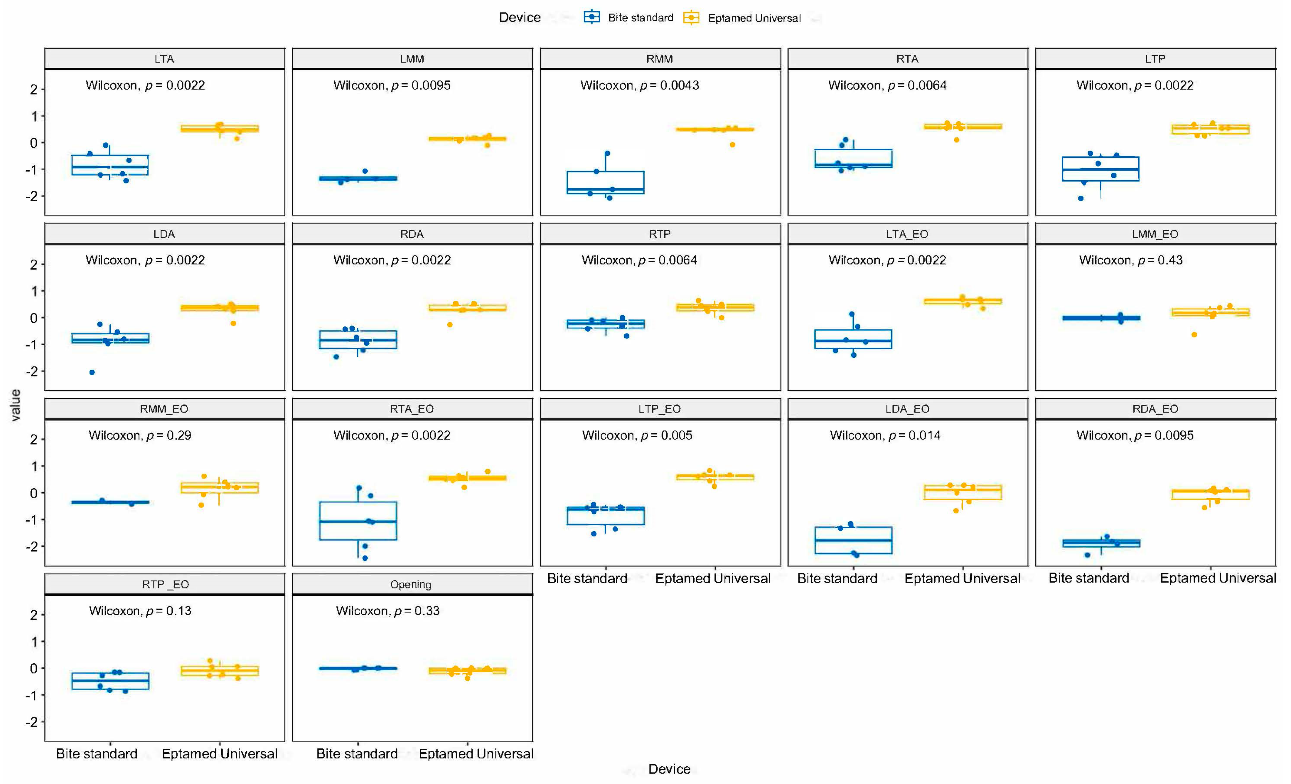
  • The elastodontic device (EQ UNIVERSAL, Eptamed srl Via Ravennate, 979, 47522 Cesena (FC) ITALY) is capable of causing, in patients with temporomandibular disorders, a reduction in muscle tone at rest (scan 9) of the examined muscles (masseter, anterior temporal, digastric and sternocleidomastoid muscles) compared to a standard bite sold in pharmacies that, instead, causes an increase in muscle tension.
  • The reduction of electromyographic activity with Eptamed is greater in the closed-eye condition than in the open-eye condition when scan 9 is recorded.
  • No statistically significant difference was assessed during the kinesiographic examination at the maximum opening of the mouth in the comparison between the two groups at T0 and T1.
  • No improvement in the electrical activity of the muscles was observed after 6 months of the use of the standard bite.
Following the results obtained from the above-mentioned study, the Eptamed device can be considered as a device to be administered to patients with TMD and orofacial pain before they start orthodontic therapy, as it is able to relax the stomatognathic muscles. These results are in line with the findings of Ortu et al. in which, comparing the electromyographic and kinesiographic examination of two elastodontic devices, it was seen that both ensured muscle relaxation, reducing the risk of TMD(s) development in treated subjects [22].
A few articles in the literature over the years called orthodontic treatment into question as a possible cause of TMD. Kononen et al. examined 166 children after orthodontic treatment, and 52% of them manifested signs and symptoms of TMDs [23]. Hirata et al. had shown that the development of dysfunction occurred equally in both orthodontically treated and untreated subjects, this means that orthodontic treatment does not always lead to the achievement of a muscular balance [24]. Also, Ricketts stated that clinical symptoms of joint derangement were noted as occlusions were changed, and he suggested that the various orthodontic forces provided during therapy may predispose patients to temporomandibular joint problems [25]. But, the most emblematic case is a 1987 Brimm vs. Malloy Michigan court case [26] in which an orthodontist was ordered to recover damages against a patient who had undergone mismanaged orthodontic treatment that caused her joint problems. Stohler and Zarb [27] state that orthodontic treatment of patients with TMD is considered radical and maximizes the risk of iatrogenic complications; that is, there is a significant risk of failure.
In this regard, guidelines based on the latest scientific evidence were drawn up, and an orthodontist must consider them in clinical practice [28]. In a nutshell, it is required of dentists to conduct a thorough examination and/or screening of TMD (presence of clicks, joint pain or mandibular deviations [29]) during orthodontic consultation and before starting orthodontic treatment both in patients who already have temporomandibular disorders and in healthy patients. However, it is necessary to stop any treatment if symptoms worsen (in patients diagnosed with TMD) or arise (in patients without TMD before treatment). In this way, the orthodontist can avoid both the worsening and the occurrence of TMD due to poorly managed orthodontic treatment.
In this circumstance, electromyographic and kinesiographic examination are essential tools that help the clinician assess whether a neuromuscular balance can be achieved following orthodontic treatment that does not compromise the health of the temporomandibular joint. According to Castroflorio et al., the electromyographic examination is considered an easily reproducible examination, so much so that the instrumentation used and a well-controlled sEMG protocol allow it to be used for longitudinal studies [30]. The presence of generalized muscle relaxation in all eight muscles analyzed by electromyographic examination is an indication of the effectiveness of the orthodontic treatment itself [22]. For this reason, the achievement of low muscle activity at rest must be a desirable outcome during treatment.
Kecik et al. [31] compared the dental and muscular changes that patients manifested following treatment of maxillary expansion with the quad-helix. In this case, in addition to clinical and cephalometric examinations, the need to contemplate electromyographic evaluations in the diagnostic setting was affirmed. The sEMG allowed appropriate diagnosis and prognosis to be formulated and also to monitor the functional impact of orthodontic therapies at various stages of treatment. In this way, the clinician can evaluate whether or not the orthodontic device is suitable in the presence of TMDs, thus avoiding worsening pain and dysfunction at the joint level.
To our knowledge, this is the first study to evaluate electromyographic and kinesiographic activity in subjects with TMD by comparing two different orthodontic devices, with the aim of assessing any muscle tension that these devices may cause. The use of the Eptamed device resulted in the achievement of greater muscle relaxation and a reduction in most of the electromyographic values considered. This did not occur in TMD patients wearing a bite sold in pharmacies. The improvement in electromyographic values in the case group is manifested mostly in the eyes-closed condition compared to the eyes-open condition. The change in electromyographic values according to visual input is in line with accumulated evidence indicating that TMD subjects show a dysregulation of the systems that control the response of the autonomic and somatomotor systems to sensory stimuli [32]. In a study by Monaco et al. [33], subjects with TMD in the open-eye condition had higher electromyographic values than healthy subjects in the same condition. These values increased by far even after standard spectacle wrapping. The lower improvement in values in the open-eye condition that occurred in our study, therefore, may be an intrinsic factor in the pathophysiology of temporomandibular disorders. TMDs are, in fact, classified as CSS and, as such, manifest a state of hyperexcitation and altered response to peripheral sensory stimuli, including those coming from the visual system [34].
However, the subjects who wore Eptamed at the opening of the eyes had electromyographic values worse than the closed eyes condition but better than the control group. This may suggest that elastodontic devices may, in this case, positively influence the responses of TMD patients to visual sensory inputs. On the other hand, the use of oral bites in orthodontic treatment in patients with TMD is controversial; several studies evaluated muscle electromyographic activity following the use of such devices, and the results are so variable that no firm conclusion can be made [35,36]. In fact, oral bites sold in pharmacies, by not requiring the cooperation of the dentist, lead the patient to a self-diagnosis that can be harmful. In fact, as was seen in this study, such devices may even worsen the patient’s clinical situation, so they need careful evaluation and use.
It is important to note that this is a pilot study, for which it inevitably has some limitations: it must be considered that patients were evaluated only for 6 months of orthodontic treatment; it would be advisable to extend the follow-up to at least 1 year. In addition, few people were selected, so the sample is too small. Future studies should, therefore, overcome these limitations by increasing the sample size and observation time of the sample.

5. Conclusions

Our pilot study suggests that the Eptamed device could be a viable orthodontic device for use in individuals with TMD as it provides a reduction in muscle tension, which individuals with TMD can present, compared to a standard bite. However, due to the limited number of patients examined, further research will be needed in the future.

Author Contributions

Conceptualization E.O., S.C. and A.M.; methodology D.P. and S.C.; validation A.M.; writing—original draft preparation S.D.N. and E.O.; writing—review and editing S.D.N., L.D.S. and E.O. All authors have read and agreed to the published version of the manuscript.

Funding

This research received no external funding.

Institutional Review Board Statement

This study was carried out in accordance with the fundamental principles of the Declaration of Helsinki and was approved on 17 May 2016 by the Internal Review Board of the University of L’Aquila (Number 16137/2016).

Informed Consent Statement

Informed consent was obtained from all participants involved in the study.

Data Availability Statement

All the experimental data to support the findings of this study are available by contacting the corresponding author upon request.

Conflicts of Interest

The authors declare no conflicts of interest.

References

  1. Ohrbach, R.; Bair, E.; Fillingim, R.B.; Gonzalez, Y.; Gordon, S.M.; Lim, P.F.; Ribeiro-Dasilva, M.; Diatchenko, L.; Dubner, R.; Greenspan, J.D.; et al. Clinical orofacial characteristics associated with risk of first-onset TMD: The OPPERA prospective cohort study. J. Pain. 2013, 14 (Suppl. S12), T33–T50. [Google Scholar] [CrossRef]
  2. Sergio Paduano, M.D.; Rosaria Bucci, D.D.; Roberto Rongo, D.D.; Silva, R.; Michelotti, A. Prevalence of temporomandibular disorders and oral parafunctions in adolescents from public schools in Southern Italy. CRANIO® 2018, 38, 370–375. [Google Scholar] [CrossRef]
  3. Dworkin, S.F.; Von Korff, M.R.; LeResche, L. Multiple pains and psychiatric disturbance: An epidemiologic investigation. Arch. Gen. Psychiat. 1990, 47, 239–244. [Google Scholar] [CrossRef]
  4. Manfredini, D.; Ahlberg, J.; Winocur, E.; Guarda-Nardini, L.; Lobbezoo, F. Correlation of RDC/TMD axis I diagnoses and axis II pain-related disability. A multicenter study. Clin. Oral Investig. 2011, 15, 749–756. [Google Scholar] [CrossRef]
  5. Reis, P.H.F.; Laxe, L.A.C.; Lacerda-Santos, R.; Münchow, E.A. Distribution of anxiety and depression among different subtypes of temporomandibular disorder: A systematic review and meta-analysis. J. Oral Rehabil. 2022, 49, 754–767. [Google Scholar] [CrossRef] [PubMed]
  6. Yap, A.U.; Chen, C.; Wong, H.C.; Yow, M.; Tan, E. Temporomandibular disorders in prospective orthodontic patients. Angle Orthod. 2021, 91, 377–383. [Google Scholar] [CrossRef] [PubMed]
  7. de Leeuw, R.; Klasser, G. (Eds.) Differential Diagnosis and Management of TMDs. Orofacial Pain: Guidelines, Assessment, Diagnosis, and Management, 6th ed.; Quintessence Publishing: Berlin, Germany, 2018; pp. 144–207. [Google Scholar]
  8. Andrews, L.F. Straightwire: The Concept and Appliance; LA Wells: San Diego, CA, USA, 1989. [Google Scholar]
  9. Masci, C.; Ciarrocchi, I.; Spadaro, A.; Necozione, S.; Marci, M.C.; Monaco, A. Does orthodontic treatment provide a real functional improvement? A case control study. BMC Oral Health 2013, 13, 57. [Google Scholar] [CrossRef] [PubMed]
  10. LeResche, L. Epidemiology of Temporomandibular disorders: Implication for investigation of etiologic factors. Crit. Rev. Oral Biol. Med. 1997, 8, 291–305. [Google Scholar] [CrossRef] [PubMed]
  11. Popa, C.; Solomon, S.M.; Rudnic, I.; Mârţu, I.; Luchian, I.; Mârţu, M.A.; Sava, N.; Mârţu, S. Evaluation of oclusal trauma as a risk factor in the ethiology of chronic periodontitis. Int. J. Med. Dent. 2018, 8, 83. [Google Scholar]
  12. Popa, C.G.; Luchian, I.; Ioanid, N.; Goriuc, A.; Martu, I.; Bosinceanu, D.; Martu, M.A.; Tirca, T.; Martu, S. ELISA Evaluation of RANKL Levels in Gingival Fluid in Patients with Periodontitis and Occlusal Trauma. Rev. Chim. 2018, 69, 1578–1580. [Google Scholar] [CrossRef]
  13. Al-Saleh, M.A.; Flores-Mir, C.; Thie, N.M. Electromyography in diagnosing temporomandibular disorders. J. Am. Dent. Assoc. 2012, 143, 351–362. [Google Scholar] [CrossRef] [PubMed]
  14. Santana-Mora, U.; López-Ratón, M.; Mora, M.J.; Cadarso-Suárez, C.; López-Cedrún, J.; Santana-Penín, U. Surface raw electromyography has a moderate discriminatory capacity for differentiating between healthy individuals and those with TMD: A diagnostic study. J. Electromyogr. Kinesiol. 2014, 24, 332–340. [Google Scholar] [CrossRef]
  15. Giannini, L.; Maspero, C.; Batia, C.; Galbiati, G. Valutazione elettromiografica ed elettrognatografica del trattamento ortodontico-chirurgico. Mondo Ortod. 2011, 36, 12–28. [Google Scholar] [CrossRef]
  16. Dworkin, S.F.; Le Resche, L. Research diagnostic criteria for temporomandibular disorders: Review, criteria, examinations and specifications, critique. J. Craniomandib. Disord. 1992, 6, 302–355. [Google Scholar]
  17. Brook, P.H.; Shaw, W.C. The development of an index of orthodontic treatment priority. Eur. J. Orthod. 1989, 11, 309–320. [Google Scholar] [CrossRef]
  18. Konchak, P.A.; Thomas, N.R.; Lanigan, D.T.; Devon, R.M. Freeway space measurement using mandibular kinesiograph and EMG before and after TENS. Angle Orthod. 1988, 58, 343–350. [Google Scholar] [PubMed]
  19. Ortu, E.; Pietropaoli, D.; Cova, S.; Marci, M.C.; Monaco, A. Efficacy of elastodontic devices in overjet and overbite reduction assessed by computer-aid evaluation. BMC Oral Health 2021, 21, 269. [Google Scholar] [CrossRef]
  20. Castroflorio, T.; Farina, D.; Bottin, A.; Piancino, M.G.; Bracco, P.; Merletti, R. Surface EMG of jaw elevator muscles: Effect of electrode location and inter-electrode distance. J. Oral Rehabil. 2005, 32, 411–417. [Google Scholar] [CrossRef]
  21. Castro, H.A.; Resende, L.A.; Berzin, F.; Konig, B. Electromyographic analysis of the superior belly of the omohyoid muscle and anterior belly of the digastric muscle in tongue and head movements. J. Electromyogr. Kinesiol. 1999, 9, 229–232. [Google Scholar] [CrossRef]
  22. Ortu, E.; Barrucci, G.; Aprile, G.; Guerrini, L.; Pietropaoli, D.; Monaco, A. Electromyographic evaluation during orthodontic therapy: Comparison of two elastodontic devices. J. Biol. Regul. Homeost. Agents 2020, 34, 1935–1939. [Google Scholar] [CrossRef]
  23. Kononen, M.; Nystrom, M.; Kleemola-Kujola, E.; Kataja, M.; Evalahti, M.; Pekka, L.; Peck, L. Signs and symptoms of craniomandibular disorders in a series of Finnish children. Acta Odontol. Scand. 1987, 45, 109–114. [Google Scholar] [CrossRef] [PubMed]
  24. Hirata, R.H.; Heft, M.W.; Hernandez, B.; King, G.J. Longitudinal study of signs of temporomandibular disorders (TMD) in orthodontically treated and nontreated groups. Am. J. Orthod. Dentofacial. Orthop. 1992, 101, 35–40. [Google Scholar] [CrossRef] [PubMed]
  25. Ricketts, R.M. Clinical implications of the temporomandibular joint. Am. J. Orthod. 1966, 52, 416–439. [Google Scholar] [CrossRef] [PubMed]
  26. Pollack, B. Cases of note: Michigan jury awards $850,000 in ortho case: A tempest in a teapot. J. Mich. Dent. Assoc. 1988, 70, 540–542. [Google Scholar] [PubMed]
  27. Stohler, C.S.; Zarb, G.A. On the management of temporomandibular disorders: A plea for a low-tech, high-prudence therapeutic approach. J. Orofac. Pain 1999, 13, 255–261. [Google Scholar] [PubMed]
  28. Sanjivan, K.; Donald, J.; Rinchuse, C.S.; Greene, L.; Johnston, E. Temporomandibular disorders and orthodontics: What have we learned from 1992–2022? Am. J. Orthod. Dentofac. Orthop. 2022, 161, 769–774. [Google Scholar] [CrossRef]
  29. Machen, D.E. Legal aspects of orthodontic practice: Risk management concepts. Excellent diagnostic informed consent practice and record keeping make a difference. Am. J. Orthod. Dentofacial. Orthop. 1990, 98, 381–382. [Google Scholar] [CrossRef]
  30. Castroflorio, T.; Titolo, C.; Deregibus, A.; Debernardi, C.; Bracco, P. The Orthodontic Treatment of TMD Patients: EMG Effects of a Functional Appliance. CRANIO® 2007, 25, 206–212. [Google Scholar] [CrossRef]
  31. Kecik, D.; Kocadereli, I.; Saatci, I. Evaluation of the treatment changes of functional posterior crossbite in the mixed dentition. Am. J. Orthod. Dentofac. Orthop. 2007, 131, 202–215. [Google Scholar] [CrossRef]
  32. Chen, H.; Nackley, A.; Miller, V.; Diatchenko, L.; Maixner, W. Multisystem dysregulation in painful temporomandibular disorders. J. Pain 2013, 14, 983–996. [Google Scholar] [CrossRef]
  33. Monaco, A.; Ortu, E.; Giannoni, M.; D’Andrea, P.; Cattaneo, R.; Mummolo, A.; Pietropaoli, D. Standard Correction of Vision Worsens EMG Activity of Pericranial Muscles in Chronic TMD Subjects. Pain Res Manag. 2020, 2020, 3932476. [Google Scholar] [CrossRef] [PubMed]
  34. Kindler, L.L.; Bennett, R.M.; Jones, K.D. Central sensitivity syndromes: Mounting pathophysiologic evidence to link fibromyalgia with other common chronic pain disorders. Pain Manag. Nurs. 2011, 12, 15–24. [Google Scholar] [CrossRef] [PubMed]
  35. Tartaglia, G.M.; Lodetti, G.; Paiva, G.; De Felicio, C.M.; Sforza, C. Surface electromyograpic assessment of patients with long lasting temporomandibular joint disorders pain. J. Electromyogr. Kinesiol. 2011, 21, 659–664. [Google Scholar] [CrossRef]
  36. Adibi, S.S.; Ogbureke, E.I.; Minavi, B.B.; Ogbureke, K.U. Why use oral splints for temporomandibular disorders(TMDs)? Tex. Dent. J. 2014, 131, 450–455. [Google Scholar] [PubMed]
Figure 1. Non−parametric Wilcoxon signed-rank test performed following electromyographic examination in the open and closed eye condition of all muscles examined in the Eptamed group (in yellow) and the standard bite group (in blue) after 6 months of treatment.
Applsci 14 01651 g001
Figure 2. Non−parametric Wilcoxon signed-rank test performed following kinesiographic examinations in the condition of maximum mouth opening (scan 1) in the two groups.
Applsci 14 01651 g002
Table 1. z−score transformation of each variable calculated as the difference between the observed value and the mean of the sample, divided by the standard deviation of the sample, described with a heatmap.
IdDeviceLtaLmmRmmRtaLtpLdaRdaRtpLta_eoLmm_eoRmm_eoRta_eoLtp_eoLda_eoRda_eoRtp_eoOpening
1BITE−1.21−1.07−1.75−0.89−0.40−0.54−0.95−0.42−0.91−4.46−4.85−2.44−0.53−2.35−3.71−0.860.00
2BITE−1.17−4.00−3.30−1.06−0.46−2.04−1.46−0.13−1.23−4.50−3.30−1.06−0.44−4.11−3.09−0.270.00
3BITE−0.10−1.50−0.400.11−2.09−0.85−0.74−0.690.140.11−0.290.18−1.54−1.17−1.64−0.160.00
4BITE−1.42−1.36−2.08−0.10−0.79−0.25−0.430.00−1.40−5.00−4.83−2.00−0.56−1.33−2.33−0.67−0.07
5BITE−0.67−1.38−1.08−0.76−1.23−0.80−0.39−0.32−0.33−4.91−3.55−1.11−0.71−2.26−1.92−0.15−0.07
6BITE−0.41−3.00−1.91−0.94−1.50−0.97−1.22−0.09−0.83−0.15−0.43−0.11−1.36−3.50−1.83−0.820.00
1EQ. U.0.690.060.550.710.740.250.270.640.780.380.620.800.67−0.33−0.33−0.28−0.21
2EQ. U.0.14−0.10−0.080.110.26−0.21−0.270.000.34−0.64−0.470.200.240.000.03−0.24−0.17
3EQ. U.0.440.190.470.740.680.460.520.240.680.04−0.070.640.83−0.67−0.56−0.390.00
4EQ. U.0.660.150.550.540.530.420.530.800.700.450.240.580.670.290.070.070.00
5EQ. U.0.400.170.460.590.530.510.290.330.630.180.410.500.610.220.120.280.00
6EQ. U.0.550.270.480.520.250.320.300.440.490.190.200.460.440.290.170.05−0.38
Disclaimer/Publisher’s Note: The statements, opinions and data contained in all publications are solely those of the individual author(s) and contributor(s) and not of MDPI and/or the editor(s). MDPI and/or the editor(s) disclaim responsibility for any injury to people or property resulting from any ideas, methods, instructions or products referred to in the content.

