1. Introduction
Epilepsy is a neurological disorder characterized by recurrent episodes of signs and symptoms caused by abnormal brain synchronization and fast neuronal activities. Excessive electrical activity in brain cells manifests as epileptic seizures, one of the major neurological chronic illnesses affecting 50 million people globally [
1]. Epileptic seizures can lead to serious cognitive, neurological, and physiological implications, including loss of consciousness and even death if thorough diagnosis and monitoring are not conducted [
2,
3]. Neurologists employ clinical EEG (electroencephalogram) to detect epileptic seizures. The EEG signals are captured by placing multiple electrodes on different areas of the patient’s scalp. The biomedical signals recorded using EEG are difficult to analyze with the naked eye. Continuously analyzing EEG signals is a significant challenge since it always requires the presence of a neurophysiologist in case long-term monitoring is required [
4]. Furthermore, accurate identification of seizures is time-consuming and requires expert knowledge. To address these challenges, various machine learning methods are used to automate the analysis of EEG data. The machine learning model is trained using the characterizing properties extracted from the EEG signal for automatic detection/classification of epileptic seizures.
The aim of this study is to employ a random neural network (RNN)-based approach to develop a novel EEG classification scheme to detect epileptic seizures. To achieve this, data are acquired from two publicly available datasets that have been extensively used in the literature for training and testing machine learning models. The datasets contain annotated single-channel and multi-channel EEG recordings acquired from patients with epilepsy. Below are the main contributions of this research:
A novel AI-based machine learning model is presented for the classification of epileptic seizures using RNN.
The proposed model is developed by running multiple RNN-based experiments using different parameters such as training and testing ratio; learning rate (LR); and frequency analysis techniques, i.e., discrete wavelet transform (DWT) and fast Fourier transform (FFT).
A comparison of the novel model is presented with traditional classification algorithms such as ANN and SVM.
A critical evaluation of the obtained results concludes that the proposed RNN-based scheme is the most efficient in terms of accuracy, sensitivity, precision, and specificity for the classification of epileptic seizures.
2. Related Work
The application of machine learning-based techniques for the classification and identification of various medical conditions has garnered significant interest among researchers [
5,
6,
7]. One specific area that has gained substantial attention is the use of these techniques for the identification and classification of epileptic seizures.
Table 1 provides a summary of some of the machine learning methods used in the detection and classification of epileptic seizures, which include support vector machine (SVM), convolutional neural networks (CNN), and extreme learning machines (ELM). The authors in [
8] utilized deep CNN to develop a model to detect mental fatigue from electroencephalography (EEG) data. The proposed model was trained on a dataset containing EEG data labeled as fatigued or rested from 20 participants. The model achieved an accuracy of 97% to classify the mental state of the participants.
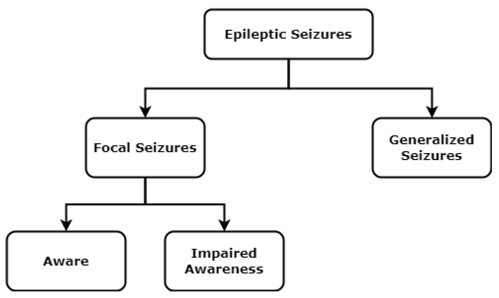
Epileptic seizures are categorized by the International League Against Epilepsy (ILEA) into two main types: focal seizures and generalized seizures. A focal seizure, also known as partial seizure, affects only a portion of the cerebral hemisphere [
9]. The level of awareness further separates focal seizures into two categories: aware (simple-partial) and impaired awareness (complex-partial). A seizure in which consciousness is intact is categorized as a focal-aware seizure. A seizure event where a person behaves abnormally, like chewing or mumbling, and is not aware of the surroundings is classified as a focal seizure with impaired awareness. The types of epileptic seizures as defined by ILAE [
10] are shown in
Figure 1.
Esbroeck et al. [
11] used a variable window approach for the segmentation of EEG data by placing boundaries where there is a sharp change in the energy of the signal. The authors propose a multi-task approach for seizure detection to address the challenge of inter-patient and intra-patient variability in the characteristic patterns found in seizure signals. The authors achieved a reduction in the false positive rate (FPR) of more than 10% in 15 cases with the proposed method. In [
12], a covariance matrix was employed to analyze EEG recordings of epileptic patients and to reduce the dimensionality of the signal before feature extraction. An accuracy of more than 99% was achieved by employing non-parametric tests to find the set with the most distinctive characteristics and subsequently fed into the adaptive boosting least square-support vector machines (AB-LS-SVM) for classification. Vicnesh et al. identified non-linear characteristics from EEG data and used the extracted features as input into a decision tree (DT) for epilepsy classification [
13]. Following the feature extraction phase, the data samples were divided into two groups for training and testing. Finally, the performance was evaluated using parameters such as sensitivity, specificity, F1-score, and accuracy.
Gill et al. [
14] proposed a model for epilepsy detection using a hybrid feature set that integrates temporal, spectral, and time–spectral features. The authors classified epilepsy data using Gaussian mixture models. The proposed approach achieved an accuracy of 86.26%. Ingolfsson et al. [
15] demonstrated that 100% sensitivity with zero false positives can be achieved by using four different classification algorithms with DWT for signal decomposition in the preprocessing phase. The classification models were trained and tested using the data from the same subjects. Donos et al. proposed a seizure detection model based on random forest that can be employed in an embedded closed-loop device. The model detected seizures with 93.84% sensitivity [
16]. Satapathy et al. [
17] employed SVM and other neural networks for classifying epileptic events using the publicly accessible BONN dataset, including multi-layer perceptrons, long short-term memory networks (LSTM), and probabilistic neural networks. The efficiency of each classification model was evaluated using a majority vote technique. The results demonstrated that SVM outperformed the other machine learning models in terms of accuracy. Zeng et al. [
18] utilized a five-level decomposition of EEG signal using empirical wavelet transform along with kurtosis-based channel selection to identify the electrodes containing the most relevant information to improve the performance of seizure detection. The proposed algorithm achieved mean sensitivity and specificity of 99.77% and 99.88%, respectively. Acharya et al. proposed a deep CNN-based model with 13 layers to analyze the EEG signal. The authors used raw EEG signals instead of feature extraction and selection for the classification of pre-ictal, normal, and seizure classes. The model achieved an accuracy of 88.67%, as reported in [
19]. Choubey et al. [
20] performed a classification of EEG data using ANN and K-nearest neighbor after extracting statistical features such as Higuchi fractal dimension and expected activity measurement from the signal. The findings demonstrate that ANN and k-NN both attained accuracy rates of 94% and 98%, respectively. Raghu et al. [
21] used multiple ML techniques such as random forest, SVM, KNN, and Adaboost to perform localization of epileptic seizures by classifying EEG data containing EEG signals of focal epilepsy using various statistical features. The results demonstrate that SVM has achieved the highest classification accuracy of 96.1%. Chen et al. [
22] employed DWT to develop a method to find the optimal DWT parameters by breaking down the EEG signal into seven commonly used mother wavelets to detect epileptic seizures with improved accuracy and a lower cost of computation. Various DWT mother wavelets such Bior6.8, coif3, db10, and haar were used to decompose the data. The highest performance in terms of accuracy of 92.30% was achieved using a coif3 mother wavelet. Zanetti et al. [
23] proposed a seizure detection technique utilizing only two bipolar channels from the CHB-MIT dataset. DWT was used to decompose the EEG signal prior to feature extraction. The proposed method detected seizures with sensitivity and specificity of 96.6% and 92.5%, respectively.
Table 1.
Machine Learning methods used for epilepsy detection sub-bands.
Table 1.
Machine Learning methods used for epilepsy detection sub-bands.
| Reference | Features | Classification Model | Dataset | Result |
|---|
| Gill et al. [14] | Hybrid feature set | Gaussian mixture models | CHB-MIT | Accuracy: 86.26% Sensitivity: 87.58% Specificity: 86.83% |
| Donos et al. [16] | Time and frequency | Random forest | EPILEPSIAE | Sensitivity: 93.84% |
| Satapathy et al. [17] | DWT | SVM and other neural networks | BONN | Accuracy: 99.1% |
| Acharya et al. [24] | | CNN | BONN | Accuracy: 88.7%, Specificity: 90%, Sensitivity: 95%. |
| Choubey et al. [20] | Higuchi fractal dimension HFD, EAM | ANN+k-NN | BONN | ANN 94%, k-NN 98% |
| Raghu et al. [21] | Time–frequency, statistical features | Random forest, SVM, KNN, and Adaboost | Bern-Barcelona | Sensitivity: 97.6% Specificity: 94.4% Accuracy: 96.1% |
| Chen et al. [22] | coif3 mother wavelet + 7 statistical features | SVM | CHB-MIT | 92.30% |
| Saminu et al. [25] | Wavelet Synchrosqueezing Transform | 2D Deep-CNN | BONN | Accruacy: 99.70% |
| | Hybrid features | SVM | BONN | Accuracy: 99.30% |
| | Hybrid features | 1D-CNN | BONN | Accuracy: 99.40% |
The identification and localization of temporal lobe epilepsy have been another topic of interest in machine learning–based epilepsy diagnosis [
26]. Seizure localization refers to the process of identifying the brain region where a seizure originates. Localized seizures that occur in either the right or left hemisphere of the brain are typically curable with surgery. Certain types of seizures may be treated with anti-epileptic medicines; however, in some cases, people with partial seizures may require surgery [
27]. Therefore, finding the location of a seizure is crucial for the treatment of epilepsy [
28,
29]. Siddiqui et al. used two algorithms based on decision forest i.e., SysFor and Forst CERN, to identify the location most affected by seizure [
30,
31,
32]. Saminu et al. [
25] proposed three different methods to perform binary classification of epilepsy data containing focal and non-focal signals using the BONN dataset to detect epileptogenic zones. The proposed method that combines deep-CNN with wavelet synchrosqueezing transform for feature extraction demonstrated the highest accuracy of 99.7% as compared with hybrid-SVM and 1D-CNN, which achieved accuracies of 99.3% and 99.4% respectively.
In this research, we studied EEG analysis and machine learning techniques for epilepsy detection. It is evident from the literature that there is increasing interest among researchers in this topic and that significant progress is being made in epilepsy detection. However, there are still significant challenges that, if addressed, will help advance the research further. Imbalanced data are a common issue with epilepsy data. If not properly addressed, the results may be inaccurate. Therefore, more work needs to be carried out on accurate epilepsy detection considering imbalanced datasets where the normal class is hours long and epileptic events are a few seconds/minutes. One of the future avenues for research in epilepsy detection [
33,
34] is seizure localization. It can assist clinicians in identifying the specific region of the brain that is impacted by seizures, which is crucial in the treatment of some types of epilepsy. Raw EEG data usually contain artifacts and noise and may not provide the necessary patterns or signatures in the signal that are required to effectively classify seizures. Therefore, it is imperative to select the most suitable statistical features for efficient detection and dimensionality reduction [
35,
36]. This research proposes a novel RNN-based epileptic seizure detection approach employing two commonly used datasets (CHB-MIT and BONN) that contain multi-channel and single-channel EEG data. The RNN-based epilepsy detection model has been optimized in multiple scenarios with different parameters to find the optimal configuration for the efficient detection of seizures. The proposed RNN is fundamentally different from the memory-based algorithms such as recurrent neural network architecture. Therefore, the RNN does not possess memory or temporal context, meaning that it does not consider or retain any information from previous inputs.
3. Epileptic Seizure Detection
An essential part of the diagnosis and classification of epilepsy is the analysis of EEG signals. EEG patterns reflect the electrophysiological condition of the brain at any given time. This section provides an overview of current epilepsy detection techniques based on several methods for the classification and analysis of EEG data. The most common steps followed by researchers for epilepsy detection from EEG signals are given below:
Data acquisition;
Pre-processing;
Extracting features;
Classification;
Evaluation.
EEG data are most commonly employed for the diagnosis and detection of epileptic seizures. The data are acquired by placing multiple electrodes on the scalp that record voltage fluctuations in the brain. Researchers usually use EEG data that are publicly available online.
After data acquisition, the next step is the extraction of features which involves obtaining relevant statistical attributes from the data. These are necessary for the classification of EEG signals since they contain distinct signatures and characteristics that help segregate epileptic and non-epileptic events [
37]. The accuracy of epilepsy detection depends upon the quality of features extracted. Therefore, improving the quality of feature extraction is critical. According to research, the quality of features can be improved by employing various dimensionality reduction techniques.
3.1. Random Neural Network (RNN)
RNN is the mathematical model of an artificial neural network, which was originally developed by Erol Gelenbe in his seminal work in 1989 [
38]. RNN has been used extensively in recent literature for data classification in a variety of fields such as 5G network simulation [
39], indoor localization based on LoRaWAN, and fall activity detection [
40,
41]. In RNN, each neuron transitions between excitatory or inhibitory states based on the potential of the received signal, which can be either positive or negative. In RNN layers, the neurons probabilistically send and receive excitation and inhibition signals. After receiving a + 1 signal, the neuron will enter an excitatory state. Similarly, a neuron will enter an inhibitory state after receiving a − 1 signal. A vector containing non-negative integers
represents the potential state of a neuron
m at time
t. A neuron enters an excited state when
, and it is considered idle when
. In the excited state, a neuron sends a potential spike signal at a rate of
, which reduces its excitation potential by 1. The mathematical expression of the activation function of RNN
is as follows.
where
and
are excitatory and inhibitory inputs, respectively:
and
The firing rates of neurons m and n are denoted as and , respectively. The probabilities of excitatory or inhibitory spikes sent from neuron n to neuron m are represented as or .
The activation function
for neuron
m can be expressed as follows:
In Equation (
5), the sum of probabilities of all the signals leaving the network must be equal to 1, where the probability of each signal is denoted by
, and
N refers to the sum of all neurons in the network. The rate at which an excited neuron sends positive or negative signals is as follows:
The equation presented by Gelenbe in [
38] derives the firing rate of neuron
m from Equations (
5)–(
7). The resulting expression is as follows:
According to Gelenbe in [
38], the weights used in RNN, i.e.,
and
follow the same conventions as those used in traditional ANNs and may be trained using conventional optimization methods such as gradient descent.
3.2. Proposed Epileptic Seizure Detection Scheme Based on Random Neural Network
The model proposed in this research involves the classification of EEG data utilizing RNN for epileptic seizure detection using two different datasets.
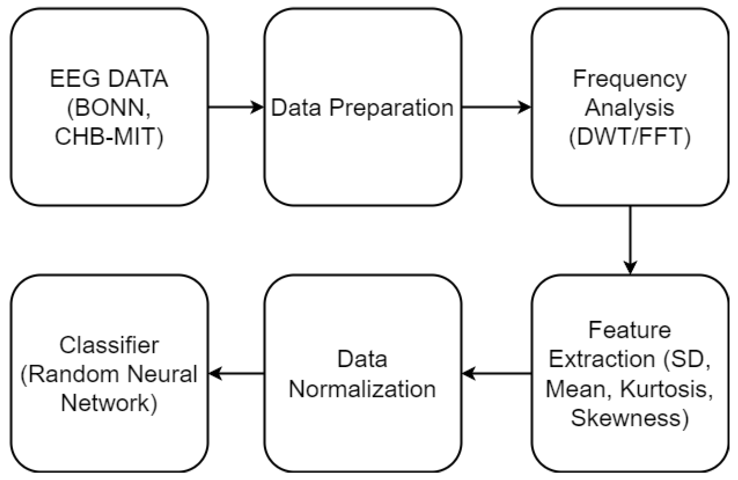
A flow diagram of the proposed scheme is illustrated in
Figure 2. The steps involved in the proposed methodology are outlined as follows:
- 1.
Data preparation;
- 2.
Pre-processing;
- 3.
Feature extraction;
- 4.
Data normalization;
- 5.
Classification using RNN.
In the first step, the data are prepared for use as input in our experiments. The CHB-MIT data have been recorded with varying numbers of EEG channels in different cases; however, to standardize the data, we have selected 23 common EEG channels as shown in
Table 2. Another channel has been added to the data and indicates the ictal and pre-ictal segments in the data. The data have been balanced to have an equal number of data samples in both classes.
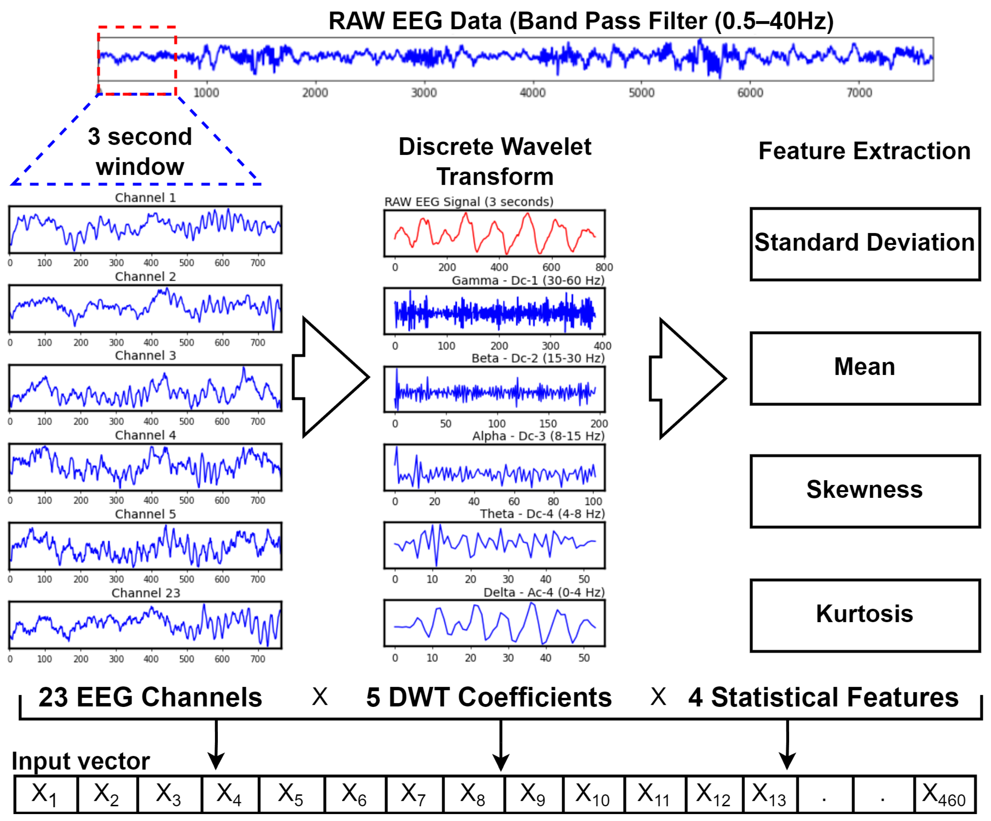
In the pre-processing stage, as illustrated in
Figure 3, a band-pass filter of 0.5 Hz to 40 Hz is applied to the EEG data to remove the high-frequency artifacts from the signal [
42]. The data have been divided into epochs of 3 s each with no overlap using MNE-Tools in Python [
43]. Each epoch is decomposed using four levels of decomposition of DWT utilizing Daubechie’s mother wavelet of the fourth order (DB4) at all 23 channels of the EEG signal. The DWT-based decomposition partitions all channels in each epoch into five frequency sub-bands: Delta Ac-4 (0.0–4.0 Hz), Theta Dc-4 (4.0–8.0 Hz), Alpha Dc-3 (8.0–15.0 Hz), Beta Dc-2 (15.0–30.0 Hz), and Gamma Dc-1 (30.0–60.0 Hz). As compared with our previous work [
44], where we used raw data for feature extraction, in the current research, we utilized DWT for signal decomposition before the feature extraction phase. DWT-based decomposition of the EEG signals provides enhanced feature representation and results in a more discriminative feature set.
In the feature extraction phase, four statistical features have been extracted from each frequency sub-band in each epoch. The statistical features obtained are standard deviation, mean, kurtosis, and skewness. These features have been used in the literature for training machine learning (ML) models for the diagnosis of various neurological disorders including Alzheimer’s disease [
45] and classification of epileptic seizures [
46]. The result is a feature vector of size 23 (EEG channels) × 5 (sub-bands) × 4 (statistical features). Therefore, the corresponding feature vector (size 460 × 1) for each EEG epoch is extracted and used as an input to train the RNN-based classification model.
After feature extraction, the resulting data contain a total of 2730 epochs. Each epoch is labeled as either 1 or 0 for “seizure” and “no seizure” data, respectively. The method used for classification involves an RNN architecture, comprising an input layer, a hidden layer, and an output layer. The first layer is set at 460 neurons due to the size of the input vector generated at the feature extraction phase. The output layer has only one neuron as the model is performing binary classification between ictal and inter-ictal data. The hidden layer has been tuned to have 20 neurons using the trial and error method. The RNN-based model has been trained and tested multiple times using different sets of training and testing ratios and LR, as illustrated in
Table 3 and
Table 4.
The RNN-based epilepsy classification model has been trained and tested in five different scenarios. In scenario 1, RNN-based epilepsy classification has been performed using the CHB-MIT dataset. Each frequency sub-band extracted from the raw data using DWT has been used separately to train the RNN-based epilepsy detection model. The number of neurons in the input, hidden, and output layers in all five experiments is 92, 10, and 1, respectively. The results from scenario 1 indicate which frequency sub-band contains the most discriminating features for efficient detection of epileptic seizures. In scenarios 2 and 3, as stated in
Table 3, the BONN dataset has been used for classification using DWT and FFT, respectively. In scenario 2, the RNN-based classification model has 460 input neurons, whereas in scenario 3, the input neurons are 92. In both instances, the number of neurons in the hidden and output layers is 20 and 1, respectively. Similarly, in scenarios 4 and 5, as illustrated in
Table 3, the CHB-MIT dataset was used for classification using DWT and FFT in pre-processing step. The number of input neurons in scenarios 4 and 5 is 460 and 92, respectively. The number of hidden neurons is 20, with one output neuron.
4. Experimental Analysis
This section describes the experimental setup for evaluating the proposed RNN-based epilepsy classification model. Two publicly accessible datasets, the BONN EEG dataset and the CHB-MIT EEG database, are utilized to assess the model’s performance. The BONN dataset contains single-channel intracranial EEG signals, while the CHB-MIT dataset consists of multi-channel surface EEG recordings. Different scenarios are considered in the experimental setup, involving variations in parameters such as training and testing ratios, learning rate, and frequency analysis methods. The proposed RNN-based model is trained and tested on both datasets using cross-validation techniques to validate the efficacy of the proposed model in classifying EEG signals and detecting epileptic seizures.
The experiments were carried out on a computer running Microsoft Windows 11 operating system, equipped with an AMD Ryzen 7 3700X 8-Core Processor, and 48 GB of RAM. The implementation of data preparation, frequency analysis, and feature extraction was performed in Python. On the other hand, the data normalization and classification using RNN were conducted using MATLAB, a widely used tool for data analysis and modeling.
4.1. Datasets
In this study, two publicly available datasets are utilized to evaluate the performance of the proposed RNN-based epilepsy classification model. The primary difference between the two datasets is the electrode type which has a direct impact on the spatial coverage and quality of the recorded EEG signals. The first one is BONN dataset, which is a smaller dataset containing single-channel intracranial EEG signals. while the second one, CHB-MIT is a larger dataset consisting of 23 channels of surface EEG recordings. Since the spatial coverage in intracranial EEG is more localized and focused on specific brain regions, it provides a higher spatial resolution as compared to scalp EEG. Therefore, the BONN dataset offers enhanced data quality as compared with CHB-MIT in terms of spatial resolution as intracranial EEG electrodes provide more direct measurements of brain activity allowing for more accurate and detailed capture of neural signals [
47]. Our proposed method was first tested for classification on the smaller BONN dataset to adjust the model parameters. Following this, the proposed method was used to classify the larger CHB-MIT dataset.
4.1.1. BONN EEG Dataset
One of the datasets used in this research is the BONN dataset, which contains single-channel intracranial EEG data from epilepsy patients. The dataset is curated at the University of Bonn by the Department of Epileptology in Germany [
48,
49]. There are five subsets in the data, with each subset consisting of 100 segments of a single-channel EEG signal. Each data segment consists of 23.6 s of EEG signals collected at a sampling rate of 173.61 Hz. The total number of sample points in each segment is 173.61 × 23.6 = ∼4097. The subsets are labeled as Z, O, N, F, and S. The data are collected from five individuals in good health with their eyes in the open and closed states using the international 10/20 electrode system. An intracranial electrode placement system is used to collect subsets N, F, and S from five patients diagnosed with epilepsy. Subset S is recorded during seizures, while subsets labeled N and F are collected during periods without any seizure events.
4.1.2. CHB-MIT EEG Dataset
The CHB-MIT dataset is an openly accessible database [
50] containing EEG recordings of epilepsy patients that were recorded using surface EEG electrodes at Children’s Hospital Boston (CHB-MIT) and provided by the Massachusetts Institute of Technology [
51,
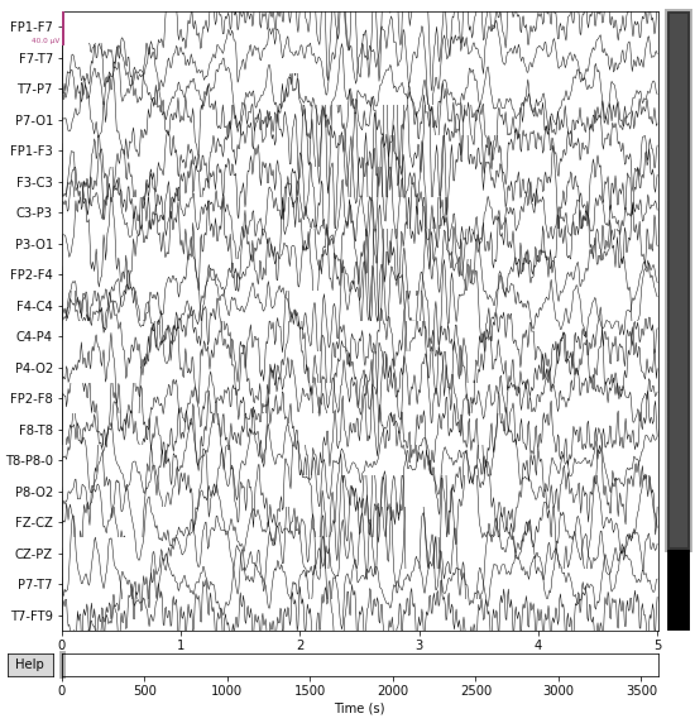
52]. The dataset contains 23 cases collected from 22 pediatric patients suffering from intractable seizures, of whom 18 are female and 5 are male, with ages ranging from 1.5 to 22 years old. A total of 686 EEG data records are collected, of which 198 contain seizure events. The dataset has been recorded with a sampling frequency of 256 Hz and contains 23 or more EEG channels.
Figure 4 shows a snapshot of 5 s of raw data in 23 EEG channels in the CHB-MIT dataset.
4.2. Experimental Setup
This study presents a novel epileptic seizure classification model that utilizes RNN and is tested on both single-channel and multi-channel datasets (BONN and CHB-MIT), which are publicly accessible online. The BONN dataset contains single-channel EEG data while CHB-MIT consists of 23 EEG channels. The RNN-based model was trained and tested in five scenarios with different sets of parameters [
53] such as learning rate, training, testing ratios, and frequency analysis methods, i.e., DWT and FFT as illustrated in Algorithm 1.
| Algorithm 1: RNN-based epileptic seizure classification. |
![Applsci 14 00599 i001]() |
A commonly used method for improving the performance and evaluating the accuracy of machine learning classifiers is known as k-fold cross-validation. This method divides the dataset into k subsets, with each subset serving as validation data for one model and the remaining subsets serving as training data. This method is repeated k times, with a different validation subset for each model. In this study, for instance, a 10-fold cross-validation is used, resulting in the dataset being divided into 10 subsets. It means that 10 unique RNN models have been trained using the following procedure: model 1 used the first subset for validation and trained on the subsequent nine subsets. Similarly, model 2 reserved the second subset for validation and trained on subsets 1, 3–9. This method was used for all 10 models. Finally, the model with the highest validation accuracy, indicating stronger generalization capabilities, was chosen for further testing.
In scenarios 2 and 3, there are a total of 320 simulations each in which the RNN-based epilepsy detection model is trained and tested on the BONN dataset using DWT and FFT. Similarly, the same two scenarios are replicated in scenarios 4 and 5 using the CHB-MIT dataset, as illustrated in
Table 3.
In scenario 1, the RNN-based classification of EEG data is performed five frequency sub-bands (Delta Ac-4 0–4.0 Hz, Theta Dc-4 4.0–8.0 Hz, Alpha Dc-3 8.0–15.0 Hz, Beta Dc-2 15.0–30.0 Hz, and Gamma Dc-1 30.0–60.0 Hz) extracted from the CHB-MIT dataset using DWT, as described in
Table 4.
4.2.1. Data Pre-Processing
DWT is used by researchers extensively in the fields of signal and image processing for the decomposition of a signal into multiple frequency bands. DWT provides multi-resolution analysis and time–frequency localization of the signal [
33,
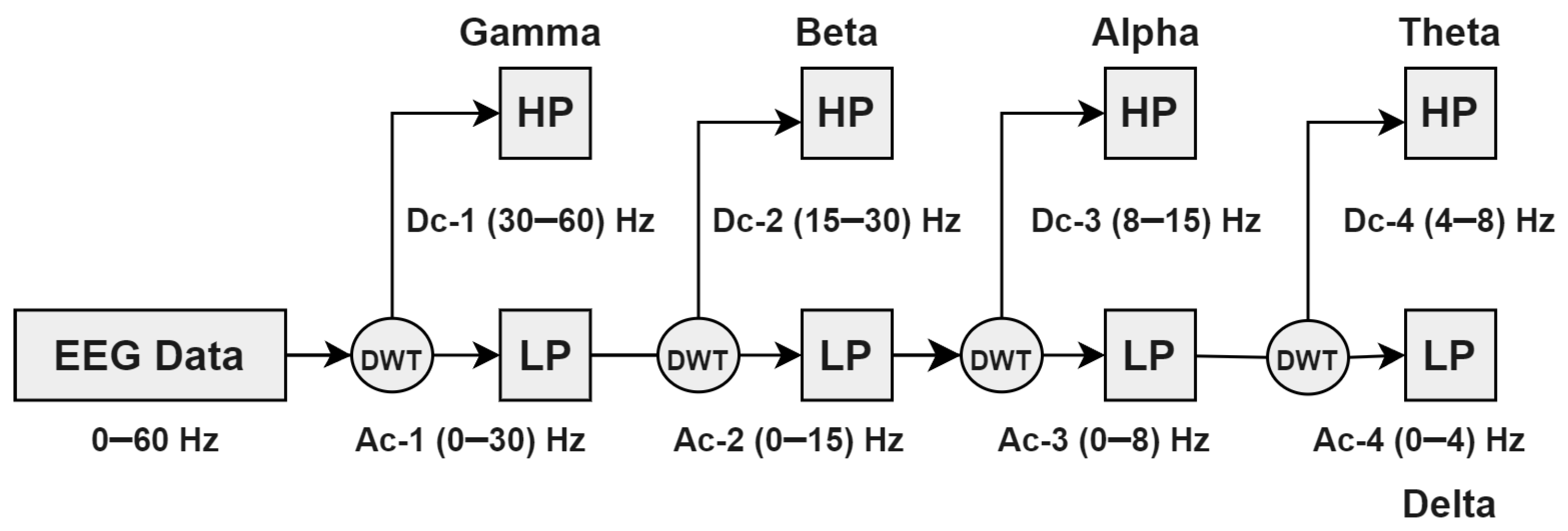
54]. A series of high-pass and low-pass filters are applied to a time-series signal such as EEG. The signal is divided into varying sizes of time windows, and the filter is applied to each window to extract approximation coefficients (ACs) and detail coefficients (DCs). The ACs can be further divided iteratively using the same process to obtain finer frequency bands. The high-frequency and low-frequency components of the signal are obtained by stretching or compressing the mother wavelet [
55]. In the first iteration of DWT, the EEG signal is simultaneously filtered using low-pass (LP) and high-pass (HP) filters to extract approximation and detail coefficients. The outputs of LP and HP are denoted in
Figure 5 as Ac-1 and Dc-1, respectively. The same process is repeated at each step, and the approximation coefficient is further divided into sub-bands.
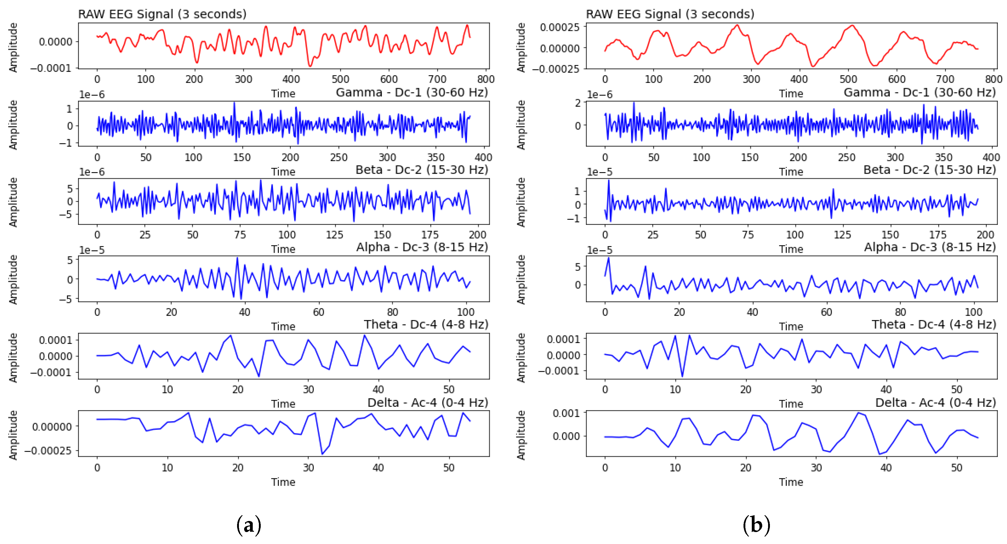
In this paper, DWT has been used at the pre-processing stage to decompose the EEG signal using the Daubechies-4 (DB-4) wavelet function to extract five EEG bands: gamma (30–60 Hz), beta (15–30 Hz), alpha (8–15 Hz), theta (4–8 Hz) and delta (0–4 Hz). The mother wavelet DB-4 is considered suitable for analyzing the EEG signal in epilepsy diagnosis due to the orthogonal shape of the wavelet that resembles the spike waves in EEG signal [
56,
57]. The DWT approximation and detail coefficients of the data segments labeled as “seizure” and “no-seizure” in the CHB-MIT dataset are depicted in
Figure 6a and
Figure 6b, respectively.
The epilepsy classification model employed in this paper uses an RNN-based neural network by decomposing the EEG signal into five frequency sub-bands (gamma, beta, alpha, theta, and delta) and by extracting statistical features (skewness, kurtosis, mean, and standard deviation) from each frequency sub-band. The proposed RNN-based classification model has been implemented on two widely used, publicly available datasets that contain EEG that are recorded from epileptic patients.
4.2.2. Experimental Results
This sub-section presents a detailed discussion of the RNN-based experiments carried out in different scenarios as well as a comparison of the results of the proposed model with traditional classification models such as ANN and SVM. The results have also been compared with other classification models available in the literature.
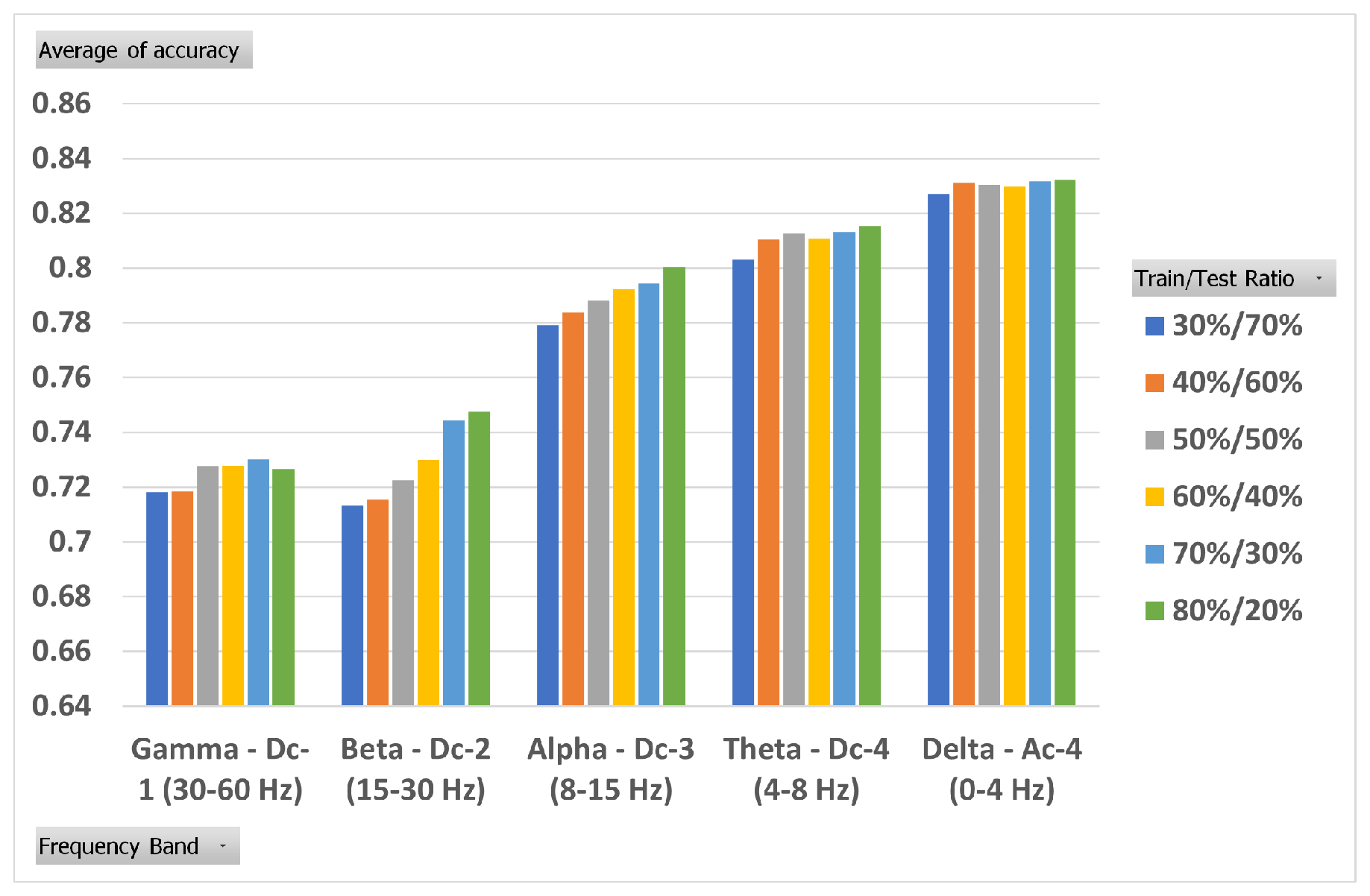
Figure 7 illustrates the results for our classification model in scenario 1 using five frequency sub-bands. It can be noted that the lower frequency sub-bands, such as delta (0–4 Hz), theta (4–8 Hz, and alpha (8–15 Hz), contain more discriminating features for epileptic seizure classification as compared with the higher frequency sub-bands. The proposed model yielded more than 70% accuracy on all of the frequency sub-bands; however, the delta band (0–4 Hz) showed the highest accuracy of 83.63%, as tabulated in
Table 5.
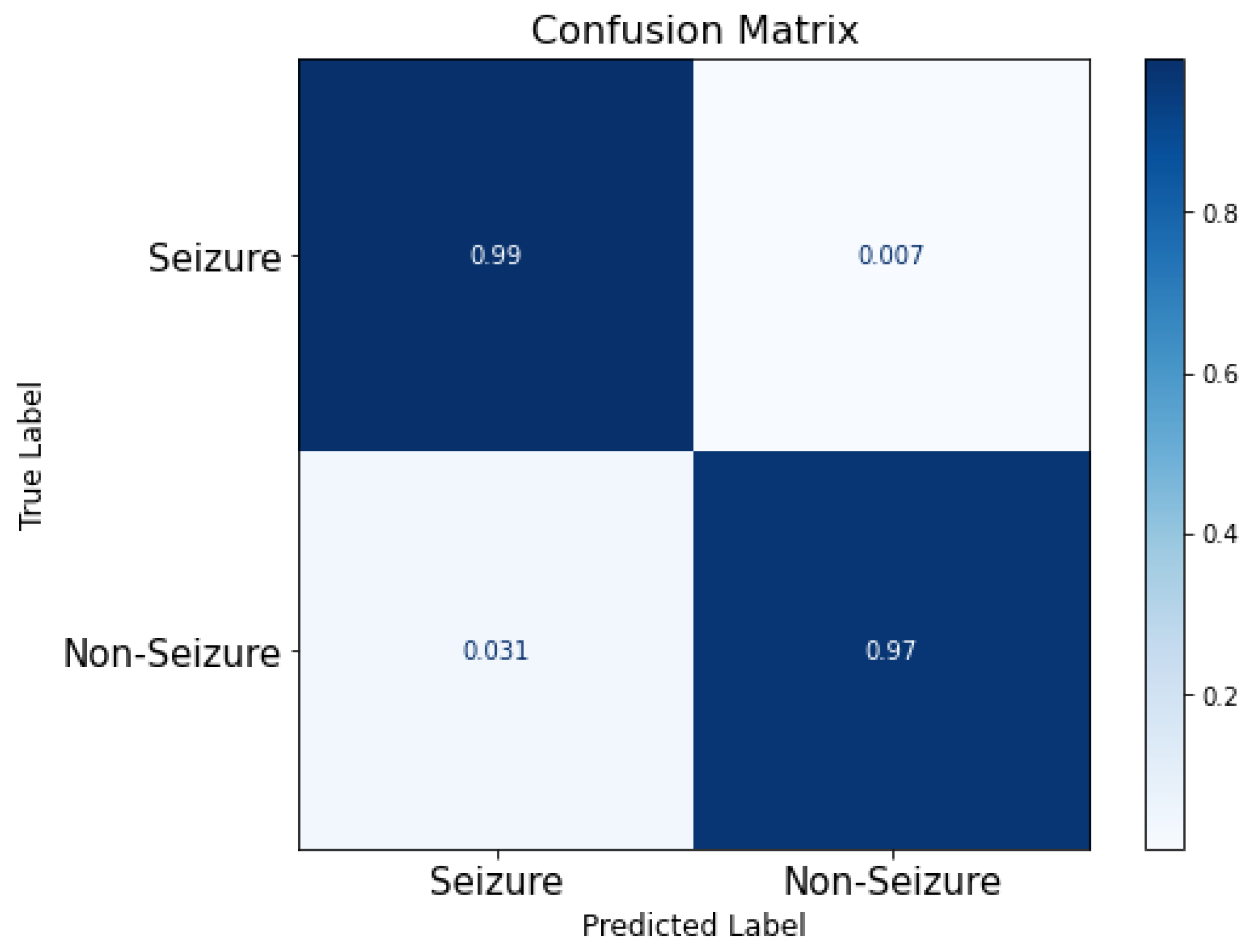
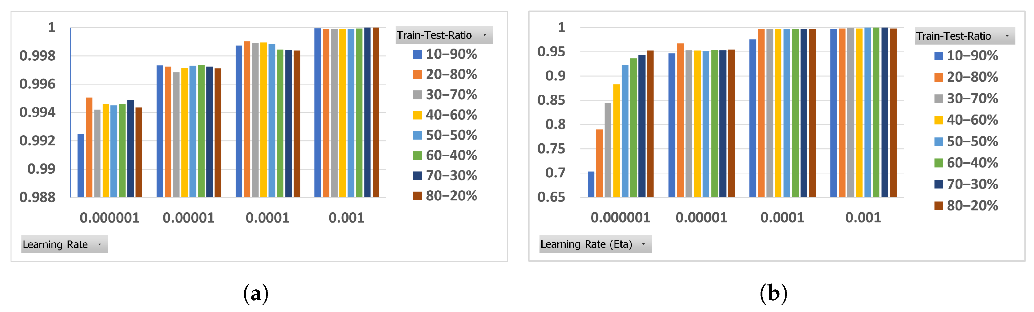
The BONN dataset contains comparatively simple data, with only one EEG channel of intracranial signals; therefore, our RNN-based model converges relatively quickly on the BONN dataset even with a lower learning rate (0.000001–0.001). The confusion matrix presented in
Figure 8 reveals that the RNN+DWT approach achieved an accuracy of 99.30% in classifying seizures on the BONN dataset while the false positive rate remained below 1%. The results were obtained with a learning rate of 0.001 and a training/testing ratio of 70%/30%.
Overall, the RNN-based model achieved an accuracy of 99.84% and 99.77% with DWT and FFT, respectively. In
Figure 9a, it can be noted that the FFT-based RNN model achieved better accuracy with a lower learning rate; however, when the learning rate was increased, the DWT-based RNN model showed better results, as illustrated in
Figure 9b. A comparison of the results obtained from RNN-based classification model on the BONN dataset is presented in
Table 6. The results show that the RNN-based epilepsy classification model performed better with DWT as compared with FFT. The proposed model also demonstrated better results when compared with the latest research found in the literature, as shown in
Table 6.
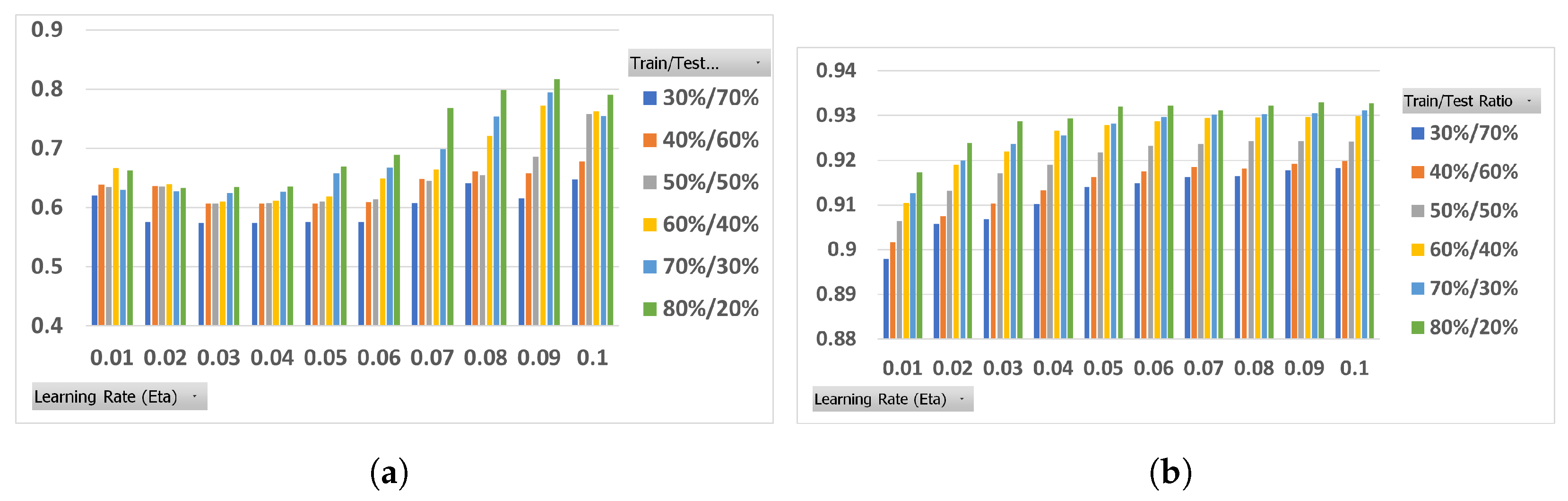
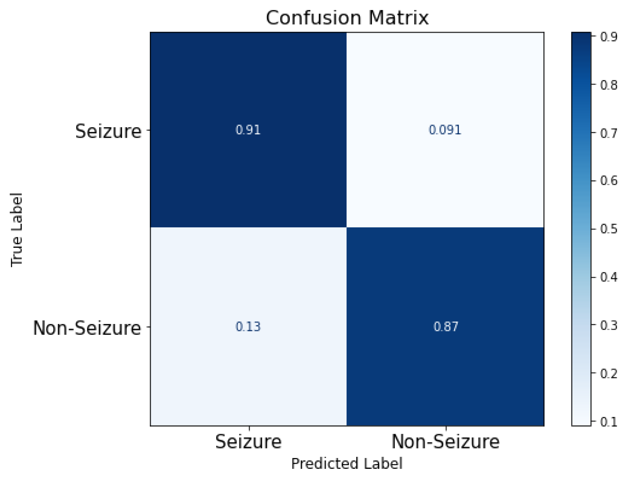
The accuracy of the proposed RNN-based classification model exhibited a comparable trend on both the CHB-MIT and BONN datasets. The RNN-based model coupled with DWT in the feature extraction pre-processing phase demonstrated a gradual increase in accuracy with each increment in the LR, as depicted in
Figure 10b, while the accuracy with FFT remained steady for the initial increments in the LR. Based on the confusion matrix illustrated in
Figure 11, it can be observed that the CHB-MIT dataset, with a training and testing ratio of 30–70% and a learning rate of 0.05, identifies 91% of seizure segments accurately while maintaining a false positive rate of 9.1%.
Overall, the DWT-based RNN model achieved better performance on both datasets.
Table 7 provides a comparison of the results obtained from the proposed RNN-based classification model on the CHB-MIT dataset. The results indicate that the proposed RNN-based model performed with an accuracy of 93.27% as compared with ANN and SVM, which achieved an accuracy of 86.10% and 90.68% respectively. The proposed RNN-based model converged faster as it was trained for 50 epochs, while the ANN-based model was trained for 200 epochs on the same data.
Table 7 presents a comparison of the proposed RNN-based model with recent studies in the literature, demonstrating superior performance in terms of accuracy, sensitivity, and specificity. For instance, Chen et al. [
22] achieved an accuracy of 89.01% with five statistical features as compared with the proposed RNN-based model’s 93.27%.
4.3. Limitations
There are certain limitations to the proposed scheme in this work, which involves combining EEG data from multiple patients. For example, one disadvantage of combining EEG data from various patients is the potential for information leakage from the training data to the testing data. Since the data are temporal, there may be an unintentional correlation between the sets used for training and testing the model’s performance. As a result, the model’s true generalization capabilities may be overestimated. Another limitation is the uncertainty around how the model would perform on completely unseen data. While the model may perform well on the given dataset, its ability to appropriately generalize to new, previously unseen data from different patients cannot be guaranteed. When applied to completely new and different datasets, factors such as inter-subject variability, unique patient characteristics, and differences in data collection techniques can all have an impact on the model’s performance.
Before deploying the proposed model for clinical decision support, real-time experimental trials should be carried out to assess the model’s performance under real-world conditions. For instance, the model can be tested with a continuous stream of EEG signals to evaluate its performance in real time. This can provide useful insights into how the model works with previously unseen data, allowing for a more accurate assessment of its performance metrics. Furthermore, feedback from clinicians and patients, as well as a collection of data, can help in identifying areas for improvement and fine-tuning the model’s performance. As new data become available, regular retraining and calibration can help maintain accuracy metrics.
5. Conclusions
This study proposed a novel method for the classification of epileptic seizures using RNN. The proposed method was compared with traditional machine learning models such as ANN and SVM, using data acquired from two widely used publicly available datasets, CHB-MIT and BONN. The proposed model presents a novel technique by integrating several stages, including EEG signal filtering, data segmentation, pre-processing, feature extraction, and classification. The data were segmented into epochs of 3 s each. The EEG signal was first pre-processed using DWT-based decomposition into five frequency sub-bands, and statistical features were extracted from each sub-band to obtain an input vector for training the classification models, including RNN. Training and testing of the classification models were performed multiple times with various parameter sets such as learning rate, training/testing ratios, and frequency analysis techniques using 10-fold cross-validation. The results conclude that the proposed RNN-based model demonstrated the highest classification accuracies of 93.27% on the CHB-MIT dataset and 99.84% on the BONN dataset. In the future, ensemble methods will be applied by combining RNN with other machine learning algorithms such as ANN and SVM to enhance classification accuracy.


