Systematic Review

Microbiota Alterations in Patients with Mucous Membrane Pemphigoid and Pemphigus Vulgaris: A Systematic Review

by
Rossella Santoro
1,*,
Antonio Romano
1,
Maria Cristina Morcaldi
1,
Fausto Fiori
1 and
Federica Di Spirito
2
1
Multidisciplinary Department of Medical-Surgical and Dental Specialties, University of Campania “Luigi Vanvitelli”, 80138 Naples, Italy
2
Department of Medicine and Surgery, University of Salerno, 84081 Salerno, Italy
*
Author to whom correspondence should be addressed.
Appl. Sci. 2023, 13(7), 4377; https://doi.org/10.3390/app13074377
Submission received: 19 February 2023 / Revised: 22 March 2023 / Accepted: 28 March 2023 / Published: 30 March 2023
(This article belongs to the Special Issue Oral Pathology and Medicine: Diagnosis and Therapy)

Abstract

:
The human oral cavity comprises an extensive range of microorganisms, viruses, bacteria, fungi, archaea, and protozoa, each having a particular role and interacting with each other and with the host in sickness or health. Changes in the microbiome composition can be crucial in balancing health and disease, locally and systemically. Various microbial species in commensal relationships form the oral microbiota, and when this commensalism undergoes variations the immune system can be pushed towards the activation of inflammatory and autoimmune processes. Through a systematic review of the literature, we set out to investigate the role that the oral microbiota can play in the development and evolution of pemphigus vulgaris and mucous membrane pemphigoid. We performed our systematic review by searching “microbiome OR microbiota” AND “pemphigus OR pemphigoid” on Medline, ISI Web of science and Embase, and we included randomized controlled trials (RCTs), prospective comparison studies, retrospective cohort studies, case–control studies, and case series. These autoimmune diseases need a genetic basis to develop, but as multifactorial pathologies they are influenced by environmental factors and the dysbiosis of the oral microbiota can be a trigger. If the human microbiome plays a critical role in the pathogenesis and manifestation of oral autoimmune diseases, the next step could be new and promising therapeutic approaches such as probiotics or prebiotics.

1. Introduction

The oral cavity comprises hundreds of microbial species, presenting different relationships with the host. Several factors can influence the microbial composition, such as diet and drugs. After the gastrointestinal tract, the human oral cavity contains the second largest collection of microorganisms. The expanded Human Oral Microbiome Database (eHOMD) includes data on around 772 species of prokaryotes, with 70% of these species being able to be cultivated in a laboratory, and the remaining 30% belonging to the class of microorganisms that cannot be cultivated [1]. As of now, the process by which various species are chosen to form a healthy and advantageous microbiome in the oral cavity remains unknown. Despite significant advancements in microbiome research and a greater understanding of its alterations, a complete comprehension of the mechanisms underlying the selection of specific species that form the healthy and beneficial microbiome has yet to be achieved [2,3]. The most abundant taxonomic group of oral microbiota is bacteria. Its richness of species, its high alpha diversity, and its stability make the oral microbiome one of the human body’s most complex [4,5]. Some authors described the importance of a core oral bacteriome composed of the same group of microorganisms in the vast majority of humans. It is essential in the dynamic equilibrium between disease and health [6,7,8]. Several authors from different medical branches have also analyzed the microbiome’s alterations after disease treatment, highlighting how clinical improvement induced a partial normalization of human microbiome or how poor clinical response after treatment could be affected by microbiome [9,10,11,12]. It is necessary to underline the important effect of age, gender, and geographical (mainly focusing on westernized people) features on the human microbiome [13]. It seems that the country affects human microbiome composition [14,15]. Gastrointestinal microbiome also represents a reservoir for antimicrobial resistance genes (resistome) [16,17]. The significant phyla comprise Actinobacteria, Bacteroidetes, Firmicutes, Fusobacteria, and Proteobacteria; Streptococcus is the most prevalent genus within the oral environment [18,19]. The oral microbiome is unique to each individual and consists of different habitats within the oral cavity, each with distinct anatomic and physiologic features [20]. However, the oral microbiome is often studied as a whole entity [21]. The complexity of the oral ecosystem depends on the various aspects of its components, including the hard tissue surfaces of the teeth, the soft tissue surfaces of the oral mucosa and tongue, and the saliva. Each of these elements provides a distinct ecosystem with favorable nutrients and conditions for colonizing microbes [22,23]. Interestingly, samples taken from the same site in different individuals exhibited more similarities than those taken from different niches in the same individual [24,25]. Furthermore, the oral microbiome shows difference based on geographical features [26]. In healthy individuals, the oral virome is highly complex, consisting of a community of double-stranded DNA viruses in the saliva [27]. Bacteriophages, which are viruses that prey on bacteria, are a significant portion of the oral viruses and may play a role in regulating microbial diversity while simultaneously serving as reservoirs of pathogenic genes in the human oral environment [28]. These microorganisms live in close proximity, forming a complex relationship that results in a variety of interspecies interactions, which can be synergistic, signaling, or antagonistic [29]. Although fungi represent a minority, they are an integral part of the healthy oral microbiota. The chemical, physical, and metabolic interactions between bacteria and fungi are crucial for establishing and maintaining good oral health [30,31]. Fungi only become opportunistic pathogens under specific conditions [30,32]. A healthy oral microbiota includes about a hundred species of fungi, among which the most widespread are Candida [33], Cladosporium, Aureobasidium, Saccharomyces, Aspergillus, Fusarium, and Cryptococcus [31]. Candidiasis is mycotic infectious stomatopathy mainly caused by Candida albicans and Candida tropicalis [34]. From a clinical point of view, Candida infection can occur in various forms: pseudomembranous candidiasis, erythematous candidiasis, and hyperplastic candidiasis (following chronic infections). The most common opportunistic fungal disease in the oral cavity is chronic erythematous candidiasis; the primary causative agent of chronic erythematous candidiasis is Candida albicans (95% of cases). This commensal microorganism is present in the microbiota of about 80% of the population, but an excessive increase can transform it into a pathogen [35]. It can become an opportunistic microorganism and therefore cause infections when there is a subversion of the oral microbiota following an alteration of tissue barriers, and when the host’s immune system is weakened [36]. Among the predisposing factors for this infection, in addition to local dysbiosis, we note smoking, nutritional deficiencies, hyposalivation, use of dental prostheses, and the use of antibiotics [37]. Numerous studies have shown that C. albicans cooperates with certain bacteria that inhabit the oral microbiota, establishing synergistic relationships [38,39]. Some bacteria influence the activity of C. albicans, such as Streptococcus oralis and Porphyromonas gingivalis, which increase the expression of a gene encoding cellular adhesin in C. albicans, thus increasing its capacity for biofilm formation [40] and, therefore, the transition from yeast to hypha. Numerous studies have shown that the commensal microbiota regulates host immunity to pathogens. However, the microbiota significance in autoimmune response regulation is yet to be a topic of discussion and research. Some authors have even highlighted the correlation between the presence of oral pathogens and gastrointestinal diseases [41]. Different authors have already discussed the importance of the microbiome in autoimmune diseases, interesting other medical specialties such as gastroenterology, ophthalmology, and gynecology. Autoimmune diseases are often influenced in their pathogenesis and in their recurrence by environmental factors. Among these, several authors have discussed the role of infectious agents as causative factors [42,43]. It seems that autoimmune diseases affecting animal models and the human gut could be influenced by the decrease of short-chain fatty acids, which have an influence on intestinal homeostasis [44]. How the oral microbiome functions, when changed, has yet to be completely clear. The disruption of microbiome composition, usually resulting in surplus commensal numbers, may force the immune system toward triggering inflammatory and autoimmune processes. Shifts in the oral microbiome may play a significant part in the evolution of autoimmune diseases. The oral cavity is often the site of the onset of autoimmune diseases like pemphigus vulgaris (PV) and mucous membrane pemphigoid (MMP) [45,46]. PV and MMP are chronic autoimmune mucocutaneous diseases affecting the mucous membranes and the skin [47]. These diseases clinically appear like oral ulcers; for this reason, differential diagnosis could be challenging for the clinician [48,49]. In PV, pathogenic autoantibodies directed against desmogleins I and III, proteins contained in desmosomes, develop [50]. The union of autoantibodies and components of the desmosomes compromises intraepidermal adhesion, leading to acantholysis and the formation of vesicles, blisters, and erosions on the mucous membranes [51,52]. PV epidemiology ranges from less than 0.76 per million to 16.1 per million based on several features, with a higher incidence in Ashkenazi Jews [53]. Some authors showed a relative prevalence between pemphigoid and pemphigus ranging from 4:1 to 1:2, correlating the different rates on geographical features [54]. It is a multifactorial disease, and the influence of genetic and immunological factors on onset is well established. In addition to genetic factors, environmental factors such as drugs, stress, diet, physical trauma like ionizing radiation, UV light, thermal burns, neoplasm, and infections may induce or impact the disease. Nevertheless, most patients lack a recognized influencing factor [55,56]. MMP predominately affects the mucous membranes, frequently involving the oral mucosae, especially the gingiva. MMP gingival manifestation is known as desquamative gingivitis [46] and can culminate in scarring and considerable morbidity. The MMP is not marked by a specific serologic marker alone. In fact, different target antigens associated with the clinical phenotype of MMP have been molecularly identified: Bullous Pemphigoid 180 (BP180), Bullous Pemphigoid 230 (BP230), laminin 332, and both subunits of α6β4 integrin and type VII collagen [57,58]. Mucous membrane pemphigoid diagnoses are based on clinical, histological, and immunopathological findings [59]. The initiating factor for the autoimmune response in MMP is unknown. Based on the evidence reported in the literature, the present work investigates what could be the role of oral microbiome in the development and evolution of these oral cavity autoimmune diseases. This study aims also to analyze correlations between PV and MMP, and qualitative and quantitative modifications of the oral microbiota, such as trigger factors for the onset of these diseases, are also investigated.

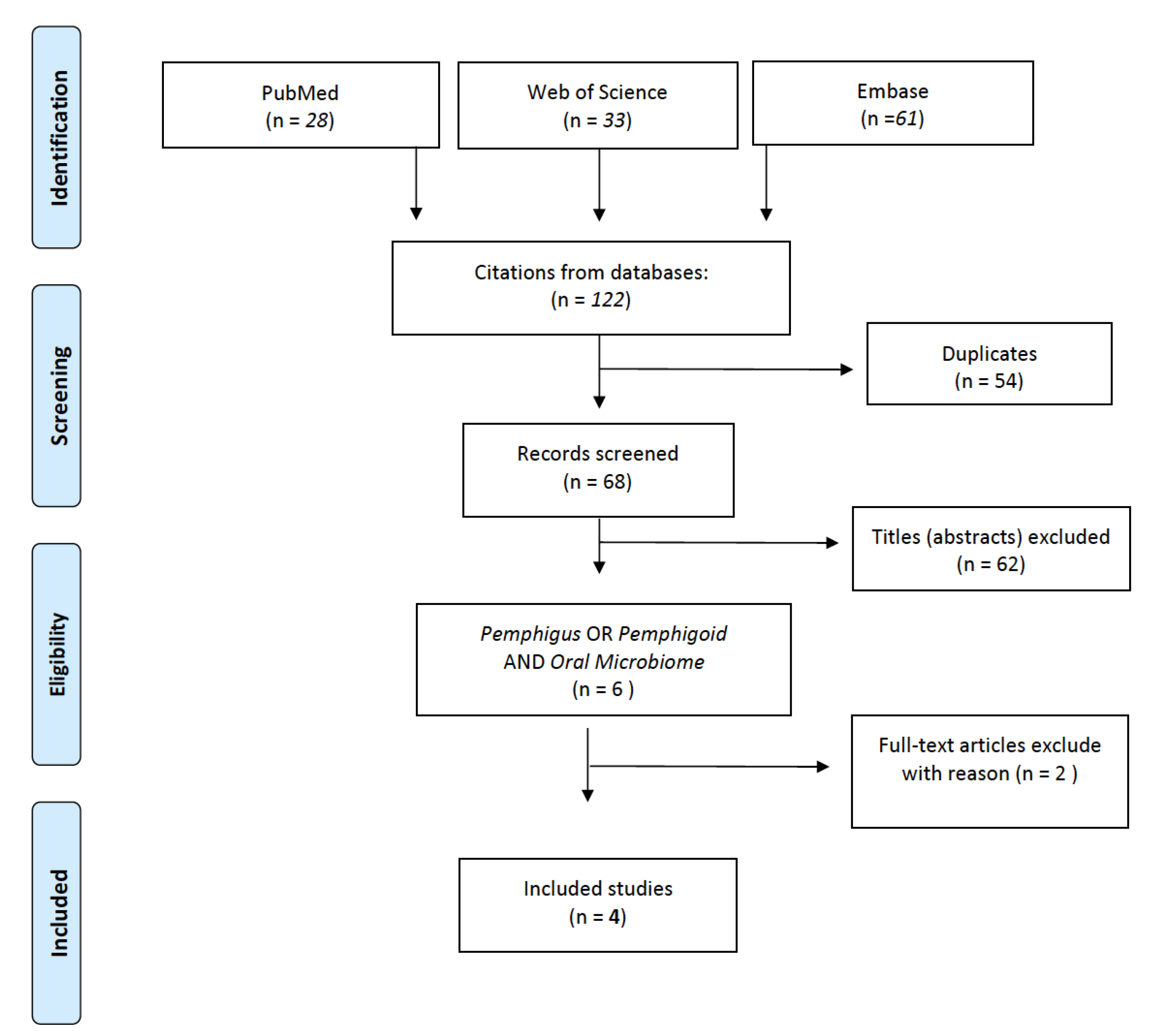
2. Materials and Methods

The approach utilized in this systematic review adhered to the guidelines of the Preferred Reporting Items for Systematic Reviews and Meta-Analyses (PRISMA) [60]. To identify relevant original studies published in English, an electronic search was conducted on the MEDLINE, EMBASE, and Web of Science databases without any publication year limit up to 30 December 2022. The search terms used (MeSH terms) were “microbiome OR microbiota” AND “pemphigus OR pemphigoid”. The study population of interest included both genders with or without pemphigus vulgaris or mucous membrane pemphigoid, and the intervention was to investigate the presence of the microbiome in patients with PV or MMP. The comparison was made with patients without PV or MMP, and the outcome was to evaluate the presence of the microbiome in patients with or without PV or MMP. The study designs included randomized controlled trials (RCTs), prospective comparison studies, retrospective cohort studies, case–control studies, and case series. The quality assessment of non-randomized studies was evaluated using the Risk of Bias in Non-randomized Studies of Interventions (ROBINS I) assessment tool (Figure 1) [61]. Seven bias domains were assessed, and each was rated on a five-grade scale: low, moderate, severe, critical, and no information. This review was registered on PROSPERO with registration number CRD42023389051. Table 1 presents a summary of the results retrieved.

3. Results

In the results, the initial electronic search identified 122 results, of which 54 were duplicates, and 62 were eliminated based on the exclusion criteria after screening the titles. Two authors (AR, FF) independently screened the titles and abstracts based on the listed criteria. From the remaining six abstracts, full-text articles were obtained for all agreed upon titles, and disagreements were resolved by discussion. After the analysis of the full text two more articles were excluded because they did not meet the inclusion criteria [66,67]. Finally, four studies were included in this review. The selection process is explained in detail and graphically summarized in Figure 2, and the included studies are briefly reported in Table 1.

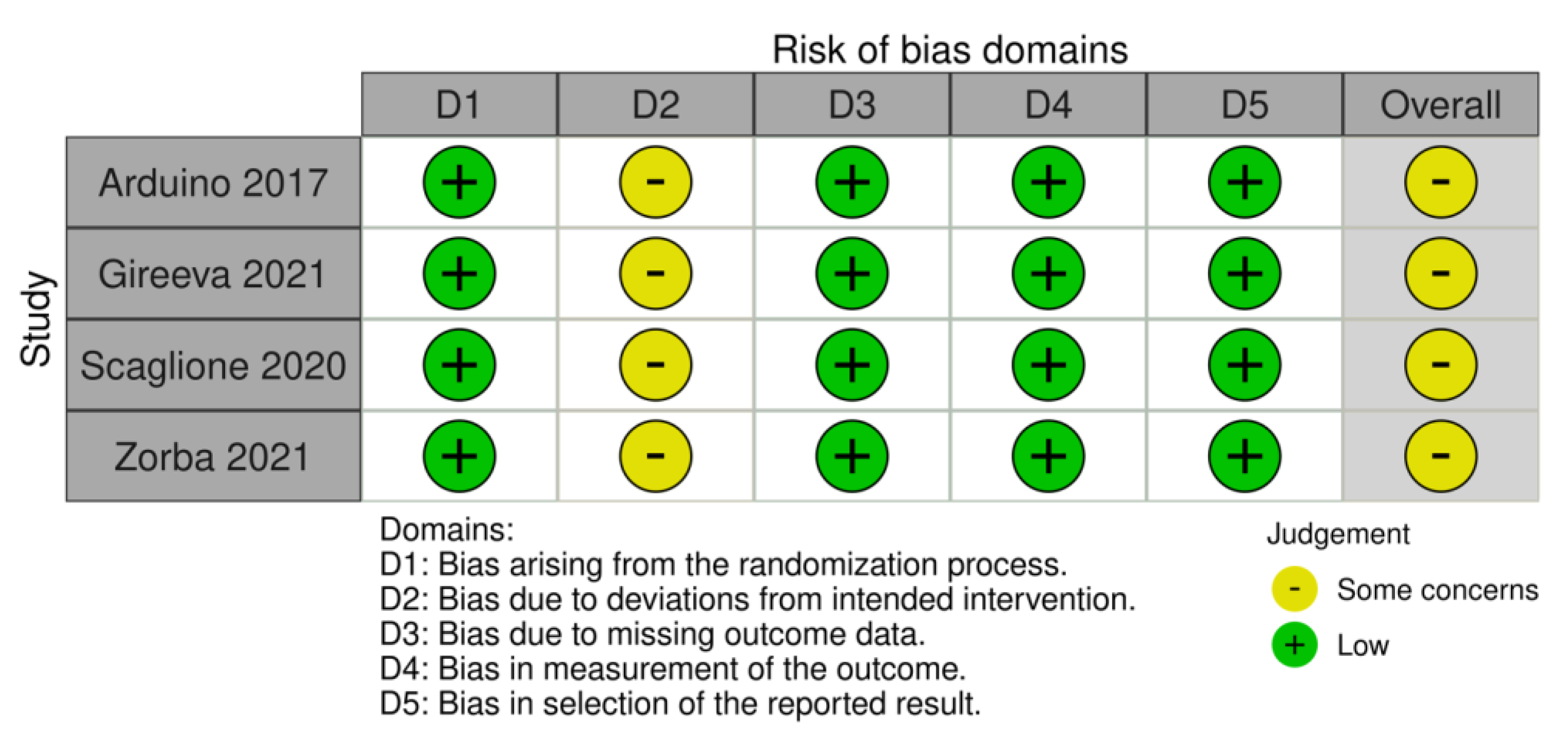
3.1. Risk of Bias in Individual Studies

Of the studies assessed with ROBINS-I, four studies were evaluated as moderate risk RoB and none as serious risk. This systematic review examines non-randomized studies. All of the studies provide sound evidence, and none presented a critical RoB in any domain.

3.2. Pemphigus Vulgaris

Three studies investigated oral microbiome in patients with pemphigus vulgaris, as reported in Table 1. In their study, Gireeva et al., utilized the polymerase chain reaction real-time (PCR) method to compare the qualitative and quantitative composition of gingival fluid microbiota in patients with PV during the remission and exacerbation phases. The authors not only examined the presence of periodontal pathogens, but also viruses and fungi in the oral cavity. They found that the bacterial counts of P. intermedia, T. denticola, T. forsythensis, and P. gingivalis were lower during exacerbation compared to remission, while the viral counts of Epstein–Barr virus were significantly higher during exacerbation. The authors suggest that these findings may be due to the frequent use of chlorhexidine-containing products during exacerbation, as well as a decrease in host response caused by increased doses of corticosteroids during the exacerbation phase [63]. Scaglione et al., used next generation sequencing (NGS), targeting the phylogenetically informative 16S ribosomal RNA gene for the characterization and evaluation of the oral microbiota in patients affected by PV. The authors reported the following data from seven oral mucosae samples: (i) Firmicutes (%)= 45.5 (27.1–72.6) in PV and 39.6 (32.3–73.4) in control samples; (ii) Fusobacteria (%) = 28.0 (10.4–41.6) in PV and 8.5 (1.9–13.2) in control samples; (iii) Bacteroidetes (%) = 7.2 (5.7–12.6) in PV and 28.4 (7.3–38.5) in control samples; (iv) Proteobacteria (%) = 15.2 (5.1–23.9) in PV and 13.3 (10.5–42.6) in control samples; (v) Actinobacteria (%) = 5.5 (2.8–27.0) in PV and 2.4 (1.4–5.3) in control samples [64]. Zorba et al., utilized deep sequencing of the bacterial 16S rRNA gene to analyze the microbial communities of the oral cavity in both healthy individuals and those with PV. They characterized the composition of the microbiota at the phylum, family, genus, and species levels. The results indicated that the most prevalent phyla were Firmicutes (60.0%) and Bacteroidetes (16.20%), followed by Proteobacteria (11.59%), Actinobacteria (7.24%), and Fusobacteria (3.94%). At the genus level, patients with PV showed a statistically significant increase in the abundance of Streptococcus (34.37% in patients vs. 33.30% in controls, p value = 0.006), Fusobacterium (4.51% in patients vs. 4.13% in controls, p value = 0.024), and Gemella (7.13% in patients vs. 5.80% in controls, p value = 0.030) [65].

3.3. Mucous Membrane Pemphigoid

Only one study investigated oral microbiome in patients with MMP as reported in Table 1. Arduino et al., investigated prevalence of periodontopathogenic microorganisms in patients with desquamative gingivitis, and compared this with the microbiologic status of control patients affected by plaque-induced gingivitis (pGI). In the desquamative gingivitis group, there were 14 patients with MMP. The periodontopathogenic microorganisms analyzed were: Aggregatibacter actinomycetemcomitans; Porphyromonas gingivalis; Prevotella intermedia; Tannerella forsythia; Treponema denticola; Parvimonas micra; Fusobacterium nucleatum/periodonticum; Campylobacter rectus; Eubacterium nodatum; Eikenella corrodens; and Capnocytophaga spp. (C. gingivalis, C. ochracea, C. sputigena). The authors reported higher levels of detection of F. nucleatum, E. corrodens, and Capnocytophaga spp. [62].

4. Discussion

Numerous studies have tried to find a possible association between microbiome and autoimmune diseases, like primary Sjögren’s syndrome [68,69], systemic lupus erythematosus [70], rheumatoid arthritis [71], and brain autoimmunity [72], trying to understand if a specific bacterial infection may play a role in the aetiopathogenesis of the disease [73]. Mucous membranous pemphigoid and pemphigus vulgaris are multifactorial genetic disorders. Genetic predisposition is necessary for developing these diseases, but it is not the only cause. Finding a possible association between these diseases and different bacteria, as well as identifying the actual prevalence of bacterial infection and whether a specific bacterial infection may play a role in the aetiopathogenesis of the disease, is a current and very intriguing topic. Not long ago, thanks to next generation sequencing, scientists began to study microbial communities and sequence them faster, making them more accessible and more searchable, and coming to define the contours of the microbiome’s contribution to health and disease. Any dearth of variety can lead to dysbiosis, a critical disproportion between commensal and pathogenic bacteria. These commensals supply an essential niche by regulating the immune response and supporting a homeostatic environment. The bacteria that make up the oral microbiota come into contact with the oral mucosal immune system with numerous consequences. The mechanisms by which the microbiome can potentially influence the host’s immune system include molecular mimicry, epitope spreading, and constitutive stimulation of Toll-like receptors [74,75]. Microbes can also cause epigenetic changes, including post-translational modification of histones, micro-RNA alteration without changes in DNA sequence, and changes in gene function through DNA methylation [76]. In the present review, we identify very few scientific studies that evaluated the oral microbiome in patients with pemphigus vulgaris and mucous membranous pemphigoid. The three studies that analyzed samples from the oral cavity of patients with PV reported the presence of P. intermedia, T. denticola, T. forsythensis, P. gingivalis, Fusobacterium nucleatum, Capnocytophaga leadbetteri and Parvimonas micra. At the phyla level, Scaglione et al., record a prevalence of Firmicutes and Fusobacteria. These data agree with those presented in a recent paper by Petruzzi et al. [66]. The authors searched for peptides common to a set of eight periodontopathogenic bacteria (Aggregatibacter actinomycetemcomitans; Campylobacter rectus; Eikenella corrodens; Fusobacterium nucleatum; Parvimonas micra; Porphyromonas gingivalis; Prevotella intermedia; Tannerella forsythia) and DSG3. The PV autoantigen and the eight bacterial proteomes share 23 heptapeptides; the bacterial proteomes most involved in sharing are E. corrodens, F. nucleatum and T. forsythia. Similarly to PV, a very small number of studies have been identified for mucus membranous pemphigoid, in this case only one [62]: that of Arduino et al., The authors reported a subgingival colonization of F. nucleatum, E. corrodens, Capnocytophaga spp. in the 14 patients analyzed with MMP. Again, these experimental data are in agreement with a recent research paper [68]. In this study, the authors investigated a possible relationship between the immune response directed against pathogenic bacteria colonizing the oral cavity and the aetiopathogenesis of MMP. They hypothesized that immune responses against these pathogens may cross-react with MMP’s autoantigens. Based on the study by Arduino et al. [68], they searched for peptides common to a set of eight oral bacteria representing periodontopathogenic bacterial species, and BP180 and BP230. Subsequently, they searched for potentially immuno-crosslinked shared sequences. The authors reported 12 peptides shared between BP180 and the bacterial proteome of Tannerella forsythia and Eikenella corrodens [67]. The presence of shared peptides between the bacteria most commonly found in the oral cavity of patients with PV and MMP and the autoantigens of these autoimmune diseases would provide further evidence to support the role of molecular mimicry in the development of autoimmune diseases [71,77,78,79].
Although the microbiota plays an essential role in the regulation of the immune system and the aetiology of autoimmune diseases, the entire population of microorganisms (bacteriome, mycobiome, and virome) of the oral cavity is not always considered as a whole. Indeed, the role of viral communities embedded in the oral microbiome has not always been investigated in the studies we have reviewed. Only Gireeva et al. [68] determined the qualitative and quantitative content of not only bacteria but also viruses and fungi in the oral cavity. In this study, the authors found alterations in the oral virome: the viral counts of Epstein–Barr virus were significantly greater in the exacerbation period. A role of corticosteroids in the decrease of a host response during the exacerbation phase has been proposed. The role of some viruses has been well characterised in the development and exacerbation of some autoimmune diseases of the oral cavity, such as herpes simplex virus in erythema multiforme [80]. The association between oral lichen planus and viral infection is well known [81,82,83]. Some authors speculate that viral infection may be an antigenic stimulus of CTL expansion that characterises severe erosive OLP [84,85]. Another player in the gut microbiome is the mycobiota, which comprises several fungal communities. A Russian study by Rabinovich et al., reported a 26.3% association between Str.pneumoniae and C. albicans in patients with pemphigus vulgaris, and 20% in those with pemphigoid. Furthermore, in subjects with pemphigus vulgaris, the association between Str.pneumoniae, C. albicans and EBV is reported in 31.6% of cases. However, the role of oral mycobiota in different autoimmune diseases needs to be further explored in other studies.

5. Conclusions

Recent literature has established the role of dysbiosis in several diseases, like type 1 diabetes [86], rheumatoid arthritis [80], psoriatic arthritis, ankylosing spondyloarthritis [87], systemic lupus erythematosus [88], multiple sclerosis, osteoporosis [34,89], Crohn’s disease (CD) and may even contribute to the late onset of schizophrenia [90]. Several mechanisms have been proposed to explain how microbiota contribute to the development of autoimmune diseases. As previously mentioned, the oral microbiota is physiologically in a state of equilibrium to maintain the state of health of the host. Without perturbations within this balance, colonization by several microorganisms cannot occur. C. albicans, for example, is not a passive player. Indeed, in a predisposing immunological condition, it promotes a subversion of the oral microbiota, increasing the prevalence of some bacterial species to the detriment of others. For example, with the increase in the number of enterococci, there is a negative impact on tissue barriers, thus facilitating their colonization [91]. Significant variations exist between the composition of the “healthy” microbiota and the one of a subject suffering from candidiasis. Colonization by C. albicans is therefore combatted by a number of factors that act synergistically. A note should be made about the microorganisms that produce short-chain saturated fatty acids (SCFA), which have a profound impact on the formation of the biofilm by C. albicans and, therefore, on its passage from yeast to hypha, both by inhibiting the metabolic activity of the fungus and by reducing the environmental pH [92]. The same goes for lactobacilli, which through the production of certain acids and inducing the production of IL-22 by the host, hinder colonization by C. albicans [93]. That said, it is therefore easy to deduce that this infection is associated with the loss of mucosal bacterial diversity [91]. It is, therefore, clear that the interactions between C. albicans, host, and microbiota have an important impact on both the development and spread of infection and its severity. Being aware of all these interactions gives us the opportunity to reflect on the appropriate use of certain drugs, such as antibiotics, antifungals, and probiotics. Hence, dysbiosis should not be viewed only as a biomarker of inflammatory diseases but even as a promoter of inflammatory diseases and autoimmune responses [94,95]. It is unclear whether it could trigger a cascade in the autoimmune response that leads to the onset of the disease. Still, some bacterial species’ predominance seems protective against pathology manifestation [96,97]. Research data are insufficient to determine whether dysbiosis of the oral cavity is the consequence or the cause of autoimmune bullous diseases. However, the role of commensal bacteria in modulating the immune system is evident, which could lead to autoimmune inflammation. This fascinating topic acquires increasing relevance and consequently encourages further analysis of the correlation between the microbiome and autoimmunity, also intending to provide additional scientific support for antibiotic therapy in treating diseases with oral involvement, such as MMP [98,99]. Additional studies are required to investigate the possible role of antibiotics as adjuvant therapy for oral autoimmune diseases and their possible clinical role. The ultimate goal is always to enhance the quantity and quality of the data to boost the translation of research into clinical practice.

Author Contributions

Conceptualization, R.S. and F.D.S.; methodology, R.S.; validation, M.C.M.; formal analysis, F.F.; investigation, A.R.; writing—original draft preparation, R.S.; writing—review and editing, F.D.S.; supervision, F.F. All authors have read and agreed to the published version of the manuscript.

Funding

This research received no external funding.

Institutional Review Board Statement

Not applicable.

Informed Consent Statement

Not applicable.

Data Availability Statement

Data is contained within the article.

Conflicts of Interest

The authors declare no conflict of interest.

References

  1. Verma, D.; Garg, P.K.; Dubey, A.K. Insights into the Human Oral Microbiome. Arch. Microbiol. 2018, 200, 525–540. [Google Scholar] [CrossRef] [PubMed]
  2. Zaura, E.; Nicu, E.A.; Krom, B.P.; Keijser, B.J.F. Acquiring and Maintaining a Normal Oral Microbiome: Current Perspective. Front. Cell. Infect. Microbiol. 2014, 4, 85. [Google Scholar] [CrossRef] [PubMed] [Green Version]
  3. Alcaraz, L.D.; Belda-Ferre, P.; Cabrera-Rubio, R.; Romero, H.; Simon-Soro, A.; Pignatelli, M.; Mira, A. Identifying a Healthy Oral Microbiome through Metagenomics. Clin. Microbiol. Infect. 2012, 18, 54–57. [Google Scholar] [CrossRef] [PubMed] [Green Version]
  4. Huttenhower, C.; Gevers, D.; Knight, R.; Abubucker, S.; Badger, J.H.; Chinwalla, A.T.; Creasy, H.H.; Earl, A.M.; Fitzgerald, M.G.; Fulton, R.S.; et al. Structure, Function and Diversity of the Healthy Human Microbiome. Nature 2012, 486, 207–214. [Google Scholar] [CrossRef] [Green Version]
  5. Zhou, Y.; Gao, H.; Mihindukulasuriya, K.A.; Rosa, P.S.L.; Wylie, K.M.; Vishnivetskaya, T.; Podar, M.; Warner, B.; Tarr, P.I.; Nelson, D.E.; et al. Biogeography of the Ecosystems of the Healthy Human Body. Genome Biol. 2013, 14, R1. [Google Scholar] [CrossRef] [Green Version]
  6. Jiang, W.X.; Hu, Y.J.; Gao, L.; He, Z.Y.; Zhu, C.L.; Ma, R.; Huang, Z.W. The Impact of Various Time Intervals on the Supragingival Plaque Dynamic Core Microbiome. PLoS ONE 2015, 10, e0124631. [Google Scholar] [CrossRef] [Green Version]
  7. Frank, D.N.; Feazel, L.M.; Bessesen, M.T.; Price, C.S.; Janoff, E.N.; Pace, N.R. The Human Nasal Microbiota and Staphylococcus Aureus Carriage. PLoS ONE 2010, 5, e10598. [Google Scholar] [CrossRef]
  8. Kort, R.; Caspers, M.; van de Graaf, A.; van Egmond, W.; Keijser, B.; Roeselers, G. Shaping the Oral Microbiota through Intimate Kissing. Microbiome 2014, 2, 41. [Google Scholar] [CrossRef] [PubMed] [Green Version]
  9. Dewhirst, F.E.; Chen, T.; Izard, J.; Paster, B.J.; Tanner, A.C.R.; Yu, W.H.; Lakshmanan, A.; Wade, W.G. The Human Oral Microbiome. J. Bacteriol. 2010, 192, 5002–5017. [Google Scholar] [CrossRef] [Green Version]
  10. Schwarz, E.; Maukonen, J.; Hyytiäinen, T.; Kieseppä, T.; Orešič, M.; Sabunciyan, S.; Mantere, O.; Saarela, M.; Yolken, R.; Suvisaari, J. Analysis of Microbiota in First Episode Psychosis Identifies Preliminary Associations with Symptom Severity and Treatment Response. Schizophr. Res. 2018, 192, 398–403. [Google Scholar] [CrossRef] [Green Version]
  11. Dominguez-Bello, M.G.; De Jesus-Laboy, K.M.; Shen, N.; Cox, L.M.; Amir, A.; Gonzalez, A.; Bokulich, N.A.; Song, S.J.; Hoashi, M.; Rivera-Vinas, J.I.; et al. Partial Restoration of the Microbiota of Cesarean-Born Infants via Vaginal Microbial Transfer. Nat. Med. 2016, 22, 250–253. [Google Scholar] [CrossRef]
  12. Goleva, E.; Jackson, L.P.; Harris, J.K.; Robertson, C.E.; Sutherland, E.R.; Hall, C.F.; Good, J.T.; Gelfand, E.W.; Martin, R.J.; Leung, D.Y.M. The Effects of Airway Microbiome on Corticosteroid Responsiveness in Asthma. Am. J. Respir. Crit. Care Med. 2013, 188, 1193–1201. [Google Scholar] [CrossRef] [PubMed] [Green Version]
  13. Clemente, J.C.; Pehrsson, E.C.; Blaser, M.J.; Sandhu, K.; Gao, Z.; Wang, B.; Magris, M.; Hidalgo, G.; Contreras, M.; Noya-Alarcón, Ó.; et al. The Microbiome of Uncontacted Amerindians. Sci. Adv. 2015, 1, e1500183. [Google Scholar] [CrossRef] [PubMed] [Green Version]
  14. Mueller, S.; Saunier, K.; Hanisch, C.; Norin, E.; Alm, L.; Midtvedt, T.; Cresci, A.; Silvi, S.; Orpianesi, C.; Verdenelli, M.C.; et al. Differences in Fecal Microbiota in Different European Study Populations in Relation to Age, Gender, and Country: A Cross-Sectional Study. Appl. Environ. Microbiol. 2006, 72, 1027–1033. [Google Scholar] [CrossRef] [Green Version]
  15. Yatsunenko, T.; Rey, F.E.; Manary, M.J.; Trehan, I.; Dominguez-Bello, M.G.; Contreras, M.; Magris, M.; Hidalgo, G.; Baldassano, R.N.; Anokhin, A.P.; et al. Human Gut Microbiome Viewed across Age and Geography. Nature 2012, 486, 222–227. [Google Scholar] [CrossRef] [Green Version]
  16. Moore, A.M.; Patel, S.; Forsberg, K.J.; Wang, B.; Bentley, G.; Razia, Y.; Qin, X.; Tarr, P.I.; Dantas, G. Pediatric Fecal Microbiota Harbor Diverse and Novel Antibiotic Resistance Genes. PLoS ONE 2013, 8, e78822. [Google Scholar] [CrossRef] [PubMed] [Green Version]
  17. Stecher, B.; Denzler, R.; Maier, L.; Bernet, F.; Sanders, M.J.; Pickard, D.J.; Barthel, M.; Westendorf, A.M.; Krogfelt, K.A.; Walker, A.W.; et al. Gut Inflammation Can Boost Horizontal Gene Transfer between Pathogenic and Commensal Enterobacteriaceae. Proc. Natl. Acad. Sci. USA 2012, 109, 1269–1274. [Google Scholar] [CrossRef] [Green Version]
  18. Li, K.; Bihan, M.; Methé, B.A. Analyses of the Stability and Core Taxonomic Memberships of the Human Microbiome. PLoS ONE 2013, 8, e63139. [Google Scholar] [CrossRef] [Green Version]
  19. Aas, J.A.; Paster, B.J.; Stokes, L.N.; Olsen, I.; Dewhirst, F.E. Defining the Normal Bacterial Flora of the Oral Cavity. J. Clin. Microbiol. 2005, 43, 5721–5732. [Google Scholar] [CrossRef] [Green Version]
  20. Sharma, N.; Bhatia, S.; Singh Sodhi, A.; Batra, N. Oral Microbiome and Health. AIMS Microbiol. 2018, 4, 42–66. [Google Scholar] [CrossRef]
  21. Gizani, S.; Papaioannou, W.; Haffajee, A.D.; Kavvadia, K.; Quirynen, M.; Papagiannoulis, L. Distribution of Selected Cariogenic Bacteria in Five Different Intra-Oral Habitats in Young Children. Int. J. Paediatr. Dent. 2009, 19, 193–200. [Google Scholar] [CrossRef] [PubMed]
  22. Sonnenburg, J.L.; Fischbach, M.A. Community Health Care: Therapeutic Opportunities in the Human Microbiome. Sci. Transl. Med. 2011, 13, 78ps12. [Google Scholar] [CrossRef] [PubMed] [Green Version]
  23. Lamont, R.J.; Koo, H.; Hajishengallis, G. The Oral Microbiota: Dynamic Communities and Host Interactions. Nat. Rev. Microbiol. 2018, 16, 745–759. [Google Scholar] [CrossRef]
  24. Papapanou, P.N.; Park, H.; Cheng, B.; Kokaras, A.; Paster, B.; Burkett, S.; Watson, C.W.M.; Annavajhala, M.K.; Uhlemann, A.C.; Noble, J.M. Subgingival Microbiome and Clinical Periodontal Status in an Elderly Cohort: The WHICAP Ancillary Study of Oral Health. J. Periodontol. 2020, 91 (Suppl. S1), S56–S67. [Google Scholar] [CrossRef] [PubMed]
  25. Mark Welch, J.L.; Rossetti, B.J.; Rieken, C.W.; Dewhirst, F.E.; Borisy, G.G. Biogeography of a Human Oral Microbiome at the Micron Scale. Proc. Natl. Acad. Sci. USA 2016, 113, E791–E800. [Google Scholar] [CrossRef] [Green Version]
  26. Contreras, M.; Costello, E.K.; Hidalgo, G.; Magris, M.; Knight, R.; Dominguez-Bello, M.G. The Bacterial Microbiota in the Oral Mucosa of Rural Amerindians. Microbiology 2010, 156, 3282–3287. [Google Scholar] [CrossRef] [Green Version]
  27. Pride, D.T.; Salzman, J.; Relman, D.A. Comparisons of Clustered Regularly Interspaced Short Palindromic Repeats and Viromes in Human Saliva Reveal Bacterial Adaptations to Salivary Viruses. Environ. Microbiol. 2012, 14, 2564–2576. [Google Scholar] [CrossRef] [Green Version]
  28. Pride, D.T.; Salzman, J.; Haynes, M.; Rohwer, F.; Davis-Long, C.; White, R.A.; Loomer, P.; Armitage, G.C.; Relman, D.A. Evidence of a Robust Resident Bacteriophage Population Revealed through Analysis of the Human Salivary Virome. ISME J. 2012, 6, 915–926. [Google Scholar] [CrossRef]
  29. Roberts, A.P.; Kreth, J. The Impact of Horizontal Gene Transfer on the Adaptive Ability of the Human Oral Microbiome. Front. Cell. Infect. Microbiol. 2014, 4, 124. [Google Scholar] [CrossRef] [Green Version]
  30. Krom, B.P.; Kidwai, S.; Ten Cate, J.M. Candida and Other Fungal Species: Forgotten Players of Healthy Oral Microbiota. J. Dent. Res. 2014, 93, 445–451. [Google Scholar] [CrossRef]
  31. Brun, A.; Nuzzo, A.; Prouvost, B.; Diallo, D.; Hamdan, S.; Meseguer, E.; Guidoux, C.; Lavallée, P.; Amarenco, P.; Lesèche, G.; et al. Oral Microbiota and Atherothrombotic Carotid Plaque Vulnerability in Periodontitis Patients. A Cross-Sectional Study. J. Periodontal Res. 2021, 56, 339–350. [Google Scholar] [CrossRef] [PubMed]
  32. Contaldo, M.; Romano, A.; Mascitti, M.; Fiori, F.; della Vella, F.; Serpico, R.; Santarelli, A. Association between Denture Stomatitis, Candida Species and Diabetic Status. J. Biol. Regul. Homeost. Agents 2019, 33, 35–42. [Google Scholar] [PubMed]
  33. Mishra, A.A.; Koh, A.Y. Adaptation of Candida Albicans during Gastrointestinal Tract Colonization. Curr. Clin. Microbiol. Rep. 2018, 5, 165. [Google Scholar] [CrossRef]
  34. Paoletti, I.; Fusco, A.; Grimaldi, E.; Perillo, L.; Coretti, L.; Di Domenico, M.; Cozza, V.; Lucchese, A.; Contaldo, M.; Serpico, R.; et al. Assessment of Host Defence Mechanisms Induced by Candida Species. Int. J. Immunopathol. Pharmacol. 2013, 26, 663–672. [Google Scholar] [CrossRef] [Green Version]
  35. Vila, T.; Sultan, A.S.; Montelongo-Jauregui, D.; Jabra-Rizk, M.A. Oral Candidiasis: A Disease of Opportunity. J. Fungi 2020, 6, 15. [Google Scholar] [CrossRef] [Green Version]
  36. della Vella, F.; Lauritano, D.; Lajolo, C.; Lucchese, A.; Di Stasio, D.; Contaldo, M.; Serpico, R.; Petruzzi, M. The Pseudolesions of the Oral Mucosa: Differential Diagnosis and Related Systemic Conditions. Appl. Sci. 2019, 9, 2412. [Google Scholar] [CrossRef] [Green Version]
  37. Di Stasio, D.; Lauritano, D.; Minervini, G.; Paparella, R.S.; Petruzzi, M.; Romano, A.; Candotto, V.; Lucchese, A. Management of Denture Stomatitis: A Narrative Review. J. Biol. Regul. Homeost. Agents 2018, 32 (Suppl. 1), 113–116. [Google Scholar]
  38. Abrantes, P.M.D.S.; Africa, C.W.J. Measuring Streptococcus Mutans, Streptococcus Sanguinis and Candida Albicans Biofilm Formation Using a Real-Time Impedance-Based System. J. Microbiol. Methods 2020, 169, 105815. [Google Scholar] [CrossRef]
  39. Xiao, J.; Grier, A.; Faustoferri, R.C.; Alzoubi, S.; Gill, A.L.; Feng, C.; Liu, Y.; Quivey, R.G.; Kopycka-Kedzierawski, D.T.; Koo, H.; et al. Association between Oral Candida and Bacteriome in Children with Severe ECC. J. Dent. Res. 2018, 97, 1468–1476. [Google Scholar] [CrossRef]
  40. Bartnicka, D.; Karkowska-Kuleta, J.; Zawrotniak, M.; Satała, D.; Michalik, K.; Zielinska, G.; Bochenska, O.; Kozik, A.; Ciaston, I.; Koziel, J.; et al. Adhesive Protein-Mediated Cross-Talk between Candida Albicans and Porphyromonas Gingivalis in Dual Species Biofilm Protects the Anaerobic Bacterium in Unfavorable Oxic Environment. Sci. Rep. 2019, 9, 4376. [Google Scholar] [CrossRef] [Green Version]
  41. Contaldo, M.; Fusco, A.; Stiuso, P.; Lama, S.; Gravina, A.G.; Itro, A.; Federico, A.; Itro, A.; Dipalma, G.; Inchingolo, F.; et al. Oral Microbiota and Salivary Levels of Oral Pathogens in Gastro-Intestinal Diseases: Current Knowledge and Exploratory Study. Microorganisms 2021, 9, 1064. [Google Scholar] [CrossRef] [PubMed]
  42. Konig, M.F. The Microbiome in Autoimmune Rheumatic Disease. Best Pract. Res. Clin. Rheumatol. 2020, 34, 101473. [Google Scholar] [CrossRef]
  43. De Luca, F.; Shoenfeld, Y. The Microbiome in Autoimmune Diseases. Clin. Exp. Immunol. 2018, 195, 74–85. [Google Scholar] [CrossRef] [Green Version]
  44. Sun, M.; Wu, W.; Liu, Z.; Cong, Y. Microbiota Metabolite Short Chain Fatty Acids, GPCR, and Inflammatory Bowel Diseases. J. Gastroenterol. 2017, 52, 1–8. [Google Scholar] [CrossRef]
  45. Kuriachan, D.; Suresh, R.; Janardhanan, M.; Savithri, V. Oral Lesions: The Clue to Diagnosis of Pemphigus Vulgaris. Case Rep. Dent. 2015, 2015, 593940. [Google Scholar] [CrossRef] [Green Version]
  46. Tofan, E.C.; Părlătescu, I.; Ţovaru, Ş.; Nicolae, C.; Preda, A.S.; Funieru, C. Desquamative Gingivitis—A Clinicopathological Review. Curr. Heal. Sci. J. 2018, 44, 331. [Google Scholar] [CrossRef]
  47. Liu, S.-D.; Chen, W.-T.; Chi, C.-C. Association between Medication Use and Bullous Pemphigoid. JAMA Dermatol. 2020, 156, 891. [Google Scholar] [CrossRef]
  48. Carey, B.; Setterfield, J. Mucous Membrane Pemphigoid and Oral Blistering Diseases. Clin. Exp. Dermatol. 2019, 44, 732–739. [Google Scholar] [CrossRef] [Green Version]
  49. Di Stasio, D.; Romano, A.; Paparella, R.S.; Gentile, C.; Serpico, R.; Minervini, G.; Candotto, V.; Laino, L. How Social Media Meet Patients Questions: YouTube Review for Mouth Sores in Children. J. Biol. Regul. Homeost. Agents 2018, 32 (Suppl. 1), 117–121. [Google Scholar]
  50. Lucchese, A.; Mittelman, A.; Tessitore, L.; Serpico, R.; Sinha, A.A.; Kanduc, D.; Lucchese, A.; Mittelman, A.; Tessitore, L.; Serpico, R.; et al. Proteomic Definition of a Desmoglein Linear Determinant Common to Pemphigus Vulgaris and Pemphigus Foliaceous. J. Transl. Med. 2006, 4, 37. [Google Scholar] [CrossRef] [PubMed] [Green Version]
  51. Kridin, K.; Zelber-Sagi, S.; Bergman, R. Mortality and Cause of Death in Patients with Pemphigus. Acta Derm. Venereol. 2017, 97, 607–611. [Google Scholar] [CrossRef] [PubMed] [Green Version]
  52. Di Zenzo, G.; Amber, K.T.; Sayar, B.S.; Müller, E.J.; Borradori, L. Immune Response in Pemphigus and beyond: Progresses and Emerging Concepts. Semin. Immunopathol. 2016, 38, 57–74. [Google Scholar] [CrossRef]
  53. Kridin, K. Pemphigus Group: Overview, Epidemiology, Mortality, and Comorbidities. Immunol. Res. 2018, 66, 255–270. [Google Scholar] [CrossRef] [PubMed]
  54. Heng, L.C.; Phoon, Y.W.; Pang, S.M.; Lee, H.Y. Pemphigoid and Pemphigus: Comparative Analysis of Clinical Epidemiology, Course and Outcome in an Asian Academic Medical Centre. Australas. J. Dermatol. 2021, 62, e288–e290. [Google Scholar] [CrossRef]
  55. Sagi, L.; Baum, S.; Agmon-Levin, N.; Sherer, Y.; Katz, B.S.P.; Barzilai, O.; Ram, M.; Bizzaro, N.; SanMarco, M.; Trau, H.; et al. Autoimmune Bullous Diseases the Spectrum of Infectious Agent Antibodies and Review of the Literature. Autoimmun. Rev. 2011, 10, 527–535. [Google Scholar] [CrossRef]
  56. Di Stasio, D.; Lauritano, D.; Romano, A.; Salerno, C.; Minervini, G.; Minervini, G.; Gentile, E.; Serpico, R.; Lucchese, A. In Vivo Characterization of Oral Pemphigus Vulgaris by Optical Coherence Tomography. J. Biol. Regul. Homeost. Agents 2015, 29, 39–41. [Google Scholar] [PubMed]
  57. Petruzzi, M.; Tampoia, M.; Serpico, R.; Lauritano, D.; Lajolo, C.; Lucchese, A.; Della Vella, F. Evaluation of BP180-NC16A ELISA in Exclusive Oral Pemphigoid Diagnosis. A Comparative Study. Oral Dis. 2021, 27, 525–531. [Google Scholar] [CrossRef]
  58. Bağcı, I.S.; Horváth, O.N.; Ruzicka, T.; Sárdy, M. Bullous Pemphigoid. Autoimmun. Rev. 2017, 16, 445–455. [Google Scholar] [CrossRef]
  59. Buonavoglia, A.; Leone, P.; Dammacco, R.; Di Lernia, G.; Petruzzi, M.; Bonamonte, D.; Vacca, A.; Racanelli, V.; Dammacco, F. Pemphigus and Mucous Membrane Pemphigoid: An Update from Diagnosis to Therapy. Autoimmun. Rev. 2019, 18, 349–358. [Google Scholar] [CrossRef]
  60. Page, M.J.; McKenzie, J.E.; Bossuyt, P.M.; Boutron, I.; Hoffmann, T.C.; Mulrow, C.D.; Shamseer, L.; Tetzlaff, J.M.; Akl, E.A.; Brennan, S.E.; et al. The PRISMA 2020 Statement: An Updated Guideline for Reporting Systematic Reviews. BMJ 2021, 88, 105906. [Google Scholar] [CrossRef]
  61. Romano, A.; Di Stasio, D.; Lauritano, D.; Lajolo, C.; Fiori, F.; Gentile, E.; Lucchese, A. Topical Photodynamic Therapy in the Treatment of Benign Oral Mucosal Lesions: A Systematic Review. J. Oral Pathol. Med. 2020, 50, 639–648. [Google Scholar] [CrossRef] [PubMed]
  62. Arduino, P.G.; Romano, F.; Sasia, D.; Broccoletti, R.; Ricceri, F.; Barbui, A.M.; Brossa, S.; Cipriani, R.; Cricenti, L.; Cabras, M.; et al. Subgingival Microbiota in White Patients with Desquamative Gingivitis: A Cross-Sectional Study. J. Periodontol. 2017, 88, 643–650. [Google Scholar] [CrossRef] [PubMed]
  63. Gireeva, A.I.; Polyakova, M.A.; Lalaev, K.V.; Babina, K.S.; Sokhova, I.; Doroshina, V.Y.; Seli-Fanova, E.I.; Eshtieva, A.A.; Kadzhoyan, A.G.; Podkhvatilina, A.S.; et al. Oral Hygiene Level and Composition of Oral Microbiota in Patents with Pemphigus Vulgaris during the Periods of Exacerbation and Remission. New Armen. Med. J. 2021, 15, 27–33. [Google Scholar]
  64. Scaglione, G.L.; Fania, L.; De Paolis, E.; De Bonis, M.; Mazzanti, C.; Di Zenzo, G.; Lechiancole, S.; Messinese, S.; Capoluongo, E. Evaluation of Cutaneous, Oral and Intestinal Microbiota in Patients Affected by Pemphigus and Bullous Pemphigoid: A Pilot Study. Exp. Mol. Pathol. 2020, 112, 104331. [Google Scholar] [CrossRef] [PubMed]
  65. Zorba, M.; Melidou, A.; Patsatsi, A.; Poulopoulos, A.; Gioula, G.; Kolokotronis, A.; Minti, F. The Role of Oral Microbiome in Pemphigus Vulgaris. Arch. Microbiol. 2021, 203, 2237–2247. [Google Scholar] [CrossRef]
  66. Petruzzi, M.; Romano, A.; Baroni, A.; Borgia, R.; Fusco, A.; Donnarumma, G.; Contaldo, M.; Serpico, R. Oral Microbioma and Pemphigus Vulgaris. J. Biol. Regul. Homeost. Agents 2022, 36, 35–42. [Google Scholar] [CrossRef]
  67. Lucchese, A.; Petruzzi, M.; Lauritano, D. Crossreactivity: The Possible Role of Oral Microbiota in Oral Mucous Membrane Pemphigoid. Autoimmun. Rev. 2021, 20, 102799. [Google Scholar] [CrossRef]
  68. Yao, Y.-F.; Dou, X.-Y. Gastrointestinal Microbiome and Primary Sjögren’s Syndrome: A Review of the Literature and Conclusions. Int. J. Ophthalmol. 2022, 15, 1864–1872. [Google Scholar] [CrossRef]
  69. Li, Y.; Li, Z.; Sun, W.; Wang, M.; Li, M. Characteristics of Gut Microbiota in Patients with Primary Sjögren’s Syndrome in Northern China. PLoS ONE 2022, 17, e0277270. [Google Scholar] [CrossRef]
  70. Ning, W.; Cheng, D.; Howe, P.H.; Bian, C.; Kamen, D.L.; Luo, Z.; Fu, X.; Ogunrinde, E.; Yang, L.; Wang, X.; et al. Staphylococcus Aureus Peptidoglycan (PGN) Induces Pathogenic Autoantibody Production via Autoreactive B Cell Receptor Clonal Selection, Implications in Systemic Lupus Erythematosus. J. Autoimmun. 2022, 131, 102860. [Google Scholar] [CrossRef]
  71. Shaheen, W.A.; Quraishi, M.N.; Iqbal, T.H. Gut Microbiome and Autoimmune Disorders. Clin. Exp. Immunol. 2022, 209, 161–174. [Google Scholar] [CrossRef] [PubMed]
  72. Hosang, L.; Canals, R.C.; van der Flier, F.J.; Hollensteiner, J.; Daniel, R.; Flügel, A.; Odoardi, F. The Lung Microbiome Regulates Brain Autoimmunity. Nature 2022, 603, 138–144. [Google Scholar] [CrossRef]
  73. Wildner, G. Antigenic Mimicry—The Key to Autoimmunity in Immune Privileged Organs. J. Autoimmun. 2022, 102942. [Google Scholar] [CrossRef] [PubMed]
  74. Kanduc, D.; Lucchese, A.; Mittelman, A. Non-Redundant Peptidomes from DAPs: Towards “The Vaccine”? Autoimmun. Rev. 2007, 6, 290–294. [Google Scholar] [CrossRef] [PubMed]
  75. Lauritano, D. Designing a Peptide-Based Vaccine against Porphyromonas Gingivalis. Front. Biosci. 2013, S5, 631–637. [Google Scholar] [CrossRef] [Green Version]
  76. Nikitakis, N.G.; Papaioannou, W.; Sakkas, L.I.; Kousvelari, E. The Autoimmunity–Oral Microbiome Connection. Oral Dis. 2017, 23, 828–839. [Google Scholar] [CrossRef] [PubMed]
  77. Mittelman, A.; Lucchese, A.; Sinha, A.A.; Kanduc, D. Monoclonal and Polyclonal Humoral Immune Response to EC HER-2/NEU Peptides with Low Similarity to the Host’s Proteome. Int. J. Cancer 2002, 98, 741–747. [Google Scholar] [CrossRef] [PubMed]
  78. Willers, J.; Lucchese, A.; Mittelman, A.; Dummer, R.; Kanduc, D. Definition of Anti-Tyrosinase MAb T311 Linear Determinant by Proteome-Based Similarity Analysis. Exp. Dermatol. 2005, 14, 543–550. [Google Scholar] [CrossRef]
  79. Lucchese, A.; Willers, J.; Mittelman, A.; Kanduc, D.; Dummer, R. Proteomic Scan for Tyrosinase Peptide Antigenic Pattern in Vitiligo and Melanoma: Role of Sequence Similarity and HLA-DR1 Affinity. J. Immunol. 2005, 175, 7009–7020. [Google Scholar] [CrossRef] [Green Version]
  80. Lucchese, A. From HSV Infection to Erythema Multiforme through Autoimmune Crossreactivity. Autoimmun. Rev. 2018, 17, 576–581. [Google Scholar] [CrossRef]
  81. Razavi, S.M.; Ghalayani, P.; Salehi, M.R.; Attarzadeh, H.; Shahmoradi, M. Human Papilloma Virus as a Possible Factor in the Pathogenesis of Oral Lichen Planus. Dent. Res. J. 2009, 6, 82–86. [Google Scholar]
  82. Yildirim, B.; Senguven, B.; Demir, C. Prevalence of Herpes Simplex, Epstein Barr and Human Papilloma Viruses in Oral Lichen Planus. Med. Oral Patol. Oral y Cir. Bucal 2011, 16, e170–e174. [Google Scholar] [CrossRef] [PubMed] [Green Version]
  83. Petti, S.; Rabiei, M.; De Luca, M.; Scully, C. The Magnitude of the Association between Hepatitis C Virus Infection and Oral Lichen Planus: Meta-Analysis and Case Control Study. Odontology 2011, 99, 168–178. [Google Scholar] [CrossRef]
  84. Viguier, M.; Bachelez, H.; Poirier, B.; Kagan, J.; Battistella, M.; Aubin, F.; Touzé, A.; Carmagnat, M.; Francès, C.; Gougeon, M.-L.; et al. Peripheral and Local Human Papillomavirus 16–Specific CD8 + T-Cell Expansions Characterize Erosive Oral Lichen Planus. J. Investig. Dermatol. 2015, 135, 418–424. [Google Scholar] [CrossRef] [PubMed] [Green Version]
  85. Lucchese, A.; Serpico, R.; Crincoli, V.; Shoenfeld, Y.; Kanduc, D. Sequence Uniqueness as a Molecular Signature of HIV-1-Derived B-Cell Epitopes. Int. J. Immunopathol. Pharmacol. 2009, 22, 639–646. [Google Scholar] [CrossRef] [PubMed]
  86. Wu, Z.; Tian, E.; Chen, Y.; Dong, Z.; Peng, Q. Gut Microbiota and Its Roles in the Pathogenesis and Therapy of Endocrine System Diseases. Microbiol. Res. 2023, 268, 127291. [Google Scholar] [CrossRef]
  87. Rosenbaum, J.T.; Lin, P.; Asquith, M.; Costello, M.E.; Kenna, T.J.; Brown, M.A. Does the Microbiome Play a Causal Role in Spondyloarthritis? Clin. Rheumatol. 2014, 33, 763–767. [Google Scholar] [CrossRef]
  88. López, P.; De Paz, B.; Rodríguez-Carrio, J.; Hevia, A.; Sánchez, B.; Margolles, A.; Suárez, A. Th17 Responses and Natural IgM Antibodies Are Related to Gut Microbiota Composition in Systemic Lupus Erythematosus Patients. Sci. Rep. 2016, 6, 24072. [Google Scholar] [CrossRef] [Green Version]
  89. Cronin, O.; Lanham-New, S.A.; Corfe, B.M.; Gregson, C.L.; Darling, A.L.; Ahmadi, K.R.; Gibson, P.S.; Tobias, J.H.; Ward, K.A.; Traka, M.H.; et al. Role of the Microbiome in Regulating Bone Metabolism and Susceptibility to Osteoporosis. Calcif. Tissue Int. 2022, 110, 273–284. [Google Scholar] [CrossRef]
  90. Lucchese, G. From Toxoplasmosis to Schizophrenia via NMDA Dysfunction: Peptide Overlap between Toxoplasma gondii and N-Methyl-d-Aspartate Receptors as a Potential Mechanistic Link. Front. Psychiatry 2017, 8, 37. [Google Scholar] [CrossRef] [Green Version]
  91. Bertolini, M.; Ranjan, A.; Thompson, A.; Diaz, P.I.; Sobue, T.; Maas, K.; Dongari-Bagtzoglou, A. Candida Albicans Induces Mucosal Bacterial Dysbiosis That Promotes Invasive Infection. PLoS Pathog. 2019, 15, e1007717. [Google Scholar] [CrossRef] [Green Version]
  92. Guinan, J.; Wang, S.; Hazbun, T.R.; Yadav, H.; Thangamani, S. Antibiotic-Induced Decreases in the Levels of Microbial-Derived Short-Chain Fatty Acids Correlate with Increased Gastrointestinal Colonization of Candida Albicans. Sci. Rep. 2019, 9, 8872. [Google Scholar] [CrossRef] [Green Version]
  93. Noverr, M.C.; Huffnagle, G.B. Regulation of Candida Albicans Morphogenesis by Fatty Acid Metabolites. Infect. Immun. 2004, 72, 6206–6210. [Google Scholar] [CrossRef] [Green Version]
  94. Coit, P.; Sawalha, A.H. The Human Microbiome in Rheumatic Autoimmune Diseases: A Comprehensive Review. Clin. Immunol. 2016, 170, 70–79. [Google Scholar] [CrossRef]
  95. Contaldo, M.; Lucchese, A.; Lajolo, C.; Rupe, C.; Stasio, D.D.; Romano, A.; Petruzzi, M.; Serpico, R. The Oral Microbiota Changes in Orthodontic Patients and Effects on Oral Health: An Overview. J. Clin. Med. 2021, 10, 780. [Google Scholar] [CrossRef] [PubMed]
  96. Belvoncikova, P.; Maronek, M.; Gardlik, R. Gut Dysbiosis and Fecal Microbiota Transplantation in Autoimmune Diseases. Int. J. Mol. Sci. 2022, 23, 10729. [Google Scholar] [CrossRef] [PubMed]
  97. Marietta, E.; Mangalam, A.K.; Taneja, V.; Murray, J.A. Intestinal Dysbiosis in, and Enteral Bacterial Therapies for, Systemic Autoimmune Diseases. Front. Immunol. 2020, 11, 573079. [Google Scholar] [CrossRef] [PubMed]
  98. Carrozzo, M.; Arduino, P.G.; Baldovino, S.; Di Zenzo, G.; Salzano, S.; Calabresi, V.; Roccatello, D. Minocycline in Combination with Mycophenolate Mofetil in Oral Mucous Membrane Pemphigoid. Eur. J. Dermatol. 2008, 18, 198–200. [Google Scholar] [CrossRef]
  99. Carrozzo, M.; Arduino, P.; Bertolusso, G.; Cozzani, E.; Parodi, A. Systemic Minocycline as a Therapeutic Option in Predominantly Oral Mucous Membrane Pemphigoid: A Cautionary Report. Int. J. Oral Maxillofac. Surg. 2009, 38, 1071–1076. [Google Scholar] [CrossRef]
Figure 1. Summary of the risk of bias assessment.
Applsci 13 04377 g001
Figure 2. PRISMA flowchart.
Applsci 13 04377 g002
Table 1. Brief report of main data extracted from included papers. Mucous membrane pemphigoid (MMP); pemphigus vulgaris (PV); plaque-induced gingivitis (PG); polymerase chain reaction (PCR); next generation sequencing (NGS).
Reference
(First Author + Year)
N° Cases (Disease) and ControlsSampleBacteriaStudy TypeSample Analysis
Arduino 2017 [62]14 MMP and 33 controls affected by PGSubgingival
plaque samples
F. nucleatum
E. corrodens
Capnocytophaga spp.
Cross-sectional studyPCR technique
Gireeva 2021 [63]30 PVGingival
fluid
P. intermedia, T. denticola, T. forsythensis,
and P. gingivalis
Observation longitudinal
study
Real-time PCR
Scaglione 2020 [64]7 PVOral cavity swabsFirmicutes
Fusobacteria
Cross-sectional studyNGS-based technologies
Zorba 2021 [65]15 PV and 15 healthy controlsOral smearFusobacterium nucleatum
Capnocytophaga leadbetteri
Parvimonas micra
Case–control studyNGS-based technologies
Disclaimer/Publisher’s Note: The statements, opinions and data contained in all publications are solely those of the individual author(s) and contributor(s) and not of MDPI and/or the editor(s). MDPI and/or the editor(s) disclaim responsibility for any injury to people or property resulting from any ideas, methods, instructions or products referred to in the content.

