Article

Antibacterial Activity of Tanacetum vulgare L. Extracts against Clinical Isolates of Bovine Mastitis

1
Department of Pharmaceutical Chemistry, Faculty of Pharmacy, Rīga Stradiņš University, LV-1007 Riga, Latvia
2
Department of Pharmaceuticals, Red Cross Medical College of Rīga Stradiņš University, LV-1007 Riga, Latvia
3
Baltic Biomaterials Centre of Excellence, Headquarters at Riga Technical University, Dzirciema Street 16, LV-1007 Riga, Latvia
4
Department of Biology and Microbiology, Rīga Stradiņš University, LV-1007 Riga, Latvia
5
Laboratory of Finished Dosage Forms, Rīga Stradiņš University, LV-1007 Riga, Latvia
6
Faculty of Veterinary Medicine, Clinical Institute, Latvia University of Life Sciences and Technologies, K. Helmana Street 8, LV-3004 Jelgava, Latvia
*
Author to whom correspondence should be addressed.
These authors contributed equally to this work.
Appl. Sci. 2023, 13(5), 3369; https://doi.org/10.3390/app13053369
Submission received: 9 February 2023 / Revised: 2 March 2023 / Accepted: 4 March 2023 / Published: 6 March 2023
(This article belongs to the Special Issue Antibacterial Activity of Plant Extracts)

Abstract

:
A bovine mastitis is an infectious disease, which is usually treated with antibiotics. Alternatively, herbal medicine has been proposed due to bacterial resistance. The aim of this study was to determine the antibacterial activity of the acetonic and ethanolic extracts of dried flowers and leaves of Tanacetum vulgare L. against bovine mastitis-inducing clinical isolates such as Escherichia coli, Streptococcus agalactiae, Streptococcus uberis, Serratia liquefaciens, Staphylococcus aureus, and reference cultures of S. aureus and E. coli. The extracts of T. vulgare showed partial antibacterial activity against tested strains of S. aureus. The MIC and MBC values of a 70% ethanol extract of flowers (MIC = 3.4 mg/mL, MBC = 3.4–6.8 mg/mL) were lower than for the 70% ethanol extract of leaves (MIC = 15.7–31.4 mg/mL, MBC = 62.9–125.9 mg/mL). The flower extracts showed low activity against E. coli (MIC = 53.9 mg/mL, MBC = 53.9–107.8 mg/mL) and S. agalactiae (MIC, MBC = 53.9 mg/mL). T. vulgare leaf extracts had minimal antibacterial effects against Streptococcus strains (MIC = 31.4–62.9 mg/mL, MBC = 53.9–125.9 mg/mL) and Serratia liquefaciens (MIC, MBC = 125.9 mg/mL). However, flower extracts had a higher phenolic content that did not correlate with antibacterial effects. T. vulgare flower and leaf extracts could be combined to obtain broader antibacterial effects.

1. Introduction

Bovine mastitis is an infectious disease of the udder, commonly caused by Staphylococcus aureus, other Staphylococci, Escherichia coli, Corynebacterium bovis, Streptococcus uberis, Streptococcus dysgalactiae, and Streptococcus agalactiae [1,2]. This disease reduces animal welfare and milk production and quality, causing financial losses to dairy industries. Due to climate changes—weather becoming warmer and more favorable for these bacteria—the issue might become even more prevalent [3,4]. Bovine mastitis is usually treated with antibiotics such as penicillin, ampicillin, tetracycline, gentamycin, etc. [5]. Consequently, the misuse and overuse of antimicrobials have led to microbial resistance and spread throughout the environment and in humans. Of particular concern is that antibiotic residue is found in milk and animals for long periods of time [5,6].
World Health Organization (WHO) guidelines [7] for the use of new and critical human antimicrobials emphasize the need to reduce antibiotic use in food-producing animals and plants [8]. To achieve the goals set, it is necessary to improve disease prevention and husbandry practices [7,8,9]. Alternative treatments and prophylactic measures have been suggested and used in conventional and organic farms [5,8,10].
As alternative therapy or co-combined with standard treatment, medicinal plants have a promising place in battling infections [5,10,11]. Although herbal medicine for the treatment and prevention of bovine mastitis has been in use for decades [11], research reports are lacking [8,12]. Additionally, Tanacetum vulgare has not been reported as an antibacterial agent against bovine mastitis and has only been mentioned once in ethnoveterinary studies previously [9].
Tanacetum vulgare syn. Chrysanthemum vulgare L., commonly known as Tansy (Common Tansy, Garden Tansy, Golden Buttons), is a perennial plant that belongs to the Asteraceae family. It has two varieties: var. vulgare which grows in Europe and North America, and var. boreale which is more typical to Russia and Asia. Typically, T. vulgare is found in meadows, along roads, and in hedges and wastelands [13,14]. T. vulgare is an aromatic herbaceous plant with a yellow inflorescence and deep green pinnatipartite to pinnatisect leaves [13]. The chemical composition of T. vulgare varies due to its high environmental adaptability [13,15,16]. Studies of T. vulgare provide information about the chemical composition and the properties of its aerial parts (flower, leaf, herb) and underground organ (root). Although secondary metabolite groups are similar, individual chemical compounds in the aerial parts of the plant differ. The aerial parts of T. vulgare contain essential oil, nonvolatile sesquiterpenes, polyphenolic compounds, phenolic acids, fatty acids, sugars, vitamins, and minerals [14,16,17,18,19].
Traditional uses of T. vulgare include anthelmintic effects, treatment of headaches, neuralgias, anorexia, respiratory infection, and rheumatisms, and some cultures have used it as an abortifacient [20,21]. Considering that T. vulgare essential oil contains toxic compounds such as thujones, it should be used with caution or externally [22]. In addition, people sensitive to the Asteraceae family experience allergic contact dermatitis to T. vulgare pollen [20]. T. vulgare has potential activity against herpes simplex [23]. Most of the studies focus on the antioxidant, anti-inflammatory, and antimicrobial effects of T. vulgare, which are thought to be linked to essential oil and phenolic compounds found in the plant [19,22,24]. Therefore, the pharmacological properties and medicinal use of T. vulgare plants vary depending on the part of the plant used and the extraction methods applied.
Extraction methods, solvent, plant material-to-solvent ratio, time, pH, and temperature can influence the outcome of the extraction. More polar solvents generally lead to a higher yield of phenolic compounds [25]. The use of solvents of varying polarities leads to differing chemical compounds that are extracted from the same plant material. The highest rosmarinic acid yield was extracted by 50% methanol, but 70% acetone extracted a high yield of 3′-hydroxy-5,6,7,4′-tetramethoxyflavone. However, chloroform extraction provided the highest amounts of sinensetin and eupatorin [26]. Additionally, the concentration of solvent can change the outcome of extract yield and composition, as well as the properties of the extract. The highest antioxidant activity and polyphenol content was observed in 100% ethanol extracts, but the highest extraction yield was from 50% acetone [27].
To assess the effects of the crude extracts of T. vulgare flowers and leaves, we aimed to compare two types of solvents with varied concentrations. The total phenolic content was determined, and Fourier transform infrared spectroscopy was used to characterize the chemical composition of the extracts. This study was aimed to determine the in vitro antimicrobial properties of different T. vulgare flower and leaf extracts against bovine mastitis-inducing bacterial pathogens that are common in Latvia [28].

2. Materials and Methods

2.1. Plant Harvest and Identification

The aerial parts of Tanacetum vulgare were harvested during flowering in July 2020, from Allaži Parish (57.083579, 24.782287), Latvia. The plants were identified by Prof. D.B., Ph.D. in Pharmacy. The voucher herbariums of plants were kept in the internal collection of Riga Stradiņš University’s Pharmaceutical chemistry department and labeled BL-2020; BZ-2020. Flowers and leaves were separated by hand and dried in the shade, at ambient temperature. The dried material was stored in paper bags and kept away from the light. All procedures were conducted according to general WHO guidelines for plant collection and processing [29].

2.2. Extract Preparation

The dried plant material was ground in a grinder and sieved through 2 mm sieves. Powdered plant material was macerated in 100 mL of 30%, 50% and 70% acetone solutions (Chempur, PL) and 30%, 50%, 70% ethanol solutions (Kalsnavas Elevators, LV), using an orbital shaker (PSU-10i Biosan, Riga, Latvia) at an ambient temperature for 80 min (Figure 1) to obtain the extracts. Afterwards, mixtures were filtered through 9 mm diameter filter paper (Sartorius, FT-3-303-110, Germany). The solvent was removed from extracts via rotary vacuum evaporation (Heidolph Laborota 4002 control, Germany). Each semi-solid extract was quantitatively placed in ambient vials and freeze-stored until determination of total phenolic content, Fourier transform infrared spectroscopy (FTIR), and agar disc diffusion tests (Figure 2). Before testing, extracts were dissolved to 5 mL with distilled water. Water was purified using the Stakpure GmpH Water System (Niederahr, GE).
For the broth microdilution method, semisolid extracts were freeze-dried (by lyophilization at −80 °C, 0.05 mBar) to obtain the dry extract. The procedure is shown in Figure 2. Plant extracts chosen for the broth microdilution method were those corresponding to the data from agar disk diffusion test (Table 2).

2.3. Bacterial Cultures

Six clinical isolates of bovine mastitis, Escherichia coli (ID. V-2019-4), E. coli (ID. V-2019-252), E. coli (ATCC 25922), Streptococcus agalactiae (ID. V-2019-171), Streptococcus uberis (ID. V-2019-243), Serratia liquefaciens (ID. V-2019-251), Staphylococcus aureus (ID. V-2019-256), and S. aureus (ATCC 25923), were obtained from the Research Laboratory of Biotechnology of Latvia University of Life Sciences and Technology. The sensitivity to antibiotics of selected bacteria is shown in Figure 3. Susceptibility of bacteria was tested using standard reference antibiotics, and categorized according to standard procedure, the EUCAST European Committee on Antimicrobial Susceptibility Testing [30]. Reference strains S. aureus ATCC 25923 and E. coli ATCC 25922 were provided by Riga Stradiņš University’s Department of Biology and Microbiology.

2.4. Antibacterial Susceptibility—Agar Disc Diffusion Test

Bacterial suspensions were prepared according to the EUCAST (European Committee on Antimicrobial Susceptibility Testing) standards [31] with an optical density of 0.5 according to the McFarland standard measured with a McFarland optical densitometer (Biosan, Riga, Latvia). Bacterial suspensions were inoculated with a sterile cotton swab on Mueller–Hinton (MH) (Oxoid, Basingstoke, UK) agar or Mueller–Hinton agar with 5% sheep blood (MHBA) (Oxoid, UK). Four sterile filter paper disks (Ø = 5 mm) were impregnated with 15 µL of semi-solid extracts of T. vulgare flowers or leaves and placed on agar plates. The MH and MHBA agars were incubated in a thermostat (Memmert, Schwabach, Germany) for 24 h at 37 °C. The antibacterial properties of the extracts were examined by measuring the diameter of the sterile zones.
Since this disc diffusion test had some limitations for the determination of antibacterial activity of the plant material [32], we used the broth microdilution method to determine quantitative effects of extracts, which is described below.

2.5. Broth Microdilution Method for Determination of Minimum Inhibitory Concentration and Minimal Bactericidal Concentration

The broth microdilution method was used to determine the MIC (minimal inhibitory concentration) and MBC (minimal bactericidal concentration) of lyophilized T. vulgare flower and leaf extracts according to the EUCAST standard [31]. Whole yields of dry extract powder (obtained by the method described in Section 2.2) were dissolved in 2 mL of dimethyl sulfoxide DMSO (Sigma Aldrich, St. Louis, MO, USA). This compound solution was mixed with 50 μL of Mueller–Hinton broth (MHB) (Oxoid, UK). Two-fold serial dilutions were made to obtain extract solutions ranging between 7.9 and 125.9 mg/mL (leaf extract) and 3.4 and 53.9 mg/mL (flower extract) placed in 96-well plates (SarsTEDT, Sarstedt, Germany). Each well was seeded with 50 μL of bacterial suspension (106 CFU/mL) which was made from suspension with 0.5 McFarland optical density. The 96-well plates were incubated in a thermostat (Memmert, Germany) for 24 h at 37 °C. Broth without added extract was used as control. The lowest concentration of extracts that did not show visual bacterial growth was considered as the MIC value. To obtain the MBC value, extra cultivation on nonselective tryptic soy agar (Oxoid, UK) or 5% sheep blood agar (Oxoid, UK) was conducted. The lowest concentration of extracts that completely inhibited bacterial growth was considered as the MBC value.

2.6. Total Phenolic Content

Analytical-grade gallic acid (Sigma Aldrich, MO, USA), sodium carbonate (Merck, Darmstadt, Germany), and Folin–Ciocalteu reagent (Merck, GE) were purchased from Biotecha Latvia. Total phenolic content was determined using the Folin–Ciocalteu method with minor modifications [33]. Defrosted semi-dry plant extracts of flowers and leaves were dissolved in up to 250 mL of distilled water. After filtering, 1 mL of this solution was diluted to 50 mL with water. A total of 5 mL of 10% Folin–Ciocalteu reagent and 4 mL of 7.5% sodium carbonate were added to 1 mL of the aforementioned final extract solution. All such preparatory steps were performed while avoiding daylight, and the analytes were kept in the dark. The absorbance was measured after 30 min at 765 nm. The calibration curve was calculated using a gallic acid solution (c = 0.120 mg/L) ranging from 0.0075 to 0.9000 mg/mL. Results are expressed as weight per 1 g of dried plant material (DW).

2.7. Fourier Transform Infrared Spectroscopy

T. vulgare parts—flowers, leaves, and extracts—were characterized by the Fourier transform infrared spectroscopy (FTIR) sampling method. All spectra were measured with the modified DRIFT method with a diamond stick and taken with PerkinElmer Spectrum One (450–4000 cm−1, resolution of 4 cm−1, 10 scans, aperture of 8.94 mm, scan speed of 0.2 cm/s) based on a previously published method [34]. The FTIR spectra analysis was performed in the fingerprint region at 850–1850 cm−1. The main task was to ensure that the fingerprints of the samples corresponded to those of the T. vulgare plant, and that the plant did not contain impurities or substances that could affect the antimicrobial properties.

2.8. Statistical Analysis

Experimental data were entered and summarized and displayed using Microsoft® Excel® 2019 (Microsoft, Redmond, DC, USA). Results were analyzed using IBM® SPSS® Statistics Software (Version 27.0; IBM Corp©, Armonk, NY, USA). At least three samples were used for calculations and analysis. Descriptive statistics were used to investigate the differences between samples using one-way analysis of variance (ANOVA, post hoc Tukey test) or a Mann–Whitney–Watt test. Furthermore, Spearman’s correlation analysis was performed to determine the relation between the total phenolic content and the antibacterial activity. In all cases, p ≤ 0.05 was accepted as denoting significance.

3. Results

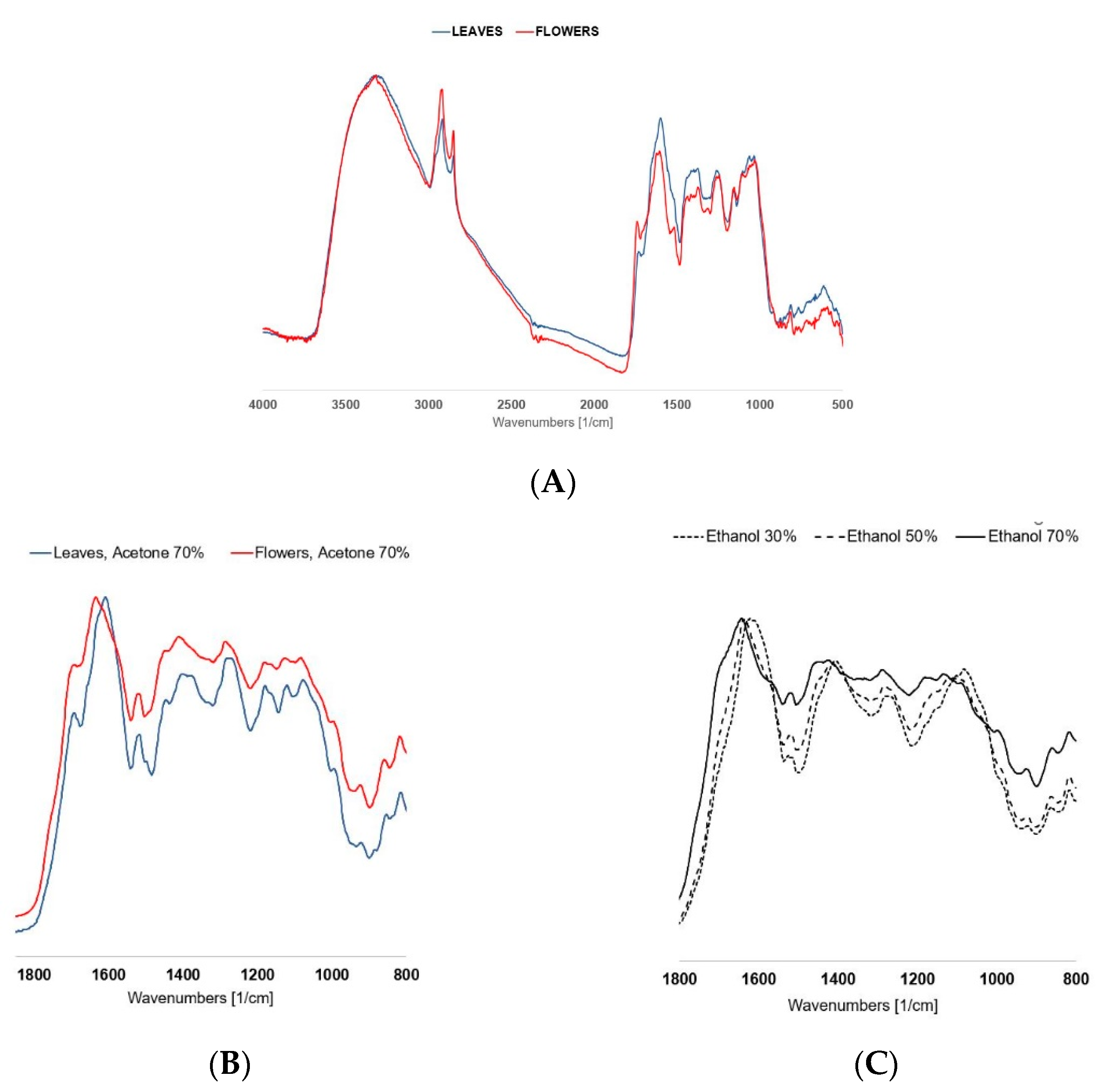
3.1. Characterization of Plant Samples by Fourier Transform Infrared Spectroscopy (FTIR)

The FTIR spectra were taken in the wavenumber range 400–4000 cm−1. The spectra were taken to demonstrate the differences between leaf and flower powder and their extracts in ethanol and acetone (Figure 4).
The complexity of the FTIR pattern limits the identification of individual components. However, using a fingerprint method [34], it is possible to observe differences in the spectrum pattern in the 850–1850 cm−1 region between powdered dried leaves and flowers, between extracts of 30%, 50%, and 70% ethanol, as well as between extracts of acetone.

3.2. Total Phenolic Content

The total phenolic content (TPC) was determined in the herbal preparations (Table 1). There was a higher TPC in the flower extracts ranging from 62.2 to 77.6 mg/g DW gallic acid equivalent (GAE), while the leaf extracts contained a maximum of 28.7 mg/g DW GAE of TPC. The extraction solvent type only affected the phenolic content in the case of the flower preparations (f = 6.984, p = 0.003). The acetone extracts of the T. vulgare flower showed higher TPC than the ethanol extracts (Mann–Whitney U = 2.5, p < 0.001), but there were no significant differences in the solvent concentration used (p = 1). All leaf extracts had similar amounts of TPC, and there were no statistical differences between solvents p < 0.05 (Table 1).

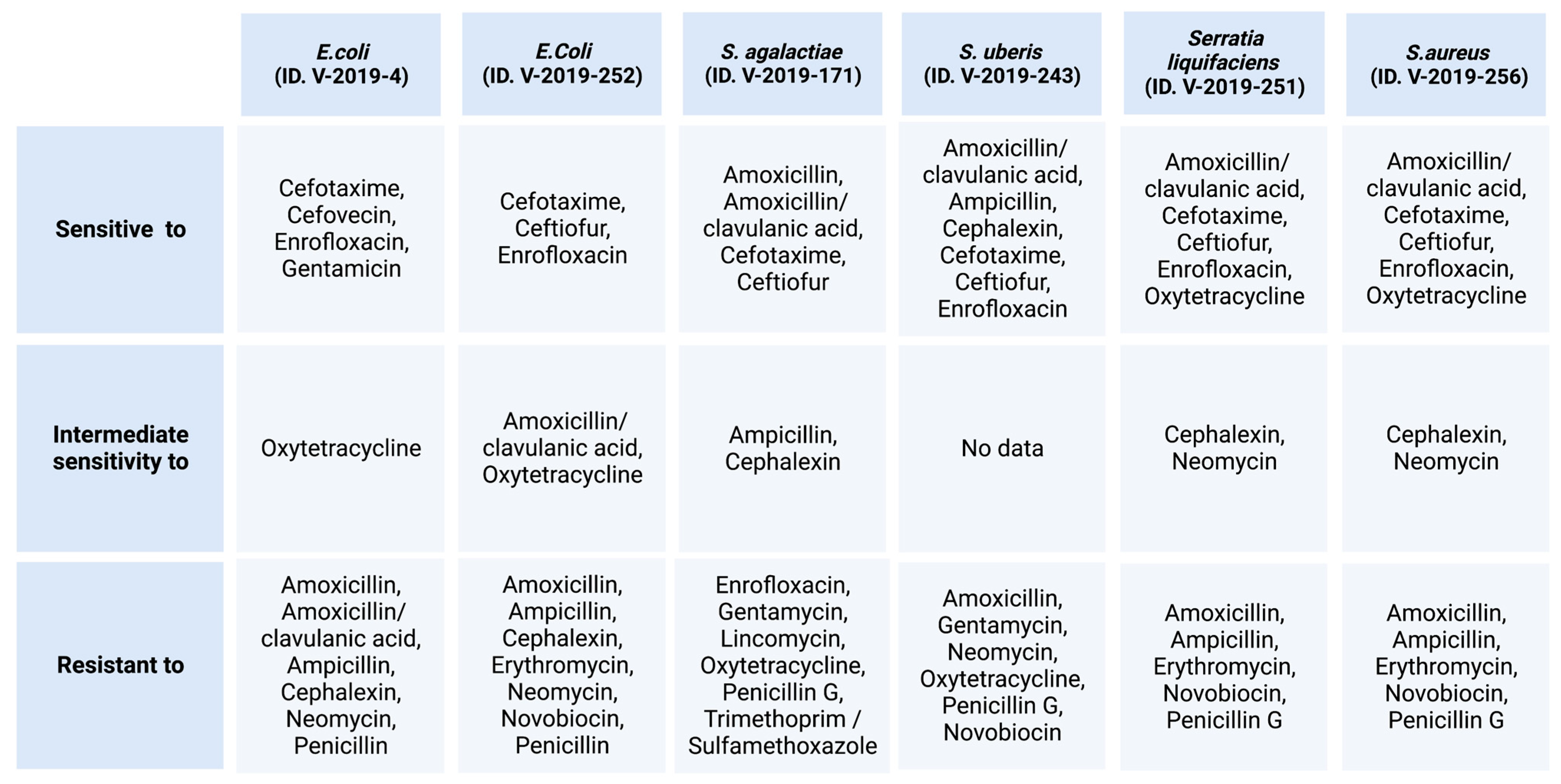
3.3. Antibacterial Effects of T. vulgare Extracts

All extracts of T. vulgare were first tested by agar disc diffusion tests. Extracts with the most antibacterial activity were tested with a broth microdilution test to obtain quantitative data.

3.3.1. Antibacterial Susceptibility—Agar Disc Diffusion Test

The results of the agar disc diffusion test for antibacterial effects of the extract are shown in the Table 1. Analysis of the inhibition zone results of T. vulgare flower extract showed that extracts made with a low-concentration solvent such as 30% ethanol and 30% acetone had no antibacterial effects. The 50% ethanol extracts of flowers were effective against the S. aureus bovine isolate and E. coli (V252). The 50% acetone extracts of flowers were effective against both S. aureus strains, E. coli (V252), and S. agalactiae. The 70% ethanol and 70% acetone extracts of flowers were both effective against S. aureus, E. coli, and S. agalactiae, but had no effect against S. uberis or Serratia liquefaciens. There was no significant difference between the results of 70% ethanol extracts and 70% acetone extracts of flowers (Table 2). Because ethanol is considered safer than acetone, it was used for further extract preparation and antibacterial analysis.
All leaf extracts of T. vulgare had an effect against the isolate of S. aureus bovine mastitis, S. uberis, and Serratia liquefaciens. The 70% ethanol extracts of leaves of T. vulgare and all types of acetone extracts showed effectiveness against S. aureus ATCC 25923 and S. agalactiae, but only the 70% acetone extract had a minimal effect against the reference culture of E. coli ATCC 25922. The best antibacterial effect was noted for 70% acetone and 70% ethanol extracts—with inhibition zone diameters ranging from 8.0 mm to 24.3 ± 2.9 mm and from 9.3 ± 1.3 mm to 24.3 ± 2.9 mm, respectively. Antibacterial effectiveness increased against S. aureus with an increase in concentration of the solvent, but no such relationship was observed for the rest of the bacterial samples. Since there were no significant differences (Table 2) between measurements amongst effective extracts, for further analysis, we chose the 70% ethanol extract of T. vulgare leaves as it is considered a safer solvent.

3.3.2. Broth Microdilution Method for Determination of Minimal Inhibitory Concentration (MIC) and Minimal Bactericidal Concentration (MBC)

To determine precise effective concentrations of most active extracts, we applied broth microdilution tests, and the results are shown in Table 3. T. vulgare flowers were effective against E. coli strains, S. aureus strains, and S. agalactiae, although the extract concentrations were high. However, T. vulgare leaves showed effects against S. aureus strains, Streptococcus strains, and Serratia liquefaciens, and was not tested against E. coli, since it did not show effectiveness in the previously mentioned agar disc diffusion test.
The 70% ethanol flower extract of T. vulgare had the lowest MIC/MBC of 3.4 mg/mL against S. aureus V256, and the second lowest MIC of 3.4 mg/mL and MBC of 6.8 mg/mL was against S. aureus ATCC 25923 which was the reference culture. For the leaf extract (70% ethanol), the lowest MIC and MBC measurements were observed against S. aureus V256 (MIC = 7.8 mg/mL and MBC = 15.73 mg/mL), and the second lowest were against S. agalactiae (MIC = 31.5 mg/mL, MBC = 62.9 mg/mL).

4. Discussion

Our investigation focused on the antibacterial effects of T. vulgare extracts against bovine mastitis-causing bacteria in vitro. The results of our study showed that 70% acetone and 70% ethanol extracts had the broadest range and the best effects against tested bacteria. Flower and leaf extracts were effective against S. aureus and S. agalactiae. The flower extracts of T. vulgare were effective against all E. coli strains tested. On the contrary, the leaf extracts of T. vulgare showed an effect against S. uberis and Serratia liquefaciens but no effect against E. coli. However, to have adequate antibacterial effects in all cases, the concentrations of extracts had to be high.
Several factors could affect the antibacterial effectiveness of extracts, such as the type of solvent, concertation, extraction process, and plant material [35]. Differing results for different extracts shown by Gevrenova et al. support the idea that the solvent polarity and the T. vulgare part used could affect the amounts and effects of the extracted phenolic compounds [19]. Ethanol extracts (70%, ratio of 1:10) of T. vulgare flowers grown in Transylvania showed low antibacterial effects against S. aureus (10 mm inhibition zone diameter), but unlike our extracts, theirs did not show any effect against E. coli. These extracts had 46.8–50.1 mg GAE/g of TPC, less than our T. vulgare flower ethanol extract [24]. An analysis of aerial parts of T. vulgare methanol extracts reported effects against S. aureus, but no effect against Gram-negative bacteria—namely E. coli [24]. Similar results were obtained in a study from Transylvania [36]. Diverse antimicrobial activity of T. vulgare parts (herb, leaf, flower, and essential oil) was noted by Devrnja et al. [37], and here, overall methanolic extracts showed better antibacterial effects against Gram-positive bacteria (S. aureus, Bacillus cereus, and Micrococcus flavus). Flower methanolic extracts inhibited Gram-negative bacteria such as E. coli and Enterobacter cloacae better than leaf extracts. These findings correspond to our results, where T. vulgare flowers had antibacterial effects against E. coli strains, but leaf extracts did not.
Previously reported characteristics of components of T. vulgare extracts showed that changing extraction solvent type and its concentration led to different extracted compositions. Additionally, T. vulgare flower extracts had distinctively different main peaks in FTIR spectra from those of T. vulgare leaf extracts, meaning that the chemical composition of both extracts differs [34]. Leaves and flowers of different T. vulgare samples had various polyphenolic profiles [38]. Similarly, when comparing leaves and discs of flower polyphenol types, it was reported that the disc contained apigenin, luteolin, and chrysoeriol, but leaves, on the other hand, had 6-hydroxyluteolin derivatives [39]. Antibacterial effects could depend on the part of the plant used in the extract, and also the solvent type used. In our study, there were no significant differences between the solvent type and the amount of TPC extracted; correlation between the amount of TPC and the antibacterial effects could not be calculated. Although 30% acetone extracts of T. vulgare flowers had the highest TPC, they did not have an antibacterial effect on bacteria. The dried flowers of T. vulgare contain less tannins than the leaves of T. vulgare (they contain 2.01% and 2.45%, respectively) [40]. Possibly, the combination of specific phenolic compounds could also influence the antibacterial activity.
Distinctive phenolic compounds have been reported to have a different effect on Gram-negative or Gram-positive bacteria [41], because they have differing mechanisms of action against bacteria. The antibacterial mechanism of phenolic compounds is mainly attributed to their ability to generate hydrogen peroxide, which, when coupled with the metal ion complexation capacity, results in the inhibition of the activity of essential enzymes and their ability to destabilize bacterial membranes, causing an increase in their permeability [42]. Differing mechanisms of action of polyphenol groups are provided by Daglia [43]. Flavonols had antibacterial effects against both Gram-positive and Gram-negative bacteria, possibly because of their aggregatory effect on all bacterial cells. Condensed tannins (proanthocyanidins) inhibit the growth of several pathogenic bacteria through several mechanisms, such as permeabilization of the cell membrane, destabilization of the cytoplasmic membrane, inhibition of extracellular microbial enzymes, direct actions on microbial metabolism, or deprivation of the substrates required for microbial growth (because proanthocyanidins chelate metals such as iron and zinc). Ellagitannins inhibited the growth of some Gram-negative intestinal bacteria, but gallotannins showed better activity against Gram-positive bacteria. Antibacterial effects are primarily due to the affinity of gallotannins for iron and the inactivation of membrane-bound proteins [43].
Other phytochemicals with antibacterial properties, such essential oils and their isolated components, could have affected the results. It was reported that the essential oil of T. vulgare flowers has antibacterial effects against E. coli, S. aureus, Enterobacter aerogenes, Sarcina lutea, Bacillus cereus, and Candida albicans. This effect varied depending on strains of the same species. It must be noted that the clinical isolates of bacteria are usually more resistant. In addition, the effect with this essential oil was found to be weaker than with the isolated α- and β-thujone mixture. However, authors of the study could not directly link the effect to α- and/or β-thujone alone. They noted possible antibacterial properties of other components of the essential oil and their synergism. Further, they explained that the composition of the essential oil of T. vulgare flowers varied in other studies (lower concentrations of β-thujone or lack of it) [44]. Varying chemotypes of T. vulgare were reported in Finland in 1987 [45], and it was observed that, based on the location, the main essential oil components (thujone, camphor, sabinene, umbellulone, bornyl acetone, 1,8-cineol, α-pinene, germacrene D) and their combinations changed. In a later study in Finland from 2001 [15], different chemotypes were reported, and they found 15 chemotypes with main components such as camphor, trans thujone, artemisia ketone, 1,8-cineole, and davadone-D. The composition of the essential oil of T. vulgare changes depending on the location; therefore, reports on medicinal effects might differ in each study.
In comparison, T. vulgare flower extracts had a lesser antibacterial effect than the essential oil obtained from it. The essential oil from T. vulgare inhibited the growth of some Gram-positive bacteria such as S. aureus, B. cereus, B. subtilis, and S. epidermidis, and had no effect on Gram-negative bacteria. It is possible that the essential oil could be responsible for antibacterial effects in the extracts made from T. vulgare flowers, or the yield of specific phenolic compounds was not substantial enough in the T. vulgare extract. It was reported that the extract of Tanacetum balsamita, which contained twice the amount of flavonoids, had a better antibacterial effect [46].
When comparing the results of the antibacterial effects in our study, it should be noted that bovine mastitis isolates were used, but other research studies focused on human disease-causing bacterial isolates or reference strains. Along with our results, a study conducted in Romania found that herbal extracts had better inhibition of S. aureus growth and a lower inhibition of E. coli, both isolated from bovine mastitis [47]. Conversely, our study revealed that T. vulgare extracts had some antibacterial effects against tested Streptococcus strains and Serratia liquefaciens. Extracts from the flowers of T. vulgare minimally inhibited the growth of S. agalactiae, but extracts from the leaves had minimal effects against S. uberis and Serratia liquefaciens. When the same bacterial isolates of bovine mastitis were tested, 70% acetone extracts of Quercus robur bark and 30% ethanol extracts of Calluna vulgaris herb had broader antibacterial spectra and lower MIC and MBC [48]. One study reported that Serratia liquefaciens was sensitive to ethanolic extracts of Mentha pulegium, Nepeta cataria, Melissa officinalis, Agastache foeniculum, Lavandula angustifolia, Origanum vulgare, Althaea officinalis, Plantago lanceolata, Artemisia absinthium, Populus nigra, and Evernia prunastri, all of which contain both essential oil and phenolic compounds. The 80% ethanol extracts of Senna macranthera, Artemisia absinthium, Cymbopogon nardus, and Baccharis dracunculifolia showed promising antibacterial effect against S. aureus strains isolated from bovine mastitis [49].
T. vulgare extracts were also effective against some resistant strains isolated from bovine mastitis-infected udders. The sensitivity of the bacteria is shown in Figure 3. The 70% ethanol extracts of T. vulgare flowers showed antibacterial activity against S. aureus V256, which is resistant to amoxicillin, ampicillin, erythromycin, novobiocin, and penicillin G. The 70% ethanol extracts of the leaves of T. vulgare had weaker effects than the flower extracts against the same S. aureus strain. The usual resistance cut-off point for penicillin is at a 26 mm inhibition zone diameter [30]. As T. vulgare extracts measured 20–24 mm, they are considered weaker than penicillin [30]. The growth of E. coli V4 (resistant to amoxicillin, amoxicillin/clavulanic acid, ampicillin, cephalexin, neomycin, penicillin) and E. coli V252 (resistant to amoxicillin, amoxicillin/clavulanic acid, cephalexin, erythromycin, neomycin, novobiocin, penicillin) was inhibited by 70% ethanol extracts of T. vulgare flowers. In this case, the necessary concentration was high and measured inhibition zones were low (8–10 mm). This is significantly lower than the breakpoint for standard treatments [30]. S. uberis (resistant to amoxicillin, gentamycin, neomycin, oxytetracycline, penicillin G, novobiocin) and S. agalactiae (resistant to enrofloxacin, gentamycin, lincomycin, oxytetracycline, penicillin G, trimethoprim/sulfamethoxazole), and Serratia liquefaciens (resistant to amoxicillin, ampicillin, erythromycin, novobiocin, penicillin G) were sensitive to T. vulgare leaf extracts in relatively high concentrations. This is the same as in the case of E. coli. T. vulgare flowers and leaves could be combined to obtain broader and better antibacterial effects.
Co-combination of herbal extracts and antibiotics has been reported. Two novel substances, guttiferone-A and 7-epiclusianone, isolated from the fruit of Garcinia brasiliensis, showed good antibacterial effects against bovine mastitis isolates, S. agalactiae and S. uberis, both of which were resistant to ampicillin and gentamicin, and S. uberis, which is resistant to ceftriaxone. Additionally, combined with traditionally used antibiotics, these substances also showed good synergism against Streptococcus spp. [50]. Different effects were observed in the study of the interactions of plant extracts and selected antibiotics affecting E. coli. Water and ethanol extracts prepared from the leaves of T. vulgare increased the MIC of ciprofloxacin, kanamycin, and ampicillin, while they did not affect the MIC of chloramphenicol and rifampicin. It was proposed that this may be due to the antioxidant effects of quercetin and tannin, which protect E. coli from the effects of ciprofloxacin [26]. This effect could also explain why some T. vulgare extracts did not inhibit E. coli, but further chemical analysis of phenolic compound types is needed to confirm this.
There is no standard cut-off point for the antibacterial effectiveness for plant extracts. Authors differentiate between extracts and derived compounds for which the cut-off point is usually lower. The suggested MIC for herbal medicine or extract is as follows: significant effect is when MIC < 0.1 mg/mL, moderate effect is when MIC 0.1—0.625 mg/mL, and weak effect is when MIC > 0.625 mg/mL [51,52]. According to this clause, our tested T. vulgare extracts have a weak antibacterial effect (MIC 3.4—125.9 mg/mL). Previously reported plant MICs had broad ranges. The antibacterial activity of northern Peruvian medicinal plants was also diverse and, in some cases, low, their MIC ranging from 0.008—256 mg/mL [53]. In this aspect, our results are comparable to other plant studies.
In vivo use of herbal preparations should be performed with caution [54,55] as bovine mammary gland tissue is sensitive [56], so development of products containing T. vulgare extracts should undergo a clinical evaluation of their effect on the bovine mammary gland.

5. Conclusions

T. vulgare has some antibacterial effects in vitro. Extracts of T. vulgare flowers minimally inhibited the growth of S. aureus, and inhibited E. coli and S. uberis even less, but the extracts of leaves, on the contrary, had no effect on E. coli. It moderately inhibited the growth of S. aureus, and had a weak effect against S. agalactiae, S. uberis, and Serratia liquefaciens. Although the necessary concentrations were high, 70% ethanol extracts of T. vulgare flowers and leaves could be combined to obtain better and broader antibacterial effects.
The antibacterial effects were dependent on the extract solvent concentration, since 30% ethanol extracts had no antibacterial effects. However, the total phenolic content was more affected by solvent type and less so by concentration. Since the types of phenolics vary in parts of T. vulgare, the activities of the extracts are different. Precise analysis of the chemical constituents and their antibacterial activity is necessary.
Considering that the obtained extracts could contain compounds with certain irritating properties, additional investigations are necessary to determine the possible risks to bovine mammary glands. In future studies, to maximize extract effects, modern extraction methods could be applied; new drug delivery forms such as nanoparticles could be researched and developed.

Author Contributions

Conceptualization, funding acquisition, resources, project administration, supervision, validation, D.B., L.K. and R.K.; methodology, R.Š., I.S. and A.B. (Agnese Brangule); formal analysis, R.Š. and A.B. (Agnese Brangule); investigation, R.Š., I.S. and A.B. (Agnese Brangule); data curation, R.Š. and A.B. (Agnese Brangule); writing—original draft preparation, R.Š.; writing—review and editing, D.B., R.K., L.K, A.B. (Agnese Brangule), I.S., L.L. and A.B. (Ance Bārzdiņa); visualization, A.B. (Ance Bārzdiņa). All authors have read and agreed to the published version of the manuscript.

Funding

This research was funded by the European Union’s Horizon 2020 research and innovation program under the grant agreement No 857287 and this research is supported by the Ministry of Agriculture and Rural Support Service Republic of Latvia with the European Agricultural Fund for Rural Development—Development of herbal plant containing medical extracts with anti-parasitic effect No 18-00-A01620-000028.

Institutional Review Board Statement

Not applicable.

Informed Consent Statement

Not applicable.

Data Availability Statement

Not applicable.

Acknowledgments

The authors thank the Research Laboratory of Biotechnology of Latvia University of Life Sciences and Technology for kindly supplying bovine mastitis-inducing bacterial samples.

Conflicts of Interest

The authors declare no conflict of interest.

References

  1. Heikkilä, A.-M.; Liski, E.; Pyörälä, S.; Taponen, S. Pathogen-Specific Production Losses in Bovine Mastitis. J. Dairy Sci. 2018, 101, 9493–9504. [Google Scholar] [CrossRef] [PubMed] [Green Version]
  2. Gonçalves, J.L.; Kamphuis, C.; Martins, C.M.M.R.; Barreiro, J.R.; Tomazi, T.; Gameiro, A.H.; Hogeveen, H.; dos Santos, M.V. Bovine Subclinical Mastitis Reduces Milk Yield and Economic Return. Livest. Sci. 2018, 210, 25–32. [Google Scholar] [CrossRef]
  3. Guzmán-Luna, P.; Nag, R.; Martínez, I.; Mauricio-Iglesias, M.; Hospido, A.; Cummins, E. Quantifying Current and Future Raw Milk Losses Due to Bovine Mastitis on European Dairy Farms under Climate Change Scenarios. Sci. Total Environ. 2022, 833, 155149. [Google Scholar] [CrossRef]
  4. Feliciano, R.; Boué, G.; Mohssin, F.; Hussaini, M.M.; Membré, J.-M. Probabilistic Modelling of Escherichia Coli Concentration in Raw Milk under Hot Weather Conditions. Food Res. Int. 2021, 149, 110679. [Google Scholar] [CrossRef]
  5. Cheng, W.N.; Han, S.G. Bovine Mastitis: Risk Factors, Therapeutic Strategies, and Alternative Treatments—A Review. Asian-Australas. J. Anim. Sci. 2020, 33, 1699–1713. [Google Scholar] [CrossRef] [PubMed]
  6. Guo, X.; Akram, S.; Stedtfeld, R.; Johnson, M.; Chabrelie, A.; Yin, D.; Mitchell, J. Distribution of Antimicrobial Resistance across the Overall Environment of Dairy Farms—A Case Study. Sci. Total Environ. 2021, 788, 147489. [Google Scholar] [CrossRef]
  7. World Health Organization. WHO Guidelines On Use Of Medically Important Antimicrobials in Food-Producing Animals; World Health Organization: Geneva, Switzerland, 2017. [Google Scholar]
  8. EIP-AGRI Agriculture and Innovation. Quality Low Input Food Project Mastitis Control in Organic Dairy. Available online: https://ec.europa.eu/eip/agriculture/en/content/mastitis-control-organic-dairy (accessed on 1 December 2022).
  9. Mayer, M.; Vogl, C.R.; Amorena, M.; Hamburger, M.; Walkenhorst, M. Treatment of Organic Livestock with Medicinal Plants: A Systematic Review of European Ethnoveterinary Research. Complement. Med. Res. 2014, 21, 375–386. [Google Scholar] [CrossRef] [Green Version]
  10. Phillips, H.N.; Heins, B.J. Alternative Practices in Organic Dairy Production and Effects on Animal Behavior, Health, and Welfare. Animals 2022, 12, 1785. [Google Scholar] [CrossRef]
  11. Mushtaq, S.; Shah, A.M.; Shah, A.; Lone, S.A.; Hussain, A.; Hassan, Q.P.; Ali, M.N. Bovine Mastitis: An Appraisal of Its Alternative Herbal Cure. Microb. Pathog. 2018, 114, 357–361. [Google Scholar] [CrossRef]
  12. Ruegg, P.L. Management of Mastitis on Organic and Conventional Dairy Farms. J. Anim. Sci. 2009, 87, 43–55. [Google Scholar] [CrossRef]
  13. Aćimović, M.; Puvača, N. Tanacetum vulgare L.—A Systematic Review. Technol. Eng. Manag. J. Agron. Technol. Eng. Manag. 2020, 2020, 416–422. [Google Scholar]
  14. Nurzyńska-Wierdak, R.; Sałata, A.; Kniaziewicz, M. Tansy (Tanacetum vulgare L.)—A Wild-Growing Aromatic Medicinal Plant with a Variable Essential Oil Composition. Agronomy 2022, 12, 277. [Google Scholar] [CrossRef]
  15. Keskitalo, M.; Pehu, E.; Simon, J.E. Variation in Volatile Compounds from Tansy (Tanacetum vulgare L.) Related to Genetic and Morphological Differences of Genotypes. Biochem. Syst. Ecol. 2001, 29, 267–285. [Google Scholar] [CrossRef]
  16. Rohloff, J.; Mordal, R.; Dragland, S. Chemotypical Variation of Tansy (Tanacetum vulgare L.) from 40 Different Locations in Norway. J. Agric. Food Chem. 2004, 52, 1742–1748. [Google Scholar] [CrossRef]
  17. Mihaylova, D.; Vrancheva, R.; Desseva, I.; Ivanov, I.; Dincheva, I.; Popova, M.; Popova, A. Analysis of the GC-MS of Volatile Compounds and the Phytochemical Profile and Antioxidant Activities of Some Bulgarian Medicinal Plants. Z. Nat. C 2018, 74, 45–54. [Google Scholar] [CrossRef] [PubMed]
  18. Mihaylova, D.; Vrancheva, R.; Petkova, N.; Ognyanov, M.; Desseva, I.; Ivanov, I.; Popova, M.; Popova, A. Carotenoids, Tocopherols, Organic Acids, Charbohydrate and Mineral Content in Different Medicinal Plant Extracts. Z. Nat. C 2018, 73, 439–448. [Google Scholar] [CrossRef]
  19. Ak, G.; Gevrenova, R.; Sinan, K.I.; Zengin, G.; Zheleva, D.; Mahomoodally, M.F.; Senkardes, I.; Brunetti, L.; Leone, S.; di Simone, S.C.; et al. Tanacetum vulgare L. (Tansy) as an Effective Bioresource with Promising Pharmacological Effects from Natural Arsenal. Food Chem. Toxicol. 2021, 153, 112268. [Google Scholar] [CrossRef]
  20. Barceloux, D.G. Tansy (Tanacetum vulgare L.). In Medical Toxicology of Natural Substances; Chapter 93; John Wiley & Sons, Inc.: Hoboken, NJ, USA, 2008; pp. 614–616. [Google Scholar]
  21. Mitich, L.W. Tansy. Weed Technol. 1992, 6, 242–244. [Google Scholar] [CrossRef]
  22. Sowa, P.; Marcinčáková, D.; Miłek, M.; Sidor, E.; Legáth, J.; Dżugan, M. Analysis of Cytotoxicity of Selected Asteraceae Plant Extracts in Real Time, Their Antioxidant Properties and Polyphenolic Profile. Molecules 2020, 25, 5517. [Google Scholar] [CrossRef]
  23. Onozato, T.; Nakamura, C.V.; Cortez, D.A.G.; Filho, B.P.D.; Ueda-Nakamura, T. Tanacetum vulgare: Antiherpes Virus Activity of Crude Extract and the Purified Compound Parthenolide. Phytother. Res. 2009, 23, 791–796. [Google Scholar] [CrossRef] [PubMed]
  24. Muresan, M.; Benedec, D.; Vlase, L.; Oprean, R.; Toiu, A.; Oniga, I. Screening of Polyphenolic Compounds, Antioxidant and Antimicrobial Properties of Tanacetum vulgare from Transylvania. Stud. Univ. Babes-Bolyai. Chem. 2015, 60, 12718. [Google Scholar]
  25. Sridhar, A.; Ponnuchamy, M.; Kumar, P.S.; Kapoor, A.; Vo, D.-V.N.; Prabhakar, S. Techniques and Modeling of Polyphenol Extraction from Food: A Review. Environ. Chem. Lett. 2021, 19, 3409–3443. [Google Scholar] [CrossRef]
  26. Smirnova, G.; Samoilova, Z.; Muzyka, N.; Oktyabrsky, O. Influence of Plant Polyphenols and Medicinal Plant Extracts on Antibiotic Susceptibility of Escherichia coli. J. Appl. Microbiol. 2012, 113, 192–199. [Google Scholar] [CrossRef] [PubMed]
  27. Do, Q.D.; Angkawijaya, A.E.; Tran-Nguyen, P.L.; Huynh, L.H.; Soetaredjo, F.E.; Ismadji, S.; Ju, Y.-H. Effect of Extraction Solvent on Total Phenol Content, Total Flavonoid Content, and Antioxidant Activity of Limnophila Aromatica. J. Food Drug Anal. 2014, 22, 296–302. [Google Scholar] [CrossRef] [Green Version]
  28. Avsejenko, J. Pieredze Un Ieteikumi Govs Mastītu Laboratoriskā Diagnostikā. Vet. Z. 2015, 1, 19–28. [Google Scholar]
  29. World Health Organization. WHO Guidelines on Good Agricultural and Collection Practices (GACP) for Medicinal Plants; World Health Organization: Geneva, Switzerland, 2003. [Google Scholar]
  30. EUCAST Clinical Breakpoints—Breakpoints and Guidance. Available online: https://www.eucast.org/clinical_breakpoints (accessed on 7 February 2023).
  31. Gajic, I.; Kabic, J.; Kekic, D.; Jovicevic, M.; Milenkovic, M.; Mitic Culafic, D.; Trudic, A.; Ranin, L.; Opavski, N. Antimicrobial Susceptibility Testing: A Comprehensive Review of Currently Used Methods. Antibiotics 2022, 11, 427. [Google Scholar] [CrossRef]
  32. Klančnik, A.; Piskernik, S.; Jeršek, B.; Možina, S.S. Evaluation of Diffusion and Dilution Methods to Determine the Antibacterial Activity of Plant Extracts. J. Microbiol. Methods 2010, 81, 121–126. [Google Scholar] [CrossRef]
  33. Singleton, V.L.; Rossi, J.A. Colorimetry of Total Phenolics with Phosphomolybdic-Phosphotungstic Acid Reagents. Am. J. Enol. Vitic. 1965, 16, 144. [Google Scholar]
  34. Brangule, A.; Šukele, R.; Bandere, D. Herbal Medicine Characterization Perspectives Using Advanced FTIR Sample Techniques—Diffuse Reflectance (DRIFT) and Photoacoustic Spectroscopy (PAS). Front. Plant. Sci. 2020, 11, 356. [Google Scholar] [CrossRef] [PubMed]
  35. Chaves, J.O.; de Souza, M.C.; da Silva, L.C.; Lachos-Perez, D.; Torres-Mayanga, P.C.; Machado, A.P.d.F.; Forster-Carneiro, T.; Vázquez-Espinosa, M.; González-de-Peredo, A.V.; Barbero, G.F.; et al. Extraction of Flavonoids From Natural Sources Using Modern Techniques. Front. Chem. 2020, 8, 507887. [Google Scholar] [CrossRef]
  36. Ivănescu, B.; Tuchiluș, C.; Corciovă, A.; Lungu, C.; Teodor Mihai, C.; Gheldiu, A.; Vlase, L. Antioxidant, Antimicrobial and Cytotoxic Activity of Tanacetum Vulgare, Tanacetum Corymbosum and Tanacetum Macrophyllum Extracts. Farmacia 2018, 66, 282–288. [Google Scholar]
  37. Devrnja, N.; Anđelković, B.; Aranđelović, S.; Radulović, S.; Soković, M.; Krstić-Milošević, D.; Ristić, M.; Ćalić, D. Comparative Studies on the Antimicrobial and Cytotoxic Activities of Tanacetum vulgare L. Essential Oil and Methanol Extracts. S. Afr. J. Bot. 2017, 111, 212–221. [Google Scholar] [CrossRef]
  38. Williams, C.A.; Harborne, J.B.; Eagles, J. Variations in Lipophilic and Polar Flavonoids in the Genus Tanacetum. Phytochemistry 1999, 52, 1301–1306. [Google Scholar] [CrossRef]
  39. Williams, C. The Flavonoids of Tanacetum parthenium and T. vulgare and Their Anti-Inflammatory Properties. Phytochemistry 1999, 51, 417–423. [Google Scholar] [CrossRef]
  40. Šukele, R.; Bandere, D.; Koka, R. Quantitative Analysis of Tannins in Some Herbs of Latvian Flora. In RSU Research Week 2021: Knowledge for Use in Practice; Rīga Stradiņš University: Rīga, Latvia, 2021; p. 395. [Google Scholar]
  41. Rauha, J.-P.; Remes, S.; Heinonen, M.; Hopia, A.; Kahkonen, M.; Kujala, T.; Pihlaja, K.; Vuorela, H.; Vuorela, P. Antimicrobial Effects of Finnish Plant Extracts Containing Flavonoids and Other Phenolic Compounds. Int. J. Food. Microbiol. 2000, 56, 3–12. [Google Scholar] [CrossRef]
  42. Morena, A.G.; Bassegoda, A.; Natan, M.; Jacobi, G.; Banin, E.; Tzanov, T. Antibacterial Properties and Mechanisms of Action of Sonoenzymatically Synthesized Lignin-Based Nanoparticles. ACS Appl. Mater. Interfaces 2022, 14, 37270–37279. [Google Scholar] [CrossRef] [PubMed]
  43. Daglia, M. Polyphenols as Antimicrobial Agents. Curr. Opin. Biotechnol. 2012, 23, 174–181. [Google Scholar] [CrossRef] [PubMed]
  44. Radulović, N.S.; Genčić, M.S.; Stojanović, N.M.; Randjelović, P.J.; Stojanović-Radić, Z.Z.; Stojiljković, N.I. Toxic Essential Oils. Part V: Behaviour Modulating and Toxic Properties of Thujones and Thujone-Containing Essential Oils of Salvia officinalis L., Artemisia absinthium L., Thuja occidentalis L. and Tanacetum vulgare L. Food Chem. Toxicol. 2017, 105, 355–369. [Google Scholar] [CrossRef] [PubMed]
  45. Holopainen, M.; Hiltunen, R.; von Schantz, M. A Study on Tansy Chemotypes. Planta Med. 1987, 53, 284–287. [Google Scholar] [CrossRef] [PubMed]
  46. Bączek, K.B.; Kosakowska, O.; Przybył, J.L.; Pióro-Jabrucka, E.; Costa, R.; Mondello, L.; Gniewosz, M.; Synowiec, A.; Węglarz, Z. Antibacterial and Antioxidant Activity of Essential Oils and Extracts from Costmary (Tanacetum balsamita L.) and Tansy (Tanacetum vulgare L.). Ind. Crops Prod. 2017, 102, 154–163. [Google Scholar] [CrossRef]
  47. Pașca, C.; Mărghitaș, L.; Dezmirean, D.; Bobiș, O.; Bonta, V.; Chirilă, F.; Matei, I.; Fiț, N. Medicinal Plants Based Products Tested on Pathogens Isolated from Mastitis Milk. Molecules 2017, 22, 1473. [Google Scholar] [CrossRef] [Green Version]
  48. Šukele, R.; Skadiņš, I.; Koka, R.; Bandere, D. Antibacterial Effects of Oak Bark (Quercus Robur) and Heather Herb (Calluna vulgaris L.) Extracts against the Causative Bacteria of Bovine Mastitis. Vet. World 2022, 15, 2315–2322. [Google Scholar] [CrossRef] [PubMed]
  49. Diaz, M.A.N.; Rossi, C.C.; Mendonça, V.R.; Silva, D.M.; Ribon, A.d.O.B.; Aguilar, A.P.; Muñoz, G.D. Screening of Medicinal Plants for Antibacterial Activities on Staphylococcus Aureus Strains Isolated from Bovine Mastitis. Rev. Bras. Farmacogn. 2010, 20, 724–728. [Google Scholar] [CrossRef] [Green Version]
  50. Maia, N.L.; de Barros, M.; de Oliveira, L.L.; Cardoso, S.A.; dos Santos, M.H.; Pieri, F.A.; Ramalho, T.C.; da Cunha, E.F.F.; Moreira, M.A.S. Synergism of Plant Compound with Traditional Antimicrobials Against Streptococcus spp. Isolated from Bovine Mastitis. Front. Microbiol. 2018, 9, 1203. [Google Scholar] [CrossRef] [PubMed]
  51. Liang, J.; Huang, X.; Ma, G. Antimicrobial Activities and Mechanisms of Extract and Components of Herbs in East Asia. RSC Adv. 2022, 12, 29197–29213. [Google Scholar] [CrossRef] [PubMed]
  52. Kuete, V.; Efferth, T. Cameroonian Medicinal Plants: Pharmacology and Derived Natural Products. Front. Pharm. 2010, 1, 123. [Google Scholar] [CrossRef] [PubMed] [Green Version]
  53. Bussmann, R.W.; Malca-García, G.; Glenn, A.; Sharon, D.; Chait, G.; Díaz, D.; Pourmand, K.; Jonat, B.; Somogy, S.; Guardado, G.; et al. Minimum Inhibitory Concentrations of Medicinal Plants Used in Northern Peru as Antibacterial Remedies. J. Ethnopharmacol. 2010, 132, 101–108. [Google Scholar] [CrossRef] [Green Version]
  54. Pinedo, P.; Karreman, H.; Bothe, H.; Velez, J.; Risco, C. Efficacy of a Botanical Preparation for the Intramammary Treatment of Clinical Mastitis on an Organic Dairy Farm. Can. Vet. J. 2013, 54, 479–484. [Google Scholar] [CrossRef]
  55. Maramulla, A.; Ambica, G.; Kosqapati, L.; Bommu, S.; Katta, P. Efficacy of Herbal Prepararions in the Therapy of Sub Clinical Mastitis in Cows of Periurban Areas of Hyderabad. Pharma Innov. J. 2019, 8, 186–188. [Google Scholar]
  56. Sanderson, C.J. The Treatment of Mastitis with Intrammary Infusions. Aust. Vet. J. 1966, 42, 47–53. [Google Scholar] [CrossRef]
Figure 1. Plant material extraction process.
Applsci 13 03369 g001
Figure 2. Extract preparation process.
Applsci 13 03369 g002
Figure 3. Antibacterial susceptibility for the bovine mastitis clinical isolates.
Applsci 13 03369 g003
Figure 4. FTIR DRIFT spectra. (A) T. vulgare parts: flowers and leaves; (B) Extract in 70% acetone of T. vulgare parts: flowers and leaves; (C) Extract in 70%, 50%, and 30% ethanol of T. vulgare flowers.
Applsci 13 03369 g004
Table 1. Total Phenolic Content of the ethanol and acetone extracts of flowers and leaves of T. vulgare.
Extract SampleExtraction SolventTPC GAE mg/g DW ± SD
T. vulgare flower
extract
Ethanol 30% 62.5 ±10.9 a
Ethanol 50%63.4 ±3.2 a
Ethanol 70%62.3 ±9.2 a
Acetone 30%77.6 ±2.9 b
Acetone 50%77.4 ±3.4 b
Acetone 70%76.7 ±3.4 b
T. vulgare leaf
extract
Ethanol 30%28.7 ±9.6 c
Ethanol 50%27.6 ±6.9 c
Ethanol 70%24.5 ±4.5 c
Acetone 30%20.9 ±4.1 c
Acetone 50%28.1 ±3.6 c
Acetone 70%22.3 ±6.2 c
TPC—Total Phenolic Content, GAE—Gallic Acid Equivalent, DW—Dry Plant weight, SD—standard deviation. Within columns, values with the same superscript letter are not significantly different at the p ≤ 0.05 level according to ANOVA and post hoc Tukey test.
Table 2. Inhibition zones of T. vulgaris extracts for antibacterial susceptibility—agar disc diffusion test.
Extract Type
by Solvent
Inhibition Zone, mm ±SD
S. aureus ATCC 25923S. aureus V256S. uberis V243S. agalactiae V171E. coli ATCC 25922E. coli V4E. coli V252Serratia liquefaciens V251
T. vulgare flowerE30%nnnnnnnn
E50%n17.0 ± 2.5 annnn8.7 ± 0.5 an
E70%14.8 ± 1.3 a20.8 ± 0.9 bn9.3 ± 1.2 a8.8 ± 0.5 a7.8 ± 0.5 a10.0 ± 1.4 an
A30%nnnnnnnn
A50%19.0 ± 2.5 b17.5 ± 2.6 an8.3 ± 0.5 ann7.5 ± 0.5 an
A70%15.3 ± 2.1 a21.8 ± 2.4 bn8.3 ± 0.5 a9.8 ± 0.9 a8.0 ± 0 a9.5 ± 1.3 an
T. vulgare leafE30%n12.8 ± 1.7 c8.8 ± 0.5 annnnn
E50%n13.8 ± 1.5 c9.8 ± 2.2 a9.5 ± 1.0 annn8.5 ± 1.0 a
E70%17.5 ± 2.1 b24.3 ± 2.9 d9.8 ± 0.5 a9.3 ± 1.3 annn10.5 ± 1.9 a
A30%11.3 ± 3.8 b17.8 ± 1.7 a9.0 ± 1.4 a8.8 ± 0.9 annn8.3 ± 0.5 a
A50%15.0 ± 0.8 a20.5 ± 0.9 b8.5 ± 0.6 a8.3 ± 0.5 annn8.5 ± 1.0 a
A70%18.3 ± 1.3 b24.3 ± 2.9 d8.0 ± 0.0 a8.3 ± 0.5 a9.3 ± 1.5 ann8.8 ± 1.0 a
Bacterial cultures: Escherichia coli (ID. V-2019-4), E. coli (ID. V-2019-252), E. coli (ATCC 25922), Streptococcus agalactiae (ID. V-2019-171), S. uberis (ID. V-2019-243), Serratia liquefaciens (ID. V-2019-251), Staphylococcus aureus (ID. V-2019-256), S. aureus (ATCC 25923). A—acetone extract and concentration, E—ethanol extract and concentration; n—not inhibited (diameter of the inhibition zone measured below 7 mm on average). SD—standard deviation. All means calculated by ANOVA from at least three measurements. Within each column, values with the same superscript letter are not significantly different at the p ≤ 0.05 level, according to the post hoc Tukey test.
Table 3. Minimal Inhibitory Concentration and Minimal Bactericidal Concentration of T. vulgare extracts by the Broth Microdilution Method.
Bacteria
Sample
T. vulgare Flower Extract,
mg/mL
T. vulgare Leaf Extract,
mg/mL
MICMBCMICMBC
E coliATCC 2592253.953.9--
V25253.9107.8--
V453.9107.8--
S. aureusATCC 259233.46.8 7.815.7
V2563.43.415.7125.9
S. agalactiaeV17153.953.931.462.9
S. uberisV243--62.9125.9
Serratia liquefaciensV251--125.9125.9
MIC—Minimal Inhibitory Concentration, MBC—Minimal Bactericidal Concentration. Bacterial cultures: Escherichia coli (ID. V-2019-4), E. coli (ID. V-2019-252), E. coli (ATCC 25922), Streptococcus agalactiae (ID. V-2019-171), S. uberis (ID. V-2019-243), Serratia liquefaciens (ID. V-2019-251), Staphylococcus aureus (ID. V-2019-256), S. aureus (ATCC 25923).
Disclaimer/Publisher’s Note: The statements, opinions and data contained in all publications are solely those of the individual author(s) and contributor(s) and not of MDPI and/or the editor(s). MDPI and/or the editor(s) disclaim responsibility for any injury to people or property resulting from any ideas, methods, instructions or products referred to in the content.

