Systematic Review

Acceleration Techniques for Teeth Movements in Extractive Orthodontic Therapy

1
Department of Interdisciplinary Medicine, School of Medicine, University of Bari “Aldo Moro”, 70124 Bari, Italy
2
Implant Dentistry College of Medicine and Dentistry, Birmingham B4 6BN, UK
*
Author to whom correspondence should be addressed.
These authors contributed equally to this work as first authors.
Appl. Sci. 2023, 13(17), 9759; https://doi.org/10.3390/app13179759
Submission received: 13 July 2023 / Revised: 24 August 2023 / Accepted: 26 August 2023 / Published: 29 August 2023

Abstract

:
For a world that is constantly trying to speed up every procedure while obtaining the maximum result, traditional orthodontics have the biological limitation of using light and constant forces that allow tooth movement in a time frame that is only sometimes short. The treatment time could be lengthened if surgical procedures are programmed in the plan. Methods to accelerate tooth movement and reduce the duration of treatment while minimising complications are investigated and reported in the dental literature (e.g., low-level laser therapy, corticotomy, and micro-osteoperforations). This systematic review aims to analyse and summarise the strategies for quickening orthodontic movement during extraction orthodontic treatment, including any potential drawbacks or adverse consequences. The review will evaluate each approach’s effectiveness, safety, and evidence quality, compare their benefits and disadvantages, and analyse the implications for clinical practice and future research. Pubmed, Science Direct, Scopus, and Web of Science were searched using the keywords “acceleration” AND “dental movement” AND “orthodontic” between 1 April 2003 and 1 April 2023. After carefully scanning the study findings, forty-four publications were chosen for the systematic review. Most therapies discussed and provided in the literature seem promising and successful in enhancing orthodontic treatments. The success of operations like corticotomies, piezo-incisions, micro-osteoperforations, osteogenic distraction, low-level laser therapy, the administration of pharmacological treatments, and infiltrations with PRF and PRP were statistically significant and appear to be promising and effective in optimising orthodontic treatments. These strategies expedite treatment and enhance the patient experience, potentially broadening orthodontic appeal and minimising issues like cavities and enamel demineralisation. Further studies, with larger samples and standardised treatment protocols, are needed to investigate the efficacy of these tooth movement acceleration modalities.

1. Introduction

Orthodontics is a fundamental speciality of dentistry that deals with diagnosing, preventing, and treating dental and facial abnormalities. Orthodontic treatment aims to improve patients’ aesthetics, function, and oral health by aligning teeth and correcting skeletal discrepancies. However, traditional orthodontic treatments can take considerable time, often between 1 and 3 years, depending on the case’s complexity. This long duration of therapy may discourage some patients from seeking orthodontic intervention and expose patients to possible complications, such as enamel demineralisation, caries, and gingival recession [1,2,3].
In recent years, there has been a growing interest in researching methods to accelerate orthodontic tooth movement (OTM) and reduce the duration of treatment. Several approaches have been proposed and studied, including corticotomies, micro-osteoperforations (MOPs), vibration therapy (VT), low-level pulsed light therapy (LLLT), platelet-rich plasma (PRP) and platelet-rich fibrinogen (PRF), drug therapy, and dentoalveolar distraction (DAD). Despite the growing body of literature, there is still no clear consensus on which methods are most effective, safe, and feasible in the clinical setting [4,5,6,7].
This systematic review aims to examine and synthesise the evidence regarding different methods of accelerating OTM in extractive orthodontic treatment. A fixed orthodontic procedure lasts two to three years, and canine retraction is a crucial and time-consuming step in fixed orthodontic treatment for individuals who have had their premolars extracted. The canine retraction process uses traditional techniques at an average rate of 0.5 to 1 mm monthly. As a result, canine retraction alone requires 5 to 9 months and raises the risk of caries, external root resorption, and decreased patient participation. Therefore, making an effort to accelerate OTM and reduce the duration of treatment can be very helpful for improving future orthodontic treatment [8,9].
This review will evaluate the efficacy, safety, and quality of the evidence for each approach, compare the advantages and disadvantages of the various methods, and discuss the implications for clinical practice and future research. Through this analysis, we aim to provide a solid foundation for orthodontists and researchers to improve orthodontic treatments further and increase patient satisfaction.
It is important to note that not all methods are suitable for all patients and should be carefully evaluated and discussed with the orthodontist before proceeding. Some of the most used methods of accelerating OTM include:
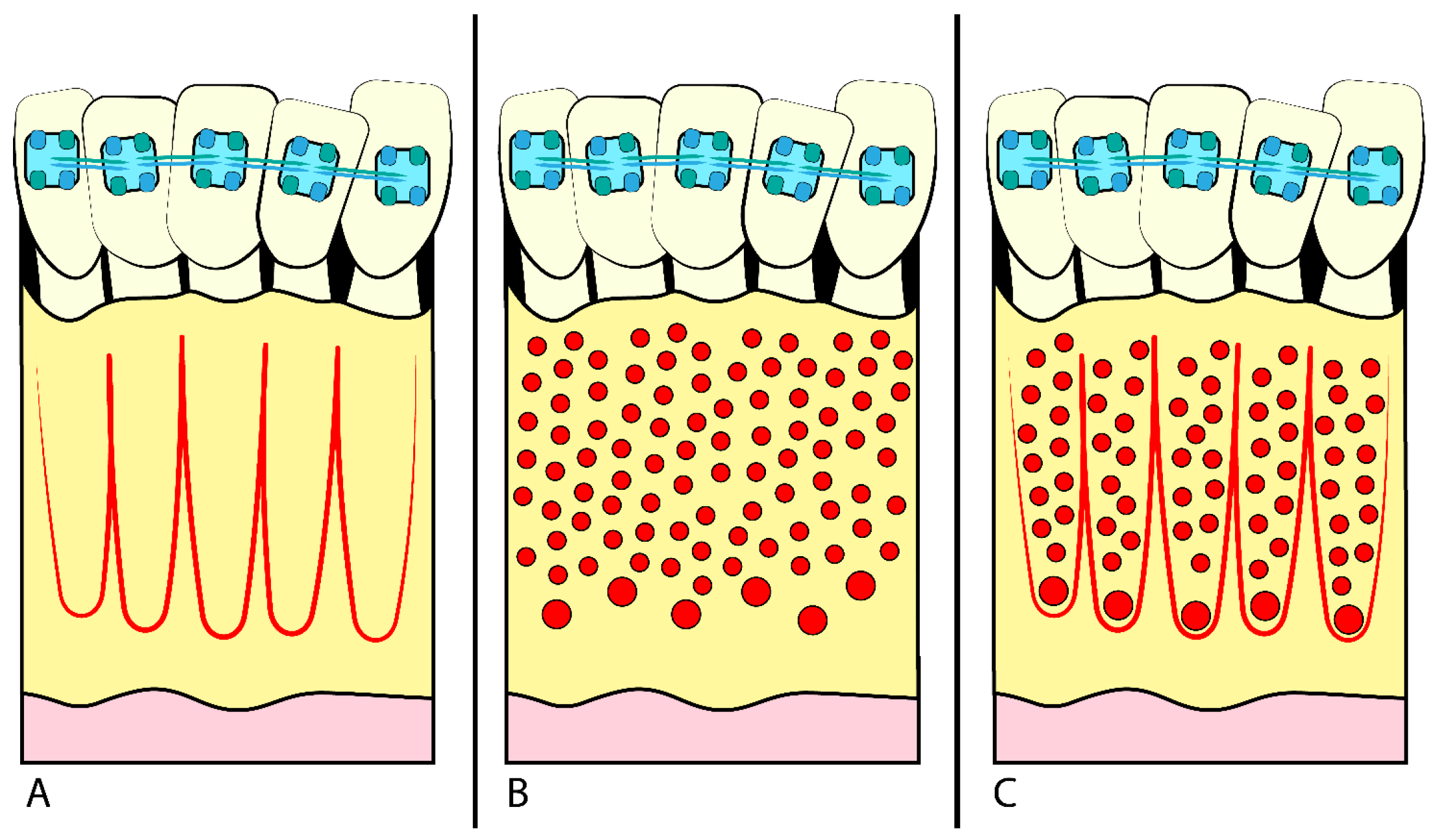
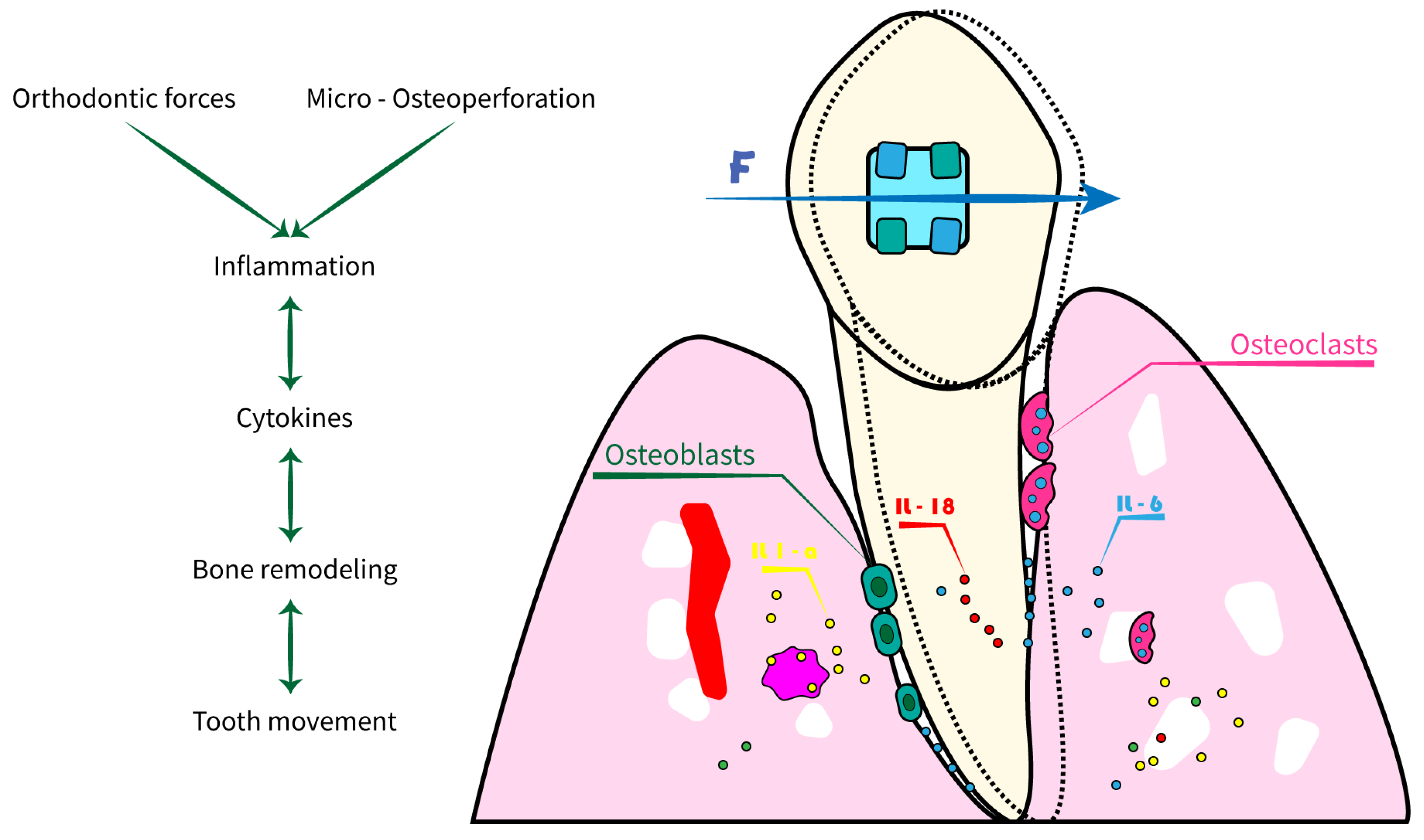
  • Corticotomy is the execution of small incisions in the alveolar bone surrounding the teeth to facilitate their movement (Figure 1) [1], using several different techniques (chisel and hammer, piezosurgery, etc.) [4]. The goal is to stimulate local biological response and bone remodelling without damaging the surrounding tissues.
  • MOPs are small perforations in the alveolar bone around the teeth, obtained using miniscrews or fine needles [8]. The drilling process stimulates the local inflammatory response and accelerates bone remodelling, allowing for faster and more efficient OTM (Figure 1B) [5]. MOPs can be performed safely and with minor patient morbidity, reducing orthodontic TT [8,10].
  • VT devices use low-frequency vibrations to stimulate bone remodelling, reduce TT [11], reduce pain and discomfort, and improve the overall patient experience [6]. Application time is around 20 min per day [12].
  • LLLT uses low-level pulsed light to stimulate tissue healing and bone remodelling by penetrating soft tissue without causing thermal damage [13]. This can help reduce TT and improve patient comfort, promoting faster recovery and reducing the risk of complications [14].
  • PRP and PRF are autologous platelet concentrates derived from the patient’s blood [15], applied locally during orthodontic procedures to accelerate healing and bone remodelling [16].
  • In some cases, nonsteroidal anti-inflammatory drugs (NSAIDs) or drugs-modulating calcium and phosphorus metabolism can also facilitate OTM and reduce pain [17].
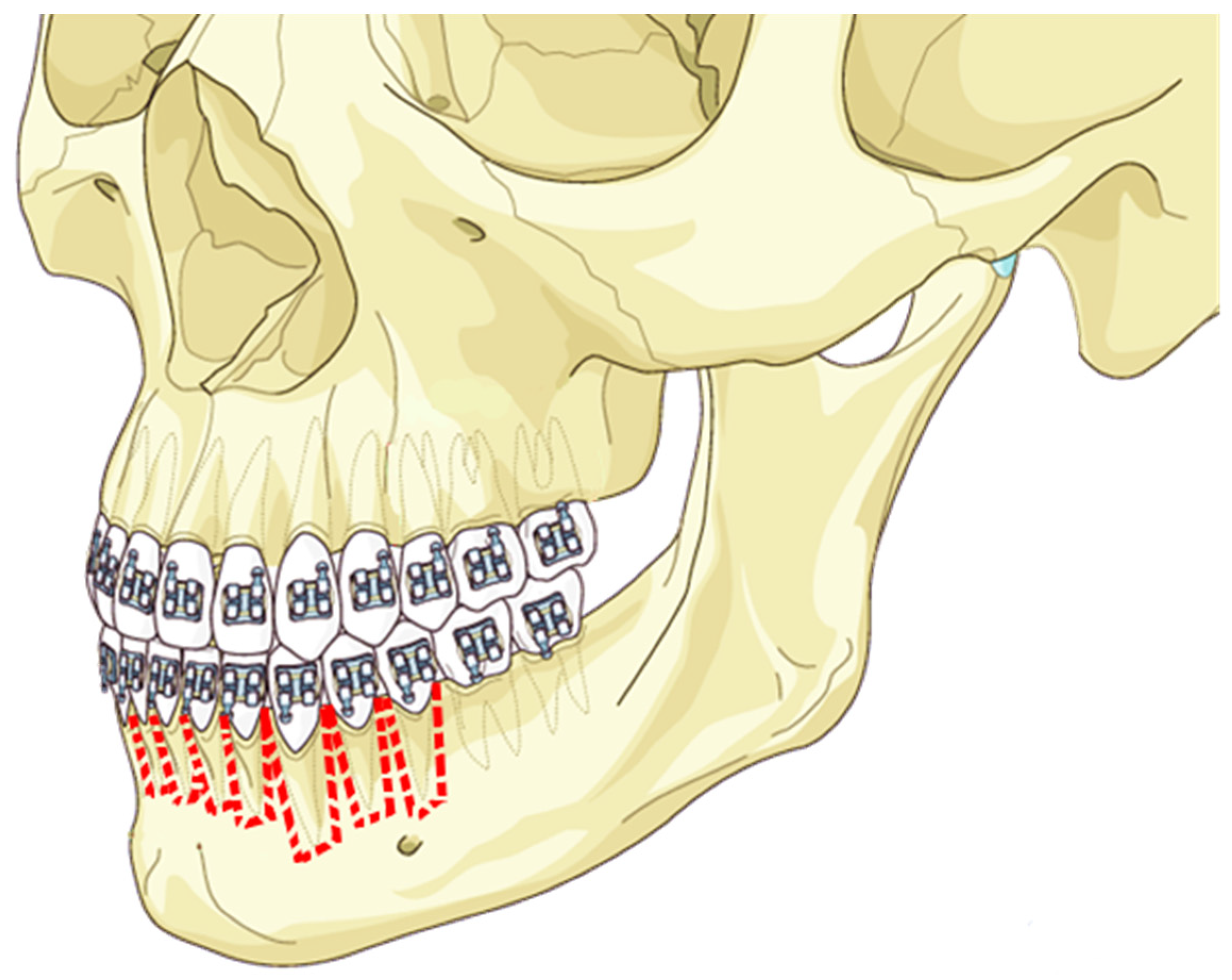
  • DAD involves a device constantly forcing the teeth to stimulate bone remodelling [18]. Osteogenic distraction is often used to treat severe skeletal discrepancies and requires close collaboration between the orthodontist and the oral-maxillofacial surgeon [19,20]. The process creates a controlled fracture in the bone, followed by applying a distraction device to lengthen the bone over time, forming new bone. This allows significant corrections of skeletal deformities and functional and aesthetic improvements [18]. Although this technique can be highly effective, it is associated with an increased risk of complications and requires careful patient management throughout the treatment process [21].

2. Materials and Methods

2.1. Protocol

The Preferred Reporting Items for Systematic Reviews and Meta-Analyses (PRISMA) guidelines were used in this systematic review (PROSPERO registration code ID 446071) [22].

2.2. Information Sources and Search Strategy

The qualifying criteria were developed using the PICOS (population, intervention, comparison, outcomes, and study design) framework. Pubmed, Science Direct, Scopus, and Web of Science databases were searched from 1 April 2003 up to 1 April 2023, using the keywords “acceleration” AND “dental movement” AND “orthodontic”(Table 1).

2.3. Eligibility Criteria

This research studies the dental movement acceleration strategies in extractive orthodontic treatment. Articles that met several criteria were included: (1) the study design selected was Randomised Clinical Trials (RCT), a case series with more than 5 case reports, clinical trials (CT), retrospective studies (R), and prospective studies (P); (2) participants were young adult and adult patients with permanent dentition; (3) patients were treated via extraction of the 1st or 2nd premolars in the upper or lower jaw; (4) extractive orthodontic treatment used included one strategy of dental movement acceleration (corticotomies, MOPs, VT, LLLT, drug therapy, and DAD); (5) the language selected was English; (6) only full-text was available.
Studies characterised by one of the following exclusion criteria were excluded: (1) the study design excluded was reviews, letters, or comments; case series with less than five case reports; case reports; in vivo and in vitro studies; (2) participants were animal models or dry skulls studies; (3) no-extractive orthodontic treatment; (4) the acceleration of the dental movement was facilitated using orthodontic miniscrews.

2.4. Synthesis Methods

The study data was selected by analysing the study design, number of patients, average age, dental acceleration technique employed, type of orthodontic treatment, and outcomes (Table 2).

3. Results

The electronic database search generated 1672 results. Following duplication elimination, 1350 studies were screened for titles and abstracts. After the abstract screening, 1252 papers were rejected, and 82 articles were chosen for the eligibility evaluation. Following the full-text examination, 37 manuscripts were eliminated: 21 were off-topic, six had wrong settings, and 10 had no outcome of interest. Finally, 44 papers were chosen for the systematic review, divided for each acceleration technique into:
  • Corticotomy, 15 articles
  • PRF/PRP, 4 articles
  • LLLT, 13 articles
  • MOPs, 4 articles
  • Vibration, 5 articles
  • DAD, 1 article
  • LLLT + corticotomy, 1 article
  • Drugs, 1 article
Figure 2 summarises the selection procedure.

4. Discussion

This systematic review examines different methods for accelerating OTM. In recent years, several techniques have been introduced that promise to speed up OTM, thereby reducing treatment duration. However, it is necessary to evaluate the effectiveness of these techniques via high-level randomised controlled studies to determine whether such devices can accelerate OTM.

4.1. Corticotomies

First introduced in 1892, corticotomy is a surgical technique involving a cortical bone cut, perforated or mechanically altered, with minimal involvement of the bone marrow [33]. It differs from osteotomy, where the cut involves bone and marrow. Regional Accleratory Phenomena (RAP) is based following corticotomy surgery on the principle that rapid bone remodelling and increased cell turnover occur following any trauma to bone tissue. There is a 4–5-month window during which the bone physiology changes, where the trabecular bone loses density and selectively offers less resistance to OTM. In AOO (Accelerated Osteogenic Orthodontics), non-activated teeth provide a relative anchorage for activated teeth that move faster (Figure 3) [33,65,66].
Several studies have been conducted to investigate the efficacy of this strategy. Bhattacharya et al. (2014) evaluated the impact of corticotomy on a sample of 20 patients [33]. In the group of ten patients who underwent surgery, after lifting a full-thickness flap, a 2 mm round bur was used to make incisions between the inter radicular spaces from premolar to premolar, starting 2 mm apical from the bone crest and ending more than 2 mm from the root apex. The cuts for horizontal corticotomy involved both labial and palatal sides, and demineralised freeze-dried bone was applied before flap closure and suturing. Once compared to the CGr, the ExGr TT was significantly shorter(131 ± 7.5 days vs. 234 ± 9) [33]. Chandran et al. (2018) compared corticotomy versus the control in a split-mouth study [34]. Two vertical incisions were made, one on the canine distal line angle and the other on the second premolar mesial line angle, and full-thickness mucoperiosteal flaps were reflected. Selective decortication was performed on the buccal and lingual cortical plates using a fissure bur (#556) in a high-speed handpiece. To standardise all corticotomy operations, decortication was conducted at three sites: the buccal plate, the crest of the alveolar ridge, and the palatal plate (Figure 1). The study concludes that corticotomy accelerates OTM by 40% [34,67]. Sakthi et al. (2014), which analysed bur decortication via 701 slit burs and number 2 round burs attached on a micromotor handpiece, reached a similar conclusion, as it measures the average space closure velocity in the maxilla as 1.8 mm/month, and in the lower jaw, as 1.57 mm/month, compared to 1.02 mm/month in the maxilla and 0.87 mm/month in the CGr [54].
Addanki et al. (2017) inserted an additional variable in the split mouth RCT study, as it compared bur corticotomy performed only on the buccal side with corticotomy performed on both the buccal and palatal side and came to the conclusion that there are no statistically notable differences within the two populations [24].
Dibart pioneered piezocision as a minimally invasive approach for accelerating OTM. Piezocision is a potentially minimally invasive tooth acceleration procedure because of its numerous periodontal, cosmetic, and orthodontic benefits [57]. Incisions are made in the buccal mucosa under local anaesthetic 2–3 mm below the interproximal papilla’s base. To decorticate the alveolar bone, the tip of the piezotome is placed at a depth of 3 mm. Subrahmanya’s study (2020) states that piezocision accelerates canine retraction movement by 1.5 times [57].
In an RCT study, Simre et al. (2021) compared traditional corticotomy with bur using piezocision. Buccal 1 cm transmucosal incisions were made distal to the canine and mesial to the second premolar area in both groups [55]. The incisions were made 5 mm apical to the papillae, and the vertical guideline bur holes were drilled and joined on the buccal cortex using a No.8 tungsten carbide round bur mounted on a straight handpiece and rotated at 30,000 rpm. At a depth of 2 mm, the groove went through the cortical bone, barely entering the spongiosa. The piezo group employed the OT7 (Mectron®, Carasco, Italy) piezo tip to produce a vertical groove over the buccal cortex.
This study assessed that corticotomy with bur was 1.5–2 times more rapid, whereas piezocision was 1.5 times faster; both are effective therapeutic options [55].
The Gibreal (2019) RCT study compared piezocision with traditional orthodontics in the mandible in 36 patients [38]. Radiographic-guided micro piezoelectric corticotomies on the labial surfaces of the alveolar bone between the six anterior teeth were performed on patients in the ExGr to accelerate alignment. Compared to traditional therapy, this approach required 59% less TT to correct highly crowded lower anterior teeth [38].
Aksakalli (2015) compared two minimally invasive techniques: in a split-mouth study on ten patients, he performed traditional corticision with a number 15 blade in the mesiobuccal and distobuccal area of the maxillary canines on one side; on the contralateral side, he performed piezocisions of a depth of 3 mm [7]. The superimposition of the 3D models, carried out using the third palatine wrinkle as a reference point revealed approximately twice as fast movement on the piezocision side as on the C Side, especially during the first month of treatment [7].
Sultana et al. (2022) evaluated the effectiveness of piezocision compared to conventional orthodontics [58]. The piezocision group completed the levelling and alignment phase substantially faster than the CGr (mean difference = 31.5 days, 95% CI 6.5, 56.5; p = 0.018). The alignment rate in the piezocision group was quicker in the first two months than in the CGr [58].
The Abbas study (2015) compared piezocision and control versus corticotomy and control [23]. They assessed that orthodontics supported via corticotomies are 1.5 to 2 times faster than traditional orthodontics, and piezocision was 1.5 more rapid than conventional orthodontics [23].
Also, Al Imam (2019) compared piezocision with traditional orthodontics: the ExGr with piezocision significantly improved the rate of incisor retraction by 53%, while the retraction time was significantly reduced by 27% [25].
Gibreal et al. (2022) used a new technique, with 3D-guided piezo-assisted orthodontic treatment, compared to conventional orthodontic [39]. In ExGr 5, incisions were made in the labial cortical between the anterior teeth. The study revealed an OAT reduced 48% in this group compared to the CGr. Through a slight incision, this procedure might enable the creation of a simple, precise, predictable, and safe localised alveolar decortication. As a result, no harm would be inflicted on any surrounding anatomical structures if a flap was not raised [39].
The RCT study by Mahmoudzadeh (2020) that analyses the influence of lasercision on OTM is exciting. Lasercision corticotomy (Er, Cr: YSGG 3.5 W, 30 Hz, 40% air, 80% water) in one maxillary quadrant required 59% less TT to correct highly crowded lower anterior teeth than traditional therapy [49].
Alfawal (2018) analysed laser-assisted flapless corticotomy (LAFC) with ER: YAG laser and piezocision [26]. In both groups, the rate of canine retraction was two-fold higher in the ExGr than in the CGr in the first month and 1.5-fold higher in the second month (p < 0.001). In addition, the total canine retraction time was decreased by nearly 25% in both groups when comparing the experimental and C Sides [26].
Baeshen (2020) conducted a partial buccal plate corticotomy distal to the lingual vertical and subapical incisions, and the lingual flap was not raised [30]. This technique revealed that the rate of canine retraction on the corticotomy side was substantially greater than on the C Side [30].
The literature [26,38,68] confirms the efficacy of corticotomy, which reduces treatment time by 40 per cent; bur corticotomy is 1.5–2 times faster, and piezocision is 1.5 times faster, with both being effective. Piezocision accelerates canine retraction by 1.5 times. Lasercision requires 59% less TT for crowded anterior teeth. Corticotomy can be used in different clinical contexts and with different types of orthodontic treatment, providing a customised acceleration solution. However, it also has some disadvantages and risks associated with surgery. It is a technique that requires a learning curve on the part of the clinician, and it can still cause discomfort with oedema and pain for the patient.

4.2. Micro-Osteo-Perforations (MOPs)

Bajaj et al. (2022) compared the effects of photobiomodulation (PBM) and micro-osteo perforations (MOPs) on the speed of canine retraction in a study group of 30 patients [31]. The extraction of the premolars was followed by a waiting period of 3 months to allow trabecular bone formation. Three vertical MOPs (approximately 19-gauge diameter) were performed distal to the canine root using Propel contra-angle perforation screws in the MOPs group. In the PBM group, the canine was stimulated using a Gallium Aluminium Arsenide (GaAlAs) semiconductor diode laser for 10 s at ten different points of the canine root. The retraction rate was approximately 1.1-fold higher in patients treated with MOPs than in comparison patients [31].
Feizbakhsh et al. (2018) analysed accelerated canine distalisation using MOPs (Figure 4) [37]. After 28 days, the movement was assessed by analysing the digital models of the two arches using the canine and second premolar at three locations as retrievals. MOPs increased OTM over twice as much as the C Side [37].
In the study by Attri et al. (2018), the retraction was activated immediately after the MOPs were executed. MOPs were carried out using a manual screwdriver (Propel device) and drilling screws. Each patient received three perforations (1.5 mm in diameter and 2–3 mm deep) in the extraction space at an equal distance from the canine and second premolar at the level of the bony cortical performed every 28 days until the space was closed entirely. When MOP patients were compared to the control individuals, there was an increase in OTM. Therefore, the authors suggest its use after carefully evaluating the risk–benefits, as there is an increase in costs and initial discomfort during the procedure [29]. Kundi et al. (2020) compared the extent of canine distalisation in patients undergoing cortical perforations without flaps or MOPs.
Patients were divided into an intervention group and a CGr. In one session, three MOPs (diameter 1.5 mm, depth 2.5 mm) were executed distal to the canine on both sides using the Propel device at the buccal cortical level. The mesial movement of the molar associated with canine retraction was also investigated using the median palatine line and the most prominent part of the palatine wrinkles as references.
The orthodontic movement was accelerated 2–3 times in the MOPs group compared to the CGr [45]. Compared to the other articles used in this section of the discussion, Bajaj’s work did not compare the improvement in the speed of canine retraction with a CGr treated only with fixed therapy. Still, it compared with a group exposed to PBM [31]. It can be deduced, however, that MOPs are a valid aid in increasing the speed of tooth retraction, although particular attention must be paid to reducing pain when performing this procedure.
MOPs have shown promise in accelerating orthodontic movement [8,69,70]. These are simple and minimally invasive procedures; however, they may cause temporary discomfort, swelling, or slight pain at the treated site.

4.3. Vibration Therapy

Several trials suggest that mechanical vibratory devices, in their current setting, do not offer significant advantages for orthodontic OTM. In the study by Khera et al. (2022), the effect of a customised vibratory device on the speed of OTM, particularly during canine retraction, was analysed [43]. The canine retraction was initiated at least four months after the first maxillary premolars extraction to eliminate any effects due to post-extraction regional acceleratory phenomena. Additionally, a customised vibratory device was used to reduce cost, which was economically advantageous compared to commercial devices and maintained a frequency of 30 Hz and a force of 0.25 N.
The study’s primary objective was to evaluate the effect of low-frequency vibrations (30 Hz) on the speed of OTM through the customised vibratory device by comparing canine retraction between the vibrated and non-vibrated sides. The present study concluded that low-frequency vibratory stimulation (30 Hz) applied for 20 min per day using a customised vibratory device does not significantly accelerate the rate of canine retraction [43]. The trial conducted by Taha et al. (2020) aimed to compare two groups of adolescents undergoing complete orthodontic treatment with and without using the AcceleDent Aura device [59,71]. The study results showed that the total amount of OTM did not show statistically significant differences between the groups at any of the three time intervals. The study attempted to minimise bias and observed an average monthly OTM rate of 1.21 ± 0.32 mm in the CGr and 1.12 ± 0.20 mm in the ExGr.
In conclusion, using the AcceleDent Aura device did not significantly affect accelerating maxillary canine retraction or reducing perceived pain during orthodontic treatment [59]. Liao et al. (2017) investigated the effects of vibration-enhanced OTM and the underlying mechanisms [48]. The vibration was applied to the buccal surface of canines for 10 min daily for 28 days, using an Oral B (USA) Hamming Bird vibrating unit. The amplitude of the vibrating force was measured at approximately 0.2 N (20 g), and the frequency of the vibrations was 50 Hz.
The results showed that the total space closure and distalisation of canines were significantly more significant on the vibration side than on the non-vibration side. The research concluded that applying low to medium-frequency vibration, such as 50 Hz in this study, can accelerate OTM without causing adverse effects like tissue necrosis or other undesirable outcomes [72]. The study suggested that the mechanism for OTM acceleration may be more biologically based than mechanically based, as the short duration of vibration application seems to stimulate OTM-related cells and factors via temporarily sustained and dynamic amplification of the pressure levels within the periodontal ligament (PDL). Therefore, applying mechanical vibrations could increase the speed of OTM and may be considered a promising method for accelerating extraction-based orthodontic treatments [48]. Kumar et al. (2020) evaluated the rate of orthodontic movement in adolescent patients combined with low-frequency mechanical vibrations in passive self-ligating and conventional appliances [44].
The customised vibrating device used had a frequency of 30 Hz and was used by patients for 20 min per day during the space closure phase. The primary objective was to measure the space closure rate (mm/month). The results showed no statistically significant differences regarding the space closure rate among the three groups.
Therefore, the null hypothesis was confirmed, namely that there was no difference in the rate of orthodontic movement between passive self-ligating and conventional appliances in patients with low-frequency vibrations [44,73]. Another method described is the pulsed electromagnetic field (PEMF) used to reduce TT, as described in the study by Bhad (Patil) and Karemore (2022) [32].
An electrical engineer designed a device to generate a weak PEMF of 0.5 mT (Tesla), at 1 Hz. PEMF therapy was initiated on the same day as the application of the closed-coil spring, and the PEMF device consisted of an integrated circuit powered via a battery and embedded in a removable acrylic appliance. Patients were required to wear the appliance for 8 h at night, and the device was checked at each appointment. The results showed that the ExGr, exposed to the PEMF, experienced a 1.2-fold increase in the rate of OTM compared to the CGr, translating to a 41% increase.
The average time for canine retraction in the ExGr was 4.5 months, while the CGr took 6–6.5 months. The study concludes that PEMF therapy can physiologically increase the rate of OTM, thereby reducing overall TT. When used in combination with closed-coil springs, 1 Hz PEMFs were successful in increasing OTM. The study suggests that PEMF therapy could be safely and routinely used during orthodontic treatment to shorten TT [32].
El-Angbawi et al. [74] analysed two studies that compared the use of OrthoAccel and Tooth Masseuse devices with standard orthodontic mechanics during alignment and canine retraction stages, respectively. The trials evaluated tooth mobility objectively, but meta-analysis was hampered by varying outcome measures at various stages of treatment. Additionally assessed were discomfort, pain, and negative consequences. There needed to be a discussion of duration or how frequently to visit. Over ten weeks, the Tooth Masseuse improved lower incisor alignment with minimal pain variations. The maxillary canine movement was slightly faster with OrthoAccel, with no clinical significance.
In summary, although vibration therapy offers a non-invasive and user-friendly approach, its effectiveness for orthodontic tooth movement remains inconclusive [75,76,77]. Despite a customised device’s economic advantage, low-frequency (30 Hz) vibratory stimulation for 20 min daily did not significantly accelerate canine retraction. Pulsed electromagnetic field (PEMF) therapy increased the OTM rate by 1.2-fold, shortening treatment time. PEMF therapy, especially when combined with the other methods, offers the potential for accelerated orthodontic treatment.

4.4. Low-Level Laser Therapy (LLLT)

LLLT has emerged as one of the most promising new supportive treatment techniques in recent years since it is a non-intervention therapy that is easy to obtain and does not require expensive equipment [47]. LLLT has been demonstrated to increase angiogenesis by up-regulating chemical mediators such as the vascular endothelial growth factor, to facilitate osteoclast and osteoblast cell proliferation and differentiation, and to accelerate OTM [35]. Furthermore, low-level laser irradiation (LLLI) has been demonstrated to benefit analgesics in various clinical and therapeutic applications.
LLLI reduces pain perception by preventing the release of arachidonic acid, which lowers prostaglandin E2 levels. It also causes the production of an endogenous opioid neuropeptide (beta-endorphin), which has powerful analgesic properties [52]. Several studies have been conducted to evaluate the impact of LLLT on improving OTM. Farhadian et al. worked an RCCT with 60 patients divided into three groups: 20 treated with LLLT, 20 treated with LED biostimulation, and 20 in the CGr. The extraction site and buccal surface of the canine were exposed to light using an intraoral LED device called Biolight®, which is comparable to Ortho-Pulse® and has a wavelength of 640 nm, an energy density of 10 j/cm2, and a power density of 40 mW/cm2 [35].
The patients were instructed to utilise the device for the maxillary dental arch for 5 min daily at the commencement of canine retraction. A GaAlAs diode laser with a wavelength of 810 nm and a power of 100 mW was used to treat the LLLT group. According to Farhadian et al., LLLT looked to help accelerate OTM by 60%, while the LED could not significantly speed up the process. In addition, patient-centred outcomes showed that neither LLLT nor LED impacted how painful the procedure was felt by the patient [35].
On a sample of 22 patients, Quamruddin et al. examined the effect of LLLI delivered at 3-week intervals on OTM and pain related to OTM using self-ligating brackets [52]. According to Qamruddin et al., LLLT is a helpful technique that, if used at intervals of three weeks, can double the rate of OTM [52]. To assess the utilisation of non-invasive or minimally invasive techniques to expedite OTM, such as LLLT, Moradinejad et al. conducted a split-mouth RCT [50]. Three parallel intervention groups were randomly assigned to 64 quadrants in 32 patients: LLLT, LLLT with piezoincision, and CGr. A 940 nm laser with 8 J and 0.5 W of power was utilised for 16 s at six sites to accomplish LLLT. This was performed on the first day and then again after three and six weeks.
This study demonstrated that although LLLT statistically and significantly sped up canine retraction and slowed anchoring loss, its effects were, at best, mild or moderate [50,78]. Lam et al. evaluated the effectiveness of LLLT on 16 patients using a split-mouth RCT [47]. A GaAlAs diode laser with an output power of 100 mW and an 810 nm wavelength was used to treat the LLLT group for 10 s on both the buccal and lingual surfaces. LLLT in the conditions was used in our study. In orthodontic therapy, a GaAlAs diode laser with a twice-monthly radiation dose of 5.1 J/cm2 positively impacted OTM speed [47]. Isola et al. assessed the effects of LLLT after extracting the first upper premolars for orthodontic purposes using a split-mouth RCT [42].
A diode laser operating in continuous wave mode at an 810 nm wavelength treated the test side at three places on the buccal and palatal sides at baseline, at 3, 7, and 14 days, and then every 15 days until the space closed. Only orthodontic traction was used on the C Side to treat the opposing chosen canine. This study shows that applying LLLT therapy successfully quickens OTM and lowers OTM-related discomfort levels [42]. In another split-mouth RCT, Qamruddin et al. evaluated the effects of LLLT [51].
After removing the first bicuspid on day 21, each canine was retracted using a 6 mm close coil NiTi spring stretched to 150 gm of force. Immediately following the spring installation on the experimental side, LLLT irradiation was used. A continuous, uninterrupted beam of light at a wavelength of 940 nm from a GaAlAs diode laser was used. Qamruddin et al. claimed that LLLT during routine orthodontic appointments spaced three weeks apart accelerates OTM and considerably lessens pain [51,79]. The combined impact of corticotomy and LLLT on the speed of OTM was examined by Farid et al. [36].
The premolar extractions were performed on the same day a surgical corticotomy was conducted. The laser was applied at the beginning of the four-month study period or on the first day of full canine retraction, as well as one week, two weeks, three weeks, and every two weeks after that. According to Farid et al., corticotomy and LLLT alone could not increase the rate of canine retraction above that of the gold-standard corticotomy approach [36]. Arumughan et al. evaluate whether the LLLT can accelerate OTM during en-masse retraction [28]. The experimental side was subjected to biostimulation by using a GaAlAs diode laser with a wavelength of 810 nm. Each site received ten irradiations for 10 s, five on the palatal side and ten on the buccal side of the tooth.
With a three-week interval between appointments, the total energy density at each application was 10 J. It was found that the rate of extraction space closing can be accelerated via biostimulation using an 810 nm diode laser. As a result, it can accelerate tooth mobility during orthodontic treatment [28]. Hasan et al. examined if LLLT might hasten the migration of crowded maxillary incisors during orthodontic treatment [40]. Patients in the laser group obtained an LLLT dosage from a GaAlAs laser device with an energy of 2 J/point at an 830 nm wavelength just after the first archwire was inserted. Due to a statistically significant difference in total TT between the two groups, Hasan et al. asserted that LLLT is a valuable technique for quickening OTM [40].
In 2020, Impellizzeri et al. used a split-mouth RCT to assess the efficacy of photobiomodulation therapy in accelerating OTM [41]. Following the extraction of the first premolar, canine retraction movement was monitored. Linear measurements of the canine’s anteroposterior position were obtained at the onset of treatment and one month later, and these values were contrasted with those from the non-irradiated side to determine the pace of orthodontic therapy. GaAlAs PBM was administered four times to the experimental segment. The results indicated a statistically significant disparity and a 32% acceleration in OTM attributable to the biostimulatory agent [41].
Cruz et al.’s work (2004) also utilised LLLT to assess the speed difference between the irradiated and C Sides of OTM [14]. The authors emphasised a notable acceleration of OTM on the side that had been exposed to radiation, and they specifically demonstrated that the laser group always experienced faster displacement with each spring activation. The authors claimed that LLLT in that region increased the region’s receptivity to biochemical alterations that help OTM [14]. Strniolo-Souza et al. (2020) monthly applied LLLT in both the upper and lower arch [56].
The LLLT technique for the upper arch varied depending on whether the side was buccal or palatal; the latter side required a larger dosage since the canine root was further from the laser application site due to the greater bony thickness of the palate. However, they discovered in their research that the LLLT procedure proved only helpful in the upper arch in the first stages of canine retraction, with speed around the same as on the C Side. The effectiveness of LLLT is correlated with both the dosage and frequency of laser application, according to scientists, who speculated that variations in bone density between the maxilla and mandible may have had differing effects on laser light absorption [56].
When LLLT photonic radiation enters the cell nucleus, it increases the production of ribonucleic acid (RNA), deoxyribonucleic acid (DNA), and protein synthesis. Enhancing the inflammatory response to specific stimuli produces biostimulating effects on the cellular metabolic processes [41].
Inflammatory responses mediate orthodontic movement; some research has sought to identify the primary inflammatory players responsible for increased OTM following LLLT. With the use of the GaAlAs diode laser, Varella’s (2018) study intended to measure and analyse the levels of IL-1β in gingival crevicular fluid during OTM [61]. In the split-mouth experiment, the canine retraction on the irradiation side progressed faster than on the C Side.
Crevicular gingival fluid samples were examined using ELISA assays, and it was discovered that the amount of IL-1β was more significant in the laser-irradiated region. The scientists hypothesised that the dental acceleration was likely caused by the increased IL-1β levels induced by LLLT [61]. In contrast, in a previous study, interleukin-6 was analysed as potentially responsible for the acceleration of OTM after LLLT [63].
Patients underwent maxillary canine retraction to close the extraction space, in which only one side had LLLT applied. During orthodontic treatment, gingival crevicular fluid samples were taken to analyse the IL-6 levels. The results showed that LLLT indeed accelerated OTM. However, the difference in the mean IL-6 concentration was not statistically significant, and it was impossible to attribute a major role to this cytokine [63].
LLLT is a non-invasive approach with potential benefits, including increased angiogenesis, osteoblast/osteoclast activity, and analgesic effects. Studies have explored LLLT’s impact on Orthodontic Tooth Movement (OTM), indicating accelerated movement. However, challenges exist, including varying outcomes, precision demands, cost, availability, treatment duration, and frequency. Notably, LLLT’s mechanism involves cellular responses and cytokine modulation, yet cytokine roles remain debated. While LLLT holds promise for enhancing OTM, carefully considering its advantages, disadvantages, patient preferences, and further research is essential for informed treatment decisions in orthodontic practice [80].

4.5. PRP and PRF

Leukocyte and platelet-rich fibrin (L-PRF) injections can accelerate OTM. The branchial vein collects 20 mL of blood, which is then centrifuged once at 700× g rpm for 3 min to obtain the L-PRF. To obtain PRF, which will subsequently be injected into the buccal and palate mucosa to quicken orthodontic movement, the yellow–orange section of the animal was employed [81]. Uday H. Barhate et al. (2022) evaluated the effect of L-PRF on the rate of canine maxillary retraction [60]. A careful analysis of the study models observed some correlation but is not statistically significant between canine retraction and the concentration of cytokines such as IL-1β and TNF-α.
Acceleration mainly occurred in the first four weeks; after that, retraction between the experimental and C sides was equal [60]. Zeitunlouian et al. (2021) performed a similar split-mouth experimental study to evaluate the effect of PRF on accelerating OTM during orthodontic treatment [64]. PRF was injected into the mucosa before canine retraction, and the procedure was repeated one month later. Results show a statistically significant acceleration occurred on the experimental side compared to the C Side [64].
On the other hand, Naji et al. (2022) compared the efficacy of PRF injection and PRP during orthodontic canine retraction [53]. A significantly faster rate of canine retraction was obtained with PRP infiltration in the first month. It was shown that PRP infiltration was more impactful with accelerated canine movement than PRF infiltration [53]. Angel et al. (2022) evaluated the effects of PRP on the acceleration of maxillary canine retraction [27,82]. The evaluation was performed by measuring the soluble receptor activator of nuclear factor κb (sRANKL) and the osteoprotegerin (OPG) ligand in the gingival crevicular fluid (GCF). After alignment, the premolar was extracted and then freshly prepared PRP was injected. The motion was assessed using digital model superposition at T0, T1 (30 days), and T3 (60 days). Movement occurred 35% more on the PRP than on the C Side, altering the OPG and sRANKL levels in GCF [27,83].
Therefore, the injection of PRF or PRP before a canine retraction in the post-extraction space may occur faster in the first few weeks. The prolonged PRF injections may be needed to achieve accelerated OTM, but this deserves more research.
Leukocyte and platelet-rich fibrin (L-PRF) injections show potential in hastening OTM initiation, with studies reporting accelerated movement [64,81,84]. Platelet-rich plasma (PRP) demonstrates initial superiority in canine retraction speed compared to PRF. Both interventions hold promise for modulating biomarkers associated with enhanced movement. Challenges encompass outcome variability, required treatment duration, and biomarker correlations. While early OTM acceleration with PRP and PRF is encouraging, a thorough evaluation considering patient preferences, long-term effects, and cost-effectiveness remains crucial. Ongoing research is vital for refining protocols and establishing sustained orthodontic benefits.

4.6. Drugs Therapy

Among strategies for OTM using drugs, the effect of vitamin D on canine distalisation and alveolar bone density using multi-slice spiral computed tomography (MSCT) has been studied. An RCT [62] used an in situ gel containing 1.25 DHC (active vitamin D metabolite) to evaluate its effect on OTM speed [85].
In the first month, the speed of canine movement was faster in the ExGr, but without a statistically significant difference compared to the CGr. In the following months, the rate of canine training in the ExGr was statistically significant. The effect of vitamin D was shown to be more significant when administered at doses close to normal physiological levels. In conclusion, the local administration of 1.25 DHC led to a significant increase in canine distalisation and a reduction in bone density in the trabecular bone tissue, suggesting that vitamin D may play a role in accelerating orthodontic movement [62].

4.7. Dentoalveolar Osteodistraction

The study by Kurt et al. (2017) aimed to evaluate the effects of the DAD device on OTM compared to conventional methods in patients with Class II malocclusion [46]. Thirty-three patients were divided into the DAD group and the Distalisation Group (DG). The patients who underwent a DAD have applied a retraction force of approximately 800 g for the upper canine. The rate of canine movement was measured in millimetres per month, and the dentoskeletal effects were assessed via an analysis of panoramic and lateral skull radiographs [86].
Furthermore, the dentoskeletal effects of DAD were limited to the treatment area, with no adverse effects on the position of the other teeth. The DAD group showed significantly faster canine movement (0.87 mm/month) concerning average speed experimented with conventional orthodontic technique (0.35/month) and no significant changes in the other maxillary and mandibular parameters except for maxillary canine retraction.
The DG group showed significant changes in the vertical dimension and mesial movement of the maxillary first molars, indicating anchorage loss [46,87]. No significant difference was found between the two groups in maxillomandibular measurement difference or root resorption. The study concludes that DAD can benefit patients with increased skeletal vertical dimensions and may reduce anterior tooth retraction time during fixed orthodontic therapy [46].
Osteodistraction, as exemplified via the DAD device, offers advantages in OTM for patients with Class II malocclusion [88]. The DAD group exhibited notably faster canine movement than conventional methods, benefiting those with increased skeletal vertical dimensions. Significantly, dentoskeletal effects were localised, avoiding negative impacts on the neighbouring teeth. However, some limitations were observed, including altered maxillary canine retraction and potential vertical dimension changes in the distalisation group. Root resorption and maxillomandibular measurements remained unaffected. While advantageous for specific cases, a careful consideration of possible anchorage loss and anatomical factors is essential.
Furthermore, this work [89] asserts that the absence of periodontal defects or endodontic lesions characterises the rapid distraction of the periodontal ligament during canine retraction. So, it is possible to rapidly distract the periodontal ligament without complications.

5. Limitation

This study reviews numerous RCTs and CSs regarding OTM acceleration techniques during extractive orthodontic treatments. However, orthodontic treatments that involved only medium and minimal anchorage appliances were considered in the literature selection, excluding orthodontic treatments that used orthodontic miniscrews as the maximum anchorage. Further studies will be needed to investigate this topic by considering this additional variable.

6. Conclusions

Most orthodontic therapies discussed in the literature seem promising and effective:
  • Techniques like corticotomies and micro-osteo perforations (MOPs) exhibit 1.5 to 2 times faster acceleration than traditional methods.
  • Piezoincisions are effective with variable success rates, offering time benefits but potential costs and discomfort.
  • Vibrational therapy’s impact on tooth movement is debated.
  • Pulsed electromagnetic field significantly shortens treatment times.
  • Low-level laser therapy speeds up tooth movement and offers analgesic benefits.
  • PRF, PRP, and Vitamin D treatments increase movement speed.
  • Dentoalveolar distraction aids shorter treatment, particularly in patients with vertical skeletal dimensions, minimizing anchoring loss.
These strategies expedite treatment, enhance patient experience, and minimise issues like cavities and enamel demineralisation. Research is needed to assess their efficacy, considering compliance, complications, risks, benefits, and efficacy in specific clinical contexts. Larger studies with standardised protocols can illuminate their impact on orthodontic care.

Author Contributions

Conceptualization, G.M.; I.F., F.V., A.M.C., A.D.I. and F.I.; methodology, A.P. (Assunta Patano), I.F., A.N., G.D. and A.M.I.; software, A.P. (Andrea Palermo), D.A., F.I. and G.D.; validation, G.M., S.C., A.D.I. and D.A.; formal analysis, G.M., A.N., D.A., A.M.C. and A.M.I.; investigation, A.D.I., A.P. (Assunta Patano), I.F., F.I. and G.D.; resources, A.D.I., F.V, I.F., A.N. and S.C.; data curation, I.F., A.N., D.A., A.M.C. and A.M.I.; writing—original draft preparation, F.V., A.P. (Assunta Patano), F.I. and A.M.I.; writing—review and editing, F.V., G.M., S.C., A.M.C. and G.D.; visualisation, A.D.I., A.N, S.C., D.A. and A.M.I.; supervision, I.F., F.V., A.N., A.P. (Andrea Palermo), S.C. and F.I.; project administration, G.M., A.P. (Assunta Patano), F.V., A.M.C. and G.D. All authors have read and agreed to the published version of the manuscript.

Funding

This research received no external funding.

Data Availability Statement

Not applicable.

Conflicts of Interest

The authors declare no conflict of interest.

Abbreviations

CGrControl Group
C SideControl Side
CTClinical trial
DADDentoalveolar Distraction
DGDistalisation group
DNADeoxyribonucleic acid
ExGrExperimental Group
GaAlAsGallium Aluminum Arsenide
GCF Gingival Crevicular Fluid
IL-1β: L’interleuchina-1 beta
LAFCLaser-assisted flapless corticotomy
LLLILow-level Laser Irradiation
LLLTLow-level Laser Therapy
L-PRF: Leukocyte and Platelet-Rich Fibrin
MOPsMicro-osteoperforations
NiTi Nickel-Titanium
NSAIDs Nonsteroidal anti-inflammatory drugs
OPGOsteoprotegerin
OTMOrthodontic tooth movement
PProspective study
PBM Photobiomodulation
PEMFPulsed Electromagnetic Field
PRFPlatelet-rich fibrinogen
PRPPlatelet-rich plasma
RAPRegional Accleratory Phenomena
RCTRandomised clinical trial
RNARibonucleic acid
sRANKLSoluble Receptor Activator of Nuclear factor κb
SWStraightwire
TNF-α: Tumor Necrosis Factor Alfa
TTTreatment Time
VTVibration therapy

References

  1. Wilcko, W.M.; Wilcko, T.; Bouquot, J.E.; Ferguson, D.J. Rapid Orthodontics with Alveolar Reshaping: Two Case Reports of Decrowding. Int. J. Periodontics Restor. Dent. 2001, 21, 9–19. [Google Scholar]
  2. Coloccia, G.; Inchingolo, A.D.; Inchingolo, A.M.; Malcangi, G.; Montenegro, V.; Patano, A.; Marinelli, G.; Laudadio, C.; Limongelli, L.; Di Venere, D.; et al. Effectiveness of Dental and Maxillary Transverse Changes in Tooth-Borne, Bone-Borne, and Hybrid Palatal Expansion through Cone-Beam Tomography: A Systematic Review of the Literature. Medicina 2021, 57, 288. [Google Scholar] [CrossRef] [PubMed]
  3. Mummolo, S.; Marchetti, E.; Albani, F.; Campanella, V.; Pugliese, F.; Di Martino, S.; Tecco, S.; Marzo, G. Comparison between Rapid and Slow Palatal Expansion: Evaluation of Selected Periodontal Indices. Head. Face Med. 2014, 10, 30. [Google Scholar] [CrossRef] [PubMed]
  4. Fischer, T.J. Orthodontic Treatment Acceleration with Corticotomy-Assisted Exposure of Palatally Impacted Canines. Angle Orthod. 2007, 77, 417–420. [Google Scholar] [CrossRef]
  5. Teixeira, C.C.; Khoo, E.; Tran, J.; Chartres, I.; Liu, Y.; Thant, L.M.; Khabensky, I.; Gart, L.P.; Cisneros, G.; Alikhani, M. Cytokine Expression and Accelerated Tooth Movement. J. Dent. Res. 2010, 89, 1135–1141. [Google Scholar] [CrossRef] [PubMed]
  6. Miles, P.; Fisher, E. Assessment of the Changes in Arch Perimeter and Irregularity in the Mandibular Arch during Initial Alignment with the AcceleDent Aura Appliance vs No Appliance in Adolescents: A Single-Blind Randomized Clinical Trial. Am. J. Orthod. Dentofac. Orthop. 2016, 150, 928–936. [Google Scholar] [CrossRef] [PubMed]
  7. Aksakalli, S.; Calik, B.; Kara, B.; Ezirganli, S. Accelerated Tooth Movement with Piezocision and Its Periodontal-Transversal Effects in Patients with Class II Malocclusion. Angle Orthod. 2015, 86, 59–65. [Google Scholar] [CrossRef]
  8. Alikhani, M.; Raptis, M.; Zoldan, B.; Sangsuwon, C.; Lee, Y.B.; Alyami, B.; Corpodian, C.; Barrera, L.M.; Alansari, S.; Khoo, E.; et al. Effect of Micro-Osteoperforations on the Rate of Tooth Movement. Am. J. Orthod. Dentofac. Orthop. 2013, 144, 639–648. [Google Scholar] [CrossRef]
  9. Quinzi, V.; Tecco, S.; Nota, A.; Caggiati, E.; Mummolo, S.; Marzo, G. Mesial Rotation of the Upper First Molar: Association with Anterior Dental Crowding in Mixed and Permanent Dentition. Appl. Sci. 2020, 10, 5301. [Google Scholar] [CrossRef]
  10. Quinzi, V.; Ferro, R.; Rizzo, F.A.; Marranzini, E.M.; Federici Canova, F.; Mummolo, S.; Mattei, A.; Marzo, G. The Two by Four Appliance: A Nationwide Cross-Sectional Survey. Eur. J. Paediatr. Dent. 2018, 19, 145–150. [Google Scholar] [CrossRef] [PubMed]
  11. Davidovitch, Z. Tooth Movement. Crit. Rev. Oral Biol. Med. 1991, 2, 411–450. [Google Scholar] [CrossRef] [PubMed]
  12. Kau, C.; Nguyen, J.T.; English, J. The Clinical Evaluation of a Novel Cyclical Force Generating Device in Orthodontics. Orthod. Pract. US 2010, 1.1, 1–4. [Google Scholar]
  13. Scarano, A.; Lorusso, F.; Inchingolo, F.; Postiglione, F.; Petrini, M. The Effects of Erbium-Doped Yttrium Aluminum Garnet Laser (Er: YAG) Irradiation on Sandblasted and Acid-Etched (SLA) Titanium, an In Vitro Study. Materials 2020, 13, 4174. [Google Scholar] [CrossRef]
  14. Cruz, D.R.; Kohara, E.K.; Ribeiro, M.S.; Wetter, N.U. Effects of Low-Intensity Laser Therapy on the Orthodontic Movement Velocity of Human Teeth: A Preliminary Study. Lasers Surg. Med. 2004, 35, 117–120. [Google Scholar] [CrossRef]
  15. Dohan Ehrenfest, D.M.; Rasmusson, L.; Albrektsson, T. Classification of Platelet Concentrates: From Pure Platelet-Rich Plasma (P-PRP) to Leucocyte- and Platelet-Rich Fibrin (L-PRF). Trends Biotechnol. 2009, 27, 158–167. [Google Scholar] [CrossRef]
  16. Alissa, R.; Esposito, M.; Horner, K.; Oliver, R. The Influence of Platelet-Rich Plasma on the Healing of Extraction Sockets: An Explorative Randomised Clinical Trial. Eur. J. Oral. Implantol. 2010, 3, 121–134. [Google Scholar] [PubMed]
  17. Kim, S.-J.; Park, Y.-G.; Kang, S.-G. Effects of Corticision on Paradental Remodeling in Orthodontic Tooth Movement. Angle Orthod. 2009, 79, 284–291. [Google Scholar] [CrossRef]
  18. Wang, X.; Mei, M.; Han, G.; Luan, Q.; Zhou, Y. Effectiveness of Modified Periodontally Accelerated Osteogenic Orthodontics in Skeletal Class II Malocclusion Treated by a Camouflage Approach. Am. J. Transl. Res. 2022, 14, 979–989. [Google Scholar] [PubMed]
  19. Boyne, P.J. Experimental Evaluation of the Osteogenic Potential of Bone Graft Materials. Annu. Meet. Am. Inst. Oral. Biol. 1969, 13–21. [Google Scholar]
  20. Quinzi, V.; Panetta, G.; Filippi, P.; Rizzo, F.A.; Mancini, L.; Mummolo, S. Autotransplatation of Immature Third Molars as Substitutes for Congenitally Missing Second Premolars: An Alternative Solution in a Young Patient with Oligodontia. J. Biol. Regul. Homeost. Agents 2020, 34, 155–163. [Google Scholar] [PubMed]
  21. Liou, E.J.-W.; Figueroa, A.A.; Polley, J.W. Rapid Orthodontic Tooth Movement into Newly Distracted Bone after Mandibular Distraction Osteogenesis in a Canine Model. Am. J. Orthod. Dentofac. Orthop. 2000, 117, 391–398. [Google Scholar] [CrossRef] [PubMed]
  22. Liberati, A.; Altman, D.G.; Tetzlaff, J.; Mulrow, C.; Gøtzsche, P.C.; Ioannidis, J.P.A.; Clarke, M.; Devereaux, P.J.; Kleijnen, J.; Moher, D. The PRISMA Statement for Reporting Systematic Reviews and Meta-Analyses of Studies That Evaluate Healthcare Interventions: Explanation and Elaboration. BMJ 2009, 339, b2700. [Google Scholar] [CrossRef] [PubMed]
  23. Abbas, N.H.; Sabet, N.E.; Hassan, I.T. Evaluation of Corticotomy-Facilitated Orthodontics and Piezocision in Rapid Canine Retraction. Am. J. Orthod. Dentofac. Orthop. 2016, 149, 473–480. [Google Scholar] [CrossRef] [PubMed]
  24. Addanki, P.; Gooty, J.R.; Palaparthy, R. Clinical and Radiographic Comparative Evaluation of Buccal and Palatal Corticotomy with Buccal Corticotomy in Periodontally Accelerated Osteogenic Orthodontics with Surgical Bur. Contemp. Clin. Dent. 2017, 8, 321–326. [Google Scholar] [CrossRef]
  25. Al-Imam, G.M.F.; Ajaj, M.A.; Hajeer, M.Y.; Al-Mdalal, Y.; Almashaal, E. Evaluation of the Effectiveness of Piezocision-Assisted Flapless Corticotomy in the Retraction of Four Upper Incisors: A Randomized Controlled Clinical Trial. Dent. Med. Probl. 2019, 56, 385–394. [Google Scholar] [CrossRef] [PubMed]
  26. Alfawal, A.M.H.; Hajeer, M.Y.; Ajaj, M.A.; Hamadah, O.; Brad, B. Evaluation of Piezocision and Laser-Assisted Flapless Corticotomy in the Acceleration of Canine Retraction: A Randomized Controlled Trial. Head. Face Med. 2018, 14, 4. [Google Scholar] [CrossRef]
  27. Angel, S.L.; Samrit, V.D.; Kharbanda, O.P.; Duggal, R.; Kumar, V.; Chauhan, S.S.; Coshic, P. Effects of Submucosally Administered Platelet-Rich Plasma on the Rate of Tooth Movement. Angle Orthod. 2022, 92, 73–79. [Google Scholar] [CrossRef]
  28. Arumughan, S.; Somaiah, S.; Muddaiah, S.; Shetty, B.; Reddy, G.; Roopa, S. A Comparison of the Rate of Retraction with Low-Level Laser Therapy and Conventional Retraction Technique. Contemp. Clin. Dent. 2018, 9, 260–266. [Google Scholar] [CrossRef]
  29. Attri, S.; Mittal, R.; Batra, P.; Sonar, S.; Sharma, K.; Raghavan, S.; Rai, K.S. Comparison of Rate of Tooth Movement and Pain Perception during Accelerated Tooth Movement Associated with Conventional Fixed Appliances with Micro-Osteoperforations—A Randomised Controlled Trial. J. Orthod. 2018, 45, 225–233. [Google Scholar] [CrossRef]
  30. Baeshen, H.A. The Effect of Partial Corticotomy on the Rate of Maxillary Canine Retraction: Clinical and Radiographic Study. Molecules 2020, 25, 4837. [Google Scholar] [CrossRef]
  31. Bajaj, I.; Garg, A.; Gupta, D. Comparative Effect of Micro-Osteoperforation and Photo-Bio-Modulation on the Rate of Maxillary Canine Retraction: A Split Mouth Randomized Clinical Trial. La Clin. Ter. 2022, 173, 39–45. [Google Scholar] [CrossRef]
  32. Bhad (Patil), W.A.; Karemore, A.A. Efficacy of Pulsed Electromagnetic Field in Reducing Treatment Time: A Clinical Investigation. Am. J. Orthod. Dentofac. Orthop. 2022, 161, 652–658. [Google Scholar] [CrossRef] [PubMed]
  33. Bhattacharya, P.; Bhattacharya, H.; Anjum, A.; Bhandari, R.; Agarwal, D.K.; Gupta, A.; Ansar, J. Assessment of Corticotomy Facilitated Tooth Movement and Changes in Alveolar Bone Thickness—A CT Scan Study. J. Clin. Diagn. Res. 2014, 8, ZC26–ZC30. [Google Scholar] [CrossRef]
  34. Chandran, M.; Muddaiah, S.; Nair, S.; Shetty, B.; Somaiah, S.; Reddy, G.; Abraham, B. Clinical and Molecular-Level Comparison between Conventional and Corticotomy-Assisted Canine Retraction Techniques. J. World Fed. Orthod. 2018, 7, 128–133. [Google Scholar] [CrossRef]
  35. Farhadian, N.; Miresmaeili, A.; Borjali, M.; Salehisaheb, H.; Farhadian, M.; Rezaei-Soufi, L.; Alijani, S.; Soheilifar, S.; Farhadifard, H. The Effect of Intra-Oral LED Device and Low-Level Laser Therapy on Orthodontic Tooth Movement in Young Adults: A Randomized Controlled Trial. Int. Orthod. 2021, 19, 612–621. [Google Scholar] [CrossRef]
  36. Farid, K.A.; Eid, A.A.; Kaddah, M.A.; Elsharaby, F.A. The Effect of Combined Corticotomy and Low Level Laser Therapy on the Rate of Orthodontic Tooth Movement: Split Mouth Randomized Clinical Trial. Laser Ther. 2019, 28, 275–283. [Google Scholar] [CrossRef]
  37. Feizbakhsh, M.; Zandian, D.; Heidarpour, M.; Farhad, S.Z.; Fallahi, H.R. The Use of Micro-Osteoperforation Concept for Accelerating Differential Tooth Movement. J. World Fed. Orthod. 2018, 7, 56–60. [Google Scholar] [CrossRef]
  38. Gibreal, O.; Hajeer, M.Y.; Brad, B. Efficacy of Piezocision-Based Flapless Corticotomy in the Orthodontic Correction of Severely Crowded Lower Anterior Teeth: A Randomized Controlled Trial. Eur. J. Orthod. 2019, 41, 188–195. [Google Scholar] [CrossRef]
  39. Gibreal, O.; Al-modallal, Y.; Al-assaf, M. Evaluation of the Efficacy of 3D-Guided Piezosurgery in Accelerating Mandibular Orthodontic Teeth Alignment: A Randomized Controlled Trial in Adults. Dentistry 3000 2022, 10, 281–288. [Google Scholar] [CrossRef]
  40. AlSayed Hasan, M.M.A.; Sultan, K.; Hamadah, O. Low-Level Laser Therapy Effectiveness in Accelerating Orthodontic Tooth Movement: A Randomized Controlled Clinical Trial. Angle Orthod. 2017, 87, 499–504. [Google Scholar] [CrossRef] [PubMed]
  41. Impellizzeri, A.; Horodynski, M.; Fusco, R.; Palaia, G.; Polimeni, A.; Romeo, U.; Barbato, E.; Galluccio, G. Photobiomodulation Therapy on Orthodontic Movement: Analysis of Preliminary Studies with a New Protocol. Int. J. Environ. Res. Public. Health 2020, 17, 3547. [Google Scholar] [CrossRef] [PubMed]
  42. Isola, G.; Matarese, M.; Briguglio, F.; Grassia, V.; Picciolo, G.; Fiorillo, L.; Matarese, G. Effectiveness of Low-Level Laser Therapy during Tooth Movement: A Randomized Clinical Trial. Materials 2019, 12, 2187. [Google Scholar] [CrossRef] [PubMed]
  43. Khera, A.; Raghav, P.; Mehra, V.; Wadhawan, A.; Gupta, N.; Phull, T. Effect of Customized Vibratory Device on Orthodontic Tooth Movement: A Prospective Randomized Control Trial. J. Orthod. Sci. 2022, 11, 18. [Google Scholar] [CrossRef]
  44. Kumar, V.; Batra, P.; Sharma, K.; Raghavan, S.; Srivastava, A. Comparative Assessment of the Rate of Orthodontic Tooth Movement in Adolescent Patients Undergoing Treatment by First Bicuspid Extraction and En Mass Retraction, Associated with Low-Frequency Mechanical Vibrations in Passive Self-Ligating and Conventional Brackets: A Randomized Controlled Trial. Int. Orthod. 2020, 18, 696–705. [Google Scholar] [CrossRef] [PubMed]
  45. Kundi, I.; Alam, M.K.; Shaheed, S. Micro-Osteo Perforation Effects as an Intervention on Canine Retraction. Saudi Dent. J. 2020, 32, 15–20. [Google Scholar] [CrossRef]
  46. Kurt, G.; İşeri, H.; Kişnişçi, R.; Özkaynak, Ö. Rate of Tooth Movement and Dentoskeletal Effects of Rapid Canine Retraction by Dentoalveolar Distraction Osteogenesis: A Prospective Study. Am. J. Orthod. Dentofac. Orthop. 2017, 152, 204–213. [Google Scholar] [CrossRef]
  47. Le, L.; Le, K.; Do, T. Influence of Low-Level Laser Treatment on Tooth Movement in Orthodontic Treatment. J. Int. Dent. Med. Res. 2023, 15, 1614–1619. [Google Scholar]
  48. Liao, Z.; Elekdag-Turk, S.; Turk, T.; Grove, J.; Dalci, O.; Chen, J.; Zheng, K.; Ali Darendeliler, M.; Swain, M.; Li, Q. Computational and Clinical Investigation on the Role of Mechanical Vibration on Orthodontic Tooth Movement. J. Biomech. 2017, 60, 57–64. [Google Scholar] [CrossRef]
  49. Mahmoudzadeh, M.; Poormoradi, B.; Alijani, S.; Farhadian, M.; Kazemisaleh, A. Efficacy of Er,Cr Laser Incision Corticotomy in Rapid Maxillary Canine Retraction: A Split-Mouth Randomized Clinical Trial. J. Lasers Med. Sci. 2020, 11, 442–449. [Google Scholar] [CrossRef]
  50. Moradinejad, M.; Chaharmahali, R.; Shamohammadi, M.; Mir, M.; Rakhshan, V. Low-Level Laser Therapy, Piezocision, or Their Combination vs. Conventional Treatment for Orthodontic Tooth Movement: A Hierarchical 6-Arm Split-Mouth Randomized Clinical Trial. J. Orofac. Orthop. 2022. [Google Scholar] [CrossRef]
  51. Qamruddin, I.; Alam, M.K.; Mahroof, V.; Fida, M.; Khamis, M.F.; Husein, A. Photobiostimulatory Effect of a Single Dose of Low-Level Laser on Orthodontic Tooth Movement and Pain. Pain. Res. Manag. 2021, 2021, 6690542. [Google Scholar] [CrossRef] [PubMed]
  52. Qamruddin, I.; Alam, M.K.; Mahroof, V.; Fida, M.; Khamis, M.F.; Husein, A. Effects of Low-Level Laser Irradiation on the Rate of Orthodontic Tooth Movement and Associated Pain with Self-Ligating Brackets. Am. J. Orthod. Dentofac. Orthop. 2017, 152, 622–630. [Google Scholar] [CrossRef] [PubMed]
  53. Naji, R.; Zeitounlouian, T.; Alomari, E.; Youssef, M. Evaluation of the Efficacy of Platelet-Rich Plasma (PRP) and Injectable Platelet-Rich Fibrin (i-PRF) in the Acceleration of Canine Retraction: A Randomized Controlled Trial. J. Int. Oral. Health 2022, 14, 243. [Google Scholar] [CrossRef]
  54. Inchingolo, F.; Ballini, A.; Cagiano, R.; Inchingolo, A.D.; Serafini, M.; De Benedittis, M.; Cortelazzi, R.; Tatullo, M.; Marrelli, M.; Inchingolo, A.M.; et al. Immediately Loaded Dental Implants Bioactivated with Platelet-Rich Plasma (PRP) Placed in Maxillary and Mandibular Region. Clin Ter 2015, 166, e146-52. [Google Scholar]
  55. Simre, S.S.; Rajanikanth, K.; Bhola, N.; Jadhav, A.; Patil, C.; Mishra, A. Comparative Assessment of Corticotomy Facilitated Rapid Canine Retraction Using Piezo versus Bur: A Randomized Clinical Study. J. Oral. Biol. Craniofac Res. 2022, 12, 182–186. [Google Scholar] [CrossRef] [PubMed]
  56. Storniolo-Souza, J.; Lima, L.M.; Pinzan, A.; Alvarez, F.; Pereira, S.C.d.C.; Janson, G. Influence of Low-Level Laser Irradiation on Orthodontic Movement and Pain Level—A Randomized Clinical Trial. Orthod. Waves 2020, 79, 105–112. [Google Scholar] [CrossRef]
  57. Inchingolo, F.; Tatullo, M.; Abenavoli, F.M.; Marrelli, M.; Inchingolo, A.D.; Gentile, M.; Inchingolo, A.M.; Dipalma, G. Non-Syndromic Multiple Supernumerary Teeth in a Family Unit with a Normal Karyotype: Case Report. Int. J. Med. Sci. 2010, 7, 378–384. [Google Scholar] [CrossRef]
  58. Sultana, S.; Ab Rahman, N.; Zainuddin, S.L.A.; Ahmad, B. Effect of Piezocision Procedure in Levelling and Alignment Stage of Fixed Orthodontic Treatment: A Randomized Clinical Trial. Sci. Rep. 2022, 12, 6230. [Google Scholar] [CrossRef]
  59. Taha, K.; Conley, R.S.; Arany, P.; Warunek, S.; Al-Jewair, T. Effects of Mechanical Vibrations on Maxillary Canine Retraction and Perceived Pain: A Pilot, Single-Center, Randomized-Controlled Clinical Trial. Odontology 2020, 108, 321–330. [Google Scholar] [CrossRef]
  60. Barhate, U.H.; Duggal, I.; Mangaraj, M.; Sharan, J.; Duggal, R.; Jena, A.K. Effects of Autologous Leukocyte-Platelet Rich Fibrin (L-PRF) on the Rate of Maxillary Canine Retraction and Various Biomarkers in Gingival Crevicular Fluid (GCF): A Split Mouth Randomized Controlled Trial. Int. Orthod. 2022, 20, 100681. [Google Scholar] [CrossRef]
  61. Varella, A.M.; Revankar, A.V.; Patil, A.K. Low-Level Laser Therapy Increases Interleukin-1β in Gingival Crevicular Fluid and Enhances the Rate of Orthodontic Tooth Movement. Am. J. Orthod. Dentofac. Orthop. 2018, 154, 535–544.e5. [Google Scholar] [CrossRef]
  62. Varughese, S.T.; Shamanna, P.U.; Goyal, N.; Thomas, B.S.; Lakshmanan, L.; Pulikkottil, V.J.; Ahmed, M.G. Effect of Vitamin D on Canine Distalization and Alveolar Bone Density Using Multi-Slice Spiral CT: A Randomized Controlled Trial. J. Contemp. Dent. Pract. 2019, 20, 1430–1435. [Google Scholar] [PubMed]
  63. Minervini, G.; Franco, R.; Marrapodi, M.M.; Fiorillo, L.; Cervino, G.; Cicciù, M. Economic Inequalities and Temporomandibular Disorders: A Systematic Review with Meta-analysis. J. Oral. Rehabil. 2023, 50, 715–723. [Google Scholar] [CrossRef] [PubMed]
  64. Minervini, G.; Franco, R.; Marrapodi, M.M.; Crimi, S.; Badnjević, A.; Cervino, G.; Bianchi, A.; Cicciù, M. Correlation between Temporomandibular Disorders (TMD) and Posture Evaluated Trough the Diagnostic Criteria for Temporomandibular Disorders (DC/TMD): A Systematic Review with Meta-Analysis. J. Clin. Med. 2023, 12, 2652. [Google Scholar] [CrossRef] [PubMed]
  65. Quinzi, V.; Saccomanno, S.; Manenti, R.J.; Giancaspro, S.; Coceani Paskay, L.; Marzo, G. Efficacy of Rapid Maxillary Expansion with or without Previous Adenotonsillectomy for Pediatric Obstructive Sleep Apnea Syndrome Based on Polysomnographic Data: A Systematic Review and Meta-Analysis. Appl. Sci. 2020, 10, 6485. [Google Scholar] [CrossRef]
  66. Inchingolo, A.D.; Ferrara, I.; Viapiano, F.; Netti, A.; Campanelli, M.; Buongiorno, S.; Latini, G.; Carpentiere, V.; Ciocia, A.M.; Ceci, S.; et al. Rapid Maxillary Expansion on the Adolescent Patient: Systematic Review and Case Report. Children 2022, 9, 1046. [Google Scholar] [CrossRef]
  67. Dinoi, M.; Marchetti, E.; Garagiola, U.; Caruso, S.; Mummolo, S.; Marzo, G. Orthodontic Treatment of an Unerupted Mandibular Canine Tooth in a Patient with Mixed Dentition: A Case Report. J. Med. Case Rep. 2016, 10, 170. [Google Scholar] [CrossRef]
  68. Bakr, A.R.; Nadim, M.A.; Sedky, Y.W.; El Kady, A.A. Effects of Flapless Laser Corticotomy in Upper and Lower Canine Retraction: A Split-Mouth, Randomized Controlled Trial. Cureus 2023, 15, e37191. [Google Scholar] [CrossRef] [PubMed]
  69. Alkebsi, A.; Al-Maaitah, E.; Al-Shorman, H.; Abu Alhaija, E. Three-Dimensional Assessment of the Effect of Micro-Osteoperforations on the Rate of Tooth Movement during Canine Retraction in Adults with Class II Malocclusion: A Randomized Controlled Clinical Trial. Am. J. Orthod. Dentofac. Orthop. 2018, 153, 771–785. [Google Scholar] [CrossRef] [PubMed]
  70. Li, J.; Papadopoulou, A.K.; Gandedkar, N.; Dalci, K.; Darendeliler, M.A.; Dalci, O. The Effect of Micro-Osteoperforations on Orthodontic Space Closure Investigated over 12 Weeks: A Split-Mouth, Randomized Controlled Clinical Trial. Eur. J. Orthod. 2022, 44, 427–435. [Google Scholar] [CrossRef]
  71. Pasini, M.; Giuca, M.R.; Ligori, S.; Mummolo, S.; Fiasca, F.; Marzo, G.; Quinzi, V. Association between Anatomical Variations and Maxillary Canine Impaction: A Retrospective Study in Orthodontics. Appl. Sci. 2020, 10, 5638. [Google Scholar] [CrossRef]
  72. Campanella, V.; Gallusi, G.; Nardi, R.; Mea, A.; Di Taranto, V.; Montemurro, E.; Marzo, G.; Libonati, A. Dentinal Substrate Variability and Bonding Effectiveness: SEM Investigation. J. Biol. Regul. Homeost. Agents 2020, 34, 49–54. [Google Scholar] [PubMed]
  73. Mummolo, S.; Nota, A.; Marchetti, E.; Padricelli, G.; Marzo, G. The 3D Tele Motion Tracking for the Orthodontic Facial Analysis. Biomed. Res. Int. 2016, 2016, 4932136. [Google Scholar] [CrossRef] [PubMed]
  74. El-Angbawi, A.; McIntyre, G.T.; Fleming, P.S.; Bearn, D.R. Non-Surgical Adjunctive Interventions for Accelerating Tooth Movement in Patients Undergoing Fixed Orthodontic Treatment. Cochrane Database Syst. Rev. 2015, 2016, CD010887. [Google Scholar] [CrossRef] [PubMed]
  75. Jing, D.; Xiao, J.; Li, X.; Li, Y.; Zhao, Z. The Effectiveness of Vibrational Stimulus to Accelerate Orthodontic Tooth Movement: A Systematic Review. BMC Oral. Health 2017, 17, 143. [Google Scholar] [CrossRef]
  76. Aljabaa, A.; Almoammar, K.; Aldrees, A.; Huang, G. Effects of Vibrational Devices on Orthodontic Tooth Movement: A Systematic Review. Am. J. Orthod. Dentofac. Orthop. 2018, 154, 768–779. [Google Scholar] [CrossRef]
  77. Qamar, Z.; Alghamdi, A.M.S.; Bin Haydarah, N.K.; Balateef, A.A.; Alamoudi, A.A.; Abumismar, M.A.; Shivakumar, S.; Cicciù, M.; Minervini, G. Impact of Temporomandibular Disorders on Oral Health-related Quality of Life: A Systematic Review and Meta-analysis. J. Oral. Rehabil. 2023, 50, 706–714. [Google Scholar] [CrossRef] [PubMed]
  78. Cirulli, N.; Inchingolo, A.D.; Patano, A.; Ceci, S.; Marinelli, G.; Malcangi, G.; Coloccia, G.; Montenegro, V.; Di Pede, C.; Ciocia, A.M.; et al. Innovative Application of Diathermy in Orthodontics: A Case Report. Int. J. Environ. Res. Public. Health 2022, 19, 7448. [Google Scholar] [CrossRef]
  79. Ballini, A.; Dipalma, G.; Isacco, C.G.; Boccellino, M.; Di Domenico, M.; Santacroce, L.; Nguyễn, K.C.D.; Scacco, S.; Calvani, M.; Boddi, A.; et al. Oral Microbiota and Immune System Crosstalk: A Translational Research. Biology 2020, 9, 131. [Google Scholar] [CrossRef]
  80. Shaadouh, R.I.; Hajeer, M.Y.; Mahmoud, G.; Murad, R.M.T. Systematic Review: Is High-Energy Laser Therapy (HELT) With Flapless Corticotomy Effective in Accelerating Orthodontic Tooth Movement? Cureus 2022, 14, e22337. [Google Scholar] [CrossRef]
  81. Erdur, E.A.; Karakaslı, K.; Oncu, E.; Ozturk, B.; Hakkı, S. Effect of Injectable Platelet-Rich Fibrin (i-PRF) on the Rate of Tooth Movement. Angle Orthod. 2021, 91, 285–292. [Google Scholar] [CrossRef]
  82. Minervini, G.; Franco, R.; Marrapodi, M.M.; Ronsivalle, V.; Shapira, I.; Cicciù, M. Prevalence of Temporomandibular Disorders in Subjects Affected by Parkinson Disease: A Systematic Review and Metanalysis. J. Oral. Rehabil. 2023. [Google Scholar] [CrossRef] [PubMed]
  83. Scarano, A.; Inchingolo, F.; Rapone, B.; Lucchina, A.G.; Qorri, E.; Lorusso, F. Role of Autologous Platelet Gel (APG) in Bone Healing: A Rabbit Study. Appl. Sci. 2021, 11, 395. [Google Scholar] [CrossRef]
  84. Krishna, V.B.; Duggal, I.; Sharan, J.; Mangaraj, M.; Duggal, R.; Jena, A.K. Effect of Leukocyte-Platelet-Rich Fibrin (L-PRF) on the Rate of Orthodontic Tooth Movement and Expression of Various Biomarkers in Gingival Crevicular Fluid. Clin. Oral. Invest. 2023, 27, 2311–2319. [Google Scholar] [CrossRef]
  85. Chackartchi, T.; Iezzi, G.; Goldstein, M.; Klinger, A.; Soskolne, A.; Piattelli, A.; Shapira, L. Sinus Floor Augmentation Using Large (1–2 Mm) or Small (0.25–1 Mm) Bovine Bone Mineral Particles: A Prospective, Intra-Individual Controlled Clinical, Micro-Computerized Tomography and Histomorphometric Study. Clin. Oral. Implant. Res. 2011, 22, 473–480. [Google Scholar] [CrossRef]
  86. Maspero, C.; Abate, A.; Inchingolo, F.; Dolci, C.; Cagetti, M.G.; Tartaglia, G.M. Incidental Finding in Pre-Orthodontic Treatment Radiographs of an Aural Foreign Body: A Case Report. Children 2022, 9, 421. [Google Scholar] [CrossRef]
  87. Inchingolo, A.D.; Inchingolo, A.M.; Bordea, I.R.; Xhajanka, E.; Romeo, D.M.; Romeo, M.; Zappone, C.M.F.; Malcangi, G.; Scarano, A.; Lorusso, F.; et al. The Effectiveness of Osseodensification Drilling Protocol for Implant Site Osteotomy: A Systematic Review of the Literature and Meta-Analysis. Materials 2021, 14, 1147. [Google Scholar] [CrossRef]
  88. İşeri, H.; Kişnişci, R.; Bzizi, N.; Tüz, H. Rapid Canine Retraction and Orthodontic Treatment with Dentoalveolar Distraction Osteogenesis. Am. J. Orthod. Dentofac. Orthop. 2005, 127, 533–541. [Google Scholar] [CrossRef] [PubMed]
  89. Liou, E.J.W.; Huang, C.S. Rapid Canine Retraction through Distraction of the Periodontal Ligament. Am. J. Orthod. Dentofac. Orthop. 1998, 114, 372–382. [Google Scholar] [CrossRef] [PubMed]
Figure 1. (A) Corticotomies; (B) MOPs; (C) combination of both techniques.
Applsci 13 09759 g001
Figure 2. The literature search’s Preferred Reporting Items for Systematic Reviews and Meta-Analyses (PRISMA) flow diagram.
Applsci 13 09759 g002
Figure 3. Corticotomies technique, as shows the red dashes.
Applsci 13 09759 g003
Figure 4. Mechanism of action of MOPs.
Applsci 13 09759 g004
Table 1. Database search indicator.
Articles
screening strategy
(Keywords: acceleration) AND (dental movement) AND (orthodontic)
Boolean Indicators: (“A” AND “B”)
Timespan: from 1 April 2003 to 1 April 2023
Electronic Database: Pubmed, Science Direct, Scopus, and Web of Science
Table 2. Descriptive summary of item selection.
Author
(Year)
Study DesignNumber of PatientsAverage Age (Years)Dental Movement Acceleration TechniquesOrthodontic Treatment Outcomes
Abbas (2016)
[23]
RCT2015–25Piezocision/control group (CGr); corticotomy/CGrRoth prescription brackets; closed coils NiTi springs 150 g forceOrthodontics supported via corticotomies and piezocision is 1.5–2 times faster than traditional orthodontics.
Addanki (2017)
[24]
RCT1620–40Buccal and palatal bur corticotomy/buccal bur corticotomy (control)SW bracketsNo difference between the two groups.
Aksakalli (2015)
[7]
RCT1016.3 ± 2.4Piezocision/corticocision (blade 15)Roth prescription brackets; elastomeric chain 150 g force and medium anchorage (transpalatal arch)Movement in the side undergoing piezocision is twice as fast as in the CGr.
Al Imam (2019)
[25]
RCT4219.15PiezocisionMBT prescription brackets; NiTi coil springs 150 g; medium anchorage (transpalatal arch)The incisor retraction time in the experimental group has decreased by 27%.
Alfawal (2018)
[26]
RCT3615–27Piezocision/CGr; laser-assisted flapless corticotomy (LAFC) ER: YAG laser MBT prescription brackets; NiTi closed coil spring 150 g forceThe experimental side had a higher rate of OTM in the first and second months and a 25% reduction in overall canine retraction duration.
Angel et al. (2022)
[27]
RCT1016–24 Injection of PRPRoth prescription brackets; medium anchorage (Nance palatal button)Movement occurred 35% more increased on the i-PRP side than on the CGr.
Arumughan et Al. (2018)
[28]
RCT12 LLLT: 810 nm wavelength laser (100 mW power, continuous wave).MBT prescription brackets; NiTi closed-coil spring 150 g force LLLT speeds OTM by 12.555% compared to the conventional retraction approach.
Attri et al. (2018)
[29]
2-arm parallel RCT6013–20MOPsMBT prescription bracketsIncreased OTM with MOPs.
Baeshen (2020)
[30]
CT with the split-mouth2016 ± 2.8Partial corticotomy SW brackets; elastomeric chain 150 g force; medium anchorage (transpalatal arch)The rate of canine retraction was significantly higher on the corticotomy side than on the CGr (p < 0.05).
Bajaj et al. (2022)
[31]
split mouth RCT3018–25 MOPs and PBMMBT prescription bracketsThe retraction rate is 1.1 times higher with MOPs than with PBM.
Bhad (Patil) e Karemore (2022)
[32]
A clinical study with a split-mouth design1918–24 PEMF therapySW; NiTi closed-coil springs. The rate of OTM in the experimental group was significantly higher than the CGr, with a mean increase in M1 of 41% and a mean increase in M2 of 31%.
Bhattacharya (2014)
[33]
RCT2018.8 ± 3.48CorticotomyMBT prescription brackets; NiTi closed coil spring 250 g force; medium anchorage (transpalatal arch)The corticotomy group’s meantime for en masse retraction was 131 ± 7.5 d, compared to 234 ± 9 d for the traditional approach.
Chandran (2018)
[34]
RCT2014.5Bur corticotomy MBT prescription brackets; active tie-back 100 g forceAlveolar corticotomy enhanced the rate of canine retraction by about 40%.
Cruz (2004)
[14]
RCT1115 LLLTRoth prescription brackets from right to left canines;12 mm NiTi closed coil springLaser Group is faster than CGr with a ratio of 1.34.
Farhadian et Al. (2021)
[35]
RCT60LLLT group (20.9 ± 5.5); LED group (21.7 ± 4.2); CGr (22.7 ± 5.3). LLLT Group:
GaAlAs
(810 nm; 100 mW) performed on days 0, 3, 30, and 60.
LED Group: intraoral LED device (wavelength: 640 nm; 10 j/cm2; 40 mW/cm2), 5 min/day
MBT and Roth prescription brackets. Medium anchorage (trans-palatal arch, on second molars) 6-mm NiTi closed-coil spring 150 g forceThe laser group had a considerably higher rate of canine retraction than the CGr (p = 0.004). This variable is also 26% higher in the LED group than in the CGr; the difference is not statistically significant (p = 0.17).
Farid et Al. (2019)
[36]
RCT Split mouth1621.5 ± 3.2 LLLT: In-Ga-As diode laser (940 nm; 0.5 W/cm2 power density, 5 J/cm2 Fluence, CW, 240 s time irradiations), weekly for the first month and twice monthly for the next three monthsRoth prescription brackets; Medium anchorage (trans-palatal arch). 6-mm NiTi closed-coil spring LLLT paired with corticotomy did not achieve a higher rate of canine retraction than the gold standard corticotomy approach alone.
Feizbakhsh et al. (2018)
[37]
RCT2028 MOPsRoth prescription bracketsThe retraction rate was twice as high in the MOPs group than in the control group.
Gibreal (2019)
[38]
RCT3416–27PiezocisionMBT prescription brackets; power chain59% less TT in piezocision group.
Gibreal
(2022)
[39]
Parallel-group RCT3420.863D-guided piezo-assisted orthodontic treatment/conventional orthodonticMBT prescription brackets; 5 incisions in the labial cortical plate between the six anterior teeth.OTM time was decreased by 48% in the experimental group. This could be explained via the regional acceleratory phenomenon (RAP) following the intentional bone injury.
Hasan et A. (2017)
[40]
RCT2620.07 ± 3.13 LLLT: 830 nm; 2.25-J/cm2 MBT prescription bracketsLLLT is an efficient way to accelerate OTM.
Impellizzeri et al. (2020)
[41]
RCT316LLLTSW brackets; lace-back After 1 month of follow-up, the laser side was 32% faster than the placebo.
Isola et Al. (2019)
[42]
RCT4113.4 ± 2.1 LLLT: 810 nm laser applied on 3 points (1 W, continuous wave 66.7 J/cm2; 8 J) at 3, 7, and 14 days and every 15 days until the space closed.Self-ligating brackets system; Closed NiTi coil spring (9 mm long, 50 N).LLLT therapy is effective in accelerating OTM.
Khera et al. (2022)
[43]
RCT2518–25 A customised vibratory device is similar to AcceleDent Aura, with a frequency of 30 Hz and force of 0.25 N (25 g).0.018” MBT prescription bracketsThere is no statistically significant difference between the experimental and CGrs.
Kumar et al. (2020)
[44]
RCT65Group 1 (17 ± 0.80), Group 2 (17.40 ± 0.7), Group 3 (16.90 ± 1.1)Low-frequency vibrations (30 Hz) using a custom-made vibratory deviceGroup 1: Passive self-ligating brackets (MBT prescription) with low-frequency vibrations
Group 2: Conventional MBT brackets with low-frequency vibrations
Group 3: Conventional MBT brackets without low-frequency vibrations
There are no significant differences in the rate of space closure between the three groups (p > 0.05).
Kundi et al. (2020)
[45]
Parallel group RCT3027.5 ± 4.4 MOPsMBT prescription bracketsAcceleration of OTM by 2–3 times.
Kurt et al. (2017)
[46]
P33DAD group: 15.8 ± 1.96; DG group: 16.02 ± 2.8The distraction of the Alveolar Bone (DAD) and Distalisation group (DG)SW BracketsDAD Group: Canines retracted 7.9 ± 1.49 mm in 11.8 ± 1.3 days;
DG group: Canine distalisation achieved 5.29 ± 2.01 mm in 200 ± 57 days; significant distal displacement of maxillary incisors (1.96 ± 2.79 mm) and canines (5.29 ± 2.01 mm).
Le et Al. (2023)
[47]
CS1622.53 ± 3.54 LLLT:
GaAlAs laser
(810 nm; 100 mW continuous mode, twice-a-month irradiation; 5.1 J/cm2
MBT prescription bracket In orthodontic therapy, LLLt had a positive influence on OTM speed.
Liao et al. (2017)
[48]
CS1313.6 Vibration using an Oral B (USA) Hamming Bird Vibrating UnitCoil springs attached to maxillary first molar and canine bracketsOTM was higher with vibration compared to non-vibration.
Mahmoudzadeh et al. (2020)
[49]
RCT1218.91 ± 3.87 Laser corticotomyMBT prescription brackets; 9-mm-long nickel-titanium closed coil springsAt one month, OTM under laser was 2.5 times higher than the control.
Moradinejad et Al. (2022)
[50]
RCT3219.13 ± 2.27 LLLT + PiezocisionMBT prescription bracket; short-size elastic chain Piezocision is superior in accelerating movement compared to LLLT.
Speed is higher with the combination of piezocision and LLLT.
Qamruddin et Al. (2021)
[51]
CS2020.25 ± 3.88 LLLT:
GaAlAs (940 nm; 100 mW for 3 s)
MBT prescription brackets; NiTi closed-coil spring 150 g force The use of LLLT at regular orthodontic sessions (3 weeks apart) speeds up OTM.
Qamruddin et Al. (2017)
[52]
RCT2219.8 ± 3.1 LLLT:
GaAlAs
laser (940 nm) applied at baseline and then repeated after three weeks for two more consecutive follow-up visits
Self-ligating brackets; 6 mm NiTi closed coil springs 150 g forceLLLT applied at 3-week intervals can accelerate OTM.
Naji et al. (2022)
[53]
RCT4021.3 ± 1.8 Injection of PRF and PRPRoth 0.018-inch brackets; Ricketts Retraction Spring (Blue-Elgiloy, 0.016 * × 0.022 inches)PRP determined a more pronounced acceleration of canine retraction than i-PRP.
Sakthi et al. (2014)
[54]
RCT40n.d.Bur decortication Roth prescription; NiTi closed coil spring 250 g force; no anchorageThe average space closure velocity in the maxilla was 1.8 mm/month, and the mandible was 1.57 mm/month, compared to 1.02 mm/month in the maxilla and 0.87 mm/month in the CGr.
Simre (2022)
[55]
RCT2420.50 ± 2.58Piezocision-conventional bur corticotomySW brackets; NiTi closed coil springsCorticotomy with bur was 1.5–2 times more rapid, whereas piezocision was 1.5 times faster.
Storniolo-Souza (2020)
[56]
RCT1114.04 LLLTSW brackets; NiTi closed coil springs
(12 mm length)
High retraction speed of the mandibular canine laser side.
Subrahmanya (2020)
[57]
P1518–26 PiezoincisionSW brackets; elastomeric chain 150 g force; medium anchorage (BTP)1.5 times acceleration of movement.
Sultana (2022)
[58]
RCT1320.83 ± 2.32PiezoincisionMBT prescription Brackets; NiTi closed coil spring 250 g force; medium anchorage (transpalatal arch)The piezocision group completed the levelling and alignment phase faster than the CGr.
Taha et al. (2020)
[59]
Single-center pilot RCT2115.09 ± 1.7 CGr and 15.9 ± 1.29 in the ExGrAcceleDent Aura (OrthoAccel Technologies Inc., Bellaire, USA) is used in the ExGr for 20 min daily.MBT prescription bracketsThere were no statistically significant differences in OTM between the control and ExGrs: 1.21 ± 0.32 mm/month in the CGr and 1.12 ± 0.20 mm/month in the ExGr.
Uday H Barhate et al. (2022)
[60]
RCT1518–25 Injection of L-PRF Standard Edgewise appliance of 0.018“slot dimension A slight acceleration was found in the first four weeks.
Varella (2018)
[61]
P1017.7 yearsLLLTMBT prescription Brackets; 9-mm-long NiTi closed coil springThe laser side is two times faster than the control side (C Side) with high production of IL-1b.
Varughese et al. (2019)
[62]
RCT1522.5Periodontal injection of calcitriol (1.25 DHC) on the experimental side and injection of placebo gel on the C Side.SW brackets; closed NiTi coil springs 150 g forceSignificantly greater canine distalisation on the experimental side compared to the C Side.
Yassaei (2016)
[63]
RCT1119 ± 4.21 LLLTedgewise appliance; NiTi closed coil springsLLT did not lead to statistically significant differences.
Zeitunlouian et al. (2021)
[64]
RCT2120.85 ± 3.85 injection of PRF MBT prescription BracketsStatistically significant orthodontic movement acceleration at T2.
CGr: Control group; RCT: Randomised clinical trial; SW: Straightwire; MBT: McLaughlin–Bennett–Trevisi; NiTi: Nickel titanium; OTM: Orthodontic tooth movement; PRP: Platelet-rich plasma; LLLT: Low-level laser therapy; MOPs: Micro-osteoperforations; GaAlAs: Gallium aluminum arsenide; CT: Clinical trial; PEMF: Pulsed electromagnetic field; LED: Laser-emitting diode; TT: Treatment time; DAD: Distraction of the alveolar bone; DG: Distalisation group; CS: Case series; ExGr: Experimental group; PRF: Platelet-rich fibrinogen; P: Prospective study.
Disclaimer/Publisher’s Note: The statements, opinions and data contained in all publications are solely those of the individual author(s) and contributor(s) and not of MDPI and/or the editor(s). MDPI and/or the editor(s) disclaim responsibility for any injury to people or property resulting from any ideas, methods, instructions or products referred to in the content.

