1. Introduction
Modern endodontics has two goals: the eradication of microorganisms and the products of their metabolism from the root canal system and the obturation of the prepared root canal space to prevent reinfection [
1,
2]. It is impossible to achieve an efficient disinfection without the appropriate irrigation protocol due to the complexity of the endodontic system (irregularities, lateral canals, ramifications, apical delta) [
2,
3]. Abundant irrigation with antiseptic and chelating agents plays a crucial complimentary role to the mechanical preparation of the canals as it leads to the removal of debris, necrotic tissues, and the remnants produced during preparation. The most used irrigating solution during root canal treatment is sodium hypochlorite applied in various concentrations (0.5–5.25%). The protocol of irrigation should result in the dissolving of the smear and a considerable reduction of pathogens [
4]. The removal of the smear layer is considered as a necessity to allow for the penetration of disinfecting agents into the dentinal tubules, which can contain microorganisms. After the removal of the smear layer, chemical agents such as concentrated sodium hypochlorite should be used to dissolve the organic remnants and bacteria in the tubules [
2,
3,
4]. Following the appropriate irrigation protocol ought to assure the optimal interaction between the sealing material and the wall of the root canal, as the sealed closure of the root system is a significant stage of endodontic treatment [
2,
5]. Endodontic sealers have important functions for successful canal treatment as they create an impenetrable barrier, which fills in the irregularities of the root canal system and the space between gutta-percha and dentin. After the removal of the smear layer, sealers penetrate into the dentinal tubules, creating resin tags, thus enclosing any microbiota that could have potentially survived the mechanical removal and antiseptic procedures [
2,
5]. There has not been any observation of the chemical connection between sealing materials and the dentin yet. Therefore, the sealer that penetrates the dentinal tubules may be considered as a micromechanical barrier that enhances the retention of the sealing material to the walls of the root canal [
2,
5,
6]. The depth of the sealer’s resins tags in dentinal tubules is inter alia a sign of the efficacy of the removal of smear layer [
2,
5,
6,
7]. Different final irrigation protocols may affect the dentinal tubule penetration of the sealer [
8]. The depth of the sealer resin tags also relies on the type of solution used in the final stages of the irrigation protocol. The most commonly used liquids for this purpose are saline, sodium hypochlorite, citric acid, alcohols and chlorhexidine. They are all characterized by various biophysical parameters. The following factors can be mentioned: hydrophilicity, surface tension and contact (wetting) angle [
9]. Alcohols (ethanol and isopropyl) are commonly used as solvents for hydrophobic substances and mostly have low surface tension [
10,
11,
12]. Various irrigating solutions can also have a positive or negative impact on the depth of the penetration of the hydrophobic epoxy sealer.
Confocal laser scanning microscopy (CLSM) is often used to detect and observe the penetration of the sealer into the dentinal tubules. CLSM is a method for imaging but also tracking the location of fluorescent molecules with submicrometer resolution and high sensitivity, commonly used in life sciences including endodontics [
13,
14,
15]. Various fluorescent compounds, upon excitation using an appropriate wavelength of light, are able to emit light (fluorescence) that can be collected in a confocal microscope system. If a given material is labeled with a fluorescent compound, CLSM is able to track its location in the nonfluorescent surrounding. Thus, the labeling of the sealer enables the detection and measurement of the depth of resin tags. The recent study carried out by Donnermeyer et al. has proven that the use of Rhodamine B can lead to the dye being leached from the sealers [
16]. This in turn may provide the distorted, inadequate and unrepresentative measurements of the sealer penetration made under CLSM compared to SEM-based measurements. The aforementioned paper by Donnermeyer et al. concluded with the words “Staining of sealers using a fluorescent dye, such as rhodamine B, is an inadequate method for the evaluation of the sealers’ penetration depth into dentinal tubules”, and a strong clinical need to develop and evaluate both new types of sealers and protocols for sealing dentinal tubules became an inspiration for the authors to do research with the use of two dyes characterized by different properties in order to minimize the research error. In this paper, the authors made the conscious decision to use fluorescein and porphyrin dyes to minimalize the potential error and to check if the type of dye used to label the sealer affects the results.
Fluorescein was used to stain the sealers in previous studies with the use of the CLSM by Macedo et al. and Ortiz-Blanco et al. [
17,
18]. The probability that the hydrophilic fluorescein dye may leak from the sealer has been taken into account and is indicated as a weakness of the study listed in a special discussion paragraph on the possible shortcomings of the studies presented. To avoid abovementioned weakness and compare the obtained more likely results, the decision to use a second, hydrophobic dye was made. In a previous study carried out by the authors, hydrophobic porphyrin (5,10,15,20-Tetraphenyl-21H,23H-porphine (TPP)) was used to stain the sealer [
19]. In contrast to the previous study, in this experiment hydrophobic porphyrin was used to verify if the results obtained with both dyes were comparable. In theory, the hydrophilic fluorescein might leach more in an aqueous environment and the hydrophobic porphyrin may leak into the alcohol in the tubules. These results show that the use of porphyrin alone may be somewhat imprecise and is another shortcoming of the present study. Since the need to study the penetration of sealants into dentinal tubules is still strong, the authors, aware of the weaknesses of currently available testing methods, used both hydrophilic and hydrophobic dyes to optimize the current research results as much as possible.
The novelty of the study is the evaluation of a modified irrigation protocol during root canal treatment in terms of penetration of sealants into the dentinal tubules as well as the attempt to look for alternative dyeing methods used to stain the sealers.
The clinical relevance of this paper is concerned with the effect of the last irrigating solution on sealer penetration into the tubules. In a typical clinical scenario, it is very probable that not all the bacteria/microbiota would be killed and flushed away by using sodium hypochlorite. The added tubule closure with the sealer might therefore be beneficial to entrap the remaining debris/microorganisms in the tubules. It is clinically important to know which of the final irrigants is more favorable prior the obturation with the hydrophobic epoxy sealer in terms of sealer’s penetration into the dentine tubules.
The null hypothesis in the presented study is that the use of alcohol in the final phase of irrigation of the root canals with sodium hypochlorite will enhance the depth of penetration of the epoxy sealer into the dentinal tubules.
The aim of this study was to compare the penetration depth of the epoxy sealer into the dentinal tubules after performing three irrigation protocols.
2. Materials and Methods
This study was based on 90 human teeth with straight single canals: incisors, canines and second premolars [
20,
21]. The teeth were collected from extractions made for orthodontic or periodontal reasons. After the extraction, they were kept in a 1% chloramine solution. After the debridement, the roots were cut at the level of the cemento-enamel junction (CEJ) with the use of reciprocal diamond-coated separator under continuous air-water cooling.
2.1. Preparation, Irrigation and Filling of the Root Canals
The root canals were negotiated using C-pilot files (VDW, Munich, Germany), size ISO#10. The working length was set by reaching the anatomical foramen and then subtracting 1 mm. After the root canals were prepared with Reciproc 25 and 40 instruments (VDW) using Silver Reciproc micromotor (VDW) and calibrated with ISO K#40 files (VDW), after which the patency was tested (recapitulation) using the C-pilot ISO#10 file. During the preparation, a lubricant (FileCare, VDW) was used on every instrument. After using each file, the root canals were rinsed with 1 mL of 5.25% sodium hypochlorite. After the instrumentation of the root canals, the apexes were closed using wax in order to simulate the periapical tissue resistance (a closed system). Then the roots were randomly allocated into 3 groups (n
1 = 30). Afterwards the root canals were irrigated according to the following irrigation protocols:
| Group 1: | 5.25% sodium hypochlorite—120 s—5 mL |
| Group 2: | 5.25% sodium hypochlorite—30 s—1 mL |
| | 40% citric acid—30 s—1 mL |
| | 5.25% sodium hypochlorite—30 s—1 mL |
| | 40% citric acid—30 s—1 mL |
| | 5.25% sodium hypochlorite—60 s—2 mL |
| Group 3: | 5.25% sodium hypochlorite—30 s—1 mL |
| | 40% citric acid—30 s—1 mL |
| | 5.25% sodium hypochlorite—30 s—1 mL |
| | 40% citric acid—30 s—1 mL |
| | 5.25% sodium hypochlorite—60 s—2 mL |
| | isopropyl alcohol—60 s—2 mL |
In groups 2 and 3, the smear layer was removed by alternating irrigation with 5.25% sodium hypochlorite (NaOCl) and 40% citric acid (CA). In previous studies performed by the authors, this method of smear layer removal was the most effective [
22,
23]. In group 2, 5.25% NaOCl was used as the final irrigant, and in group 3 isopropyl alcohol was used as the last fluid. The liquids were applied to each canal using a needle (size 0.4 × 19.0 mm) in 0.5 mL portions. The needle was inserted with up-and-down movements up to 1–2 mm shorter than the working length. Subsequently, each liquid was passively activated with ultrasounds for 5 s using the spreader ISO 35 (VDW) with the E1 tip of a Smart Piezo scaler (Mectron, Carasco, Italy). During the spreader activation up-and-down movements were performed, limited to the maximal depth 1–2 mm shorter than the working length [
8,
24]. Each cycle of fluid exchange and activation lasted 15 s, thus the stages that lasted 30, 60 and 120 s were carried out in 2, 4 and 8 exchange-activation cycles, respectively.
After irrigation, the root canals were dried using paper points (VDW). Following that, the roots from all of the groups were randomly allocated into two equal subgroups (F and P), each containing 15 roots (n2 = 15). The subgroups differed in the type of marker added to the sealer.
In F subgroups, the sealer was modified by adding a saturated alcohol solution of fluorescein to obtain its concentration of 0.1%. In the studies carried out by Macedo et al. and Ortiz-Blanco et al., fluorescein was added to the sealers to achieve 0,1% concentration [
17,
18]. In P subgroups, the sealer was modified using saturated alcohol solution of porphyrin to obtain a concentration of 0.1%. In both subgroups, excess alcohol was evaporated from the sealer by blowing dry air at room temperature. The same protocol was used in the previous experiment carried out by the authors, which proved to be an effective way of introducing the dye into the epoxy sealer [
19].
The root canals were filled with gutta-percha with marked epoxy sealer AH Plus (Dentsply, Bensheim, Germany) using the thermoplastic vertical condensation technique with the B&L SuperEndo (B&L, Jeonju, Republic of Korea). The 0.4 taper gutta-percha cones were calibrated to ISO 40 size using the gutta-percha tip gauge A0186 (Maillefer (Ecublens, Switzerland), Dentsply). After that procedure, the calibrated cones, coated with the stained sealer, were introduced into the root canals with 2–3 up–down strokes until reaching the working length. The tug back was tested and when the cones were plasticized and cut approximately 3 mm from the working length with a 50/50 tip of the Alpha pen from the B&L SuperEndo set. Then the softened gutta-percha remnants in the root canals were condensed with the #1 plugger—ISO 50 (VDW). The remaining space of the root canals was filled within 2 injection-condensation cycles using Beta gun from B&L SupeEndo and #2 and #3 pluggers—ISO 60 and 80, respectively (VDW).
2.2. Cutting of the Samples and Analysis of the Pictures
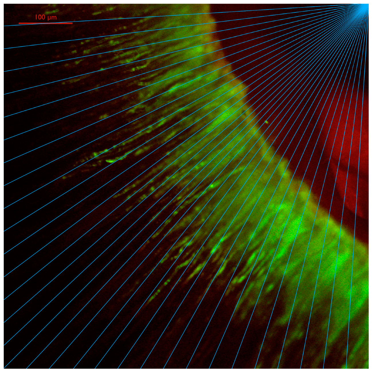
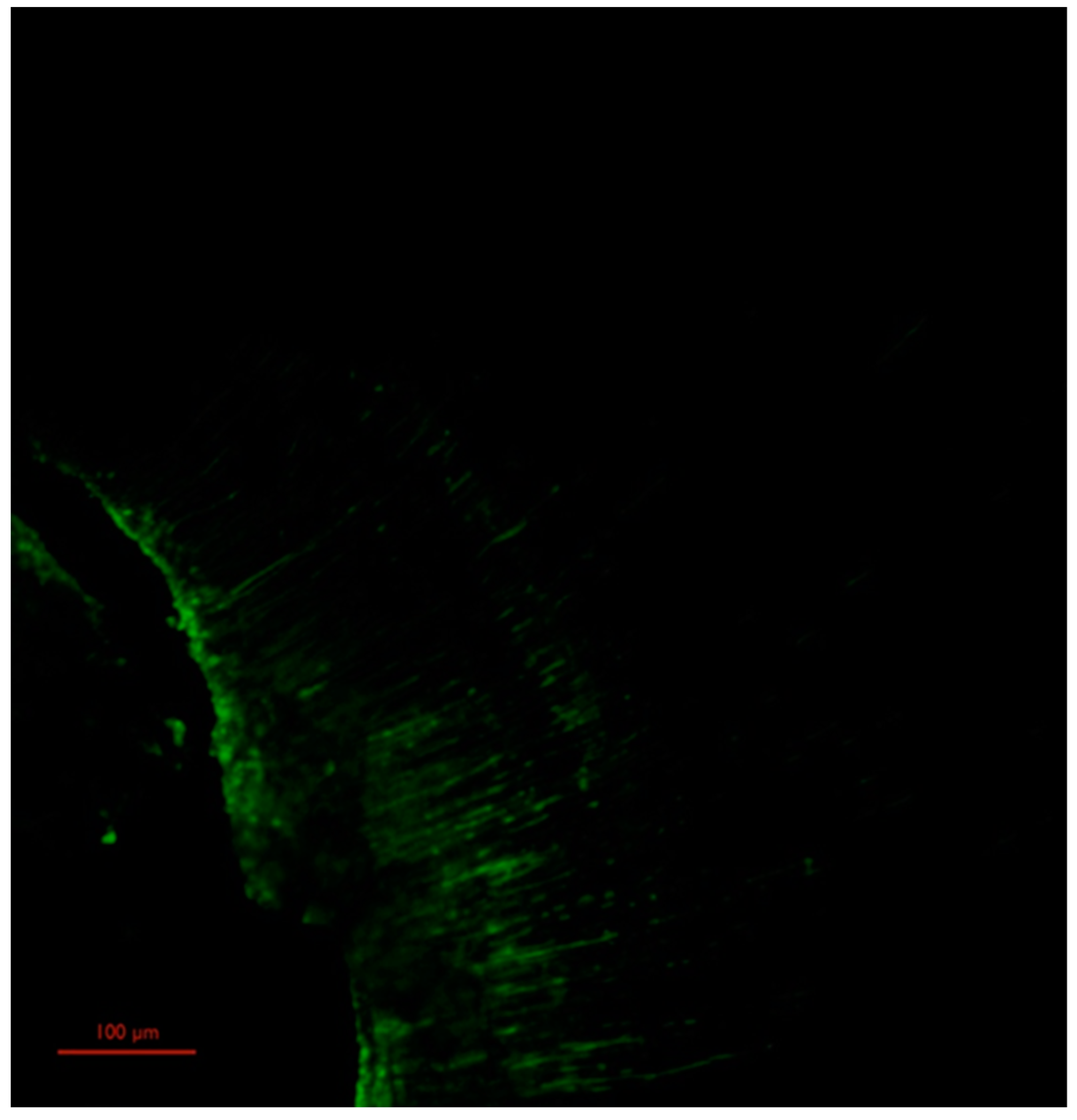
After obturation of the canals, the roots were stored for 72 h in a moist environment with the temperature set at 37 °C. Then, using the IsoMet 1000 Precision Saw (Buehler, USA), three cross-sections were cut out from every root, each 1 mm wide, at the lengths of 2 mm (apical section), 5 mm (mid-section), and 8 mm (coronal section) from the root apex. The samples were numbered, encoded, observed and analyzed using a reversed microscope Nikon Ti-E with confocal system Nikon A1 (Nikon, Tokyo, Japan). Objectives of 10× and 20× magnifications were used, while the excitation was realized using 488 nm (for fluorescein) and 561 nm (for porphyrin) laser sources. Each sample was portrayed in four clockwise quarters. Digital pictures were obtained through combining 50 average cross-sections taken every 0.5 μm, starting with 10 μm from the surface of the sample up to 35 μm inside the sample (the pictured width was 25 μm). The summary resolution was 1024 × 1024 which resulted in 0.62 μm/pixel. The pictures were encoded and underwent measurements with ImageJ 1.45 s program (National Institutes of Health, Bethesda, MD, USA). The measurements of the sealer’s depth of resin tags in dentinal tubules were carried out in all four quarters for each cross-section in the same manner as in the previous study made by the authors [
19]. Each picture was accompanied by a series of thirty-two measurements in constant fields placed radially to the canal’s axis every 2.8 degrees (
Figure 1).
The results for each measurement for each field were a mean value of the resin tag lengths in that field.
2.3. Data Collection and Statistical Analysis
The results were collected, properly encoded, and stored in a database for further statistical analysis. One-way analysis of variance (ANOVA), concluded by Tukey’s honestly significant difference (HSD) test at the a posteriori analysis stage, turned out to be unfulfilled even after the Box–Cox transformation (the normality Kolmogorov–Smirnov test, Shapiro–Wilk test, Lilliefors test, and even the variance homogeneity tests—Levene’s, Hartley’s, Fisher’s and Bartlett’s—all failed). The decision to carry out the analysis, using the nonparametric Kruskal–Wallis test, followed by relevant nonparametric multiple comparison tests, was made due to the failure of the tests commonly used in similar cases. Despite the fact that the suppositions for parametric tests were not met, the numerical results were in full agreement with the results of the nonparametric tests. This can be attributed to the very large sample size. The obtained results were also consistent with an additional check based on the reversed decision tree technique. In this case, the usual roles of the dependent and independent variables (predictors) are exchanged, namely the irrigation protocol was selected as the dependent variable, and penetration depth was one of the predictors. Because of the intrinsic characteristic of the data (the qualitative data ordered on 3 integer levels—the number of the canal sections), the correlation analysis based on Pearson’s r parametric test would lead to wrong statements. Thus, for the correlation analysis, Kendall’s tau nonparametric test was carried out. In all tests, a significance level of p ≤ 0.05 was assumed.
3. Results
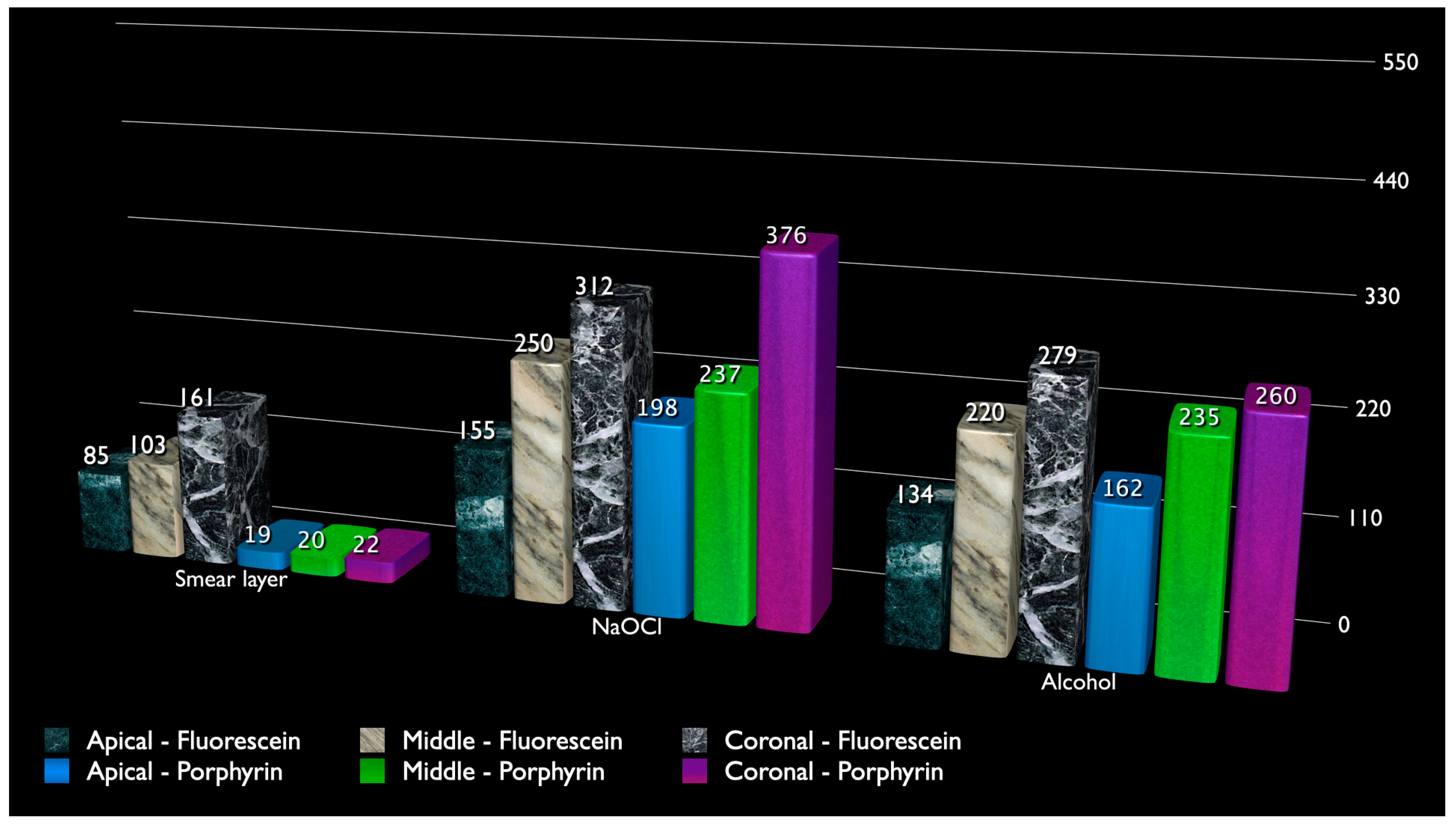
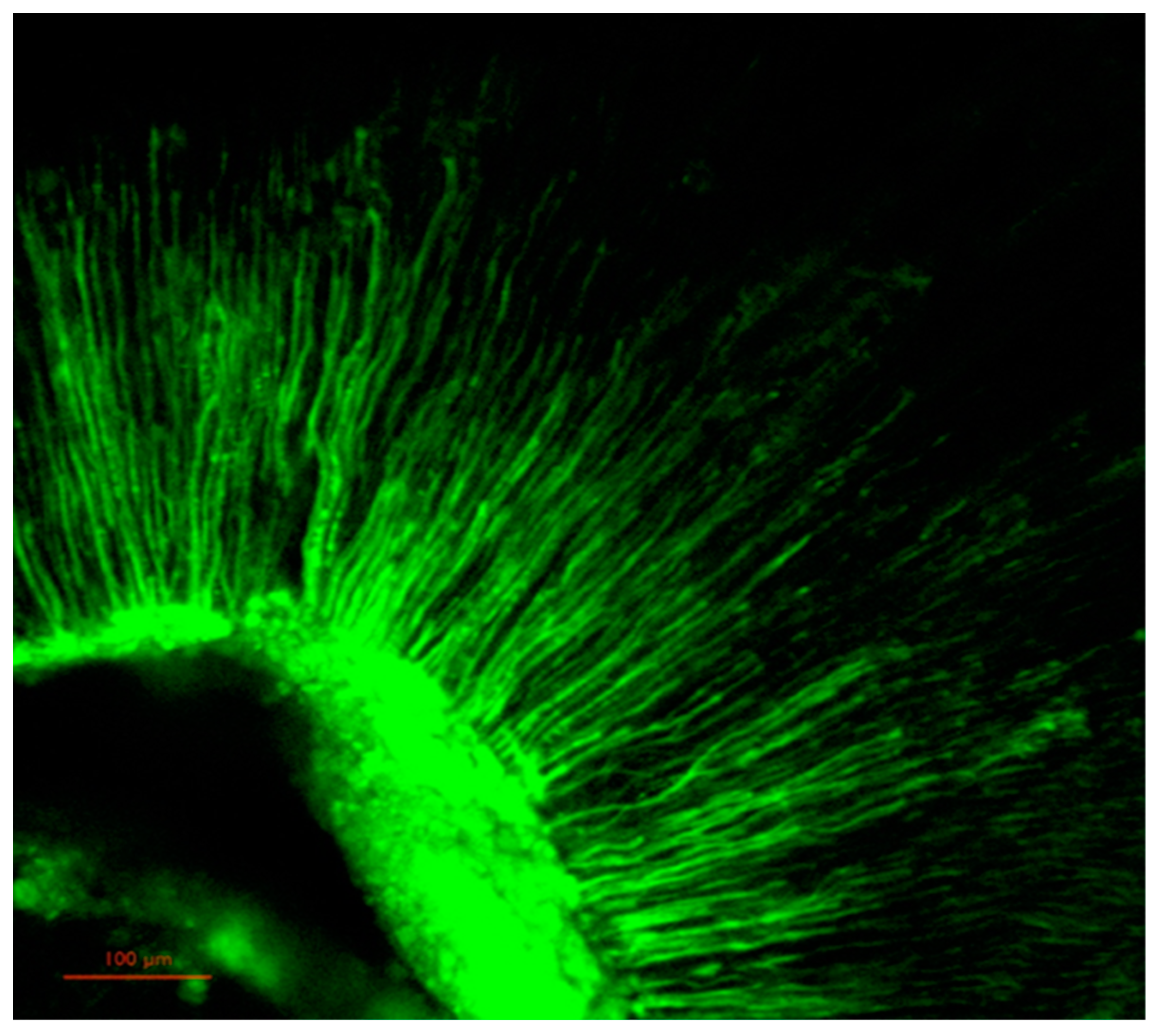
In the case of the sealer marked with porphyrin, a statistical significance was recorded between all experimental groups and in all studied lengths (sections) (p = 0.0). The deepest sealer’s penetration was recorded in group 2 (sodium hypochlorite), and the shallowest in group 1 (smear layer).
When it comes to the sealer marked with fluorescein, the statistically significant differences were recorded between all of the groups in the mid-section. In the apical and coronal section statistically important differences were recorded between group 1 (smear layer) and the rest of the groups (p = 0.0). No significant differences were recorded between group 2 (sodium hypochlorite) and group 3 (alcohol) in the apical and coronal sections (pa = 0.97 and pc = 0.89, respectively).
For both markers (fluorescein and porphyrin), a statistically significant positive correlation was recorded between the part of the root and the depth of the sealer’s penetration (p = 0.0). The hydrophobic or hydrophilic nature of the used dye affected the measured values, but the alcoholic environment in group 3 did not promote more leakage of porphyrin, compared to the aqueous environment in group 2 (sodium hypochlorite).
4. Discussion
Sealers with gutta-percha determine a tight seal of the whole space of the root canal system with the apical delta [
2,
5]. The robustness of such sealing aims at hermetic closing from remains and bacteria as well as prevention of re-infection [
2,
5,
6,
7]. The failure of the endodontic obturation may result in periapical inflammation leading to surgical treatment or even tooth loss, the latter requiring replacement with dental implants [
25,
26]. One of the parameters that defines the sealer interaction with the dentin is the sealer penetration range into the dentinal tubules [
5,
6,
7]. It depends on factors such as the presence of the smear layer, the chosen filling technique, the physio-chemical properties of the sealer (e.g., its thickness, stickiness to the root canal tissues and hydrophilic properties) as well as its possible interaction with dentin [
5,
6,
7,
27,
28,
29,
30]. An epoxy sealer was used in this investigation as it is the most frequently used type of sealer in endodontic therapy and to this day is considered as the golden standard in endodontics. According to Kuci et al. a type of sealer used, may also differ in its penetration into dentinal tubules, when the smear layer is present or absent [
31].
The smear layer, created by a mechanical preparation, establishes a mechanical barrier against irrigating solutions and sealers. Its removal is obligatory for effective decontamination of the dentin and for the sealers penetration into the dentinal tubules [
4,
5,
6,
7,
22,
23]. In the above studies, the smear layer was removed in experimental groups (2 and 3) using two different irrigation protocols with citric acid and sodium hypochlorite. The other studies of the authors revealed that this method of canal irrigation was the most effective way to remove the smear layer [
22,
23]. During the alternating irrigation with sodium hypochlorite (NaOCl) and citric acid (CA), probably irrigation with NaOCl after the first cycle of CA increases the effectiveness of the subsequent CA irrigation cycle. The partial removal of the smear layer (inorganic matter) during the first irrigation cycle with the chelating agents resulted in the exposure of the organic compounds in the deeper parts of the smear layer and the dentin (collagen). Subsequent irrigation with NaOCl most likely dissolves exposed organic substances (organic matter in the deeper parts of the smear layer), enhancing the effect of subsequent application of CA. This phenomenon was tested by the authors twice with the use of different methodologies [
22,
23]. There are some recently invented solutions for the final rinse that can provide higher sealer penetration and more opened dentinal tubules than sequent application of EDTA and chlorhexidine [
32].
Although there is no consensus on the sequence of the irrigation protocols, the most common protocol is using sodium hypochlorite (NaOCl) during the whole mechanical preparation. When used alone, NaOCl can dissolve pulpal remnants and dentin debris. However, many studies have revealed its ineffectiveness in removing the entire smear layer when used as the only irrigant. Other studies proved that alternating the use of EDTA and NaOCl is an effective method for smear layer removal [
33,
34,
35]. Zou et al. researched the effect of NaOCl concentration, its active duration and temperature on its penetration into the dentinal tubules while in direct contact with dentine. The results proved that the potential of sodium hypochlorite to penetrate into the dentinal tubules was dependent on time, its percentage and temperature, but the comparative effect of the these three factors was less effective than supposed [
36]. In a systematic review of the literature, Tonnini et al. evaluated the irrigating solutions and activation methods used in clinical endodontics. They concluded that existing protocols have not yet been defined for the real clinical state and context among those actually available. The review showed that activation methods of the irrigants could provide significantly higher microbiological flora removal. However, activation methods, time of the application and volume of irrigating solutions should be standardized to find out the optimal irrigating protocols [
37].
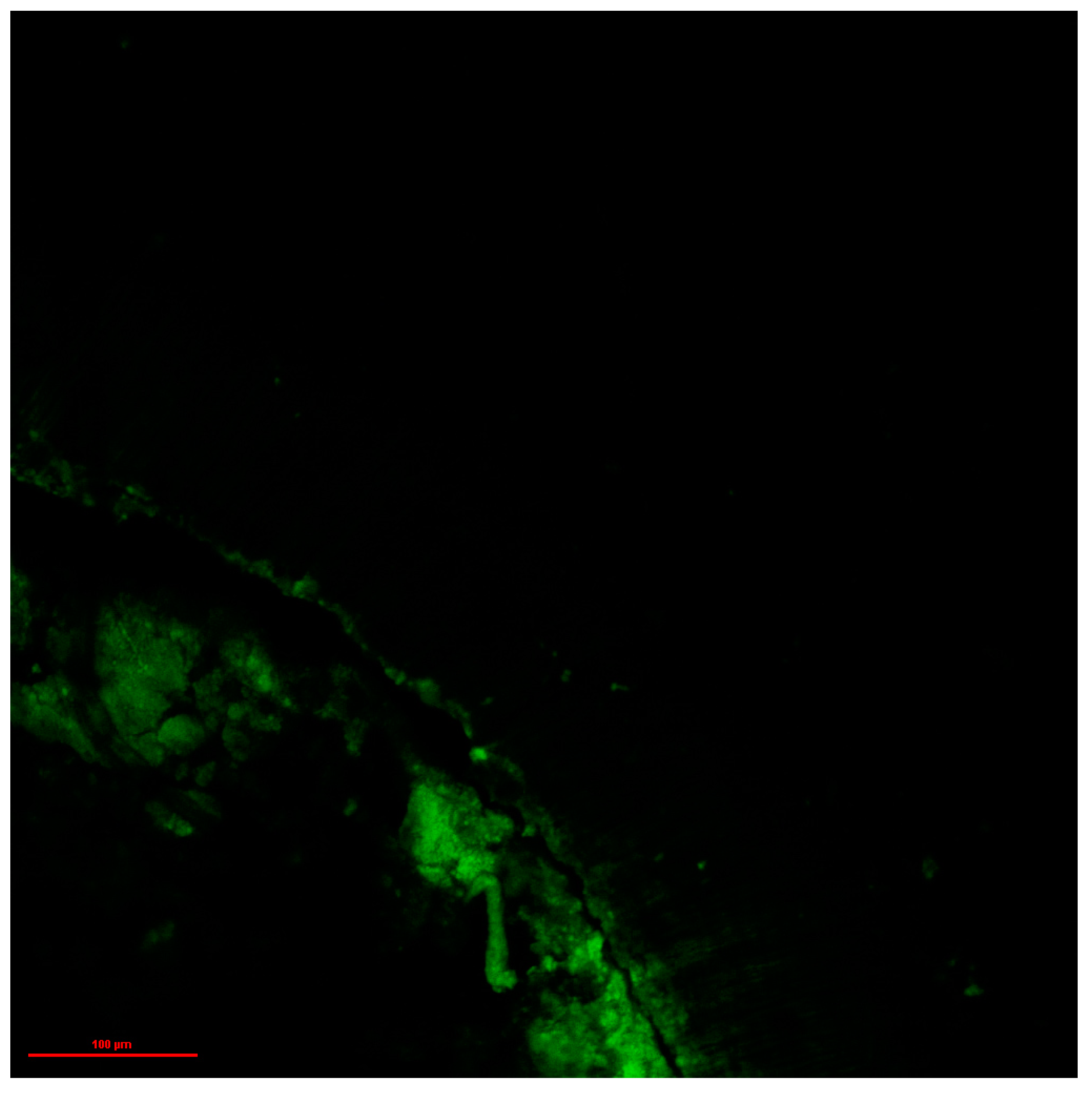
In this study some penetration of the sealer marked with fluorescein in control group (group 1) into dentinal tubules was observed. In this group the irrigation was carried out with sodium hypochlorite only. It is likely that there was some dye leakage of the fluorescein from the sealer, or the probability that the lubricant containing 17% EDTA used during the preparation may have resulted in partial removal of the smear layer. Passive ultrasonic activation could have influenced the initial and incomplete elimination of the smear layer. Cameron investigated ultrasonics to remove a smear layer and confirmed its usefulness in the process [
38]. During the thermoplastic vertical gutta-percha compaction, some pressure is applied to the materials against the canal walls. It could have also influenced the mechanical pressing of the sealer into partly opened dentinal tubules after the irrigating process. It is important to point out that these explanations require long term research using additional and different methodologies to verify the results presented in this study. In the case of a porphyrin marked sealer, the penetration into the dentinal tubules in this group was undoubtedly limited by the smear layer in group 1. It is probably due to the different physio-chemical characteristics of a sealer marked with two different substances. Fluorescein is soluble in basic solutions, where it forms highly fluorescent salt. Thus, it could partially migrate from the sealer into dentinal tubules even in the samples of group 1.
The authors supposed that the applied fluorescein dye may migrate and leak from the sealer, as it is soluble in water (in basic solutions). As mentioned by Donnermeyer et al., rhodamine B is also highly soluble in water, making it susceptible to leakage to the hydrophilic environment, even disregarding its pH [
16]. Thus, the use of two types of dye (hydrophilic and hydrophobic) was necessary to verify if the choice of a dye affects the results of this study. The applied porphyrin (TPP), unlike fluorescein and rhodamine B, is hydrophobic, which significantly limits its leakage to the surrounding aqueous medium, presumably making the presented results more reliable. Thus, using hydrophobic porphyrin as a fluorescent tracker seems to be more appropriate in this type of study, but it still seems to remain a weakness of the method. After filling, the roots were exposed to a humid environment and partial diffusion of fluorescein with the water vapor (the environment after NaOCl is alkaline) into the tubules may have occurred. The use of porphyrin appears to be more beneficial due to its higher sensitivity in comparison to the fluorescein (there is a smaller standard deviation, there is more emphasis on the differences in results between the groups and the statistical significance).
The initial doubt concerned the question of whether the porphyrin would leach from the sealer into the alcoholic environment in the tubules of the samples from group 3, but the results showed that the penetration of the stained sealer was greater in an aqueous environment (sodium hypochlorite group). Also, the hydrophilic fluorescein did not leak into the aqueous environment as much as anticipated in the sodium hypochlorite group. The initial assumption that each dye could leak into its favorable medium was rejected. This might be correlated with the type of the sealer. In this paper, the well-documented, chemically stable and hydrophobic AH Plus epoxy sealer was used. Donnermeyer et al. used three different sealers, and AH Plus yielded the least leakage of rhodamine B into the PBS solution in comparison to the two bioceramic sealers (BioRoot RCS and Total Fill BC) [
16]. Nevertheless considerable leakage of the dye into the tubules was recorded, even from the AH Plus sealer [
16]. In this paper, the hydrophobic AH Plus sealer was used, which in turn is probably the main factor limiting the leaching of both of the used dyes in this experiment, however these assumptions require further investigations with different methodologies.
The AH Plus epoxy sealer was used in this study. It is characterized by its effective tightness, superior spatial stability and low density [
39], but its hydrophobicity can be a theoretical problem. According to Nagas et al., it is more beneficial to leave slightly moisturized dentin, even if a hydrophobic sealer is used [
40]. However, the researchers revealed that using alcohol for the final stages of irrigation could improve the penetration of a hydrophobic sealer into the dentinal tubules in relation to a hypochlorite used as an irrigant. On the other hand, Engel et al. proved that a final rinse of the canal with either 70% isopropyl alcohol or Peridex did not increase sealer penetration, or significantly affected apical microleakage compared to NaOCl. However, in this model of the study, light microscopy was used to investigate the sealer penetration in vitro, so the results cannot be directly compared with those using confocal microscopes [
41]. An in vitro study, which investigated the use of 95% ethanol as the final irrigant, in smear layer free dentin, revealed (within the limitations of these studies) an increased sealer penetration into dentinal tubules and decreased leakage [
42].
The main shortcomings of the conducted study relate to the physical and chemical properties of the dyes used in the process of dyeing the sealer. Anatomical conditions of the root canals and histological differences between the samples taken from natural teeth may also be a risk factor for the presented research. The reader has to take these results with criticism and concern, as the sample preparation technique and sample variability can strongly influence the results obtained. The results of this experiment allowed for the null hypothesis to be rejected. The use of an isopropyl alcohol as the final irrigant had a different effect than previously assumed. The greatest sealer depth penetration measurement into the dentinal tubules was observed in the group, where a water-based solution of sodium hypochlorite was used in the final stages of irrigation. The differences were statistically significant mainly in the case of the porphyrin marked sealer.
In this study, independently from the experimental group, different sealer depth penetrations were obtained in each section of the root canal. The differences can be influenced by the structure of the dentinal tubules in different sections of the root canal, as well as varied effectiveness of the irrigation and activation of solutions before obturation. Similar research points revealed a comparable relationship between these factors [
2,
5,
28,
29,
43].


 ,
, 


