
Petrographic and Chemical Characterization of the Frescoes by Saturnino Gatti (Central Italy, 15th Century)
 , , , ,
  , and
, and
Abstract
:1. Introduction
2. Materials and Methods
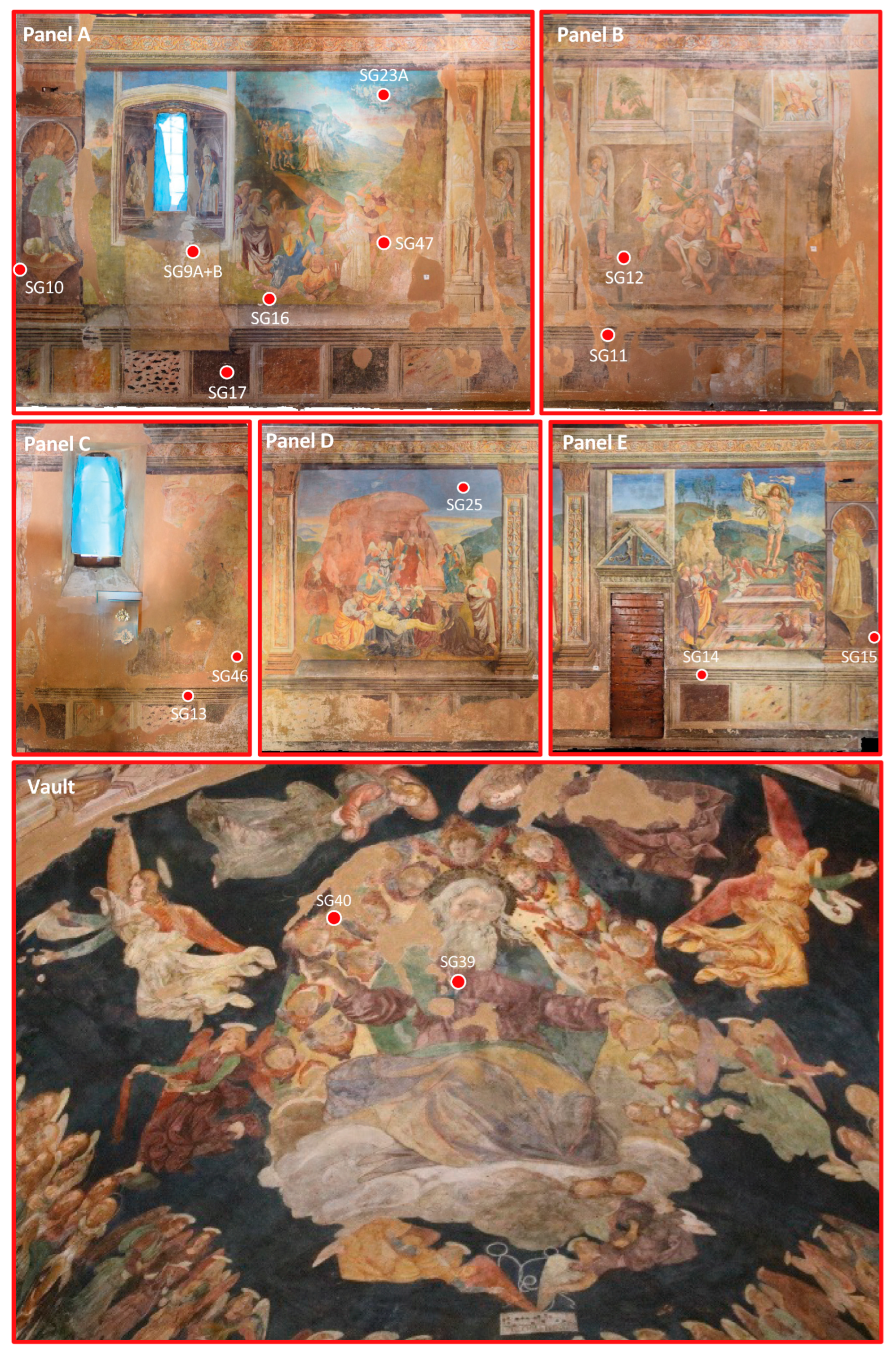
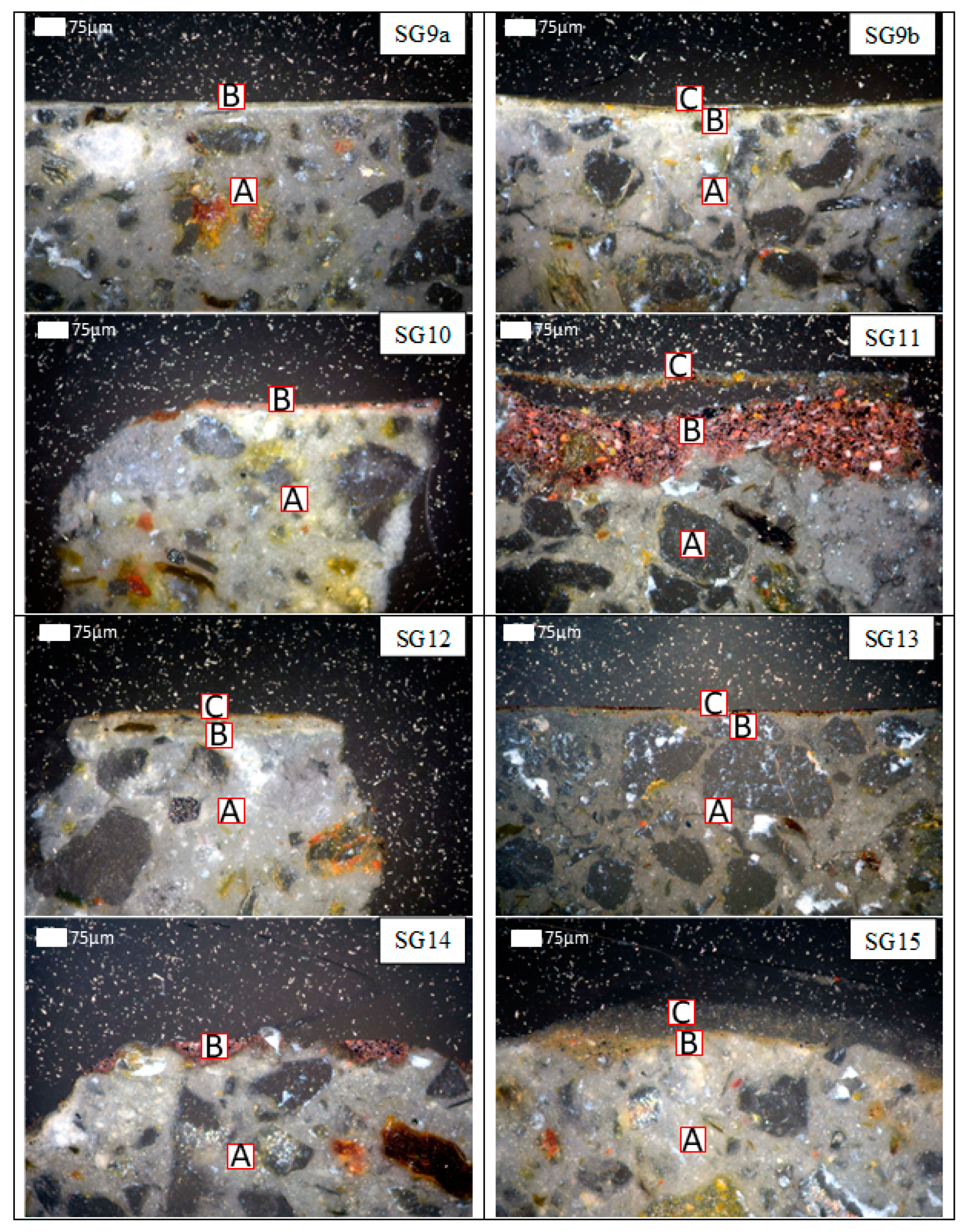
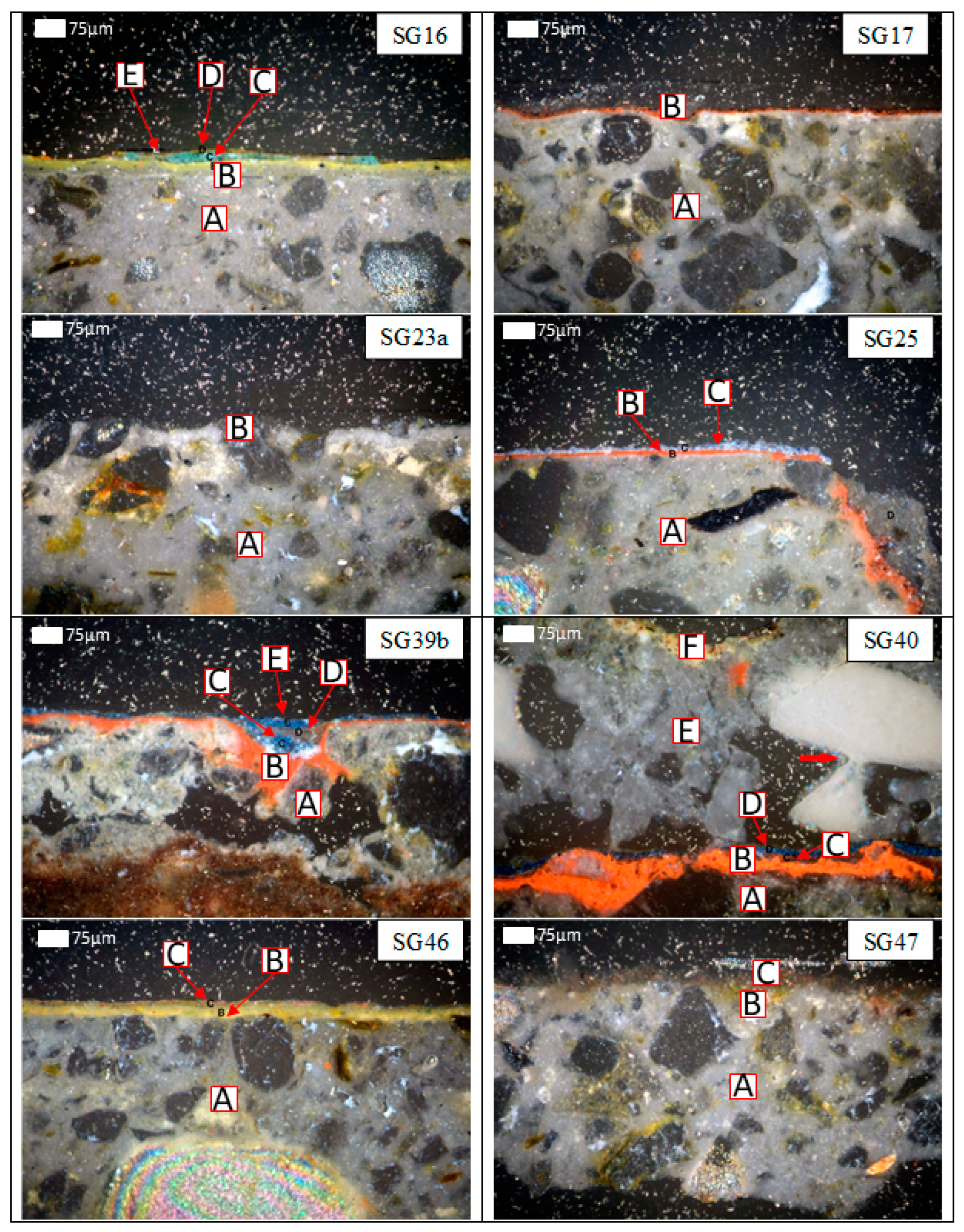
2.1. Sampling
2.2. Methods
3. Results
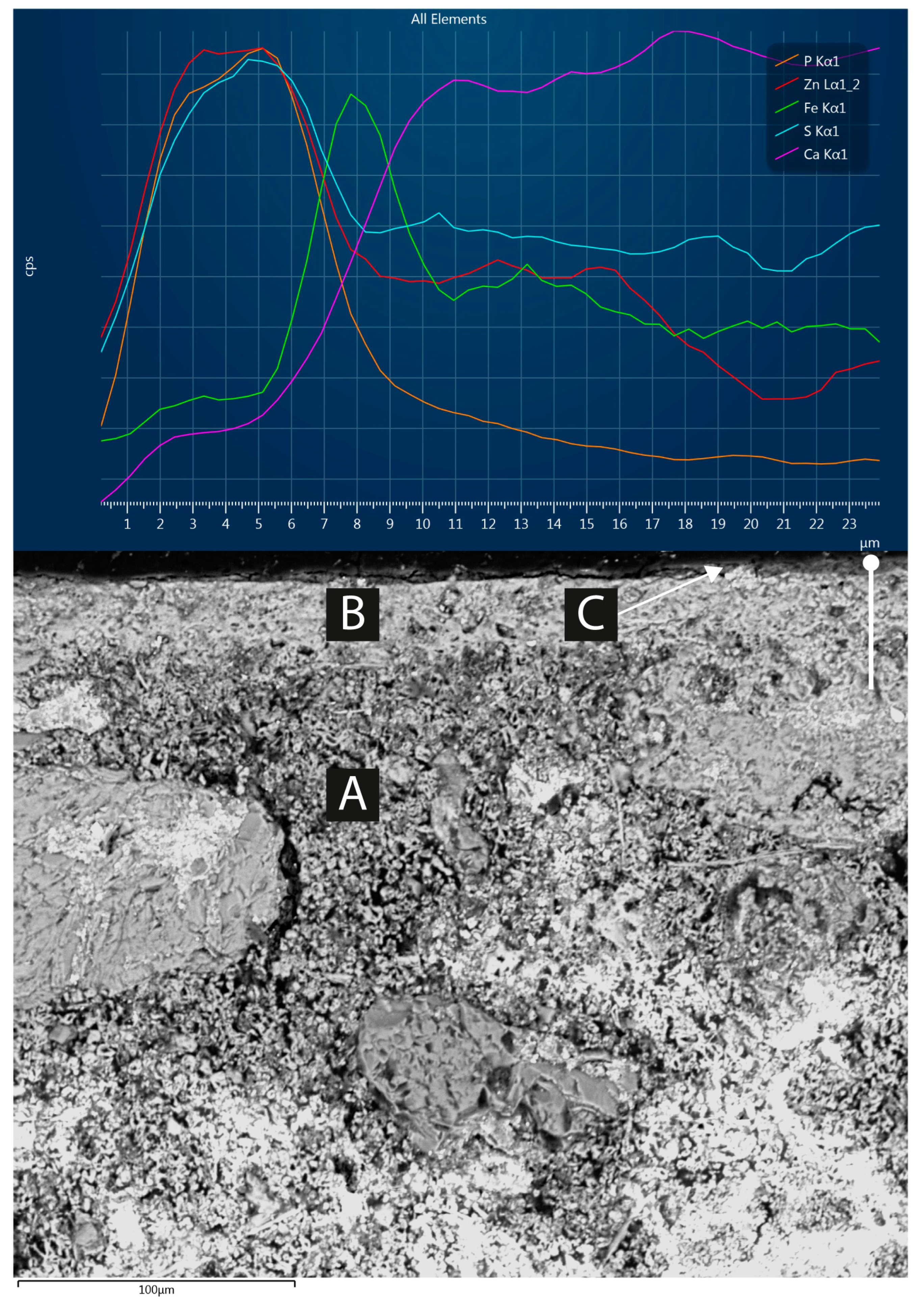
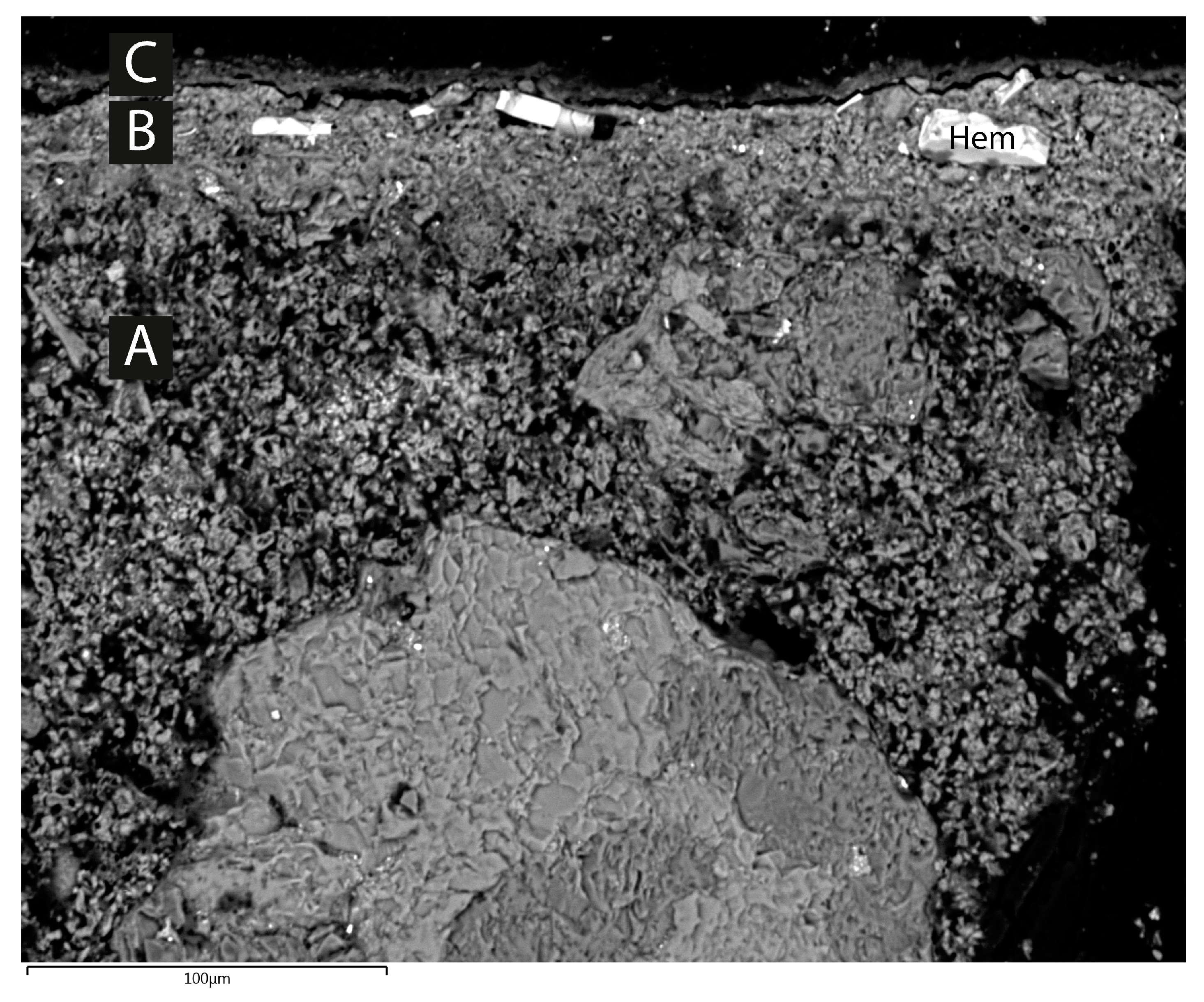
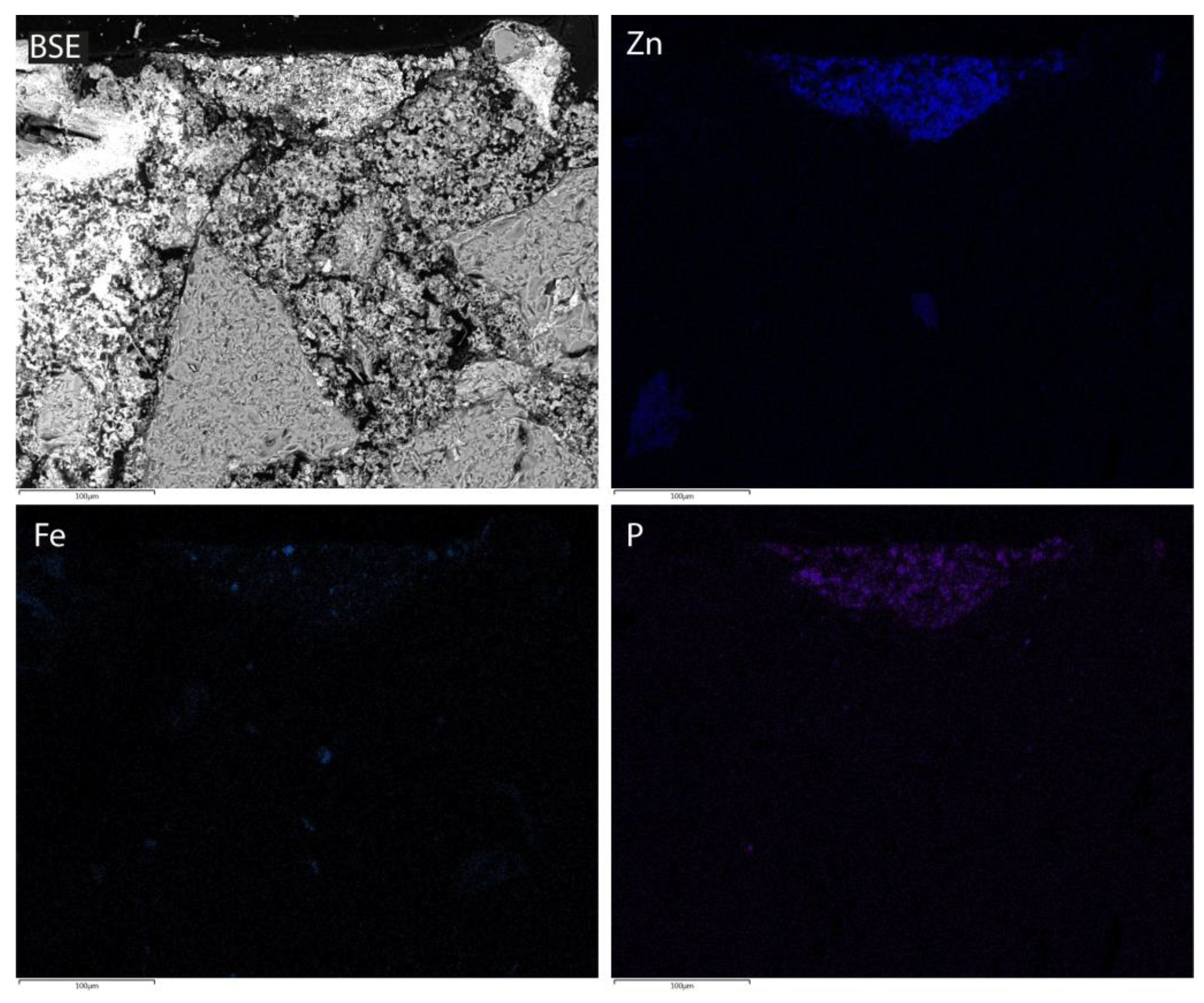
3.1. Petrography (OM, CL)
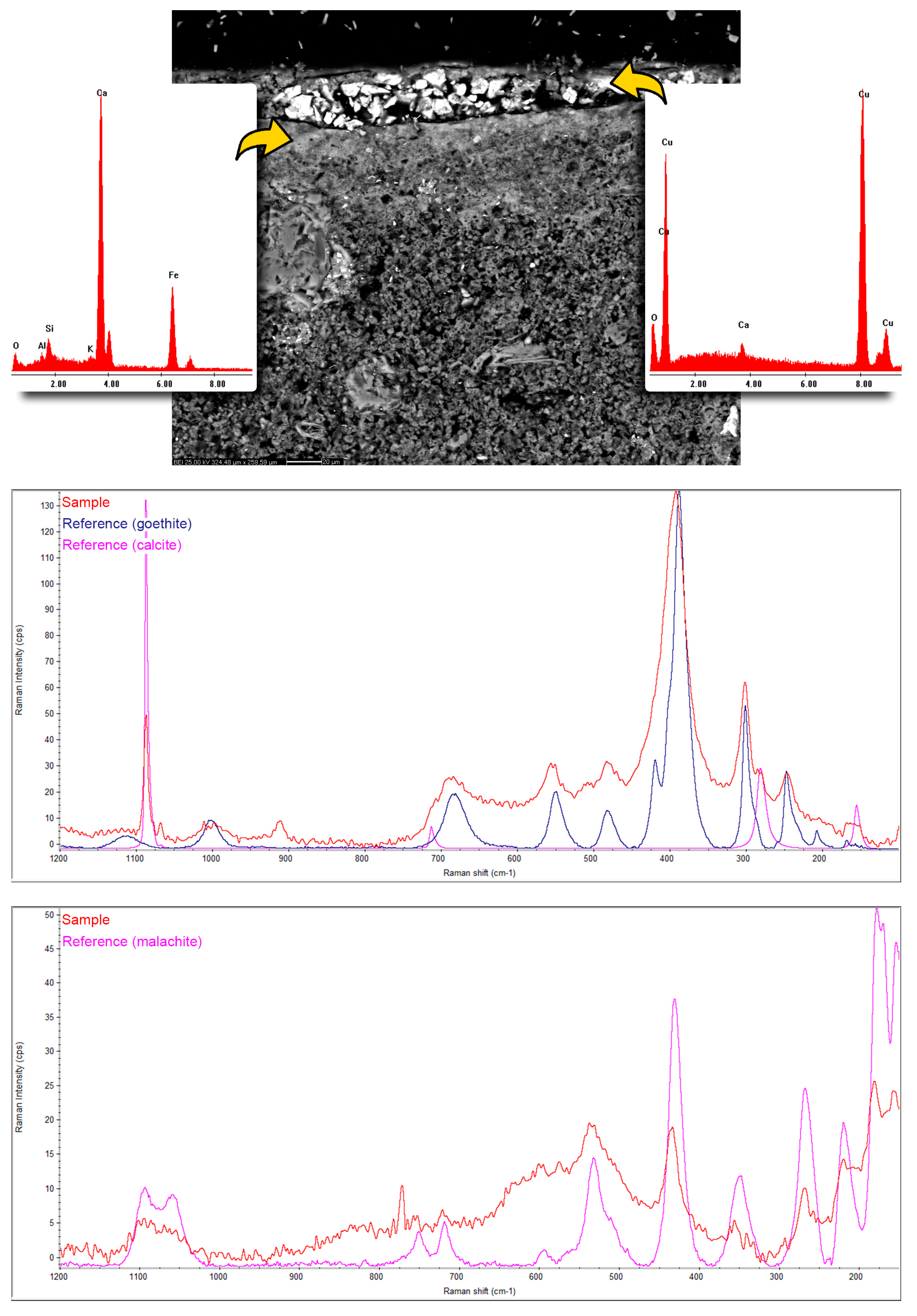
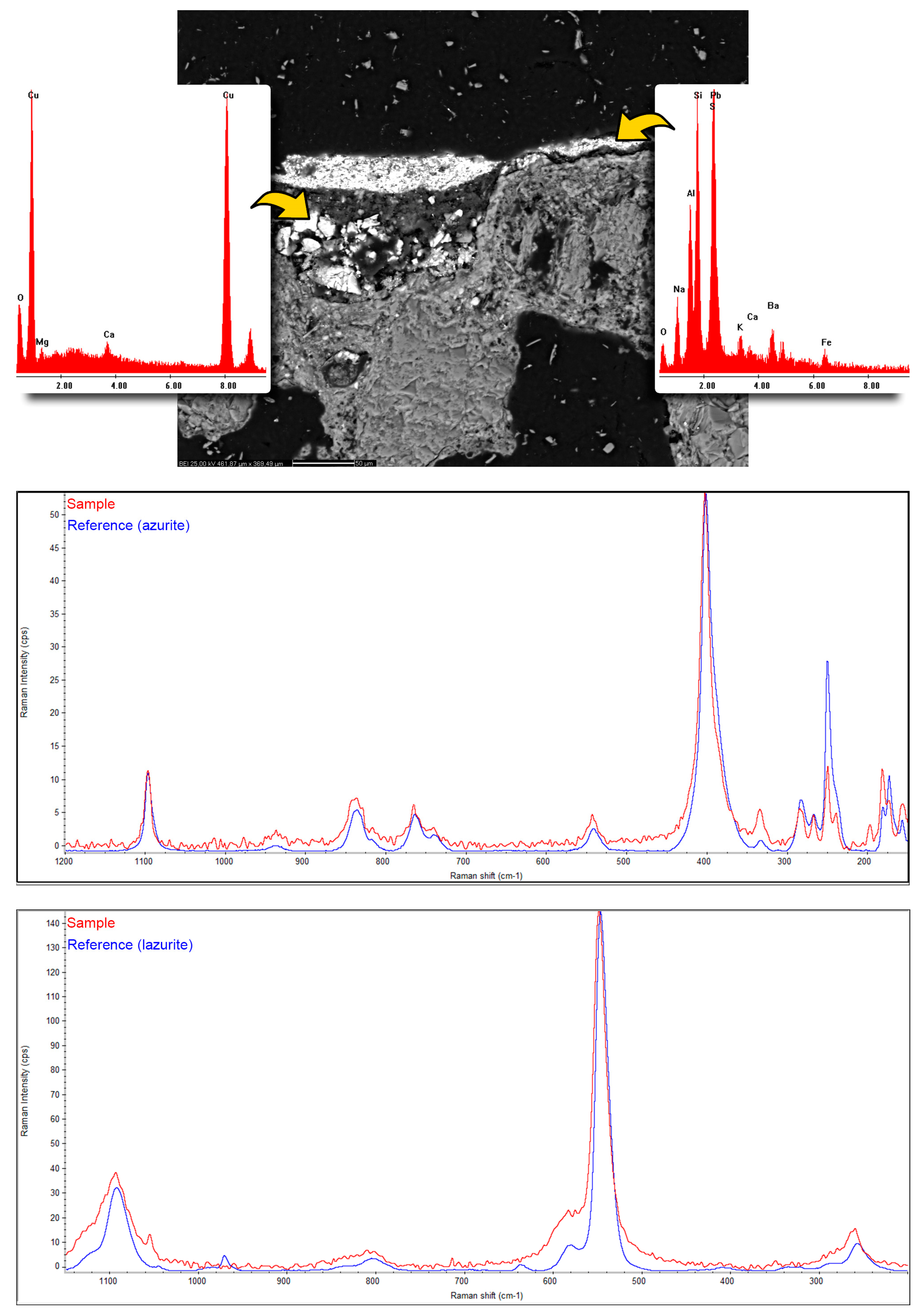
3.2. SEM-EDS and µ-Raman
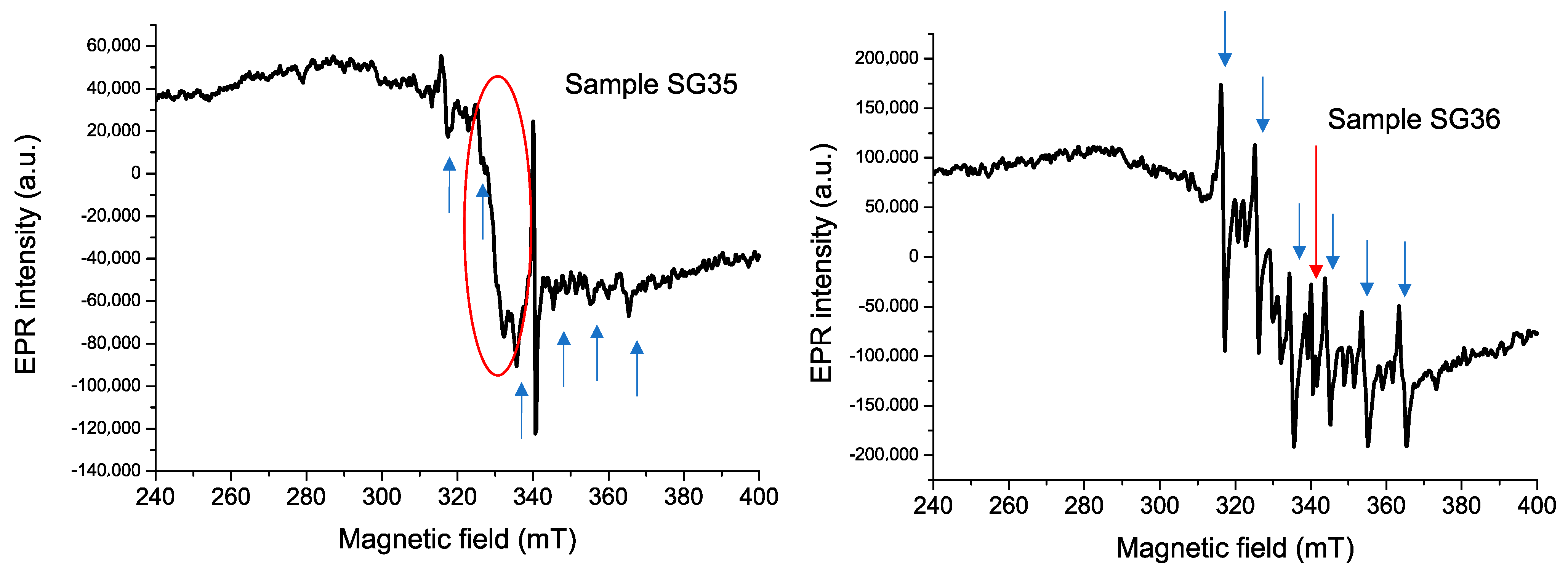
3.3. EPR
4. Discussion
4.1. Stratigraphy: Original and Successive Interventions
4.2. Lime Plasters: Raw Materials and Technological Aspects
4.3. Pigments, Binders, and Painting Techniques
4.4. Alteration and Decay
5. Conclusions
Author Contributions
Funding
Data Availability Statement
Acknowledgments
Conflicts of Interest
References
- Arbace, L. I Volti dell’Anima. Saturnino Gatti. In Vita e Opere di un Artista del Rinascimento; Paolo De Siena Editore: Pescara, Italy, 2012; ISBN 88-96341-11-6. [Google Scholar]
- Zezza, A. Paintings, Frescoes, and Cycles. In A Companion to the Renaissance in Southern Italy (1350–1600); Di Vitiis, B., Ed.; Brill: New York, NY, USA, 2022; pp. 591–617. ISBN 978-90-04-52637-2. [Google Scholar]
- Vittorini, A. L’Aquila 2009–2019: Back to the Future. Cultural Heritage and Post-Seismic Reconstruction Challenges. In Invisible Reconstruction. Cross-Disciplinary Responses to Natural, Biological and Man-Made Disasters; Patrizio Gunning, L., Rizzi, P., Eds.; Fringe; UCL Press: London, UK, 2022; pp. 11–28. [Google Scholar]
- Quagliarini, E.; Lenci, S.; Seri, E. On the Damage of Frescoes and Stuccoes on the Lower Surface of Historical Flat Suspended Light Vaults. J. Cult. Herit. 2012, 13, 293–303. [Google Scholar] [CrossRef]
- Lermé, N.; Hégarat-Mascle, S.L.; Zhang, B.; Aldea, E. Fast and Efficient Reconstruction of Digitized Frescoes. Pattern Recognit. Lett. 2020, 138, 417–423. [Google Scholar] [CrossRef]
- Bruno, N.; Mikolajewska, S.; Roncella, R.; Zerbi, A. Integrated Processing of Photogrammetric and Laser Scanning Data for Frescoes Restoration. Int. Arch. Photogramm. Remote Sens. Spat. Inf. Sci. ISPRS Arch. 2022, 46, 105–112. [Google Scholar] [CrossRef]
- Piovesan, R.; Maritan, L.; Amatucci, M.; Nodari, L.; Neguer, J. Wall Painting Pigments of Roman Empire Age from Syria Palestina Province (Israel). Eur. J. Mineral. 2016, 28, 435–448. [Google Scholar] [CrossRef]
- Pecchioni, E.; Pallecchi, P.; Giachi, G.; Calandra, S.; Santo, A.P. The Preparatory Layers in the Etruscan Paintings of the Tomba dei Demoni Alati in the Sovana Necropolis (Southern Tuscany, Italy). Appl. Sci. 2022, 12, 3542. [Google Scholar] [CrossRef]
- Mangone, A.; Colombi, C.; Eramo, G.; Muntoni, I.M.; Forleo, T.; Giannossa, L.C. Pigments and Techniques of Hellenistic Apulian Tomb Painting. Molecules 2023, 28, 1055. [Google Scholar] [CrossRef]
- Holclajtner-Antunović, I.; Stojanović-Marić, M.; Bajuk-Bogdanović, D.; Žikić, R.; Uskoković-Marković, S. Multi-Analytical Study of Techniques and Palettes of Wall Paintings of the Monastery of Žiča, Serbia. Spectrochim. Acta Part A Mol. Biomol. Spectrosc. 2016, 156, 78–88. [Google Scholar] [CrossRef] [PubMed]
- Miriello, D.; Bloise, A.; Crisci, G.M.; De Luca, R.; De Nigris, B.; Martellone, A.; Osanna, M.; Pace, R.; Pecci, A.; Ruggieri, N. Non-Destructive Multi-Analytical Approach to Study the Pigments of Wall Painting Fragments Reused in Mortars from the Archaeological Site of Pompeii (Italy). Minerals 2018, 8, 134. [Google Scholar] [CrossRef] [Green Version]
- Garavelli, A.; Andriani, G.F.; Fioretti, G.; Iurilli, V.; Marsico, A.; Pinto, D. The “Sant’Angelo in Criptis” Cave Church in Santeramo in Colle (Apulia, South Italy): A Multidisciplinary Study for the Evaluation of Conservation State and Stability Assessment. Geosciences 2021, 11, 382. [Google Scholar] [CrossRef]
- Carlomagno, G.M.; Meola, C. Comparison between Thermographic Techniques for Frescoes NDT. NDT E Int. 2002, 35, 559–565. [Google Scholar] [CrossRef]
- Gusella, V.; Cluni, F.; Liberotti, R. Feasibility of a Thermography Nondestructive Technique for Determining the Quality of Historical Frescoed Masonries: Applications on the Templar Church of San Bevignate. Appl. Sci. 2021, 11, 281. [Google Scholar] [CrossRef]
- Almaviva, S.; Fantoni, R.; Colao, F.; Puiu, A.; Bisconti, F.; Fiocchi Nicolai, V.; Romani, M.; Cascioli, S.; Bellagamba, S. LIF/Raman/XRF Non-Invasive Microanalysis of Frescoes from St. Alexander Catacombs in Rome. Spectrochim. Acta Part A Mol. Biomol. Spectrosc. 2018, 201, 207–215. [Google Scholar] [CrossRef]
- Sáez-Hernández, R.; Antela, K.U.; Gallello, G.; Cervera, M.L.; Mauri-Aucejo, A.R. A smartphone-based innovative approach to discriminate red pigments in roman frescoes mock-ups. J. Cult. Herit. 2022, 58, 156–166. [Google Scholar] [CrossRef]
- Merello, P.; Beltrán, P.; García-Diego, F.J. Quantitative Non-Invasive Method for Damage Evaluation in Frescoes: Ariadne’s House (Pompeii, Italy). Environ. Earth Sci. 2016, 75, 165. [Google Scholar] [CrossRef]
- Fioretti, G.; Campobasso, C.; Capotorto, S. Digital Photogrammetry as Tool for Mensiochronological Analysis: The Case of St. Maria Veterana Archaeological Site (Triggiano, Italy). Digit. Appl. Archaeol. Cult. Herit. 2020, 19, e00158. [Google Scholar] [CrossRef]
- Horgnies, M.; Bayle, M.; Gueit, E.; Darque-Ceretti, E.; Aucouturier, M. Microstructure and Surface Properties of Frescoes Based on Lime and Cement: The Influence of the Artist’s Technique. Archaeometry 2015, 57, 344–361. [Google Scholar] [CrossRef]
- Helvaci, Y.Z.; Dias, L.; Manhita, A.; Martins, S.; Cardoso, A.; Candeias, A.; Gil, M. Tracking Old and New Colours: Material Study of 16th Century Mural Paintings from Évora Cathedral (Southern Portugal). Color Res. Appl. 2016, 41, 276–282. [Google Scholar] [CrossRef]
- Vasco, G.; Serra, A.; Manno, D.; Buccolieri, G.; Calcagnile, L.; Buccolieri, A. Investigations of Byzantine Wall Paintings in the Abbey of Santa Maria Di Cerrate (Italy) in View of Their Restoration. Spectrochim. Acta Part A Mol. Biomol. Spectrosc. 2020, 239, 118557. [Google Scholar] [CrossRef]
- Graziano, S.F.; Rispoli, C.; Guarino, V.; Balassone, G.; Di Maio, G.; Pappalardo, L.; Cappelletti, P.; Damato, G.; De Bonis, A.; Di Benedetto, C.; et al. The Roman Villa of Positano (Campania Region, Southern Italy): Plasters, Tiles and Geoarchaeological Reconstruction. Int. J. Conserv. Sci. 2020, 11, 319–344. [Google Scholar]
- De Benedetto, G.E.; Savino, A.; Fico, D.; Rizzo, D.; Pennetta, A.; Cassiano, A.; Minerva, B. A Multi-Analytical Approach for the Characterisation of the Oldest Pictorial Cycle in the 12th Century Monastery Santa Maria Delle Cerrate. Open J. Archaeom. 2013, 1, e12. [Google Scholar] [CrossRef] [Green Version]
- Bersani, D.; Berzioli, M.; Caglio, S.; Casoli, A.; Lottici, P.P.; Medeghini, L.; Poldi, G.; Zannini, P. An Integrated Multi-Analytical Approach to the Study of the Dome Wall Paintings by Correggio in Parma Cathedral. Microchem. J. 2014, 114, 80–88. [Google Scholar] [CrossRef]
- Romani, M.; Capobianco, G.; Pronti, L.; Colao, F.; Seccaroni, C.; Puiu, A.; Felici, A.C.; Verona-Rinati, G.; Cestelli-Guidi, M.; Tognacci, A.; et al. Analytical Chemistry Approach in Cultural Heritage: The Case of Vincenzo Pasqualoni’s Wall Paintings in S. Nicola in Carcere (Rome). Microchem. J. 2020, 156, 104920. [Google Scholar] [CrossRef]
- Alberghina, M.F.; Schiavone, S.; Greco, C.; Saladino, M.L.; Armetta, F.; Renda, V.; Caponetti, E. How Many Secret Details Could a Systematic Multi-Analytical Study Reveal about the Mysterious Fresco Trionfo Della Morte? Heritage 2019, 2, 145. [Google Scholar] [CrossRef] [Green Version]
- Crupi, V.; Fazio, B.; Fiocco, G.; Galli, G.; La Russa, M.F.; Licchelli, M.; Majolino, D.; Malagodi, M.; Ricca, M.; Ruffolo, S.A.; et al. Multi-Analytical Study of Roman Frescoes from Villa Dei Quintili (Rome, Italy). J. Archaeol. Sci. Rep. 2018, 21, 422–432. [Google Scholar] [CrossRef]
- Angelini, I.; Asscher, Y.; Secco, M.; Parisatto, M.; Artioli, G. The Pigments of the Frigidarium in the Sarno Baths, Pompeii: Identification, Stratigraphy and Weathering. J. Cult. Herit. 2019, 40, 309–316. [Google Scholar] [CrossRef]
- Mohanu, I.; Mohanu, D.; Gomoiu, I.; Barbu, O.H.; Fechet, R.M.; Vlad, N.; Voicu, G.; Truşcă, R. Study of the Frescoes in Ioneştii Govorii Wooden Church (Romania) Using Multi-Technique Investigations. Microchem. J. 2016, 126, 332–340. [Google Scholar] [CrossRef]
- Hakobyan, Z.A. The Frescoes of Haghpat Monastery in the Historical-Confessional Context of the 13th Century. Actual Probl. Theory Hist. Art 2021, 11, 256–267. [Google Scholar] [CrossRef]
- Luvidi, L.; Prestileo, F.; De Paoli, M.; Riminesi, C.; Del Fà, R.M.; Magrini, D.; Fratini, F. Diagnostics and Monitoring to Preserve a Hypogeum Site: The Case of the Mithraeum of Marino Laziale (Rome). Heritage 2021, 4, 235. [Google Scholar] [CrossRef]
- Ranalli, G.; Alfano, G.; Belli, C.; Lustrato, G.; Colombini, M.P.; Bonaduce, I.; Zanardini, E.; Abbruscato, P.; Cappitelli, F.; Sorlini, C. Biotechnology applied to cultural heritage: Biorestoration of frescoes using viable bacterial cells and enzymes. J. Appl. Microbiol. 2005, 98, 73–83. [Google Scholar] [CrossRef]
- Galli, A.; Alberghina, M.F.; Re, A.; Magrini, D.; Grifa, C.; Ponterio, R.C.; La Russa, M.F. Special Issue: Results of the II National Research project of AIAr: Archaeometric study of the frescoes by Saturnino Gatti and workshop at the church of San Panfilo in Tornimparte (AQ, Italy). Appl. Sci. 2023. to be submitted. [Google Scholar]
- Čiuladienė, A.; Luckutė, A.; Kiuberis, J.; Kareiva, A. Investigation of the Chemical Composition of Red Pigments and Binding Media. Chemija 2018, 29, 243–256. [Google Scholar] [CrossRef]
- Cennini, C. Il Libro Dell’arte, o Trattato della Pittura di Cennino Cennini; Milanesi, G., Milanesi, C., Eds.; Le Monnier: Florence, Italy, 1859. [Google Scholar]
- Mora, P.; Mora, L.; Philippot, P. La Conservazione Delle Pitture Murali; Editrice Compositori: Bologna, Italy, 1999. [Google Scholar]
- Piovesan, R.; Mazzoli, C.; Maritan, L.; Cornale, P. Fresco and lime paint: An experimental study and objective criteria for distinguishing between these painting techniques. Archaeometry 2012, 54, 723–736. [Google Scholar] [CrossRef]
- Ergenç, D.; Fort, R.; Varas−Muriel, M.J.; Alvarez de Buergo, M. Mortars and Plasters—How to Characterize Aerial Mortars and Plasters. Archaeol. Anthr. Sci. 2021, 13, 197. [Google Scholar] [CrossRef]
- Bonizzoni, L.; Caglio, S.; Galli, A.; Lanteri, L.; Pelosi, C. Materials and technique: The first look at Saturnino Gatti. Appl. Sci. 2023, 13, 6842. [Google Scholar] [CrossRef]
- Bonizzoni, L.; Caglio, S.; Galli, A.; Germinario, C.; Izzo, F.; Magrini, D. Identifying original and restoration materials through spectroscopic analyses on Saturnino Gatti mural paintings: How far a non-invasive approach can go. Appl. Sci. 2023, 13, 6638. [Google Scholar] [CrossRef]
- Briani, F.; Caridi, F.; Ferella, F.; Gueli, A.M.; Marchegiani, F.; Nisi, S.; Paladini, G.; Pecchioni, E.; Politi, G.; Santo, A.P.; et al. Multi-technique characterization of painting drawings of the pictorial cycle at the San Panfilo Church in Tornimparte (AQ). Appl. Sci. 2023, 13, 6492. [Google Scholar] [CrossRef]
- Armetta, F.; Giuffrida, D.; Ponterio, R.C.; Falcon Martinez, M.F.; Briani, F.; Pecchioni, E.; Santo, A.P.; Ciaramitaro, V.C.; Saladino, M.L. Looking for the original materials and evidence of restoration at the Vault of the San Panfilo Church in Tornimparte (AQ). Appl. Sci. 2023, 13, 6492. [Google Scholar] [CrossRef]
- Andreotti, A.; Izzo, F.C.; Bonaduce, I. Archaeometric study of the mural paintings by Saturnino Gatti and workshop in the Church of San Panfilo—Tornimparte (AQ). The study of organic materials. Appl. Sci. 2023, 13, 7153. [Google Scholar] [CrossRef]
- Centamore, E.; Dramis, F. Note Illustrative Della Carta Geologica d’Italia Alla Scala 1:50.000, Foglio 358-Pescorocchiano. ISPRA-Serv. Geol. D’Italia 2010, 147. [Google Scholar]
- Murat, Z. Wall Paintings through the Ages: The Medieval Period (Italy, Twelfth to Fifteenth Century). Archaeol. Anthr. Sci. 2021, 13, 191. [Google Scholar] [CrossRef]


| Sample | Panel | Description | Investigation Technique |
|---|---|---|---|
| SG9A | A | Plaster with pale ochre paint | OM, SEM-EDS (t.s.) |
| SG9B | A | Plaster with pale ochre paint and decay products (?) | OM, SEM-EDS (t.s.) |
| SG10 | A | Plaster with violaceous paint | OM, SEM-EDS (t.s.) |
| SG11 | B | Plaster with dark-brown paint | OM, SEM-EDS (t.s.) |
| SG12 | B | Plaster with blue–gray paint and makeover (?) | OM, SEM-EDS (t.s.) |
| SG13 | C | Plaster with white paint | OM, SEM-EDS (t.s.) |
| SG14 | E | Plaster with dark paint and protective coating (?) | OM, SEM-EDS (t.s.) |
| SG15 | E | Plaster with blue layer | OM, SEM-EDS (t.s.) |
| SG16 | A | Pale-green paint and plaster (reworked area?) | OM, CL, SEM-EDS, μ-Raman (t.s.) |
| SG17 | A | Porphyry-like paint and plaster | OM, CL, SEM-EDS, μ-Raman (t.s.) |
| SG23A | A | Plaster | OM, CL, SEM-EDS, μ-Raman (t.s.) |
| SG25 | D | Pale-blue paint on a red–brown layer (morellone?) and plaster | OM, CL, SEM-EDS, μ-Raman (t.s.) |
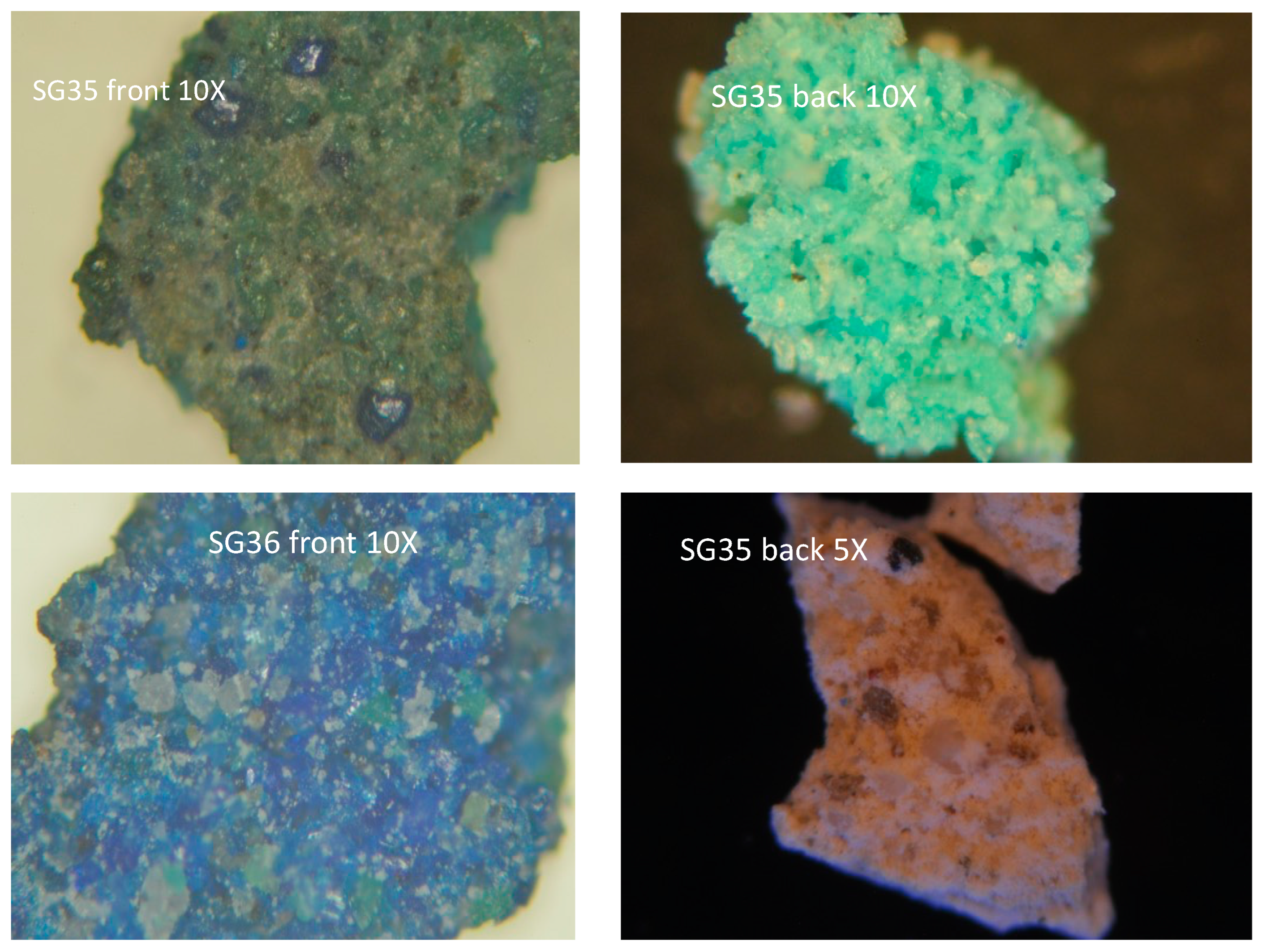
| SG35 | D/E | Green layer | EPR (f.) |
| SG36 | D/E | Light-blue layer | EPR (f.) |
| SG39B | Vault | Blue paint on a red–brown layer (morellone?) and plaster | OM, CL, SEM-EDS, μ-Raman (t.s.) |
| SG40 | Vault | Filling mortar | OM, CL, SEM-EDS, μ-Raman (t.s.) |
| SG46 | C | Plaster with remains of a brown preparation layer | OM, CL, SEM-EDS, μ-Raman (t.s.) |
| SG47 | A | Plaster with remains of a brown preparation layer | OM, CL, SEM-EDS, μ-Raman (t.s.) |
| Sample | Layer A (Plaster) | Layer B | Layer C | Layer D | Layer E | Layer F |
|---|---|---|---|---|---|---|
| SG9A | Air lime with quartz, alkali–feldspars, and plagioclases, micas with minor carbonates as aggregate | red bolus | bone black | / | / | / |
| SG9B | red bolus | bone black, red bolus, ZnO | / | / | / | |
| SG10 | hematite, C black | red bolus, ZnO | / | / | / | |
| SG11 | bone black, red ochre, ZnO | organic? | / | / | / | |
| SG12 | C black, sienna | / | / | / | / | |
| SG13 | C black, ochre | bone black, ochre | / | / | / | |
| SG14 | bone black + ochre + ZnO | / | / | / | / | |
| SG15 | C black + ochre | / | / | / | / | |
| SG16 | yellow ochre + C black | malachite | Cr yellow? | / | / | |
| SG17 | red ochre + C black | / | / | / | / | |
| SG23A | / | / | / | / | / | |
| SG25 | red ochre | ultramarine + Ti white | / | / | / | |
| SG35 | azurite 1 | / | / | / | ||
| SG36 | azurite | / | / | / | ||
| SG39B | red ochre | azurite | resins/org. polymers? | ultramarine + Pb white | / | |
| SG40 | cinnabar | resins/org. polymers? | ultramarine + Pb white | lime plaster | yellow ochre + C black | |
| SG46 | yellow ochre | red ochre + C black | / | / | / | |
| SG47 | yellow ochre + C black | red ochre | / | / | / |
Disclaimer/Publisher’s Note: The statements, opinions and data contained in all publications are solely those of the individual author(s) and contributor(s) and not of MDPI and/or the editor(s). MDPI and/or the editor(s) disclaim responsibility for any injury to people or property resulting from any ideas, methods, instructions or products referred to in the content. |
© 2023 by the authors. Licensee MDPI, Basel, Switzerland. This article is an open access article distributed under the terms and conditions of the Creative Commons Attribution (CC BY) license (https://creativecommons.org/licenses/by/4.0/).
Share and Cite
Germinario, L.; Giannossa, L.C.; Lezzerini, M.; Mangone, A.; Mazzoli, C.; Pagnotta, S.; Spampinato, M.; Zoleo, A.; Eramo, G. Petrographic and Chemical Characterization of the Frescoes by Saturnino Gatti (Central Italy, 15th Century). Appl. Sci. 2023, 13, 7223. https://doi.org/10.3390/app13127223
Germinario L, Giannossa LC, Lezzerini M, Mangone A, Mazzoli C, Pagnotta S, Spampinato M, Zoleo A, Eramo G. Petrographic and Chemical Characterization of the Frescoes by Saturnino Gatti (Central Italy, 15th Century). Applied Sciences. 2023; 13(12):7223. https://doi.org/10.3390/app13127223
Chicago/Turabian StyleGerminario, Luigi, Lorena C. Giannossa, Marco Lezzerini, Annarosa Mangone, Claudio Mazzoli, Stefano Pagnotta, Marcello Spampinato, Alfonso Zoleo, and Giacomo Eramo. 2023. "Petrographic and Chemical Characterization of the Frescoes by Saturnino Gatti (Central Italy, 15th Century)" Applied Sciences 13, no. 12: 7223. https://doi.org/10.3390/app13127223