


Archaeometric Study of the Mural Paintings by Saturnino Gatti and Workshop in the Church of San Panfilo, Tornimparte (AQ): The Study of Organic Materials in Original and Restored Areas

Abstract
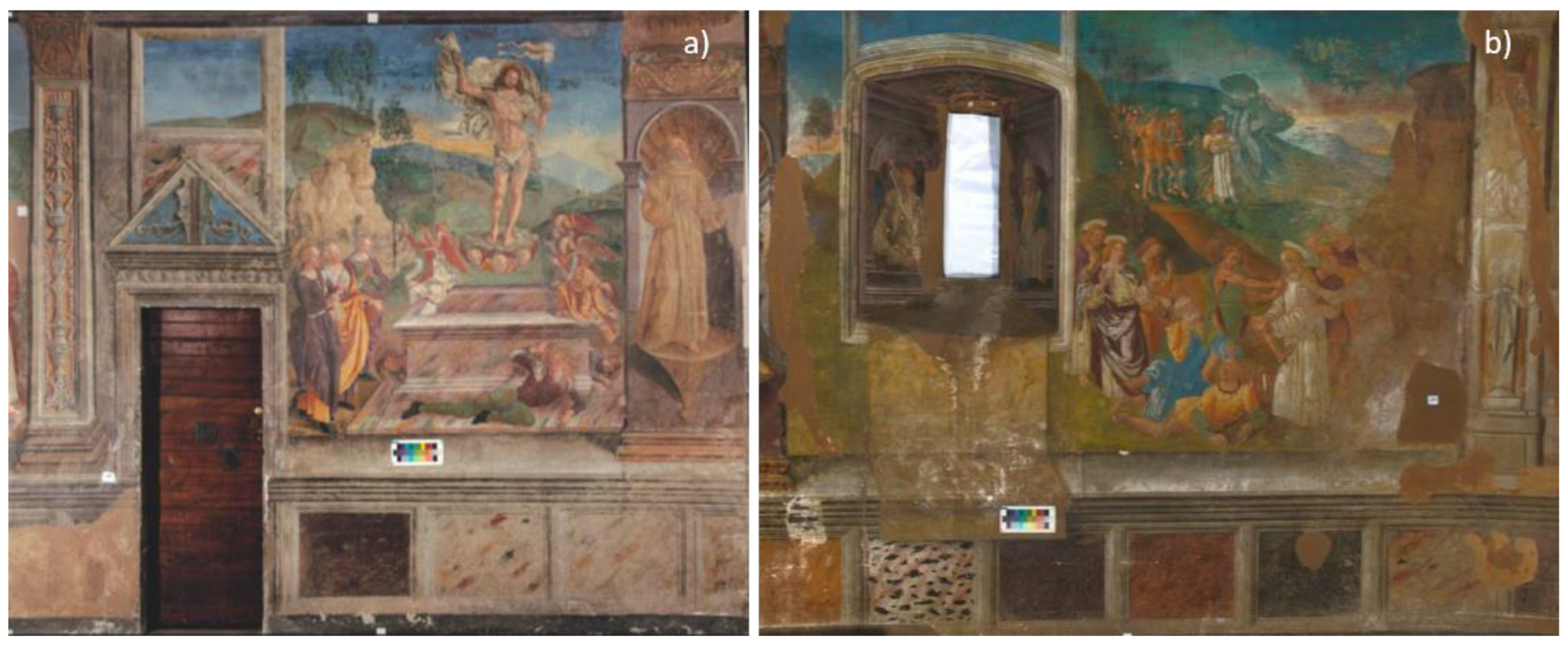
:1. Introduction
2. Materials and Methods
2.1. Samples
2.1.1. Original Binding Media and Aged Coatings
2.1.2. Lacunae, Retouched and Restored Areas
2.2. Analytical Instrumentation
2.2.1. Instrumentation for the Analysis of Original Painting Materials
2.2.2. Instrumentation for the Investigation of Restoration/Consolidation Treatment
- -
- A Trace GC 1300 system equipped with an ISQ 7000 MS detector was used (ThermoFisher Scientific, Waltham, MI, USA) for the analysis of approximately 80–100 µg of sample. The transesterification reaction and the complete methodology are described elsewhere [16,17,18]. The data interpretation was performed using NIST and MS Search 1.7 libraries and ad hoc databases created by the authors. The Chromeleon 7 software was used for the data acquisition and processing.
- -
- Thermally Assisted Hydrolysis and Methylation (THM)–Single Shot Pyrolysis–Gas Chromatography/Mass Spectrometry (TMH–SS-Py–GC/MS) was performed on small aliquots of samples (around 30–80 µg) in eco-cup pyrolysis crucibles. The samples were then treated with 3 µL of tetramethylammonium hydroxide (TMAH), 25%, in methanol.
- -
- A PY-3030D pyrolizer (Frontier Lab, Koriyama, Japan), connected to a Trace 1310 gas chromatograph (ThermoFisher Scientific, Waltham, MA, USA) with an ISQ7000 mass spectrometer (ThermoFisher Scientific, Waltham, MA, USA), was used. The analytical parameters and software for collecting, processing and interpreting the mass spectral data are reported in the literature [19,20].
3. Results and Discussion
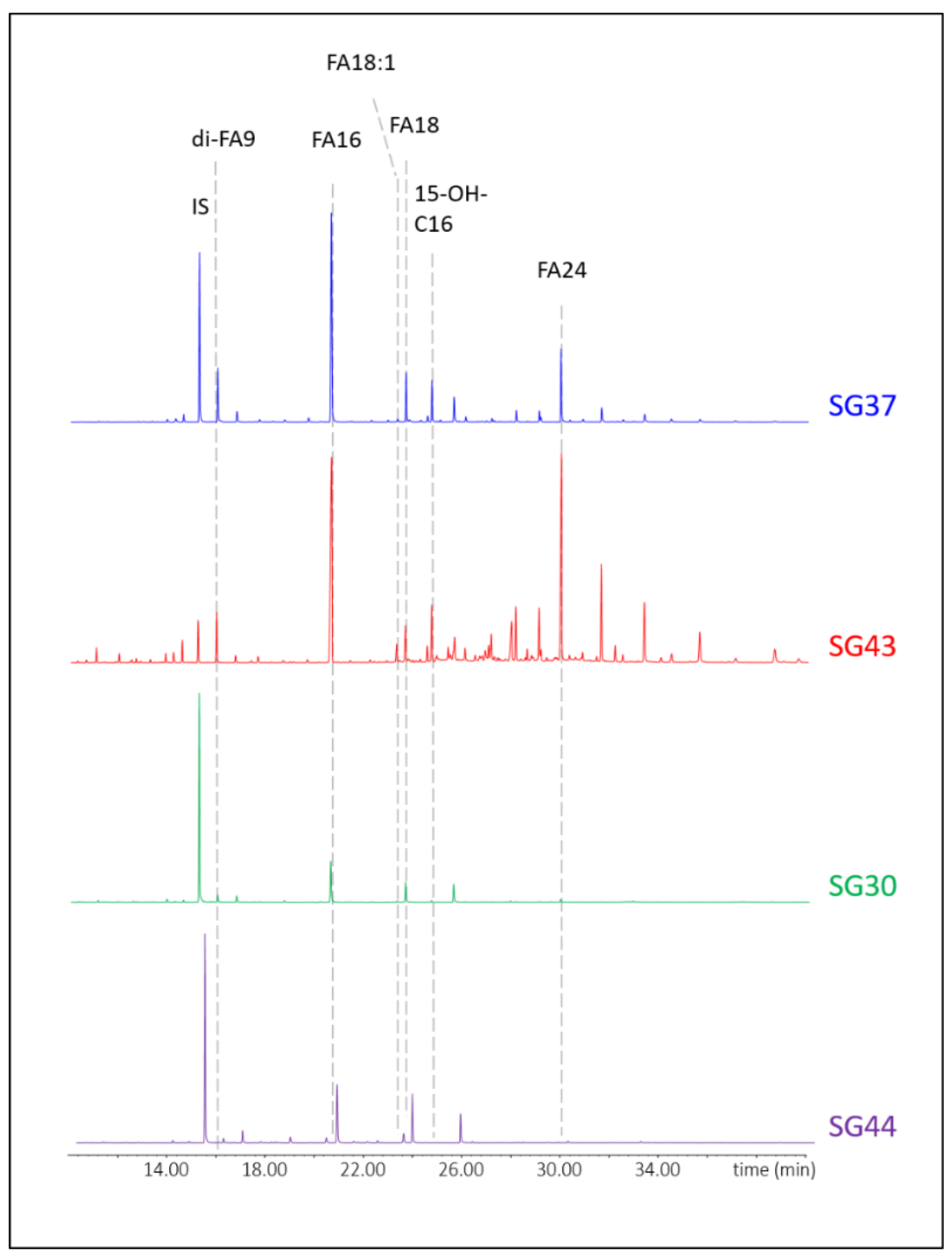
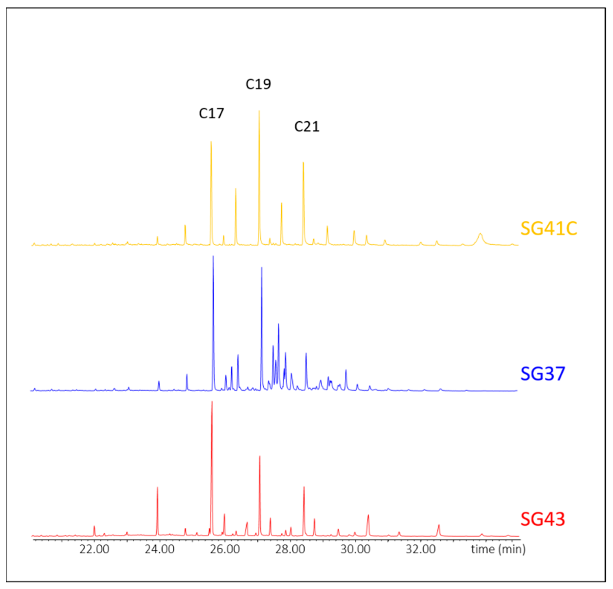
3.1. Original Binding Media, Aged Coatings and Consolidants
3.2. Lacunae, Retouched and Restored Areas
4. Conclusions
Supplementary Materials
Author Contributions
Funding
Institutional Review Board Statement
Informed Consent Statement
Data Availability Statement
Acknowledgments
Conflicts of Interest
References
- Magrini, D.; Bracci, S.; Cantisani, E.; Conti, C.; Rava, A.; Sansonetti, A.; Shank, W.; Colombini, M. A multi-analytical approach for the characterization of wall painting materials on contemporary buildings. Spectrochim. Acta-Part A Mol. Biomol. Spectrosc. 2017, 173, 39–45. [Google Scholar] [CrossRef] [PubMed]
- Bracci, S.; Iannaccone, R.; Magrini, D. The application of multiband imaging integrated with non-invasive spot analysis for the examination of archeological stone artifacts. CONSERVATION 360° 2020, 141–160. Available online: https://monografias.editorial.upv.es/index.php/con_360/article/view/71 (accessed on 12 February 2022).
- Bracci, S.; Cantisani, E.; Conti, C.; Magrini, D.; Vettori, S.; Tomassini, P.; Marano, M. Enriching the knowledge of Ostia Antica painted fragments: A multi-methodological approach. Spectrochim. Acta Part A Mol. Biomol. Spectrosc. 2022, 265, 120260. [Google Scholar] [CrossRef] [PubMed]
- Comite, V.; Bergomi, A.; Lombardi, C.A.; Borelli, M.; Fermo, P. Characterization of Soluble Salts on the Frescoes by Saturnino Gatti in the Church of San Panfilo in Villagrande di Tornimparte (L’Aquila). Appl. Sci. 2023, 13, 6623. [Google Scholar] [CrossRef]
- Da Filicaia, E.G.; Evershed, R.P.; Peggie, D.A. Review of recent advances on the use of mass spectrometry techniques for the study of organic materials in painted artworks. Anal. Chim. Acta 2023, 1246, 340575. [Google Scholar] [CrossRef] [PubMed]
- Calvano, C.D.; van der Werf, I.D.; Palmisano, F.; Sabbatini, L. Revealing the composition of organic materials in polychrome works of art: The role of mass spectrometry-based techniques. Anal. Bioanal. Chem. 2016, 408, 6957–6981. [Google Scholar] [CrossRef]
- Mazurek, J.; Svoboda, M.; Schilling, M. GC/MS characterization of beeswax, protein, gum, resin, and oil in romano-egyptian paintings. Heritage 2019, 2, 1960–1985. [Google Scholar] [CrossRef] [Green Version]
- Castellá, F.; Pérez-Estebanez, M.; Mazurek, J.; Monkes, P.; Learner, T.; Niello, J.F.; Tascon, M.; Marte, F. A multi-analytical approach for the characterization of modern white paints used for Argentine concrete art paintings during 1940–1960. Talanta 2020, 208, 120472. [Google Scholar] [CrossRef]
- Rogge, C.E.; Mazurek, J.; Schilling, M. The Nucleus of Color: Analysis of Hélio Oiticica’s Studio Materials. Stud. Conserv. 2022. [Google Scholar] [CrossRef]
- Schilling, M.R. Paint media analysis. In Scientific Examination of Art: Modern Techniques in Conservation and Analysis; National Academies Press: Washington, DC, USA, 2005; pp. 186–205. [Google Scholar] [CrossRef]
- Chiavari, G.; Galletti, G.C.; Lanterna, G.; Mazzeo, R. The potential of pyrolysis—Gas chromatography/mass spectrometry in the recognition of ancient painting media. J. Anal. Appl. Pyrolysis 1993, 24, 227–242. [Google Scholar] [CrossRef]
- Wei, S.; Pintus, V.; Schreiner, M. A comparison study of alkyd resin used in art works by Py-GC/MS and GC/MS: The influence of aging. J. Anal. Appl. Pyrolysis 1970, 104, 441–447. [Google Scholar] [CrossRef]
- Wei, S.; Pintus, V.; Schreiner, M. Photochemical degradation study of polyvinyl acetate paints used in artworks by Py-GC/MS. J. Anal. Appl. Pyrolysis 2012, 97, 158–163. [Google Scholar] [CrossRef] [PubMed] [Green Version]
- Peris-Vicente, J.; Baumer, U.; Stege, H.; Lutzenberger, K.; Gimeno Adelantado, J.V. Characterization of Commercial Synthetic Resins by Pyrolysis-Gas Chromatography/Mass Spectrometry: Application to Modern Art and Conservation. Anal. Chem. 2009, 81, 3180–3187. [Google Scholar] [CrossRef] [PubMed]
- Bonaduce, I.; Cito, M.; Colombini, M.P. The development of a gas chromatographic-mass spectrometric analytical procedure for the determination of lipids, proteins and resins in the same paint micro-sample avoiding interferences from inorganic media. J. Chromatogr. A 2009, 1216, 5931–5939. [Google Scholar] [CrossRef] [PubMed]
- Izzo, F.C.; Lodi, G.C.; Vázquez de Ágredos Pascual, M.L. New insights into the composition of historical remedies and pharmaceutical formulations: The identification of natural resins and balsams by gas chromatographic-mass spectrometric investigations. Archaeol. Anthropol. Sci. 2021, 13, 2. [Google Scholar] [CrossRef]
- Izzo, F.C.; Ferriani, B.; Van den Berg, K.J.; Van Keulen, H.; Zendri, E. 20th century artists’ oil paints: The case of the Olii by Lucio Fontana. J. Cult. Herit. 2014, 15, 557–563. [Google Scholar] [CrossRef]
- Morales Toledo, E.G.; Raicu, T.; Falchi, L.; Barisoni, E.; Piccolo, M.; Izzo, F.C. Critical Analysis of the Materials Used by the Venetian Artist Guido Cadorin (1892–1976) during the Mid-20th Century, Using a Multi-Analytical Approach. Heritage 2023, 6, 600–627. [Google Scholar] [CrossRef]
- Izzo, F.; Van Keulen, H.; Carrieri, A. Assessing the Condition of Complex Poly-Material Artworks by Py-GC-MS: The Study of Cellulose Acetate-Based Animation Cels. Separations 2022, 9, 131. [Google Scholar] [CrossRef]
- Van Keulen, H.; Schilling, M. AMDIS & EXCEL: A Powerful Combination for Evaluating THM-Py-GC/MS Results from European Lacquers. Stud. Conserv. 2019, 64, S74–S80. [Google Scholar]
- Colombini, M.P.; Andreotti, A.; Bonaduce, I.; Modugno, F.; Ribechini, E. Analytical strategies for characterizing organic paint media using gas chromatography/mass spectrometry. Acc. Chem. Res. 2010, 43, 715–727. [Google Scholar] [CrossRef]
- Dallongeville, S.; Garnier, N.; Rolando, C.; Tokarski, C. Proteins in art, archaeology, and paleontology: From detection to identification. Chem. Rev. 2016, 116, 2–79. [Google Scholar] [CrossRef]
- Bonaduce, I.; Colombini, M.P. Characterisation of beeswax in works of art by gas chromatography-mass spectrometry and pyrolysis-gas chromatography-mass spectrometry procedures. J. Chromatogr. A 2004, 1028, 297–306. [Google Scholar] [CrossRef]
- Secco-Suardo, G. Manuale Ragionato per la Parte Meccanica Dell’arte del Restauratore dei Dipinti; Forgotten Books: London, UK, 1866. [Google Scholar]
- Mora, P.; Mora, L.; Philippot, P. Conservazione delle Pitture Murali, 2nd ed.; HOEPLI: Milan, Italy, 2003. [Google Scholar]
- Bertorello, C. Materiali a Confronto sui Dipinti Murali Nell’esperienza dei Restauratori Tra’800 e’900; Bollettino d’arte: Rome, Italy, 1996. [Google Scholar]
- Vanni, T. Le Decorazioni Pittoriche del Centro di Portogruaro; Associazione Accordi Portogruaro: Portogruaro, Italy, 2004. [Google Scholar]
- Prisco, G. Tecnica esecutiva e conservazione delle pitture murali di epoca romana. Il dibattito tra fine ’800 e prima metà del ’900. Boll. ICR 2013, 27, 50–69. [Google Scholar]
- La Nasa, J.; Modugno, F.; Degano, I. Liquid chromatography and mass spectrometry for the analysis of acylglycerols in art and archeology. Mass Spectrom. Rev. 2021, 40, 381–407. [Google Scholar] [CrossRef] [PubMed]
- Asperger, A.; Engewald, W.; Fabian, G. Advances in the analysis of natural waxes provided by thermally assisted hydrolysis and methylation (THM) in combination with GC/MS. J. Anal. Appl. Pyrolysis 1999, 52, 51–63. [Google Scholar] [CrossRef]
- Pintus, V.; Schreiner, M. Characterization and identification of acrylic binding media: Influence of UV light on the ageing process. Anal. Bioanal. Chem. 2011, 399, 2961–2976. [Google Scholar] [CrossRef]
- Fardi, T.; Pintus, V.; Kampasakali, E.; Pavlidou, E.; Schreiner, M.; Kyriacou, G. Analytical characterization of artist’s paint systems based on emulsion polymers and synthetic organic pigments. J. Anal. Appl. Pyrolysis 2018, 135, 231–241. [Google Scholar] [CrossRef]
- Orsini, S.; Parlanti, F.; Bonaduce, I. Analytical pyrolysis of proteins in samples from artistic and archaeological objects. J. Anal. Appl. Pyrolysis 2017, 124, 643–657. [Google Scholar] [CrossRef]


| Sample | Area | Description | Aim of the Investigations |
|---|---|---|---|
| SG_30 | Panel E | Fragments of white plaster + greyish pictorial finish taken from an area which under UV appears characterized by an uneven yellowish response. Macroscopically, the surface appears chromatically altered (with a spotted effect). | Identification of the degraded protective |
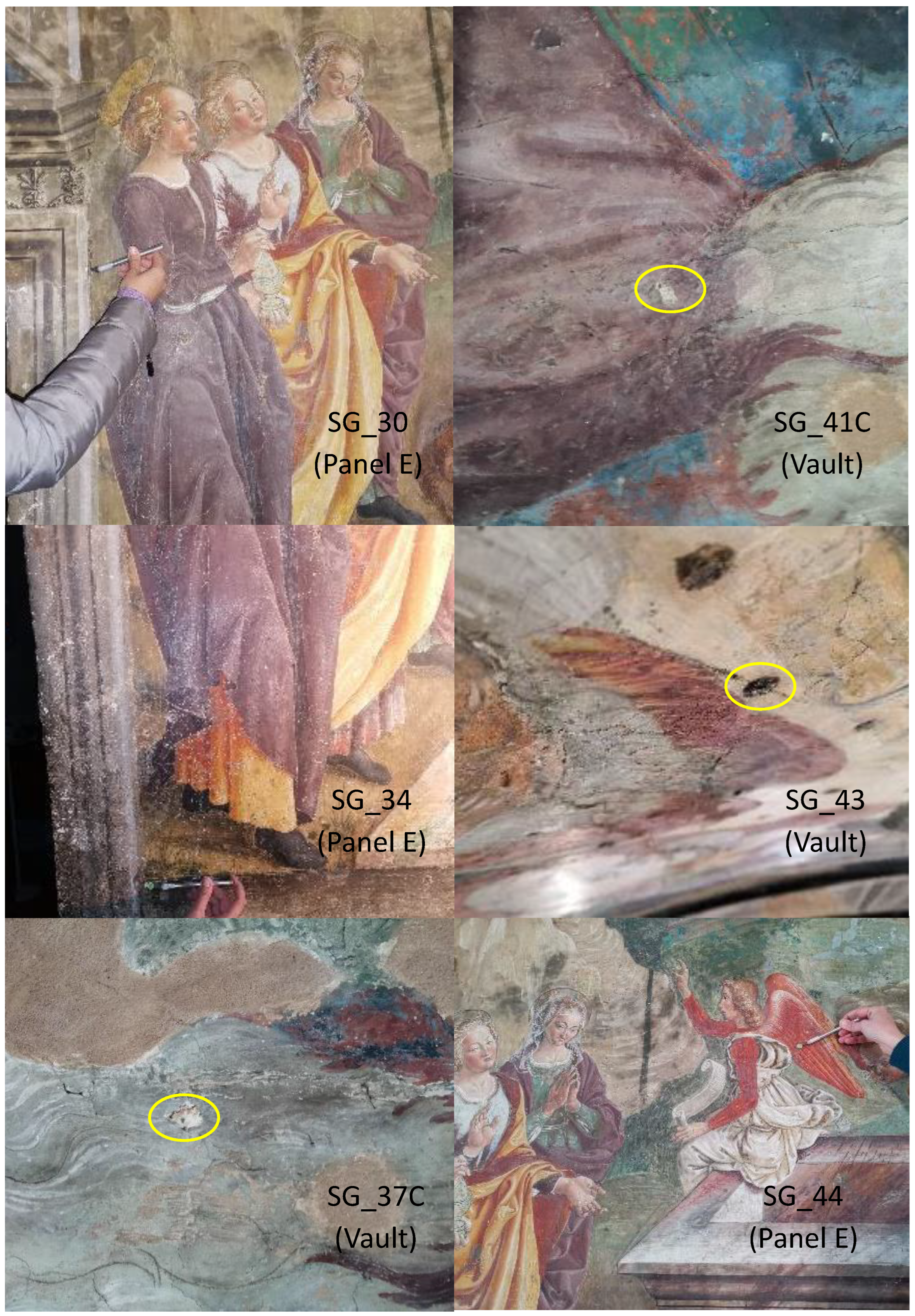
| SG_34 | Panel E | Fragments of white plaster + grayish paint finish taken from an area that shows no fluorescence response under UV. Macroscopically, the surface appears glossy. | Identification of altered protective and/or other consolidating treatments undergone in the past |
| SG_37c | Vault, top part | Gray pictorial spread on a layer of degraded plaster, taken from a degraded area due to detachments and lifting of the surface layers. | Identification of organic binders and protective consolidants |
| SG_41C | Vault, top part | Purplish pictorial spread on a layer of degraded plaster, taken from a degraded area with lifting of the surface layers. | Identification of organic binders and protective consolidants |
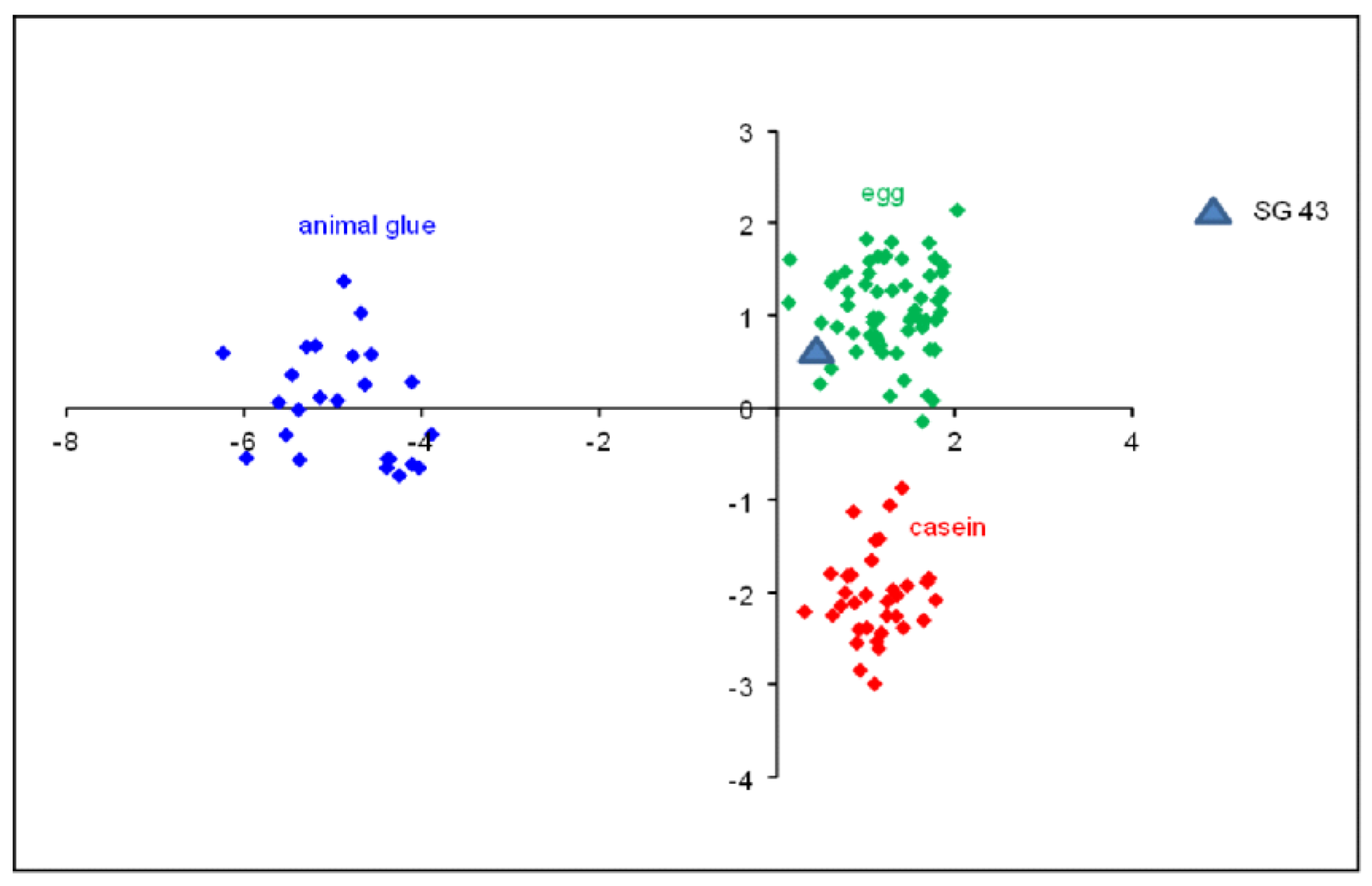
| SG_43 | Vault, top part | Relief layer of material of an organic nature selectively taken from the pictorial surface. These are tablets to simulate the golden decorations of the halos and robes of the Almighty (or relief base for the application of gold leaf), now detached. | Understanding of painting technique |
| SG_44 | Panel E | Traces of a layer of preparation for the application of gold leaf for the creation of rays of the risen Christ. The surface of the layer taken is raised and tenaciously adheres to the plaster. Given the small number of traces still present, the sampling has been reduced to minimal quantities. | Understanding of the gold leaf application technique |
| Sample | Area | Description | Image |
|---|---|---|---|
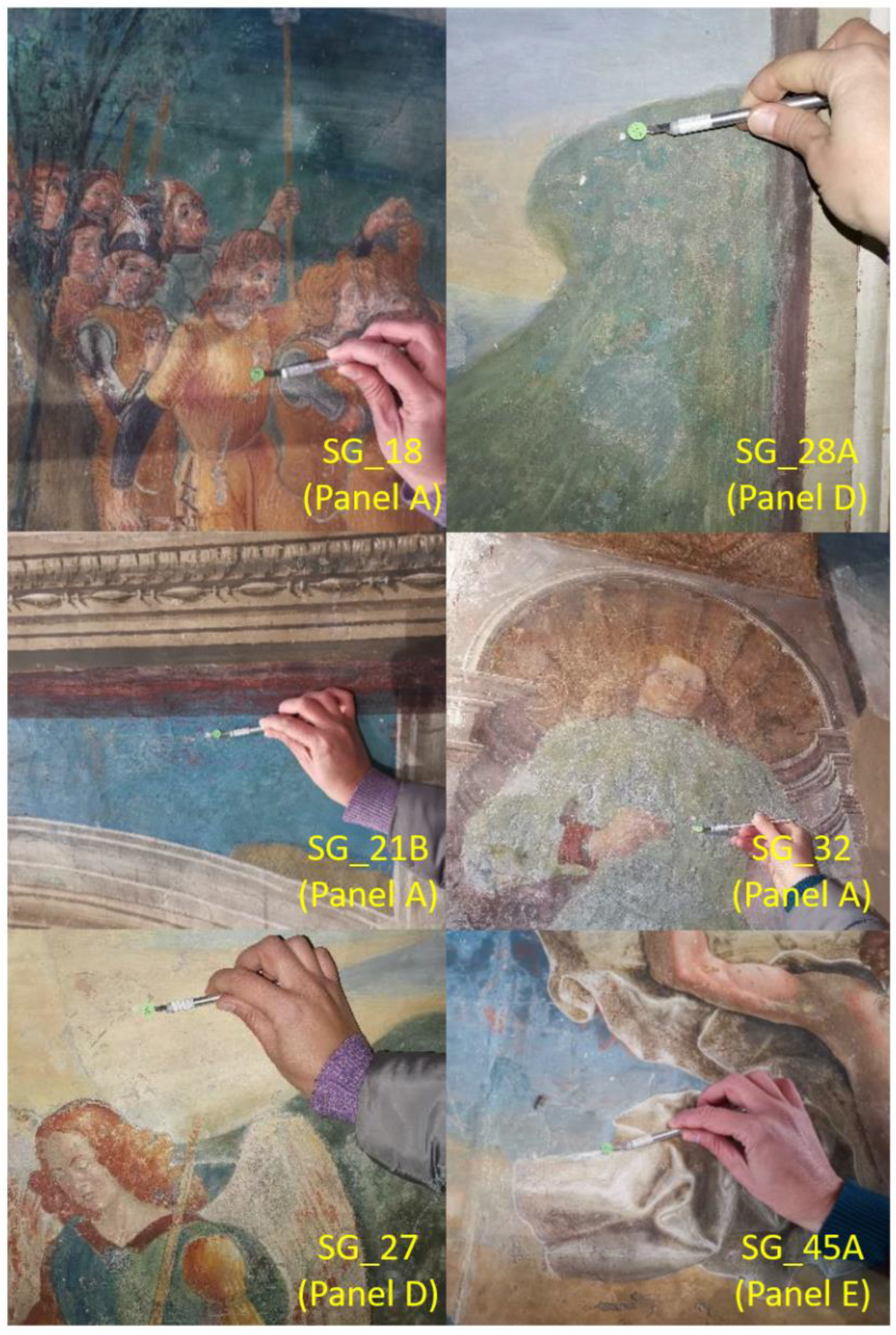
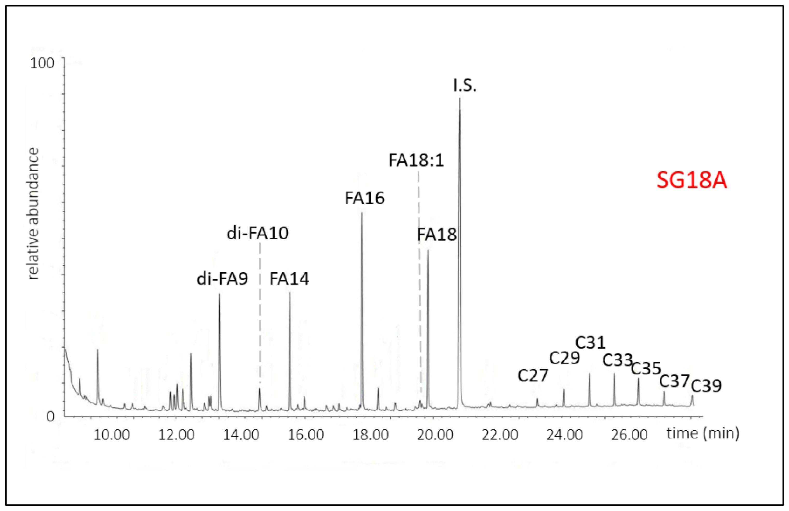
| SG_18A | Panel A | Yellow-orange pictorial layer and whitish preparation/primer, taken along an existing gap. |  |
| SG_21B | Panel A | Blue pictorial layer on a red-brown layer (the so-called ‘morellone’) and underlying plaster fragments. |  |
| SG_27A | Panel D | Yellow-blue pictorial application and white preparation/primer. |  |
| SG_28A | Panel D | Dark green pictorial layer and fragments of plaster. The area also has pictorial integrations with glazes. |  |
| SG_32 | Panel A | Green pictorial layer and white preparation/primer, taken along the edge of a gap. |  |
| SG_45A | Panel E | White layer (‘lumeggiatura’) (highlighting) applied on the underlying blue pictorial surface. |  |
Disclaimer/Publisher’s Note: The statements, opinions and data contained in all publications are solely those of the individual author(s) and contributor(s) and not of MDPI and/or the editor(s). MDPI and/or the editor(s) disclaim responsibility for any injury to people or property resulting from any ideas, methods, instructions or products referred to in the content. |
© 2023 by the authors. Licensee MDPI, Basel, Switzerland. This article is an open access article distributed under the terms and conditions of the Creative Commons Attribution (CC BY) license (https://creativecommons.org/licenses/by/4.0/).
Share and Cite
Andreotti, A.; Izzo, F.C.; Bonaduce, I. Archaeometric Study of the Mural Paintings by Saturnino Gatti and Workshop in the Church of San Panfilo, Tornimparte (AQ): The Study of Organic Materials in Original and Restored Areas. Appl. Sci. 2023, 13, 7153. https://doi.org/10.3390/app13127153
Andreotti A, Izzo FC, Bonaduce I. Archaeometric Study of the Mural Paintings by Saturnino Gatti and Workshop in the Church of San Panfilo, Tornimparte (AQ): The Study of Organic Materials in Original and Restored Areas. Applied Sciences. 2023; 13(12):7153. https://doi.org/10.3390/app13127153
Chicago/Turabian StyleAndreotti, Alessia, Francesca Caterina Izzo, and Ilaria Bonaduce. 2023. "Archaeometric Study of the Mural Paintings by Saturnino Gatti and Workshop in the Church of San Panfilo, Tornimparte (AQ): The Study of Organic Materials in Original and Restored Areas" Applied Sciences 13, no. 12: 7153. https://doi.org/10.3390/app13127153