Review

Plant Extracellular Vesicles: Investigating Their Utilization as Beneficial Nutrients in Diet

Department of Biological Sciences and Technologies (Di.S.Te.B.A.), University of Salento, 73100 Lecce, Italy
*
Authors to whom correspondence should be addressed.
These authors contributed equally to this work.
Appl. Sci. 2023, 13(11), 6656; https://doi.org/10.3390/app13116656
Submission received: 30 March 2023 / Revised: 9 May 2023 / Accepted: 29 May 2023 / Published: 30 May 2023

Abstract

:
Plant-derived extracellular vesicles (EVs) isolated from seeds, leaves, and fruits have shown a significant therapeutic potential for their anticancer, anti-inflammatory, and antioxidant properties. The ability to transport bioactive molecules and the low toxicity give EVs remarkable versatility in the field of nanomedicine for the development of drug delivery systems. Moreover, the physicochemical stability in gastric and intestinal fluids makes them the ideal candidate as nutritional carriers in oral formulations. It is well known that the consumption of antioxidant molecules from dietary plant sources, such as fruits and vegetables, can prevent pathologies caused by oxidative damage, including inflammatory and cardiovascular disease, neurodegeneration, aging, and cancer. EVs present in plant juices are receiving a lot of interest concerning their biological relevance in terms of their health benefits. EVs from food might be new components participating in body homeostasis, as they are in contact with the intestinal tract. This review aims to report and discuss the main biological properties and nutraceutical use of plant-derived EVs as promising therapeutic tools, with a focus on anti-oxidant effect and as a basis in developing new food-derived technology.

1. Introduction

The beneficial functions of food and its bioactive constituents on human health are widely recognized; consumption of fruits and vegetables is associated with a risk reduction in a number of leading human diseases, likely due to plant antioxidant content [1]. Recently, it has been reported that food-derived vesicles containing bioactive molecules enhance the efficacy of natural compounds by improving their bioavailability, due to biological protection of the content obtained by the presence of a membrane [2].
Plants secrete a multitude of molecules in the extracellular environment, facilitating cell-to-cell signaling, which is crucial to plant defense and immunity. Based on current knowledge, secreted extracellular vesicles (EVs) represent a major way to achieve this crosstalk [3]. Furthermore, it is known that they mediate different mechanisms of interspecies communication, gaining increasing attention as a valuable therapeutic tool [4].
Mammalian EVs are categorized as exosomes, microvesicles, and apoptotic bodies (known ad classical EVs), and autophagic EVs, stressed EVs, and matrix vesicles (recently considered as new EVs types) [5]. Since it is very difficult to categorize the different types of EVs, as specific markers to distinguish them are lacking, the International Society for Extracellular Vesicles (ISEV) recommends the use of the term “extracellular vesicles” (EVs) to generically refer to particles delimited by a lipid bilayer naturally released from mammalian cells. The classification of EVs can be based on size, biogenesis mechanism, and biochemical composition. In line with guidelines reported in Thery et al., 2018, the authors are recommended to consider (a) physical characteristics of EVs, such as size (“small EVs” (sEVs) and “medium/large EVs” (m/lEVs)), with ranges defined, for instance, respectively, <100 nm or <200 nm (small), or >200 nm (large and/or medium) or density (low, middle, high, with each range defined); (b) biochemical composition (CD63+/CD81+ EVs, Annexin A5-stained EVs, etc.); or (c) descriptions of conditions or cell of origin [6]. However, in general, the experts have recommended calling exosomes as small EVs (sEV) originating from the endosomal compartment regardless of the generating cells, while for EVs budding from plasma membrane and known as microvesicles (MVs) or ectosomes or microparticles, it is recommended to use the term large EVs (lEVs). Apoptosis-derived vesicles, which are characterized by their large size range from 800 nm to 5 μm, result from cell death and contain a mixture of damaged or unnecessary cellular components [5].
Similar to mammalian ones, plant-derived extracellular vesicles (P-EVs) are small vesicles surrounded by a lipid-enriched membrane, transporting a variety of bioactive substances such as nucleic acids, vitamins, antioxidant molecules, proteins, and metabolites [7]. Due to current insufficient knowledge, it is still difficult to classify plant EVs with the established nomenclature used for mammalian EVs [8]. In order to standardize the literature, the term “extracellular vesicles” was proposed to include all vesicles isolated from extracellular compartments (e.g., apoplastic fluid or growth medium), and “plant-derived vesicles” for vesicular fractions obtained by disrupting plant tissues, when natural release into the apoplast cannot be established [9]. Moreover, the name “exosomesf, widely used for mammalian vesicles, is barely adopted for plants due to limited knowledge of surface protein markers [10].
Although the biogenesis pathway of plant EVs is not completely clarified, it is believed that these vesicles are mainly produced by three specific organelles: MVBs, exosome-positive organelles (EXPO), and vacuolar bodies [11]. In the plant, exosomes are secreted by the fusion of MVBs with the plasma membrane [12]. A specific marker for these vesicles type has been identified in the integral protein tetraspanin-8 (TET8), which is the orthologue for the human exosomal marker CD63 [13].
EXPO are double-membrane structures, similar to autophagosomes, that fuse with the plasma membrane and release vesicles into the apoplast. EXPO-derived vesicles have a size range of 200–500 nm and are characterized by the presence of the exocytosis complex EXO70E2 [14].
The passage of EVs through the plant cell wall is a key event in the release of P-EVs due to the composition of the cell wall consisting of lignin, pectin, and (hemi-)cellulose fibrils, which make up a dense network with spaces that are smaller than the EVs size. Both a passive and an active process occur in EV passage: the first is based on the dynamics of the cell wall, and the second relies on cell wall-degrading enzymes present in the cytoplasm. Moreover, the spherical structure of P-EVs can change into a tubular one, which could promote cell wall passage [15].
Other types of P-EVs include vesicles originating from vacuoles that the plants use to respond to environmental attacks. For example, the vacuolar content is released outside the cell membrane in vesicular form during pathogen infections, to counteract the dissemination of pathogens within the plant [16]. In Figure 1, a schematic representation of P-EVs biogenesis and release is reported.
Besides their physiological functions in plants, it has been reported that P-EVs may provide beneficial effects on human health, by exerting anti-cancer and anti-inflammatory effects, thus gaining a growing interest in the field of biomedicine [17]. For instance, P-EVs from grapefruit, grapes, and ginger have been shown to contribute to gut barrier homeostasis, promoting the enterocytes renewal and alleviating inflammation [13]. In addition, citrus-derived vesicles, particularly those from lemon, have shown antineoplastic effects, by inhibiting cancer cell proliferation through the activation of the programmed cell death mechanisms, both in vitro and in vivo [4].
P-EVs have been reported to enhance the efficacy of phytochemicals by improving their bioavailability, primarily due to the biological protection that the lipid bilayer exerts on its content, thus preserving both stability and activity [18]. Moreover, plant vesicles show excellent biocompatibility, are minimally cytotoxic, and may be derived from any part of plants, especially the leaves and fruits, for application in large-scale production [19]. While mammal EVs are highly characterized, plant vesicles still remain poorly investigated.
In this review, we summarize the recent knowledge about the type, nature, and composition of P-EVs, with particular consideration about the antioxidant properties of vesicles obtained from vegetables.

2. Structure and Biological Composition of P-EVs

Extracellular vesicles are a heterogeneous group of membrane-enclosed particles that are different in size and shape [20]. Their membrane consists of a phospholipid bilayer with integrated proteins, which protects the molecular content from enzymatic degradation, as well as from environmental conditions (e.g., extreme temperatures, pH, salinity) [2].
The lipid composition of P-EVs varies depending on the plant species and the physiological state of the producing cells. However, the P-EVs membrane typically contains a mixture of phospholipids, glycolipids, and phytosterols [21]. Lipidomic analyses have revealed that phosphatidic acid, phosphatidylcholine, and phosphatidylethanolamine account for most of the phospholipid content [2]. In particular, phosphatidic acid is a major component in ginger- and grape-derived vesicles, and it has been demonstrated its involvement in the mechanism of membrane fusion [22]. Moreover, since it is a cell-signaling lipid involved in the activation of the mammalian target of rapamycin (mTOR), as well as mitogen-activated protein kinase (MAPK) pathways, phosphatidic acid can be considered the main effector on mammalian cells growth and proliferation upon treatment with P-EVs [8]. Besides their role in vesicle internalization, lipids may also contribute to P-EVs’ intrinsic therapeutic properties [23]. For instance, phosphatidylcholine- and phosphatidylethanolamine-enriched grapefruit-derived vesicles have shown antioxidant, anticolitic, and anti-inflammatory activities [24]. Differences in the composition of the lipid bilayer can affect vesicle uptake by specific gut bacteria, suggesting that specific lipids could signal for preferential uptake by specific cells, playing a key role in the reported therapeutic activities of these vesicles [25]. The main lipid families found in P-EVs are reported in Table 1.
Similarly to lipids, P-EVs’ protein profile depends on the origin of vesicles and the physiological conditions of the plant, and the main role of the proteins is in facilitating plant vesicle uptake by animal cells [22]. P-EVs contain significantly lower protein content compared to mammalian vesicles, and the main proteins present belong to the families of aquaporins, involved in the regulation of cell turgor; heat-shock proteins (HSPs), related to biotic and abiotic stress responses and plant growth; metabolic enzymes; and annexins, implicated in intracellular vesicle transport and exosome secretion [11] (Table 2).
Plant vesicles contain a considerable number of small RNAs (sRNAs), including microRNAs (miRNA), which are short non-coding RNA of about 15 to 27 nucleotides, capable of regulating gene expression by binding and cleaving target mRNA molecules [26]. In the case of pathogen infections, plants use EVs to secrete miRNA into the host and silence virulence-related genes. Emerging data demonstrate that EVs deriving from edible plants are also able to control target gene expression in the mammalian genome via cross-kingdom interactions, by transferring sRNAs [4]. Consistent with these findings, plant miRNAs are naturally modified at their 3′ ends by methylation to acquire greater stability and protection against degradation [7]. Zhang et al. [27] proposed that ingested plant miRNAs are absorbed by the intestinal epithelial cells of consuming animals and are then packaged into extracellular vesicles. These structures protect miRNAs from degradation and deliver them to distant cells, where they interact with endogenous RNAs and regulate their expression. Several studies also show that these molecules can prevent human diseases. For instance, miRNA-156a, which is abundantly expressed in green vegetables such as cabbage, spinach, and lettuce, has a protective effect against the progression of atherosclerosis [28]. Recent evidence also shows an emerging function of P-EVs miRNA in the treatment of COVID-19, by binding to multiple sites of the SARS-CoV-2 viral genome and subsequently inhibiting viral replication [29]. The table below (Table 3) summarizes the therapeutic activities reported for plant-derived miRNA.
It is well known that plants produce a plethora of secondary metabolites, such as carotenoids, flavonoids, saponins, and glucosinolates, having antioxidant, antitumor, and immunomodulatory effects and that support the stability of intestinal microflora [4]. Most of these natural compounds are packaged into P-EVs during biogenesis, conferring additional value on health and its beneficial impact [25].
Moreover, these active metabolites are more concentrated in EVs than in cell plants, as demonstrated in ginger-derived EVs that show much higher 6-gingerol, 8-gingerol, and 10-gingerol amounts. These EVpacked gingerols are efficiently delivered to the intestines of rats and are absorbed [39]. Similarly, Perut demonstrated a high content of ascorbic acid (vitamin C) in strawberry-derived EVs with a strong antioxidant effect [40], and Deng suggested the efficiency of broccoli-derived EVs that carried sulforaphane by using a model of mouse colitis [41].
Considering that P-EVs represent a source of phytochemicals, the effects of these vesicles on animal cells may be predictable, although further investigation is needed.

3. Conventional Methods for P-EV Isolation and Characterization

To date, a major challenge associated with P-EV research is the lack of standardized protocols for good-quality purification [4]. Despite this inconvenience, EVs can be isolated in large amounts from many parts of the plant including the fruits and apoplastic fluid from the leaves, seeds, and roots [7]. Although different methods have recently been developed for plant vesicle isolation, including size exclusion chromatography, immunoprecipitation, and polymer-based precipitation [13], ultracentrifugation remains the preferred one, due to the greater amount of material that can be recovered [22]. Differential ultracentrifugation is a separating technique that involves centrifugation cycles of different centrifugal forces and duration to separate particles in the homogenate or the juice of plants, proportionally to their molecular weight and density [42]. During the EV isolation process, high-density particles such as dead cells and apoptotic bodies are first removed by low-speed centrifugation, followed by higher speed spins to remove larger vesicles and debris. In the final purification step, a supernatant containing EVs is pelleted and concentrated by high-speed centrifugation (40,000–200,000× g) [22].
Ultracentrifugation is often supplemented by additional purifying methods [11], such as ultrafiltration, which consists in the use of ultrafine nano-membranes with different pore sizes, to trap particles above a certain dimension threshold, while allowing the smaller ones to flow through [7]. Moreover, since differential ultracentrifugation sediments contain contaminants such as proteins and other vesicles [13], it can be combined with density gradient ultracentrifugation to isolate P-EVs with higher purity [42]. Briefly, gradient ultracentrifugation uses a previously developed medium with a gradually decreasing density gradient, from the bottom to the top [22]. Under the application of the centrifugal force, solutes in the sample, including vesicles, separate into different zones according to the specific sedimentation rate [13].
Size exclusion chromatography is based on particle size for separation [43]. Larger vesicles are eluted earlier, as they cannot enter the pores of the column, while vesicles with a smaller hydrodynamic radius are retained, resulting in late elution [13]. Although this approach has great potential to preserve the integrity and biological activities of vesicles, as it relies on passive gravity flow, the long run time limits its application in large-scale production [44].
Immunoprecipitation consists of magnetic beads coated with antibodies that target vesicle surface markers, such as the tetraspanins CD63 and CD81, allowing for the isolation and identification of specific extracellular vesicles [25]. Despite the high-purity isolation, the immunoaffinity method is not widely used, probably because protein markers for the plant vesicle surface are not yet well known [45]. In addition, the integrity of vesicles may be disrupted if the antibodies cannot be easily removed after precipitation [25].
Polymer-based precipitation methods have been successfully used to isolate P-EVs [43]. Highly hydrophilic polymers, such as polyethylene glycol (PEG), are used as crowding reagents to form a net-like structure that traps vesicles, allowing them to precipitate at low-speed centrifugation by reducing their solubility in solution [13]. In this procedure, PEG with different molecular weights (6000–20,000 Da) [46] is added to the samples in an appropriate amount (5–12%) to recover between 60–90% of EVs, with a yield comparable to the differential centrifugation method [42]. Therefore, this precipitation method provides a scalable and cost-effective alternative for the purification of P-EVs. Polymers with higher molecular weight are more efficient, but they may increase the viscosity of fluids, exacerbating the purification process [46]. Moreover, due to similar solubility, other biological contaminants are co-precipitated, thereby compromising the purity of the isolated vesicles [47].
In plants, EVs were first detected in carrot cell cultures in 1967, using transmission electron microscopy (TEM) [34]. Since then, various physical and biochemical approaches have been developed for vesicle characterization in terms of size, morphology, and zeta potential to determine the type and stability of the particles [11]. TEM is one of the most common imaging techniques for observing nanoscale samples because its resolution, of about 0.5 nm, is smaller than that of exosomes, providing detailed structural information about EVs [13]
A good alternative to TEM is cryo-electron microscopy, in which samples are vitrified and cryo-immobilized at extremely low temperatures (−175 °C) by cooling with liquid ethane, preserving the native hydrated status of samples and thus avoiding artifacts usually caused by conventional TEM [48]. In combination with immunogold labeling, cryo-TEM has been successfully used to distinguish EVs containing specific proteins and to track their uptake by recipient cells [25].
Scanning electron microscopy (SEM) scans the surface of samples by using a focused beam of high-energy electrons to provide information about the three-dimensional structure of the vesicle surface [49]. However, as the sample needs to be dried for imaging, the natural morphology may change, typically resulting in the formation of cup-shaped structures. In addition, during the preparation, the sample is coated with a thin layer of conductive material, which may affect the surface structure of EVs [49].
Recently, a novel technique in the form of nanoparticle tracking analysis (NTA) has been developed to analyze a population of polydisperse nanoparticles in solution, ranging from 10 nm up to 2 μm in size [50]. During NTA, the sample is irradiated with a laser beam, and based on the light scattered by individual particles and relative Brownian motion in the solution, a camera tracks the path of each particle to determine the average concentration and size [13]. The size distribution of P-EVs can be measured by dynamic light scattering (DLS), exploiting the intensity of light scattered by each particle [11].
The identification of protein markers is considered a method of choice for the identification of specific P-EVs. In general, immunodetection methods (e.g., Western blot, immunofluorescence) using antibodies specific for marker proteins such as HSP70 and TET8 reveal high-specificity plant-derived nanovesicle fractions [13].

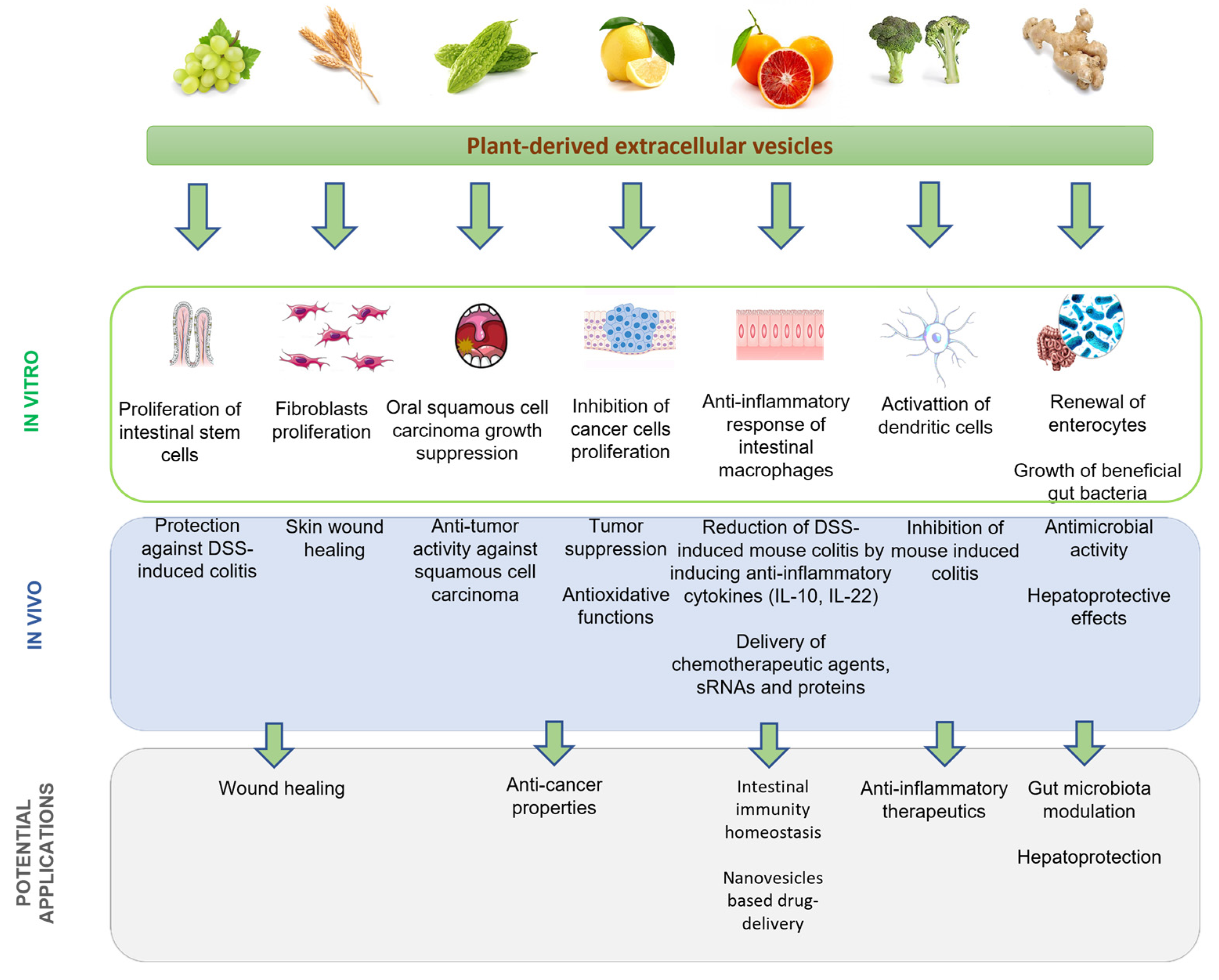
4. P-EV Biological Properties and Therapeutic Applications

In the last decade, plant extracellular vesicles have been exploited as excellent candidates for nanomedicine and nutraceutical applications due to their unique biological functions; thus, the research in this field has increased exponentially [2]. As already mentioned, because P-EVs can establish cell-to-cell communication between different species, they can be internalized and modulate host cell processes [7]. As such, the intrinsic bioactive cargos are believed to account for the beneficial effects of these vesicles, including anti-inflammatory, anticancer, antioxidant, and regenerative properties [18]. In addition to the promising application as a new class of therapeutics, P-EVs can also be developed as novel biologically derived drug vehicles with further advantages over current synthetic carriers, exhibiting in vitro and in vivo high biocompatibility, as most of them are isolated from edible plants, and stability in biological fluids even when administered orally [51]. Their great potential as oral drug carriers stems from their ability to withstand the harsh gastrointestinal environments and cross-biological barriers [51]. In vitro studies have demonstrated that plant vesicles can remain stable at different temperatures and pH values, mainly due to the protective functions against degradation exerted by the lipid bilayer on the bioactive cargos [51]. In particular, aquaporins were reported to play a key role in preserving the integrity of plasma membrane in broccoli-derived vesicles under high salt stress, which induced a significant accumulation of these proteins to ensure permeability as an adaptive mechanism [52]. In a previous study, the same authors demonstrated that changes in the lipid/protein ratio of Brassica species confer different physical properties to the lipid bilayer, leading to a degree of salt tolerance related to the presence of membrane proteins [53]. Because of their stability in the gastrointestinal tract, P-EVs can be administered orally and reach the target site efficiently as intact structures [54]. For example, Zhang et al. showed that ginger-derived EVs were taken up by intestinal stem cells and macrophages, and after oral administration for the treatment of inflammatory bowel disease, they promoted intestinal wound healing [55]. These authors also used ginger-nanoparticles to deliver siRNA-CD98 into colon tissue, and they found that administration of these vesicles decreased CD98 expression and prevented inflammation [55]. In addition, orally given vesicles, isolated from broccoli extracts, can regulate intestinal immune homeostasis in mice by targeting dendritic cells [41].
Inflammation is part of the innate immune response that, under damaging stimuli, may evolve toward acute or chronic inflammatory diseases, such as colitis [11]. In particular, plant vesicles have shown immunomodulatory effects on gastrointestinal homeostasis [56]. For example, Teng et al. [57] demonstrated that grapefruit-derived vesicles could alleviate dextran sulfate sodium (DSS)-induced colitis in mice without exerting toxic effects. It is likely that these vesicles enhance the anti-inflammatory response of intestinal macrophages by upregulating the expression of anti-inflammatory cytokines such as IL-10, which in turn decreases the expression of proinflammatory cytokines and chemokines. In addition, the same study reported that miRNAs from ginger and grapefruit vesicles can target genes of the probiotic Lactobacillus rhamnosus in mice gut, thus promoting antimicrobial immunity [57]. Similar studies have also been reported on broccoli, grapes, and carrots [41,58].
Several studies have demonstrated the anti-proliferative and pro-apoptotic effects of P-EVs, which account for their anticancer properties [59]. Vesicles isolated from lemon juice were found to inhibit cancer cells growth in vitro, without affecting healthy cells, by selective activation of the Trail-mediated apoptotic pathway. This finding was confirmed by intratumoral administration of lemon-derived exosomes in mice with chronic myeloid leukemia. Tumor growth was suppressed by regulating apoptosis and by inhibiting angiogenesis [60]. Recently, Cao et al. [61] demonstrated that EVs purified from ginseng roots induce apoptosis of murine melanoma cells by inducing the formation of ROS. This result suggests that plant EVs may participate as a crucial modulator in the mammalian immune response. It was also proven that P-EVs may exhibit synergistic effects with anticancer therapies [56]. Co-administration of bitter-melon-derived vesicles with the chemotherapeutic agent 5-fluorouracil in oral squamous cell carcinoma treatment showed increased antiproliferative and cytotoxic effects [35].
The effects of plant vesicles on in vitro skin regeneration have been studied, suggesting new potential applications. Wheatgrass-derived EVs have been shown to induce cell regeneration in wound healing, by triggering the proliferation and migration of epithelial, endothelial, and dermal fibroblast in a dose-dependent manner and by stimulating type I collagen expression [62]. The regenerative functions of grape-derived vesicles were investigated in mice with induced colitis. In this study, it was reported that these vesicles can penetrate the intestinal barrier and stimulate stem cell proliferation, by inducing the expression of genes responsible for pluripotency (SOX2, Oct4, Klf4), thus resulting in a strong acceleration of mucosal epithelial renewal [63]. Therefore, the proliferation of intestinal stem cells mediated by grape vesicles could potentially be used for the treatment of patients with intestinal epithelial injuries.
A summary of the anticancer, anti-inflammatory and regenerative properties of plant-derived vesicles evaluated through in vitro and in vivo experiments is shown in Figure 2 and Table 4.

Antioxidant Functions

ROS are produced in cells during aerobic respiration, and in response to xenobiotics, cytokines, and bacterial invasion, they have a physiological role in cell survival and proliferation [74]. Oxidative stress occurs in the cells when the balance between free radical concentration and antioxidant defense is altered in favor of the former. The overproduction of ROS may cause oxidative damage to biological molecules such as DNA, proteins and lipids, playing a crucial role in the etiology of many common diseases, including neurodegenerative disorders, heart diseases, and diabetes, due to oxidative damage to biomolecules [75]. Natural antioxidants, such as polyphenols, carotenoids, and vitamins have a variety of biological functions, including anti-aging, anti-inflammatory and antibacterial effects [7]. Considering their impact on human health, phytochemicals have become increasingly important in the prevention and treatment of chronic diseases in recent years [76]. Polyphenols (flavonoids, phenolic acids, anthocyanins, lignans, stilbenes) and carotenoids (carotenes and xantophylls) account for the main antioxidant properties of these compounds and can be found in foods and plants [77]. Anthocyanin extracts from Vaccinium floribundum and Aristotelia chilensis have been reported to limit adipogenesis and lipid accumulation in vitro and to exhibit anti-inflammatory properties on Raw 264.7 macrophages [78]. Dietary intake of vitamin C, vitamin E and β-carotene has been associated with a lower risk of gastric cancer and inhibition of breast cancer cell proliferation [79].
Neurological disorders are characterized by the deposition of misfolded proteins in the brain, with evidence of increased oxidative stress in tissue samples from patients, leading to direct protein modifications [80]. In a recent study, Latif et al. investigated the use of natural antioxidants in the treatment of Parkinson’s disease. In particular, hydroxytyrosol, a phenolic compound extracted from olive oil and leaves, and curcumin showed neuroprotective effects on SH-SY5Y cells, which were associated with a reduction in neurotoxin-induced ROS production and modulation of apoptosis pathways [76]. A separate study demonstrated the potential antitumor effects of green-synthesized nanoparticles containing berberine, an alkaloid molecule derived from various medicinal plants, whose pharmacological effects are widely recognized. Berberine treatment significantly increased the survival rate of mice injected with Ehrlich ascites tumor cells and reduced tumor growth, showing antioxidant and proapoptotic activities [81].
Taking into account that plants are rich in antioxidant compounds, research is therefore increasingly focusing on the isolation of vesicles that enclose and transport these molecules, not only for medicinal purposes but also for anti-aging formulations and food preservation. One of the antioxidant metabolites present in P-EVs is vitamin C, a powerful free radical scavenger, possessing a significant protective effect against oxidative damage, and it is also a cofactor for important enzymatic reactions. Its high molecular instability under physiological conditions reduces the bioavailability; thus, the vitamin C packaged in P-EVs can be considered very important [82]. Exosomal vesicles from citrus fruits and berries have been shown to resist in the gastric environment before being absorbed in the intestine [38]. The vesicles extracted from these fruits contain elevated concentrations of vitamin C (e.g., 50 µg/mL of lemon exosomes contain about 7 µM vitamin C); therefore, the ability to effectively deliver high doses to target cells may explain the antioxidant properties of these vesicles [25]. In support of this finding, mesenchymal stem cells pretreated with H2O2 showed a significantly increased survival after the internalization of lemon EVs, in a dose-dependent manner and reduced ROS production [83]. The high contents of vitamin C and other active metabolites, including anthocyanins, folic acid, and flavonols, have been detected in EVs isolated from Fragaria x ananassa strawberry juice. The adipose-derived mesenchymal stem cells internalize Fragaria-derived vesicles that do not negatively affect cell viability; moreover, the pretreatment of cells with Fragaria-derived vesicles prevents oxidative damage in a dose-dependent manner. This is mainly due to the presence of vitamin C in the nanovesicle membrane, indicating the potential health-promoting activity of Fragaria-derived vesicles [40].
In addition, carrot-derived vesicles were shown to have antioxidant properties in H9C2 cardiomyoblasts and the human neuroblastoma SH-SY5Y cell line. Carrot EVs show low cytotoxicity in both cell lines and a high uptake ratio. The treatment significantly inhibited ROS formation and apoptosis in vitro in myocardial infarction and Parkinson’s disease models by inhibiting the reduction of antioxidant molecules expression, including Nrf-2 nuclear factor erythroid 2-related factor 2, HO-1, and NQO-1, in both models [84]. Another study confirmed that carrot-derived EVs have antioxidant properties by increasing nuclear translocation of Nrf2, a key regulator of the HO1 gene involved in anti-inflammation and anti-oxidation activity, in Raw 264.7 macrophages [58].
An effect on the Nrf2 expression increment leading to the expression of detoxifying/antioxidant genes has been demonstrated in mice alcoholic liver model. In particular, ginger-derived EVs protect the liver against alcohol-induced damage. Specifically, the researchers have identified in the compound 6-shogaol the active molecule acting on Nrf2 expression and contributing to hepatoprotection [73].
Berry fruits have been reported to exhibit the highest total antioxidant capacity among fruits, mainly due to their elevated content of phytochemicals and polyphenols [85]. In particular, blueberry consumption is associated with a reduced risk of cardiovascular diseases and hypertension by modulating several mechanisms, including the prevention of cholesterol-induced atherosclerosis and the reduction of oxidative and inflammatory damage to the endothelium by suppressing the release of inflammatory mediators [86]. This suggests that consumption of berry fruits prevents vascular diseases and disorders caused by oxidative damage [87]. Blueberry-derived EVs ameliorate oxidative stress in rotenone-induced HepG2 cells and high-fat diet (HFD)-fed C57BL/6 mice. Preincubation with EVs decreased the level of ROS by acting on Bcl-2 mitochondrial protein functionality that, in turn, prevents cell apoptosis in HepG2 cells. Moreover, in vivo, the administration of blueberry-derived EVs prevents liver dysfunction by improving insulin resistance, and expression of detoxifying/antioxidant genes regulated by Nrf2. These findings suggested that blueberry-derived EVs can be used for the treatment of nonalcoholic fatty liver disease (NAFLD) because of their antioxidative activity [88]. Finally, tea leaf-derived EVs contain large amounts of galactose-functionalized proteins on their surface, mediating specific internalization by Raw 264.7 macrophages and inducing the reduction of ROS production [69].

5. Conclusions and Future Perspectives

Extracellular vesicles play a key role in cell-signaling processes, in both intraspecies and interspecies communication. Besides mammalian cells, plants also produce and release vesicles rich in lipids, proteins, and nucleic acids that contribute to growth, response to environmental conditions, and defense against pathogens.
Given the abundance of these natural molecules in diet, their role in regulating human body homeostasis has been widely investigated. Many efforts are underway to determine the most reliable method for isolating EVs, in terms of yield, purity, reproducibility, and cost-effectiveness. To date, differential ultracentrifugation is the method of choice concerning these parameters, although it has some limitations requiring expensive lab equipment and time-consuming.
Aging and inflammation strictly depend on oxidative stress, and several molecules are important in preventing oxidation. In plants, a panoply of molecules with anti-oxidant properties are present, and the consumption of fruit is recommended in the diet. The lipid bilayer structure of plant-derived EVs protects powerful antioxidants in the vesicles that can be easily delivered. The antioxidant effect of P-EVs has great potential both in cosmetology and medicine. Moreover, P-EVs can be considered as ingredients in food with health benefits due to the possibility that plant-derived EVs can be taken up by intestinal epithelial cells and can exert beneficial healthy effects on the intestinal barrier (i.e., antioxidant protective properties), which in case of damage may lead to the pathogenesis of many chronic diseases.
At the moment, P-EVs also represent an attractive alternative in nanosized drug delivery.
P-EVs have the advantage over synthetic nanocarriers, such as liposomes and lipid nanoparticles, in that they can transport bioactive substances to the target site while being biocompatible, safe, and non-toxic. Issues about toxicity and immunogenicity, poor biodistribution, and costly production are the major limitations to the use of synthetic nanocarriers. Thus, the researchers have addressed their interest in the use of natural carriers, and several studies supported by ongoing clinical trials have supported mammalian-derived EVs as potential carriers. Unfortunately, the risk of immunogenicity, pathogen propagation, adverse genes or protein transmission renders the use of mammalian-derived EVs as very limited. In contrast to mammalian-derived EVs, plant-derived ones display important advantages: P-EVs do not harbor zoonotic or human pathogens [23], their natural provenance avoids adverse reactions; P-EVs are efficiently internalized by cells, are stable in the gastrointestinal environment, and specifically reach their target; and plants represent a cost-effective, sustainable and renewable resource for EVs [71]. Multiple studies have demonstrated the efficiency of P-EVs derived from grapefruit, ginger, bitter melon, and cabbage as carriers for paclitaxel, curcumin, doxorubicin, miRNA, methotrexate, fluorouracil, etc., as recently reviewed in Lian et al. [11].
These properties are currently being exploited for the development of nanomedicine therapeutics. However, even with studies constantly emerging, the use of plant vesicles as therapeutic supplements requires further detailed analysis, in vivo studies, and clinical trials to better assess their long-term stability and effects.

Author Contributions

S.D.G. and E.P. contributed to the conception of the manuscript. S.D.G. and E.P. wrote the first draft of the manuscript. E.C. and S.M. wrote sections of the manuscript and revised it critically. E.P. proofreads the manuscript. All authors have read and agreed to the published version of the manuscript.

Funding

This research received no external funding.

Institutional Review Board Statement

Not applicable.

Informed Consent Statement

Not applicable.

Data Availability Statement

Not applicable.

Conflicts of Interest

The authors declare no conflict of interest.

References

  1. Stanaway, J.D.; Afshin, A.; Ashbaugh, C.; Bisignano, C.; Brauer, M.; Ferrara, G.; Garcia, V.; Haile, D.; Hay, S.I.; He, J.; et al. Health effects associated with vegetable consumption: A Burden of Proof study. Nat. Med. 2022, 28, 2066–2074. [Google Scholar] [CrossRef] [PubMed]
  2. Kameli, N.; Dragojlovic-kerkache, A.; Savelkoul, P.; Stassen, F.R. Plant-Derived Extracellular Vesicles: Current Findings, Challenges, and Future Applications. Membranes 2021, 11, 411. [Google Scholar] [CrossRef]
  3. Choi, W.G.; Miller, G.; Wallace, I.; Harper, J.; Mittler, R.; Gilroy, S. Orchestrating rapid long-distance signaling in plants with Ca2+, ROS and electrical signals. Plant J. 2017, 90, 698–707. [Google Scholar] [CrossRef] [PubMed]
  4. Urzì, O.; Raimondo, S.; Alessandro, R. Extracellular Vesicles from Plants: Current Knowledge and Open Questions. Int. J. Mol. Sci. 2021, 22, 5366. [Google Scholar] [CrossRef] [PubMed]
  5. Sheta, M.; Taha, E.A.; Lu, Y.; Eguchi, T. Extracellular Vesicles: New Classification and Tumor Immunosuppression. Biology 2023, 12, 110. [Google Scholar] [CrossRef] [PubMed]
  6. Théry, C.; Witwer, K.W.; Aikawa, E.; Alcaraz, M.J.; Anderson, J.D.; Andriantsitohaina, R.; Antoniou, A.; Arab, T.; Archer, F.; Atkin-Smith, G.K.; et al. Minimal information for studies of extracellular vesicles 2018 (MISEV2018): A position statement of the International Society for Extracellular Vesicles and update of the MISEV2014 guidelines. J. Extracell. Vesicles 2018, 7, 1535750. [Google Scholar] [CrossRef]
  7. Nemati, M.; Singh, B.; Mir, R.A.; Nemati, M.; Babaei, A.; Ahmadi, M.; Rasmi, Y.; Golezani, A.G.; Rezaie, J. Plant-derived extracellular vesicles: A novel nanomedicine approach with advantages and challenges. Cell Commun. Signal. 2022, 20, 69. [Google Scholar] [CrossRef] [PubMed]
  8. Kocholata, M.; Maly, J.; Martinec, J.; Auer Malinska, H. Plant extracellular vesicles and their potential in human health research, the practical approach. Physiol. Res. 2022, 71, 327–339. [Google Scholar] [CrossRef] [PubMed]
  9. Pinedo, M.; de la Canal, L.; de Marcos Lousa, C. A call for Rigor and standardization in plant extracellular vesicle research. J. Extracell. Vesicles 2021, 10, e12048. [Google Scholar] [CrossRef]
  10. Alfieri, M.; Leone, A.; Ambrosone, A. Plant-Derived Nano and Microvesicles for Human Health and Therapeutic Potential in Nanomedicine. Pharmaceutics 2021, 13, 498. [Google Scholar] [CrossRef]
  11. Lian, M.Q.; Chng, W.H.; Liang, J.; Yeo, H.Q.; Lee, C.K.; Belaid, M.; Tollemeto, M.; Wacker, M.G.; Czarny, B.; Pastorin, G. Plant-derived extracellular vesicles: Recent advancements and current challenges on their use for biomedical applications. J. Extracell. Vesicles 2022, 11, 12283. [Google Scholar] [CrossRef]
  12. An, Q.; van Bel, A.J.E.; Hückelhoven, R. Do Plant Cells Secrete Exosomes Derived from Multivesicular Bodies? Plant Signal. Behav. 2007, 2, 4. [Google Scholar] [CrossRef]
  13. Shkryl, Y.; Tsydeneshieva, Z.; Degtyarenko, A.; Yugay, Y.; Balabanova, L.; Rusapetova, T.; Bulgakov, V. Plant Exosomal Vesicles: Perspective Information Nanocarriers in Biomedicine. Appl. Sci. 2022, 12, 8262. [Google Scholar] [CrossRef]
  14. Wang, J.; Ding, Y.; Wang, J.; Hillmer, S.; Miao, Y.; Lo, S.W.; Wang, X.; Robinson, D.G.; Jiang, L. EXPO, an Exocyst-Positive Organelle Distinct from Multivesicular Endosomes and Autophagosomes, Mediates Cytosol to Cell Wall Exocytosis in Arabidopsis and Tobacco Cells. Plant Cell 2010, 22, 4009. [Google Scholar] [CrossRef]
  15. Ruf, A.; Oberkofler, L.; Robatzek, S.; Weiberg, A. Spotlight on plant RNA-containing extracellular vesicles. Curr. Opin. Plant Biol. 2022, 69, 102272. [Google Scholar] [CrossRef] [PubMed]
  16. Farley, J.T.; Eldahshoury, M.K.; de Marcos Lousa, C. Unconventional Secretion of Plant Extracellular Vesicles and Their Benefits to Human Health: A Mini Review. Front. Cell Dev. Biol. 2022, 10, 1150. [Google Scholar] [CrossRef] [PubMed]
  17. Logozzi, M.; di Raimo, R.; Mizzoni, D.; Fais, S. The Potentiality of Plant-Derived Nanovesicles in Human Health—A Comparison with Human Exosomes and Artificial Nanoparticles. Int. J. Mol. Sci. 2022, 23, 4919. [Google Scholar] [CrossRef]
  18. di Gioia, S.; Hossain, M.N.; Conese, M. Biological properties and therapeutic effects of plant-derived nanovesicles. Open Med. 2020, 15, 1096. [Google Scholar] [CrossRef]
  19. Kim, J.; Li, S.; Zhang, S.; Wang, J. Plant-derived exosome-like nanoparticles and their therapeutic activities. Asian J. Pharm. Sci. 2022, 17, 53. [Google Scholar] [CrossRef]
  20. Phillips, W.; Willms, E.; Hill, A.F. Understanding extracellular vesicle and nanoparticle heterogeneity: Novel methods and considerations. Proteomics 2021, 21, 13–14. [Google Scholar] [CrossRef]
  21. Woith, E.; Guerriero, G.; Hausman, J.F.; Renaut, J.; Leclercq, C.C.; Weise, C.; Legay, S.; Weng, A.; Melzig, M.F. Plant Extracellular Vesicles and Nanovesicles: Focus on Secondary Metabolites, Proteins and Lipids with Perspectives on Their Potential and Sources. Int. J. Mol. Sci. 2021, 22, 3719. [Google Scholar] [CrossRef] [PubMed]
  22. Wang, Y.; Wang, J.; Ma, J.; Zhou, Y.; Lu, R. Focusing on Future Applications and Current Challenges of Plant Derived Extracellular Vesicles. Pharmaceuticals 2022, 15, 708. [Google Scholar] [CrossRef] [PubMed]
  23. Dad, H.A.; Gu, T.W.; Zhu, A.Q.; Huang, L.Q.; Peng, L.H. Plant Exosome-like Nanovesicles: Emerging Therapeutics and Drug Delivery Nanoplatforms. Mol. Ther. 2021, 29, 13–31. [Google Scholar] [CrossRef] [PubMed]
  24. Karamanidou, T.; Tsouknidas, A. Plant-Derived Extracellular Vesicles as Therapeutic Nanocarriers. Int. J. Mol. Sci. 2021, 23, 191. [Google Scholar] [CrossRef]
  25. Subha, D.; Harshnii, K.; Madhikiruba, K.G.; Nandhini, M.; Tamilselvi, K.S. Plant derived exosome- like Nanovesicles: An updated overview. Plant Nano Biol. 2023, 3, 100022. [Google Scholar] [CrossRef]
  26. Baldrich, P.; Rutter, B.D.; Karimi, H.Z.; Podicheti, R.; Meyers, B.C.; Innes, R.W. Plant Extracellular Vesicles Contain Diverse Small RNA Species and Are Enriched in 10- to 17-Nucleotide “Tiny” RNAs. Plant Cell 2019, 31, 315. [Google Scholar] [CrossRef]
  27. Zhang, L.; Hou, D.; Chen, X.; Li, D.; Zhu, L.; Zhang, Y.; Li, J.; Bian, Z.; Liang, X.; Cai, X.; et al. Exogenous plant MIR168a specifically targets mammalian LDLRAP1: Evidence of cross-kingdom regulation by microRNA. Cell Res. 2011, 22, 107–126. [Google Scholar] [CrossRef]
  28. Hou, D.; He, F.; Ma, L.; Cao, M.; Zhou, Z.; Wei, Z.; Xue, Y.; Sang, X.; Chong, H.; Tian, C.; et al. The potential atheroprotective role of plant MIR156a as a repressor of monocyte recruitment on inflamed human endothelial cells. J. Nutr. Biochem. 2018, 57, 197–205. [Google Scholar] [CrossRef]
  29. Teng, Y.; Xu, F.; Zhang, X.; Mu, J.; Sayed, M.; Hu, X.; Lei, C.; Sriwastva, M.; Kumar, A.; Sundaram, K.; et al. Plant-derived exosomal microRNAs inhibit lung inflammation induced by exosomes SARS-CoV-2 Nsp12. Mol. Ther. 2021, 29, 2424–2440. [Google Scholar] [CrossRef]
  30. Cavalieri, D.; Rizzetto, L.; Tocci, N.; Rivero, D.; Asquini, E.; Si-Ammour, A.; Bonechi, E.; Ballerini, C.; Viola, R. Plant microRNAs as novel immunomodulatory agents. Sci. Rep. 2016, 6, 25761. [Google Scholar] [CrossRef]
  31. Xiao, J.; Feng, S.; Wang, X.; Long, K.; Luo, Y.; Wang, Y.; Ma, J.; Tang, Q.; Jin, L.; Li, X.; et al. Identification of exosome-like nanoparticle-derived microRNAs from 11 edible fruits and vegetables. PeerJ 2018, 6, e5186. [Google Scholar] [CrossRef] [PubMed]
  32. Liu, J.; Wang, F.; Weng, Z.; Sui, X.; Fang, Y.; Tang, X.; Shen, X. Soybean-derived miRNAs specifically inhibit proliferation and stimulate apoptosis of human colonic Caco-2 cancer cells but not normal mucosal cells in culture. Genomics 2020, 112, 2949–2958. [Google Scholar] [CrossRef] [PubMed]
  33. Urzì, O.; Gasparro, R.; Ganji, N.R.; Alessandro, R.; Raimondo, S. Plant-RNA in Extracellular Vesicles: The Secret of Cross-Kingdom Communication. Membranes 2022, 12, 352. [Google Scholar] [CrossRef] [PubMed]
  34. Zhou, Q.; Ma, K.; Hu, H.; Xing, X.; Huang, X.; Gao, H. Extracellular vesicles: Their functions in plant–pathogen interactions. Mol. Plant Pathol. 2022, 23, 760. [Google Scholar] [CrossRef] [PubMed]
  35. Yang, M.; Luo, Q.; Chen, X.; Chen, F. Bitter melon derived extracellular vesicles enhance the therapeutic effects and reduce the drug resistance of 5-fluorouracil on oral squamous cell carcinoma. J. Nanobiotechnology 2021, 19, 259. [Google Scholar] [CrossRef]
  36. Potestà, M.; Roglia, V.; Fanelli, M.; Pietrobono, E.; Gismondi, A.; Vumbaca, S.; Nguedia Tsangueu, R.G.; Canini, A.; Colizzi, V.; Grelli, S.; et al. Effect of microvesicles from Moringa oleifera containing miRNA on proliferation and apoptosis in tumor cell lines. Cell Death Discov. 2020, 4, 6–43. [Google Scholar] [CrossRef]
  37. Chin, A.R.; Fong, M.Y.; Somlo, G.; Wu, J.; Swiderski, P.; Wu, X.; Wang, S.E. Cross-kingdom inhibition of breast cancer growth by plant mir159. Cell Res. 2016, 26, 217–228. [Google Scholar] [CrossRef]
  38. Li, M.; Chen, T.; Wang, R.; Luo, J.Y.; He, J.J.; Ye, R.S.; Xie, M.-Y.; Xi, Q.-Y.; Jiang, Q.-Y.; Sun, J.-J.; et al. Plant MIR156 regulates intestinal growth in mammals by targeting the Wnt/β-catenin pathway. Am. J. Physiol. 2019, 317, 434–448. [Google Scholar] [CrossRef]
  39. Man, F.; Meng, C.; Liu, Y.; Wang, Y.; Zhou, Y.; Ma, J.; Lu, R. The Study of Ginger-Derived Extracellular Vesicles as a Natural Nanoscale Drug Carrier and Their Intestinal Absorption in Rats. AAPS PharmSciTech 2021, 22, 206. [Google Scholar] [CrossRef]
  40. Perut, F.; Roncuzzi, L.; Avnet, S.; Massa, A.; Zini, N.; Sabbadini, S.; Giampieri, F.; Mezzetti, B.; Baldini, N. Strawberry-Derived Exosome-Like Nanoparticles Prevent Oxidative Stress in Human Mesenchymal Stromal Cells. Biomolecules 2021, 11, 87. [Google Scholar] [CrossRef]
  41. Deng, Z.; Rong, Y.; Teng, Y.; Mu, J.; Zhuang, X.; Tseng, M.; Samykutty, A.; Zhang, L.; Yan, J.; Miller, D.; et al. Broccoli-Derived Nanoparticle Inhibits Mouse Colitis by Activating Dendritic Cell AMP-Activated Protein Kinase. Mol. Ther. 2017, 25, 1641–1654. [Google Scholar] [CrossRef] [PubMed]
  42. Huang, Y.; Wang, S.; Cai, Q.; Jin, H. Effective methods for isolation and purification of extracellular vesicles from plants. J. Integr. Plant Biol. 2021, 63, 2020–2030. [Google Scholar] [CrossRef]
  43. Liangsupree, T.; Multia, E.; Riekkola, M.L. Modern isolation and separation techniques for extracellular vesicles. J. Chromatogr. A 2021, 1636, 461773. [Google Scholar] [CrossRef] [PubMed]
  44. Liu, D.S.K.; Upton, F.M.; Rees, E.; Limb, C.; Jiao, L.R.; Krell, J.; Frampton, A.E. Size-Exclusion Chromatography as a Technique for the Investigation of Novel Extracellular Vesicles in Cancer. Cancers 2020, 12, 3156. [Google Scholar] [CrossRef] [PubMed]
  45. Liu, G.; Kang, G.; Wang, S.; Huang, Y.; Cai, Q. Extracellular Vesicles: Emerging Players in Plant Defense Against Pathogens. Front. Plant Sci. 2021, 12, 757925. [Google Scholar] [CrossRef] [PubMed]
  46. Ludwig, A.K.; De Miroschedji, K.; Doeppner, T.R.; Börger, V.; Ruesing, J.; Rebmann, V.; Durst, S.; Jansen, S.; Bremer, M.; Behrmann, E.; et al. Precipitation with polyethylene glycol followed by washing and pelleting by ultracentrifugation enriches extracellular vesicles from tissue culture supernatants in small and large scales. J Extracell Vesicles. 2018, 7, 1528109. [Google Scholar] [CrossRef]
  47. Konoshenko, M.Y.; Lekchnov, E.A.; Bryzgunova, O.E.; Kiseleva, E.; Pyshnaya, I.A.; Laktionov, P.P. Isolation of Extracellular Vesicles from Biological Fluids via the Aggregation–Precipitation Approach for Downstream miRNAs Detection. Diagnostics 2021, 11, 384. [Google Scholar] [CrossRef]
  48. Thompson, R.F.; Walker, M.; Siebert, C.A.; Muench, S.P.; Ranson, N.A. An introduction to sample preparation and imaging by cryo-electron microscopy for structural biology. Methods 2016, 100, 3. [Google Scholar] [CrossRef]
  49. Chuo, S.T.Y.; Chien, J.C.Y.; Lai, C.P.K. Imaging extracellular vesicles: Current and emerging methods. J. Biomed. Sci. 2017, 25, 91. [Google Scholar] [CrossRef]
  50. Auger, C.; Brunel, A.; Darbas, T.; Akil, H.; Perraud, A.; Bégaud, G.; Bessette, B.; Christou, N.; Verdier, M. Extracellular Vesicle Measurements with Nanoparticle Tracking Analysis: A Different Appreciation of Up and Down Secretion. Int. J. Mol. Sci. 2022, 23, 2310. [Google Scholar] [CrossRef]
  51. Cai, Y.; Zhang, L.; Zhang, Y.; Lu, R. Plant-Derived Exosomes as a Drug-Delivery Approach for the Treatment of Inflammatory Bowel Disease and Colitis-Associated Cancer. Pharmaceutics 2022, 14, 822. [Google Scholar] [CrossRef]
  52. Martínez-Ballesta, M.D.C.; García-Gomez, P.; Yepes-Molina, L.; Guarnizo, A.L.; Teruel, J.A.; Carvajal, M. Plasma membrane aquaporins mediates vesicle stability in broccoli. PLoS ONE 2018, 13, e0192422. [Google Scholar] [CrossRef] [PubMed]
  53. Chalbi, N.; Martínez-Ballesta, M.C.; Youssef, N.B.; Carvajal, M. Intrinsic stability of Brassicaceae plasma membrane in relation to changes in proteins and lipids as a response to salinity. J Plant Physiol. 2015, 175, 148–156. [Google Scholar] [CrossRef] [PubMed]
  54. Mao, Y.; Han, M.; Chen, C.; Wang, X.; Han, J.; Gao, Y.; Wang, S. A biomimetic nanocomposite made of a ginger-derived exosome and an inorganic framework for high-performance delivery of oral antibodies. Nanoscale. 2021, 13, 20157–20169. [Google Scholar] [CrossRef] [PubMed]
  55. Zhang, M.; Viennois, E.; Prasad, M.; Zhang, Y.; Wang, L.; Zhang, Z.; Han, M.K.; Xiao, B.; Xu, C.; Srinivasan, S.; et al. Edible ginger-derived nanoparticles: A novel therapeutic approach for the prevention and treatment of inflammatory bowel disease and colitis-associated cancer. Biomaterials 2016, 101, 321–340. [Google Scholar] [CrossRef] [PubMed]
  56. Tan, Z.L.; Li, J.F.; Luo, H.M.; Liu, Y.Y.; Jin, Y. Plant extracellular vesicles: A novel bioactive nanoparticle for tumor therapy. Front. Pharmacol. 2022, 13, 4070. [Google Scholar] [CrossRef]
  57. Teng, Y.; Ren, Y.; Sayed, M.; Park, J.W.; Egilmez, N.K.; Zhang, H.G. Plant-derived exosomal microRnas shape the gut microbiota. Cell Host Microbe 2018, 24, 637–652. [Google Scholar] [CrossRef]
  58. Mu, J.; Zhuang, X.; Wang, Q.; Jiang, H.; Deng, Z.-B.; Wang, B.; Zhang, L.; Kakar, S.; Jun, Y.; Miller, D.; et al. Interspecies communication between plant and mouse gut host cells through edible plant derived exosome-like nanoparticles. Mol. Nutr. Food Res. 2014, 58, 1561–1573. [Google Scholar] [CrossRef]
  59. Cirmi, S.; Maugeri, A.; Ferlazzo, N.; Gangemi, S.; Calapai, G.; Schumacher, U.; Navarra, M. Anticancer Potential of Citrus Juices and Their Extracts: A Systematic Review of Both Preclinical and Clinical Studies. Front. Pharmacol. 2017, 8, 420. [Google Scholar] [CrossRef]
  60. Raimondo, S.; Naselli, F.; Fontana, S.; Monteleone, F.; lo Dico, A.; Saieva, L.; Zito, G.; Flugy, A.; Manno, M.; di Bella, M.A.; et al. Citrus limon-derived nanovesicles inhibit cancer cell proliferation and suppress CML xenograft growth by inducing TRAIL-mediated cell death. Oncotarget 2015, 6, 19514–19527. [Google Scholar] [CrossRef]
  61. Cao, M.; Yan, H.; Han, X.; Weng, L.; Wei, Q.; Sun, X.; Lu, W.; Wei, Q.; Ye, J.; Cai, X.; et al. Ginseng-derived nanoparticles alter macrophage polarization to inhibit melanoma growth. J. Immunother. Cancer 2019, 7, 326. [Google Scholar] [CrossRef] [PubMed]
  62. Şahin, F.; Koçak, P.; Güneş, M.Y.; Özkan, İ.; Yıldırım, E.; Kala, E.Y. In Vitro Wound Healing Activity of Wheat-Derived Nanovesicles. Appl. Biochem. Biotechnol. 2019, 188, 381–394. [Google Scholar] [CrossRef] [PubMed]
  63. Ju, S.; Mu, J.; Dokland, T.; Zhuang, X.; Wang, Q.; Jiang, H.; Xiang, X.; Deng, Z.-B.; Wang, B.; Zhang, L.; et al. Grape exosome-like nanoparticles induce intestinal stem cells and protect mice from DSS-induced colitis. Mol. Ther. 2013, 21, 1345–1357. [Google Scholar] [CrossRef] [PubMed]
  64. Stanly, C.; Alfieri, M.; Ambrosone, A.; Leone, A.; Fiume, I.; Pocsfalvi, G. Grapefruit-derived micro and nanovesicles show distinct metabolome profiles and anticancer activities in the A375 human melanoma cell line. Cells 2020, 9, 2722. [Google Scholar] [CrossRef]
  65. Savcı, Y.; Kırbaş, O.K.; Bozkurt, B.T.; Abdik, E.A.; Taşlı, P.N.; Şahin, F.; Abdik, H. Grapefruit-derived extracellular vesicles as a promising cell-free therapeutic tool for wound healing. Food Funct. 2021, 12, 5144–5156. [Google Scholar] [CrossRef]
  66. Yang, M.; Liu, X.; Luo, Q.; Xu, L.; Chen, F. An efficient method to isolate lemon derived extracellular vesicles for gastric cancer therapy. J. Nanobiotechnology 2020, 18, 100. [Google Scholar] [CrossRef]
  67. Berger, E.; Colosetti, P.; Jalabert, A.; Meugnier, E.; Wiklander, O.P.B.; Jouhet, J.; Errazurig-Cerda, E.; Chanon, S.; Gupta, D.; Rautureau, G.J.P.; et al. Use of nanovesicles from orange juice to reverse diet-induced gut modifications in diet-induced obese mice. Mol. Ther. Methods Clin. Dev. 2020, 18, 880–892. [Google Scholar] [CrossRef]
  68. Chen, Q.; Li, Q.; Liang, Y.; Zu, M.; Chen, N.; Canup, B.S.B.; Luo, L.; Wang, C.; Zeng, L.; Xiao, B. Natural exosome-like nanovesicles from edible tea flowers suppress metastatic breast cancer via ROS generation and microbiota modulation. Acta Pharm. Sin. B 2022, 12, 907–923. [Google Scholar] [CrossRef]
  69. Zu, M.; Xie, D.; Canup, B.S.B.; Chen, N.; Wang, Y.; Sun, R.; Zhang, Z.; Fu, Y.; Dai, F.; Xiao, B. Green nanotherapeutics from tea leaves for orally targeted prevention and alleviation of colon diseases. Biomaterials 2021, 279, 121178. [Google Scholar] [CrossRef]
  70. Song, H.; Canup, B.S.B.; Ngo, V.L.; Denning, T.L.; Garg, P.; Laroui, H. Internalization of garlic-derived nanovesicles on liver cells is triggered by interaction with CD98. ACS Omega 2020, 5, 23118–23128. [Google Scholar] [CrossRef]
  71. You, J.Y.; Kang, S.J.; Rhee, W.J. Isolation of cabbage exosome-like nanovesicles and investigation of their biological activities in human cells. Bioact. Mater. 2021, 6, 4321–4332. [Google Scholar] [CrossRef] [PubMed]
  72. Chen, X.; Zhou, Y.; Yu, J. Exosome-like nanoparticles from ginger rhizomes inhibited NLRP3 inflammasome activation. Mol. Pharm. 2019, 16, 2690–2699. [Google Scholar] [CrossRef] [PubMed]
  73. Zhuang, X.; Deng, Z.-B.; Mu, J.; Zhang, L.; Yan, J.; Miller, D.; Feng, W.; McClain, C.J.; Zhang, H.G. Ginger-derived nanoparticles protect against alcohol-induced liver damage. J. Extracell. Vesicles 2015, 4, 28713. [Google Scholar] [CrossRef]
  74. Sinenko, S.A.; Starkova, T.Y.; Kuzmin, A.A.; Tomilin, A.N. Physiological Signaling Functions of Reactive Oxygen Species in Stem Cells: From Flies to Man. Frontiers in Cell and Dev. Biol. 2021, 9, 714370. [Google Scholar] [CrossRef]
  75. Sharifi-Rad, M.; Anil Kumar, N.V.; Zucca, P.; Varoni, E.M.; Dini, L.; Panzarini, E.; Rajkovic, J.; Tsouh Fokou, P.V.; Azzini, E.; Peluso, I.; et al. Lifestyle, Oxidative Stress, and Antioxidants: Back and Forth in the Pathophysiology of Chronic Diseases. Front. Physiol. 2020, 11, 694. [Google Scholar] [CrossRef]
  76. Abdul-Latif, R.; Stupans, I.; Allahham, A.; Adhikari, B.; Thrimawithana, T. Natural antioxidants in the management of Parkinson’s disease: Review of evidence from cell line and animal models. J. Integr. Med. 2021, 19, 300–310. [Google Scholar] [CrossRef] [PubMed]
  77. Zhang, Y.J.; Gan, R.Y.; Li, S.; Zhou, Y.; Li, A.N.; Xu, D.P.; Li, H.B. Antioxidant Phytochemicals for the Prevention and Treatment of Chronic Diseases. Molecules. 2015, 20, 21138–21156. [Google Scholar] [CrossRef] [PubMed]
  78. Schreckinger, M.E.; Wang, J.Z.; Yousef, G.; Lila, M.A.; de Mejia, E.G. Antioxidant capacity and in vitro inhibition of adipogenesis and inflammation by phenolic extracts of Vaccinium floribundum and Aristotelia chilensis. J. Agric. Food Chem. 2010, 58, 8966–8976. [Google Scholar] [CrossRef]
  79. Li, P.; Zhang, H.; Chen, J.; Shi, Y.; Cai, J.; Yang, J.; Wu, Y. Association between dietary antioxidant vitamins intake/blood level and risk of gastric cancer. Int. J. Cancer 2014, 135, 1444–1453. [Google Scholar] [CrossRef]
  80. Poprac, P.; Jomova, K.; Simunkova, M.; Kollar, V.; Rhodes, C.J.; Valko, M. Targeting Free Radicals in Oxidative Stress-Related Human Diseases. Trends Pharmacol. Sci. 2017, 38, 592–607. [Google Scholar] [CrossRef]
  81. Othman, M.S.; Obeidat, S.T.; Al-Bagawi, A.H.; Fareid, M.A.; Fehaid, A.; Abdel Moneim, A.E. Green-synthetized selenium nanoparticles using berberine as a promising anticancer agent. J. Integr. Med. 2022, 20, 65–72. [Google Scholar] [CrossRef]
  82. Yin, X.; Chen, K.; Cheng, H.; Chen, X.; Feng, S.; Song, Y.; Liang, L. Chemical Stability of Ascorbic Acid Integrated into Commercial Products: A Review on Bioactivity and Delivery Technology. Antioxidants 2022, 11, 153. [Google Scholar] [CrossRef] [PubMed]
  83. Baldini, N.; Torreggiani, E.; Roncuzzi, L.; Perut, F.; Zini, N.; Avnet, S. Exosome-like Nanovesicles Isolated from Citrus limon L. Exert Antioxidative Effect. Curr. Pharm. Biotechnol. 2018, 19, 877–885. [Google Scholar] [CrossRef] [PubMed]
  84. Kim, D.K.; Rhee, W.J. Antioxidative Effects of Carrot-Derived Nanovesicles in Cardiomyoblast and Neuroblastoma Cells. Pharmaceutics 2021, 13, 1203. [Google Scholar] [CrossRef] [PubMed]
  85. Onuh, J.O.; Dawkins, N.L.; Aluko, R.E. Cardiovascular disease protective properties of blueberry polyphenols (Vaccinium corymbosum): A concise review. Food Prod. Process Nutr. 2023, 5, 27. [Google Scholar] [CrossRef]
  86. Kalt, W.; Cassidy, A.; Howard, L.R.; Krikorian, R.; Stull, A.J.; Tremblay, F.; Zamora-Ros, R. Recent research on the health benefits of blueberries and their anthocyanins. Adv. Nutr. 2020, 11, 224–236. [Google Scholar] [CrossRef]
  87. Neto, C.C. Cranberry and blueberry: Evidence for protective effects against cancer and vascular diseases. Mol Nutr Food Res. 2007, 51, 652–664. [Google Scholar] [CrossRef]
  88. Zhao, W.-J.; Bian, Y.-P.; Wang, Q.-H.; Yin, F.; Yin, L.; Zhang, Y.-L.; Liu, J.-H. Blueberry-derived exosomes-like nanoparticles ameliorate nonalcoholic fatty liver disease by attenuating mitochondrial oxidative stress. Acta Pharmacol. Sin. 2022, 43, 645–658. [Google Scholar] [CrossRef]
Figure 1. Schematic representation of P-EVs biogenesis. MVBs fuse with the plasma membrane and release exosome-like vesicles into the extracellular space. Plant-EVs are also released in the apoplast by EXPOs and vacuolar vesicles, which also fuse with the cell membrane. These vesicles contain not only bioactive molecules but also defense proteins and sRNAs to counteract pathogen infections.
Applsci 13 06656 g001
Figure 2. Summary of biological functions and applications of plant-derived EVs.
Applsci 13 06656 g002
Table 1. Main lipid families found in plant EVs by lipidomic profiling.
Lipid Families in P-EVsTypeRoleSource
Phosphatidic acidMembrane phospholipidActivation of cellular signaling pathways
Activation of cell signaling pathways
Ginger and grape EVs
Extracellular fluid of sunflower seeds
PhosphatidylcholineMembrane phospholipidAnti-inflammatory propertiesGrapefruit and ginger EVS
PhosphatidylethanolamineMembrane phospholipidMembrane fusionGrapefruit and ginger EVS
GalactolipidsGlycolipid found in edible plantsAnti-inflammatory and anti-tumor effectsGinger EVs rich in monogalactosylmonoacylglycerol and digalactosyldiacylglycerol
Table 2. Main protein families found in plant EVs by proteomic profiling.
Protein Families in P-EVsTypeRoleSource
Heat-shock
proteins (HSPs)
Membrane proteinsProduced under cells undergoing stress
Plant growth and nutrient internalization
HSP70 and HSP90 highly expressed in citrus species EVs
HSP60, HSP70, and HSP90 found in sunflower and grapefruit EV
AquaporinsTrans-membrane proteinsWater transport across the membrane
Regulation of cell turgor
Citrus species and leaf apoplast
AnnexinsMembrane proteinsEV biogenesis
Vesicle trafficking
Grape and grapefruit EVs
Table 3. Potential therapeutic activities and plant sources of miRNAs packaged in P-EVs.
Potential Therapeutic ActivitiesmicroRNAP-EVs SourceRef
Reduced risk of cardiovascular diseasemiR-156aGreen leafy vegetables (i.e., cabbage, spinach, lettuce)[28]
Immuno-modulationmiR-168Strawberry[30]
Modulation of inflammatory response by targeting cytokine genes (IL-16, IL-1A, IL-6 and IL-5 respectively)miR-164aHami melon[31]
miR-398-b, miR-1078Orange
miR-4995Tomato
Antiproliferative effects on human colon Caco-2 cancer cells
Inhibition of SARS-CoV-2 replication by targeting viral genes
Suppression of SARS-CoV-2-induced cytopathic effect by inhibition of Nsp12 and spike genes expression
gma-miR159a-3p, gma-miR159e-3p
gma-miR-6300, mtr-miR-156a
Soybean[32]
zma-miR-398b-5pBlueberry[33]
bdi-miR-5059, osa-miR-5077Grapefruit
gma-miR-4995Coconut
gma-miR-6300, aqc-miR-159
aly-miR396a-5p
Ginger[29]
Anti-viral effect against influenza virusmiR-2911Honeysuckle[34]
Anti-inflammatory effects by downregulation of NLRP3 inflammasome expression, enhancing the therapeutic efficacy of 5-FU against OSCCmiR-156 d, miR-162, miR-166 5p, miR-167, miR-172, miR-390, miR-394, miR-396 3p, miR-399, miR-529, miR-2111 5pBitter melon[35]
Antiproliferative and proapoptotic mechanisms in cancer cellsmol-miR160h, mol-mir482b, mol-mir166, mol-mir 159c, mol-mir2118a, mol-mir167f-3p, mol-mir156e, mol-mir395d, mol-mir393a, mol-mir397a, mol-mir858b, mol-mir396aMoringa oleifera[36]
miR160Broccoli
Inhibition of breast cancer growth in mouse modelmir-159Arabidopsis thaliana[37]
Intestinal epithelium homeostasismir-156Wheat and corn[38]
Table 4. Activity of different sources of P-EVs in in vitro and in vivo models.
SourceTherapeutic ActivityIn Vitro EffectIn Vivo EffectRef.
Bitter melonAnticancerAntiproliferation, apoptosis induction, ROS generation induction on oral squamous cell carcinoma CAL27 and WSU-HN6 cellsEnhancement of the cytotoxic effect of 5-FU in
female BALB/c nude mice
[35]
GrapefruitAnticancerCell cycle arrest at G2/M checkpoint, reduction of cyclins B1 and B2 expression levels, upregulation of cell cycle inhibitor p21 in A375 human melanoma cells [64]
Anti-inflammatorySuppression of pro-inflammatory cytokine and chemokyne expression and increment in anti-inflammatory cytokine expression in Raw 264.7 macrophagesProtective effect against dextran sulfate sodium (DSS)-induced colitis in mice via increased expression of the β-galactosidase in the crypts[58]
RegenerativeIncrement of cell viability and migration of keratinocyte HaCaT cells
Upregulation of wound-healing genes
Promotion of tube formation ability of HUVEC cells
[65]
LemonAnticancerInhibition of cancer cell proliferation in chronic myeloid leukemia LAMA84 cells, human colorectal adenocarcinoma SW480 cells, human lung carcinoma A549 cellsAbility to reach tumor sites in NOD/SCID mice[60]
Cell cycle S-phase arrest and induction of apoptosis in BGC-823 and SGC-7901 gastric cancer cellsSuppression of gastric cancer growth in BALB/c nude mice[66]
OrangeAnti-obesity Increment in villi size, reduction of triglyceride content, and modulation of mRNA levels of genes involved in immune response, barrier permeability, fat absorption, and chylomicron release in high-fat, high-sucrose diet (HFHSD) mice model.
Amelioration of liver steatosis
[67]
GinsengAnticancer Switch of macrophages polarization from M2 to M1 phenotype in mice bearing B16F10 melanoma
Increment of ROS production resulting in increasing apoptosis
Suppression of melanoma growth
[61]
BroccoliAnti-inflammatory Decrement in expression of TNF-α, IL 17A and IFN-γ pro-inflammatory cytokines and increment in expression of IL 10 anti-inflammatory cytokine in DSS colitis C57BL/6 (B6) mice model.
Intestinal dendritic cells activation inhibition
[41]
Tea flowerAnticancerAnti-proliferation, anti-migration, and anti-invasion activities against breast cancer MCF-7 and 4T1cells, lung cancer A549 cells, and cancer HeLa cellsAccumulation in breast cancer and lung metastatic sites, inhibition of the growth and metastasis in BALB/c xenograft lung and breast tumor mice model[68]
Tea leafAnti-inflammatoryInhibition of the expression of pro-inflammatory cytokines and increase in Raw 264.7 macrophagesPrevention and mitigation of inflammatory bowel disease and colitis-associated colon cancer in FVB and C57BL/6 female mice[69]
GarlicAnti-inflammatoryDownregulation of IFN-γ and IL-6 proinflammatory factors in LPS-treated HepG2 cells [70]
CarrotAnti-inflammatoryIncrement in expression of IL 10 anti-inflammatory cytokine in Raw 264.7 macrophages [58]
CabbageAnti-inflammatoryReduction of pro-inflammatory IL 6 e IL 1β cytokines and COX-2 in LPS-treated Raw 264.7 macrophages [71]
GingerAnti-inflammatoryInhibition of NLRP3 inflammasome assembly and activation in primary
macrophages from C57BL/6J mice
Inhibition of NLRP3 inflammasome-mediated IL-1β and IL-18 secretion and pyroptosis
[72]
Protection against alcohol-induced liver damage in C57BL/6j mice[73]
AnticancerInduction of apoptosis in colon-26 tumor and HT-29 adenocarcinoma cells [55]
GrapeAnti-inflammatory Protection against DSS-induced colitis in female C57BL/6 mice[63]
Regenerative Promotion of intestinal stem cells in DSS-induced colitis in female C57BL/6 mice
Wheat Induction of proliferation and migration in HUVEC endothelial and HaCaT epithelial cells and human dermal fibroblast
Downregulation of collagen type I gene
Reduction of apoptosis
Increment in tube formation capability in HUVEC cells
[62]
Disclaimer/Publisher’s Note: The statements, opinions and data contained in all publications are solely those of the individual author(s) and contributor(s) and not of MDPI and/or the editor(s). MDPI and/or the editor(s) disclaim responsibility for any injury to people or property resulting from any ideas, methods, instructions or products referred to in the content.

