Article

In Vitro Antibacterial Activity of Marine Microalgae Extract against Vibrio harveyi

by
Mohd Ridzwan Jusidin
1,
Rafidah Othman
1,*,
Sitti Raehanah Muhamad Shaleh
1,
Fui Fui Ching
1,
Shigeharu Senoo
2 and
Siti Nur Hazwani Oslan
3
1
Borneo Marine Research Institute, Universiti Malaysia Sabah, Kota Kinabalu 88400, Sabah, Malaysia
2
UMS-Kindai Aquaculture Development Center, Kindai University, 3153 Shirahama, Wakayama 649-211, Japan
3
Faculty of Food Science and Nutrition, Universiti Malaysia Sabah, Kota Kinabalu 88400, Sabah, Malaysia
*
Author to whom correspondence should be addressed.
Appl. Sci. 2022, 12(3), 1148; https://doi.org/10.3390/app12031148
Submission received: 20 December 2021 / Revised: 13 January 2022 / Accepted: 18 January 2022 / Published: 22 January 2022
(This article belongs to the Special Issue Discovery and Research on Aquatic Microorganisms)

Abstract

:

Featured Application

Potential application of N. oceanica, Isochrysis sp. and T. weissflogii as inhibitory bacteria and probiotics in controlling bacterial diseases in the aquaculture industry.

Abstract

Marine microalgae may produce antibacterial substances. At the exponential phase of growth, four species of marine microalgae were examined for their potential to create secondary metabolites that limit the growth of Vibrio harveyi: Nannochloropsis oceanica, Chaetoceros gracilis, Isochrysis sp., and Thalassiosira weissflogii. V. harveyi is a pathogenic bacteria that can cause severe mortality and loss in aquaculture. Disc diffusion assay and co-culture assay were used to determine antibacterial activity. On TSA % NaCl media, the disc impregnated with microalgae and extracted with ethanol, methanol, saline water, and dimethyl sulfoxide (DMSO) was tested against V. harveyi at concentrations of 1.0 × 10⁵, 106 and 107 CFU mL−1. The disc diffusion assay revealed that N. oceanica extracted with ethanol had the largest inhibitory zone against V. harveyi. Meanwhile, only N. oceanica, Isochrysis sp., and T. weissflogii reduced the growth of V. harveyi (105 CFU mL−1) in the co-culture assay (p < 0.05). The current findings reveal that the hydrophilic chemicals in microalgae extract have antibiotic activity against the highly virulent V. harveyi, which causes vibriosis, a serious disease in farmed fish and aquaculture cultivation around the world.

1. Introduction

In recent years, the aquaculture business has grown quickly and is predicted to supply approximately 62% of fish for human demand and consumption by 2030 [1,2]. However, diseases in aquaculture could impact the aquaculture production function by destroying basic resources, lowering the physical output or unit value of a production process, lowering the efficiency of a production process, and ultimately leading to economic losses in the aquaculture sector [2]. Vibriosis is one of the most prevalent bacterial diseases and is claimed to have damaged farmed marine fish in Malaysia, resulting in a USD 7.4 million loss in 1990 [3] and severe economic loss to Asian seabass farmers in 2017 [4].
Vibriosis is associated with infections in fish, such as skin necrosis, ulceration, and scale drops on the abdomen caused by a variety of Vibrio spp., including Vibrio harveyi, V. vulnificus, V. parahaemolyticus, V. alginolyticus, V. penaeicida, and V. splendidus [5]. V. harveyi, the primary bacterium responsible for catastrophic death in fish farming, has led to massive economic loss [6]. Antibiotics and chemicals are frequently used by farmers to combat harmful organisms. However, they have been used sparingly since they are expensive, non-biodegradable, highly biomagnified, and antibiotic resistance has grown [7]. Antibiotic overuse can result in various environmental problems, including contamination of the culture environment, organism harm, and the development of bacterial resistance that can extend to the food chain [8]. Alternatively, numerous techniques for controlling pathogenic vibriosis have been proposed, including phage therapy, short-chain fatty acid inhibition of bacterial growth, quorum-sensing disruption, probiotics, immunostimulants, vaccinations, and green water [9].
Pathogen management in aquaculture, particularly disease prevention employing herbs and phytochemicals, has garnered a considerable amount of attention in the previous decade [10]. Furthermore, microalgae can produce a variety of bioactive compounds such as carotenoids, polysaccharides, vitamins, and lipids [11]. Recently, microalgae have been frequently utilised in aquaculture as nutritional supplements, immunostimulants to strengthen immune systems, and to increase disease resistance against harmful bacteria [2]. Furthermore, they contain antibacterial and antiviral properties that could be exploited for disease prevention and management in the aquaculture industry [11]. Thus, because the habitat is in a trapped and limited area, such as in fish-rearing tanks or cage culture, the transmission of numerous viral, fungal, parasitic, and bacterial diseases easily occurs among cultured fish, which can further cause co-infections and mortality in cultured fish [12,13]. It has been found that adding microalgae to farmed fish reduces the bacterial load of larval rearing systems by reducing the number of opportunistic bacteria [14].
Furthermore, the growing desire for more ecologically friendly disease control strategies has prompted academics to investigate alternate ways with little negative effects. Thus, the primary goal of this work was to investigate the antibacterial activity effects of several marine microalgae cultures, including Nannochloropsis oceanica, Chaetoceros gracilis, Isochrysis sp., and Thalassiosira weissflogii, against V. harveyi at different cell viability levels.

2. Materials and Methods

2.1. Cultures and Preparation of Microalgae Extract

The species of unicellular marine microalgae were tested. N. oceanica, C. gracilis, Isochrysis sp., and T. weissflogii microalgae strains were collected from the Culture Collection of the Live Feed Culture Laboratory, Borneo Marine Research Institute, University Malaysia Sabah. Each microalgae culture was produced and maintained in 250 mL Erlenmeyer flasks with 100 mL Guillard’s F/2 media [15]. The microalgae culture was reactivated as an inoculum for four days and the starting culture concentration was 1 × 10⁵ cells mL−1. The cultivation conditions were set at 25 ± 1 °C with 24 h of continuous illumination at 1000 μmol photon m−2 s−1 given by cool white fluorescent lights. The microalgae inoculum culture was put into 500 mL Erlenmeyer flasks with 200 mL of Walney’s media [16]. After 10 days in culture, the cultivation reached the exponential phase of growth and cell density (1 × 10⁷ cells mL−1). The cell viability of cell microalgae was evaluated using a Malassez haemocytometer on a daily basis. By plating 50 µL samples to tryptic soy agar (TSA) with 2% (w/v) NaCl and Thiosulfate Citrate Bile Salt Sucrose (TCBS) agar, the viability of cell microalgae culture was tested for contamination. The microalgae culture was afterwards recovered and harvested by centrifugation at low speed (2000× g) for 10 min at 15 °C, and the algal pellet was filtered through Whatman no. 1 filter paper [17]. The extraction of microalgae biomass was carried out with four different solvents: methanol, ethanol, Dimethyl sulfoxide (DMSO), and saline water, with a ratio of 0.1 g of algal biomass for 1.0 mL of solvent. Every microalgae extract was fixed into 106 cells mL−1. Before further usage, the crude extracts were promptly refrigerated at 4 °C.

2.2. Preparation of Vibrio harveyi Inoculums

Vibrio strains were collected from the Live Feed Culture Laboratory Culture Collection at the Borneo Marine Research Institute, University Malaysia Sabah. V. harveyi strains were obtained from an epidemic of vibriosis in Asian seabass, Lates calcarifer (Bloch). V. harveyi stock culture was subcultured into TCBS agar plates and incubated overnight at 28 °C. After 24 h of incubation, pure single colonies were selected and subcultured in 1.5 percent NaCl Tryptic Soy Broth (TSB). The bacterium was then cultured for 24 h at an incubator shaker. Then, 1 mL of inoculum was transferred to a 1.5 mL microcentrifuge tube and centrifuged for 5 min at 2000 rpm (10,000× g). After discarding the supernatant, 700 μL of TSB containing 1.5% NaCl was added to the tube and vortexed to mix the bacterial pellet with the TSB. To obtain varying amounts of inoculum, appropriate dilutions were made, and the viable cell count was validated using a spread plate test using a serial dilution of the bacterial suspension.

2.3. Disc Diffusion Antibacterial Assay

The various microalgae extractions were tested for their antibacterial activity against the pathogenic bacteria strains of V. harveyi. A modified agar disc diffusion assay method was used to investigate the antibacterial activity of algal extracts [18]. Petri dishes containing TSA and 2% (w/v) NaCl were seeded with V. harveyi inoculum at three different concentrations: 105 CFU mL−1, 106 CFU mL−1, and 107 CFU mL−1. To test the extracts’ activity, sterile filter paper discs (6 mm) were impregnated with 20 μL of the various algal extracts. The discs were allowed to dry at room temperature before being placed on test plates inoculated with V. harveyi. As a control, discs with the same volume of extractants (20 μL) were made. The plates were incubated for 48 h at 35 °C. Extracts containing antibacterial components produced distinct, clear, and circular zones of inhibition around the filter discs, and this positive activity was quantified by measuring the growth inhibition zone (mm) surrounding the discs after 24 h and scoring them based on the diameter of the inhibition zone [19].

2.4. Co-Culture Antibacterial Assay

Cultures of four microalgae species, N. oceanica, C. gracilis, Isochrysis sp., and T. weissflogii, were utilised at 106 CFU mL−1 concentration to evaluate their potential to inhibit the various viability of cell growth of V. harveyi by incubating this bacteria in these microalgae cultures (co-culture). The concentration of V. harveyi used in this co-culture experiment was determined by the disc diffusion assay result that indicated the broadest inhibitory zone. Co-culture assay samples were collected at 0, 12, 24, 48, 72, 96, and 120 h after the experiment began, and 50 μL of 10-fold dilutions were spread on TCBS agar plates to assess the bacterial content in the samples. All studies were performed in triplicate, and each microalga studied, as well as V. harveyi, received a control treatment.

2.5. Statistical Analysis

All data for the concentration of V. harveyi at each point of time for co-culture assay were statistically analyzed by one-way ANOVA using software SPSS (version 22), and graphs have been plotted using Microsoft Excel.

3. Results

3.1. Disc Diffusion Antibacterial Assay

According to the findings of this investigation, four microalgae extracts showed varying degrees of inhibitory impact against V. harveyi, as shown in Table 1, and the extraction had broad-spectrum activity against pathogenic bacteria. On the other hand, ethanolic extracts inhibited all microalgae with the broadest (+++) range of inhibition against the lowest concentration of V. harveyi at 105 CFU mL−1. However, at the maximum concentration of V. harveyi (107 CFU mL−1), N. oceanica, Isochrysis sp., and T. weissflogii extracts displayed a moderate (++) spectrum of inhibition, whilst C. gracilis showed low (+) inhibition. Interestingly, the ethanolic extract of N. oceanica demonstrated the broadest (+++) inhibitory zone against all V. harveyi concentrations tested; 105, 106, and 107 CFU mL−1.

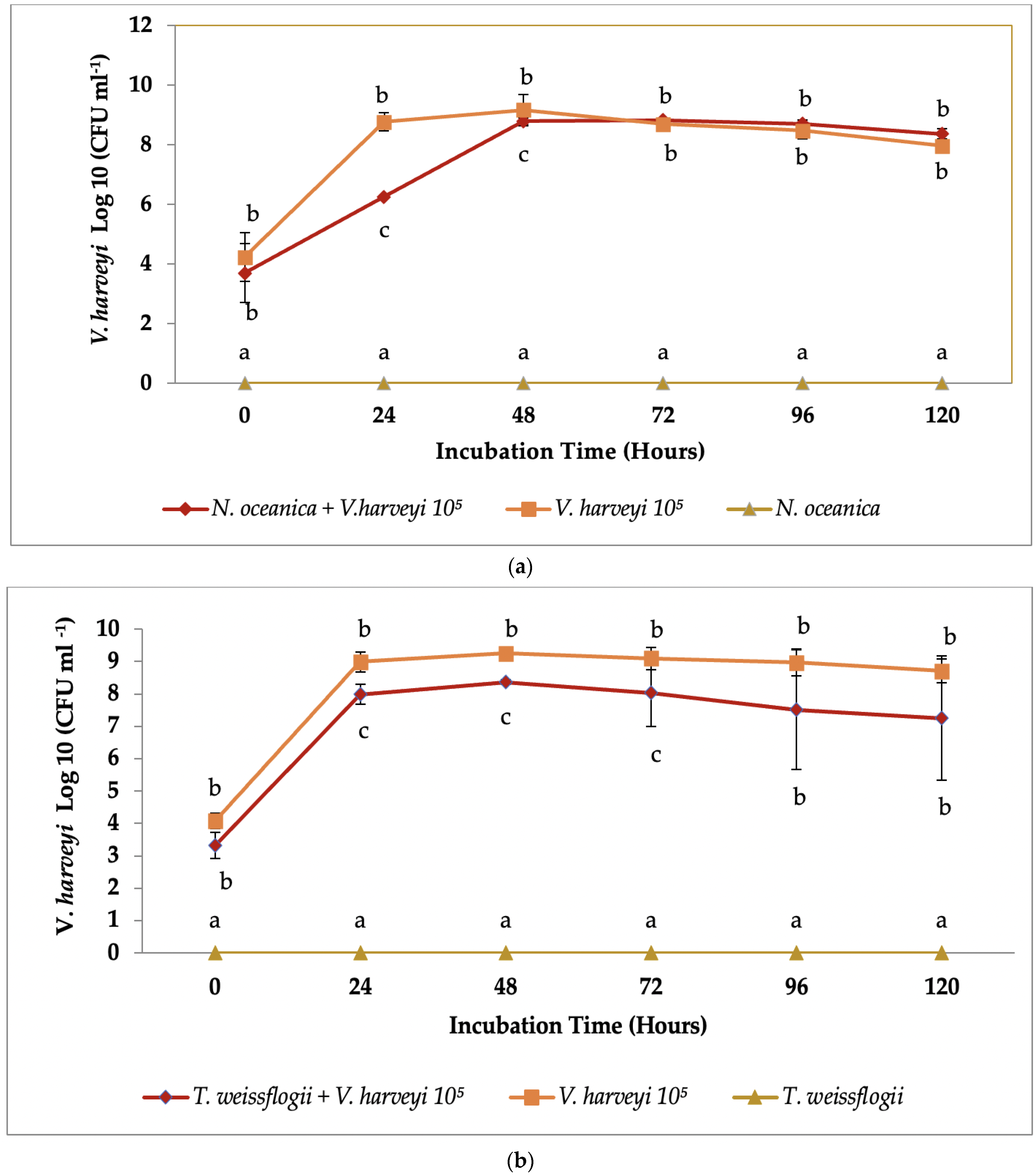
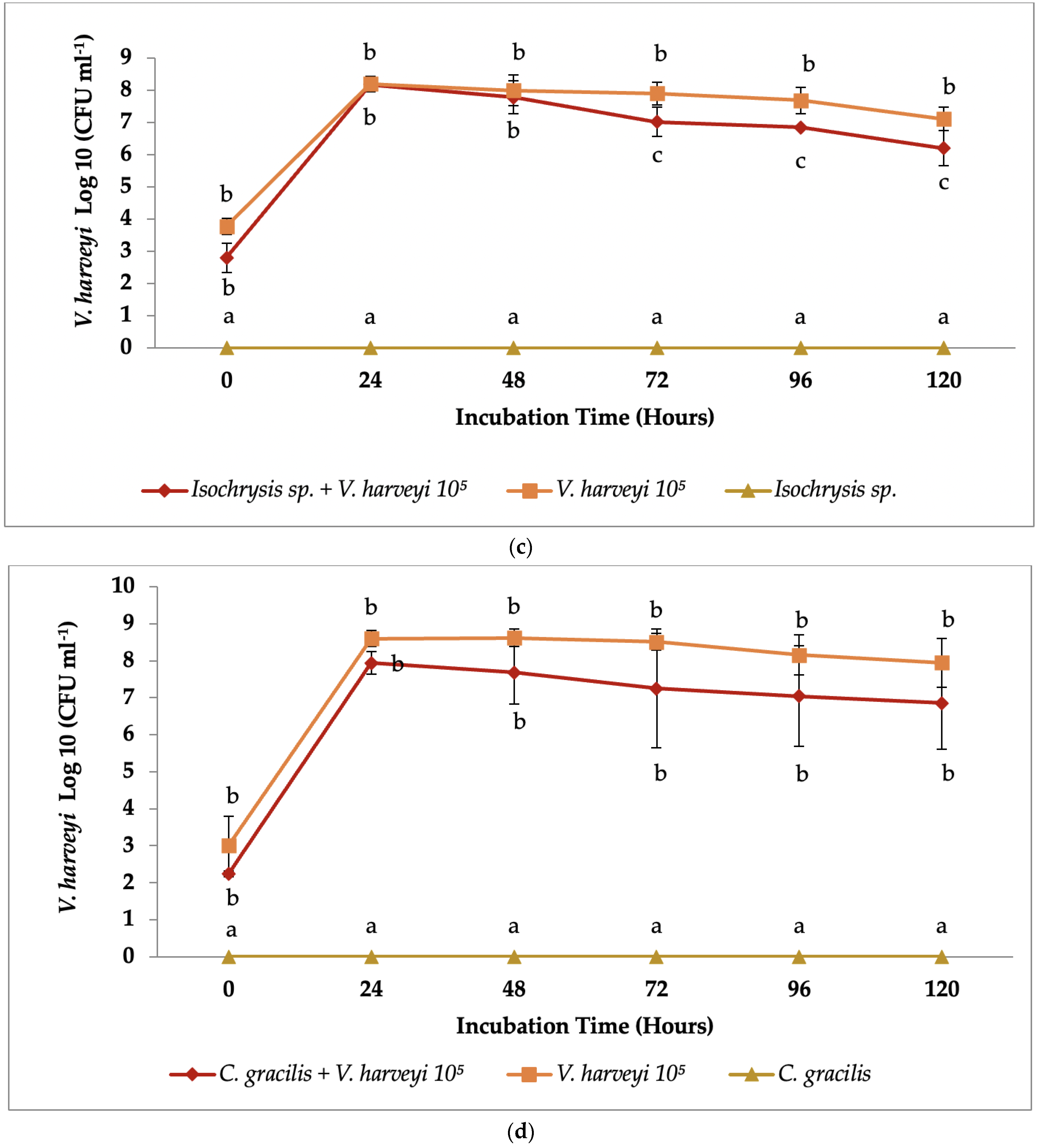
3.2. Co-Culture Antibacterial Assay

In the co-culture assay, three (3) microalgae species—co-culture assays of N. oceanica (Figure 1a), T. weissflogii (Figure 1b), and Isochrysis sp. (Figure 1d)—demonstrated minimal antibacterial activity by preventing significant (p < 0.05) proliferation of V. harveyi at different time intervals when compared to the control group culture of V. harveyi with no microalgae.
In the co-culture V. harveyi with N. oceanica (Figure 1a), the concentration of V. harveyi was significantly lower (p < 0.05) at 24 to 48 h incubation as compared to control. However, there was no significant difference (p > 0.05) in V. harveyi concentration at 72, 96, and 120 h incubation.
For the co-culture V. harveyi with T. weissflogii (Figure 1b), the concentration of V. harveyi was significantly lower (p < 0.05) at 24, 48, and 72 h incubation, followed by no significant difference (p > 0.05) at 96 and 120 h incubation as compared to control.
Meanwhile, the co-culture V. harveyi with Isochrysis sp in Figure 1d demonstrated a significantly lower (p < 0.05) concentration of V. harveyi at 72, 96, and 120 h, as compared to the control. In comparison to the control, co-culture V. harveyi with C. gracilis (Figure 1c) exhibited no significant difference (p > 0.05) in V. harveyi concentration. In fact, no bibliographic data on the antibacterial activity of C. gracilis described in the prior study is available. The growth of V. harveyi strains increased steadily with time up to 48 h until reaching a plateaued condition and subsequently decreased insignificantly (p > 0.05) at the end of the experiment in the control group culture with no microalgae added.

4. Discussion

Many bioactive and pharmacologically active chemicals, particularly antibacterial compounds, are potentially produced by marine microalgae [6,20,21]. Furthermore, microalgae are gaining popularity due to their ability to create bioactive metabolites with anticancer, anti-inflammatory, antibacterial, and antioxidant effects [11,22]. Previous studies have suggested that fatty acids [23], terpenoids, carbohydrates [24], peptides, polysaccharides, and alkaloids are responsible for antibacterial activity in microalgae [25]. Novel antibacterial substances were also discovered in microalgae, with Coccomyxa onubensis fatty acid extracts inhibiting E.coli and P. mirabilis [26]. Furthermore, microalgae have been increasingly treated with other compounds such as cyanovirin, oleic acid, linoleic acid, palmitoleic acid, -carotene, or phycocyanin, which have antioxidant or anti-inflammatory properties, as well as antimicrobial activity, such as against Staphylococcus aureus and methicillin-resistant Staphylococcus aureus (MRSA) [22,23].
Table 1 shows variable degrees of inhibition on several species of microalgae extract against V. harveyi from this investigation. As a result, the generation of antibiotics is heavily reliant on microalgal species [27]. Furthermore, the availability of antibiotic agents can vary greatly between various species within the same class or within a single species, depending on which ecotypes are adapted to certain conditions [27,28]. The green microalga Dunaliella sp. isolated from severely polluted waters, for example, was found to be more active against bacteria than ecotypes obtained from less polluted waters [29]. Recently, microalgae of the genus Nannochloropsis have been shown to be rich in polyunsaturated fatty acid compounds (PUFAs), carotenoids, polyphenols, and vitamins, and have previously been used in aquaculture [30], while the microalgae strain of Isochrysis sp. was shown to be high in fucoxanthin and DHA [31]. T. weissflogii, on the other hand, has antibacterial properties due to its high presence of substances such as betain, lipids, phospolipid, polyunsaturated fatty acid, fucoxanthin (FX), and eicosapentaenoic acid (EPA) [32]. C. gracilis showed a limited inhibited zone as compared to other microalgae extracts, particularly at higher concentrations of V. harveyi. This could be because the compound composition of C. gracilis is minimal and contains only non-essential amino acids [33].
Apart from microalgae species, the presence of antibacterial chemicals in microalgae extracts is also strongly reliant on the extraction process and solvent utilised. According to the findings of this investigation, ethanol is the best extractant since it demonstrated the broadest inhibition against V. harveyi in the disc diffusion method. Based on prior research, ethanol was also used to extract the thalli of Gracilaria fisheri from India, the extract of which exhibited a high inhibitory effect against V. harveyi [34]. An ethanol extract of Gracilaria corticata from India was found to be highly active against V. cholerae and V. parahaemolyticus, but less active against Pseudomonas aeruginosa and Shigella flexneri [35]. Another study on ethanolic extract of Spiriluna also demonstrated more effective against two species of Vibrio [36]. These findings suggest that molecules with antibacterial action in microalgae are generally hydrophobic and might even be extracted more easily with organic solvents [27]. This could indicate that ethanol is a competent extraction method capable of extracting lipids with a specific set of chemicals present during extraction.
The methanolic extract is a well-established and reported method to isolate active antimicrobial components from microalgae [37,38]. In another study, methanol was found to be the best organic solvent to obtain bioactive compounds from Dunaliella sp. [39]. The methanolic extract of freshwater microalgae exhibited antibacterial activity against important human bacterial pathogens [40]. Methanolic extract in the current study inhibited V. harveyi moderately, whereas the antibiotic synergism with the methanolic extract of C. vulgaris exerted enhanced anti-bacterial activity in E. coli. [41]. Therefore, the selection of a solvent is also important for the further utilisation of post-extracted microalgal biomass. According to Navarro et al., 2017 [26], it was suggested that fatty acids could be involved in the antibacterial activity due to increased activity from green microalga extracts obtained with non-polar solvents compared to polar solvents.
In the present study, microalgae extraction by saline water and DMSO demonstrated a comparable less effective antibacterial activity against V. harveyi based on the concentration of V. harveyi. Nevertheless, it was reported that the extraction of antimicrobial compounds from Scenedesmus subspicatus using DMSO inhibited the growth of Klebsiella pneumoniae and Escherichia coli [42]. As observed in previous studies, water extracts of Dunaliella sp. had lower bioactive properties than the ones obtained from organic solvents [43]. Hence, the selection of a solvent is important for optimisation of the extraction process and generation of its bioactive compounds, which have diverse applications not only in aquaculture but also against human pathogens. According to Pradhan et al., 2012 [36] no antimicrobial activity was detected in the aqueous extracts, which was probably because of the low polar nature of the active components.
The number of V. harveyi increased exponentially, especially in the first 24 h during the experimental period, without the presence of microalgae cells (control) demonstrating that the bacteria cells were able to utilise the growth medium of the microalgae cultures. The inhibition of V. harveyi in co-culture assay with N. oceanica, Isochrysis sp., and T. weissflogii indicated the production of antibacterial compounds by these microalgae cells. Previous research has shown that Isochrysis sp. and Nannochloropsis exhibit antibacterial action against the majority of vibriosis pathogens, including V. alginolyticus, V. lentus, V. splendidus, V. scophthalmi, V. parahaemolyticus, and V. anguillarum [44]. The microalgae species Nannochloropsis sp. and Isochrysis sp. have been demonstrated to synthesise short-chain fatty acids, which are implicated in antibacterial activity [45,46]. Thus, microalgae biomass is regarded as a source of valuable chemical elements that can be used in animals, particularly farmed fish, as pharmaceuticals in aquaculture industries.
Another element that could explain these discrepancies is that antibacterial activity is linked to the culture conditions and growth phase of the microalgae cultures. Although all microalgae species were at the same concentration in all trials in this study, the growth phase for each microalgae culture could have been different and may have influenced the results. Previous studies suggested that modifying the culture conditions of green microalgae exhibited differences in antibacterial activity [47,48,49], whereas modifications on microalgae culture conditions could be stimulated to produce secondary metabolites with antibacterial activity, as well as potentially larger quantities of these secondary metabolites [50]. This may include modifications on media composition, pH, light, and temperature [51,52]. As an example, the highest light intensity (4800 lux) exposure effectively induced the production of antibacterial compounds by Dunaliella sp. and suggested that the higher antimicrobial effect was related to the content of bioactive compound produced in stress conditions [39]. Other than the culture environment, the period of microalgae cultivation also plays an important parameter in producing bioactive compounds [39,53]. Therefore, the culture environment could be artificially manipulated in order to allow for increased production of antibacterial compounds in microalgae.

5. Conclusions

Out of four species of microalgae, only N. oceanica, Isochrysis sp., and T. weissflogii demonstrated a significant ((p < 0.05) inhibitory effect against V. harveyi in an in vitro co-culture assay. This indicated a positive effect of the addition of these microalgae in the rearing of fish larvae and implicated the production of antibacterial compounds. Ethanolic extract from N. oceanica, Isochrysis sp., and T. weissflogii demonstrated promising antibacterial activity against V. harveyi. Meanwhile, C. gracilis inhibited the development of V. harveyi with minimal inhibition but no significant difference (p > 0.05) in the co-culture assay and disc diffusion method, indicating that it had less potential to limit the growth of V. harveyi. Therefore, in the aquaculture industry, N. oceanica, Isochrysis sp., and T. weissflogii act as potential inhibitory bacteria and probiotics in controlling the disease. More comprehensive studies are recommended to optimize its cultivation conditions, the extraction process, and the purification of its antibacterial compounds to unleash their potential.

Author Contributions

Conceptualization, R.O., S.R.M.S., and M.R.J.; methodology, R.O. and M.R.J.; validation, S.R.M.S. and R.O.; formal analysis, M.R.J.; investigation, M.R.J.; resources, F.F.C. and S.R.M.S.; data curation, M.R.J. and R.O.; writing—original draft preparation, M.R.J., R.O., and S.N.H.O.; writing—review and editing, R.O., S.N.H.O., and S.S.; visualization, M.R.J. and F.F.C.; supervision, R.O. All authors have read and agreed to the published version of the manuscript.

Funding

This project was funded by the Minister of Higher Education Malaysia under the Fundamental Research Grant Scheme FRGS0469-2017.

Institutional Review Board Statement

Not applicable.

Informed Consent Statement

Not applicable.

Data Availability Statement

The datasets generated during and/or analyzed during the current study are available from the corresponding author on reasonable request.

Acknowledgments

This project was funded by the Minister of Higher Education Malaysia under the Fundamental Research Grant Scheme FRGS0469-2017. The publication fee was provided by the Research Management Centre, Universiti Malaysia Sabah.

Conflicts of Interest

The authors declare no conflict of interest.

References

  1. Pauly, D.; Zeller, D. Comments on FAOs State of World Fisheries and Aquaculture (SOFIA 2016). Mar. Policy 2017, 77, 176–181. [Google Scholar] [CrossRef]
  2. Ma, K.; Bao, Q.; Wu, Y.; Chen, S.; Zhao, S.; Wu, H.; Fan, J. Evaluation of Microalgae as Immunostimulants and Recombinant Vaccines for Diseases Prevention and Control in Aquaculture. Front. Bioeng. Biotechnol. 2020, 8, 1331. [Google Scholar] [CrossRef] [PubMed]
  3. Wei, L.S.; Wee, W. Diseases in aquaculture. Res. J. Ani. Vet. Sci. 2014, 7, 1–6. [Google Scholar]
  4. Mohd Yazid, S.H.; Mohd Daud, H.; Azmai, M.N.A.; Mohamad, N.; Mohd Nor, N. Estimating the Economic Loss Due to Vibriosis in Net-Cage Cultured Asian Seabass (Lates calcarifer): Evidence from the East Coast of Peninsular Malaysia. Front. Vet. Sci. 2021, 8, 1111. [Google Scholar] [CrossRef]
  5. Ina-Salwany, M.Y.; Al-Saari, N.; Mohamad, A.; Mursidi, F.A.; Mohd-Aris, A.; Amal, M.N.A.; Kasai, H.; Mino, S.; Sawabe, T.; Zamri-Saad, M. Vibriosis in Fish: A Review on Disease Development and Prevention. J. Aquat. Anim. Health 2019, 31, 3–22. [Google Scholar] [CrossRef]
  6. Farisa, M.Y.; Namaskara, K.E.; Yusuf, M.B.; Desrina. Antibacterial Potention of Extract of Rotifers Fed with Different Microalgae to Control Vibrio harveyi. IOP Conf. Ser. Earth Environ. Sci. 2019, 246, 012058. [Google Scholar] [CrossRef]
  7. Aftabuddin, S.; Siddique, M.A.M.; Habib, A.; Akter, S.; Hossen, S.; Tanchangya, P.; Abdullah, M.A.l. Effects of seaweeds extract on growth, survival, antibacterial activities, and immune responses of Penaeus monodon against Vibrio parahaemolyticus. Ital. J. Anim. Sci. 2021, 20, 243–255. [Google Scholar] [CrossRef]
  8. Manyi-Loh, C.; Mamphweli, S.; Meyer, E.; Okoh, A. Antibiotic Use in Agriculture and Its Consequential Resistance in Environmental Sources: Potential Public Health Implications. Molecules 2018, 23, 795. [Google Scholar] [CrossRef] [PubMed] [Green Version]
  9. Molina-Cárdenas, C.A.; Sánchez-Saavedra, M.P.; Lizárraga-Partida, M.L. Inhibition of pathogenic Vibrio by the microalgae Isochrysis galbana. J. Appl. Phycol. 2014, 26, 2347–2355. [Google Scholar] [CrossRef]
  10. Reverter, M.; Bontemps, N.T.; Sasal, P.; Saulnier, D. Use of medicinal plants in aquaculture. In Diagnosis and Control of Diseases of Fish and Shellfish; Austin, B., Newaj-Fyzul, A., Eds.; Wiley: Hoboken, NJ, USA, 2017; pp. 223–261. [Google Scholar]
  11. Khavari, F.; Saidijam, M.; Taheri, M.; Nouri, F. Microalgae: Therapeutic potentials and applications. Mol. Biol. Rep. 2021, 48, 4757–4765. [Google Scholar] [CrossRef]
  12. Abdullah, A.; Ramli, R.; Ridzuan, M.S.M.; Murni, M.; Hashim, S.; Sudirwan, F.; Abdullah, S.Z.; Mansor, N.N.; Amira, S.; Zamri-Saad, M.; et al. The presence of Vibrionaceae, Betanodavirus and Iridovirus in marine cage-cultured fish: Role of fish size, water physicochemical parameters and relationships among the pathogens. Aquac. Rep. 2017, 7, 57–65. [Google Scholar] [CrossRef]
  13. Mohamad, N.; Mustafa, M.; Amal, M.N.A.; Saad, M.Z.; Md-Yasin, I.S.; Al-saari, N. Environmental factors associated with the presence of Vibrionaceae in tropical cage-cultured marine fishes. J. Aquat. Anim. Health 2019, 31, 154–167. [Google Scholar] [CrossRef]
  14. Salvesen, I.; Reitan, K.I.; Skjermo, J.; Oie, G. Microbial environments in marine larviculture: Impacts of algal growth rates on the bacterial load in six microalgae. Aquac. Int. 2000, 8, 275–287. [Google Scholar] [CrossRef]
  15. Guillard, R.R.; Ryther, J.H. Studies on marine planktonic diatoms. I. Cyclotella nana Hustedt and Detonula confervacaea (Cleve) Gran. Can. J. Microbiol. 1962, 8, 229–239. [Google Scholar] [CrossRef]
  16. Sasireka, G.; Muthuvelayudham, R. Effect of Salinity and Iron Stressed on Growth and Lipid Accumulation in Skeletonema costatum for Biodiesel Production. Res. J. Chem. Sci. 2015, 5, 69–72. [Google Scholar]
  17. Gonzalez, D.V.A.; Platas, G.; Basilio, A. Screening of antimicrobial activities in red, green and brown macroalgae from Gran Canaria (Canary Islands, Spain). Int. Microbiol. 2001, 4, 35–40. [Google Scholar]
  18. Bauer, A.N.; Kirby, W.M.M.; Sherries, J.C.; Truck, M. Antibiotic susceptibility testing by a standardized single disk method. Am. J. Clin. Pathol. 1966, 45, 493–496. [Google Scholar] [CrossRef] [PubMed]
  19. Corona, E.; Fernandez-Acero, J.; Bartual, A. Screening study for antibacterial activity from marine and freshwater microalgae. Int. J. Pharm. Bio Sci. 2017, 8, 189–194. [Google Scholar]
  20. Prasetiya, F.S.; Sunarto, S.; Bachtiar, E.; Mochamad, U.K.A.; Nathanael, B.; Pambudi, A.C.; Lestari, A.D.; Astuty, S.; Mouget, J.-L. Effect of the blue pigment produced by the tropical diatom Haslea nusantara on marine organisms from different trophic levels and its bioactivity. Aquac. Rep. 2020, 17, 100389. [Google Scholar] [CrossRef]
  21. Soto-Rodriguez, S.A.; Lopez-Vela, M.; Soto, M.N. 2021 Inhibitory effect of marine microalgae used in shrimp hatcheries on Vibrio parahaemolyticus responsible for acute hepatopancreatic necrosis disease. Aquac. Res. 2021, 1–11. [Google Scholar]
  22. Barone, M.E.; Murphy, E.; Parkes, R.; Fleming, G.T.A.; Campanile, F.; Thomas, O.P.; Touzet, N. Antibacterial Activity and Amphidinol Profiling of the Marine Dinoflagellate Amphidinium carterae (Subclade III). Int. J. Mol. Sci. 2021, 22, 12196. [Google Scholar] [CrossRef]
  23. Desbois, A.P.; Mearns-Spragg, A.; Smith, V.J. A fatty acid from the diatom Phaeodactylum tricornutum is antibacterial against diverse bacteria including multi-resistant Staphylococcus aureus (MRSA). Mar. Biotechnol. 2009, 11, 45–52. [Google Scholar] [CrossRef]
  24. Duff, D.C.B.; Bruce, D.L. The antibacterial activity of marine planktonic algae. Can. J. Microbiol. 1966, 12, 877–884. [Google Scholar] [CrossRef] [PubMed]
  25. Borowitzka, M.A. Microalgae as sources of pharmaceuticals and other biologically active compounds. J. Appl. Phycol. 1995, 7, 3–15. [Google Scholar] [CrossRef]
  26. Navarro, F.; Forján, E.; Vázquez, M.; Toimil, A.; Montero, Z.; Ruiz-Domínguez, M.D.C.; Garbayo, I.; Castaño, M.A.; Vilchez, C.; Vega, J.M. Antimicrobial activity of the acidophilic eukaryotic microalga Coccomyxa onubensis. Phycol Res. 2017, 65, 38–43. [Google Scholar] [CrossRef]
  27. Falaise, C.; François, C.; Travers, M.A.; Morga, B.; Haure, J.; Tremblay, R.; Turcotte, F.; Pasetto, P.; Gastineau, R.; Hardivillier, Y.; et al. Antimicrobial Compounds from Eukaryotic Microalgae against Human Pathogens and Diseases in Aquaculture. Mar. Drugs 2016, 14, 159. [Google Scholar] [CrossRef] [Green Version]
  28. Pawlik-Skowrońska, B. Resistance, accumulation and allocation of zinc in two ecotypes of the green alga Stigeoclonium tenue Kütz. coming from habitats of different heavy metal concentrations. Aquat. Bot. 2003, 75, 189–198. [Google Scholar] [CrossRef]
  29. Lustigman, B. Comparison of antibiotic production from four ecotypes of the marine alga, Dunaliella Bull. Environ. Contam. Toxicol. 1988, 40, 18–22. [Google Scholar] [CrossRef] [PubMed]
  30. Zanella, L.; Vianello, F. Microalgae of the genus Nannochloropsis: Chemical composition and functional implications for human nutrition. J. Funct. Foods 2020, 68, 103919. [Google Scholar] [CrossRef]
  31. Sun, Z.; Wang, X.; Liu, J. Screening of Isochrysis strains for simultaneous production of docosahexaenoic acid and fucoxanthin. Algal Res. 2019, 41, 101545. [Google Scholar] [CrossRef]
  32. Marella, T.K.; Tiwari, A. Marine diatom Thalassiosira weissflogii based biorefinery for co-production of eicosapentaenoic acid and fucoxanthin. Bioresour. Technol. 2020, 307, 123245. [Google Scholar] [CrossRef]
  33. Merchie, G.; Lavens, P.; Sorgeloos, P. Optimization of dietary vitamin C in fish and crustacean larvae: A review. Aquaculture 1997, 155, 165–181. [Google Scholar] [CrossRef]
  34. Kanjana, K.; Radtanatip, T.; Asuvapongpatana, S.; Withyachumnarnkul, B.; Wongprasert, K. Solvent extracts of the red seaweed Gracilaria fisheri prevent Vibrio harveyi infections in the black tiger shrimp Penaeus monodon, Fish. Shellfish Immunol. 2011, 30, 389–396. [Google Scholar] [CrossRef]
  35. Padmakumar, K.; Ayyakkannu, K. Seasonal variation of antibacterial and antifungal activities of marine algae from southern coast of India. Bot. Mar. 1997, 40, 507–515. [Google Scholar] [CrossRef]
  36. Pradhan, J.; Das, B.K.; Sahu, S.; Marhual, N.P.; Swain, A.K.; Mishra, B.K.; Eknath, A.E. Traditional antibacterial activity of fresh water microalga Spirulina platensis to aquatic pathogens. Aquac. Res. 2012, 43, 1287–1295. [Google Scholar] [CrossRef]
  37. Patil, L.; Kaliwal, B.B. Microalga Scenedesmus bajacalifornicus BBKLP-07, a new source of bioactive compounds with in vitro pharmacological applications. Bioprocess Biosyst. Eng. 2019, 42, 979–994. [Google Scholar] [CrossRef] [PubMed]
  38. Zea-Obando, C.; Tunin Ley, A.; Turquet, J.; Culioli, G.; Briand, J.F.; Bazire, A.; Réhel, K.; Faÿ, F.; Linossier, I. Anti-bacterial adhesion activity of tropical microalgae extracts. Molecules 2018, 23, 2180. [Google Scholar] [CrossRef] [Green Version]
  39. Kilic, N.K.; Erdem, K.; Donmez, G. Bioactive compounds produced by Dunaliella species, antimicrobial effects and optimization of the efficiency. Turk. J. Fish. Aquat. Sci. 2018, 19, 923–933. [Google Scholar]
  40. Prakash, J.W.; Antonisamy, J.M.; Jeeve, S. Antimicrobial activity of certain fresh water microalgae from Thamirabarani River, Tamil Nadu, South India. Asian Pac. J. Tro. Biomed. 2011, 1, S170–S173. [Google Scholar] [CrossRef]
  41. Pradhan, B.; Patra, S.; Dash, S.R.; Nayak, R.; Behera, C.; Jena, M. Evaluation of the anti-bacterial activity of methanolic extract of Chlorella vulgaris Beyerinck [Beijerinck] with special reference to antioxidant modulation. Futur. J. Pharm. Sci. 2021, 7, 17. [Google Scholar] [CrossRef]
  42. Dantas, D.M.D.M.; Oliveira, C.Y.B.D.; Costa, R.M.P.B.; Carneiro-da-Cunha, M.D.G.; Gálvez, A.O.; Bezerra, R.D.S. Evaluation of antioxidant and antibacterial capacity of green microalgae Scenedesmus subspicatus. Food Sci. Technol. Int. 2019, 25, 318–326. [Google Scholar] [CrossRef] [PubMed]
  43. Herrero, M.; Jaime, L.; Martin-Alvarez, J.M.; Cifuentes, A.; Ibanez, E. Optimization of the Extraction of Antioxidants from Dunaliella salina Microalga by Pressurized Liquids. J. Agric. Food Chem. 2006, 54, 5597–5603. [Google Scholar] [CrossRef] [PubMed]
  44. Kokou, F.; Makridis, P.; Kentouri, M.; Divanach, P. Antibacterial activity in microalgae cultures. Aquac. Res. 2012, 43, 1520–1527. [Google Scholar] [CrossRef]
  45. Roncarati, A.; Meluzzi, A.; Acciari, S.; Tallarico, N.; Melotti, P. Fatty acid composition of different microalgae strains (Nannochloropsis sp., Nannochloropsis oculata (Droop) Hibberd, Nannochloropsis atomus Butcher and Isochrysis sp. according to the culture phase and the carbon dioxide concentration. J. World Aquac. Soc. 2004, 35, 401–411. [Google Scholar] [CrossRef]
  46. Defoirdt, T.; Boon, N.; Sorgeloos, P.; Verstraete, W.; Bossier, P. Alternatives to antibiotics to control bacterial infections: Luminescent vibriosis in aquaculture as an example. Trends Biotechol. 2007, 25, 472–479. [Google Scholar] [CrossRef] [Green Version]
  47. Elkomy, R.; Ibraheem, I.B.M.; Shreadah, M.; Mohammed, R. Optimal conditions for antimicrobial activity production from two microalgae Chlorella marina and Navicula f. delicatula. J. Pure Appl. Microbiol. 2015, 9, 2725–2732. [Google Scholar]
  48. Dineshkumar, R.; Narendran, R.; Jayasingam, P.; Sampath-kumar, P. Cultivation and chemical composition of microalgae Chlorella vulgaris and its antibacterial activity against human pathogens. J. Aquac. Mar. Biol. 2017, 5, 00119. [Google Scholar]
  49. Hamouda, R.A.E.; Abou-El-Souod, G.W. Influence of various concentrations of phosphorus on the antibacterial, antioxidant and bioactive components of green microalgae Scenedesmus obliquus. Int. J. Pharmacol. 2018, 14, 99–107. [Google Scholar]
  50. Ruffell, S.E.; Müller, K.M.; McConkey, B.J. Comparative assessment of microalgal fatty acids as topical antibiotics. J. Appl. Phycol. 2016, 28, 1695–1704. [Google Scholar] [CrossRef]
  51. Schuelter, A.R.; Kroumov, A.D.; Hinterholz, C.L.; Fiorini, A.; Trigueros, D.E.G.; Vendruscolo, E.G.; Zaharieva, M.M.; Módenes, A.N. Isolation and identification of new microalgae strains with antibacterial activity on food-borne pathogens: Engineering approach to optimize synthesis of desired metabolites. Biochem. Eng. J. 2019, 144, 28–39. [Google Scholar] [CrossRef]
  52. Santhakumaran, P.; Kookal, S.K.; Mathew, L.; Ray, J.G. Experimental evaluation of the culture parameters for optimum yield of lipids and other nutraceutically valuable compounds in Chloroidium saccharophillum (Kruger) comb. Nov. Renew. Energy 2020, 147, 1082–1097. [Google Scholar] [CrossRef]
  53. Noaman, N.H.; Fattah, A.; Khaleafa, M.; Zaky, S.H. Factors affecting antimicrobial activity of Synechococcus leopoliensis. Microbiol. Res. 2004, 159, 395–402. [Google Scholar] [CrossRef] [PubMed]
Figure 1. (a) V. harveyi concentration was significantly lower (p < 0.05) at 24–48 h incubation in co-culture assay with N. oceanica as compared to control V. harveyi with no microalgae added. (b) V. harveyi concentration was significantly lower (p < 0.05) at 24 to 72 h incubation in co-culture assay with T. weissflogii as compared to control V. harveyi with no microalgae added. (c) V. harveyi concentration was significantly lower (p < 0.05) at 72 to 120 h incubation in co-culture assay with Isochrysis sp as compared to control V. harveyi with no microalgae added. (d) V. harveyi concentration showed no significant difference (p > 0.05) in co-culture assay with C. gracilis as compared to control V. harveyi with no microalgae added. Note: Values are presented as mean ±SD. Mean values of V. harveyi within the same time (hours) with different lowercase letters are significantly different (p < 0.05).
Applsci 12 01148 g001aApplsci 12 01148 g001b
Table 1. Antibacterial susceptibility test of the microbial extract against V. harveyi.
Concentration of V. harveyExtractantDiameter of the Inhibition Zone (D) in mm by Marine Microalgae Extract (106 Cell/mL) and Control (Extractant) after 24 h Incubation with V. harveyi
N. oceanicaT. weissflogiiC. gracilisIsochrysis sp.Control (Extractant)
105 CFU/mLEthanol++++++++++++
Methanol++++++++
Saline water+++++++
DMSO+++++
106 CFU/mLEthanol++++++++++
Methanol+++++++
Saline water++++++
DMSO++++
107 CFU/mLEthanol++++++++
Methanol++++
Saline water++++
DMSO++++
Note: (−): No activity, (+): D < 6 mm, (++): 6 < D < 8.5 mm, (+++): D > 8.5 mm. D: Diameter of the inhibition zone in milimeters.
Publisher’s Note: MDPI stays neutral with regard to jurisdictional claims in published maps and institutional affiliations.

