Article

In Vitro Study Comparing Retention of Custom Post and Cores Fabricated Using Conventional, CAD/CAM Milling and 3D-Printing Techniques

1
Restorative and Prosthetic Dental Science Department, College of Dentistry, King Saud Bin Abdulaziz University for Health Sciences, Riyadh 14611, Saudi Arabia
2
King Abdullah International Medical Research Center, Riyadh 14611, Saudi Arabia
3
Department of Prosthetic Dental Sciences, College of Dentistry, King Saud University, Riyadh 11545, Saudi Arabia
4
Advanced Specialty Education Program in Prosthodontics, Loma Linda University School of Dentistry, Loma Linda, CA 92350, USA
5
Department of Prosthetic Dental Sciences, College of Dentistry, Jazan University, Jazan 45142, Saudi Arabia
6
Rutgers School of Dental Medicine, Rutgers University, Newark, NJ 07103, USA
7
Advanced Specialty Education Program in Endodontics, University of California School of Dentistry, Los Angeles, CA 92350, USA
8
Private Practice, Newport Beach, CA 92660, USA
9
School of Nursing, Loma Linda University, Loma Linda, CA 92354, USA
*
Authors to whom correspondence should be addressed.
Appl. Sci. 2022, 12(23), 11896; https://doi.org/10.3390/app122311896
Submission received: 17 October 2022 / Revised: 11 November 2022 / Accepted: 19 November 2022 / Published: 22 November 2022

Abstract

:
This study aimed to evaluate the differences in the retention of custom-cast non-precious post and cores (CCNPPCs) (control group), custom-milled titanium post and cores (CMTPCs), custom-printed titanium post and cores (CPTPCs), and custom-milled zirconia post and cores (CMZPCs), and to evaluate their mode of failure. The tested null hypothesis was that there were no differences in the retention of the various custom post and cores tested. A total of 80 post-and-core patterns were made using pattern resin and were divided into four groups: Group 1—fabricated via conventional casting using a non-precious casting alloy; Group 2—fabricated using a computer-aided design/computer-aided manufacturing (CAD/CAM) subtractive technique using titanium; Group 3—fabricated using a CAD/CAM additive (3D printing) technique using titanium; and Group 4—fabricated using a CAD/CAM subtractive technique using zirconia. The post and cores were cemented with resin cement and a universal pull-out test was used to check the retention. The data were statistically analyzed using one-way ANOVA tests, post hoc tests, and Tukey’s adjustment for multiple comparisons. The pull-out test revealed higher retention values for CPTPCs and CMTPCs. When compared with CMZPCs, the conventional CCNPPCs revealed significantly better retention values (p < 0.05). Cohesive failure was observed in Groups 1, 2, and 4. However, Group 3 revealed a mixed type of failure. The CCNPPCs revealed clinically acceptable values, while the CPTPC and CMTPC groups revealed better overall values of retention and time to failure. The titanium alloy was assessed to be a promising choice for fabricating dental post-and-core restorations.

1. Introduction

Post and cores are commonly used to restore endodontically treated teeth with the extensive coronal structural loss [1]. The retention of the post is a fundamental factor influencing definitive restoration longevity and success. Post length, shape, diameter and surface texture, and the type of cement used are associated factors that may affect the retention and stability of the post [2].
Custom-cast post and cores (CCPCs) have been reported to have superior adaptation and fit the radicular post-space walls when compared with prefabricated posts [3,4]. Compared with other post-and-core systems, CCPCs are advantageous, as they exhibit higher resistance to rotational movement forces [5], have a superior success rate [6,7], and have better retrievability for endodontic re-treatment [6,8]. Various materials can be used for fabricating custom post and cores. These include gold alloys [9,10,11], titanium alloy [9,11], base-metal alloys [9,12,13,14], zirconium oxide [11,15,16], glass fiber-reinforced composites [11,17], etc., which can be fabricated by different techniques such as casting [9,10,11,12,13], CAD/CAM milling [11,12,15,16,17], and 3D printing [11,12,14]. Various alloys are used for the fabrication of CCPCs, and most of them exhibit good retention and fracture strength [18,19]. However, CCPCs have been associated with catastrophic abutment fractures due to their high stiffness [4,5].
Custom post and cores have also been reported to be fabricated out of zirconia to overcome the esthetic limitations associated with metal CCPCs. Zirconia has been reported to have high flexural strength, high fracture toughness, chemical stability, biocompatibility, favorable optical properties, greater toughness, and maximum adaptability to the canal, as well as good esthetic characteristics [20,21]. As discussed by Baba et al. [22] and Ozkurt et al. [23], zirconia as a post-and-core material has a few disadvantages. These include more frequent root fractures (due to high rigidity) than fiber posts, decreased retention of the post (due to poor bonding between zirconia and resin cement), and poor retrievability in cases that need endodontic re-treatment.
Titanium has also gained wide acceptance in dentistry due to its biocompatibility, excellent corrosion resistance, reduced cost, ease of fabrication, and superior mechanical properties, which make it suitable as a post-and-core material [24,25,26,27]. Computer-aided design/computer-aided manufacturing (CAD/CAM) additive (3D printing) and CAD/CAM subtractive manufacturing techniques (milling) can be used to manufacture custom post and cores.
To the best of our knowledge, the current literature lacks the relevant information assessing the relationship between the retention of custom-cast non-precious post and cores (CCNPPCs), custom-milled titanium post and cores (CMTPCs), custom-printed titanium post and cores (CPTPCs) and custom-milled zirconia post and cores (CMZPCs). Therefore, this study aims to evaluate the differences in the retention of CCNPPCs (control group), CPTPCs, CMTPCs, and CMZPCs. The tested null hypothesis was that there were no differences in the retention values of the various tested custom post and cores.

2. Materials and Methods

2.1. Materials

In this study, four different techniques were used to fabricate custom post and cores (simulating the protocols for post-and-core fabrication) for the maxillary central incisors. The fabrication techniques used were: the conventional lost-wax casting technique (using a non-precious alloy), the CAD/CAM subtractive technique (using Grade 5 titanium and zirconia), and the CAD/CAM additive technique (3D printing) (using Grade 5 titanium 6AL4V). The details of each material used in the study are listed in Table 1.

2.2. Specimen Preparation

Eighty sound human maxillary central incisors, extracted for periodontal reasons, were selected for this study. The exclusion criteria included the presence of caries, restoration, root canal treatment (RCT), crack/s, attrition, very long or very short teeth, and/or severe root curve. Teeth were thoroughly cleaned with a brush after extraction, and a scalpel and a periodontal curette were used to remove any remaining hard and soft tissues from the root surfaces. Teeth were subsequently stored in 0.2% sodium azide (Merck KGaA, Frankfurter Str. 250, Darmstadt, Germany) prior to specimen preparation (ISO 28399; 2011) [28]. Each tooth was randomly assigned a number (from 1 to 80) and allocated into one of 4 groups (n = 20 each). Group 1 teeth were restored with custom-cast non-precious post and cores (CCNPPCs); Group 2 teeth were restored with custom-milled titanium post and cores (CMTPCs); Group 3 teeth were restored with custom-printed titanium post and cores (CPTPCs), and Group 4 teeth were restored with custom-milled zirconia post and cores (CMZPCs).

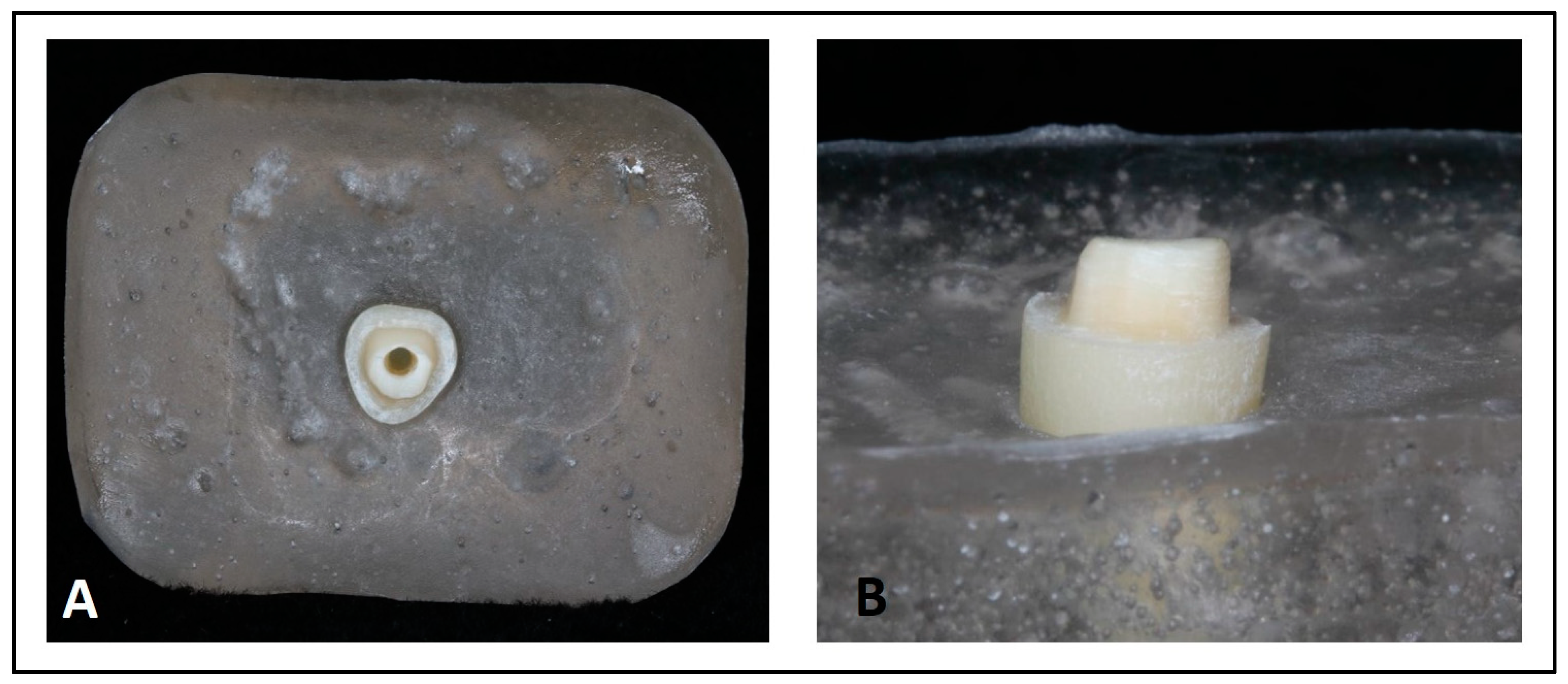
2.2.1. Mounting Teeth in Acrylic-Resin Blocks

Teeth were individually mounted in a special specimen holder using epoxy resin (Exakto-form; Bredent, Derbyshire, UK) (with an elastic modulus of 12 GPa, which is similar to the elastic modulus of human bone (18 GPa)) [29] with the use of a test mount former of 2 cm3 in dimension 2–3 mm below the cementoenamel junction (CEJ). A prefabricated jig was used to position each tooth in the test mount former during the immersion of the tooth in acrylic resin to standardize the tooth position to be centralized within the test mounts. The test mount jig was used to standardize the tooth position while performing tooth preparation and RCT (Figure 1). To avoid the dehydration of natural teeth due to the heat generated during the polymerization of acrylic resin, the resin block was cooled in water [30]. The teeth were prepared to have a 2 mm ferrule and a 1 mm shoulder finish line (Figure 2) [31,32,33].

2.2.2. Root-Canal Preparation and Obturation

Access-cavity preparation was performed using a size 2 diamond round bur on a high-speed handpiece with copious water. The working length was established as 1 mm shorter than the root apex. Root-canal preparation for each tooth was performed up to size 40/0.06 with a Vortex Blue rotary file (Dentsply Sirona, Tulsa, OK, USA) in a crown-down fashion. Canals were irrigated with NaOCl 5.25% and EDTA 17% (chelating agent) (Ultradent Products Inc., South Jordan, UT, USA) to remove organic and inorganic debris and smear layers [34]. Upon completion of the cleaning and shaping and before obturation, the prepared root canals were dried with sterile paper points (Sure-endo, Gyeonngi-do, Republic of Korea). A matching size 40/0.06 gutta-percha master cone (Dentsply Sirona, Tulsa, OK, USA) coated with AH Plus sealer (Dentsply Sirona GmbH, De-Trey-Straße, Konstanz, Germany) was used for obturation [34]. Root canals were obturated using the warm vertical compaction technique with System-B and Obtura (Kerr/Sybron Endo Corp., Brea, CA, USA).

2.2.3. Post-Space Preparation

To obtain a standardized length for the posts, the coronal portion of gutta-percha was removed with System-B (Kerr corporation, Brea, CA, USA) until an adequate length (11 mm) was achieved [35,36,37,38]. Definitive post length and width were prepared and established with the use of Peeso reamers (Maillefer S.A., Ballaigues, Switzerland) up to size 3. Each canal was then cleaned using an air/water spray and EDTA to remove debris and then dried with paper points.

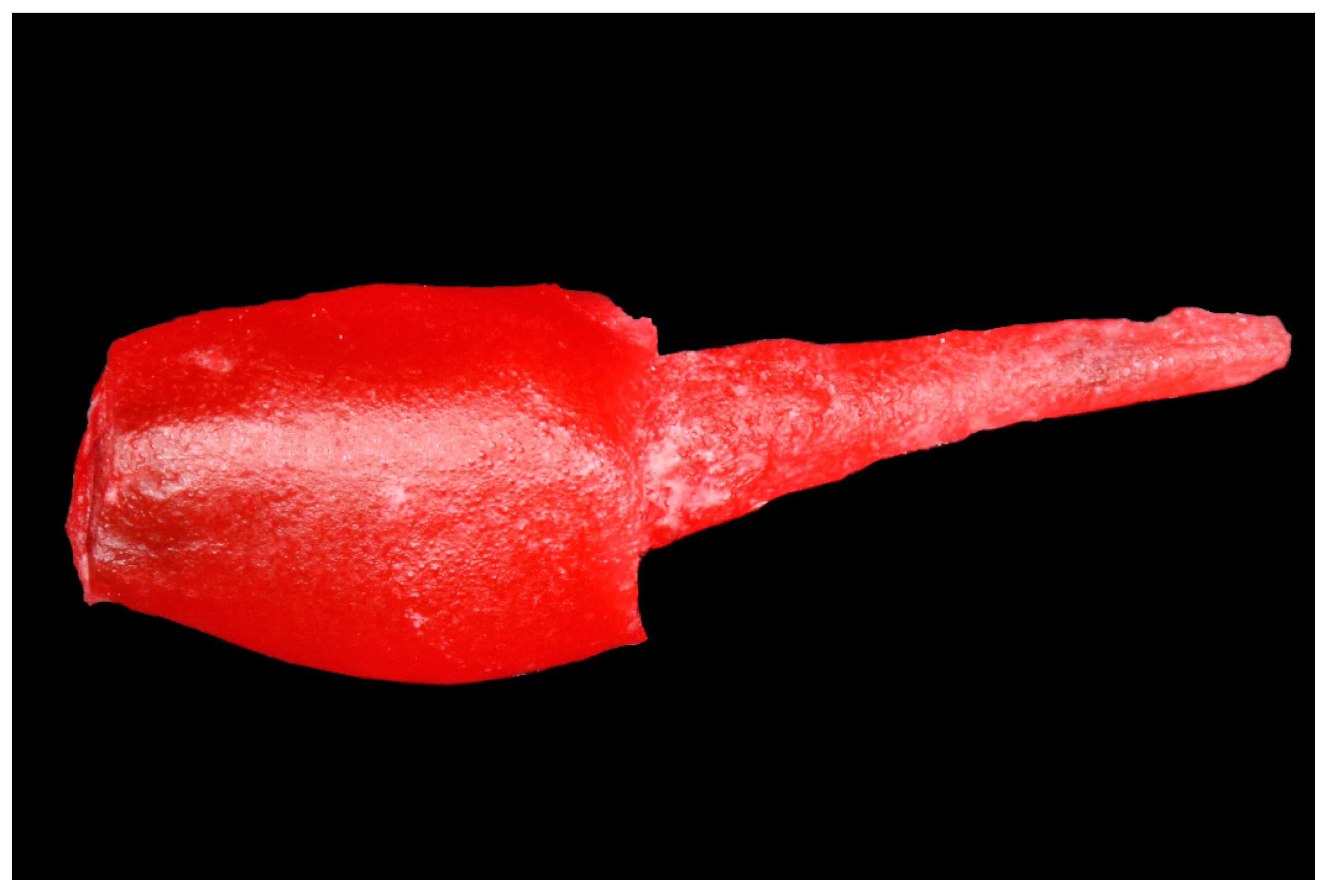
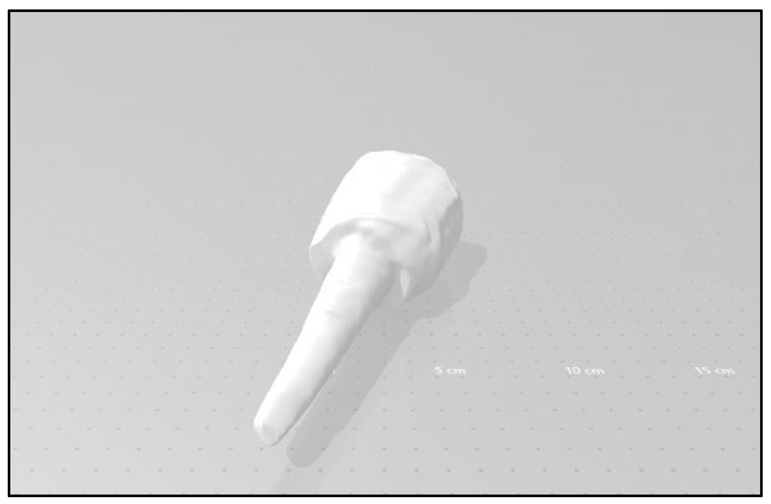
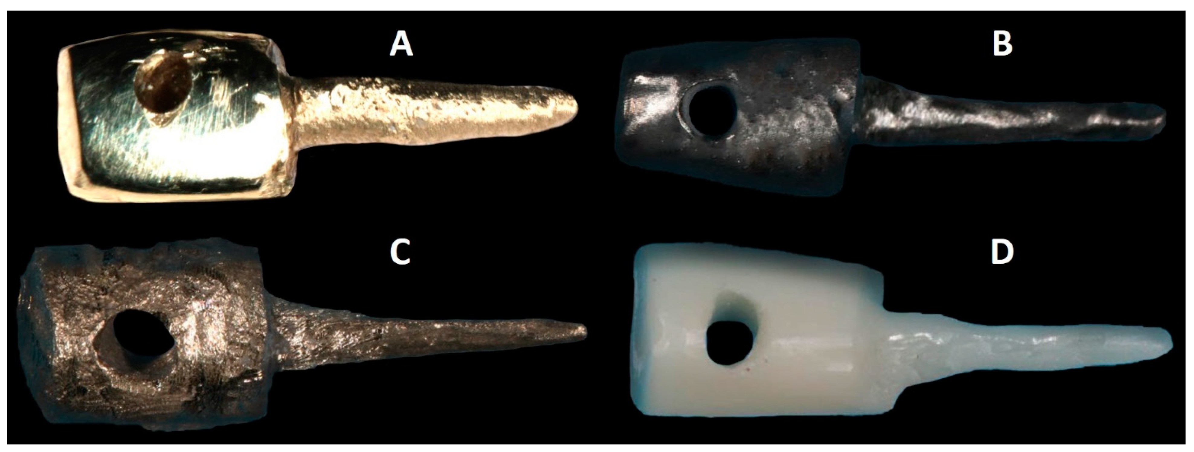
2.2.4. Post-and-Core Fabrication

Eighty custom post-and-core patterns were fabricated with the use of auto-polymerizing acrylic resin (Pattern Resin LS; GC America, Alsip, IL, USA) and serrated plastic posts. Each serrated plastic post was relined with acrylic resin and then inserted into the root canal until the canal space was bound to the walls of the prepared teeth. Then, the core was built up using a GC pattern and prepared using a diamond bur (ISO No. 010; Brasseler USA Dental, Georgia) to achieve a core with a 4 mm height (Figure 3). A hole was made in the coronal part of the GC pattern core and was used to attach the post and core to an Instron machine (Instron; Model 5585H; Instron Corp., Norwood, MA, USA) during the pull-out test.
Specimens with a post-and-core pattern were randomly divided into 4 groups. Twenty acrylic-resin pattern posts were cast with a non-precious alloy using the conventional lost-wax casting technique. Sixty acrylic-resin pattern posts were scanned with a desktop scanner (3Shape D900L; 3Shape Dental System, Copenhagen, Denmark). The STL files for each scanned post and core were sent to Core 3D and Renovis for the fabrication of 20 milled titanium (Grade 5) (Ti6AL4V), 20 printed titanium (Grade 5) (Ti6AL4V), and 20 milled zirconia post and cores (Figure 4). The prepared specimens were stored with 100% humidity at room temperature to simulate the in vivo humidity until they were returned for testing [39] (Figure 5).

2.2.5. Surface Treatment of Post and Core

According to the manufacturer’s recommendation, Group 1 (CCNPPCs), Group 2 (CMTPCs), and Group 3 (CPTPCs) post and cores were first treated using ultrasonic cleaning solution in 96% isopropyl (3 min) and were then airborne-particle abraded (50 μm Al2O3 at 2.8 bar for 5 s). Group 4 (CMZPCs) post and cores were treated using ultrasonic cleaning in 96% isopropyl (3 min) and Rocatec soft (30 μm airborne-particle abrasion at 2.8 bar for 12 s) over the entire zirconia surface; finally, silane coupling agent (Espe-Sil; 3M ESPE; Seefeld, Germany) was applied [40].

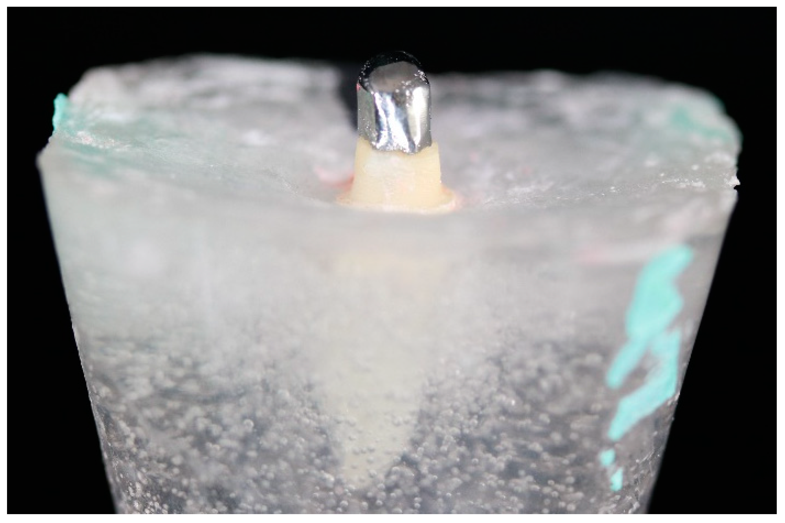
2.2.6. Post-and-Core Cementation

Resin luting cement, Rely X Unicem resin cement (3M ESPE; Seefeld, Germany), was used to cement all post and cores. The cement was mixed according to the manufacturer’s instructions and was coated on the post. Cement was also applied into the root canals by attaching an elongation tip to the nozzle. After that, posts were gently inserted into the root canals to reduce the hydrostatic pressure; they were positioned in place under firm finger pressure, and excess cement was removed (Figure 6). Then, specimens of each group (n = 20) were kept in normal saline for 24 h in a refrigerator before testing. Thermocycling (Thermocycling test apparatus; Durant) was performed for all specimens in 2 water tanks (cold, warm) by immersion at the temperatures of 5 °C and 55 °C and fixed time intervals (16 s cold, 16 s warm) for a total of 6000 cycles, which represented 7 months of clinical use [41].

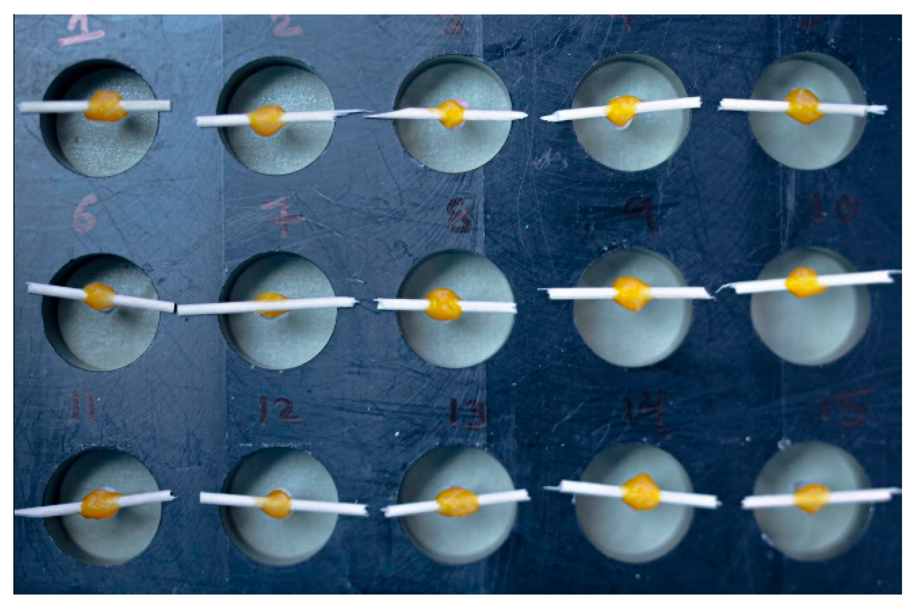
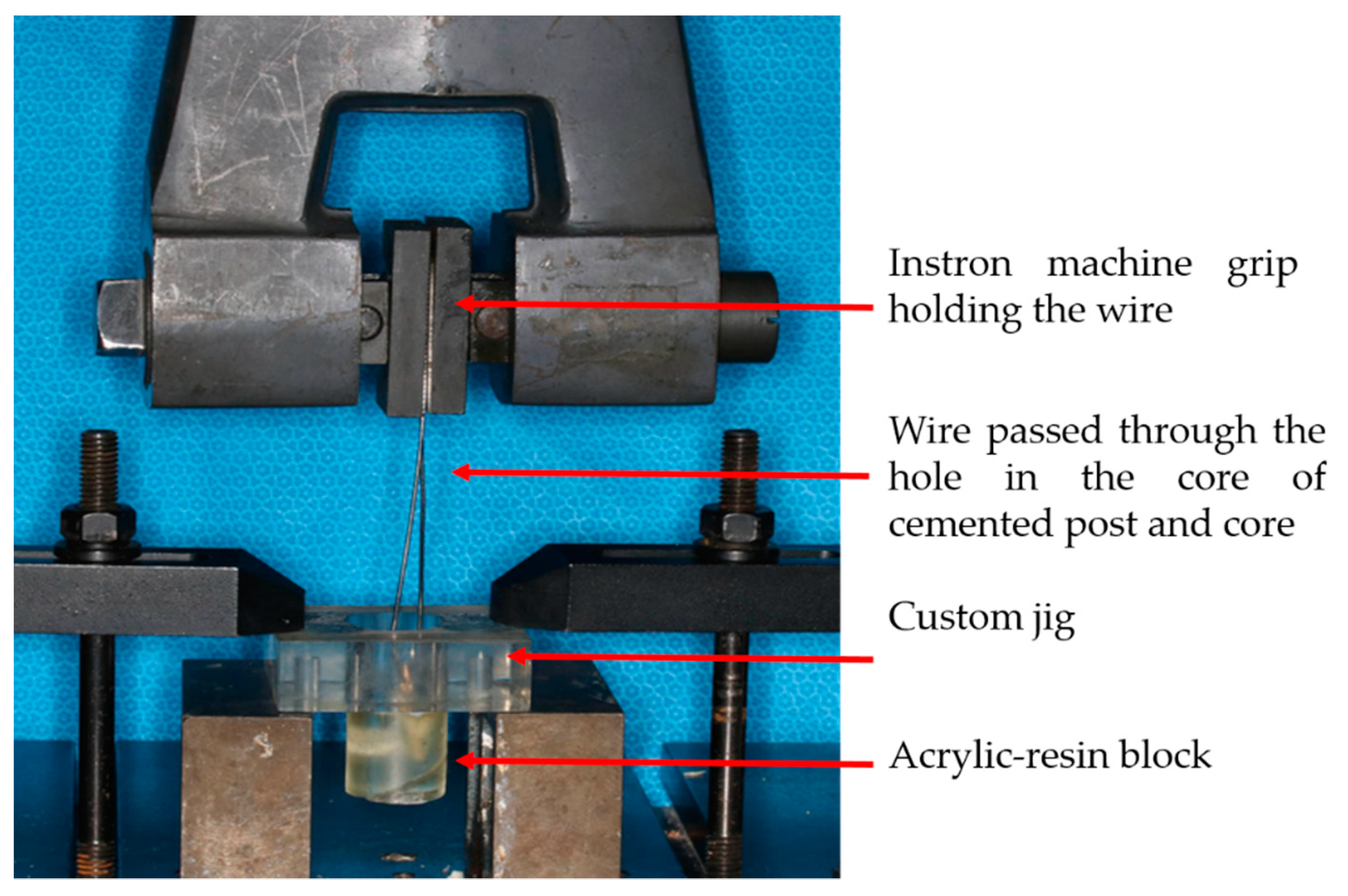
2.3. Placing Specimens on the Measuring Machine (Instron Testing Machine)

Each tooth with a post and core was subjected to a pull-out test by a universal Instron testing machine (Instron Corp.) at a crosshead speed of 0.5 mm/min. The device was calibrated before placing each sample.
To standardize the location and direction of the specimens in the machine, samples were placed in a custom-made, self-aligning device. A hook-shaped attachment was passed through the hole created in the custom post and core, which was attached to the Instron testing machine (ITM). The acrylic-resin blocks held the teeth securely during retention testing (Figure 7). Force was applied until the cemented post was removed from the prepared post space. The retention values were recorded as the amount of force required to dislodge the post and core from the prepared post space [30]. Test specimens were considered to have failed when the post and core separated from the tooth.

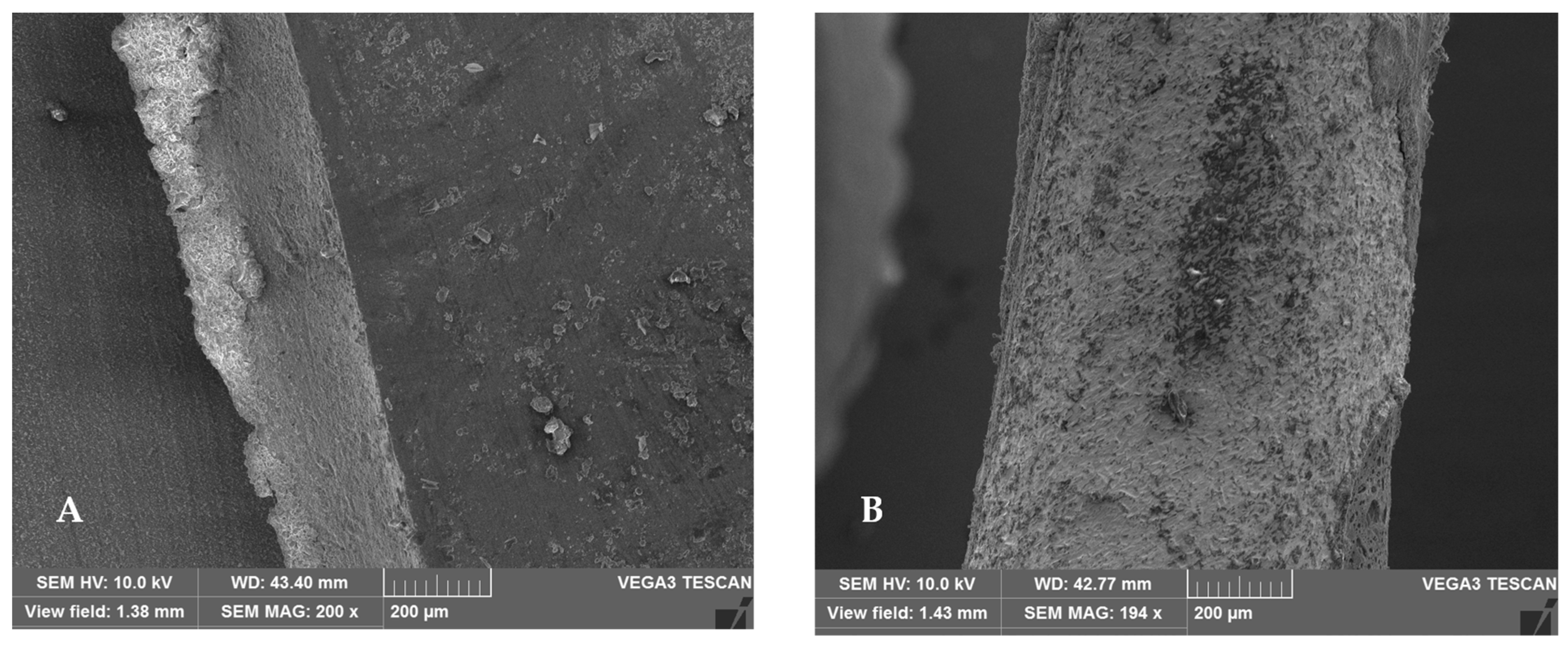
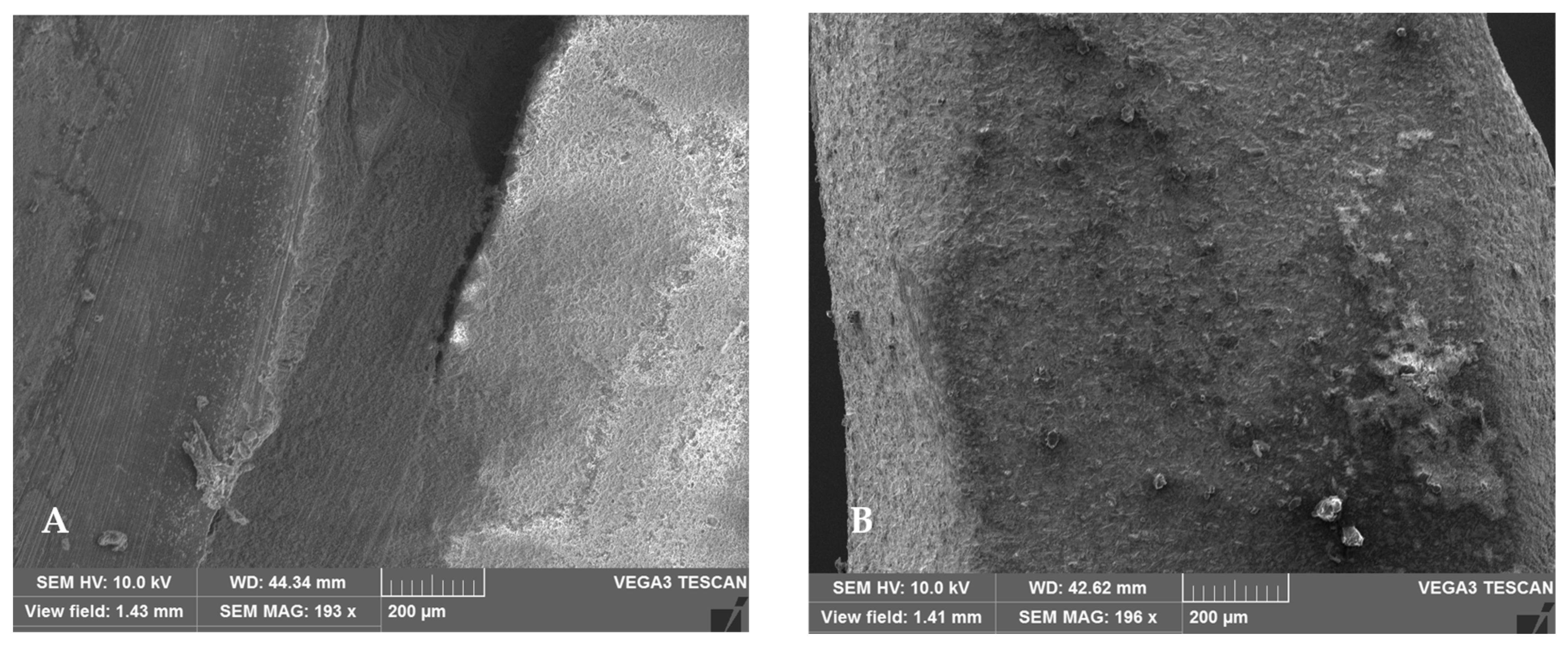
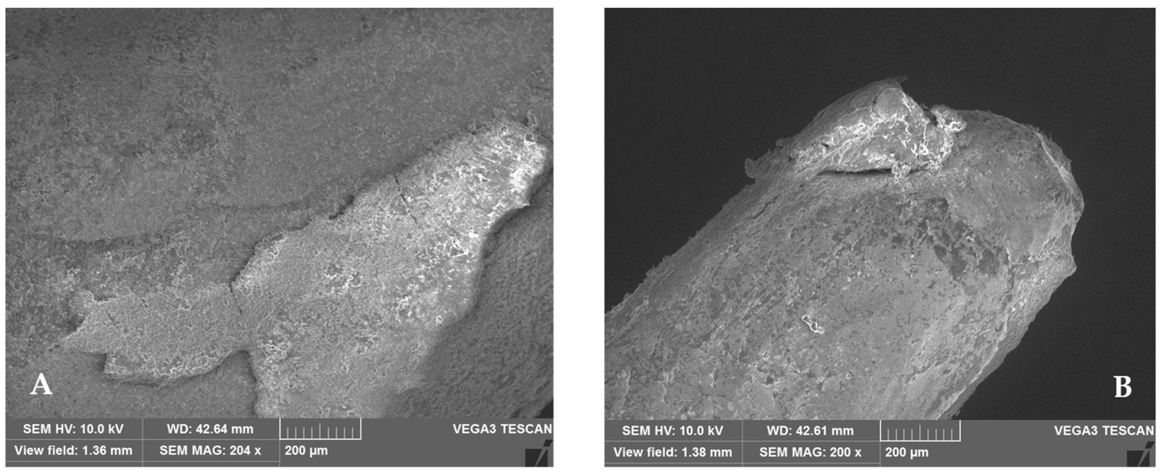
2.4. Scanning Electron Microscopy (SEM) Analysis

The modes of failure were classified as follows: (1) adhesive (clean break at the bond), (2) cohesive (full break in the base material or tooth), and (3) mixed (combination of adhesive and cohesive failure modes). A precision low-speed Techcut 4tm saw (ALLIED High-Tech Product, Inc., Compton, CA, USA) was used to slice the roots along the long axis under constant water irrigation. A stereomicroscope at 50× magnification and a scanning electron microscope (SEM) (FEI QUANTA 250 FEG, Thermo Fisher Scientific, Waltham, MA, USA) were used to examine the sectioned root canals, and displaced posts were examined using a stereomicroscope at the same magnification [42].

2.5. Statistical Analysis
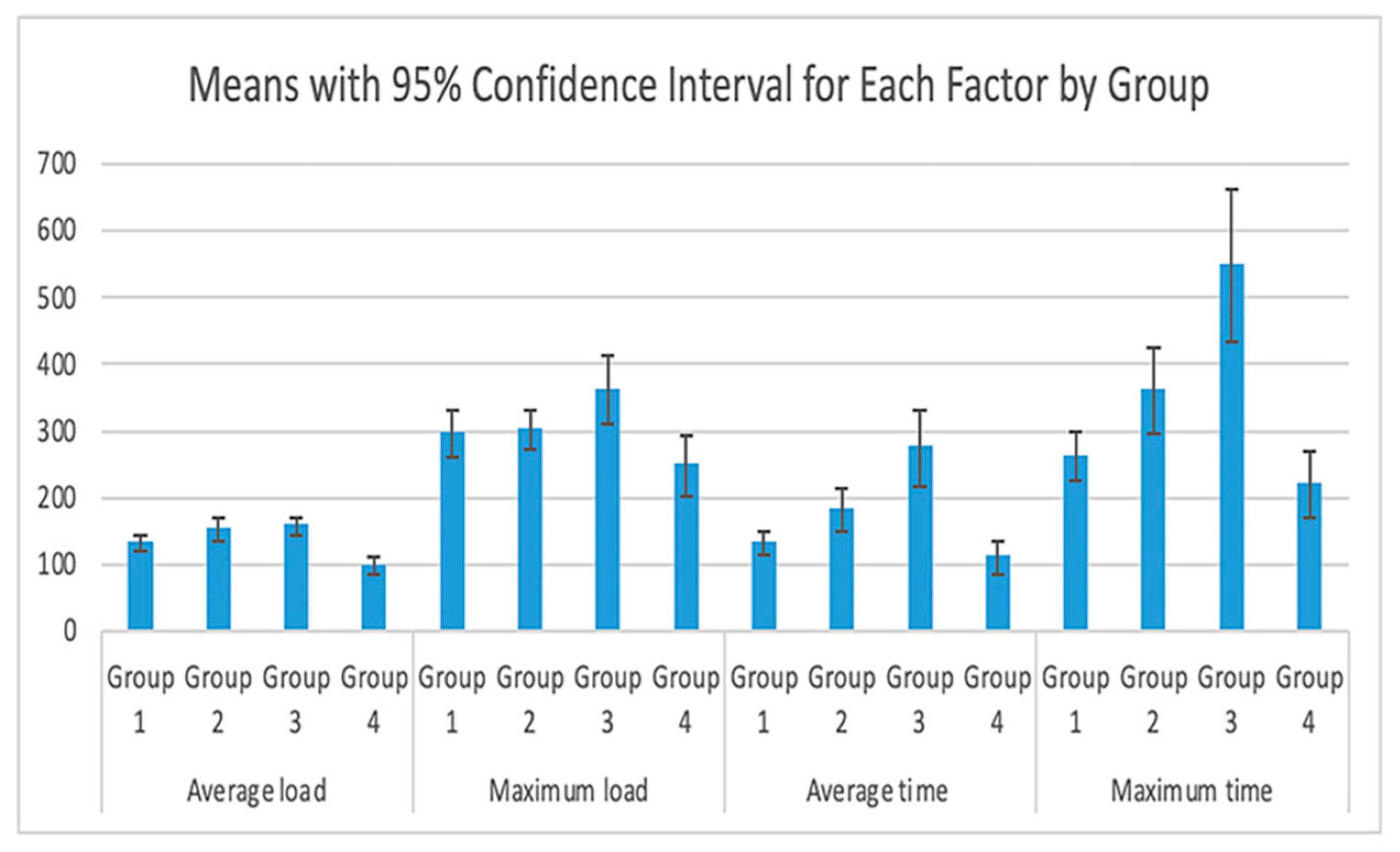
The descriptive statistics were reported as means ± standard deviation and medians with minimum and maximum values for all variables for each dental-material group. The one-way ANOVA procedure was used to test if there was a difference in total time and average load among the treatment groups [43]. Post hoc tests were performed using Tukey’s adjustment for multiple comparisons. The Games–Howell test was used for multiple comparisons instead of Tukey’s test if the assumption of equal variances was not assumed.
The independent-sample Mann–Whitney U test with Bonferroni adjustment was used to compare the medians of the displacement scores of each group with the gold-standard control group. Statistical analyses were performed using IBM SPSS Statistics (Version 25; IBM Corporation 1989, 2018. IBM Corp., Armonk, NY, USA).

3. Results

Descriptive statistics are given as means with 95% confidence intervals (CIs) for the variables of maximum total time, average total time, average load, and maximum load for each group (Table 2).
Using the one-way ANOVA with Tukey’s adjustment, significant differences in the average-load variables were observed between the four different groups, with higher values recorded for Group 3 (CPTPC) in comparison with all other groups (Figure 8).
The mean difference values are shown in Table 2. The load values necessary to create failure in Group 4 (CMZPCs) were lower when compared with those of Group 3 (CPTPCs), Group 2 (CMTPCs), and Group 1 (CCNPPCs); furthermore, all groups showed different distributions at the time of failure and were statistically significantly higher for Group 3 (CPTPCs) and Group 2 (CMTPCs) (p < 0.05). Group 3 (CPTPCs) and Group 1 (CCNPPC group) required loads of 361.5 N and 295.9 N, respectively, to fail, and the difference was statistically significant (p < 0.05). When considering Group 4 (CMZPC group) and Group 3 (CPTPCs), they required loads of 361.5 N and 248.1 N, respectively, and this difference was statistically significant (p < 0.05). The differences in the averages of maximum time and average total time between Group 3 (CPTPCs), Group 2 (CMTPCs), Group 1 (CCNPPCs), and Group 4 (CMZPCs) were significant (p < 0.05), with higher values recorded for Group 3 (CPTPCs) in comparison with all other groups (Figure 8).
Using the Mann–Whitney U test with Bonferroni adjustment, no significant differences in the medians of the displacement scores were observed between each group and the control group (Group 1), as shown in Table 3. All Bonferroni adjusted p-values were >0.05.
The failure mode varied between the groups, according to the scanning-electron-microscopy images. The majority of failures for all tested custom post-and-core systems were cohesive failures, except for Group 4, which revealed a mixed type of failure, as shown in Figure 9, Figure 10, Figure 11 and Figure 12.

4. Discussion

This study evaluated the differences in the retention of different types of custom post and cores. Significant differences in retention were observed when the groups were compared. Therefore, the tested null hypothesis could be rejected; however, retention varied according to the fabrication technique and material used for manufacturing custom post and cores.
Several studies have reported acceptable clinical outcomes with CCNPPCs, which have been used for many years; hence, they were used as the control group in this study [2,4,5,6,7,8,18]. Advancements in materials science and the use of CAD/CAM have transformed the approach to fabricating indirect restorations in the field of prosthodontics, including the fabrication of custom post and cores. These new materials are easier to fabricate when compared with non-precious alloys. The current literature lacks evidence to support that the newly introduced custom-printed and -milled titanium post and cores offer comparable retention and effectiveness.
Various studies have reported that the success of teeth restored with custom post and cores correlated with the design, technique, and material used for the fabrication of the post and cores [6,7,42,44,45]. Recently, Wei Liu et al. found that post and cores fabricated with the CAD-CAM milled technique using a cobalt–chromium alloy could be an alternative to conventional casting for metal-post-and-core fabrication [46]. However, the retention of CAD/CAM milled or printed post and cores was neither evaluated nor tested.
The results of the present study showed that there was a significant difference (p < 0.05) between the retention of teeth restored with custom-printed and -milled titanium post and cores and that of teeth restored with CCNPPCs and CMZPCs. The retention difference between the two groups of custom-printed and -milled titanium post and cores was not significant (p > 0.05), and the CMZPC group revealed significantly lower retention (p < 0.05). The possible reason for the difference between custom post and cores and the retention of the zirconia group could lie in the post-surface configuration and roughness of the printed titanium post, which allows the post-and-core material to form micromechanical retention locks, whereas the smooth surface of zirconia reduces mechanical retention. Maya et al. reported similar results, with metal posts revealing significantly greater retention (495.5 N ± 75.9 N) than zirconia posts (241 ± 89.3 N) [47]. In addition, the study by Cohen et al. reported that zirconia posts had extremely low retention values (104.5 ± 34.8) [48]. These findings are consistent with the results of our study. Previous research has already demonstrated that resin cement can provide greater retention than non-resin cement [49,50]. However, this observation was not confirmed in this study, as we did not evaluate post and cores cemented with non-resin cements.
The finishing and polishing of post-and-core surfaces may improve the fit; however, this could affect the retention. It was noted that among the custom post and cores, those that were made from printed titanium required less adjustment and showed a better fit than the other groups. Studies have reported that differences in retention may also be due to differences in the adaptation of the posts [51,52,53]. Poor adaptation can lead to an increase in resin-matrix cement layer thickness which can increase in cracks, pores, and micro-spaces. These structural defects can cause stress concentration leading to a reduction in interface strength [51,52,53]. Amin et al. [54] reported that a higher volume of resin cement can also cause higher polymerization shrinkage leading to poor bond strength.
To date, due to the complexity of casting post and cores and the wide range of available materials, few studies have quantitatively evaluated the fit and accuracy of custom post and cores using micro-CT scanning, nor have they evaluated the fit and accuracy of custom post and cores via a visual inspection or direct measurements of the gap filled with cement material, as the latter has only been used to evaluate the internal fit and adaption of dental restorations [55,56].
The limitations of this study and recommendations for future ones are listed below:
  • Thermocycling was used for a short period. Further studies with longer thermocycling periods should be conducted;
  • In the current study, only one type of luting cement was used. Additional studies using different types of cements should be performed to assess the effect of the type of cement on the retention of these post and cores;
  • The effects of saliva and temperature changes in the oral cavity were not replicated in this study. The simulated clinical situations might have affected the results; hence, further studies that simulate the oral environment are recommended.

5. Conclusions

Within the limitations of the present study and based on the findings, it can be concluded that custom-printed and -milled titanium post and cores revealed significantly higher retention and bonding to resin cement than CMZPCs and similar to the control group. The CMZPCs revealed the lowest retention and adhesion to the resin cement. Therefore, the use of titanium in the fabrication of printed and milled post and cores was effective in terms of retention and could be an alternative material of choice to non-precious alloys. Further studies with in vivo testing are required to confirm the conclusions of this study.

Author Contributions

Conceptualization, H.A., R.J., M.T.K., S.A.V. and S.D.; methodology, H.A., R.J., M.T.K., S.A.V., S.D. and A.A.A.; software, H.A., S.A.V., S.D. and A.A.A.; validation, H.A., R.J., M.T.K., S.A.V. and S.D.; formal analysis, H.A., R.J., M.T.K., S.A.V., S.D. and A.A.A.; investigation, H.A., R.J., M.T.K., S.A.V. and S.D.; resources, H.A., R.J., M.T.K., S.A.V., M.E.S., S.J. and S.D.; data curation, H.A., R.J., M.T.K., S.A.V. and S.D.; writing—original draft preparation, H.A., R.J., M.T.K., S.A.V., S.D., M.E.S., S.J. and A.A.A.; writing—review and editing, H.A., R.J., M.T.K., S.A.V., S.D., M.E.S., S.J. and A.A.A.; visualization, H.A., R.J., M.T.K., S.A.V., M.E.S., S.J. and S.D.; supervision, H.A., R.J., M.T.K., S.A.V. and S.D.; project administration, H.A., S.A.V., S.D., M.E.S., S.J. and A.A.A. All authors have read and agreed to the published version of the manuscript.

Funding

This research received no external funding.

Institutional Review Board Statement

IRB# 5220414.

Informed Consent Statement

Not applicable.

Data Availability Statement

The data that support the findings of this study are available from the corresponding authors upon reasonable request.

Acknowledgments

The authors thank and acknowledge Ozgul Yasar-Inceoglu (California State University, Chico, CA, USA) for support with scanning electron microscopy in this investigation.

Conflicts of Interest

The authors declare no conflict of interest.

References

  1. The Glossary of Prosthodontic Terms: Ninth Edition. J. Prosthet. Dent. 2017, 117, e1–e105. [CrossRef] [Green Version]
  2. Raedel, M.; Fiedler, C.; Jacoby, S.; Boeing, K.W. Survival of teeth treated with cast post and cores: A retrospective analysis over an observation period of up to 19.5 years. J. Prosthet. Dent. 2015, 114, 40–45. [Google Scholar] [CrossRef] [PubMed]
  3. Goto, Y.; Nicholls, J.I.; Phillips, K.M.; Junge, T. Fatigue resistance of endodontically treated teeth restored with three dowel-and-core systems. J. Prosthet. Dent. 2005, 93, 45–50. [Google Scholar] [CrossRef]
  4. Schillingburg, H.T.; Hobo, S.; Whitsett, L.D.; Jacobi, R.; Brackett, S.E. Fundamentals of Fixed Prosthodontics, 3rd ed.; Quintessence Publishing: Chicago, IL, USA, 1997; p. 40. [Google Scholar]
  5. Fernandes, A.S.; Shetty, S.; Coutinho, I. Factors determining post selection: A literature review. J. Prosthet. Dent. 2003, 90, 556–562. [Google Scholar] [CrossRef]
  6. Bergman, B.; Lundquist, P.; Sjögren, U. Restorative and endodontic results after treatment with cast posts and cores. J. Prosthet. Dent. 1989, 61, 10–15. [Google Scholar] [CrossRef]
  7. Creugers, N.H.; Mentink, A.G.; Käyser, A.F. An analysis of durability data on post and core restorations. J. Dent. 1993, 21, 281–284. [Google Scholar] [CrossRef]
  8. Pontius, O.; Hutter, J.W. Survival rate and fracture strength of incisors restored with different post and core systems and endodontically treated incisors without coronoradicular reinforcement. J. Endod. 2002, 28, 710–715. [Google Scholar] [CrossRef]
  9. Venkataraman, K.J.; Thanapathi, S.; Balasubramanian, S.; Gandhi, S.A.; Sarojinikutty, A.C. Fracture Resistance of Titanium, Chrome-Cobalt, and Gold Alloy as Post and Core Materials: A Comparative Evaluation. J. Pharm. Bioallied. Sci. 2020, 12 (Suppl. S1), S583–S588. [Google Scholar] [CrossRef] [PubMed]
  10. Jung, R.E.; Kalkstein, O.; Sailer, I.; Roos, M.; Hämmerle, C.H. A comparison of composite post buildups and cast gold post-and-core buildups for the restoration of nonvital teeth after 5 to 10 years. Int. J. Prosthodont. 2007, 20, 63–69. [Google Scholar]
  11. Al-Qarni, F.D. Customized Post and Cores Fabricated with CAD/CAM Technology: A Literature Review. Int. J. Gen. Med. 2022, 15, 4771–4779. [Google Scholar] [CrossRef] [PubMed]
  12. Piangsuk, T.; Dawson, D.V.; El-Kerdani, T.; Lindquist, T.J. The Accuracy of Post and Core Fabricated with Digital Technology. J. Prosthodont. 2022. ahead of print. [Google Scholar] [CrossRef] [PubMed]
  13. Khiavi, H.A.; Habibzadeh, S.; Safaeian, S.; Eftekhar, M. Fracture Strength of Endodontically treated Maxillary Central Incisors restored with Nickel Chromium and Nonprecious Gold Alloy Casting Post and Cores. J. Contemp. Dent. Pract. 2018, 19, 560–567. [Google Scholar] [CrossRef] [PubMed]
  14. Kanduti, D.; Korat, L.; Kosec, T.; Legat, A.; Ovsenik, M.; Kopač, I. Comparison between accuracy of posts fabricated using a digital CAD/CAM technique and a conventional direct technique. Int. J. Prosthodont. 2021, 34, 212–220. [Google Scholar] [CrossRef] [PubMed]
  15. Libonati, A.; Di Taranto, V.; Gallusi, G.; Montemurro, E.; Campanella, V. CAD/CAM customized glass fiber post and core with digital intraoral impression: A case report. Clin. Cosmet. Investig. Dent. 2020, 12, 17–24. [Google Scholar] [CrossRef] [PubMed] [Green Version]
  16. Lee, J.H. Fabricating a custom zirconia post-and-core without a post-and-core pattern or a scan post. J. Prosthet. Dent. 2018, 120, 186–189. [Google Scholar] [CrossRef] [PubMed]
  17. Pang, J.; Feng, C.; Zhu, X.; Liu, B.; Deng, T.; Gao, Y.; Li, Y.; Ke, J. Fracture behaviors of maxillary central incisors with flared root canals restored with CAD/CAM integrated glass fiber post-and-core. Dent. Mater. J. 2019, 38, 114–119. [Google Scholar] [CrossRef] [PubMed] [Green Version]
  18. Balkenhol, M.; Wöstmann, B.; Rein, C.; Ferger, P. Survival time of cast post and cores: A 10-year retrospective study. J. Dent. 2007, 35, 50–58. [Google Scholar] [CrossRef]
  19. Heydecke, G.; Butzm, F.; Hussein, A.; Strub, J.R. Fracture strength after dynamic loading of endodontically treated teeth restored with different post-and-core systems. J. Prosthet. Dent. 2002, 87, 438–445. [Google Scholar] [CrossRef] [PubMed]
  20. Awad, M.A.; Marghalani, T.Y. Fabrication of a custom-made ceramic post and core using CAD-CAM technology. J. Prosthet. Dent. 2007, 98, 161–162. [Google Scholar] [CrossRef]
  21. Lee, J.H.; Sohn, D.S.; Lee, C.H. Fabricating a fiber-reinforced post and zirconia core with CAD/CAM technology. J. Prosthet. Dent. 2014, 112, 683–685. [Google Scholar] [CrossRef] [PubMed]
  22. Baba, N.Z.; Golden, G.; Goodacre, C.J. Nonmetallic prefabricated dowels: A review of compositions, properties, laboratory, and clinical test results. J. Prosthodont. 2009, 18, 527–536. [Google Scholar] [CrossRef] [PubMed]
  23. Ozkurt, Z.; Işeri, U.; Kazazoğlu, E. Zirconia ceramic post systems: A literature review and a case report. Dent. Mater. J. 2010, 29, 233–245. [Google Scholar] [CrossRef] [PubMed] [Green Version]
  24. Massa, F.; Dias, C.; Blos, C.E. Resistance to fracture of mandibular premolars restored using post and-core systems. Quintessence Int. 2010, 41, 49–57. [Google Scholar] [PubMed]
  25. Ayad, M.F.; Bahannan, S.A.; Rosenstiel, S.F. Influence of Irrigant, Dowel Type, and RootReinforcing Material on Fracture Resistance of Thin-Walled Endodontically Treated Teeth. J. Prosthodont. 2011, 20, 180–189. [Google Scholar] [CrossRef]
  26. Alhajj, M.N.; Qi, C.H.; Sayed, M.E.; Johari, Y.; Ariffin, Z. Fracture Resistance of Titanium and Fiber Dental Posts: A Systematic Review and Meta-Analysis. J. Prosthodont. 2022, 31, 374–384. [Google Scholar] [CrossRef]
  27. Ranjkesh, B.; Leo, M.; Vafaei, A.; Lovschall, H. Pull-Out Bond Strength of Titanium Post Cemented with Novel Fast-Setting Calcium Silicate Cement. Eur. Endod. J. 2021, 6, 314–318. [Google Scholar] [CrossRef] [PubMed]
  28. International Standard 28399; Dentistry—Products for External Tooth Bleaching. International Organization for Standardization: Geneva, Switzerland, 2011.
  29. Rho, J.Y.; Ashman, R.B.; Turner, C.H. Young’s modulus of trabecular and cortical bone material: Ultrasonic and microtensile measurements. J. Biomech. 1993, 26, 111–119. [Google Scholar] [CrossRef]
  30. Khaledi, A.A.; Sheykhian, S.; Khodaei, A. Evaluation of Retention of two Different Cast Post-Core Systems and Fracture Resistance of the Restored Teeth. J. Dent. 2015, 16, 121–128. [Google Scholar]
  31. Sorensen, J.A.; Engelman, M.J. Ferrule design and fracture resistance of endodontically treated teeth. J. Prosthet. Dent. 1990, 63, 529–536. [Google Scholar] [CrossRef]
  32. Eissman, H.F.; Radke, R.A. Postendodontic restoration. In Pathways of the Pulp, 4th ed.; Cohen, S., Burns, R.C., Eds.; Mosby: St. Louis, CV, USA, 1987; pp. 640–643. [Google Scholar]
  33. Bakirtzoglou, E.; Kamalakidis, S.N.; Pissiotis, A.L.; Michalakis, K. In vitro assessment of retention and resistance failure loads of complete coverage restorations made for anterior maxillary teeth restored with two different cast post and core designs. J. Clin. Exp. Dent. 2019, 11, e225–e230. [Google Scholar] [CrossRef]
  34. De-Deus, G.; Belladonna, F.G.; Silva, E.J.N.L.; Souza, E.M.; Carvalhal, J.C.A.; Perez, R.; Lopes, R.T.; Versiani, M.A. Micro-CT assessment of dentinal micro-cracks after root canal filling procedures. Int. Endod. J. 2017, 50, 895–901. [Google Scholar] [CrossRef]
  35. Sorensen, J.A.; Martinoff, J.T. Clinically significant factors in dowel design. J. Prosthet. Dent. 1984, 52, 28–35. [Google Scholar] [CrossRef]
  36. Goodacre, C.J.; Spolnick, K.J. The prosthodontic management of endodontically treated teeth: A literature review. Part II. Maintaining the apical seal. J. Prosthod. 1995, 4, 51–53. [Google Scholar] [CrossRef]
  37. Morgano, S.M. Restoration of pulpless teeth: Application of traditional principles in present and future contexts. J. Prosthet. Dent. 1996, 75, 375–380. [Google Scholar] [CrossRef]
  38. Mattison, G.D.; Delivanis, P.D.; Thacker, R.W., Jr.; Hassell, K.J. Effect of post preparation on the apical seal. J. Prosthet. Dent. 1984, 51, 785–789. [Google Scholar] [CrossRef]
  39. Sreedevi, S.; Sanjeev, R.; Raghavan, R.; Abraham, A.; Rajamani, T.; Govind, G.K. An In Vitro Study on the Effects of Post-Core Design and Ferrule on the Fracture Resistance of Endodontically Treated Maxillary Central Incisors. J. Int. Oral Health 2015, 7, 37–41. [Google Scholar] [PubMed]
  40. Blatz, M.B.; Chiche, G.; Holst, S.; Sadan, A. Influence of surface treatment and simulated aging on bond strengths of luting agents to zirconia. Quintessence Int. 2007, 38, 745–753. [Google Scholar]
  41. Amaral, F.L.; Colucci, V.; Palma-Dibb, R.G.; Corona, S.A. Assessment of in vitro methods used to promote adhesive interface degradation: A critical review. J. Esthet. Restor. Dent. 2007, 19, 340–353. [Google Scholar] [CrossRef]
  42. Stegaroiu, R.; Yamada, H.; Kusakari, H.; Miyakawa, O. Retention and failure mode after cyclic loading in two post and core systems. J. Prosthet. Dent. 1996, 75, 506–511. [Google Scholar] [CrossRef]
  43. Ahmad, M.; Tarmeze, A.; Abdul Rasib, A. Capability of 3D Printing Technology in Producing Molar Teeth Prototype. Int. J. Eng. Appl. 2020, 8, 64–70. [Google Scholar] [CrossRef]
  44. Hudis, S.I.; Goldstein, G.R. Restoration of endodontically treated teeth: A review of the literature. J. Prosthet. Dent. 1986, 55, 33–38. [Google Scholar] [CrossRef]
  45. Bolhuis, H.P.B.; De Gee, A.J.; Feilzer, A.J.; Davidson, C.L. Fracture strength of different core build-up designs. Am. J. Dent. 2001, 14, 286–290. [Google Scholar]
  46. Liu, W.; Qing, H.; Pei, X.; Wang, J. Internal adaptation of cobalt-chromium posts fabricated by selective laser melting technology. J. Prosthet. Dent. 2019, 121, 455–460. [Google Scholar] [CrossRef] [PubMed]
  47. Maya, A.; Millsten, P.; Freeman, Y. Determining post-core retention of smooth surface metal, non-metal posts. J. Dent. Res. 1998, 77, 160. [Google Scholar]
  48. Cohen, B.I.; Pagnillo, M.K.; Newman, I.; Musikant, B.L.; Deutsch, A.S. Retention of three endodontic posts cemented with five dental cements. J. Prosthet. Dent. 1998, 79, 520–525. [Google Scholar] [CrossRef]
  49. Liberman, R.; Ben-Amar, A.; Urstein, M.; Gontar, G.; Fitzig, S. Conditioning of root canals prior to dowel cementation with composite luting cement and two dentine adhesive systems. J. Oral Rehabil. 1989, 16, 597–602. [Google Scholar] [CrossRef] [PubMed]
  50. Chapman, K.W.; Worley, J.L.; von Fraunhofer, J.A. Effect of bonding agents on retention of posts. Gen. Dent. 1985, 33, 128–130. [Google Scholar] [PubMed]
  51. Fernandes, V.; Silva, A.S.; Carvalho, O.; Henriques, B.; Silva, F.S.; Özcan, M.; Souza, J.C.M. The resin-matrix cement layer thickness resultant from the intracanal fitting of teeth root canal posts: An integrative review. Clin. Oral Investig. 2021, 25, 5595–5612. [Google Scholar] [CrossRef] [PubMed]
  52. Penelas, A.G.; Piedade, V.M.; Borges, A.C.; Poskus, L.T.; da Silva, E.M.; Guimarães, J.G. Can cement film thickness influence bond strength and fracture resistance of fiber reinforced composite posts? Clin. Oral Investig. 2016, 20, 849–855. [Google Scholar] [CrossRef]
  53. Gomes, G.M.; Rezende, E.C.; Gomes, O.M.; Gomes, J.C.; Loguercio, A.D.; Reis, A. Influence of the resin cement thickness on bond strength and gap formation of fiber posts bonded to root dentin. J. Adhes. Dent. 2014, 16, 71–88. [Google Scholar]
  54. Amin, R.A.; Mandour, M.H.; Abd El-Ghany, O.S. Fracture strength and nanoleakage of weakened roots reconstructed using relined glass fiber-reinforced dowels combined with a novel prefabricated core system. J. Prosthodont. 2014, 23, 484–494. [Google Scholar] [CrossRef] [PubMed]
  55. Tamac, E.; Toksavul, S.; Toman, M. Clinical marginal and internal adaptation of CAD/CAM milling, laser sintering, and cast metal ceramic crowns. J. Prosthet. Dent. 2014, 112, 909–913. [Google Scholar] [CrossRef] [PubMed]
  56. Scotti, N.; Forniglia, A.; Bergantin, E.; Paolino, D.S.; Pasqualini, D.; Berutti, E. Fibre post adaptation and bond strength in oval canals. Int. Endod. J. 2014, 47, 366–372. [Google Scholar] [CrossRef] [PubMed]
Figure 1. Teeth were mounted in acrylic-resin blocks using a test mount former.
Applsci 12 11896 g001
Figure 2. Preparation of tooth to have a 2 mm ferrule and a 1 mm shoulder finish line. (A) Occlusal view. (B) Lateral view.
Applsci 12 11896 g002
Figure 3. Pattern resin post-and-core build-up.
Applsci 12 11896 g003
Figure 4. Sample of STL file of the proposed design of the specimen.
Applsci 12 11896 g004
Figure 5. (A) Custom-cast non-precious post and core. (B) Custom-milled titanium post and core. (C) Custom-printed titanium post and core. (D) Custom-milled zirconia post and core.
Applsci 12 11896 g005
Figure 6. View of a cemented post and core.
Applsci 12 11896 g006
Figure 7. Specimen of mounted post and core in testing assembly for retention test.
Applsci 12 11896 g007
Figure 8. Comparison between fabrication materials with the control group.
Applsci 12 11896 g008
Figure 9. Scanning-electron-microscope image of (A) Sectioned root canal, and (B) displaced post, at 200× magnification, showing cohesive failure mode in Group 1 (CCNPPCs) sample at root/post interface.
Applsci 12 11896 g009
Figure 10. Scanning-electron-microscope image of (A) Sectioned root canal, and (B) displaced post, at 200× magnification, showing cohesive failure mode in Group 2 (CMTPCs) sample at root/post interface.
Applsci 12 11896 g010
Figure 11. Scanning-electron-microscope image of (A) Sectioned root canal, and (B) displaced post, at 200× magnification, showing cohesive failure mode in Group 3 (CPTPCs) sample at root/post interface.
Applsci 12 11896 g011
Figure 12. Scanning-electron-microscope image of (A) Sectioned root canal, and (B) displaced post, at 200× magnification, showing cohesive failure mode in Group 4 (CMZPCs) sample at root/post interface.
Applsci 12 11896 g012
Table 1. Commercial names and details of materials used in the study.
GroupMaterial Trade NameManufacturerMain CompositionManufacturing Technique Used
Group 1NPGAalba Dent, Inc., Fulton Drive, Fairfield, CA, USACu, 80.7%; Al, 7.8%; Ni, 4.3% Casting (lost-wax technique)
Group 2KERA Ti-5 DiscEisenbacher Dentalwaren ED GmbH
Dr.-Konrad-Wiegand-Straße, Wörth am Main, Germany
Ti, 89%; Al, 6.4%; V, 4.1%Milling
Group 3Ti-6Al-4VRenovis Surgical, West Lugonia Ave, Austin, TX, USATi, 89%; Al, 6.4%; V, 4.1%3D printing
Group 4BruxZir Full-Strength ZirconiaBruxZir; Glidewell Laboratory Inc., Newport Beach, CA, USAMonolithic zirconia (zirconium oxide)Milling
Table 2. Means with 95% confidence intervals for each factor and differences in clinical factors by the group.
CharacteristicGroup 1 Group 2 Group 3 Group 4 p-ValueDifferences in Clinical Factors by the Group
(Mean Difference)
(95% CI)
Mean (95% CI)Mean (95% CI)Mean (95% CI)Mean (95% CI)
Total Time—Average (Seconds)130.3 (112.6, 148.1)180.4 (147.7, 213.0)274.0 (217.3, 330.6)109.6 (84.0, 135.2)<0.001Total time Average *
Group 1 vs. Group 2: 20.8 (−19.4, 61.0)
Group 1 vs. Group 3: −143.6 (−222.2, −65.1) #
Group 1 vs. Group 4: −50.0 (−98.3, 1.7) #
Group 2 vs. Group 3: −164.4 (−245.8, −83.1) #
Group 2 vs. Group 4: −70.8 (−124.2, −17.4) #
Group 3 vs. Group 4: 93.6 (8.8, 178.5) #
Total Time—Max.
(Seconds)
261.6 (226.1, 297.1)360.7 (295.4, 426.0)547.9 (434.6, 661.1)219.2 (168.0, 270.3)<0.001Total Time Max. *
Group 1 vs. Group 2: 42.5 (−37.9, 122.8)
Group 1 vs. Group 3: −286.2 (−443.3, −129.2) #
Group 1 vs. Group 4: −99.1 (−195.8, −2.4) #
Group 2 vs. Group 3: −328.7 (−491.4, −166.0) #
Group 2 vs. Group 4: −141.6 (−248.3, −34.8) #
Group 3 vs. Group 4: 187.1 (17.4, 356.8) #
Average Load
(N)
131.1 (118.2, 144.0)150.9 (133.5, 168.2)156.5 (142.8, 170.1)96.9 (83.4, 110.5)<0.001Average Load **
Group 1 vs. Group 2: −5.6 (−31.2, 20.1)
Group 1 vs. Group 3: 19.7 (−5.9, 45.4)
Group 1 vs. Group 4: 53.9 (28.3, 79.6)
Group 2 vs. Group 3: 25.3 (−0.30, 51.0)
Group 2 vs. Group 4: 59.5 (33.9, 85.2) #
Group 3 vs. Group 4: 34.2 (8.5, 59.8) #
Max. Load
(N)
295.9 (261.0, 330.8)302.7 (273.1, 332.3)361.5 (309.8, 413.2)248.1 (202.9, 293.3)<0.002Max. Load **
Group 1 vs. Group 2: −6.8 (−65.6, 52.0)
Group 1 vs. Group 3: −65.6 (−146.2, 15.0)
Group 1 vs. Group 4: 47.8 (−25.8, 121.3)
Group 2 vs. Group 3: −58.8 (−132.0, 14.5)
Group 2 vs. Group 4: 54.6 (−18.6, 127.8)
Group 3 vs. Group 4: 113.4 (40.1, 132.0) #
Max.: maximum; CI, confidence interval; Group 1 (CCNPPCs), Group 2 (CMTPCs), Group 3 (CPTPCs), Group 4 (CMZPCs); * Games–Howell test; ** Tukey’s adjusted post hoc test; # p-value significant at p < 0.05.
Table 3. Comparison of different post-and-core fabrication materials with the control group.
Group 1Group 2Group 3Group 4
Median (min–max)Median (min–max)Median (min–max)Median (min–max)
Displacement
(mm)
0.2 (0.1–1.0) 0.5 (0.1–1.0) 0.2 (0.1–0.8)0.1 (0.1–1.0)
p-value significant at p < 0.05.
Publisher’s Note: MDPI stays neutral with regard to jurisdictional claims in published maps and institutional affiliations.

