Article

Linear Dimensional Change in Acrylic Denture Teeth Positions Factored by Different Processing Techniques and Occlusal Forms: An In Vitro Study

by
Mohammed E. Sayed
1,*,
Amit Porwal
1,
Saurabh Jain
1,*,
Abdulkarim Hussain Alshehri
1,
Nasser M. Alqahtani
2,
Ashwaq Hadi Ali Hadadi
3,
Rawan Abdulwadoud Zakri
3,
Sahar Mahdi Musa Zeed
3,
Saswan Ibrahim Nahari
3,
Fatimah H. Alsurayyie
1,
Hatem Alqarni
4,
Saeed M. Alqahtani
2 and
Saad Saleh AlResayes
5
1
Department of Prosthetic Dental Sciences, College of Dentistry, Jazan University, Jazan 45142, Saudi Arabia
2
Department of Prosthodontics, College of Dentistry, King Khalid University, Abha 62529, Saudi Arabia
3
Private Dental Practice, Jazan 45142, Saudi Arabia
4
Restorative and Prosthetic Dental Science Department, College of Dentistry, King Abdullah International Medical Research Center, King Saud Bin Abdulaziz University for Health Science, Riyadh 11545, Saudi Arabia
5
Department of Prosthetic Dental Sciences, College of Dentistry, King Saud University, Riyadh 11545, Saudi Arabia
*
Authors to whom correspondence should be addressed.
Appl. Sci. 2022, 12(14), 7058; https://doi.org/10.3390/app12147058
Submission received: 18 June 2022 / Revised: 4 July 2022 / Accepted: 11 July 2022 / Published: 13 July 2022

Abstract

:
The current literature lacks substantial evidence for the effect of denture base processing techniques and posterior denture tooth forms on denture tooth shifts due to denture base resin polymerization. The aim of this study was to evaluate the combined effect of PMMA-based denture processing techniques (compression packing and injection molding) and posterior tooth forms (semi-anatomic and non-anatomic) on the linear dimensional shift of denture teeth following denture processing in both horizontal and vertical dimensions. Two different complete denture fabrication techniques were used to prepare forty ideal maxillary complete dentures using two different types of posterior tooth forms. The used fabrication techniques were conventional heat polymerized compression packing and injection molding. The posterior tooth forms used in the current study were non-anatomic tooth (0 degrees) and semi-anatomic tooth forms (approximately 20 degrees). Initial linear measurements (vertical and horizontal) were taken from pre-specified points for the central incisor and first molar. Specimens were randomly divided into four groups (n = 10), and denture processing was performed using the two techniques. Final linear measurements were recorded. The linear change in dimension for all six parameters was calculated by deducting the after values from the before values. Since the discrepancies were both positive and negative in magnitude, the absolute value of the difference was taken for further analysis. This value represents the dimensional change. T-tests were used to compare the mean dimensional changes. Furthermore, the mean dimensional changes for all the six parameters were compared using a two-way analysis of variance. The alpha error was set at 5%, and a p-value of less than 0.05 was considered statistically significant. The injection molding technique showed significantly fewer tooth movements in both the vertical and horizontal measurements as compared to the conventional compression packing technique. The non-anatomic tooth showed significantly fewer changes in tooth movement as compared to semi-anatomic teeth in both the compression and injection techniques. This study can guide the selection of a proper processing technique for a particular posterior tooth form, thus minimizing occlusal discrepancies and reducing occlusal corrections during laboratory and clinical remount procedures.

1. Introduction

Complete edentulism, as defined by a loss of all permanent dentition, is a global health condition that was reported to affect a range of 7–69% of adult populations worldwide [1]. Even though implant dentistry has changed the approach of treatment planning for completely edentulous cases, conventional complete dentures remain a valid and sometimes first option, especially when implants are contraindicated in such cases. Balanced complete denture occlusion is crucial for the stability and retention of complete dentures in patients with moderate to severe alveolar ridge resorption [2]. Excessive denture tooth movements due to processing may compromise the form, esthetics and occlusion and, therefore, mean that dentures need to be remade [3]. Several factors have been reported to affect the positional stability of a denture tooth during processing. These factors include the investment method [4], investment material [5], flasking and polymerization technique [6,7], denture base thickness [8], flask closure and cooling method [9], palatal form [10] and box preparation on the heel of the master cast [11]. In addition, Goodacre et al. [12] studied the effect of the denture processing technique on denture tooth movement and found that CAD–CAM technology was the most accurate and reproducible in comparison to conventional packing and injection techniques. The study also reported that the packing technique resulted in higher vertical tooth movement. However, CAD–CAM technology for denture fabrication was only implemented in 10% or less of the cases treated in US dental schools [13]. Therefore, the focus should be directed toward perfecting the techniques that are commonly used in complete denture practice. Jackson and colleagues [14] reported that the presence of acrylic teeth has a significant effect on the accuracy of denture bases fabricated using compression and injection techniques. Several studies have indicated that the injection technique is more accurate with respect to changing vertical dimension and occlusal errors [15,16]. The case-specific selection approach of the posterior tooth form is important for maintaining masticatory efficiency and the success of denture treatment [17]. The available evidence is lacking with regard to the effect of posterior denture tooth form (i.e., cuspal height) on denture tooth movement due to acrylic resin processing. Hence, the goal of this study was to evaluate the combined effect of processing techniques (compression and injection molding) and posterior tooth forms (semi-anatomic and non-anatomic) on linear dimension changes (horizontal and vertical dimensions) in denture tooth positions following processing. The null hypothesis of the study was that the variation in posterior tooth forms and complete denture processing technique has no effect on the dimensional position of anterior and posterior teeth following processing.

2. Materials and Methods

The research protocol was approved by the research board at the College of Dentistry, Jazan University (reference number: CODJU-2003I).

2.1. Materials

In this study, two different complete denture fabrication techniques were used to prepare forty ideal maxillary dentures using two different types of posterior tooth forms. The fabrication techniques used were: conventional heat polymerized compression molding and injection molding. The posterior tooth forms used in the current study were non-anatomic tooth (0 degrees) and semi-anatomic tooth forms (approx. 20 degree). Details of the materials used in the fabrication of maxillary complete dentures are listed in Table 1.

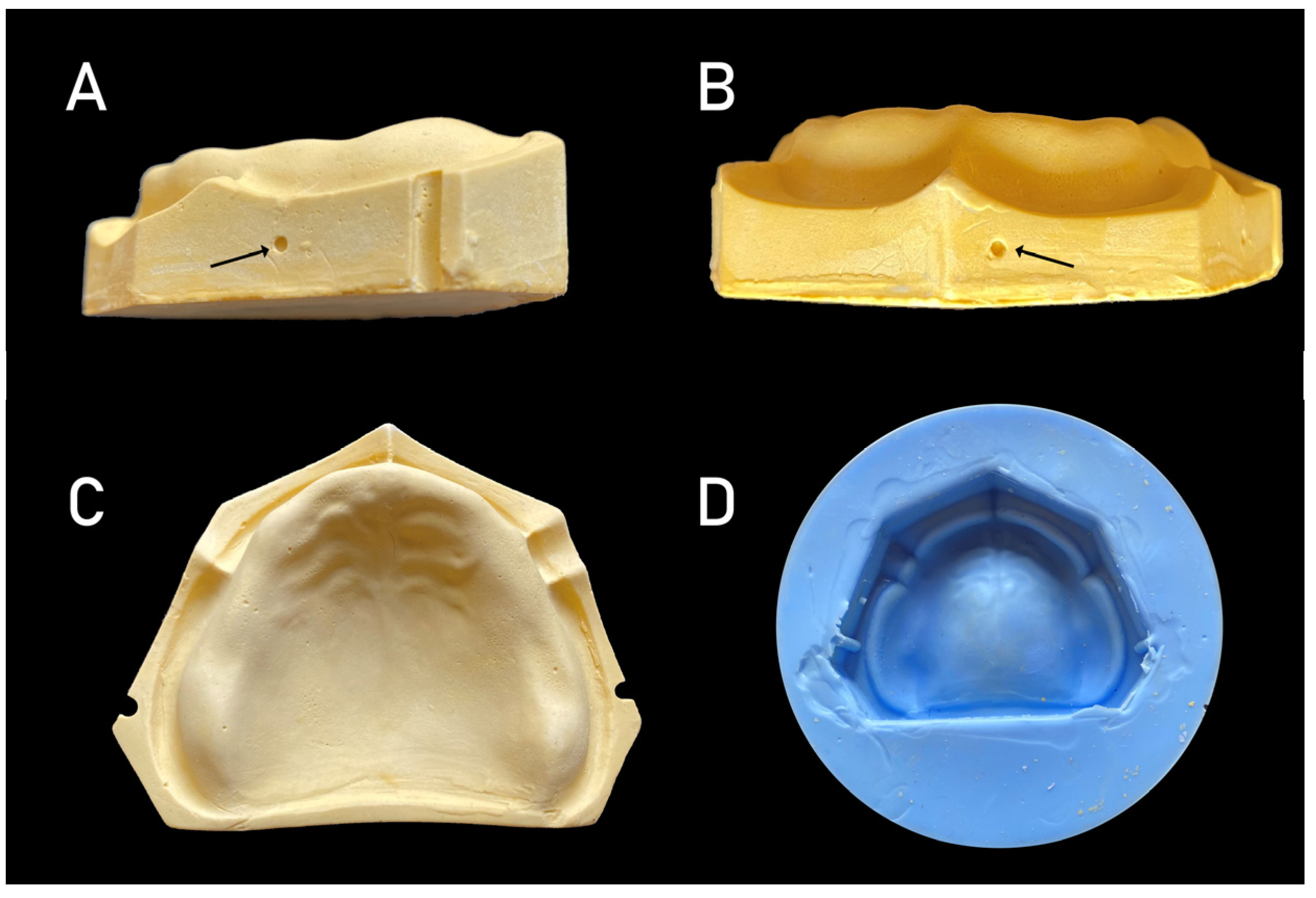
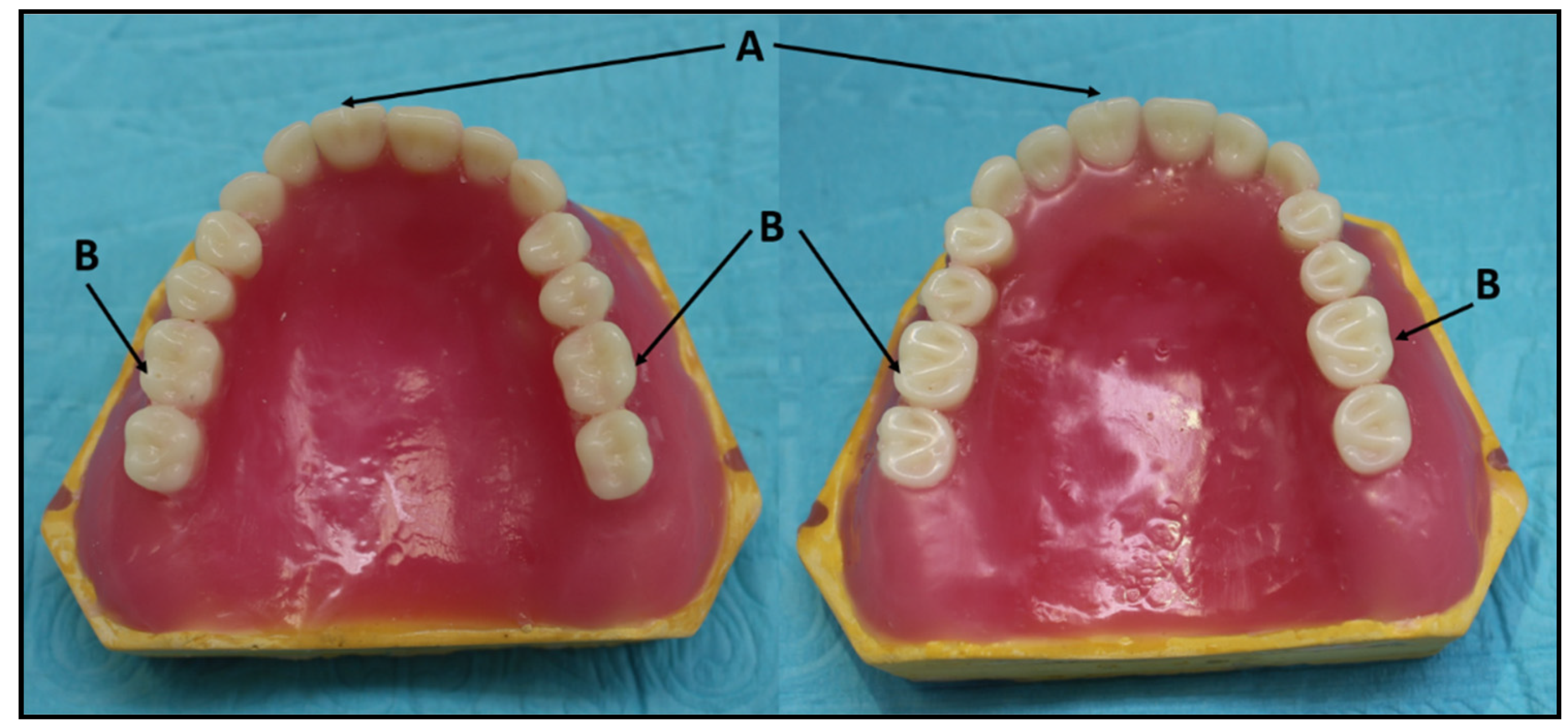
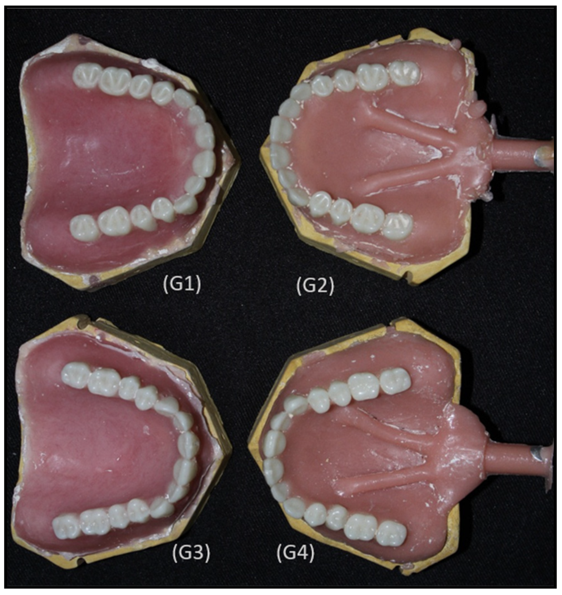
2.2. Pouring of the Cast and the Arrangement of Teeth

To standardize the reference points for measurements, a round bur (# 001/027, Quattro, DFS-Diamon GmbH, Ländenstraße, Riedenburg, Germany) was used to make holes on the land area of the ideal edentulous maxillary cast, corresponding to the disto-buccal cusps of the right and left first molars and maxillary right central incisor. The silicone mold made using this reference cast was used to pour forty ideal casts with grooves in the land area (Figure 1).
Two sheets of modelling wax were adapted on the cast, and ideal teeth arrangements for complete dentures were performed. The arrangements of the teeth on all the casts were obtained by the duplication of the ideal teeth arrangement from the initial cast, i.e., twenty sets of semi-anatomical teeth and twenty non-anatomic teeth. Following this, vertical grooves were placed on the right central incisor incisal edge (tooth number 1.1) (bur (# 114/023, Quattro, DFS-Diamon GmbH, Ländenstraße, Riedenburg, Germany)) and the round holes on the disto-buccal cusps or right and left first molars (tooth number 1.6 and 2.6) (bur (# 001/023, Quattro, DFS-Diamon GmbH, Ländenstraße, Riedenburg, Germany)) (Figure 2).

2.3. Initial Measurements

Linear measurements were taken for all the sets of semi-anatomical and non-anatomical teeth. Vertical measurements were taken between the mesio-buccal grooves of the first molar on both the right and left sides, and a round hole was placed on the land area of the cast and also between the groove placed on the right central incisal edge and the round hole on the corresponding land area of the cast [11]. Furthermore, horizontal measurements were taken between the groove on the labial surface of the right central incisor and the holes placed on the distobuccal cusps of the right and left first molars with the heel area of the casts. A metallic ruler was snugly fitted on the heel area of the casts to facilitate these horizontal measurements [11]. Measurements were taken three times for each tooth position and were then averaged.

2.4. Denture Processing

PMMA denture base resin was selected for the acrylization of the dentures using two different techniques, namely, the conventional compression molding technique and the injection molding technique. Following measurements, all forty teeth arrangements were randomly divided into 4 groups (n = 10) and were processed by conventional compression molding and injection molding techniques.
G1: Conventional compression molding technique with non-anatomical tooth form;
G2: Injection molding technique with non-anatomical tooth form;
G3: Conventional compression molding technique with semi-anatomical tooth form;
G4: Injection molding technique with semi-anatomical tooth form.
A standardized procedure was followed for the processing of all the dentures according to the manufacturer’s instructions. Conventional metal denture flasks were used for the compression molding technique, and a long curing cycle (74 °C for 8 h followed by 100 °C for 1 h) was used to polymerize the resin. For the injection molding technique, special metallic flasks were used. A Thermopress 400 system 2.62 (2019, Bredent GmbH & Co., Senden, Germany) was used. The equipment was programmed at a temperature setting of 265 °C, a heating time of 15 min, an injecting pressure of 9.5 bar and a pressing time of 60 s. After processing, the dentures were left to cool completely and were not retrieved from their respective casts (Figure 3).

2.5. Final Measurements

All the plaster over the surface was cleaned for each specimen, and their measurements (post-processing) were performed at exactly the same positions for all the samples with the help of the Vernier caliper (SE 784EC Digital Caliper; SE Tools, Lapeer, MI, USA) with an accuracy of 0.01 mm. All the initial and final measurements were performed by a single trained investigator. Measurements were taken three times in each dimension and were then averaged to obtain the final reading.

2.6. Data Analysis

The collected data were tabulated in a Microsoft Excel spreadsheet (Microsoft Inc., Redmond, WA, USA), and statistical analysis was performed using SPSS version 24.0 (IBM Corp. Released 2016; IBM SPSS Statistics for Windows, Version 24.0. Armonk, NY, USA: IBM Corp.). The changes in the dimensions for all six parameters were calculated by deducting the after values from the before values. These data were mostly normally distributed, as checked by the Shapiro–Wilk Test (Supplementary Table S1). Since the discrepancies were both positive and negative in magnitude, the absolute value of the difference was taken for further analysis. This value represents the dimensional change. These data showed some deviation from a normal distribution, as shown in Supplementary Table S2. T-tests were used to compare the mean dimensional changes. Furthermore, the mean dimensional changes for all the six parameters were compared using a two-way analysis of variance. The alpha error was set at 5%, and a p-value of less than 0.05 was considered statistically significant.

3. Results

The mean linear dimensional changes among the groups are presented in Table 2 and Figure 4. The maximum linear dimensional changes in the positions of the molar in the vertical direction were observed in complete dentures with semi-anatomic teeth and that were processed using the conventional compression molding technique (0.27 ± 0.46 and 0.24 ± 0.61). The smallest changes were observed in complete dentures with non-anatomic teeth and that were processed using the injection molding technique (0.02 ± 0.59 and 0.017 ± 0.75). The maximum linear dimensional changes in the positions of the incisor in the vertical direction were observed in complete dentures with semi-anatomic teeth and that were processed using the conventional compression molding technique (0.21 ± 0.89), whereas complete dentures with non-anatomic teeth that were processed using the injection molding technique showed the smallest changes (0.03 ± 0.44).
When the linear dimensional changes in the positions of the molar in the horizontal direction were assessed, it was observed that complete dentures with semi-anatomic teeth that were processed using the conventional compression molding technique showed the maximum movement (0.65 ± 0.79 and 0.78 ± 0.63). The least movement was observed in complete dentures with non-anatomic teeth that were processed using the injection molding technique (0.02 ± 0.49 and 0.02 ± 0.54). The maximum linear dimensional changes in the positions of the incisor in the horizontal direction were observed in complete dentures with semi-anatomic teeth that were processed using the conventional compression molding technique (0.62 ± 0.46), whereas complete dentures with non-anatomic teeth that were processed using the injection molding technique showed the smallest changes (0.004 ± 0.32).
Table 3 depicts the two-way ANOVA comparison among the four groups with respect to dimensional change. The dimensional change was found to be greater in the conventional technique as compared to the injection molding technique. Additionally, there was a statistically significant difference in the non-anatomic teeth as compared to the semi-anatomic teeth.

4. Discussion

With respect to their impact on dimensional changes, there was a significant interaction between the components, i.e., the posterior tooth forms and processing techniques applied. The statistical analysis revealed that the results were rather pronounced, with the injection molding technique exhibiting fewer changes during the transition between the waxing stage and post-processing. Another observed pattern was that the non-anatomic teeth positional change was less than that for the semi-anatomic teeth, regardless of the dimension examined and the processing technique used. The outcomes of the study revealed that changes in the positions of the molar were the highest in the horizontal dimension as compared to other dimensions tested for all the groups, when semi-anatomic teeth were processed using the conventional molding technique. However, the smallest differences were observed in the non-anatomic group, when the injection processing technique was used. The significant positional changes in semi-anatomic teeth can be explained by the complex interlock between the denture teeth and the investment material, which may create excessive stresses during acrylic polymerization that lead to significant teeth movement. The results of this study negate the proposed null hypothesis.
In the annals of contemporary dentistry, the introduction of acrylic resins in 1937 was a watershed moment. Their adoption and acknowledgment in prosthodontics are quite remarkable, as they are proven to be more esthetic and simpler to manipulate to be used in the laboratory as well as in clinics [18]. Methacrylates, particularly methyl methacrylate (MMA) acrylic resin, have been used in dentistry as a denture foundation material since their inception and have been widely employed, since they are regarded as suitable substances for use in the oral environment [19,20]. Recently, the usage of pour-type (fluid) resins in the manufacturing of denture bases has expanded dramatically [21].
The dimensional shift that happens during polymerization shrinkage is crucial to the preservation and stability of the complete denture [22]. The movement of teeth occurs during and after the production of complete dentures, according to research published in the literature [9,23]. A careful understanding and consideration of this may allow for the creation of functioning complete dentures, which require fewer occlusal modifications on the articulator and minimal corrections in the patient’s mouth. Previous research has found that the highest amount of tooth movement occurs in the posterior teeth [24]; however, this study found that the maximal teeth movement occurs solely in the compression molding semi-anatomic teeth group for this study and also for the molar teeth. Final measurements were performed after the dentures were recovered from flasking but without removal from the casts. The observed dimensional changes were greater, as the dentures were not immersed in water, as suggested previously in the literature [24]. The dimensional changes produced by water sorption cause expansion, presumably due to the entrance of water between polymethyl methacrylate molecules [25]. The movement of teeth during the production of a complete denture is also influenced by a variety of other factors. The influence of base thickness [8], geometric palatal form [10] and closing flask pressure [26] has been studied. The flask-closing procedure may cause tooth displacements, and the post-pressing time association was partially acknowledged. Denture imperfections, including base distortion and false tooth displacement, were addressed through meticulous measurements in the current study.
The literature includes many studies [16,27,28,29] related to the comparison of the conventional and injection molding processing techniques. Although, in recent decades, the conventional technique has shown promising results and is still widely used, there is still continuous ongoing research related to the development of other processing techniques. The recently introduced injection molding technique has shown promising results, with a minimal incisal pin opening and reduced adjustments required later. However, the movement of the teeth was in both the horizontal and vertical directions; it showed significant differences in the vertical dimension when the compression molding technique was used with PMMA as the material in both the techniques [16]. Bahra et al. [30] studied both linear and volumetric dimensional changes in six injection molding PMMA denture base resins and found that the IvoBase system was more accurate as compared to other resins processed by other curing techniques. Strohaver [29] compared compression and injection molded complete dentures and found fewer vertical dimension changes in the injection molding technique. When a comparison was conducted for the accuracy of the linear measurements of chemically distinct injection molding materials (PMMA, nylon and styrene) to that of compression molding acrylic resin (PMMA) by Parvizi et al. [27], they concluded that nylon had the greatest overall distortion and that styrene had the smallest. According to Sykora [28], the injection molding technique’s higher dimensional accuracy compared to the conventional method may be due to smaller resin particles, lower polymerization temperature, the lack of resin layer formation between the flask counterparts and the lack of the movement of the two halves of the flask during resin packing.
When lingualized balanced occlusion was compared with conventional balanced occlusion, the increase in the vertical dimension was found to be similar [31]. In 2004, Parvizi [27] performed a study on dimensional changes between compression and injection molding techniques using different denture base materials. All the materials used in the study responded differently with different dimensional changes in antero-posterior and cross arch measurements. The results of the current study are also in accordance with this study, showing different changes in both the horizontal and vertical dimensions for both the type of teeth and the processing technique used. However, in the complete dentures with non-anatomic teeth that were processed using injection molding technology, the lowest linear dimensional changes in the incisal and molar positions were found, whereas the highest dimensional changes were found in the semi-anatomic teeth with the conventional processing technique.
Venus [32] compared the denture base resins, processing methods and the denture bases with or without teeth and concluded that the processing technique is a more dominant variable than denture base resins. Peyton and Craig [33] reported difficulty in controlling the changes in the vertical dimension and identified pressure during flask closure as the most significant factor. Other mentioned factors that may be related are due to varied temperatures of the flask, dough consistency, stone mold strength and other factors. Additionally, Zakhari mentioned that teeth are under considerable pressure during packing and curing in the conventional curing technique [34]. Shippee [35] concluded from his experiment that, when the volume of acrylic used during final closure is increased, the teeth are more likely to be intruded; however, it can be reduced if proper venting is provided for acrylic. Atkinson and Grant (1962) [36] found in their investigation that the movement of the teeth is mainly due to a change in the dimension of the mold and also due to the pressure of the acrylic on the teeth and mold. They also proposed that teeth in the mold are carried to a different position due to the reconstitution of the gypsum on heating, cooling and compression. Additionally, the short curing technique has shown an increase in the vertical dimension and a lower number of occlusal contacts [37]. However, in the present study, utmost precaution was taken to maintain the same amount of acrylic used for each denture, standardized pressure during flask closure and a long curing cycle to reduce the effect of these factors on the movement of teeth. However, in the injection molding processing technique, the continuous flow of the non-polymerized material from the reservoir sprue would have compensated for the polymerization shrinkage, as also mentioned by Anderson [38].
The literature has shown that maximum changes after polymerization and the release of internal strains occur after polishing and water storage for 24 h. Additionally, it was shown by Venus [32] that intermolar width increases after water storage and decreases by shrinkage, suggesting that the fit of the denture improves after storage in water. Additionally, Chuchulska [39] studied four thermoplastic injection molding materials (Bre.flex 2nd edition, Vertex ThermoSens, Perflex Biosens and Polyan IC) and concluded that shrinkage is present in all the materials tested for the injection molding technique and that storage in water affects all the materials. In the current study, the post-processing measurements were performed after the removal of the dentures from the flasks but without removal from the casts and without any storage in water, which may be attributed to the more dimensional changes that were recorded. Complete dentures with non-anatomic teeth that were manufactured using the injection molding technique showed the smallest changes. However, dentures with semi-anatomic teeth that were processed using the conventional compression molding technique showed the greatest linear dimensional changes in the incisor and molar positions in both the horizontal and vertical directions in the present study.
The prevailing literature lacks studies related to the effects of posterior tooth forms and processing techniques on linear dimensional changes in the teeth. Zakhari [34] used non-anatomic acrylic teeth and established a combination of plaster and stone to reduce the change in the vertical dimension. However, this study also suggested stone with a lower water–powder ratio as an investing medium in order to control expanding acrylic resin. Woelfel et al. [40] compared different denture base materials in their study and noted that the sharper defined anatomy of Pilkington–Turner teeth seemed to coincide with more tooth movement. Carr et al. compared 0-degree teeth and 33-degree teeth and found less vertical tooth movement in the 0-degree teeth as compared to the 33-degree teeth, when plaster of Paris was used as compared to stone as an investing medium [5]. They attributed this to the greater setting expansion of plaster of Paris pushing the teeth into the wax followed by intrusion back in the mold due to the hydraulic pressure of the acrylic. In the current study, non-anatomic teeth also showed less tooth movement in both the vertical and horizontal dimensions as compared to the semi-anatomic teeth. This may be attributed to the sharper occlusal anatomy of the semi-anatomic teeth as compared to the uniform occlusal surface of the non-anatomic teeth, and it may also be related to the pressure during acrylic packing and flask closure.
Although there are continuous advancements in the field of polymers, the acrylization process and the digitization of procedures, the conventional compression molding technique is still an easier and cost-friendly substitute that is favored and practiced by a majority of laboratories. The effect observed in this in vitro study should be checked in a clinical setup for its clinical relevance. This study did not compare cross arch measurements, palatal adaptation measurements, the effect of different investing media, etc. Therefore, further studies should be planned with an increased sample size and various other parameters to check the effects of different tooth forms on denture tooth movement and their clinical relevancy. This may help in the selection of a proper processing technique for a particular tooth form, thus minimizing occlusal discrepancies and reducing occlusal corrections during laboratory and clinical remount procedures.

5. Conclusions

This study aimed to evaluate the combined effect of processing techniques (compression and injection molding) and posterior tooth forms (semi-anatomic and non-anatomic) on linear dimension changes (horizontal and vertical dimensions) in denture tooth positions following processing. Within the limitations of the study, the following conclusions can be drawn:
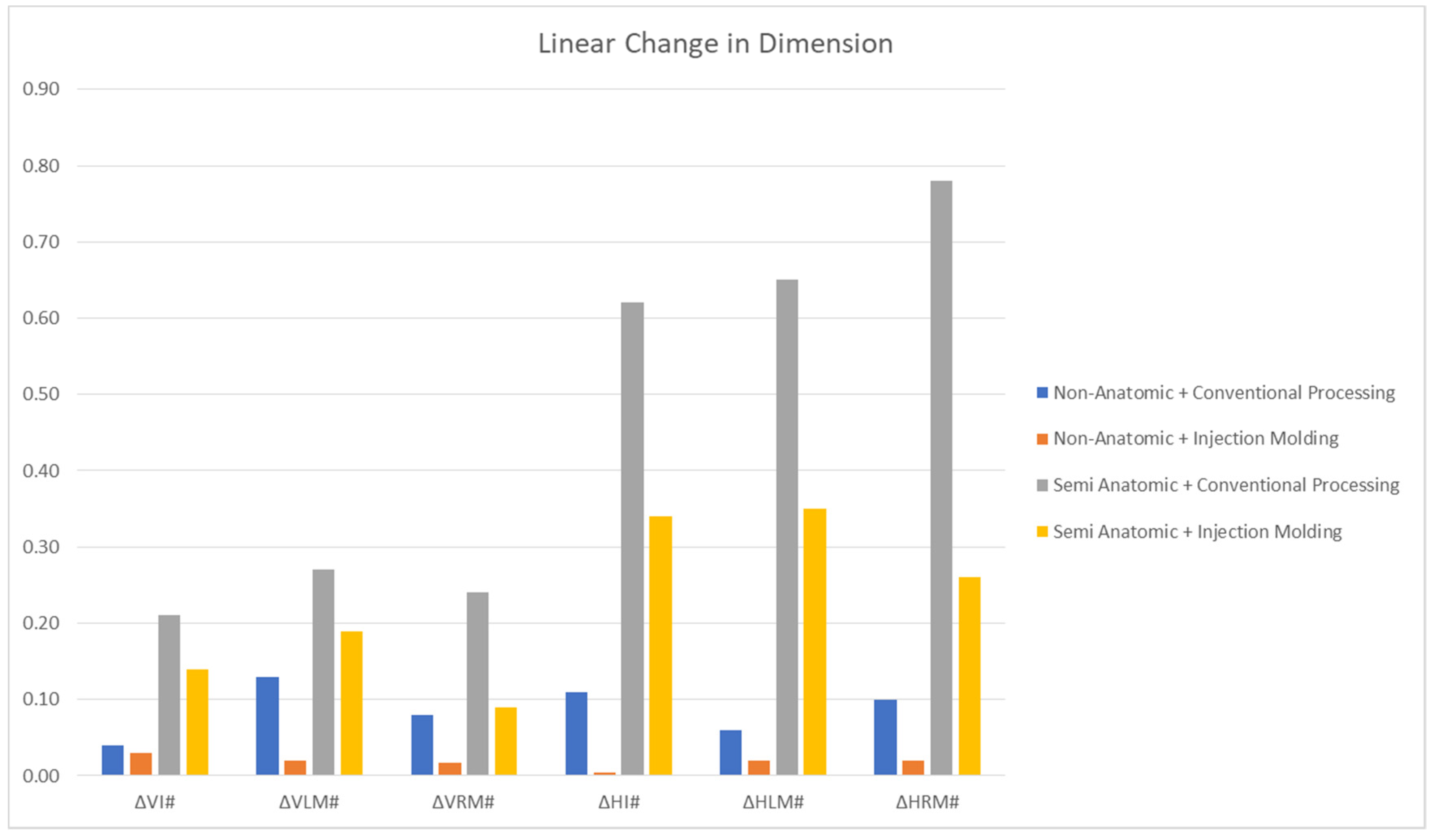
  • In relation to dimensional changes, there was a significant interaction between the components, i.e., the posterior tooth forms and the processing technique applied.
  • The injection molding technique showed smaller tooth movements in both the vertical and horizontal measurements as compared to the conventional compression molding technique.
  • Non-anatomic teeth show fewer changes in tooth movement as compared to semi-anatomic teeth in both the compression and injection molding techniques.
  • The changes in the position of the molar were highest in the horizontal dimension as compared to other dimensions tested for all the groups, when semi-anatomic teeth were processed using the conventional molding technique.
  • The smallest differences were observed in the non-anatomic group, when the injection processing technique was used.
The clinical guideline that can be drawn from this research is that, when non-anatomic teeth are selected for complete dentures, any denture processing technique can be selected, as both cause minimal teeth movement. However, when semi-anatomic teeth are selected, an injection molding processing technique should be preferred to keep tooth movements to a minimum.

Supplementary Materials

The following supporting information can be downloaded at: https://www.mdpi.com/article/10.3390/app12147058/s1, Table S1: Shapiro Wilk Normality test used to check the normality of data without taking into account the absolute value of differences. Table S2: Shapiro Wilk Normality test used to check the normality of data after absolute value of differences was taken into account.

Author Contributions

Conceptualization, M.E.S., A.P. and S.J.; methodology, M.E.S., A.P., S.J., A.H.A.H., R.A.Z., S.M.M.Z. and S.I.N.; software, A.H.A. and N.M.A.; validation, M.E.S., A.P., S.J., F.H.A. and S.S.A.; formal analysis, M.E.S., A.P., S.J., A.H.A.H., R.A.Z., S.M.M.Z. and S.I.N.; investigation, M.E.S., A.P., S.J., A.H.A.H., R.A.Z., S.M.M.Z. and S.I.N.; resources, F.H.A., A.H.A., N.M.A., H.A., S.M.A. and S.S.A.; data curation, M.E.S., A.P. and S.J.; writing—original draft preparation, M.E.S., A.P. and S.J.; writing—review and editing, M.E.S., A.P., S.J., A.H.A.H., R.A.Z., S.M.M.Z., S.I.N., F.H.A., A.H.A., N.M.A., H.A., S.M.A. and S.S.A.; visualization, F.H.A., A.H.A., N.M.A., H.A., S.M.A. and S.S.A.; supervision, M.E.S., A.P. and S.J.; project administration, M.E.S., A.P., H.A., S.M.A., A.H.A.H., R.A.Z., S.M.M.Z., S.I.N. and S.J.; funding acquisition, F.H.A., A.H.A., N.M.A., H.A., S.M.A. and S.S.A. All authors have read and agreed to the published version of the manuscript.

Funding

This research received no external funding.

Institutional Review Board Statement

The research protocol was approved by the research board at the College of Dentistry, Jazan University (Reference number: CODJU-2003I).

Informed Consent Statement

Not applicable.

Data Availability Statement

The data that support the findings of this study are available from the corresponding author upon reasonable request.

Acknowledgments

Authors would like to thank Arwa Jaber I. Mohana and Fatimah Yahya Arif for their assistance with the manuscript review process.

Conflicts of Interest

The authors declare no conflict of interest.

References

  1. Petersen, P.E.; Bourgeois, D.; Bratthall, D.; Ogawa, H. Oral health information systems—Towards measuring progress in oral health promotion and disease prevention. Bull. World Health Organ. 2005, 83, 686–693. [Google Scholar] [PubMed]
  2. Goldstein, G.; Kapadia, Y.; Campbell, S. Complete denture occlusion: Best evidence consensus statement. J. Prosthodont. Off. J. Am. Coll. Prosthodont. 2021, 30, 72–77. [Google Scholar] [CrossRef] [PubMed]
  3. McCord, J.F.; Grant, A.A. Identification of complete denture problems: A summary. Br. Dent. J. 2000, 189, 128–134. [Google Scholar] [CrossRef] [PubMed]
  4. Mainieri, E.T.; Boone, M.E.; Potter, R.H. Tooth movement and dimensional change of denture base materials using two investment methods. J. Prosthet. Dent. 1980, 44, 368–373. [Google Scholar] [CrossRef]
  5. Carr, L.; Cleaton-Jones, P.; Fatti, P.; Wolfaardt, J. An experimental comparison of vertical tooth movement of 33 degrees and 0 degree teeth after denture processing procedures. J. Oral Rehabil. 1985, 12, 263–278. [Google Scholar] [CrossRef]
  6. Shibayama, R.; Gennari Filho, H.; Mazaro, J.V.; Vedovatto, E.; Assuncao, W.G. Effect of flasking and polymerization techniques on tooth movement in complete denture processing. J. Prosthodont. Off. J. Am. Coll. Prosthodont. 2009, 18, 259–264. [Google Scholar] [CrossRef]
  7. de Negreiros, W.A.; Consani, R.L.; Verde, M.A.; da Silva, A.M.; Pinto, L.P. The role of polymerization cycle and post-pressing time on tooth movement in complete dentures. Braz. Oral Res. 2009, 23, 467–472. [Google Scholar] [CrossRef] [Green Version]
  8. Jamani, K.D.; Moligoda Abuzar, M.A. Effect of denture thickness on tooth movement during processing of complete dentures. J. Oral. Rehabil. 1998, 25, 725–729. [Google Scholar] [CrossRef]
  9. Consani, R.L.; Domitti, S.S.; Mesquita, M.F.; Consani, S. Influence of flask closure and flask cooling methods on tooth movement in maxillary dentures. J. Prosthodont. Off. J. Am. Coll. Prosthodont. 2006, 15, 229–234. [Google Scholar] [CrossRef]
  10. Abuzar, M.A.; Jamani, K.; Abuzar, M. Tooth movement during processing of complete dentures and its relation to palatal form. J. Prosthet. Dent. 1995, 73, 445–449. [Google Scholar] [CrossRef]
  11. Sayed, M.E.; Swaid, S.M.; Porwal, A. Effect of cast modification on linear dimensional change of acrylic tooth position following maxillary complete denture processing. J. Prosthodont. Off. J. Am. Coll. Prosthodont. 2017, 26, 659–663. [Google Scholar] [CrossRef] [PubMed]
  12. Goodacre, B.J.; Goodacre, C.J.; Baba, N.Z.; Kattadiyil, M.T. Comparison of denture tooth movement between CAD-CAM and conventional fabrication techniques. J. Prosthet. Dent. 2018, 119, 108–115. [Google Scholar] [CrossRef] [PubMed]
  13. Fernandez, M.A.; Nimmo, A.; Behar-Horenstein, L.S. Digital denture fabrication in pre- and postdoctoral education: A survey of U.S. dental schools. J. Prosthodont. Off. J. Am. Coll. Prosthodont. 2016, 25, 83–90. [Google Scholar] [CrossRef] [PubMed]
  14. Jackson, A.D.; Lang, B.R.; Wang, R.F. The influence of teeth on denture base processing accuracy. Int. J. Prosthodont. 1993, 6, 333–340. [Google Scholar]
  15. Chintalacheruvu, V.K.; Balraj, R.U.; Putchala, L.S.; Pachalla, S. Evaluation of three different processing techniques in the fabrication of complete dentures. J. Int. Soc. Prev. Commun. Dent. 2017, 7, S18–S23. [Google Scholar] [CrossRef] [Green Version]
  16. Nogueira, S.S.; Ogle, R.E.; Davis, E.L. Comparison of accuracy between compression- and injection-molded complete dentures. J. Prosthet. Dent. 1999, 82, 291–300. [Google Scholar] [CrossRef]
  17. Yoshida, K.; Okane, H.; Nagasawa, T.; Tsuru, H. A criterion for the selection of artificial posterior teeth. J. Oral Rehabil. 1988, 15, 373–378. [Google Scholar] [CrossRef]
  18. Murthy, S.S.; Murthy, G.S. Argon ion laser polymerized acrylic resin: A comparative analysis of mechanical properties of laser cured, light cured and heat cured denture base resins. J. Int. Oral Health 2015, 7, 28–34. [Google Scholar]
  19. Nik, T.H.; Shahroudi, A.S.; Eraghihzadeh, Z.; Aghajani, F. Comparison of residual monomer loss from cold-cure orthodontic acrylic resins processed by different polymerization techniques. J. Orthod. 2014, 41, 30–37. [Google Scholar] [CrossRef]
  20. Price, C.A. A history of dental polymers. Aust. Prosthodont. J. 1994, 8, 47–54. [Google Scholar]
  21. Praveen, B.; Babaji, H.V.; Prasanna, B.G.; Rajalbandi, S.K.; Shreeharsha, T.V.; Prashant, G.M. Comparison of impact strength and fracture morphology of different heat cure denture acrylic resins: An in vitro study. J. Int. Oral Health 2014, 6, 12–16. [Google Scholar] [PubMed]
  22. Becker, C.M.; Smith, D.E.; Nicholls, J.I. The comparison of denture-base processing techniques. Part I. Material characteristics. J. Prosthet. Dent. 1977, 37, 330–338. [Google Scholar] [CrossRef]
  23. Consani, R.L.; Mesquita, M.F.; Sinhoreti, M.A.; Consani, S. Influence of the deflasking delay time on the displacements of maxillary denture teeth. J. Appl. Oral Sci. 2003, 11, 332–336. [Google Scholar] [CrossRef] [PubMed] [Green Version]
  24. Wesley, R.C.; Henderson, D.; Frazier, Q.Z.; Rayson, J.H.; Ellinger, C.W.; Lutes, M.R.; Rahn, A.O.; Haley, J.V. Processing changes in complete dentures: Posterior tooth contacts and pin opening. J. Prosthet. Dent. 1973, 29, 46–54. [Google Scholar] [CrossRef]
  25. Campbell, R.L. Effects of water sorption on retention of acrylic resin denture bases. J. Am. Dent. Assoc. 1956, 52, 448–454. [Google Scholar] [CrossRef]
  26. Negreiros, W.A.; Consani, R.L.; Mesquita, M.F.; Sinhoreti, M.A.; Faria, I.R. Effect of flask closure method and post-pressing time on the displacement of maxillary denture teeth. Open Dent. J. 2009, 3, 21–25. [Google Scholar] [CrossRef] [Green Version]
  27. Parvizi, A.; Lindquist, T.; Schneider, R.; Williamson, D.; Boyer, D.; Dawson, D.V. Comparison of the dimensional accuracy of injection-molded denture base materials to that of conventional pressure-pack acrylic resin. J. Prosthodont. Off. J. Am. Coll. Prosthodont. 2004, 13, 83–89. [Google Scholar] [CrossRef]
  28. Sykora, O.; Sutow, E.J. Comparison of the dimensional stability of two waxes and two acrylic resin processing techniques in the production of complete dentures. J. Oral Rehabil. 1990, 17, 219–227. [Google Scholar] [CrossRef]
  29. Strohaver, R.A. Comparison of changes in vertical dimension between compression and injection molded complete dentures. J. Prosthet. Dent. 1989, 62, 716–718. [Google Scholar] [CrossRef]
  30. El Bahra, S.; Ludwig, K.; Samran, A.; Freitag-Wolf, S.; Kern, M. Linear and volumetric dimensional changes of injection-molded PMMA denture base resins. Dent. Mater. Off. Publ. Acad. Dent. Mater. 2013, 29, 1091–1097. [Google Scholar] [CrossRef]
  31. Basso, M.F.; Nogueira, S.S.; Arioli-Filho, J.N. Comparison of the occlusal vertical dimension after processing complete dentures made with lingualized balanced occlusion and conventional balanced occlusion. J. Prosthet. Dent. 2006, 96, 200–204. [Google Scholar] [CrossRef] [PubMed]
  32. Venus, H.; Boening, K.; Peroz, I. The effect of processing methods and acrylic resins on the accuracy of maxillary dentures and toothless denture bases: An in vitro study. Quintessence Int. 2011, 42, 669–677. [Google Scholar] [PubMed]
  33. Peyton, F.; Craig, R. Restorative Dental Materials, 4th ed.; The CV Mosby Company: St. Louis, MO, USA, 1971; p. 48. [Google Scholar]
  34. Zakhari, K.N. Relationship of investing medium to occlusal changes and vertical opening during denture construction. J. Prosthet. Dent. 1976, 36, 501–509. [Google Scholar] [CrossRef]
  35. Shippee, R.W. Control of increased vertical dimension of compression-molded dentures. J. Prosthet. Dent. 1961, 11, 1080–1085. [Google Scholar] [CrossRef]
  36. Atkinson, H.; Grant, A.J.A.D.J. An investigation into tooth movement during the packing and polymerizing of acrylic resin denture base materials. Austr. Dent. J. 1962, 7, 101–108. [Google Scholar] [CrossRef]
  37. Atashrazm, P.; Alavijeh, L.Z.; Afshar, M.S. Influence of the fast-processing technique on the number of the occlusal contacts and occlusal vertical dimension of complete dentures. J. Contemp. Dent. Pract. 2011, 12, 84–90. [Google Scholar] [CrossRef] [PubMed]
  38. Anderson, G.C.; Schulte, J.K.; Arnold, T.G. Dimensional stability of injection and conventional processing of denture base acrylic resin. J. Prosthet. Dent. 1988, 60, 394–398. [Google Scholar] [CrossRef]
  39. Chuchulska, B.; Yankov, S.; Todorov, R.; Ivanova, D.; Kalachev, Y. Injection Shrinkage and Water Sorption of Some Thermoplastic Dental Materials. Pesqui. Bras. Odontopediatr. Clín. Integr. 2019, 19, e4474. [Google Scholar] [CrossRef]
  40. Woelfel, J.B.; Paffenbarger, G.C.; Sweeney, W.T. Dimensional changes occurring in dentures during processing. J. Am. Dent. Assoc. 1960, 61, 413–430. [Google Scholar] [CrossRef]
Figure 1. Steps of duplicating cast and teeth setup: (A,B) round holes made on cast corresponding to maxillary right central incisor and disto-buccal cusps of the right and left first molars; (C) ideal edentulous cast with orientation grooves; (D) custom edentulous silicone mold.
Applsci 12 07058 g001
Figure 2. Dentures before processing: (A) vertical groove placed on the right central incisor incisal edge; (B) round holes on the disto-buccal cusps or right and left first molars.
Applsci 12 07058 g002
Figure 3. Dentures after processing: G1: conventional compression molding technique with non-anatomical tooth form; G2: injection molding technique with non-anatomical tooth form; G3: conventional compression molding technique with semi-anatomical tooth form, G4: injection molding technique with semi-anatomical tooth form.
Applsci 12 07058 g003
Figure 4. Linear change in dimension of non-anatomic and semi-anatomic processing groups. # Difference calculated by subtracting the post-processing values from the pre-processing values; ΔVI, mean linear dimensional change in the position of the incisor in the vertical direction; ΔVLM, mean linear dimensional change in the position of the left molar in the vertical direction; ΔVRM, mean linear dimensional change in the position of the right molar in the vertical direction; ΔHI, mean linear dimensional change in the position of the incisor in the horizontal direction; ΔHLM, mean linear dimensional change in the position of the left molar in the horizontal direction; ΔHRM, mean linear dimensional change in the position of the right molar in the horizontal direction.
Applsci 12 07058 g004
Table 1. Materials used in the study.
Denture Base Resin
Trade NameManufacturerMain CompositionFabrication Technique
Meliodent, Heraeus KulzerKulzer GmbH, Hanau, GermanyPolymethylmethacrylateCompression molding, heat polymerizing technique
Breflex Polyan IC, Bredent, GermanyBredent GmbH &
Co. KG, Senden, Germany
PolymethylmethacrylateInjection molding technique (Thermopress 400 system 2.62)
Acrylic Teeth
Trade NameTooth FormManufacturerMain Composition
Acrylic TruSmile teeth setNon-anatomic and semi-anatomicJayna Industries, Ghaziabad, Uttar Pradesh, IndiaPrepolymerized
PMMA resin
Other Materials Used in the Study
Material NameTrade NameManufacturerMain Composition
Dental stoneDurguixProtechno, Vilamalla, SpainAlpha hemihydrate
Modelling waxSpezial sculpturing waxYETI Dentalprodukte GmbH, Engen, GermanyParaffin wax
Duplicating siliconePrecisil duplicating siliconeYETI Dentalprodukte GmbH, Engen, GermanyAdditional curing silicone (Vinylsiloxane, Platincomplexes and Siliciumdioxid)
Table 2. Mean, standard deviation (linear change in dimension in mm) and intergroup comparison with respect to non-anatomic and semi-anatomic processing groups.
GroupnΔVI #ΔVLM #ΔVRM #ΔHI #ΔHLM #ΔHRM #
Non-anatomic teethConventional processing technique
(G1)
100.04 ± 0.880.13 ± 1.310.08 ± 0.570.11 ± 0.330.06 ± 0.430.10 ± 0.34
Injection molding technique
(G2)
100.03 ± 0.440.02 ± 0.590.017 ± 0.750.004 ± 0.320.02 ± 0.490.02 ± 0.54
p-value-0.560.003 *0.0550.0001 *0.630.004 *
Semi-anatomic teethConventional processing technique
(G3)
100.21 ± 0.890.27 ± 0.460.24 ± 0.610.62 ± 0.460.65 ± 0.790.78 ± 0.63
Injection molding technique
(G4)
100.14 ± 0.330.19 ± 0.650.09 ± 0.780.34 ± 0.570.35 ± 0.640.26 ± 0.50
p-value-0.040.020.001 *0.0001 *0.0001 *0.0001 *
# Difference calculated by subtracting the post-processing values from the pre-processing values; * significant (p < 0.005); ΔVI, mean linear dimensional change in the position of the incisor in the vertical direction; ΔVLM, mean linear dimensional change in the position of the left molar in the vertical direction; ΔVRM, mean linear dimensional change in the position of the right molar in the vertical direction; ΔHI, mean linear dimensional change in the position of the incisor in the horizontal direction; ΔHLM, mean linear dimensional change in the position of the left molar in the horizontal direction; ΔHRM, mean linear dimensional change in the position of the right molar in the horizontal direction.
Table 3. Two-way ANOVA comparing the tooth forms and processing techniques.
SourceType III Sum of SquaresdfMean SquareFSig.
InterceptHypothesis26.045126.04525.9690.001
Error9.3728.9401.048 a
Tooth formHypothesis17.056117.05624.1040.000
Error117.4371760.660 b
Processing techniqueHypothesis5.90822.9545.6890.001
Error117.4371760.660 b
Dependent variable: dimensional change; a 167 MS (technique) + 0.833 MS (error); b MS (error).
Publisher’s Note: MDPI stays neutral with regard to jurisdictional claims in published maps and institutional affiliations.

